the eh-domain-containing protein pan1 is required for normal
TRANSCRIPT

MOLECULAR AND CELLULAR BIOLOGY, Sept. 1996, p. 4897–4914 Vol. 16, No. 90270-7306/96/$04.0010Copyright q 1996, American Society for Microbiology
The EH-Domain-Containing Protein Pan1 Is Required forNormal Organization of the Actin Cytoskeleton
in Saccharomyces cerevisiaeHSIN-YAO TANG AND MINGJIE CAI*
Institute of Molecular and Cell Biology, National University ofSingapore, Singapore 0511, Singapore
Received 30 April 1996/Returned for modification 7 June 1996/Accepted 24 June 1996
Normal cell growth and division in the yeast Saccharomyces cerevisiae involve dramatic and frequent changesin the organization of the actin cytoskeleton. Previous studies have suggested that the reorganization of theactin cytoskeleton in accordance with cell cycle progression is controlled, directly or indirectly, by the cyclin-dependent kinase Cdc28. Here we report that by isolating rapid-death mutants in the background of theStart-deficient cdc28-4 mutation, the essential yeast gene PAN1, previously thought to encode the yeast poly(A)nuclease, is identified as a new factor required for normal organization of the actin cytoskeleton. We show thatat restrictive temperature, the pan1 mutant exhibited abnormal bud growth, failed to maintain a properdistribution of the actin cytoskeleton, was unable to reorganize actin the cytoskeleton during cell cycle, and wasdefective in cytokinesis. The mutant also displayed a random pattern of budding even at permissive temper-ature. Ectopic expression of PAN1 by the GAL promoter caused abnormal distribution of the actin cytoskeletonwhen a single-copy vector was used. Immunofluorescence staining revealed that the Pan1 protein colocalizedwith the cortical actin patches, suggesting that it may be a filamentous actin-binding protein. The Pan1 proteincontains an EF-hand calcium-binding domain, a putative Src homology 3 (SH3)-binding domain, a regionsimilar to the actin cytoskeleton assembly control protein Sla1, and two repeats of a newly identified proteinmotif known as the EH domain. These findings suggest that Pan1, recently recognized as not responsible forthe poly(A) nuclease activity (A. B. Sachs and J. A. Deardorff, erratum, Cell 83:1059, 1995; R. Boeck, S. Tarun,Jr., M. Rieger, J. A. Deardorff, S. Muller-Auer, and A. B. Sachs, J. Biol. Chem. 271:432–438, 1996), plays animportant role in the organization of the actin cytoskeleton in S. cerevisiae.
Cellular morphogenesis has been a long-standing researchinterest for biologists (26, 57). One of the key factors involvedin cellular morphogenesis is the organization of the actin cy-toskeleton. Perhaps no other organisms manifest more vividlythan the yeast Saccharomyces cerevisiae the significance of actincytoskeleton to cellular function and reproduction. S. cerevisiaecells reproduce by polarized growth, or budding, which re-quires the actin cytoskeleton on the cortex (known as corticalactin patches) to undergo dramatic reorganization in accor-dance with different stages of cell growth throughout the cellcycle (22, 34, 37, 38). The cortical actin cytoskeleton in S.cerevisiae, therefore, is a very dynamic structure. G1 cells thathave not yet passed a cell cycle stage called Start distributetheir cortical actin cytoskeleton randomly in order to growisotropically. Cell cycle progression through Start occurs in lateG1 and is dependent on the activity of Cdc28, a cyclin-depen-dent kinase, in complex with the Cln type of cyclins. Shortlyafter Start, the cortical actin patches polarize to a small regionunderneath the plasma membrane. This step sets a transitionof the growth mode from isotropic to apical, since all of theefforts that cells make for growth, in the form of secretoryvesicles, will be channeled to the bud site and, later, into thebud. The cortical actin patches remain at the tip of the budduring the initial phase of bud growth with few or no actinpatches left inside the mother. The growth pattern in the budshifts some time later in the budding phase from apical to
isotropic, by reorganizing the cortical actin patches from thetip of the bud to even distribution inside the bud. This apical/isotropic switch, permitting the bud to grow into a sphericalshape, is thought to be triggered by the activity of Cdc28 incomplex with G2 cyclins (Clb1 and Clb2) (37, 38), and may alsorequire the function of the Clb2-interacting protein Nap1 (33).At the time corresponding to mitosis, cortical actin structuresundergo another redistribution, first becoming evenly spreadover mother and bud, then followed quickly by congregationon both sides of the neck. The latter step is a prerequisite forcytokinesis and probably requires the inactivation of theCdc28/Clb complex (37, 38). Although Cdc28 kinase is impli-cated in regulation of the actin cytoskeleton network, its directcytoskeletal targets have not yet been identified. Screening forgenes required for viability in cells that had been crippled inCdc28 kinase activity has led to the identification of severalfactors, such as Slt2, Cla2, and Cla4, that are found to beinvolved in polarized growth, cytoskeleton organization, andcytokinesis (7, 18, 19, 41). Conceivably, studies of these factorswill provide more insights into the regulation of cytoskeletonorganization by Cdc28. In this report, we describe the iden-tification of a new factor required for normal organizationof the actin cytoskeleton in S. cerevisiae by a similar screeningmethod. By isolating mutants that lose viability rapidly in com-bination with a Start-deficient mutation (cdc28-4) at restrictivetemperature, we obtained a yeast mutant, pan1, that exhibitedvarious defects in the organization of the actin cytoskeleton.Genetic and molecular studies showed that the Pan1 proteinplays an important role in actin cytoskeleton organization inS. cerevisiae.
* Corresponding author. Mailing address: Institute of Molecularand Cell Biology, National University of Singapore, 10 Kent RidgeCrescent, Singapore 0511, Singapore. Phone: (65) 7723382. Fax: (65)7791117. Electronic mail address: [email protected].
4897
Dow
nloa
ded
from
http
s://j
ourn
als.
asm
.org
/jour
nal/m
cb o
n 26
Dec
embe
r 20
21 b
y 10
3.23
7.58
.122
.

MATERIALS AND METHODS
Strains, media, and genetic techniques. The yeast strains used in this work arelisted in Table 1. Rich medium (YPD) contained 1% yeast extract, 2% peptone,and 2% glucose. Minimal, synthetic complete (SC), and dropout media wereprepared as described previously (50). SC-Leu was SC but lacked leucine andwas supplemented as indicated with 2% of glucose (SC-Leu Glu), raffinose(SC-Leu Raf), or galactose (SC-Leu Gal). Restriction enzymes, ligase, and allother DNA-modifying enzymes were from either New England Biolabs or Am-ersham. Wild-type cells were grown at 308C. Temperature-sensitive mutantswere propagated at the permissive temperature of 238C and analyzed at therestrictive temperature of 378C, unless indicated otherwise. Genetic manipula-tions were performed according to standard methods described by Rose et al.(50). The lithium acetate method (31) was used in yeast transformation. DNAmanipulations were performed as described by Sambrook et al. (53). Electro-transformation of bacterial cells was done on a GenePulser (Bio-Rad) withXL1-Blue as the host Escherichia coli strain (Stratagene). PCR was performedwith Vent polymerase (New England Biolabs) as recommended by the manu-facturer. DNA was sequenced by using Sequenase (U.S. Biochemicals), andsequences were compared to the GenBank database by using the BLAST serverat the National Center for Biotechnology Information (National Institutes ofHealth, Bethesda, Md.).Isolation of mutants. The strain US100 (cdc28-4) was obtained from U.
Surana (56) and mutagenized with ethyl methanesulfonate (EMS) to 60% le-thality. After being grown on YPD plates at a density of about 500 colonies perplate at 238C, the colonies were replica plated onto fresh YPD plates which werethen incubated at 378C for 7 h before being allowed to grow at 238C. Thereplica-plated colonies that grew poorly, if at all, on the 378C-treated plates wereselected from the master plates and tested once again for the rapid-death-at-378C phenotype. Fifteen out of 50,000 colonies screened were confirmed to dierapidly at 378C. Each of the rapid-death mutants was transformed with pMC186,a centromere plasmid carrying the wild-type CDC28 gene, and reexamined forthe rapid-death phenotype by comparing with the parental strain US100. Goodviability was restored in two of these mutants after they acquired the CDC28gene and the rest showed little or no difference in their viability at 378C. Bothmutants were found to be temperature sensitive by themselves after they werecrossed to a wild-type strain isogenic to US100. One of them, named YMC387(pan1-4), was analyzed in this study.Viability and cell morphology. Appropriate yeast strains were grown to ;106
cells per ml (A600 5 0.5) in liquid YPD at 238C before being shifted to 378C.Aliquots of cells were taken at various time points for further analysis. In viabilitystudies, cells were plated onto YPD plates and allowed to form colonies for 4days at 238C. Aliquots of cells from the same cultures were fixed with formal-dehyde (3.7%), and the number of cells per milliliter was determined with ahemocytometer. Percentage of viability was determined by dividing the numberof colonies on the YPD plates by the total number of cells that were plated. Inmorphology studies, cells were fixed in formaldehyde, sonicated to disruptclumps, and analyzed with a phase-contrast microscope.Fluorescence studies. Procedures used for fluorescence microscopy have been
described previously (2, 34). Briefly, cells were grown in liquid culture in theconditions described and fixed by the addition of formaldehyde directly to theculture to a final concentration of 3.7%. After 10 min, cells were collected bybrief centrifugation and resuspended in KPi buffer (0.1 M KH2PO4, pH 6.4) plus3.7% of formaldehyde. Cells were collected after 60 min, washed three times inKPi, and resuspended in KPi plus 1.2 M sorbitol; however, to visualize theinfluenza virus hemagglutinin (HA)-tagged Pan1, cells were collected after 30
min and washed. Cellular DNA was visualized by staining with 49,6-diamidino-2-phenylindole (DAPI; Sigma). Actin was visualized by staining with rhodamine-phalloidin (Molecular Probes Inc.). To label chitin, cells were stained with 1 mgof Calcofluor (Fluorescent Brightener 28; Sigma) per ml for 5 min and thenwashed five times in distilled water.For indirect immunofluorescence, fixed and washed cells were permeabilized
by incubation with 10 mg of Zymolyase 100T (ICN Biomedicals, Inc.) per ml for60 min at 308C. After incubation, the cells were washed once with KPi–1.2 Msorbitol, and resuspended in the same solution. Then, 10 ml of the cell suspensionwas transferred to a polylysine-coated well on a multiwell slide. After 1 min, thesuspension was removed and the slide was immersed in 2208C methanol for 6min and 2208C acetone for 30 s. The slide was air dried and incubated with therelevant primary antibodies followed by the secondary antibodies and subsequentantibodies if needed. All antibodies were diluted in phosphate-buffered saline(PBS) containing 1 mg of bovine serum albumin (BSA) per ml (PBS-BSA) andincubated for 60 min each, followed by four PBS-BSA washes. Slides were thenmounted in 90% glycerol containing p-phenylenediamine. To visualize microtu-bule structures, the rat monoclonal YOL1/34 (Serotec) antibody was used as theprimary antiserum and rhodamine-conjugated goat anti-rat immunoglobulin G(IgG) (Jackson ImmunoResearch) was used as the secondary antibody. Actinwas visualized by using guinea pig anti-actin (a gift of D. Botstein, StanfordUniversity, Stanford, Calif.) and fluorescein-conjugated donkey anti-guinea pigIgG (Jackson ImmunoResearch). The HA-Pan1 was visualized by using mousemonoclonal antibody 12CA5 (Boehringer Mannheim), which recognizes the HAepitope, and rhodamine-conjugated goat anti-mouse IgG (Jackson ImmunoRe-search). To costain HA-Pan1 and actin, the antibodies were used in the followingorder: mouse anti-HA, guinea pig anti-actin, rhodamine-conjugated goat anti-mouse IgG, and finally fluorescein-conjugated donkey anti-guinea pig IgG. Incontrol experiments, no cross-reactivity was observed between mouse anti-HAand fluorescein-conjugated donkey anti-guinea pig IgG, between guinea piganti-actin and rhodamine-conjugated goat anti-mouse IgG, or between rhodam-ine-conjugated goat anti-mouse IgG and fluorescein-conjugated donkey anti-guinea pig IgG. All cells were observed and photographed with a Zeiss Axioplanmicroscope.Measurement of DNA content of yeast cells. At each time point, cells were
fixed with 70% ethanol for 1 h or overnight at 48C. Cells were then washed withwater and resuspended in 0.1 ml of 10 mM Tris-HCl (pH 8.0)–10 mM NaCl–50mg of propidium iodide per ml–1 mg of RNase A per ml for 4 h at 378C. Stainedcells were subsequently diluted into 1 ml of PBS, and, for each sample, the DNAcontent of 10,000 cells was determined with a FACScan flow cytometer (BectonDickinson).Cloning of PAN1. The pan1-4 mutant (YMC387) cells were transformed with
a low-copy-number YCp50 yeast genomic library (49), and the transformantswere screened for plasmid-dependent growth at 378C. Five Ura1 Ts1 transfor-mants were analyzed. Upon recovery from E. coli, all five could retransformYMC387 to Ura1 Ts1. Restriction digestion indicated that all five plasmidscontained overlapping regions of DNA. Various parts of the insert from aplasmid containing a 7-kb EcoRI fragment were subcloned into yeast centromereplasmid pRS315 (55) for further analysis. The smallest complementing fragmentwas localized to a 2.4-kb EcoRI-PstI fragment (see Fig. 6A). Sequence analysisrevealed that this fragment was identical to the previously described PAN1 gene(52). Subsequently, the plasmid pHT837, containing the complete PAN1 gene onthe 5.8-kb BamHI (blunted)-EcoRI fragment (see Fig. 6A), was used for allexperiments requiring the wild-type gene.To construct the PAN1 gene under the control of the GAL1 promoter, an
EcoRI site was first created in front of the ATG start codon by PCR. The 1.2-kbPCR product, extending from the start codon to the next EcoRI site, wasdigested with EcoRI and ligated to a vector containing the GAL1 promoter. Theresulting plasmid with the EcoRI fragment in the correct orientation was di-gested with MscI-NotI (blunted) to remove most of the PCR-generated PAN1and replaced with theMscI-KpnI (blunted) fragment of the wild-type PAN1 gene.Both NotI and KpnI sites are from the multiple cloning site of the vector. Thecomplete GAL-PAN1 was then excised by PvuI digestion and cloned into PvuI-digested pRS315 and pRS425 (13) to generate pMC213 and pMC214, respec-tively.Integration mapping and gene disruptions. To confirm that the cloned DNA
represents the authentic PAN1 gene, the 3.8-kb EcoRI fragment containing the59 truncated PAN1 gene was cloned into the EcoRI site of the URA3-containingintegrating vector pRS306 (55). The resulting plasmid was cleaved within thePAN1 gene at the unique NheI site and used to transform a wild-type haploidstrain YMW1 to uracil prototrophy. The structure of this genomic integrationwas confirmed by Southern hybridization. The resultant haploid strain wascrossed to YMC387 (pan1-4), and the diploid was sporulated. A total of 30tetrads were dissected, and all segregated as 2 Ura1 Ts1:2 Ura2 Ts2.The PAN1 gene was disrupted by the one-step gene replacement method (51).
pHT837 was digested withMscI-BamHI to remove most of the PAN1 sequences,and a 1.4-kb BamHI-EcoRV fragment containing the HIS3 gene was cloned intoit. This leaves 287 and 143 nucleotides from the 59 and from the 39 codingsequences of PAN1, respectively. The HIS3 gene together with PAN1 flankingsequences (pan1D::HIS3) was then excised by EcoRI, gel purified, and trans-formed into a wild-type diploid strain, YNN413. The deletion was confirmed by
TABLE 1. Yeast strains
Strain Relevant genotype Reference
US100 MATa cdc28-4 56US52 MATa cdc28-1N 56YNN413 MATa/a ade2/ade2 his3/his3 leu2/leu2 trp1/
trp1 ura3/ura39
YMW1 MATa ade2 ade3 his3 leu2 trp1 ura3 This studyYMW2 MATa ade2 ade3 his3 leu2 trp1 ura3 This studyYMC387 MATa pan1-4 This studyYMC388 MATa cdc28-4 pan1-4 This studyYMC389 MATa cdc28-1N pan1-4 This studyYMC392 YMW2::pMC213(GAL-PAN1 CEN6) This studyYMC393 YMW2::pMC214(GAL-PAN1 2mm) This studyYMC394 YMW2::pMC215(GAL 2mm) This studyYMC395 MATa pan1D::HIS3 pMC216(GAL-HA-PAN1
CEN6)This study
YMC396 MATa/a pan1-4/pan1-4 This studyYMC397 MATa/a pan1-4/PAN1 sla1D::HIS3/SLA1 This studyYMC398 MATa pan1D::HIS3 pMC213(GAL-PAN1
CEN6)This study
4898 TANG AND CAI MOL. CELL. BIOL.
Dow
nloa
ded
from
http
s://j
ourn
als.
asm
.org
/jour
nal/m
cb o
n 26
Dec
embe
r 20
21 b
y 10
3.23
7.58
.122
.

Southern hybridization. When sporulated and dissected, this diploid pan1D strainproduced only two viable spores.To create the sla1 deletion strain, the complete SLA1 gene (accession no.
Z22810) was first obtained from the yeast genome by PCR. This fragment,extending from nucleotides 50 to 4391 of the database sequence, was cloned intothe vector pBluescript KSII1 (Stratagene). The resulting plasmid was then di-gested with MscI and XbaI (blunted) to remove most of the coding region ofSLA1, and a 1.4-kb BamHI (blunted)-XhoI (blunted) HIS3 fragment was clonedinto it. This sla1D construct has 64 and 4 nucleotides of the SLA1 codingsequence remaining at the 59 and 39 ends, respectively. The construct togetherwith SLA1 flanking sequences was excised by digesting with XhoI-SacII on thepolylinker, gel purified, and transformed into a wild-type haploid strain YMW1.The deletion was then confirmed by Southern hybridization. The resultant strainwas crossed to YMC387 (pan1-4) to generate the heterozygous diploid YMC397(Mata/a pan1-4/PAN1 sla1D::HIS3/SLA1).Epitope tagging, protein extraction, and cellular fractionation of Pan1. For
tagging Pan1 at the N terminus, PCR was used to generate a unique Asp718(Boehringer Mannheim) site after the initiation codon in the plasmid pMC213(GAL-PAN1). Subsequently, an Asp718 cassette containing three tandem repeatsof the HA epitope (YPYDVPDYAG) was inserted in frame to generate theGAL-HA-PAN1 construct. This construct was then moved into the vectorpRS316 by PvuI digestion to generate the plasmid pMC216.Two truncated versions of pMC216 were generated to confirm the HA-Pan1
construct. The HA-Pan1D1208-1480 was generated by removing the PAN1 DNAsequence downstream of the PstI site in pMC216. This generates a deletion fromamino acid 1208 to 1480 but includes 27 amino acids due to the translation of thepolylinker sequences before a stop codon is encountered. The HA-Pan1D1055-1480 was generated similarly by removing the sequences downstream of the XhoIsite. This generates a deletion from amino acid 1055 to 1480 but includes 14amino acids from the polylinker.To obtain crude extracts from various yeast strains, the cells were grown under
selective conditions to mid-log phase (A600 5 0.9 to 1.2). Cells were harvested,washed once in stop mixture (0.9% NaCl, 1 mM NaN3, 10 mM EDTA, 50 mNNaF), and then resuspended in 0.2 ml of ice-cold lysis buffer (1% Triton X-100,1% sodium deoxycholate, 0.1% sodium dodecyl sulfate [SDS], 50 mM Tris-HCl[pH 7.2], 0.1 mM sodium orthovanadate, 1 mM phenylmethylsulfonyl fluoride,and protease inhibitors). Cells were then lysed at 48C by vortexing with 400- to500-mm acid-washed glass beads. After centrifugation, loading buffer was addedto the cell lysate and the mixture was analyzed by SDS-polyacrylamide gelelectrophoresis (SDS-PAGE).Cell fractionation experiments were based on methods described by Kuchler et
al. (36). Briefly, yeast strain YMC395 cells containing the GAL-HA-PAN1 gene(pMC216) were grown under selective conditions in 2% galactose to mid-expo-nential phase (A600 5 0.9 to 1.2). Cells were harvested, washed once, andresuspended in 0.5 ml of ice-cold lysis buffer (10 mM Tris-HCl, 1 mM EDTA [pH7.8], containing 2% 2-mercaptoethanol, 1 mM phenylmethylsulfonyl fluoride,and protease inhibitors). Cells were then lysed at 48C by vortexing with 400- to500-mm acid-washed glass beads. The resulting cell lysate was diluted 5- to10-fold with lysis buffer, and unbroken cells, glass beads, and large debris wereremoved by centrifugation at 300 3 g for 5 min at 48C. The 300 3 g supernatant(S1) was then spun at 10,000 3 g for 10 min at 48C, and the pellet (P2) wasresuspended in the same volume of lysis buffer. Finally, the 10,000 3 g super-natant (S2) was spun at 100,000 3 g for 1 h at 48C, producing the third super-natant (S3) and pellet (P3) which was resuspended in the same volume of lysisbuffer. To assess the relative amount of Pan1 in each fraction, equal volumes ofeach fraction were loaded onto an SDS-PAGE gel (7.5% polyacrylamide). Theresolved proteins were electrotransferred to an Immobilon-P membrane (Milli-pore) according to instructions provided by the manufacturer (Bio-Rad). TheHA-tagged Pan1 protein was subsequently detected by using anti-HA antibody(clone 12CA5; Boehringer Mannheim) and horseradish peroxidase-conjugatedgoat anti-mouse IgG secondary antibody. Antibody-antigen complexes were vi-sualized with the ECL system (Amersham).To determine the nature of the interaction between Pan1 and the particulate
fraction, the 10,0003 g pellet was resuspended in 10% glycerol–1 mM EDTA–50mM Tris-HCl (pH 7.8)–10 mM MgCl2. Equal portions were then treated withone of the following reagents: 1% SDS, 1% Triton X-100, 0.1 M Na2CO3 (pH11), 2 M urea, or water as a control. After 1 h at 48C, each mixture was spun at10,000 3 g for 10 min. Each pellet was resuspended in the same volume as thesupernatant. All samples were then analyzed by Western blotting (immunoblotting).
RESULTS
Isolation of mutants that died rapidly in combination withcdc28-4. Start is the initiation point of the yeast cell cycle andrequires the function of Cdc28 (48). Passage through Start is aprerequisite for budding, DNA synthesis, and other eventsnecessary for cell division and is under strict control by growth-related factors such as cell size and nutritional supplies (43). Inorder to gain more insight into the control of Start, we at-tempted the isolation of mutants that could undergo rapid
death in the presence of the cdc28-4 mutation at the restrictivetemperature of 378C. The cdc28-4mutant arrests preferentiallyat Start, before bud emergence and DNA synthesis (48). Al-though arrested in cell cycle progression, the cdc28-4 cellsincubated at 378C are still able, within a limited period of time,to maintain normal rates of protein and RNA synthesis, con-tinue to grow in size, and remain viable after temperaturedown-shift (48). We reasoned that perturbation of the cellcycle arrest in cdc28-4 cells due to additional mutations mayresult in a rapid-death phenotype at 378C, and mutations thatcould interfere with the cell cycle arrest of cdc28-4 may definegenes that are required for proper control of the Start events.From a total of 50,000 EMS-mutagenized colonies, 15 mu-
tants that died rapidly at 378C were isolated. To distinguish themutants that would die rapidly only when combined with thecdc28-4 mutation from those whose rapid-death phenotypewas irrespective of cdc28-4, these mutants were transformedwith the wild-type CDC28 gene carried on a centromere plas-mid. Thirteen of them still exhibited rapid death at 378C in thepresence of the CDC28 gene and were therefore discarded.The other two mutants that regained viability for 378C treat-ment and yet remained temperature sensitive after they ac-quired CDC28 were back-crossed to an isogenic wild-typestrain. One of the mutants, named pan1-4 (see below), was thesubject of this study.pan1-4 was a recessive temperature-sensitive allele. The mu-
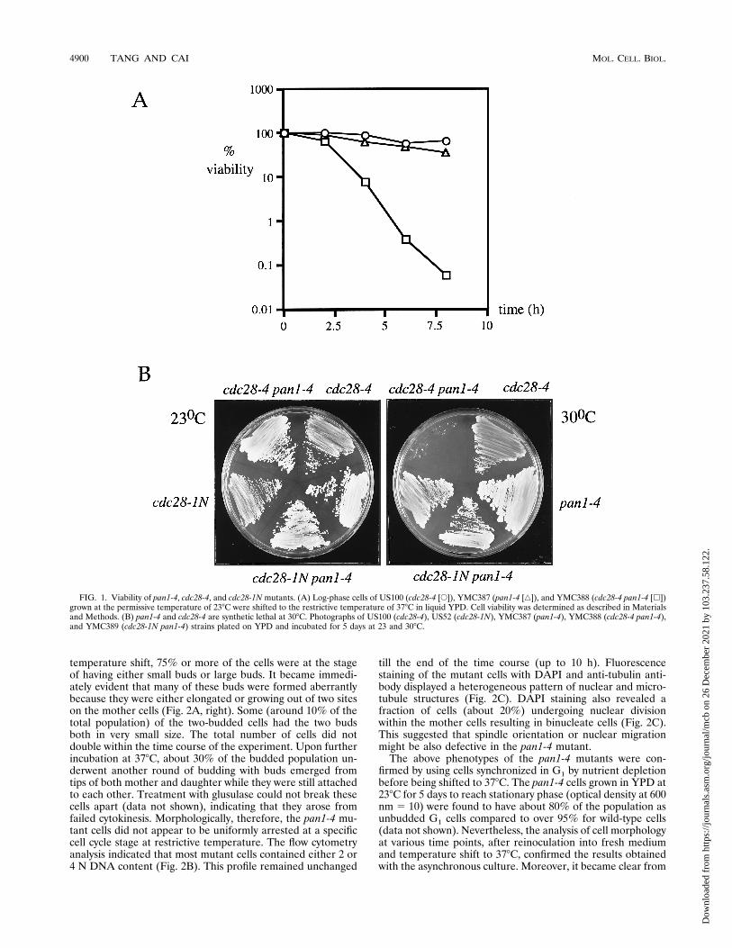
tant grew well at 238C but could not survive when incubated at378C. It did, however, show good viability if the incubation at378C was limited to just a few hours, comparable to that of thecdc28-4 mutant (Fig. 1A). The pan1-4 mutation was back-crossed into cdc28-4 (US100 [Table 1]) again to examine itseffects on the cdc28-4 mutant’s viability and cell cycle arrest atrestrictive temperature. The double mutant, though growingwell at 238C, could not withhold its viability at 378C and diedrapidly (less than 0.1% of the cells were viable after 8 h oftemperature shift, compared to about 50% for the single mu-tants) (Fig. 1A). Moreover, the double mutant could not sur-vive at 308C, a semi-permissive temperature for both singlemutants (Fig. 1B). If the steep drop of viability at 378C and/orsynthetic lethality at 308C in the double mutant was specific toG1-arrested cells, one could predict that another mutant alleleof CDC28, cdc28-1N, which conferred cell cycle arrest at G2/Mrather than Start (56), may not constitute synthetic lethalitywith pan1-4. Indeed, as shown in Fig. 1B, the cdc28-1N pan1-4double mutant could survive well at 308C. Similarly, the doublemutants formed between pan1-4 and each of the cdc mutants,cdc9, cdc13, and cdc15, which were arrested at cell cycle stageswell after Start (48), displayed neither a significant decline inviability at 378C compared with the cdc single mutants (datanot shown) nor synthetic lethality at 308C (except for cdc13,which as a single mutant could not survive at 308C; data notshown). These data suggested that pan1-4 may cause lesionsmore hazardous to cells that are defective in cell cycle progres-sion through G1 than that defective in other stages of cell cycle.Although the pan1-4 mutation had a dramatic effect on the
cell viability of the cdc28-4 mutant, it did not enable cdc28-4cells to escape from being arrested as unbudded cells with 1 Ncontent of DNA at 378C (see below).Phenotypic characterization of the pan1-4 mutant. At the
permissive temperature of 238C, the morphology of pan1-4cells, as judged by phase-contrast microscopy, resembled theisogenic wild-type cells except for the occasional appearance oftwo-budded cells (less than 5% of the population) which weremore prominent at 378C (Fig. 2A, left). Transfer of asynchro-nous cultures of the pan1-4 mutant from 23 to 378C led to arapid increase in the population of budded cells. Within 2 h of
VOL. 16, 1996 CONTROL OF ACTIN CYTOSKELETON ASSEMBLY 4899
Dow
nloa
ded
from
http
s://j
ourn
als.
asm
.org
/jour
nal/m
cb o
n 26
Dec
embe
r 20
21 b
y 10
3.23
7.58
.122
.
temperature shift, 75% or more of the cells were at the stageof having either small buds or large buds. It became immedi-ately evident that many of these buds were formed aberrantlybecause they were either elongated or growing out of two siteson the mother cells (Fig. 2A, right). Some (around 10% of thetotal population) of the two-budded cells had the two budsboth in very small size. The total number of cells did notdouble within the time course of the experiment. Upon furtherincubation at 378C, about 30% of the budded population un-derwent another round of budding with buds emerged fromtips of both mother and daughter while they were still attachedto each other. Treatment with glusulase could not break thesecells apart (data not shown), indicating that they arose fromfailed cytokinesis. Morphologically, therefore, the pan1-4 mu-tant cells did not appear to be uniformly arrested at a specificcell cycle stage at restrictive temperature. The flow cytometryanalysis indicated that most mutant cells contained either 2 or4 N DNA content (Fig. 2B). This profile remained unchanged
till the end of the time course (up to 10 h). Fluorescencestaining of the mutant cells with DAPI and anti-tubulin anti-body displayed a heterogeneous pattern of nuclear and micro-tubule structures (Fig. 2C). DAPI staining also revealed afraction of cells (about 20%) undergoing nuclear divisionwithin the mother cells resulting in binucleate cells (Fig. 2C).This suggested that spindle orientation or nuclear migrationmight be also defective in the pan1-4 mutant.The above phenotypes of the pan1-4 mutants were con-
firmed by using cells synchronized in G1 by nutrient depletionbefore being shifted to 378C. The pan1-4 cells grown in YPD at238C for 5 days to reach stationary phase (optical density at 600nm 5 10) were found to have about 80% of the population asunbudded G1 cells compared to over 95% for wild-type cells(data not shown). Nevertheless, the analysis of cell morphologyat various time points, after reinoculation into fresh mediumand temperature shift to 378C, confirmed the results obtainedwith the asynchronous culture. Moreover, it became clear from
FIG. 1. Viability of pan1-4, cdc28-4, and cdc28-1N mutants. (A) Log-phase cells of US100 (cdc28-4 [E]), YMC387 (pan1-4 [Ç]), and YMC388 (cdc28-4 pan1-4 [h])grown at the permissive temperature of 238C were shifted to the restrictive temperature of 378C in liquid YPD. Cell viability was determined as described in Materialsand Methods. (B) pan1-4 and cdc28-4 are synthetic lethal at 308C. Photographs of US100 (cdc28-4), US52 (cdc28-1N), YMC387 (pan1-4), YMC388 (cdc28-4 pan1-4),and YMC389 (cdc28-1N pan1-4) strains plated on YPD and incubated for 5 days at 23 and 308C.
4900 TANG AND CAI MOL. CELL. BIOL.
Dow
nloa
ded
from
http
s://j
ourn
als.
asm
.org
/jour
nal/m
cb o
n 26
Dec
embe
r 20
21 b
y 10
3.23
7.58
.122
.
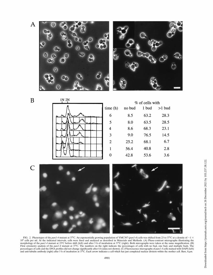
FIG. 2. Phenotypes of the pan1-4 mutant at 378C. An exponentially growing population of YMC387 (pan1-4) cells was shifted from 23 to 378C at a density of ;1 3106 cells per ml. At the indicated intervals, cells were fixed and analyzed as described in Materials and Methods. (A) Phase-contrast micrographs illustrating themorphology of the pan1-4 mutant at 238C before shift (left) and after 5 h of incubation at 378C (right). Both micrographs were taken at the same magnification. (B)Flow cytometry analysis of the pan1-4 mutant at 378C. The numbers on the right indicate the percentages of cells with no bud, one bud, and multiple buds. Thepercentages of cells and the DNA profiles did not change significantly after 6 h (data not shown). (C) Fluorescence micrographs of pan1-4 cells stained with DAPI (left)and anti-tubulin antibody (right) after 5 h of incubation at 378C. Each arrow indicates a cell which has just completed nuclear division within the mother cell. Bars, 8mm.
4901
Dow
nloa
ded
from
http
s://j
ourn
als.
asm
.org
/jour
nal/m
cb o
n 26
Dec
embe
r 20
21 b
y 10
3.23
7.58
.122
.
this experiment that some cells could generate two small budseither simultaneously or in a very short interval (data notshown).It can be summarized from these analyses that although the
pan1-4 mutation may cause rapid death and/or a colethal phe-notype for cells defective in G1 progression, the mutation itselfapparently leads to defect or delay well after Start but unre-stricted to a specific cell cycle stage.The pan1-4 mutant was defective in proper organization of
the actin cytoskeleton. Screening methods similar to ours havebeen employed previously by others to identify genes that arerequired for cell viability either in Cln1- and Cln2-deficientmutants (7, 19) or in the Start-deficient cdc28 mutants at thesemipermissive temperature (41). Interestingly, genes identi-fied this way were all found to be involved in polarized growthand cytoskeleton organization (7, 19, 41). The pan1-4 mutantphenotypes described above, namely, the presence of binucle-ate cells and defects in bud growth and cytokinesis, could beattributed to abnormalities in the organization of the actincytoskeleton (20). It is known that both astral microtubules andthe actin cytoskeleton are required for proper spindle orien-tation and migration (46). Since the astral microtubules ap-peared normal in the pan1-4 mutant (Fig. 2C), it is likely thatthe presence of binucleate cells could be due to perturbation inthe actin cytoskeleton. On the basis of these reasons, we de-cided to examine the cytoskeleton distribution in pan1-4 cellsby using rhodamine-conjugated phalloidin which selectivelybinds to filamentous actin (16). The actin staining of wild-typecells displayed two typical structures, namely, the cortical actinpatches which were highly polarized and mostly confined to thebud with little or no actin patches seen in the mother and theactin cables which were stained only in the mother and mostlyaligned along the axis of bud growth (Fig. 3A). The pan1-4mutant grown at permissive temperature (238C) also exhibitedsimilar pattern of polarized cortical actin patches (Fig. 3B).However in many cells, actin cables were apparently not allaligned parallel to the mother-bud axis (Fig. 3B). The abnor-mality of actin structures of the pan1-4 mutant was more thor-oughly exposed when the cells were incubated at 378C. Asshown in Fig. 3C, in a striking contrast to wild-type and thepan1-4 cells grown at 238C, cortical actin patches in the pan1-4cells incubated at 378C were not confined to the bud, and alarge amount of them was retained in the mother (Fig. 3C).Many of the cortical actin patches seen in the mutant cells at378C were also larger in size. In addition, the actin cytoskeletonin some cells aggregated into thick cables or bars very similarto those seen in the act1 mutants (44) but never seen in wild-type or the pan1-4 cells grown at 238C. The actin cables in themother, when visible, were not arrayed orderly as in wild-typecells. It is important to note that the distribution of the actincytoskeleton in mutant cells incubated at 378C did not displayany regular pattern in respect of bud size, suggesting that themutant had lost its ability to reorganize the actin structure inaccordance with cell cycle progression.The pan1-4 mutant showed abnormal chitin deposition and
budding pattern. Defects in the actin cytoskeleton organiza-tion can lead to abnormal budding patterns and changed chitindeposition (20, 44). Yeast cells can select bud sites in one ofthe two distinct spatial patterns: axial for haploid a and a cellsand bipolar for diploid a/a cells (11, 12). When yeast cells bud,a ring of chitin is deposited in the cell wall at the base of theneck (14). This chitin ring remains on the mother cell as a budscar after cell division. We decided to examine the pattern ofchitin deposition in the pan1-4 mutant by Calcofluor staining,which permits the visualization of bud scars on the cell wall andhence allows the determination of the budding pattern.
The haploid (YMC387) and homozygous diploid (YMC396)pan1-4 mutants were processed for staining with Calcofluorafter growth at 258C to log phase and temperature shift to 378Cfor 4 h. The budding patterns of YMC387 and YMC396 cellsdiffered dramatically from their isogenic wild-type counter-parts (YMW2 and YNN413) (Fig. 4). At 258C, most of the budscars (75%) in the wild-type haploid YMW2 cells were ar-ranged either in a line or clustered around one pole, indicativeof axial budding (Fig. 4A, left). A small population (23%) hasa bipolar pattern, and only 2% have the random buddingpattern. However, the pan1-4 haploid cells displayed a signif-icant increase in the random budding pattern (40%), while47% were of an axial pattern and 13% were of a bipolarpattern (Fig. 4B, left). The wild-type diploid YNN413 cells hadmost bud scars (83%) clustered at both ends of the cell, indic-ative of bipolar budding (Fig. 4A, right), while 11% of thepopulation had a unipolar pattern and 6% had a randombudding pattern. In contrast, the pan1-4 homozygous diploidmutant had 68% of the bud scars distributed randomly overtheir surface (Fig. 4B, right), while only 26% had a bipolarpattern and 6% had a unipolar pattern at 258C.At 378C, the chitin distribution in pan1-4 haploid cells be-
came more delocalized. The bud scars were difficult to recog-nize and some cells (19%) displayed an intense staining at thetip of the bud (Fig. 4C, left). The same was true for the diploidpan1-4 cells, in which chitin delocalization became more pro-nounced in both mother and daughter cells, and many cellsdisplayed staining in patches rather than circular bud scars(Fig. 4C, right).pan1-4 perturbed the distribution of the actin cytoskeleton
in the cdc28 mutant. Analysis of the pan1-4 single mutant withphalloidin and Calcofluor suggested that the mutant was de-fective in the organization of the actin cytoskeleton. We de-cided to investigate the cytoskeletal defects in the cdc28-4pan1-4 double mutant. As pointed out earlier, the pan1-4 mu-tation, although it affected cell viability of the cdc28-4 mutantdramatically, did not let the cdc28-4 cells escape from beingarrested as unbudded cells at 378C. At 378C, the cdc28-4pan1-4 double mutant was still predominately unbudded(81%), with G1 DNA content (Fig. 5A and B, right), comparedwith 93% arrested as unbudded G1 cells for the cdc28-4 singlemutant (Fig. 5A and B, left). It was also more pointed in shapethan the cdc28-4 single mutant. The fluorescence staining byphalloidin revealed striking difference between these twostrains in distribution of the cortical actin cytoskeleton. Stain-ing of the cdc28-4 cells arrested at G1 (3 h after temperatureshift to 378C) displayed a pattern of random distribution of thecortical actin patches (Fig. 5C, left), in agreement with theearlier report (37). In a sharp contrast, however, the staining ofthe double mutant from the culture that had been similarlyincubated for 3 h at 378C exhibited aggregated actin in most ifnot all of the unbudded cells (Fig. 5C, right). Furthermore, theactin was commonly seen to aggregate near or at the tip ofpointed cells, similar to the polarized cortical actin patches atthe pre-bud site in wild-type cells, although there were othercells that showed blocks of aggregated actin that did not situateat the tip of pointed cells. This result indicated that althoughthe pan1-4 mutation did not release cdc28-4 cells from the G1state in terms of budding and DNA replication at restrictivetemperature, it had made the mutant unable to arrest withrandomized cortical actin cytoskeleton. Transformation of thedouble mutant with a single-copy plasmid carrying the wild-type PAN1 gene (see below) resulted in cells with randomdistribution of actin patches identical to that seen in cdc28-4cells at 378C (data not shown). It was therefore concluded thatthe aggregation of cortical actin patches in the double mutant
4902 TANG AND CAI MOL. CELL. BIOL.
Dow
nloa
ded
from
http
s://j
ourn
als.
asm
.org
/jour
nal/m
cb o
n 26
Dec
embe
r 20
21 b
y 10
3.23
7.58
.122
.

FIG. 3. Organization of the actin cytoskeleton in wild-type and pan1-4 cells. Actin staining of exponentially growing wild-type (YMW2) cells at 308C (A) andYMC387 (pan1-4) cells at 238C (B) and then shifted to 378C for 5 h (C) is shown. The arrows in panel B indicate cells with disoriented actin cables. Cells were fixedand stained with rhodamine-phalloidin to visualize the actin cytoskeleton as described in Materials and Methods. Bar, 8 mm.
VOL. 16, 1996 CONTROL OF ACTIN CYTOSKELETON ASSEMBLY 4903
Dow
nloa
ded
from
http
s://j
ourn
als.
asm
.org
/jour
nal/m
cb o
n 26
Dec
embe
r 20
21 b
y 10
3.23
7.58
.122
.

FIG. 4. Chitin and bud scar distribution in wild-type and pan1-4 cells. Cells growing in YPD were fixed in 3.7% formaldehyde and stained for chitin with Calcofluor.(A) Wild-type YMW2 haploid (left) and YNN413 diploid (right) cells incubated at 258C. (B) YMC387 pan1-4 haploid (left) and YMC396 pan1-4 diploid (right) cellsincubated at 258C. (C) YMC387 (left) and YMC396 (right) cells incubated at 378C for 4 h. Bar, 8 mm.
4904 TANG AND CAI MOL. CELL. BIOL.
Dow
nloa
ded
from
http
s://j
ourn
als.
asm
.org
/jour
nal/m
cb o
n 26
Dec
embe
r 20
21 b
y 10
3.23
7.58
.122
.

FIG. 5. Phenotypes of cdc28-4 and cdc28-4 pan1-4mutants. Exponentially growing cells of US100 (cdc28-4) and YMC388 (cdc28-4 pan1-4) at 238C were shifted to 378Cand incubated for the times indicated before fixation. (A) Phase-contrast micrographs showing the morphology of US100 (left) and YMC388 (right) strains after 3 h of incu-bation at 378C. Many of the YMC388 cells are ovoid, while most of the US100 cells are round. (B) Flow cytometry analysis of US100 (left) and YMC388 (right) cells at 378C.The rightward drift of the G1 peak in US100 is a result of cell enlargement and cytoplasmic autofluorescence during each time course. (C) Fluorescence micrographs showingactin distribution in US100 (left) and YMC388 (right) after 3 h of incubation at 378C. Cortical actin patches are dispersed in US100 and polarized in YMC388. Bars, 8mm.
4905
Dow
nloa
ded
from
http
s://j
ourn
als.
asm
.org
/jour
nal/m
cb o
n 26
Dec
embe
r 20
21 b
y 10
3.23
7.58
.122
.
at restrictive temperature was due to the pan1-4 mutation, notto the differences in strain background.Cloning and sequencing of PAN1. The wild-type PAN1 gene
was cloned from a YCp50 genomic library by complementationof the pan1-4 Ts2 phenotype (Materials and Methods). Oneplasmid containing a 7-kb EcoRI fragment capable of comple-mentation was digested with various restriction enzymes tolocalize the complementing gene. The smallest region of com-plementing activity was localized to a 2.4-kb EcoRI-PstI frag-ment (Fig. 6A). DNA sequencing of this fragment revealed itto be a previously described gene named PAN1 (52). PAN1,standing for poly(A) nuclease, was an essential gene and ini-tially thought to encode a protein responsible for poly(A) tailshortening and translation initiation (52). Recently, however, itwas found that the PAN1 gene was in fact not required for thepoly(A) nuclease activity (8, 52a). It had been demonstratedthat only one third of the Pan1 protein was required for cellviability and a mutant protein containing deletions on both theN and C termini was also functional in a pan1 deletion strain(52). Although the 2.4-kb EcoRI-PstI fragment contained theessential region of PAN1 (Fig. 6A), it was unusual that thisfragment was able to complement the pan1-4 mutant withoutthe PAN1 promoter. However, many yeast genes such asCDC44 (30) and DBF2 (32) have also been cloned withouttheir promoter and the N terminus. Presumably, the adjacentDNA sequences were able to provide some promoter activityneeded to express these genes. Incidentally, another Ts2 alleleof pan1 (mdp3-8) was recently reported to be complementedby a;4-kb EcoRI fragment containing the PAN1 gene withoutits N-terminal and promoter sequences (64).Several lines of evidence supported the conclusion that the
pan1-4mutation occurred in the PAN1 gene. First, the effort toisolate the DNA sequences that were able to complement thetemperature sensitivity of pan1-4 was repeated a number oftimes with different yeast genomic libraries made in single-copy vectors (such as YCp50 and pRS315). Invariably, thepositive clones obtained every time all contained the PAN1gene. Second, the pan1-4/pan1D::HIS3 diploid, constructed bycrossing the pan1-4 mutant with a pan1 deletion haploid strainkept alive by the PAN1 gene on a plasmid, could not rescue theTs2 phenotype when the plasmid was lost (data not shown).Third, the PAN1 gene and the pan1-4 allele were deemedallelic to each other by the gene targeting method. The yeastURA3 gene was targeted to the PAN1 locus of the wild-typestrain YMW1 by homologous recombination using the clonedPAN1 sequence. One of the transformants which had theURA3 gene correctly integrated into the PAN1 locus (as de-termined by Southern hybridization) was made diploid bycrossing with the pan1-4 strain YMC387. After sporulation,tetrads were dissected and markers were analyzed. All of the30 tetrads examined segregated as 2 Ura1 Ts1: 2 Ura2 Ts2.This demonstrated a very tight linkage between the PAN1locus and the original pan1-4 mutation. These data had led usto conclude that the pan1-4 mutation occurred in the PAN1gene.The sequence of the PAN1 gene originally published by
Sachs and Deardorff contained several errors. Voss et al. re-cently published the nucleotide sequence of the centromericregion of yeast chromosome IX which comprised the PAN1locus (59). The revised PAN1 sequence (EMBL/GenBank ac-cession numbers Z38062 and X79743) differed from the orig-inal one in (i) a frameshift over 9 amino acids due to deletionsof nucleotides 1300, 1325, and 1326 (the numbers refer to thePAN1 sequence determined by Sachs and Deardorff); (ii) aninsertion of 42 bp at 1943 resulting in addition of the peptideQTTGMMPQTTGMMP; (iii) an insertion of 15 bp at 2423
resulting in addition of the pentapeptide LNKQE; and (iv) asingle base insertion at 4636 generating a frameshift over 85amino acids in the C terminus (59). These changes in the PAN1gene sequence, confirmed by our own sequencing experiments,have revealed several structural features of Pan1 which werenot identified previously. As shown in Fig. 6, the insertion ofthe QTTGMMPQTTGMMP peptide constituted a motif thatwas similar to Sla1, a yeast protein controlling actin cytoskel-eton assembly on the cortical membrane (29). The insertion ofLNKQE peptide enabled identification of the EF-hand cal-cium binding motif (Fig. 6C). Both motifs were located in theessential region of Pan1 as mentioned above. The frameshift atthe C terminus expanded a proline-rich domain from 70 to 170amino acids which contained a motif matching the core se-quence of SH3 domain binding site (X-P-P-X-P, where X is analiphatic residue; see reference 47). In addition, Pan1 con-tained a consensus sequence for the binding of phosphatidyl-inositol 4,5-bisphosphate (PIP2) towards the C terminus whichwas not affected by the sequence changes (Fig. 6B).The sequence similarities between Pan1 and Eps15, a sub-
strate for the mammalian epidermal growth factor receptor(EGFR) and other receptor tyrosine kinases, have alreadybeen noticed (23, 59–61). Both Pan1 and Eps15 belong to anewly identified family called the EH domain (Eps15 homol-ogy domain)-containing proteins (61). The EH domain spans70 or so amino acids and shows 60% overall sequence conser-vation (Fig. 6E; also see reference 61). Pan1 contained two EHdomains, both of which were located in the essential region asidentified by Sachs and Deardorff. Figure 6E shows the align-ment of EH domains of Pan1 with that of other EH domain-containing proteins using the correct Pan1 sequence whichmatched the EH domain consensus better than the incorrectsequence used by Wong et al. (61).PAN1 was essential for cell viability. The diploid strain with
one copy of PAN1 deleted by the one-step gene replacementmethod (51) gave rise to only two viable spores per tetrad (datanot shown). All of the viable spores were free of the disruptionmarker (HIS3), demonstrating that disruption of the PAN1gene was lethal. This result confirmed the previous report thatPAN1 is an essential gene (52). The spores that carried thePAN1 disruption mutation could germinate but died after oneor two cycles of cell division (data not shown). To examine thephenotype of the pan1 null mutant, the above diploid contain-ing a PAN1 deletion (constructed in YNN413) was trans-formed with a yeast URA3 centromere plasmid containing thePAN1 gene under the control of the GAL1 promoter (seeMaterials and Methods). After sporulation, Ura1 and His1
haploids were selected and analyzed. Unfortunately, thesehaploid cells were still viable on glucose medium, suggestingthat some low level of PAN1 expression was occurring. Never-theless, these haploid cells could not grow on complete syn-thetic medium containing 5-FOA, indicating that they requiredthe plasmid for viability. Therefore, the inability to turn off theexpression of PAN1 completely precluded the analysis of thenull mutant phenotype.The pan1-4 mutant was synthetic lethal with the Sla1 null
mutant. The homology observed between Pan1 and Sla1prompted us to look for any genetic interactions between thesetwo genes. Overexpression of SLA1 under the GAL1 promotercould not rescue the Ts2 phenotype of the pan1-4 mutation(data not shown). We then tested to see whether these twogenes were involved in a common process by looking for syn-thetic lethality between them. A diploid strain heterozygousfor both pan1 and sla1 mutations (PAN1/pan1-4 SLA1/sla1D::HIS3) was generated by crossing the pan1-4 mutant with thesla1 deletion mutant (see Materials and Methods). The hap-
4906 TANG AND CAI MOL. CELL. BIOL.
Dow
nloa
ded
from
http
s://j
ourn
als.
asm
.org
/jour
nal/m
cb o
n 26
Dec
embe
r 20
21 b
y 10
3.23
7.58
.122
.
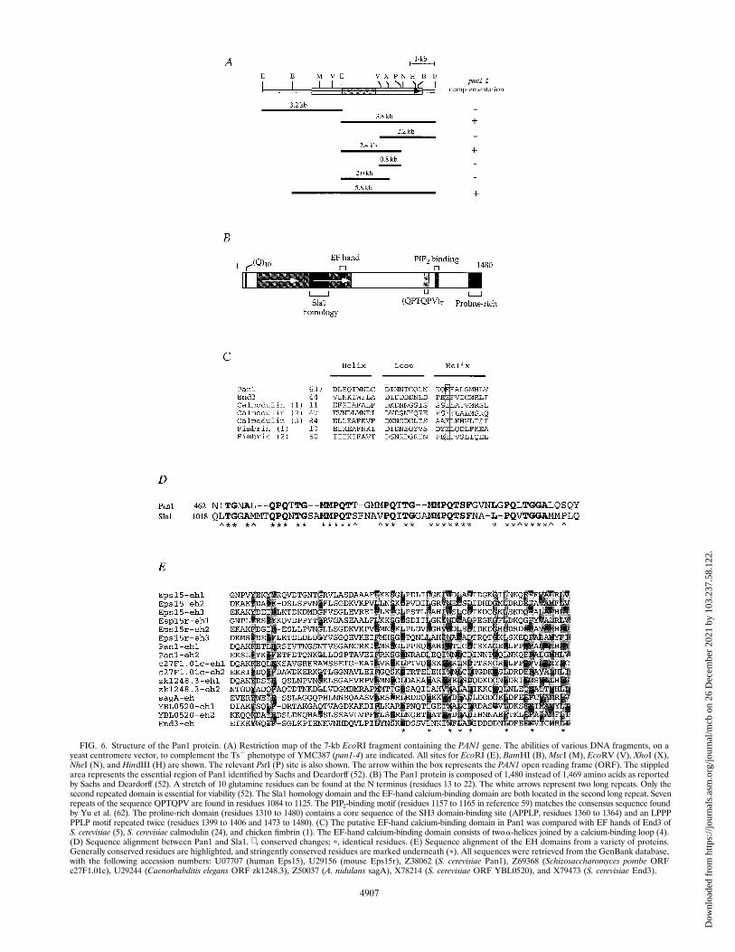
FIG. 6. Structure of the Pan1 protein. (A) Restriction map of the 7-kb EcoRI fragment containing the PAN1 gene. The abilities of various DNA fragments, on ayeast centromere vector, to complement the Ts2 phenotype of YMC387 (pan1-4) are indicated. All sites for EcoRI (E), BamHI (B), MscI (M), EcoRV (V), XhoI (X),NheI (N), and HindIII (H) are shown. The relevant PstI (P) site is also shown. The arrow within the box represents the PAN1 open reading frame (ORF). The stippledarea represents the essential region of Pan1 identified by Sachs and Deardorff (52). (B) The Pan1 protein is composed of 1,480 instead of 1,469 amino acids as reportedby Sachs and Deardorff (52). A stretch of 10 glutamine residues can be found at the N terminus (residues 13 to 22). The white arrows represent two long repeats. Only thesecond repeated domain is essential for viability (52). The Sla1 homology domain and the EF-hand calcium-binding domain are both located in the second long repeat. Sevenrepeats of the sequence QPTQPV are found in residues 1084 to 1125. The PIP2-binding motif (residues 1157 to 1165 in reference 59) matches the consensus sequence foundby Yu et al. (62). The proline-rich domain (residues 1310 to 1480) contains a core sequence of the SH3 domain-binding site (APPLP, residues 1360 to 1364) and an LPPPPPLP motif repeated twice (residues 1399 to 1406 and 1473 to 1480). (C) The putative EF-hand calcium-binding domain in Pan1 was compared with EF hands of End3 ofS. cerevisiae (5), S. cerevisiae calmodulin (24), and chicken fimbrin (1). The EF-hand calcium-binding domain consists of twoa-helices joined by a calcium-binding loop (4).(D) Sequence alignment between Pan1 and Sla1. ∧, conserved changes; p, identical residues. (E) Sequence alignment of the EH domains from a variety of proteins.Generally conserved residues are highlighted, and stringently conserved residues are marked underneath (p). All sequences were retrieved from the GenBank database,with the following accession numbers: U07707 (human Eps15), U29156 (mouse Eps15r), Z38062 (S. cerevisiae Pan1), Z69368 (Schizosaccharomyces pombe ORFc27F1.01c), U29244 (Caenorhabditis elegans ORF zk1248.3), Z50037 (A. nidulans sagA), X78214 (S. cerevisiae ORF YBL0520), and X79473 (S. cerevisiae End3).
4907
Dow
nloa
ded
from
http
s://j
ourn
als.
asm
.org
/jour
nal/m
cb o
n 26
Dec
embe
r 20
21 b
y 10
3.23
7.58
.122
.
loid sla1 deletion mutant had a wide permissive temperaturerange (29). In our hands, it was temperature sensitive at 388Cbut viable at 368C and below (data not shown). The pan1-4mutant, on the other hand, was not viable at 368C. Uponsporulation of the diploid at 238C, very few tetrads with fourviable spores were obtained. When all the viable spores weretested, none of them were found to exhibit both temperaturesensitivity at 368C and His1 at 238C (Fig. 7). This indicated thatthe pan1-4 and sla1 deletion mutations constituted noncondi-tional colethality, suggesting that these two genes may sharecertain functions.High expression of PAN1. In an attempt to identify more
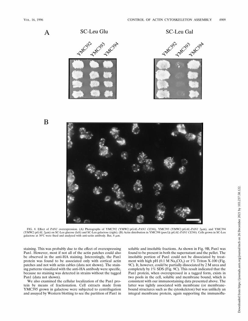
phenotypes which may further assist the characterization ofPAN1 function, we tried overexpression of the PAN1 gene withthe inducible GAL1 promoter. Although induction of theGAL1-driven PAN1 gene carried on a centromeric vector(pMC213) did not result in obvious retardation of growth ofwild-type cells (Fig. 8A), staining with anti-actin antibody re-vealed that high expression of PAN1 resulted in abnormalorganization of the actin cytoskeleton. As shown in Fig. 8B,pMC213-carrying cells (YMC398, Table 1) showed delocaliza-tion of the actin cytoskeleton similar to that observed in thepan1-4 mutants incubated at restrictive temperature. This re-sulted in cells with a more rounded shape than the wild-typecells (Fig. 8B and 3A). Many of the cortical actin patches werealso larger, and many of the cells (30%) exhibited thick actincables which were not seen in wild-type cells. Same stainingpatterns were observed when rhodamine-conjugated phalloi-din was used, except that the abnormal actin cables were not asapparently stained as with anti-actin antibody (data notshown). Even though a significant amount of cortical actinpatches was seen in the mothers of budded cells, the budscontained more cortical actin patches than the mothers. There-fore, the cortical actin distribution was still partially underregulation throughout the cell cycle to ensure normal growth instandard culture conditions. On the other hand, the wild-typecells (YMC393) containing the GAL1-driven PAN1 gene on amulticopy plasmid (pMC214) were found to be inviable ingalactose medium (Fig. 8A, right).Cellular localization of the Pan1 protein. To further shed
light on the mechanism of PAN1 function, the cellular local-ization of the Pan1 protein was determined. The PAN1 geneexpressed from its native promoter and tagged with the HAepitope (58) could not provide a sufficient amount of protein
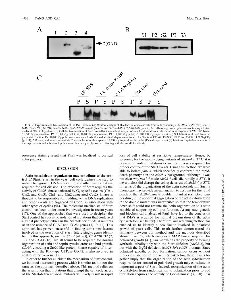
for detection with the anti-HA antibody (12CA5mAb) (datanot shown). Therefore, we used the GAL1 promoter in orderto facilitate its detection. The PAN1 gene expressed from theGAL1 promoter in pMC213 was tagged with the HA epitopeinserted immediately after the methionine initiation codon togenerate pMC216. To test the expression of this construct,which was able to complement the pan1 deletion mutation(data not shown), protein extract was made from cells carryingpMC216 and was analyzed by Western blotting. On the basis ofthe revised sequence of PAN1 (59), the gene should encode aprotein of 160 kDa (1,480 amino acids). However, on SDS-polyacrylamide gels, a band migrating near 200 kDa was de-tected (Fig. 9A, lane 2). This band was not observed from cellextract made from yeast cells carrying plasmid pMC213, whichdid not contain the HA tag (Fig. 9A, lane 1). In addition, twoC-terminal truncated versions of HA-Pan1 were constructedand tested (see Materials and Methods). Both constructs, HA-Pan1D1055-1480 and HA-Pan1D1208-1480, displayed lowerrelative molecular masses when analyzed on SDS-polyacryl-amide gels (Fig. 9A, lanes 3 and 4). This confirmed that the200-kDa band was indeed HA-Pan1. This apparent discrep-ancy in the size of Pan1 could be attributed to its high prolinecontent (10.1%). Abp1, which is similarly rich in proline(11.2%), has also been reported to exhibit a higher-than-cal-culated relative molecular mass on SDS-polyacrylamide gels(21).To localize HA-Pan1, a yeast strain (YMC395) from which
the chromosomal PAN1 gene was deleted and which was keptalive by pMC216 was grown in galactose and prepared forimmunofluorescence staining. Plasmid pMC216 had no ob-servable effect on the viability of the cells cultured in galactosebut, like pMC213, caused more delocalized cortical actinpatches (Fig. 10). Using the anti-HA antibody, the Pan1 pro-tein was found to form, or associate with, punctate structuresvery similar to the cortical actin patches (Fig. 10). By adjustingthe focus, it was clear that these patches were localized to theplasma membrane. The staining was not exclusively confined tothe membrane, and some diffusive staining was also found inthe cytosol. Double immunofluorescence staining with an-ti-HA and anti-actin antibodies (see Materials and Methods)confirmed that the Pan1 protein was indeed associated withcortical actin patches, since both antibodies stained the samepunctate structures (Fig. 10). More punctate structures couldbe seen from the anti-HA staining than from the anti-actin
FIG. 7. Synthetic lethality between pan1-4 and sla1D mutants at 238C. The heterozygous diploid YMC397 (MATa/a pan1-4/PAN1 sla1D::HIS3/SLA1) wassporulated, dissected, and grown on a YPD plate at 238C. Viable colonies were then patched onto a fresh YPD plate and incubated at 238C (left). The plate wassubsequently replica plated to another YPD plate incubated at 368C (middle) and to a His2 plate incubated at 238C (right). No viable colonies were found to be bothTs2 at 368C (representing pan1-4) and His1 at 238C (representing sla1D).
4908 TANG AND CAI MOL. CELL. BIOL.
Dow
nloa
ded
from
http
s://j
ourn
als.
asm
.org
/jour
nal/m
cb o
n 26
Dec
embe
r 20
21 b
y 10
3.23
7.58
.122
.
staining. This was probably due to the effect of overexpressingPan1. However, most if not all of the actin patches could alsobe observed in the anti-HA staining. Interestingly, the Pan1protein was found to be associated only with cortical actinpatches and not with actin cables (data not shown). The stain-ing patterns visualized with the anti-HA antibody were specific,because no staining was detected in strains without the taggedPan1 (data not shown).We also examined the cellular localization of the Pan1 pro-
tein by means of fractionation. Cell extracts made fromYMC395 grown in galactose were subjected to centrifugationand assayed by Western blotting to see the partition of Pan1 in
soluble and insoluble fractions. As shown in Fig. 9B, Pan1 wasfound to be present in both the supernatant and the pellet. Theinsoluble portion of Pan1 could not be dissociated by treat-ment with high pH (0.1 M Na2CO3) or 1% Triton X-100 (Fig.9C). It, however, could be partially dissociated by 2 M urea andcompletely by 1% SDS (Fig. 9C). This result indicated that thePan1 protein, when overexpressed in a tagged form, exists intwo pools in the cell, soluble and membrane bound, which isconsistent with our immunostaining data presented above. Thelatter was tightly associated with membrane (or membrane-bound structures such as the cytoskeleton) but was unlikely anintegral membrane protein, again supporting the immunoflu-
FIG. 8. Effect of PAN1 overexpression. (A) Photographs of YMC392 (YMW2::pGAL-PAN1 CEN6), YMC393 (YMW2::pGAL-PAN1 2mm), and YMC394(YMW2::pGAL 2mm) on SC-Leu glucose (left) and SC-Leu galactose (right). (B) Actin distribution in YMC398 (pan1D::pGAL-PAN1 CEN6). Cells grown in SC-Leugalactose at 308C were fixed and analyzed with anti-actin antibody. Bar, 8 mm.
VOL. 16, 1996 CONTROL OF ACTIN CYTOSKELETON ASSEMBLY 4909
Dow
nloa
ded
from
http
s://j
ourn
als.
asm
.org
/jour
nal/m
cb o
n 26
Dec
embe
r 20
21 b
y 10
3.23
7.58
.122
.
orescence staining result that Pan1 was localized to corticalactin patches.
DISCUSSION
Actin cytoskeleton organization may contribute to the con-trol of Start. Start in the yeast cell cycle defines the step toinitiate bud growth, DNA replication, and other events that arerequired for cell division. The execution of Start requires theactivity of Cdc28 kinase activated by G1-specific cyclins (Cln1,Cln2, and Cln3). Cln1- and Cln2-associated Cdc28 kinase isthought to be responsible for budding, while DNA replicationand other events are triggered by Cdc28 in association withother types of cyclins (54). The molecular mechanism of Startcontrol has been under intensive investigation in recent years(17). One of the approaches that were used to decipher theStart control has been the isolation of mutations that conferreda lethal phenotype either in the Start-deficient cdc28 mutantsor in the absence of CLN1 and CLN2 genes (7, 19, 41). Thisapproach has proven successful in finding some new factorsinvolved in the execution of Start. Interestingly, genes identi-fied by this approach, such as SLT2 (41, 63), CLA2/BUD2 (7,19), and CLA4 (18), are all found to be required for normalorganization of actin and septin cytoskeletons and bud growth.CLA4, encoding a Ste20-like protein kinase capable of inter-acting with the Rho-type GTPase Cdc42, is also involved incontrol of cytokinesis (18).In order to further elucidate the mechanism of Start control,
we initiated a screening process which is similar to, but not thesame as, the approach described above. This method relies onthe assumption that mutations that disrupt the cell cycle arrestof the Start-deficient cdc28 mutants will likely result in rapid
loss of cell viability at restrictive temperature. Hence, byscreening for the rapidly dying mutants of cdc28-4 at 378C, it ispossible to isolate mutations occurring in genes required forproper control of the Start events. Using this method, we wereable to isolate pan1-4, which specifically conferred the rapid-death phenotype in the cdc28-4 background. Although it wasnot clear why pan1-4 made cdc28-4 cells die rapidly at 378C, itnevertheless did disrupt the cell cycle arrest of cdc28-4 at 378Cin terms of the organization of the actin cytoskeleton. Such aphenotype may provide an explanation to account for the rapiddeath of the cdc28-4 pan1-4 double mutant at restrictive tem-perature, if the abnormal aggregation of the actin cytoskeletonin the double mutant was irreversible so that the temperaturedown-shift could not resume the actin organization to a statecapable of supporting cell proliferation. At any rate, geneticand biochemical analyses of Pan1 have led to the conclusionthat PAN1 is required for normal organization of the actincytoskeleton (see below). Therefore, our screening method hasenabled us to identify a new factor involved in polarizedgrowth of yeast cells. This result further demonstrated thesimilarity between our method and the methods describedabove. Like slt2, which encodes a MAP kinase required forpolarized growth (41), pan1-4 exhibits the property of formingsynthetic lethality only with the Start-deficient (cdc28-4), butnot with the G2/M-deficient (cdc28-1N) cdc28 mutants. Sincepolarized growth, or bud formation, cannot occur withoutproper distribution of the actin cytoskeleton, these results to-gether imply that the organization of the actin cytoskeletonresponsible for control of polarized growth may constitute animportant aspect of Start. Indeed, reorganization of the actincytoskeleton from randomization to polarization prior to budformation requires the activity of Cdc28 kinase (37, 38). It is
FIG. 9. Expression and fractionation of the Pan1 protein. (A) Western analysis of HA-Pan1 in crude extracts from cells containing GAL-PAN1 (pMC213; lane 1),GAL-HA-PAN1 (pMC216; lane 2), GAL-HA-PAN1D1055-1480 (lane 3), and GAL-HA-PAN1D1208-1480 (lane 4). All cells were grown in galactose-containing selectivemedia at 308C to log phase. (B) Cellular fractionation of Pan1. Anti-HA immunoblot analysis of samples derived from differential centrifugation of YMC395 lysate.S1, 300 3 g supernatant; P2, 10,000 3 g pellet; S2, 10,000 3 g supernatant; P3, 100,000 3 g pellet; S3, 100,000 3 g supernatant. (C) Solubilization of Pan1 from theparticulate fraction. The 10,0003 g pellet was resuspended in buffer and identical aliquots were treated for 60 min at 48C with 1% SDS, 1% Triton X-100, 0.1 M Na2CO3(pH 11), 2 M urea, and water (untreated). The samples were then spun at 10,000 3 g to produce the pellet (P) and supernatant (S) fractions. Equivalent amounts ofthe supernatants and solubilized pellets were then analyzed by Western blotting with the anti-HA antibody.
4910 TANG AND CAI MOL. CELL. BIOL.
Dow
nloa
ded
from
http
s://j
ourn
als.
asm
.org
/jour
nal/m
cb o
n 26
Dec
embe
r 20
21 b
y 10
3.23
7.58
.122
.

reasonable to speculate that the function of the budding-re-lated factors identified by using Start-deficient mutations maybe under direct or indirect control of Cdc28. As a support forthis speculation, Slt2 MAP kinase has been found recently tobe partially dependent on Cdc28 for its activation (63).PAN1 is required for normal organization of the actin cy-
toskeleton. PAN1 was originally discovered as a gene codingfor the yeast poly(A) nuclease required for initiation of trans-lation by Sachs and Deardorff (52). Although these authorsand their coworkers have reported recently that Pan1 was nolonger responsible for the poly(A) nuclease activity (8, 52a), itsrole in initiation of translation has not yet been clarified. Totest whether inhibition of protein synthesis would give rise to aphenotype similar to that of the pan1-4 mutation, we per-formed the following experiments. If pan1-4’s phenotype wasdue to inhibition of protein synthesis, the cdc28-4 mutantwhich we used to isolate the pan1-4 mutation would be ex-pected to die rapidly with aggregated actin at 378C in thepresence of cycloheximide, a drug that inhibits protein synthe-sis (28). However, this was not the case. The cdc28-4 cells stillshowed randomized cortical actin patches at 378C in the pres-ence of cycloheximide and in fact survived better in the drug-
containing medium at 378C than in the normal medium (datanot shown). Therefore, actin aggregation in the cdc28 mutantin the pan1-4 background was not the consequence of inhibi-tion of protein synthesis. This result cast doubts on the originalreport that PAN1 had an important role in initiation of trans-lation.Instead, our data strongly suggest that Pan1 is directly in-
volved in the normal organization of the actin cytoskeleton.Firstly, analysis of the pan1-4 single mutant at restrictive tem-perature revealed that the mutant had an abnormal actin cy-toskeleton. In wild-type cells, the actin cytoskeleton showedtwo identifiable structures. The cortical actin patches werehighly polarized, being mostly confined to the bud during thephase of bud growth, whereas the actin cables were arrayedalong the mother-bud axis. In the pan1-4 mutant, however,both structures were disturbed at nonpermissive temperature.The cortical actin patches were spread all over the mother andthe daughter, and the actin cables were disarrayed from themother-bud axis. Moreover, the mutant was no longer capableof reorganizing its cortical actin patches according to the cellcycle stages and therefore experienced lesions late in the cellcycle which might include the inability to proceed through
FIG. 10. Colocalization of the Pan1 protein and the cortical actin cytoskeleton. Log-phase cells of YMC395 (pan1D::pGAL-HA-PAN1 CEN6) grown in SC-Uracontaining 2% galactose at 308C were processed for double labelling as described in Materials and Methods. Left, indirect immunofluorescence of the actin cytoskeletonwith anti-actin antibody. Right, localization of Pan1 by indirect immunofluorescence with anti-HA monoclonal antibody 12CA5. Bar, 4 mm.
VOL. 16, 1996 CONTROL OF ACTIN CYTOSKELETON ASSEMBLY 4911
Dow
nloa
ded
from
http
s://j
ourn
als.
asm
.org
/jour
nal/m
cb o
n 26
Dec
embe
r 20
21 b
y 10
3.23
7.58
.122
.

cytokinesis. Consistent with this notion, the actin staining ofthe pan1-4 mutant at 378C never showed congregation of actinat the mother-daughter junction, a step necessary for cytoki-nesis (34, 37, 38). However, the possibility that Pan1 is alsodirectly involved in cytokinesis had not been ruled out.Secondly, the pan1-4 mutant showed serious defects in its
budding pattern. It frequently generated two-budded cells andshowed random budding patterns. Even at permissive temper-ature (258C), both haploid and homozygous diploid pan1-4mutants demonstrated a random budding pattern as visualizedwith Calcofluor staining. At 378C, the chitin distribution be-came delocalized and caused the whole cell surface to bebrightly stained. This phenotype was in agreement with thewell-established phenomenon that defects in the actin cy-toskeleton organization lead to abnormal budding patterns andchanged chitin deposition (3, 5, 15, 21, 25, 39, 44, 45).Thirdly, the pan1 mutation disrupted the pattern of actin
cytoskeleton distribution in cdc28-4 arrested cells. It has beenreported that, although actin polarization in wild-type cellsrequired the activity of Cdc28 kinase, prolonged incubation ofthe Start-deficient cdc28 mutants at restrictive temperaturewould eventually result in polarization of the cortical actinpatches (40). This phenomenon has been interpreted to beunrelated to the Start function because it happened well afterthe wild-type cells had formed buds (38). The cdc28-4 mutantused in our experiments (and previously used in reference 56),however, did not polarize the actin patches during the ob-served time course (up to 8 h) at 378C and remained more orless a spherical shape other than forming long projections, asseen in some cdc28 strains (27). Nevertheless, this strain was agenuine cdc28 mutant since the temperature sensitivity couldbe complemented by a copy of the wild-type CDC28 gene (datanot shown). Therefore, we attributed the discrepancy in cdc28-arrested morphology to the different strain background. Thefacts that the pan1-4 cdc28-4 double mutant aggregated itsactin patches within the first three hours at the restrictivetemperature and that transformation of the double mutantwith the wild-type PAN1 gene reversed the phenotype back torandomized actin structures, as in the cdc28-4mutant (data notshown), suggested that pan1-4 was solely responsible for dis-ruption of the randomized actin distribution in the cdc28-4cells.Fourthly, the PAN1 gene sequence itself reveals some fea-
tures indicative of cytoskeletal proteins. For example, there isa motif of 19 amino acids in Pan1 (MMPQTSFGVNLGPQLTGGA) that is repeated twice in another yeast protein, Sla1.While it has not been known whether the motif shared by Pan1is important for the function of Sla1, it is located in the essen-tial region of Pan1 (Fig. 6). SLA1 was isolated as a syntheticallylethal mutation with a null mutation of ABP1 encoding anSH3-containing cortical actin-binding protein (29). Sla1 itselfcontains three SH3 domains and has been implicated in thecontrol of cortical actin cytoskeleton assembly. At permissivetemperature, the sla1 null mutant displayed large actin spotsinstead of the punctate actin structures seen in wild-type cells.When shifted to the restrictive temperature, the cortical actinstructures became delocalized (29). Both large actin spots anddelocalization of the cortical actin structures were also ob-served in the pan1-4 mutant at nonpermissive temperature, aswell as in cells overexpressing the Pan1 protein. This suggeststhat Pan1 and Sla1 may share certain functions. In support ofthis, the sla1 and pan1-4 mutations were found to form non-conditional synthetic lethality. It is reasonable to speculate thatSla1 could interact physically with Pan1 through its SH3 do-mains binding to the proline-rich domain of Pan1. However,coimmunoprecipitation experiments have so far failed to dem-
onstrate such interaction (data not shown). Another structuralfeature of Pan1, located also in the essential region, is theEF-hand calcium-binding domain, which is found in someother proteins implicated in organization of the actin cytoskel-eton, such as End3 (5; also see below). Furthermore, Pan1 hasa putative PIP2-binding motif which is again a common featurefound in some known actin-binding proteins such as profilin,cofilin, and gelsolin (62).Finally, and mostly strongly, the cellular localization of the
Pan1 protein suggested that Pan1 was directly involved in actincytoskeleton organization. Using epitope-tagged Pan1, wewere able to show that Pan1 was co-localized with cortical actinpatches. Pan1, therefore, may be a novel actin-binding protein,although direct interaction between Pan1 and actin has yet tobe demonstrated. The protein fractionation experiment alsosuggested that at least some fractions of the Pan1 protein wereassociated with membrane-bound structures, supporting thedata obtained with immunofluorescence staining.These data have led us to conclude that Pan1 is directly
involved in the organization of the actin cytoskeleton. A recentreport by Zoladek et al., who isolated the pan1 mutation alongwith other mutations that affected the organization of the actincytoskeleton from a screening for mutants that failed to dis-tribute yeast subcellular proteins to their destinations, furtherlends support to our conclusion (64). Since the finding thatPAN1 was not responsible for poly(A) nuclease activity was notknown to these authors at the publication of their report, theirconclusion that both the actin cytoskeleton and mRNA 39 endsare involved in subcellular protein distribution is apparentlypartially incorrect (64).The EH domain may define a subset of cytoskeleton-inter-
acting proteins. A recently identified protein motif, the EHdomain, has been found in proteins from humans, mice, nem-atodes, and S. cerevisiae (61). A database search for more EHdomain-containing proteins yielded one from Schizosaccharo-myces pombe and another from the filamentous fungus As-pergillus nidulans (Fig. 6). The putative fission yeast gene,c27F1.01c, encodes an open reading frame whose product isabout 20% identical to Pan1 in a 1,379-amino-acid overlap(data not shown). The EH domain has been reported to beinvolved in protein-protein interactions (61). However, itsphysiological function is still unknown at present. Here wepropose that the EH domain may define a new family ofproteins whose functions are related to, or dependent on, theactin cytoskeleton. This proposal is based on studies of threeEH domain-containing proteins whose functions are at leastpartially known, namely, Eps15, End3, and Pan1. Both Eps15(6) and End3 (5) are involved in clathrin-mediated endocyto-sis. The END3 gene was isolated from a screen for mutantsdefective in endocytosis, along with other genes such as ACT1(encodes actin), SAC6 (encodes fimbrin, an actin filament-bundling protein), SLA2/END4, VRP1, RVS161, and RVS167,all of which were involved in actin cytoskeleton organization(5, 35, 42). Interestingly, SAC6 and SLA2/END4 were inde-pendently isolated, along with SLA1, from a screen for mutantswhich formed synthetic lethality with a null mutant of ABP1(29). These results suggest that endocytosis is a process thatrequires the function of the actin cytoskeleton. Loss of theEND3 function resulted in cytoskeletal defects similar to thatobserved in the pan1 mutant (5). Indeed, we have found thatthe END3 gene was a multicopy suppressor of the pan1-4mutation (data not shown), suggesting that End3 and Pan1interact with each other and/or play some overlapping roles inthe organization of the actin cytoskeleton. There are still otherEH domain-containing proteins (as shown in Fig. 6E) whosefunctions are completely unknown. It is tempting to speculate
4912 TANG AND CAI MOL. CELL. BIOL.
Dow
nloa
ded
from
http
s://j
ourn
als.
asm
.org
/jour
nal/m
cb o
n 26
Dec
embe
r 20
21 b
y 10
3.23
7.58
.122
.
that they may also function in actin cytoskeleton-dependentprocesses.In summary, we have isolated PAN1 as a new factor required
for normal organization of the actin cytoskeleton in yeast cells.The genetic analysis of the mutant and cellular localization ofthe protein support the suggestion that PAN1 is directly in-volved in controlling actin cytoskeleton organization. PAN1may prove important in our understanding of cellular morpho-genesis and control of cell polarity in eukaryotic organisms.
ACKNOWLEDGMENTS
We thank U. Surana for yeast strains and D. Botstein for the anti-actin antibody. We also thank F. C. Aw and P. S. Tan for generaltechnical assistance.This work was supported by the Singapore National Science and
Technology Board.
REFERENCES1. Adams, A. E. M., D. Botstein, and D. G. Drubin. 1991. Requirement of yeastfimbrin for actin organization and morphogenesis in vivo. Nature (London)354:404–408.
2. Adams, A. E. M., and J. R. Pringle. 1984. Relationship of actin and tubulindistribution to bud growth in wild-type and morphogenetic-mutant Saccha-romyces cerevisiae. J. Cell Biol. 98:934–945.
3. Amatruda, J. F., J. F. Cannon, K. Tatchell, C. Hug, and J. A. Cooper. 1992.Disruption of the actin cytoskeleton in yeast capping protein mutants. Na-ture (London) 344:352–354.
4. Babu, Y. S., C. E. Bugg, and W. J. Cook. 1988. Structure of calmodulinrefined at 2.2 A resolution. J. Mol. Biol. 204:191–204.
5. Benedetti, H., S. Raths, F. Crausaz, and H. Riezman. 1994. The END3 geneencodes a protein that is required for the internalization step of endocytosisand for actin cytoskeleton organization in yeast. Mol. Biol. Cell 5:1023–1037.
6. Benmerah, A., J. Gagnon, B. Begue, B. Megarbane, A. Dautry-Varsat, and N.Cerf-Bensussan. 1995. The tyrosine kinase substrate eps15 is constitutivelyassociated with the plasma membrane adaptor AP-2. J. Cell Biol. 131:1831–1838.
7. Benton, B. K., A. H. Tinkelenberg, D. Jean, S. D. Plump, and F. R. Cross.1993. Genetic analysis of Cln/Cdc28 regulation of cell morphogenesis inbudding yeast. EMBO J. 12:5267–5275.
8. Boeck, R., S. Tarun, Jr., M. Rieger, J. A. Deardorff, S. Muller-Auer, and A. B.Sachs. 1996. The yeast Pan2 protein is required for poly(A)-binding protein-stimulated poly(A)-nuclease activity. J. Biol. Chem. 271:432–438.
9. Cai, M., and R. W. Davis. 1990. Yeast centromere binding protein CBF1, ofthe helix-loop-helix protein family, is required for chromosome stability andmethionine prototrophy. Cell 61:437–446.
10. Chant, J. 1994. Cell polarity in yeast. Trends Genet. 10:328–333.11. Chant, J., and I. Herskowitz. 1991. Genetic control of bud site selection in
yeast by a set of gene products that constitute a morphogenetic pathway. Cell65:1203–1212.
12. Chant, J., and J. R. Pringle. 1995. Patterns of bud-site selection in the yeastSaccharomyces cerevisiae. J. Cell Biol. 129:751–765.
13. Christianson, T. W., R. S. Sikorski, M. Dante, J. H. Shero, and P. Hieter.1992. Multifunctional yeast high-copy-number shuttle vectors. Gene 110:119–122.
14. Cid, V. J., A. Duran, F. D. Rey, M. P. Snyder, C. Nombela, and M. Sanchez.1995. Molecular basis of cell integrity and morphogenesis in Saccharomycescerevisiae. Microbiol. Rev. 59:345–386.
15. Cleves, A. E., P. J. Novick, and V. A. Bankaitis. 1989. Mutation in the SAC1gene supress defects in yeast golgi and yeast actin function. J. Cell Biol.109:2939–2950.
16. Cooper, J. A. 1987. Effects of cytochalasin and phalloidin on actin. J. CellBiol. 105:1473–1478.
17. Cross, F. R. 1995. Starting the cell cycle: what’s the point? Curr. Opin. CellBiol. 7:790–797.
18. Cvrckova, F., C. De Virgilio, E. Manser, J. R. Pringle, and K. Nasmyth. 1995.Ste20-like protein kinases are required for normal localization of cell growthand for cytokinesis in budding yeast. Genes Dev. 9:1817–1830.
19. Cvrckova, F., and K. Nasmyth. 1993. Yeast G1 cyclins CLN1 and CLN2 anda GAP-like protein have a role in bud formation. EMBO J. 12:5277–5286.
20. Drubin, D. G., H. D. Jones, and K. F. Wertman. 1993. Actin structure andfunction: roles in mitochondrial organization and morphogenesis in buddingyeast and identification of the phalloidin-binding site. Mol. Biol. Cell 4:1277–1294.
21. Drubin, D. G., J. Mulholland, Z. M. Zhu, and D. Botstein. 1990. Homologyof a yeast actin-binding protein to signal transduction proteins and myosin-I.Nature (London) 343:388–390.
22. Drubin, D. G., and W. J. Nelson. 1996. Origins of cell polarity. Cell 84:335–344.
23. Fazioli, F., L. Minichiello, B. Matoskova# , W. T. Wong, and P. P. Di Fiore.1993. eps15, a novel tyrosine kinase substrate, exhibits transforming activity.Mol. Cell. Biol. 13:5814–5828.
24. Geiser, J. R., D. van Tuinen, S. E. Brockerhoff, M. M. Neff, and T. N. Davis.1991. Can calmodulin function without binding calcium? Cell 65:949–959.
25. Haarer, N. K., S. H. Lillie, A. E. M. Adams, A. E. M. Magdolen, W. Bandlow,and S. S. Brown. 1990. Purification of profilin from Saccharomyces cerevisiaeand analysis of profilin-deficient cells. J. Cell Biol. 110:105–114.
26. Harold, F. M. 1990. To shape a cell: an inquiry into the causes of morpho-genesis of microorganisms. Microbiol. Rev. 54:381–431.
27. Hartwell, L. H., R. K. Mortimer, J. Culotti, and M. Culotti. 1973. Geneticcontrol of the cell division cycle in yeast: V. Genetic analysis of cdc mutants.Genetics 74:267–286.
28. Hereford, L. M., and L. H. Hartwell. 1973. Role of protein synthesis in thereplication of yeast DNA. Nature (London) New Biol. 244:129–131.
29. Holtzman, D. A., S. Yang, and D. G. Drubin. 1993. Synthetic-lethal interac-tions identify two novel genes, SLA1 and SLA2, that control membranecytoskeleton assembly in Saccharomyces cerevisiae. J. Cell Biol. 122:635–644.
30. Howell, E. A., M. A. McAlear, D. Rose, and C. Holm. 1994. CDC44, aputative nucleotide-binding protein required for cell cycle progression thathas homology to subunits of replication factor C. Mol. Cell. Biol. 14:255–267.
31. Ito, H., Y. Jukada, K. Murata, and A. Kinura. 1983. Transformation of intactyeast cells treated with alkali cations. J. Bacteriol. 153:163–168.
32. Johnston, L. H., S. L. Eberly, J. W. Chapman, H. Araki, and A. Sugino. 1990.The product of the Saccharomyces cerevisiae cell cycle gene DBF2 has ho-mology with protein kinases and is periodically expressed in the cell cycle.Mol. Cell. Biol. 10:1358–1366.
33. Kellogg, D. R., and A. W. Murray. 1995. NAP1 acts with Clb2 to performmitotic functions and to suppress polar bud growth in budding yeast. J. CellBiol. 130:675–685.
34. Kilmartin, J. V., and A. E. M. Adams. 1984. Structural rearrangements oftubulin and actin during the cell cycle of the yeast Saccharomyces. J. CellBiol. 98:922–933.
35. Kubler, E., and H. Riezman. 1993. Actin and fimbrin are required for theinternalization step of endocytosis in yeast. EMBO J. 12:2855–2862.
36. Kuchler, K., H. G. Dohlman, and J. Thorner. 1993. The a-factor transporter(STE6 gene product) and cell polarity in the yeast Saccharomyces cerevisiae.J. Cell Biol. 120:1203–1215.
37. Lew, D. J., and S. I. Reed. 1993. Morphogenesis in the yeast cell cycle:regulation by Cdc28 and cyclins. J. Cell Biol. 120:1305–1320.
38. Lew, D. J., and S. I. Reed. 1995. Cell cycle control of morphogenesis inbudding yeast. Curr. Opin. Genet. Dev. 5:17–23.
39. Liu, H., and A. Bretscher. 1989. Disruption of the single tropomyosin genein yeast results in the disappearance of actin cables from the cytoskeleton.Cell 57:233–242.
40. Madden, K., and M. Snyder. 1992. Specification of sites for polarized growthin Saccharomyces cerevisiae and the influence of external factors on siteselection. Mol. Biol. Cell 3:1025–1035.
41. Mazzoni, C., P. Zarzov, A. Rambourg, and C. Mann. 1993. The SLT2(MPK1) MAP kinase homolog is involved in polarized cell growth in Sac-charomyces cerevisiae. J. Cell Biol. 123:1821–1833.
42. Munn, A. L., B. J. Stevenson, M. I. Geli, and H. Riezman. 1995. end5, end6,and end7: mutations that cause actin delocalization and block the internal-ization step of endocytosis in Saccharomyces cerevisiae. Mol. Biol. Cell6:1721–1742.
43. Nasmyth, K. 1993. Control of the yeast cell cycle by the Cdc28 proteinkinase. Curr. Opin. Cell Biol. 5:166–179.
44. Novick, P., and D. Botstein. 1985. Phenotypic analysis of temperature-sen-sitive yeast actin mutants. Cell 40:405–416.
45. Novick, P., B. C. Osmond, and D. Botstein. 1989. Suppressors of yeast actinmutations. Genetics 121:659–674.
46. Palmer, R. E., D. S. Sullivan, T. Huffaker, and D. Koshland. 1992. Role ofastral microtubules and actin in spindle orientation and migration in thebudding yeast, Saccharomyces cerevisiae. J. Cell Biol. 119:583–593.
47. Pawson, T. 1995. Protein modules and signalling networks. Nature (London)373:573–580.
48. Pringle, J. R., and L. H. Hartwell. 1981. The Saccharomyces cerevisiae cellcycle, p. 97–142. In J. N. Strathern, E. W. Jones, and J. R. Broach (ed.), Themolecular biology of the yeast Saccharomyces: life cycle and inheritance.Cold Spring Harbor Laboratory, Cold Spring Harbor, N.Y.
49. Rose, M. D., P. Novick, J. H. Thomas, D. Botstein, and G. R. Fink. 1987. ASaccharomyces cerevisiae genomic plasmid bank based on a centromere-containing shuttle vector. Gene 60:237–243.
50. Rose, M. D., F. Winston, and P. Hieter. 1990. Methods in yeast genetics: alaboratory course manual. Cold Spring Harbor Laboratory Press, ColdSpring Harbor, N.Y.
51. Rothstein, R. 1991. Targeting, disruption, replacement, and allele rescue:integrative DNA transformation in yeast. Methods Enzymol. 194:281–301.
52. Sachs, A. B., and J. A. Deardorff. 1992. Translation initiation requires thePAB-dependent poly(A) ribonuclease in yeast. Cell 70:961–973.
52a.Sachs, A. B., and J. A. Deardorff. 1995. Erratum. Cell 83:1059.53. Sambrook, J., E. F. Fritsch, and T. Maniatis. 1989. Molecular cloning: a
VOL. 16, 1996 CONTROL OF ACTIN CYTOSKELETON ASSEMBLY 4913
Dow
nloa
ded
from
http
s://j
ourn
als.
asm
.org
/jour
nal/m
cb o
n 26
Dec
embe
r 20
21 b
y 10
3.23
7.58
.122
.
laboratory manual, 2nd ed. Cold Spring Harbor Laboratory Press, ColdSpring Harbor, N.Y.
54. Schwob, E., T. Bohm, M. D. Mendenhall, and K. Nasmyth. 1994. The B-typecyclin kinase inhibitor p40SIC1 controls the G1 to S transition in S. cerevisiae.Cell 79:233–244.
55. Sikorski, R. S., and P. Hieter. 1989. A system of shuttle vectors and yeasthost strains designed for efficient manipulation of DNA in Saccharomycescerevisiae. Genetics 122:19–27.
56. Surana, U., H. Robitsch, C. Price, T. Schuster, I. Fitch, A. B. Futcher, andK. Nasmyth. 1991. The role of CDC28 and cyclins during mitosis in thebudding yeast S. cerevisiae. Cell 65:145–161.
57. Thompson, D. W. 1961. On growth and form. Abridged [J. T. Bonner (ed.)].Cambridge University Press, London.
58. Tyers, M., G. Tokiwa, R. Nash, and B. Futcher. 1992. The Cln3-Cdc28 kinasecomplex of S. cerevisiae is regulated by proteolysis and phosphorylation.EMBO J. 11:1773–1784.
59. Voss, H., J. Tamames, C. Teodoru, A. Valencia, C. Sensen, S. Wiemann, C.Schwager, J. Zimmermann, C. Sander, and W. Ansorge. 1995. Nucleotidesequence and analysis of the centromeric region of yeast chromosome IX.Yeast 11:61–78.
60. Wong, W. T., M. H. Kraus, F. Carlomagno, A. Zelano, T. Druck, C. M. Croce,K. Huebner, and P. P. Di Fiore. 1994. The human eps15 gene, encoding atyrosine kinase substrate, is conserved in evolution and maps to 1p31-p32.Oncogene 9:1591–1597.
61. Wong, W. T., C. Schumacher, A. E. Salcini, A. Romano, P. Castagnino, P. G.Pelicci, and P. P. Di Fiore. 1995. A protein-binding domain, EH, identifiedin the receptor tyrosine kinase substrate Eps15 and conserved in evolution.Proc. Natl. Acad. Sci. USA 92:9530–9534.
62. Yu, F., H. Sun, P. A. Janmey, and H. L. Yin. 1992. Identification of apolyphosphoinositide-binding sequence in an actin monomer-binding do-main of gelsolin. J. Biol. Chem. 267:14616–14621.
63. Zarzov, P., C. Mazzoni, and C. Mann. 1996. The SLT2(MPK1) MAP kinaseis activated during periods of polarized cell growth in yeast. EMBO J.15:83–91.
64. Zoladek, T., G. Vaduva, L. A. Hunter, M. Boguta, B. D. Go, N. C. Martin,and N. K. Hopper. 1995. Mutations altering the mitochondrial-cytoplasmicdistribution of Mod5p implicate the actin cytoskeleton and mRNA 39 endsand/or protein synthesis in mitochondrial delivery. Mol. Cell. Biol. 15:6884–6894.
4914 TANG AND CAI MOL. CELL. BIOL.
Dow
nloa
ded
from
http
s://j
ourn
als.
asm
.org
/jour
nal/m
cb o
n 26
Dec
embe
r 20
21 b
y 10
3.23
7.58
.122
.