the effects of yy1 knockout on germinal center formation ... · the effects of yy1 knockout on...
TRANSCRIPT

MQP-BIO-DSA-1832
THE EFFECTS OF YY1 KNOCKOUT ON GERMINAL
CENTER FORMATION IN MICE
A Major Qualifying Project Report
Submitted to the Faculty of the
WORCESTER POLYTECHNIC INSTITUTE
in partial fulfillment of the requirements for the
Degree of Bachelor of Science
in
Biology and Biotechnology
by
_________________________
Brittany A. Ciesluk
April 30, 2015
APPROVED:
_________________________ _________________________
Hong Zhang, PhD David Adams, PhD
Department of Cell Biology Dept. Biology and Biotechnology
UMass Medical School WPI Project Advisor
MAJOR ADVISOR
This report represents the work of WPI undergraduate students submitted to the faculty as
evidence of completion of a degree requirement. WPI routinely publishes these reports on its
website without editorial or peer review. For more information about the projects program at
WPI, please see http://www.wpi.edu/academics/ugradstudies/project-learning.html

2
ABSTRACT
This MQP researched transcription factor YY1 in B-lymphocytes, which is thought to be
a master regulator of germinal center formation. Germinal centers within lymphoid tissues are
important areas of B-lymphocyte proliferation and differentiation, and are essential for a B-
lymphocyte immune response. This immune response was studied by creating YY1-null mice
lacking this key transcription factor. Using a cre/lox recombination system, mice were bred to
become germinal center YY1-null. Fluorescence activated cell sorting (FACS) was performed for
germinal center surface proteins GL-7 and CD-95 to quantitate changes in germinal center
numbers. The results showed that stimulation of the mouse immune system with an injection of
sheep red blood cells increases germinal center numbers, and deletion of YY1 causes germinal
center numbers to decrease, suggesting that YY1 is important for germinal center formation.

3
TABLE OF CONTENTS
Signature Page ………………………………………………………………………. 1
Abstract ……………………………………………………………………………… 2
Table of Contents ……………………………………………………………….…… 3
Acknowledgements ………………………………………………………………….. 4
Background ………………………………………………………………………….. 5
Project Purpose ………………………………………………………………………. 12
Methods ……………………………………………………………………………… 13
Results ……………………………………………………………………………….. 18
Discussion …………………………………………………………………………… 27
Bibliography ………………………………………………………………………… 31

4
ACKNOWLEDGEMENTS
I would like to thank Dr. Hong Zhang for giving me the opportunity to intern in his lab at
University of Massachusetts Medical School and for all of his help throughout my internship and
my Major Qualifying Project. I would like to especially thank Sally Trabucco for helping me
define my Major Qualifying Project, all the time she spent in lab with me teaching me various lab
techniques, and providing helpful insights during my time interning in the Zhang Lab. I would
also like to thank Professor David Adams for advising this project, continually providing support
and feedback, and editing my final Major Qualifying Project report.
5
BACKGROUND
B-Cell Development and Germinal Centers
Germinal centers (GCs) are sites within secondary lymphoid organs (such as spleen, lymph
nodes, and intestinal Peyer’s patches) where mature B-lymphocytes proliferate, differentiate, and
mature (MacLennan, 1994; Thorbecke et al., 1994; Natkunam, 2007; Victoria, 2014). During an
infection, this maturation process includes the mutation of their antibody genes (through somatic
hyper-mutation), and the switching of antibody classes (i.e. from IgM to IgG). Thus, GCs are
important regions of B-lymphocyte ontogeny.
B-lymphocytes, otherwise referred to as B-cells, are types of white bloods cells that play a
role in antibody-mediated responses of the adaptive immune system ("Immune System B-Cells,"
2015). Adaptive immunity is a subset of the immune system that allows the body to respond and
adapt to specific foreign antigens. This system uses highly specialized cells to allow the body to
remember different antigens it has encountered. During an infection, B-lymphocytes become
activated by foreign antigens and undergo many stages of differentiation. The process of B-
lymphocyte specialization and development is controlled in germinal centers.
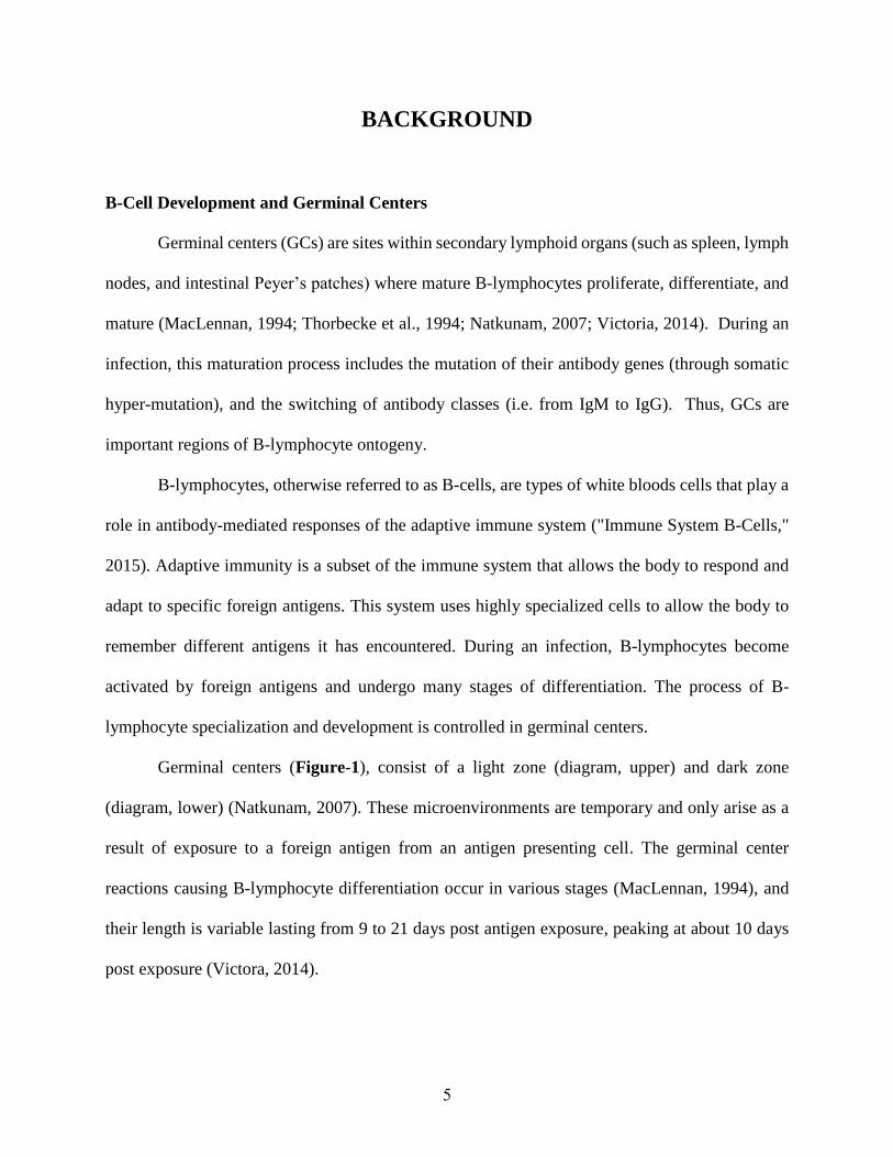
Germinal centers (Figure-1), consist of a light zone (diagram, upper) and dark zone
(diagram, lower) (Natkunam, 2007). These microenvironments are temporary and only arise as a
result of exposure to a foreign antigen from an antigen presenting cell. The germinal center
reactions causing B-lymphocyte differentiation occur in various stages (MacLennan, 1994), and
their length is variable lasting from 9 to 21 days post antigen exposure, peaking at about 10 days
post exposure (Victora, 2014).

6
Figure-1: Diagram of a Germinal Center. Shown are the main processes of a germinal
center reaction to produce mature B-lymphocytes. The dark zone and light zone areas of
the germinal center are shown, as well as the flow of B-lymphocytes through the
maturation process. Clonal expansion and class switching take place in the dark zone.
The selection of high affinity B-lymphocytes occurs in the light zone. (Heesters et al.,
2014)
B-lymphocyte development begins with stem cells originating from the bone marrow.
These stem cells undergo V(D)J recombination, or antigen receptor gene rearrangement, that
results in fully functional antigen receptors on naïve B-lymphocytes through genetic
recombination. This random recombination creates B-lymphocyte diversity and allows the cells to
appropriately encounter antigens (Alt et al., 1992). B-lymphocytes at this stage in their
development only secrete and present low-affinity antibodies on their surface, so they are only
capable of binding small amounts of antigen over longer periods of time. In order for high affinity
antibodies to be made, the naïve B-lymphocyte cells need to continue through various stages of
specialization. Once an antigen is encountered from an antigen-presenting cell, the B-lymphocyte
enters the germinal center microenvironment for additional proliferation and differentiation. The

7
process of antigen presentation involves a portion of the antigen, an epitope, coupled with a major
histocompatibility complex (MHC). Once B-lymphocytes process and present the antigen bound
MHC on the surface of their cells, T-cells recognize these cells and deliver signals that promote
the beginnings of B-lymphocyte proliferation and eventual differentiation (Victora, 2014). These
critical signals cause naïve B-lymphocytes to begin rapid clonal expansion and immunoglobulin
class switching recombination (CSR), which take place in the germinal center dark zone.
Simultaneously, the B-lymphocytes go through AID-driven somatic hyper-mutation, an essential
step in increasing antibody affinity. The activation-induced cytidine deaminase (AID) is a germinal
center B-lymphocyte specific enzyme that facilitates somatic hyper-mutation. Somatic hyper-
mutation (SHM) is the introduction of non-random, single base changes in the genes encoding the
antigen-binding sites on the surface of B-lymphocytes. This mutational process powerfully
diversifies B-lymphocyte cells, a vital characteristic of high-affinity antigen-binding cells
(Natkunam, 2007).
SHM and CSR create a variety of B-lymphocyte clones within the germinal center with
different maturation states. At this point in the B-lymphocyte differentiation process, the cells
move into the light zone of the germinal center. The movement process is not well characterized,
but only the highest affinity cells are selected to go into the germinal center light zone (Victora,
2014). Affinity selection is mediated by survival signals from follicular dendritic and T-helper cell
signals which create a cycle where cells that are unable to make it into the light zone as a result of
low affinity go through the process again to continue gaining increased antibody affinity
(Natkunam, 2007). Once the final differentiation stages are completed, some B-lymphocytes
differentiate into plasma cells, others differentiate into memory B-lymphocyte cells, and others
that are no longer functional undergo apoptosis. The entire germinal center reaction process of B-
8
lymphocyte maturation peaks at about 10 days post antigen exposure, and after 21 days post
antigen exposure the germinal centers begin to dissolve.
Overall, the germinal centers are an important part of the B-lymphocyte humoral immune
response, acting as factories for the production of affinity-matured B-lymphocytes that effectively
recognize infectious agents, and for the production of durable memory B-lymphocytes
(MacLennan, 1994; Thorbecke et al., 1994; Natkunam, 2007).
YY1 Transcription Factor
Transcription factor Ying Yang-1 (YY1) is a ubiquitous transcription factor that plays a
role in a variety of biological processes, including cellular proliferation, differentiation, relocation,
and embryogenesis. Contingent upon the different environments it is in, YY1 can either stimulate
or repress transcription, but the mechanisms by which it does so have not yet been defined (Gordon
et al., 2005).
Some evidence exists that YY1 plays a role in B-lymphocyte development, especially in
early development during V(D)J recombination (Liu et al., 2007). YY1 has been postulated to be
a master regulator of germinal center reactions. Gene expression profiling of mouse B-
lymphocytes showed that YY1 is a prominent transcription factor up-regulated in germinal center
reactions, and is likely one of its key regulators (Green et al., 2011). Over-expression of Smurf2
was shown to increase ubiquitination and degradation of YY1, and suppress B-lymphocyte
proliferation (Ramkumar et al., 2013.

9
Gene Editing Using the Cre-Lox Recombination System
This MQP project will use two different systems for knocking out the transcription factor
YY1 in B-lymphocytes, the Cre-lox system for in vivo knockout (KO), and the CRISPR-Cas9
system for in vitro knockout as a negative control and B-lymphocyte in vitro stimulation. Cre-lox
recombination is a site-specific recombination technology that allows gene editing (insertions,
deletions, inversions) at specific loci. The Cre-lox system was discovered in 1988 as part of the
P1 bacteriophage reproductive replication system (Sauer and Henderson, 1988). This system has
been used in mammalian models for performing specific gene deletions, insertions, inversions, and
translocations. Cre recombinase is an enzyme that catalyzes recombination between two loxP
restriction sites, each approximately 35 base pairs in length that are inserted to flank a gene of
interest (Figure-2). Transgenic technology is used in advance to insert loxP sites left and right of
(flanking) the gene to be edited. The orientation of the floxed sites, sites flanked by loxP sites,
determines which gene is edited. Floxed sites inserted in different directions result in inversions
of a gene (diagram, left), loxP sites inserted on different chromosomes results in gene translocation
(diagram, center), while floxed sites in the same direction flanking a gene results in gene deletion
(diagram, right) (The Jackson Laboratory, 2015).

10
Figure-2: Diagram of the Cre-Lox Recombination System for Gene Editing. Shown
are different uses of cre-lox recombination for gene editing. (A) Inversion: flanking loxP
sites face each other, (B) Translocation: loxP sites are located on different chromosomes,
and (C) Deletion: flanking loxP sites face the same direction. (The Jackson Laboratory,
2015)
Gene Editing Using the CRISPR/Cas9 System
Clustered regularly interspaced short palindromic repeats (CRISPR) is another gene editing
tool that has gained widespread use in the past few years due to its high efficiency and ease of use
(Mali et al., 2013; Shen et al., 2013; AddGene, 2014). This gene editing tool is composed of a
specifically designed duplex guide RNA (red in the diagram) and the Cas9 endonuclease (green in
the diagram) (Figure-3). The guide RNA is individually designed to hybridize to a sequence
location for editing, and this binding directs the Cas9 endonuclease to the location. The Cas9
endonuclease unwinds the DNA and cleaves the DNA causing a double-stranded break (Addgene,
2014). The CRISPR-Cas9 system can be used to disrupt genes of interest, by either insertion or
deletion, or can modify endogenous genomes to activate specific genes (Shen et al., 2013).

11
Figure-3: Diagram of the CRISPR/Cas9 System for Specific Gene Editing. The Cas-9
guide RNA (gRNA) (red) is designed to recognize and hybridize to the gene of interest,
targeting the Cas9 nuclease (green) to the location, causing a double stranded break
(DSB). The break is repaired, causing editing or disruption of the target gene (Addgene,
2014).

12
PROJECT PURPOSE
Germinal centers (GCs) are sites within secondary lymphoid organs (such as spleen, lymph
nodes, and intestinal Peyer’s patches) where mature B-lymphocytes proliferate, differentiate, and
mature. Although they are thought to play an important role in B-lymphocyte development,
questions remain about exactly which pathways are involved, how the reactions work, and which
genes and transcription factors are critical. In diseases such as diffuse large B-lymphocyte
lymphoma, the B-lymphocytes no longer respond to standard chemotherapy, so a deeper
understanding of B-lymphocyte maturation in the germinal center pathways could lead to effective
therapies for B-lymphocyte diseases (Ramkumar et al., 2013).
Transcription factor Ying Yang-1 (YY1) is thought to serve as a key regulator of B-
lymphocyte maturation in germinal centers. Gene profiling studies indicate that YY1 is a key
regulator of germinal center reactions (Green et al., 2011). Over-expression of Smurf2 was shown
to increase ubiquitination and degradation of YY1, and suppress B-lymphocyte proliferation
(Ramkumar et al., 2013. But how YY1 regulates B-lymphocyte differentiation and proliferation,
and by which reactions is unknown. The purpose of this MQP project was to investigate, in vivo
and in vitro, the relationship between YY1 and germinal center reactions by deleting the YY1
gene, and in particular determining whether deleting the YY1 gene causes a decrease in germinal
center numbers. In B6 mice, YY1 was conditionally deleted in germinal center B-lymphocytes
using AID-cre/lox recombination, and also through the design of a gRNA for CRISPR/Cas9 in
vitro usage. YY1 conditional knockout (CKO) mice were stained with fluorescent antibodies for
B220 (a pan B-lymphocyte marker), and GL7 and CD95 germinal center surface proteins, and then
analyzed through flow cytometry FACS analysis to quantitate GC numbers.
13
METHODS
YY1-Null Mice
All mouse studies were performed according to guidelines approved by the Institutional
Animal Care and Use Committee (IACUC) of the University of Massachusetts Medical School.
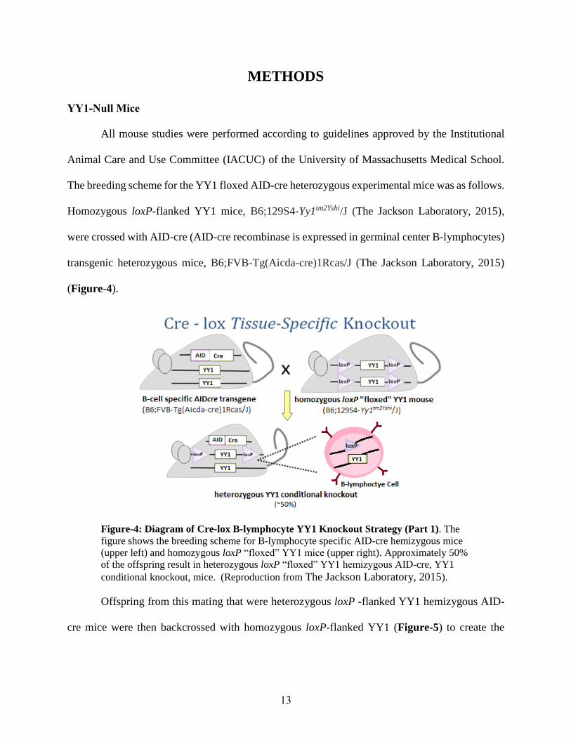
The breeding scheme for the YY1 floxed AID-cre heterozygous experimental mice was as follows.
Homozygous loxP-flanked YY1 mice, B6;129S4-Yy1tm2Yshi/J (The Jackson Laboratory, 2015),
were crossed with AID-cre (AID-cre recombinase is expressed in germinal center B-lymphocytes)
transgenic heterozygous mice, B6;FVB-Tg(Aicda-cre)1Rcas/J (The Jackson Laboratory, 2015)
(Figure-4).
Figure-4: Diagram of Cre-lox B-lymphocyte YY1 Knockout Strategy (Part 1). The
figure shows the breeding scheme for B-lymphocyte specific AID-cre hemizygous mice
(upper left) and homozygous loxP “floxed” YY1 mice (upper right). Approximately 50%
of the offspring result in heterozygous loxP “floxed” YY1 hemizygous AID-cre, YY1
conditional knockout, mice. (Reproduction from The Jackson Laboratory, 2015).
Offspring from this mating that were heterozygous loxP -flanked YY1 hemizygous AID-
cre mice were then backcrossed with homozygous loxP-flanked YY1 (Figure-5) to create the

14
desired homozygous loxP-flanked YY1 hemizygous AID-cre mice experimental mice.
Homozygous loxP -flanked YY1, AID-cre nullizygous mice were used as the control.
Figure-5: Diagram of Cre-lox B-lymphocyte YY1 Knockout Strategy (Part 2).
Shown is the breeding scheme for heterozygous loxP “floxed” YY1 AID-cre hemizygous
mice (upper left) and homozygous loxP “floxed” YY1 mice (upper right). Approximately
25% of the offpring result in the desired homozygous loxP “floxed” YY1 hemizygous
AID-cre, YY1 knockout mice. (Reproduction from The Jackson Laboratory, 2015).
PCR genotyping was used to confirm the genotype of the mice. Mouse tail clippings were
used for DNA isolation, and the isolated DNA was used for genotyping PCR with YY1 and AID-
cre primers. Primers were ordered through the University of Massachusetts Medical School
Oligonucleotide Service (www.umassmed.edu/oligo/).
Primers used for YY1 genotyping (The Jackson Laboratory, 2015):
5’ ACC TGG TCT ATC GAA AGG AAG CAC 3’ (Forward)
5’ GCT TCG CCT ATT CCT CGC TCA TAA 3’(Reverse)
Primers used for AID-cre genotyping (The Jackson Laboratory, 2015):
5’ CCG TAA CCT GGA TAG TGA AAC AG 3’ (Transgene Forward)
5’ CCA TGC GAG TCT TAA GAT GTT G 3’(Transgene Reverse)
5’ GTA GGT GGA AAT TCT AGC ATC ATC C 3’ (Internal Reverse)

15
Intraperitoneal (IP) Injections
In order to boost the immune response in general, and B-lymphocyte production
specifically, at approximately six to eight weeks of age, mice were injected intraperitoneally with
500 µl of sheep red blood cells (SRBC) at a concentration of 1.5x10⁹ cells per injection (Cocalico
Biological, Inc., Stevens, PA, USA). Three homozygous loxP “floxed” YY1 hemizygous AID-
cre, YY1 knockout, mice and three homozygous loxP -flanked YY1, AID-cre nullizygous mice
were injected.
Flow Cytometry Analysis
Spleens were collected at 10 day after SRBC injections from all six injected mice and one
non-immunized mouse. Spleens were ground between frosted glass slides, red blood cells were
lysed, then the remaining intact splenocyte cells were filtered through a 70-μm nylon mesh and
incubated with blocking solution, anti-CD16/32 antibody, to block Fc receptors (10 minutes on
ice). Cells were incubated with primary monoclonal antibodies, in different groupings (Figure-6),
for 20 minutes and washed three times with staining media [biotin-, flavin- and phenol red-
deficient RPMI-1640 medium with 10 mM, pH 7.2, HEPES, 0.02% sodium azide,
1 mM EDTA and 2% fetal bovine serum (FBS)]. The antibodies used included B220-FITC (a pan
B-lymphocyte marker) (1:200 dilution), and germinal center protein antibodies GL7-APC (1:100
dilution), and CD95-PE (1:25 dilution) (Figure-6). Cells were then resuspended in
1 μg ml−1 propidium iodide (PI) to exclude dead cells. Flow cytometry was performed on a 5-laser,
18-detector LSR II FACS machine (BD Biosciences, San Jose, CA, USA), and the data were
analyzed using FlowJo FACS software (Treestar, Ashland, OR, USA).

16
Sample Antibody 1 141212A B220 GL7 CD95
2 1478-3 B220 GL7 CD95
3 1478-6 B220 GL7 CD95
4 1478-10 B220 GL7 CD95
5 1478-7 B220 GL7 CD95
6 1478-8 B220 GL7 CD95
7 1553-5 B220 GL7 CD95
8 florescence minus one (FMO) control
B220 GL7
9 florescence minus one (FMO) control
B220 CD95
10 florescence minus one (FMO) control
CD95 GL7
11 control PI
12 control B220
13 control GL7
14 control CD95
Figure-6: Table of Spleen Cell Staining. Shown are the antibodies used to stain each
sample analyzed by the LSR II flow cytometer. Red samples are experimental samples,
black samples are control and florescence minus one (FMO) control samples.
CRISPR Design
Three guide RNAs (gRNAs) for the YY1 first exon were designed using the Zhang Lab
CRISPR Design System, crispr.mit.edu, (MIT, Cambridge, MA, USA). The gRNAs were found
using a human hg19 target genome and an input of YY1 Exon 1.
YY1 Exon 1
Tggcggcggagccctcagccatggcctcgggcgacaccctctacatcgccacggacggctcggagatgccggccgag
atcgtggagctgcacgagatcgaggtggagaccatcccggtggagaccatcgagaccacagtggtgggcgaggagga
ggaggaggacgacgacgacgaggacggcggcggtggcgaccacggcggcgggggcggccacgggcacgccggccacc
accaccaccaccatcaccaccaccaccacccgcccatgatcgctctgcagccgctggtcaccgacgacccgacccag
gtgcaccaccaccaggaggtgatcctggtgcagacgcgcgaggaggtggtgggcggcgacgactcggacgggctgcg
cgccgaggacggcttcgaggatcagattctcatcccggtgcccgcgccggccggcggcgacgacgactacattgaac
aaacgctggtcaccgtggcggcggccggcaagagcggcggcggcggctcgtcgtcgtcgggaggcggccgcgtcaag
aagggcggcggcaagaagagcggcaagaagagttacctcagcggcggggccggcgcggcgggcggcggcggcgccga
cccgggcaacaagaagtgggagcagaagcaggtgcagatcaagaccctggagggcgagttctcggtcaccatgtggt
cctcagatgaaaaaaaagatattgacc

17
Three separate sequences of 20 base pairs were chosen for gRNAs based on a predicted
low number of off-target sites, high number of mismatches, and guide location not in a gene.
Guides chosen:
Guide #1: CTACATCGCCACGGACGGCT
Guide #2: GTCGGGTCGTCGGTGACCAG
Guide #3: AATGTAGTCGTCGTCGCCGC
Guide RNAs and their reverse complements were ordered through the University of
Massachusetts Medical School Oligonucleotide Service (www.umassmed.edu/oligo/). Guide
RNA oligos were annealed to their respective reverse complements to form gRNA oligo duplexes,
and two different sets of each guide RNA were ligated into BSMB1 digested lentiCRISPR v2
vectors (lentiCRISPR v2 was a gift from Feng Zhang (Addgene plasmid # 52961). 8µl of ligation
of product was transformed into 80µl of E. coli XL10 cells. Transformed cells were plated on
ampicillin plates, colonies were selected and cultured in LB + ampicillin. QIAprep Spin Minipreps
were performed on transformed cultures to purify plasmid DNA. Purified plasmid was sent to
Genewiz (South Plainfield, NJ, USA) for sequencing.

18
RESULTS
Germinal centers (GCs) are sites within secondary lymphoid organs (such as spleen, lymph
nodes, and intestinal Peyer’s patches) where mature B-lymphocytes proliferate, differentiate, and
mature. Gene profiling studies indicate that YY1 is a key regulator of germinal center reactions
(Green et al., 2011). Over-expression of Smurf2 was shown to increase ubiquitination and
degradation of YY1, and suppress B-lymphocyte proliferation (Ramkumar et al., 2013). However,
the pathway by which YY1 regulates B-lymphocyte differentiation and proliferation remains
unknown.
The purpose of this MQP project was to investigate, in vivo and in vitro, the relationship
between YY1 and germinal center reactions by deleting the YY1 gene, and in particular to
determine whether deleting the YY1 gene causes a decrease in germinal center numbers. In B6
mice, YY1 was conditionally deleted in germinal center B-lymphocytes using AID-cre/lox
recombination, and also through the design of a gRNA for CRISPR/Cas9 in vitro usage.
Splenocytes from YY1 conditional knockout mice were stained with fluorescent antibodies for
germinal center surface proteins, and then analyzed by flow cytometry FACS analysis to quantitate
GC numbers.
YY1 Knockout by the Cre-Recombinase System
For the first part of this project, the YY1 gene was knocked out in germinal center B-
lymphocytes using the Cre-recombinase system. Mice containing Cre-recombinase under the
control of an AID promoter (to switch on Cre expression in germinal center B-lymphocytes) were
bred to mice containing the YY1 gene flanked by LoxP sites (floxed). Splenocyte cells were

19
isolated and stained with surface antibodies conjugated with fluorescent molecules, B220-FITC
(pan B-lymphocyte marker), and GL7-APC and CD95-PE (germinal center markers) (Figure 6).
Cells were then run through the LSR II flow cytometer and analyzed with FlowJo software.
Before comparative analysis of the data could be performed, each sample (Methods
section, Figure-6), needed to undergo signal compensation and gating within the software to avoid
any overlapping florescence to ensure the correct population of cells was being viewed. Figure-7,
below, shows the full gating procedure on a representative sample, using mouse 1478-3. The
gating of all other samples was performed as described for 1478-3. Compensation was done on
all samples using the control and florescence minus one control samples. Compensation was used
to set the gating, areas of interest, within the plots given the parameters of the experiment (Figure
7). Given the experimental parameters, cells were first gated on the population of live cells based
on propidium iodide (PI) uptake and size. Cells that contained PI and that were very small were
excluded and the remaining population was gated on (Figure 7-1). The cells were then gated for
forward scatter on both the X-axis and Y-axis to exclude doublet cells, clumps and debris (Figure
7-2). Next, the cells were gated based on their complexity, side scatter verse forward scatter. This
plot ensures the right population of cells is being selected by excluding cells that are not the correct
size of B-lymphocytes based on granule presence within the cell (side scatter) and size (forward
scatter) (Figure 7-3). At this point the cells were gated on the first monoclonal antibody parameter
for pan-B-lymphocyte marker B220 staining (Figure 7-4). Cells that are B220 positive, the B-
lymphocyte population, represent a clear population that is quite distinct from cells that are B220
negative. From the subset of B220 positive B-lymphocytes, germinal center cells can be gated on
based on the two germinal center monoclonal antibody parameters, CD95 and GL7 (Figure 7-5).
Double CD95/GL7+/+ positive cells represent the germinal center population being measured in

20
this project. The percentage shown in red in the upper right quadrant, is the % of GC cells of the
live B-lymphocyte cells in that sample.
Figure-7: Example of FACS Gating Using Representative Mouse 1478-3. Shown is
the FACS gating of immunized splenocyte cells in a YY1 fl/fl AID-Cre-Null mouse
(1478-3). Model gating for each sample of cells immunized with SRBCs to induce B-
lymphocyte formation. Live cell gating (1), doublet exclusion gating (2), complexity
gating (3), B-lymphocytes-B220+ gating (4) and germinal center cells CD95/GL7+/+
gating (5). In this example, germinal center cells were 3.44% of live B-lymphocyte cells.
As a negative control, one WT mouse was non-immunized (to not induce B-lymphocyte
formation), and B220-FITC, GL7-APC, CD95-PE staining was performed as for the other
immunized mice (Figure-8). This data shows a B-lymphocyte, B220+ population, of 60.5% and a
very low germinal center, CD95/GL7+/+, population of 0.741% of live B-lymphocyte cells.

21
Figure-8: Non-immunized Splenocyte Control in WT Mouse. B220-FITC and CD95-
APC/GL7-PE staining of non-immunized control mouse (CD19cre/+). In this case in
which B-lymphocyte formation was not stimulated, the B220+ population measured at
60.5%, and germinal center positive cells measured low at 0.741% of live B-lymphocyte
cells.
As another form of negative control, three homozygous loxP “floxed” YY1, AID-cre
nullizygous mice (thus no YY1 gene removal) were immunized, injected with sheep red blood
cells and analyzed by FACS (Figure-9). This data shows an average B-lymphocyte, B220+
population, of 66.1%, and an average germinal center CD95/GL7+/+ population of 3.26% of live
B-lymphocyte cells.

22
Figure-9: FACS Analysis of Immunized Negative Control (YY-1 Positive) B-Cells.
B220-FITC (B-lymphocyte) and CD95-APC/GL7-PE (germinal center) staining of
immunized homozygous loxP “floxed” YY1, AID-cre nullizygous control mice. B220+
populations were 66.8%, 65.8% and 65.9%. Germinal center cell positive populations,
CD95/GL7+/+, were 3.44%, 2.41% and 3.93% of live B-lymphocyte cells.
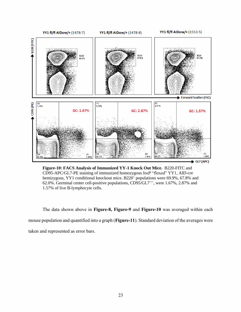
The experimental group (YY1-conditional knockout mice) consisted of three homozygous
loxP “floxed” YY1, AID-cre hemizygous mice that were immunized with sheep red blood cells
(Figure-10). This data shows an average B-lymphocyte B220+ population of 66.2% and an average
germinal center CD95/GL7+/+ population of 2.03% of live B-lymphocyte cells.
23
Figure-10: FACS Analysis of Immunized YY-1 Knock Out Mice. B220-FITC and
CD95-APC/GL7-PE staining of immunized homozygous loxP “floxed” YY1, AID-cre
hemizygous, YY1 conditional knockout mice. B220+ populations were 69.9%, 67.8% and
62.0%. Germinal center cell-positive populations, CD95/GL7+/+, were 1.67%, 2.87% and
1.57% of live B-lymphocyte cells.
The data shown above in Figure-8, Figure-9 and Figure-10 was averaged within each
mouse population and quantified into a graph (Figure-11). Standard deviation of the averages were
taken and represented as error bars.

24
Figure-11: Summary Graph of Germinal Center Average Percentages Per B-cell for
Each Mouse Population. Non-immunized (n=1), immunized homozygous loxP “floxed”
YY1, AID-cre nullizygous (n=3) (containing YY1), and immunized homozygous loxP
“floxed” YY1, hemizygous AID-cre (n=3) (YY1 knockout) mice populations shown.
Means of each population’s germinal center percentage was taken and depicted as well as
standard deviation of the averages in the form of error bars.
Overall, the non-immunized mouse (N=1) (left histobar) had a very low germinal center
population, 0.741%, the YY1 fl/fl immunized homozygous loxP “floxed” YY1, AID-cre
nullizygous population (middle histobar, containing YY1) had an average germinal center
population of 3.26%, and the YY1 fl/fl AID-cre/+ immunized homozygous loxP “floxed” YY1,
hemizygous AID-cre population (right histobar, YY1-knockout) had a reduced average germinal
center population of 2.03%. All germinal center percentages were the population of germinal
center cells relative to the total population of live B-lymphocyte cells.
CRISPR System for YY1 Knockout
The second part of this project dealt with the design of a guide RNAs for CRISPR/Cas9
usage to knockout the YY1 gene in B-lymphocyte cell lines, in vitro. The purpose of designing

25
this gRNA was for insertion into a lentiCRISPR vector and transformation into cells to create large
quantities of lentiCRISPR plasmid with the correct gRNA insertion. After transformation of the
lentiCRISPR, gRNA oligo duplex ligation product, sequencing of the purified plasmid from the
transformation was conducted. The plasmids from five different cultures were purified and sent
for sequencing: two for the first designed guide RNA (G1A & G1B), one for the second designed
guide RNA (G2B), and two for the third designed guide RNA (G3A & G3B). Once sequencing
data was received, 4Peaks software (Nucleobytes, Aalsmeer, Netherlands) was used to visualize
sequencing data and verify the insertion of the designed gRNA sequence within the five sets of
purified plasmid. The data (Figure-12), showed that 3 of the 5 plasmid samples correctly contained
the guide RNA within the lentiCRISPR plasmid.

26
Figure-12: Sequence Data Obtained from the Lenti-CRISPR Guide RNA Insertion
Plasmids. Sequencing was performed to confirm the correct insertion of the guide RNA into the
lentiCRISPR plasmid. Three of the plasmid samples had the correct insertion of the designed
guide RNA for the YY1 exon 1. (Guide 1, CTACATCGCCACGGACGGCT, inserted in G1A
and G1B and Guide, GTCGGGTCGTCGGTGACCAG, inserted in G2B)
The successful insertion of the guide RNA sequences into the lenti-CRISPR vector is
shown in the diagram below in Figure-13.
Figure-13: Diagram of the LentiCRISPR v2 Vector Containing the YY1 Guide
RNA. Shown is a linear view of the lentiCRISPR vector with the insertion of the YY1
gRNA sequences shown as blue in the center of the diagram. Different plasmid names
and their specific YY1 target sequences are shown at their insertion sites.

27
DISCUSSION
This project sought to determine whether a knockout of transcription factor YY1 had an
effect on germinal center formation in mice. Germinal center formation and their reactions are not
well characterized. A deeper understanding of these reactions and how they affect B-lymphocyte
formation could improve our understanding of antibody formation. Transcription factor YY1 was
focused on in this research because it was previously hypothesized to be a master regulator of
germinal center formation based on gene expression profiling (Green et al., 2011). To study
transcription factor YY1, the YY1 gene was knocked out using an AID-cre/lox. AID-cre is a
germinal center B-lymphocyte-specific recombination system that creates YY1 conditional
knockout mice. Expression of the Cre-recombinase is driven by AID promoter, a gene for an
enzyme expressed in germinal centers. These mice were injected with sheep red blood cells to
stimulate their immune system, and then slenocyte cells were isolated to perform FACS flow
cytometry analysis. Slenocyte cells were stained with a pan B-lymphocyte antibody, B220, and
germinal center specific antibodies, CD95 and GL7. Additionally, CRISPR/cas technology was
utilized for performing the YY1 knockout. The YY1 deletion construct was made in a
lentiCRISPR vector to be used in the future for deletion of YY1 through the CRISPR/cas 9 system
in vitro.
In the first part of the project, FACS analysis was performed on various Cre mice to
monitor germinal center B-lymphocyte numbers. Wild Type (WT) non-immunized control mouse
revealed a low germinal center percentage of 0.741% (percentage of germinal center positive cells
relative to the total live B-lymphocyte cells). A low germinal center population is expected in non-
immunized mice because the B-lymphocyte cells are not presented with any antigen so they do not

28
respond, therefore little germinal center function is required. A small baseline percentage occurs,
due to baseline immune activity, which is normal.
Next, the FACS analysis was performed on negative control mice. These mice were
homozygous loxP “floxed” YY1, AID-cre nullizygous mice (YY1 fl/fl AID-cre null), that
contained homozygous loxP “floxed” YY1 genes but no AID-cre gene to activate the “floxed”
YY1 gene, therefore there was no YY1 deletion. Since there was no YY1 deletion, these mice
should behave as WT mice when presented with antigen. The responses of these immunized mice
(Figure-9) do show germinal center reaction increases relative to non-immunized mice. These
three YY1 f/fl AID-cre null mice, 1478-3, 1478-6 and 1478-10, show a heightened germinal center
response compared to that of the non-immunized mouse suggesting there is in fact germinal center
responses similar to WT immunized mice. These mice had individual germinal center responses
of 3.44%, 2.41% and 3.93%, respectively, with an average germinal center response of 3.26%
germinal center cells of the total live B-lymphocyte cells.
It should be noted at an average germinal center response in an immunized WT mouse
averages about 5-6% at day 10 after immunization (Béguelin et al., 2013). The average response
seen here for our immunized negative control, 3.26%, is a bit low compared to the average WT
immunized mouse. This slight difference may be due to the condition of sheep red blood cells
injected into these mice. A follow up experiment to this one, done by the Hong Zhang lab using
the same strain but a different stock of sheep red blood cells revealed a higher, approximately 5%,
germinal center percentage, a more normal germinal center response to sheep red blood cell
injection (data not shown).
The experimental set of mice lacking YY1, homozygous loxP “floxed” YY1, AID-cre
hemizygous mice (YY1 fl/fl AID-cre/+), behaved differently than that of the negative control YY1

29
fl/fl AID-cre-null mice. Given the decrease in germinal center positive B-lymphocytes, YY1 was
likely deleted within the germinal center B-lymphocyte cells and germinal center reactions are
likely affected by deletion of the YY1 gene within B-lymphocyte cells, however studies are
ongoing to confirm these facts. Figure-10 reveals germinal center responses of the three
immunized conditional knockout (CKO) mice, 1478-7, 1478-8 and 1553-5, to be 1.67%, 2.87%
and 1.57%, respectively. The average germinal center response in these YY1 fl/fl AID-cre/+ mice
was 2.03%. This decrease in average germinal center response from 3.26% to 2.03%, though
minimal, appears to be a decrease in germinal center formation, which suggests that the deletion
of YY1 plays a role in germinal center formation, though what exact role YY1 plays is still
unknown.
Although there appears to be a difference in germinal center response in this data with YY1
knockout, the difference was not statistically significant (Figure-11). The lack of statistical
significance could be attributed to the low germinal center response in the immunized negative
control due the poor condition of sheep red blood cells used for injection. In the subsequent
experiment, referenced to earlier, a statistical significance was in fact seen between the YY1-
positive and YY1-knockout mice when the same experiment was repeated in the Zhang lab (data
not shown).
This FACS data, shown in this report and the unpublished Zhang lab data, suggest that the
YY1 transcription factor does play a role as a regulator of germinal center formation, in vivo, as
hypothesized by the gene expression profiling analysis done in 2011 (Green et al., 2011). The next
step would be to understand and characterize exactly what role YY1 is playing in these germinal
center formation by performing a transcriptional profile on YY1 knockout cells to deduce which
genes are affected. Antibody titering for this experiment was completed (data not shown).
30
With respect to the CRISPR portion of the project, the guide RNA plasmid was
successfully constructed and verified by DNA sequencing. Of the five purified plasmid sample
samples submitted for sequencing, three were confirmed to contain the correct insertion of the
designed guide RNA into the lentiCRISPR vector through the transformation process (Figure-12).
With the successful insertion of the designed gRNAs into the lentiCRISPR vector, the next steps
would be to package the construct into a virus for insertion into a cell line to completely delete the
YY1 gene using the CRISPR/Cas9 system (Figure-3). Given the success of the mouse breeding
strategy for the Cre-recombinase system, this in vitro system of deletion would act merely as a
positive control of YY1 knockout in subsequent FACS flow cytometry experiments.
In summary, the preliminary data outlined in this report (and supplemented by Zhang lab
data) suggest that the YY1 transcription factor likely plays a role in germinal center formation as
seen by the decrease in germinal center numbers after its knockout using the AID-cre/lox
recombination system. Ongoing experimentation is currently validating the deletion of YY1 in
germinal center cells, and further experimentation is necessary to show exactly what role the
YY1 transcription factor plays in germinal center reactions.

31
BIBLIOGRAPHY
Addgene (2014) Addgene: CRISPR in the Lab: A Practical Guide.
https://www.addgene.org/CRISPR/guide/
Alt FW, Oltz EM, Young F, Gorman J, Taccioli G, & Chen J (1992) VDJ recombination.
Immunology Today, 13(8), 306-314. doi: 10.1016/0167-5699(92)90043-7
Béguelin W, Popovic R, Teater M, Jiang Y, Bunting KL, Rosen M, Melnick AM (2013) EZH2 is
Required for Germinal Center Formation and Somatic EZH2 Mutations Promote
Lymphoid Transformation. Cancer Cell, 23(5), 677-692. doi:
http://dx.doi.org/10.1016/j.ccr.2013.04.011
Gordon S, Akopyan G, Garban H, Bonavida B (2005) Transcription factor YY1: structure,
function, and therapeutic implications in cancer biology. Oncogene, 25(8), 1125-1142.
Green MR, Monti S, Dalla-Favera R, Pasqualucci L, Walsh NC, Schmidt-Supprian M, Kutok JL,
Rodig SJ, Neuberg DS, Rajewsky K, Golub TR, Alt FW, Shipp MA, Manis JP (2011)
Signatures of murine B-cell development implicate Yy1 as a regulator of the germinal
center-specific program. Proceedings of the National Academy of Science, USA, 108(7),
2873-2878. doi: 10.1073/pnas.1019537108
Heesters BA, Myers RC, Carroll MC (2014) Follicular dendritic cells: dynamic antigen libraries.
Nature Reviews Immunology, 14(7), 495-504. doi: 10.1038/nri3689
Immune System B-Cells (2015) http://www.niaid.nih.gov/topics/immunesystem/
immunecells/Pages/bcells.aspx
Liu H, Schmidt-Supprian M, Shi Y, Hobeika E, Barteneva N, Jumaa H, Pelanda R, Reth M,
Skok J, Rajewsky K, Shi Y (2007) Yin Yang 1 is a critical regulator of B-cell
development. Genes and Development, 21(10), 1179-1189. doi: 10.1101/gad.1529307
MacLennan IC (1994) Germinal centers. Annual Review of Immunology, 12, 117-139. doi:
10.1146/annurev.iy.12.040194.001001
Mali P, Yang L, Esvelt KM, Aach J, Guell M, DiCarlo JE, Norville JE, Church GM (2013)
RNA-guided human genome engineering via Cas9. Science, 339(6121): 823-826. doi:
10.1126/science.1232033.
Natkunam Y (2007) The Biology of the Germinal Center. ASH Education Program Book,
2007(1), 210-215. doi: 10.1182/asheducation-2007.1.210
Ramkumar C, Cui H, Kong Y, Jones SN, Gerstein RM, & Zhang H (2013) Smurf2 suppresses B-
cell proliferation and lymphomagenesis by mediating ubiquitination and degradation of
YY1. Nature Communications, 4, 2598. doi: 10.1038/ncomms3598
32
Sanjana NE, Shalem O, Zhang F (2014) Improved vectors and genome-wide libraries for
CRISPR screening. Nature Methods, 11(8): 783-784. doi: 10.1038/nmeth.3047.
10.1038/nmeth.3047 PubMed 25075903
Sauer B, Henderson N (1988) Site-specific DNA recombination in mammalian cells by the Cre
recombinase of bacteriophage P1. Proceedings of the National Academy of Science,
USA, 85: 5166–5170.
Shen B, Zhang J, Wu H, Wang J, Ma K, Li Z, Zhang X, Zhang P, Huang X (2013) Generation of
gene-modified mice via Cas9/RNA-mediated gene targeting. Cell Research, 23(5), 720-
723. doi: 10.1038/cr.2013.46
The Jackson Laboratory (2015) Introduction to Cre-lox technology.
http://cre.jax.org/introduction.html
Thorbecke GJ, Amin AR, Tsiagbe VK (1994) Biology of germinal centres in lymphoid tissue.
FASEB, 8 (11): 832–840.
Victora GD (2014) SnapShot: the germinal center reaction. Cell, 159(3), 700-700.e701. doi:
10.1016/j.cell.2014.10.012