the effects of vitamin c on mg-63 cancer cell survivorship joe ziccarelli grade 12 central catholic...
TRANSCRIPT

The Effects of Vitamin C on MG-63 Cancer Cell Survivorship
Joe ZiccarelliGrade 12Central Catholic High SchoolPJAS 2015

An Overview of Cancer Cells
• Cancer cells are cells that grow and divide at an irregular, unregulated pace.
• Apoptosis does not occur in cancerous cells; their mutations are passed on to the second generation, eventually clustering and forming tumors.
• Tumors can be malignant (aggressive) or benign.
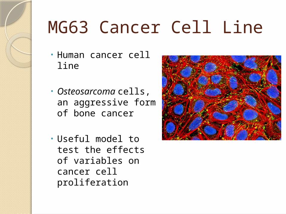
MG63 Cancer Cell Line• Human cancer cell
line
• Osteosarcoma cells, an aggressive form of bone cancer
• Useful model to test the effects of variables on cancer cell proliferation

Antioxidants
• Molecules capable of slowing or preventing the oxidation of other molecules
• May be able to prevent cancer and coronary heart disease
• Body produces antioxidants • Can obtain through Diet• Past studies have shown that the
antioxidant nature of Vitamin C has anti-tumoregenic effects

Variable: Ascorbic Acid (Vitamin C)
• Antioxidant• Enzyme cofactor • In Oranges, Strawberries,
and Grapefruit• Recommended daily intake: 60
mg• The disease scurvy occurs from
lack of Vitamin C
Purpose• To determine the effects of
ascorbic acid on MG-63 cell survivorship.

Hypotheses
• Null Hypothesis: Ascorbic will not have a significant effect on MG63 cell survivorship.
• Alternative Hypothesis: Ascorbic Acid will significantly alter the survivorship of MG63 cells.
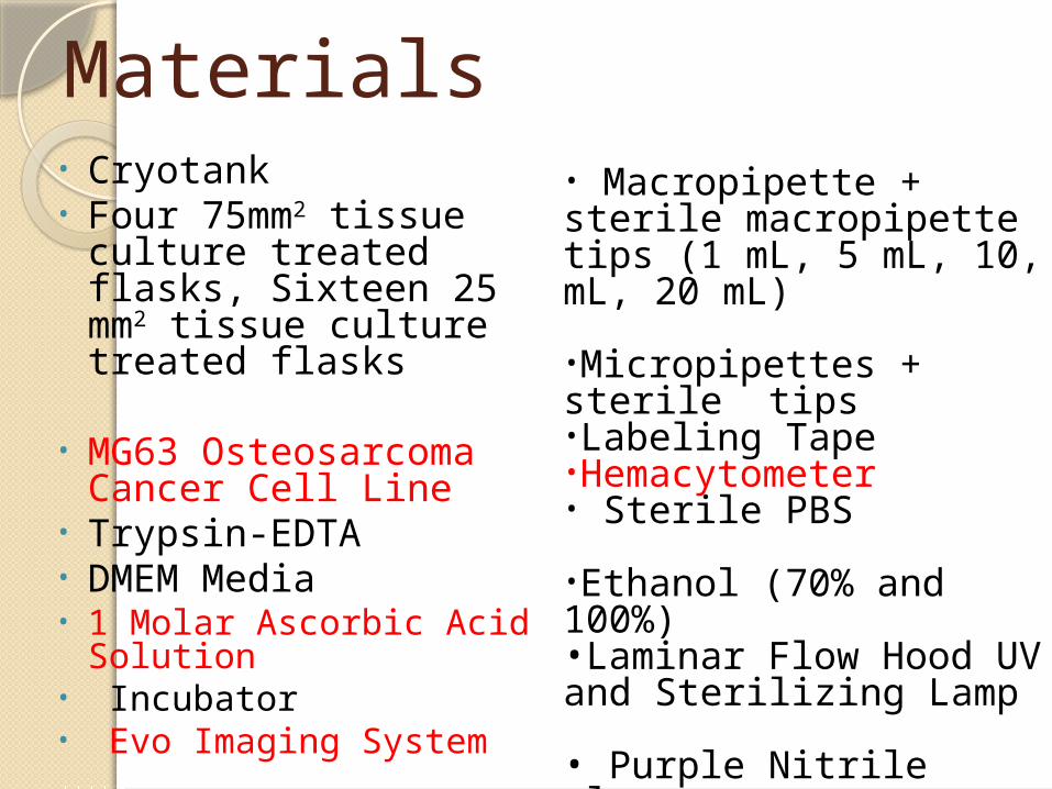
Materials• Cryotank• Four 75mm2 tissue
culture treated flasks, Sixteen 25 mm2 tissue culture treated flasks
• MG63 Osteosarcoma Cancer Cell Line
• Trypsin-EDTA• DMEM Media• 1 Molar Ascorbic Acid
Solution• Incubator• Evo Imaging System
• Macropipette + sterile macropipette tips (1 mL, 5 mL, 10, mL, 20 mL)
•Micropipettes + sterile tips•Labeling Tape•Hemacytometer• Sterile PBS
•Ethanol (70% and 100%)•Laminar Flow Hood UV and Sterilizing Lamp
• Purple Nitrile gloves•Safety goggles

Procedure 1: Cell Culturing• A 1 mL aliquot of MG63 cells from a Cryotank
was used to inoculate 30 mL of 10% serum DMEM media in one 75mm2 culture flask yielding a cell density of approximately 106 to 2 x 10⁶ cells.
• The media was replaced with 15 mL of fresh media to remove cryo-freezing fluid and incubated (37° C, 5% CO2) for 2 days until a cell density of approximately 4 x 10⁶ to 5 x 10⁶ cells/mL was reached.
• The culture was passed into two sets of 3 flasks in preparation for experiment and incubated for 2 days at 37° C, 5% CO2.

Procedure 1: Proliferation Experiment- Day 0 (Addition of Variable)• After trypsinization, cells from all of the flasks were
pooled into 1 common 75mm2 flask (cell density of approximately 1 million cells/mL).
• 0.1 mL of the cell suspension was added to eight 25 mm2 tissue culture treated flasks containing 5 mL of DMEM (com) media, creating a cell density of approximately 105 cells per flask.
• The stock solution of ascorbic acid (one molar) was created using 50 mL of ethanol and 10 g of ascorbic acid. This was then used in a serial dilution to create a 10-2, 10-4, 10-6 M concentration.
• The cells were incubated at 37°C, 5% CO2 for the remainder of the study.

Procedure 2: Cell Culturing• A 1 mL aliquot of MG63 cells from a Cryotank
was used to inoculate 30 mL of 10% serum DMEM media in one 75mm2 culture flask yielding a cell density of approximately 106 to 2 x 10⁶ cells.
• The media was replaced with 15 mL of fresh media to remove cryo-freezing fluid and incubated (37° C, 5% CO2) for 2 days until a cell density of approximately 4 x 10⁶ to 5 x 10⁶ cells/flask was reached.
• The culture was passed into two sets of 3 flasks in preparation for experiment and incubated for 2 days at 37° C, 5% CO2.
Procedure 2: Proliferation Experiment- Day 0 (Addition of Variable)• After trypsinization, cells from all of the flasks were
pooled into 1 common 75mm2 flask (cell density of approximately 1 million cells/mL).
• 0.1 mL of the cell suspension was added to eight 25 mm2 tissue culture treated flasks containing 5 mL of DMEM (com) media, creating a cell density of approximately 105 cells per flask.
• The stock solution of ascorbic acid (one molar) was created using 50 mL of ethanol and 10 g of ascorbic acid. This was then used in a serial dilution to create a 10-2, 10-4, 10-6 M concentration.
• The cells were incubated at 37°C, 5% CO2 for the remainder of the study.
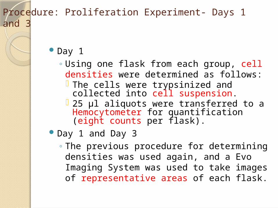
Procedure: Proliferation Experiment- Days 1 and 3
Day 1◦ Using one flask from each group, cell
densities were determined as follows: The cells were trypsinized and collected
into cell suspension. 25 µl aliquots were transferred to a
Hemocytometer for quantification (eight counts per flask).
Day 1 and Day 3◦ The previous procedure for determining
densities was used again, and a Evo Imaging System was used to take images of representative areas of each flask.

Results of Proliferation Analysis (MG63)
Control 10^-6 M 10^-4 M 10^-2 M0
50
100
150
200
250
300
350
400
450
500
Effect of Vitamin C on MG63 Survivorship (Vitamin C Exposure after two days)
Day 1 Day 3
Concentrations of Ascorbic Acid
Cell C
ou
nt
(Cells/
Fla
sk)
in t
hou
san
ds
P-value
7.25E-22
P-value
4.15E-15

Dunnett’s Test (Vitamin C Exposure After Two Days)
Concentration
T-Value T-Critical (0.05)
Variation
MG63 - - -
10^-6 M 2.057 2.88 Insignificant
10^-4 M 18.322 2.88 Significant
10^-2 M 26.357 2.88 Significant

Results of Proliferation Analysis (MG63)
Control 10^-6 M 10^-4 M 10^-2 M0
50
100
150
200
250
300
350
400
450
Effect of Vitamin C on MG63 Survivorship
Day 1 Day 3
Concentrations of Ascorbic Acid
Cell C
ou
nt
(Cells/F
lask)
in
thou
san
ds
P-value
2.15E-13
P-value
2.09E-13

Dunnett’s Test (Variable Added Without Two Day Proliferation)
Concentration
T-Value T-Critical (0.05)
Variation
MG63 - - -
10^-6 M 0.095 2.88 Insignificant
10^-4 M 19.205 2.88 Significant
10^-2 M 26.551 2.88 Significant

Conclusions Proliferation
◦ MG63 (variable added after two day proliferation) Based upon the results gathered from the ANOVA and
Dunnett’s statistical analyses, it appears that the addition of ascorbic acid at higher concentrations significantly affects cancer cell proliferation. Therefore, the null hypothesis can be rejected.
◦ MG63 (variable added without two day proliferation) Based upon the results gathered from the ANOVA and
Dunnett’s statistical analyses, it appears that the addition of ascorbic acid at higher concentrations significantly affects cancer cell proliferation. Therefore, the null hypothesis can be rejected.

Future Changes
Limitations Extensions• Hemacytometer counts
in proliferation experiment were subject to clumping error
• Limited range of concentrations
• Limited days of exposure• Only accessed
proliferation/survivorship
• Include further trials in order to maximize data accuracy
• Use a wider range of concentrations
• Test synergistic effects of other vitamins

Acknowledgements• Mark Krotec, PTEI
• Merisant Company

Works Citedhttp://cancerres.aacrjournals.org/
content/40/3/734.full.pdfhttp://www.cancer.gov/cancertopi
cs/pdq/cam/highdosevitaminc/patient/page2
http://www.mayoclinic.org/diseases-conditions/cancer/expert-answers/alternative-cancer-treatment/faq-20057968

Works Cited (cont.)http://lpi.oregonstate.edu/s-s00/v
itaminc.htmlhttp://www.webmd.com/cancer/
news/20140205/intravenous-vitamin-c-may-boost-chemos-cancer-fighting-power

Statistical Analyses of the Proliferation Results• ANOVA• Compares variation within groups to variation
between groups.
• Using the ANOVA, a p-value less than the alpha of .05 was gathered (significant variation). • Reject the null hypothesis.
• Dunnett’s test• Compares each experimental group to control
individually.• 0.05 alpha was used, and the t-value compared to
the t-critical value of 2.88

Additional Information• DMEM Serum - 1% and Complete
Media (4 mM L-glutamine, 4500 mg/L glucose, 1 mM sodium pyruvate, and 1500 mg/L sodium bicarbonate + [ 10% fetal bovine serum for complete])