the effect of water hardness on surfactant deposition
TRANSCRIPT
Accepted Manuscript
The Effect of Water Hardness on Surfactant Deposition Following Washing andSubsequent Skin Irritation in Atopic Dermatitis Patients and Healthy Controls
Simon G. Danby, Kirsty Brown, Andrew M. Wigley, John Chittock, Phyoe K. Pyae,Carsten Flohr, Michael J. Cork
PII: S0022-202X(17)32938-X
DOI: 10.1016/j.jid.2017.08.037
Reference: JID 1065
To appear in: The Journal of Investigative Dermatology
Received Date: 20 June 2017
Revised Date: 10 August 2017
Accepted Date: 27 August 2017
Please cite this article as: Danby SG, Brown K, Wigley AM, Chittock J, Pyae PK, Flohr C, Cork MJ, TheEffect of Water Hardness on Surfactant Deposition Following Washing and Subsequent Skin Irritation inAtopic Dermatitis Patients and Healthy Controls, The Journal of Investigative Dermatology (2017), doi:10.1016/j.jid.2017.08.037.
This is a PDF file of an unedited manuscript that has been accepted for publication. As a service toour customers we are providing this early version of the manuscript. The manuscript will undergocopyediting, typesetting, and review of the resulting proof before it is published in its final form. Pleasenote that during the production process errors may be discovered which could affect the content, and alllegal disclaimers that apply to the journal pertain.
MANUSCRIP
T
ACCEPTED
ACCEPTED MANUSCRIPT
Page 1 of 27
THE EFFECT OF WATER HARDNESS ON SURFACTANT
DEPOSITION FOLLOWING WASHING AND SUBSEQUENT SKIN
IRRITATION IN ATOPIC DERMATITIS PATIENTS AND HEALTH Y
CONTROLS
Simon G. Danby1,*, [email protected], ORCID: 0000-0001-7363-140X
Kirsty Brown1, [email protected]
Andrew M. Wigley1, [email protected]
John Chittock1, [email protected], ORCID: 0000-0002-1595-7441
Phyoe K. Pyae1, [email protected]
Carsten Flohr2,** , [email protected]
Michael J. Cork1,3,**, [email protected], ORCID: 0000-0003-4428-2428
1Sheffield Dermatology Research, The University of Sheffield Medical School, Sheffield,
UK;
2Unit for Population-Based Dermatology Research, St. John’s Institute of Dermatology,
King’s College London and Guy’s & St Thomas’ Hospital, London, UK
3The Paediatric Dermatology Clinic, Sheffield Children's Hospital, Sheffield, UK;
*Corresponding author: Sheffield Dermatology Research, Department of Infection &
Immunity & Cardiovascular Disease, Faculty of Medicine, Dentistry & Healthy, University of
Sheffield Medical School, Beech Hill Road, Sheffield S10 2RX, UK. Tel: +44 (0)114 21
59563, Email: [email protected]
**Both authors have equally contributed to this publication
Short Title: Water hardness and skin irritation in atopic dermatitis patients

MANUSCRIP
T
ACCEPTED
ACCEPTED MANUSCRIPT
Page 2 of 27
Abbreviations: Atopic Dermatitis, AD; Interleukin, IL; Sodium Lauryl Sulfate, SLS;
Transepidermal water loss, TEWL
ABSTRACT
Living in a hard water area is associated with an increased risk of atopic dermatitis (AD).
Greater skin barrier impairment following exposure to surfactants in wash products combined
with high calcium, and/or chlorine, levels in hard water is a compelling mechanism for this
increase. The purpose of this study was to investigate this mechanism in individuals with and
without a predisposition to skin barrier impairment.
We recruited 80 subjects; healthy controls and AD patients with and without FLG mutations.
The skin of each participant was washed with sodium lauryl sulfate (SLS) in water of varying
hardness and chlorine concentration, rinsed and covered with chambers to determine the
effects of surfactant residues.
Sites washed with hard water exhibited significantly increased SLS deposits. These deposits
increased transepidermal water loss and caused irritation, particularly in AD patients carrying
FLG mutations. A clear effect of chlorine was not observed. Water softening by ion-exchange
mitigated the negative effects of hard water.
Barrier impairment resulting from the interaction between hard water and surfactants is a
contributory factor to the development of AD. Installation of a water softener in early life may
be able to prevent AD development. An intervention study is required to test this hypothesis.

MANUSCRIP
T
ACCEPTED
ACCEPTED MANUSCRIPT
Page 3 of 27
INTRODUCTION
Atopic dermatitis/eczema (AD) is a common inflammatory disease of the skin, affecting 15–
30% of children and 2–10% of adults (Odhiambo et al., 2009). An important pathophysiologic
event in the development of AD is impairment of the skin barrier (Cork et al., 2009). Loss-of-
function mutations in the filaggrin (FLG) gene are an important cause of skin barrier
impairment, and represent the strongest genetic risk factor for AD (McAleer and Irvine, 2013).
Nevertheless, genetics alone cannot fully explain a person’s susceptibility to AD, and it is
believed that environmental factors play an important role by contributing to skin barrier
impairment. Washing the skin with hard water is one such environmental factor purported to
increase the risk of developing AD.(Ewence et al., 2011)
Hard water contains high levels (≥100mg/l) of calcium and magnesium carbonates, such as
the minerals calcite, gypsum and dolomite (Ewence et al., 2011). Domestic water hardness
varies throughout the world depending on the geography of the land. A number of studies
have now reported an increased prevalence of AD amongst infants and school children living
in hard, compared to soft, water areas (Arnedo-Pena and Bellido-Blasco, 2007, Chaumont et
al., 2012, Engebretsen et al., 2016, McNally et al., 1998, Miyake et al., 2004). Moreover, a
predisposition to skin barrier impairment, due to carriage of a FLG loss-of-function mutation,
additively increased the risk of developing AD for those living in a hard water area (Perkin et
al., 2016). A cogent pathological process is suggested whereby the effects of washing with
hard water contribute to skin barrier impairment in addition to genetic factors to determine an
individual’s overall risk. What is still unclear is how hard water impairs the skin barrier, and a
better understanding of the underlying mechanisms will aid the design of future intervention
studies aimed at reducing the incidence of AD by eliminating water hardness.

MANUSCRIP
T
ACCEPTED
ACCEPTED MANUSCRIPT
Page 4 of 27
As part of a systematic review for the UK Drinking Water Inspectorate, we identified several
possible mechanisms by which hard water may damage the skin barrier that need further
investigation (Ewence et al., 2011). The most promising of these is the interaction between
hard water and the surfactants (detergents) used in wash products. High calcium levels are
thought to reduce the solubility of surfactants and thereby potentially increase their deposition
on the skin following washing (Young and Matijevic, 1977). Common surfactants used in
wash products, such as the harsh anionic surfactant sodium lauryl sulfate (SLS), are widely
established skin irritants and important environmental stressors contributing to the severity of
AD (Ananthapadmanabhan et al., 2004).
We therefore conducted a case-control study to investigate the effects of water type on
surfactant skin deposition following washing and subsequently assess the effects of the
deposits on skin barrier function and skin irritation in individuals with healthy skin compared
to AD patients with and without FLG loss-of-function mutations. In addition to the high
calcium and magnesium levels, hard domestic water often also exhibits a high chlorine level
(Perkin et al., 2016). Chlorine is often added to domestic water and is a known skin irritant,
which could potentially modify or contribute to the effects of water hardness on the skin
(Ewence et al., 2011). We therefore controlled for both water hardness and chlorine levels.
We also wanted to evaluate the potential of an ion-exchange water softener to mitigate, or
eliminate, the effects of high calcium and magnesium levels on skin barrier function.
RESULTS
We recruited and screened 304 participants (154 with healthy skin and 150 with AD) between
November 2015 and July 2016 to establish their FLG gene status (5 most common mutations
in Europeans, Sandilands et al., 2007). An overview of recruitment is provided in Figure 1.
During the pre-defined recruitment period, we were able to fill 3 of the 4 groups. Owing to

MANUSCRIP
T
ACCEPTED
ACCEPTED MANUSCRIPT
Page 5 of 27
the rarity of individuals with healthy skin who carry a FLG mutation (only 7% of the eligible
population) we were unable to fully fill group 2. In total 12 participants were lost-to-follow (8
didn't show up for visit 1 of the patch testing, 3 cancelled their appointment for visit 1 and
withdrew themselves owing to time commitments, and 1 withdrew after visit 1 due to
discomfort of the patches), and 3 completed healthy participants were excluded because they
were later found to be atopic, which was a predefined exclusion criterion for group 1. A
summary of the study groups (completed participants) is presented in Table 1. With the
exception of group 2, the study groups were evenly sized and matched for gender, age and
Fitzpatrick skin type. As expected, the participants in the AD groups reported dryer skin
compared to the healthy groups. A high rate of adverse reactions to wash products was
reported by both AD groups (75 and 73%), while no such reactions were reported in the
groups with healthy skin irrespective of FLG gene status.
The deposition of surfactants on the skin following washing
The type of wash water significantly affected surfactant deposition following washing (Figure
2). Hard water was associated with the greatest deposition of SLS, which was 2.8±0.6 fold
greater than when deionized water was used for washing. The level of chlorine in the water
had no effect on SLS deposition. Softening the water, to remove calcium and magnesium ions,
significantly reduced the level of SLS deposition. Upon stratification of the cohort, no effect
of AD or FLG mutations on SLS deposition was found (Figure 2d).
Based upon the FTIR spectra collected from the skin sites, washing with hard water was
associated with a significant shift in the location of the CH2 symmetric stretching band
(approx. 2850 cm-1) to a higher wavenumber compared to washing with deionized water,
indicative of an increase in lipid disordering/fluidity (Figure 2e). Similarly, washing with hard
MANUSCRIP
T
ACCEPTED
ACCEPTED MANUSCRIPT
Page 6 of 27
water was associated with a shift in the location of the amide I bond (C=O) compared to
washing with deionized water, indicative of protein denaturation (Figure 2f). Both the change
in lipid and protein structure correlated significantly with SLS deposition on the skin surface
(Spearman’s r = 0.392 and 0.354, p = <0.0001 and <0.0001 respectively).
Skin irritation from surfactant residues
Patch testing was performed to determine whether SLS deposits left on the skin can damage
the skin barrier and induce irritation (Figure 3). TEWL was significantly elevated at all test
sites compared to the untreated control. This indicates reduced skin barrier function as a result
of SLS deposits on the skin irrespective of the wash water used. Importantly, the reduction in
skin barrier function was significantly greater at sites where hard water was used for washing
(10.19 ±0.74 g/m2/h without chlorine and 9.45±0.80 g/m2/h with chlorine) compared to
deionized water (7.43 ±0.74 g/m2/h without chlorine and 7.51 ±0.92 g/m2/h with chlorine).
Moreover, the increase in TEWL directly correlated with the amount of SLS deposited on the
skin following washing (Figure 3c), and the amount of residue remaining on the skin
following patch removal (Spearman’s r=0.4928 and 0.4108, p<0.0001 and <0.0001
respectively). Water softening, in line with the reduction in SLS deposits on the skin,
mitigated the negative effect of hard water on skin barrier function. The level of chlorine had
no significant effect on skin barrier function. Upon stratification by group, AD patients
carrying the FLG gene mutation were affected by SLS deposits to a significantly greater
extent compared to individuals with no FLG mutation and healthy skin (Figure 3b).
Following washing with SLS in hard water, TEWL increased by 7.12 ±0.84 g/m2/h in people
with healthy skin compared to an increase of 13.84 ±1.68 g/m2/h in people with AD carrying
a FLG mutation.
MANUSCRIP
T
ACCEPTED
ACCEPTED MANUSCRIPT
Page 7 of 27
A similar picture emerged for the effects of SLS deposits on objective skin redness (Figure
3d). All test sites, except the sites washed with deionized water, exhibited significantly
elevated redness compared to the untreated control. Additionally, there was a significant
difference between the test sites, with the hard water and the deionized water with high
chlorine test sites showing the greatest increases in redness. Redness was also significantly
correlated with SLS levels, however this was most evident for residues quantified following
patch removal (Figure 3f, r=0.411) compared to deposits quantified before patch application
(r=0.238, p<0.0001). Again, the increase in objective redness was significantly different in
each group (Figure 3e).
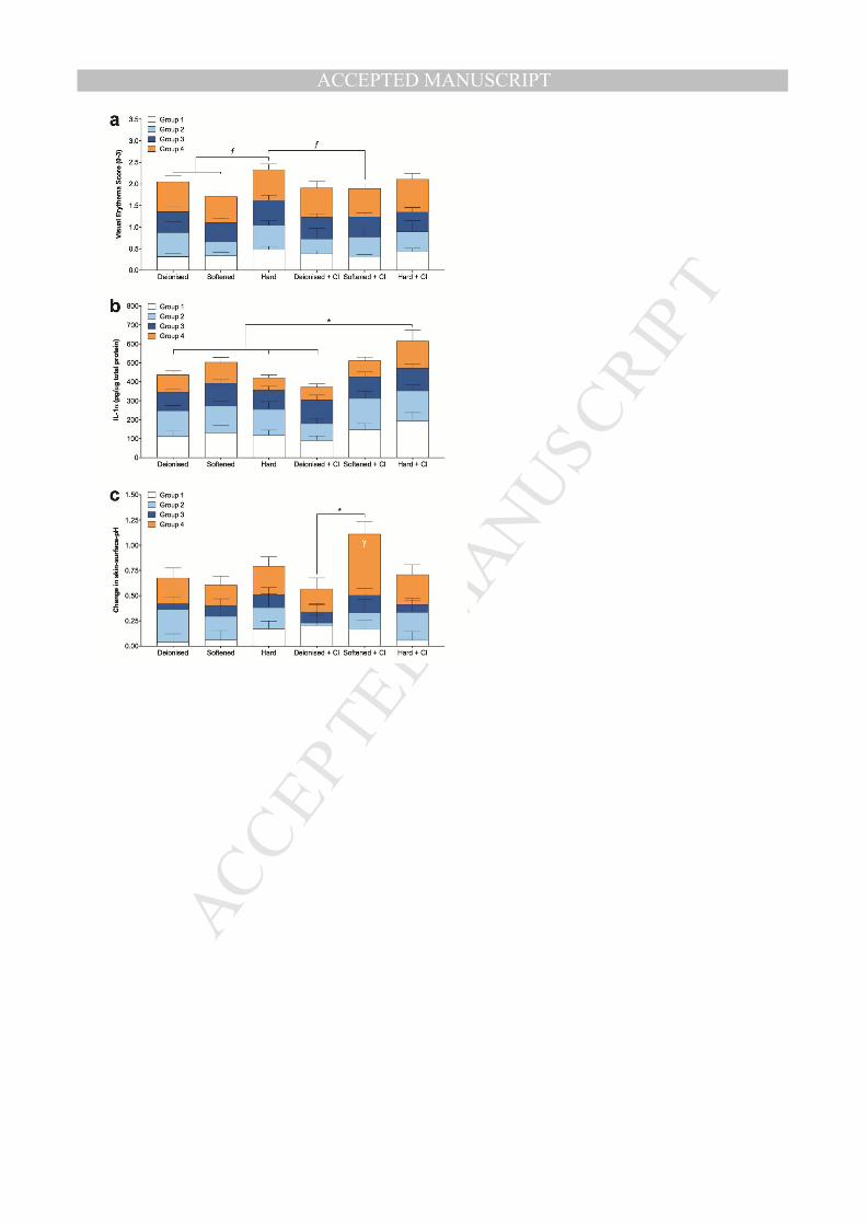
The secondary outcome measures are presented in Figure 4. Visual scoring of erythema
followed a similar pattern to objective skin redness, with the 2 parameters showing significant
association (r=0.508, p<0.0001). However, as expected the visual score demonstrated reduced
sensitivity to detect differences between the water types. Stratum corneum levels of the pro-
inflammatory cytokine Interleukin (IL)-1α were significantly different between the test sites,
with the hard water + chlorine wash water showing the highest levels. The use of hard water
without chlorine did not lead to elevated IL-1α levels compared to the deionized water control.
The levels of IL-1α did not correlate with skin redness (visual or objective), and were
consistently lower (not significant) in the AD FLGnull group compared to the other groups.
The type of wash water also significantly affected the change in skin surface pH following
patching. There was a significant association between the change in skin surface pH and SLS
deposits on the skin (r=0.3649, p<0.0001) and a weak association with the change in TEWL
and objective redness. In contrast to TEWL and objective redness, skin-surface pH was most
affected by the softened water containing chlorine compared to all other water types.
Additionally, this exaggerated response was predominantly displayed by the AD FLGnull

MANUSCRIP
T
ACCEPTED
ACCEPTED MANUSCRIPT
Page 8 of 27
group. Notably, softened water with chlorine displayed the highest alkalinity of all the water
types tested (Table 2).
DISCUSSION
Skin exposure to SLS is enhanced by washing in hard water, compared to deionized water,
due to an increased persistence of surfactant residues on the skin following rinsing. By using
an ion-exchange water softener to reduce hardness down to <25 mg/l CaCO3, SLS residues
were dramatically reduced, indicating that it is the metal ion (Ca2+ and Mg2+) concentration in
the water that affects deposition. No effect of chlorine level in the water, or the study
population, on surfactant deposition was found.
The SLS residues left on the skin following washing altered protein secondary structure,
solubilized stratum corneum lipids, and elevated skin surface pH in a dose-dependent manner.
Moreover the SLS residues caused skin irritation and skin barrier impairment, the extent of
which was dependent on the hardness of the wash water and could be directly related to the
level of SLS deposits on the skin. Patients with AD and a FLG mutation displayed
significantly greater skin barrier damage and irritation in response to SLS residues compared
to healthy individuals without FLG mutations, suggesting an increased sensitivity to SLS. The
use of an ion-exchange water softener to remove calcium and magnesium ions protected
against skin barrier damage and irritation by reducing SLS deposits on the skin.
The strength of this study is the very controlled nature of the intervention, which has enabled
us to focus in on a single exposure and assess the effects of varying the key properties of wash
water associated with the development of AD. As a result the effects of confounders such as
age, skin type, water composition (beyond hardness and chlorine levels) has been controlled.

MANUSCRIP
T
ACCEPTED
ACCEPTED MANUSCRIPT
Page 9 of 27
A limitation of our study is the small sample size of the healthy group carrying FLG
mutations, which stems from the low number of these cases in the population (<10%)
(Bandier et al., 2015). The effect of this limitation is a reduction in statistical power to
compare group means, so whilst we may have missed some potentially significant differences
we can be confident that the differences we have reported are true.
Our findings are supported by a number of epidemiological studies that have identified a link
between living in a hard water area and the prevalence of AD. Furthermore, we offer a
mechanism by which water hardness contributes to AD development, by increasing skin
exposure to harmful surfactants. While our study illustrates the deposition of the common
synthetic surfactant SLS, a previous study reported similar increases in skin deposition of
surfactants found in traditional soaps (alkyl carboxylates) when hard water is used for
washing compared to ultrapure soft water (Tanaka et al., 2015). An explanation for the
increased skin deposition of surfactants is their reduced solubility in solutions containing
metal ions such as calcium (Young and Matijevic, 1977). For instance, greater precipitates of
‘metallic surfactants’ (precipitates comprising calcium salts of anionic surfactants) form on
clothes fabrics when washed with SLS in hard versus soft water (Gotoh et al., 2016).
Moreover, as a result of this precipitation, wash products produce less foam in hard water
compared to soft water necessitating the use of more wash product to produce the same
amount of foam. In this regard our results are likely to underestimate the real impact of hard
water on surfactant deposits in every day washing habits because of the controlled use of SLS
in this study.
Harsh surfactants are known to have a broad range of effects that contribute to both their
cleansing efficacy and potential to cause skin irritation/barrier damage (Ananthapadmanabhan

MANUSCRIP
T
ACCEPTED
ACCEPTED MANUSCRIPT
Page 10 of 27
et al., 2004). The direct negative effects of SLS residues we report here on the stratum
corneum are consistent with the effects of SLS reported in the literature (Saad et al., 2012).
The low level residues of SLS left on the skin were sufficient to elicit mild irritation and skin
barrier damage, consistent with the effects of higher concentrations reported previously.
Topical products causing this level of skin barrier damage are associated with a high rate of
adverse skin reactions (Danby et al., 2011). In infants at 2 months of age an increase in
TEWL of just 1.4 g/m2/h above the mean is a predictive biomarker for AD (Kelleher et al.,
2015). This suggests that washing in hard water, through an interaction with surfactants in
wash products, could damage the skin barrier sufficiently to increase the risk of developing
AD in this age group.
Importantly the skin barrier damage and irritation caused as a result of washing in hard water
was significantly different between the study populations. In line with previous studies
patients with AD displayed the greatest response to SLS (Bandier et al., 2015, Darlenski et al.,
2013, Jungersted et al., 2010). Whilst AD patients exhibit a skin barrier defect irrespective of
their FLG gene status, the extent of the defect is significantly greater in those carrying a FLG
mutation, leaving them more susceptible to the effects of irritants, as established in this study
for SLS (Scharschmidt et al., 2009, Winge et al., 2011). This increase in sensitivity to SLS
helps explain the additive effect of FLG mutations on the association between living in a hard
water area and the risk of developing AD reported by Perkins et al (Perkin et al., 2016).
Intriguingly, and in agreement with a previous study, we found no significant difference in
effect of SLS and hard water between the healthy groups with and without a FLG mutation
(Bandier et al., 2015). This suggests that, whilst an important contributory factor, loss of
functional filaggrin alone isn’t sufficient to increase a person’s sensitivity to SLS.

MANUSCRIP
T
ACCEPTED
ACCEPTED MANUSCRIPT
Page 11 of 27
Carrying a FLG mutation has been associated with altered stratum corneum cytokine levels
that may orchestrate the increased skin response to SLS (Kezic et al., 2012). We did not
observe a significant association between IL-1α levels at the skin surface and FLG status, but
did observe a trend for reduced levels in AD patients with FLG mutations compared to all
other groups. We did not quantify baseline levels, and therefore cannot directly relate these
findings with the basal levels found in other study populations. Whilst contrary to the increase
in inflammation, decreased IL-1α levels in response to prolonged or repeated SLS exposures
have been reported previously (Angelova-Fischer et al., 2012). IL-1α plays an important role
in skin barrier repair (Man et al., 1999), and this finding may suggest an impeded repair
response in AD patients carrying a FLG mutation. AD patients carrying a FLG mutation also
displayed an increased propensity for changes to skin surface pH. Skin surface pH is an
important regulator of skin barrier homeostasis (Hachem et al., 2003). Moreover increasing
evidence supports a prominent role of skin pH in the pathogenesis of AD as a driver for
increased Kalikrein (KLK) 5 protease activity, with subsequent activation of the protease
activated receptor (PAR) 2 receptor, increased expression and release of the pro-allergic
cytokine thymic stromal lymphopoietin (TSLP), and consequently development of dermatitis
(Jang et al., 2016). Notably mice with a filaggrin defect exhibit heightened activity of this
pathway (Moniaga et al., 2013). Nevertheless differences in basal skin surface pH have been
inconsistently reported when comparing AD patients with and without FLG mutations
(Bandier et al., 2015, Jungersted et al., 2010). The increased susceptibility of the FLGnull AD
patients to pH changes reported here is consistent with the lower levels of skin acidifying
agents, such as urocanic acid and pyrrolidone carboxylic acid, in this population type reported
elsewhere (Kezic et al., 2008). Based on the observation that metallic surfactants can induce
TSLP expression when applied to the skin of mice, activation of the pH-protease-PAR2
pathway by surfactants combined with hard water in the context of a FLG gene defect is a

MANUSCRIP
T
ACCEPTED
ACCEPTED MANUSCRIPT
Page 12 of 27
plausible mechanism for promoting AD development (Tanaka et al., 2015). Our findings add
to an increasing body of evidence suggesting that FLG mutation carriers represent an
important sub-group of AD patients with increased skin sensitivity.
We report that use of an ion-exchange water softener to reduce calcium and magnesium levels
mitigated the adverse effects of metallic surfactants formed during washing with hard water
and the synthetic detergent SLS. Whilst ion-exchange water softeners do not completely
remove calcium and magnesium ions our findings suggests that the residual levels remaining
(<0.1 mg/l calcium and <0.05 magnesium) have a negligible effect on the skin. Water
alkalinity (the pH buffering capacity of water) is a property closely related to hardness, and so
it has been implicated as a factor in the association between hard water and AD risk (Ewence
et al., 2011). Whilst the water softening process did not appear to affect alkalinity of the water,
the softened water supplemented with additional chlorine did display a higher alkalinity. It
was the use of this water, with the highest alkalinity, that led to the most dramatic change in
skin surface pH following washing. The observed increase in pH was also associated with
decreased skin barrier function. This suggests that whilst calcium levels appear to be the key
driver for the skin barrier impairment observed, water alkalinity also needs to be controlled to
prevent the negative consequences of elevated skin surface pH (Hachem et al., 2003). Whilst
washing with acidic water appears to be beneficial for maintaining skin homeostasis (Hachem
et al., 2010), it is necessary to maintain domestically supplied water at neutral-alkaline pH to
control plumbosolvency (Ewence et al., 2011). The focus therefore needs to be on reducing
water alkalinity and/or strategies for acidifying wash water during washing, with
appropriately designed wash products for example.

MANUSCRIP
T
ACCEPTED
ACCEPTED MANUSCRIPT
Page 13 of 27
Chlorine levels are another parameter of water previously associated with skin effects
(Ewence et al., 2011). Whilst considered a skin irritant, the level of chlorine tested in this
study is at the top-end of the levels found in domestic water supplies, which is well within the
safe limits permitted in swimming pools to avoid adverse skin effects. Neither the level of
deposition or the skin response to SLS appeared to be consistently affected by chlorine under
the conditions tested. Yet, chlorine in deionized water, but not hard or softened water, did
appear to increase the level of skin irritation observed in this study. This suggests a specific
irritant effect of free chlorine in ultrapure water independent of surfactants. It’s worth noting
that the swimming pool attendance is inconsistently associated with the development of AD
in the literature, and like the association between chlorine in domestic water and AD is
confounded by whether study participants live in a hard water area (Chaumont et al., 2012,
Font-Ribera et al., 2014).
Four studies of varying quality have assessed the effect of water softeners on the severity of
established AD in humans and dogs with varying success (Ohmori et al., 2010, Tanaka et al.,
2015, Thomas et al., 2011, Togawa et al., 2014). Of these the only statistically powered
randomized controlled trial found no benefit of installing an ion-exchange water softener on
established moderate-severe AD (Thomas et al., 2011). In established AD, inflammation is a
key driver of skin barrier impairment, and may overshadow the effects of negative
environmental factors like water hardness (Kim et al., 2008). Furthermore, current guidance
on the management of AD recommends the avoidance of soap and detergents (replacing them
with emollient wash products), meaning that AD patients are already likely to take steps that
avoid exposure to metallic surfactants (Lewis-Jones and Mugglestone, 2007). The results of
this work, and those of more recent birth-cohort studies suggest that rather than affecting the
severity of established AD, hard water is likely to play a greater role in the primary

MANUSCRIP
T
ACCEPTED
ACCEPTED MANUSCRIPT
Page 14 of 27
development of AD in the first few months of life (Engebretsen et al., 2016, Perkin et al.,
2016).
In conclusion washing the skin with hard water increases exposure to potentially irritant
metallic surfactants that can impair the functioning of the skin barrier, especially in people
with a predisposition to a skin barrier defect. By additively impairing skin barrier function,
washing with hard water is likely to contribute to the early development of AD. Ion-exchange
water softeners could help reduce the risk of developing AD by reducing the deposition of
metallic surfactants on the skin during washing.
MATERIALS AND METHODS
Study site and randomization
This case-control observational study was conducted at the Royal Hallamshire Hospital in
Sheffield (UK). A sample size of 80 split evenly between 4 defined populations was set:
(Group 1) 20 participants with healthy skin (no current or past AD), no atopy, and FLGwt/wt;
(Group 2) 20 participants FLGnull/null or FLGwt/null without current or past AD; (Group 3) 20
participants with AD and FLGwt/wt; (Group 4) 20 participants with AD and FLGnull/null or
FLGwt/null. The study is powered at 80% (p = 0.05) to detect a difference in TEWL of 2.0
g/m2/h and in skin redness of 30 mexameter units, based upon an unpublished pilot study and
existing literature (Danby et al., 2011). To achieve the target samples, we set out to screen
500 volunteers over a 9-month period. Inclusion and exclusion criteria are presented in
Supplementary Table S1. Following group allocation participants were enrolled onto the skin
washing/patch testing procedure on a first come first served basis. Written informed consent
was obtained from all participants. The NHS Trent Research Ethics Committee approved the
study, including the consent procedure employed (#04/MREC/70).

MANUSCRIP
T
ACCEPTED
ACCEPTED MANUSCRIPT
Page 15 of 27
Preparation and testing of the study water
There were 2 sources of water: deionized water and hard water obtained from a domestic
supply in Essex, UK, where the water hardness is high, on 5 separate occasions during the 9-
month study period. Table 1 provides the summary data for the 5 batches. NRM laboratories
(Bracknell, UK) undertook the analysis of the deionized, hard and softened water samples.
Water hardness and alkalinity were determined by titration for each of the 6 samples
separately (MColortestTM, Merck Millipore, Darmstadt, Germany). The softened water was
prepared by running the hard water through an ion-exchange water softener (Harvey’s
Drinking Water Filter by Harvey Water Softeners Ltd., Surrey, UK and installed at the
source), which brought calcium carbonate (total hardness), calcium, and magnesium levels
down from 403.5 (>300mg/l = very hard), 113.1 and 28.3 mg/l, to 1.0 (<50 mg/l = soft), <0.1,
and <0.05 mg/l respectively. The water samples without chlorine were prepared by filtering
the hard or softened water through a carbon filter (Q5586, Omnipure, USA) at the time of
collection. The chlorinated water samples were prepared by supplementing each water type
with chlorine to a concentration of 1.5 ppm, immediately before use each study day, to
provide a consistent level at the upper end of the spectrum found in domestic water supplies.
The final chlorine level of all water samples was determined on the day of use, using the
Palintest Chlorimeter according to the manufacturers instructions (Pailintest Ltd., Gateshead,
UK). All water samples were stored at 4oC.
Skin washing
At the start of each study day, the 6 different test water samples (Table 2) and respective 10%
SLS (Sigma Aldrich Co., St Louis, USA) wash solutions were prepared by an independent
technician not involved in the data collection, and labeled only with a letter code to facilitate

MANUSCRIP
T
ACCEPTED
ACCEPTED MANUSCRIPT
Page 16 of 27
blinding. For each participant, 8 test sites (5x4 cm) were clearly marked on the volar side of
the forearms (4 on each forearm). Two sites were reserved as controls: a no treatment
negative control and a positive control for subsequent patch testing. Baseline measurements
were taken at all sites and then each of the 6 test sites underwent washing using one of the 6
test water types. Allocation of the test water to the test areas was randomized using a
randomization list generated online (http://www.randomization.com) and conducted double
blind to avoid site dependent effects and bias. The procedure for washing was: (1) Pre-wet the
test sites with the appropriate water type pre-warmed to 35oC using a wash bottle for 5s; (2)
place a 12mm diameter wash chamber over the test site (separate chambers for each treatment
condition); (3) apply 0.5ml of the appropriate wash solution, pre-warmed to 35oC, to the test
site using a pipette and massage the wash solution into the skin for 5s with a sterile swab
using circular motions; (4) leave the wash solution on the skin for 30s; (5) rinse the test site
with the appropriate water type pre-warmed to 35oC using a wash bottle for 5s; (6) gently blot
the skin dry with a paper towel (no rubbing); (7) wait 2 minutes for the skin to dry completely.
The aim was to replicate normal skin washing in a controlled manner using a defined
concentration of surfactant.
Patch Testing
After washing, the test sites were covered with 12mm Finn chambers on Scanpor tape (Smart
Practice, Phoenix, USA). One of the untreated sites was also covered with an empty chamber
as a negative control. The final site was covered with a chamber containing 50µl 0.5% SLS
prepared in deionized water on a filter disc insert (Whatman, Maidstone, UK), as a positive
control. The chambers were then covered with PatchProtect (Smart Practice) water resistant
adhesive dressings and left in place for 48h, before being carefully removed by the study team.
Visual grading of erythema was independently performed by 2 graders, both before patch

MANUSCRIP
T
ACCEPTED
ACCEPTED MANUSCRIPT
Page 17 of 27
application and again 24h following patch removal using a 4 point scale (0-3, where 0 is no
erythema and 3 is strong/marked erythema). The visual scores from each grader were
averaged before analysis.
Biophysical measurements
Transepidermal Water Loss (TEWL) measurements were performed using an AquaFlux
AF200 condensing chamber probe (Biox Systems Ltd., London, UK). Objective redness and
skin surface pH were measured using a Mexameter MX18 and Skin pH Meter PH905
respectively (CK electronic GmbH, Cologne, Germany). All assessments were performed in a
room maintained at 21±2°C and 38-50% relative humidity according to published guidelines
(Pinnagoda et al., 1990). All test sites were acclimatised to room conditions for 20 minutes
before assessment.
FTIR-spectroscopy
FTIR spectra were collected using a silver halide tipped fibre-optic probe (FTIR Flexispec
PIR 900, Art Photonics, Berlin, Germany) attached to a Nicolet iS50 FTIR spectrometer
(Thermo Fisher Scientific Inc., Waltham, USA), equipped with a cooled mercury-cadmium-
telluride detector and purged with dry N2. An average of 32 scans were collected for each
measurement at a resolution of 4 wavenumbers. Integration of peak intensities and locations
was performed using Omnic 9.0 software (Thermo Electron Corp., Madison, USA). Peak
intensities for the spectral region centred at ~1230 cm-1 and corresponding to sulfate groups
(SLS) were normalised relative to Amide II (1520-1560 cm-1) to account for changes in
contact pressure. To prepare a standard curve for SLS concentration, a dilution series of SLS
in deionised water was prepared. The locations of the spectral peaks corresponding to lipids
(methyl groups, CH2) and protein (amide I group, C=O), sensitive to changes in lipid and
MANUSCRIP
T
ACCEPTED
ACCEPTED MANUSCRIPT
Page 18 of 27
protein structure respectively, were analyzed in accordance with previously published works
(Boncheva et al., 2008, Saad et al., 2012).
Measurement of IL-1α
Samples of soluble stratum corneum proteins were collected 24h following patch removal by
rubbing a sterile swab dipped in phosphate buffered saline across each test site. Samples were
stored at -20oC before analysis by ELISA according to the manufacturers instructions
(BioLegend Inc., San Diego, California, USA). Protein concentrations were determined using
the bicinchoninic assay according to the manufacturers instructions (Pierce Biotechnology,
Rockford, Illinois, USA), and the levels of IL-1α expressed as pg/µg total protein.
FLG genotyping
Genomic DNA was extracted from Buccal swabs using the QIAamp DNA mini kit (Qiagen,
Hilden, Germany). The Mentype® multiplex PCR amplification kit was used to screen
individuals for FLG gene status in accordance with the manufacturer’s instructions (Biotype
Diagnostic GmbH, Dresden, Germany). 2ng of gDNA was used per reaction. PCR products
were run on a 3730 DNA analyzer, and genotypes were scored using GeneMapper® software
(Applied Biosystems, California, USA).
Statistical Analysis
The results were analysed in Prism v7 (Graphpad Software Inc., CA, USA). The significance
threshold was p<0.05. Results are presented as mean ± standard error of the mean (SEM). All
data were tested for normality visually and using the Shapiro-Wilk test and for equality of
variance using the Levene’s test in SPSS Statistics v22 (IBM United Kingdom Ltd.,
Portsmouth, UK) prior to statistical analysis, and the results used to inform the need for a non-

MANUSCRIP
T
ACCEPTED
ACCEPTED MANUSCRIPT
Page 19 of 27
parametric test. Where variances were unequal, transformation was applied to normalise the
variance before conducting analyses. Comparisons by treatment were made using a repeated
measures one-way ANOVA with a Tukey post-test or a Friedman test with Dunns post-test
for non-parametric data. Comparisons by group were made using a two-way ANOVA with
Tukey post-test or Kruskal-Wallis test with Dunns post-test for non-parametric data.
Associations were assessed by correlations (Pearson or Spearman depending upon normality).

MANUSCRIP
T
ACCEPTED
ACCEPTED MANUSCRIPT
Page 20 of 27
CONFLICT OF INTEREST
Harvey Water Softeners, who manufacture the ion-exchange water softener under
investigation, provided the funding for this investigator-led study. The authors have no other
conflict of interest to report.
ACKNOWLEDGEMENTS
We thank Harvey Bowden, Tony Frost and Mike Ledger, for their helpful advice and
thoughtful discussion on the composition and possible effects of water and the process of
softening it, and Harvey Water Softeners for providing the test water and funding that made
this study possible. We are grateful to all our volunteers who gave up their time to take part in
the study. Thanks also go to Les Hunter for the recruitment of volunteers, Jon Kilby for his
technical assistance with the FLG genotyping, and William Sargeant for delivering the water.
We are also grateful to Eric Aichinger and Biotype Diagnostic GmbH for their technical
support and permitting us to test out their new multiplex genotyping kit.

MANUSCRIP
T
ACCEPTED
ACCEPTED MANUSCRIPT
Page 21 of 27
TABLES
Table 1: Cohort demographics
Group 1 (Healthy FLGwt)
Group 2 (Healthy FLGnull)
Group 3 (AD FLGwt)
Group 4 (AD FLGnull)
n 26 8 24 22 AD 0 (0%) 0 (0%) 24 (100%) 22 (100%) Asthma, allergic rhinitis or food allergy?
0 (0%) 0 (0%) 16 (67%) 16 (73%)
FLG-/+ 0 (0%) 8 (100%) 0 (0%) 21 (95%) FLG-/- 0 (0%) 0 (0%) 0 (0%) 1 (5%) FLG mutations:
2282del4 2 9 3702delG 0 0 R2447X 3 3 R501X 3 11 S3247X 0 0
Female 16 (62%) 8 (100%) 17 (71%) 14 (64%) Age 24 ±7 (18-46) 29 ±14 (20-55) 27 ±9 (18-46) 25 ±9 (19-56) Fitzpatrick skin type (1-6)
2 ±1 (1-3) 2 ±1 (1-3) 2 ±1 (1-3) 2 ±1 (1-3)
Self-reported general skin dryness (1-5)
2.0 ±0.8 (1-4) 1.5 ±0.5 (1-2) 3.1 ±1.0 (1-5) 3.5 ±1.0 (2-5)
SCORAD NA NA 15.6 ±10.9 20.4 ±10.1 Participant-reported reactions to wash products
0 (0%) 0 (0%) 18 (75%) 16 (73%)
MANUSCRIP
T
ACCEPTED
ACCEPTED MANUSCRIPT
Page 22 of 27
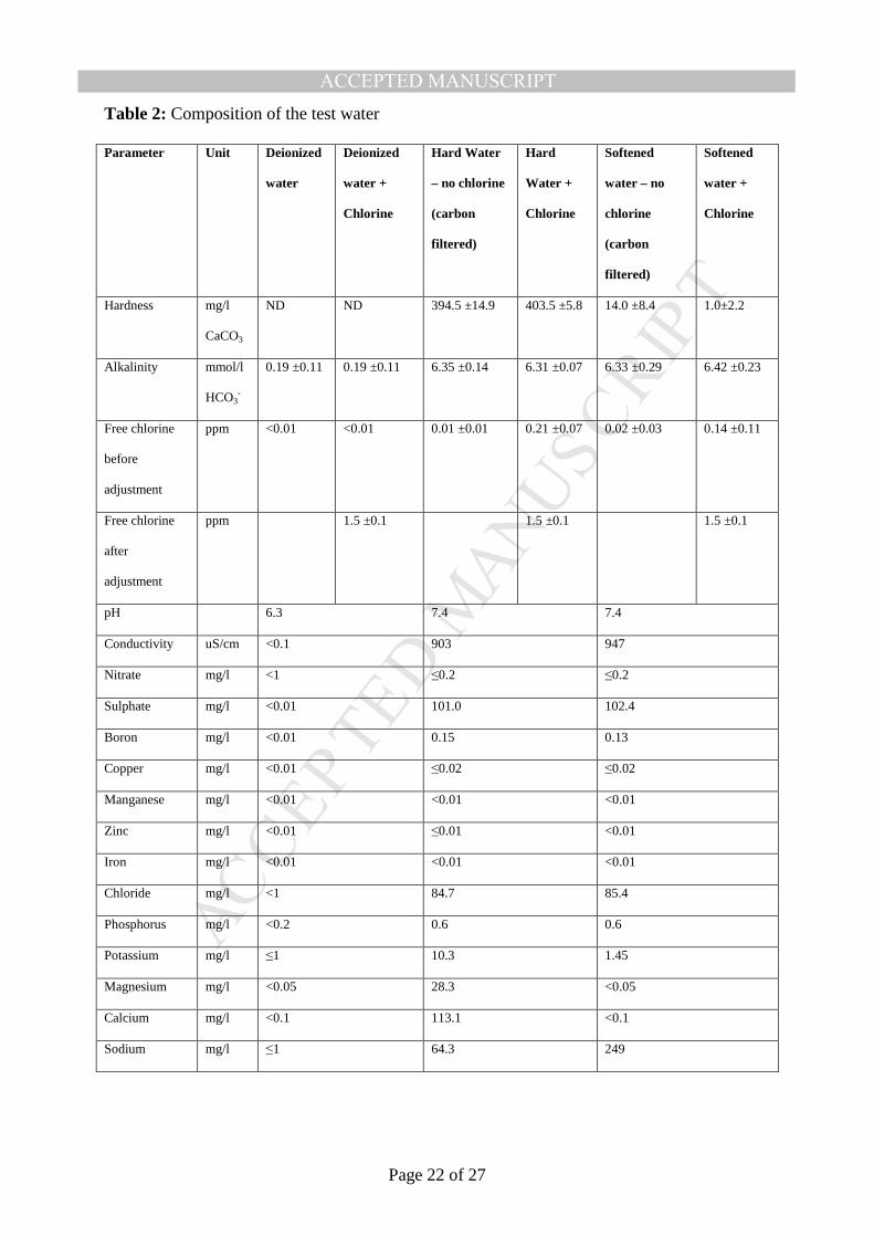
Table 2: Composition of the test water
Parameter Unit Deionized
water
Deionized
water +
Chlorine
Hard Water
– no chlorine
(carbon
filtered)
Hard
Water +
Chlorine
Softened
water – no
chlorine
(carbon
filtered)
Softened
water +
Chlorine
Hardness mg/l
CaCO3
ND ND 394.5 ±14.9 403.5 ±5.8 14.0 ±8.4 1.0±2.2
Alkalinity mmol/l
HCO3-
0.19 ±0.11 0.19 ±0.11 6.35 ±0.14 6.31 ±0.07 6.33 ±0.29 6.42 ±0.23
Free chlorine
before
adjustment
ppm <0.01 <0.01 0.01 ±0.01 0.21 ±0.07 0.02 ±0.03 0.14 ±0.11
Free chlorine
after
adjustment
ppm 1.5 ±0.1 1.5 ±0.1 1.5 ±0.1
pH 6.3 7.4 7.4
Conductivity uS/cm <0.1 903 947
Nitrate mg/l <1 ≤0.2 ≤0.2
Sulphate mg/l <0.01 101.0 102.4
Boron mg/l <0.01 0.15 0.13
Copper mg/l <0.01 ≤0.02 ≤0.02
Manganese mg/l <0.01 <0.01 <0.01
Zinc mg/l <0.01 ≤0.01 <0.01
Iron mg/l <0.01 <0.01 <0.01
Chloride mg/l <1 84.7 85.4
Phosphorus mg/l <0.2 0.6 0.6
Potassium mg/l ≤1 10.3 1.45
Magnesium mg/l <0.05 28.3 <0.05
Calcium mg/l <0.1 113.1 <0.1
Sodium mg/l ≤1 64.3 249
MANUSCRIP
T
ACCEPTED
ACCEPTED MANUSCRIPT
Page 23 of 27
FIGURE LEGENDS
Figure 1: Recruitment flowchart
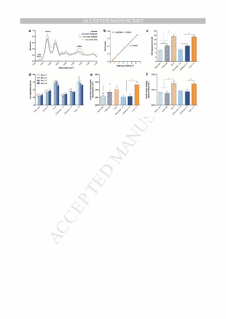
Figure 2: SLS deposition on the skin following washing with different water types. (a)
Representative spectra of the skin before and after washing. (b) The relationship between the
peak intensity at 1230 cm-1 and the concentration of SLS in aqueous solution. (c) The levels
of SLS, quantified in vivo by FTIR spectroscopy, deposited on the skin by test site. A
significant difference between the test sites was found (Friedman test p<0.0001, square root
transformation of SLS deposition to equalize variance). (d) SLS deposition by study
population. No difference between the groups was identified. (e) Lipid chain conformation, as
indicated by the position of the spectral band for CH2 symmetric stretching (approx. 2850 cm-
1), at the skin surface following washing. A higher band position indicates a more disordered
lipid chain conformation associated with surfactant damage. A significant difference between
the treatments was found (Friedman test p<0.0001). (f) Protein denaturation indicated by the
change in location of the peak associated with the amide I bond (1610-1690 cm-1). A
significant difference between the treatments was found (Friedman test p<0.0001).
*Significant differences identified using Dunn’s post-test. For simplicity only differences
within the no-chlorine and high-chlorine sets are displayed (no significant differences
between chlorine/no chlorine pairs).
Figure 3: The effect of surfactant residues on the skin: primary outcome measures. (a)
The effect of water type on the change in TEWL. There was a significant effect of the water
type on the change in TEWL (Friedman test p<0.05). (b) TEWL stratified by group. There
was a significant difference between the groups for hard, softened + chlorine and hard +
chlorine (Kruskal-Wallis p<0.0001). (c) The amount of SLS left on the skin following

MANUSCRIP
T
ACCEPTED
ACCEPTED MANUSCRIPT
Page 24 of 27
washing was significantly associated with TEWL (Spearman’s r=0.4928). (d) The effect of
water type on objective skin redness. There was a significant effect of water type on skin
redness (repeated measures ANOVA p<0.0001). (e) Skin redness stratified by group. There
was a significant effect of the group on the change in redness identified by two-way repeated
measures ANOVA (p<0.0001). (f) The amount of SLS residing on the skin following patch
removal was significantly associated with skin redness (Spearman’s r=0.411). *Significant
differences between treatments identified using Dunn’s or Tukey post-test respectively.
fSignificant differences identified using a protected Fishers LSD test. For simplicity only
differences within the no-chlorine and high-chlorine sets are displayed (no significant
differences between chlorine/no chlorine pairs). αSignificant differences to the negative
control. βSignificant differences to all other treatments.
Figure 4: The effect of surfactant residues on the skin: secondary outcome measures. (a)
visual scoring of erythema, (b) IL-1a, and (c) change in skin-surface-pH by treatment, and
stratified by group (stacked), 24h following patch removal. A significant difference in the log
transformed IL-1α levels between the test sites was found using a repeated measures ANOVA
(p=0.0114). Significant differences in visual erythema and skin-surface pH between the test
sites was found using the Friedman test (p<0.0001 and p<0.0001 respectively). *Significant
differences between treatments identified using Tukey or Dunn’s post-test respectively.
fSignificant differences identified using a protected Fishers LSD test. γA significant difference
between the groups (group 1 and 4) was found for skin-surface pH only (Kruskal-Wallis with
Dunn’s post-test).

MANUSCRIP
T
ACCEPTED
ACCEPTED MANUSCRIPT
Page 25 of 27
REFERENCES Ananthapadmanabhan KP, Moore DJ, Subramanyan K, Misra M, Meyer F. Cleansing without
compromise: the impact of cleansers on the skin barrier and the technology of mild cleansing. Dermatol Ther 2004;17 Suppl 1:16-25.
Angelova-Fischer I, Becker V, Fischer TW, Zillikens D, Wigger-Alberti W, Kezic S. Tandem repeated irritation in aged skin induces distinct barrier perturbation and cytokine profile in vivo. The British journal of dermatology 2012;167:787-93.
Arnedo-Pena A, Bellido-Blasco J. Dureza del agua de consumo doméstico y prevalencia de eczema atópico en escolares de Castellón , España. 2007;49:295-301.
Bandier J, Carlsen BC, Rasmussen MA, Petersen LJ, Johansen JD. Skin reaction and regeneration after single sodium lauryl sulfate exposure stratified by filaggrin genotype and atopic dermatitis phenotype. Br J Dermatol 2015;172(6):1519-29.
Boncheva M, Damien F, Normand V. Molecular organization of the lipid matrix in intact Stratum corneum using ATR-FTIR spectroscopy. Biochimica et biophysica acta 2008;1778(5):1344-55.
Chaumont A, Voisin C, Sardella A, Bernard A. Interactions between domestic water hardness, infant swimming and atopy in the development of childhood eczema. Environmental research 2012;116:52-7.
Cork MJ, Danby SG, Vasilopoulos Y, Hadgraft J, Lane ME, Moustafa M, et al. Epidermal barrier dysfunction in atopic dermatitis. J Invest Dermatol 2009;129(8):1892-908.
Danby S, Al Enezi T, Sultan A, Chittock J, Kennedy K, Cork MJ. The effect of Aqueous cream BP on the skin barrier in volunteers with a previous history of atopic dermatitis. Br J Dermatol 2011;165(2):329-34.
Darlenski R, Kazandjieva J, Tsankov N, Fluhr JW. Acute irritant threshold correlates with barrier function, skin hydration and contact hypersensitivity in atopic dermatitis and rosacea. Exp Dermatol 2013;22(11):752-3.
Engebretsen KA, Bager P, Wohlfahrt J, Skov L, Zachariae C, Nybo Andersen AM, et al. Prevalence of atopic dermatitis in infants by domestic water hardness and season of birth: Cohort study. J Allergy Clin Immunol 2016.
Ewence A, Rumsby P, Danby S, Cork MJ, Williams H. A Review of Skin Irritation and Tap Water. Swindon2011.
Font-Ribera L, Villanueva CM, Gracia-Lavedan E, Borras-Santos A, Kogevinas M, Zock JP. Indoor swimming pool attendance and respiratory and dermal health in schoolchildren--HITEA Catalonia. Respiratory medicine 2014;108(7):1056-9.
Gotoh K, Horibe K, Mei Y, Tsujisaka T. Effects of Water Hardness on Textile Detergency Performance in Aqueous Cleaning Systems. Journal of oleo science 2016;65(2):123-33.
Hachem JP, Crumrine D, Fluhr J, Brown BE, Feingold KR, Elias PM. pH directly regulates epidermal permeability barrier homeostasis, and stratum corneum integrity/cohesion. J Invest Dermatol 2003;121(2):345-53.
Hachem JP, Roelandt T, Schurer N, Pu X, Fluhr J, Giddelo C, et al. Acute acidification of stratum corneum membrane domains using polyhydroxyl acids improves lipid processing and inhibits degradation of corneodesmosomes. J Invest Dermatol 2010;130(2):500-10.
Jang H, Matsuda A, Jung K, Karasawa K, Matsuda K, Oida K, et al. Skin pH Is the Master Switch of Kallikrein 5-Mediated Skin Barrier Destruction in a Murine Atopic Dermatitis Model. Journal of Investigative Dermatology 2016;136(1):127-35.

MANUSCRIP
T
ACCEPTED
ACCEPTED MANUSCRIPT
Page 26 of 27
Jungersted JM, Scheer H, Mempel M, Baurecht H, Cifuentes L, Hogh JK, et al. Stratum corneum lipids, skin barrier function and filaggrin mutations in patients with atopic eczema. Allergy 2010;65(7):911-8.
Kelleher M, Dunn-Galvin A, Hourihane JOB, Murray D, Campbell LE, Irwin McLean WH, et al. Skin barrier dysfunction measured by transepidermal water loss at 2 days and 2 months predates and predicts atopic dermatitis at 1 year. Journal of Allergy and Clinical Immunology 2015:1-7.
Kezic S, Kemperman PM, Koster ES, de Jongh CM, Thio HB, Campbell LE, et al. Loss-of-function mutations in the filaggrin gene lead to reduced level of natural moisturizing factor in the stratum corneum. J Invest Dermatol 2008;128(8):2117-9.
Kezic S, O'Regan GM, Lutter R, Jakasa I, Koster ES, Saunders S, et al. Filaggrin loss-of-function mutations are associated with enhanced expression of IL-1 cytokines in the stratum corneum of patients with atopic dermatitis and in a murine model of filaggrin deficiency. The Journal of allergy and clinical immunology 2012;129(4):1031-9 e1.
Kim BE, Leung DY, Boguniewicz M, Howell MD. Loricrin and involucrin expression is down-regulated by Th2 cytokines through STAT-6. Clin Immunol 2008;126(3):332-7.
Lewis-Jones S, Mugglestone Ma. Management of atopic eczema in children aged up to 12 years: summary of NICE guidance. BMJ (Clinical research ed) 2007;335:1263-4.
Man MQ, Wood L, Elias PM, Feingold KR. Cutaneous barrier repair and pathophysiology following barrier disruption in IL-1 and TNF type I receptor deficient mice. Exp Dermatol 1999;8(4):261-6.
McAleer MA, Irvine AD. The multifunctional role of filaggrin in allergic skin disease. J Allergy Clin Immunol 2013;131(2):280-91.
McNally NJ, Williams HC, Phillips DR, Smallman-Raynor M, Lewis S, Venn A, et al. Atopic eczema and domestic water hardness. Lancet 1998;352(9127):527-31.
Miyake Y, Yokoyama T, Yura A, Iki M, Shimizu T. Ecological association of water hardness with prevalence of childhood atopic dermatitis in a Japanese urban area. Environ Res 2004;94(1):33-7.
Moniaga CS, Jeong SK, Egawa G, Nakajima S, Hara-Chikuma M, Jeon JE, et al. Protease activity enhances production of thymic stromal lymphopoietin and basophil accumulation in flaky tail mice. Am J Pathol 2013;182(3):841-51.
Odhiambo JA, Williams HC, Clayton TO, Robertson CF, Asher MI, Group IPTS. Global variations in prevalence of eczema symptoms in children from ISAAC Phase Three. J Allergy Clin Immunol 2009;124(6):1251-8 e23.
Ohmori K, Tanaka A, Makita Y, Takai M, Yoshinari Y, Matsuda H. Pilot evaluation of the efficacy of shampoo treatment with ultrapure soft water for canine pruritus. Veterinary dermatology 2010;21:477-83.
Perkin MR, Craven J, Logan K, Strachan D, Marrs T, Radulovic S, et al. Association between domestic water hardness, chlorine, and atopic dermatitis risk in early life: A population-based cross-sectional study. J Allergy Clin Immunol 2016;138(2):509-16.
Pinnagoda J, Tupker RA, Agner T, Serup J. Guidelines for transepidermal water loss (TEWL) measurement. A report from the Standardization Group of the European Society of Contact Dermatitis. Contact Dermatitis 1990;22(3):164-78.
Saad P, Flach CR, Walters RM, Mendelsohn R. Infrared spectroscopic studies of sodium dodecyl sulphate permeation and interaction with stratum corneum lipids in skin. International journal of cosmetic science 2012;34:36-43.
Sandilands A, Terron-Kwiatkowski A, Hull PR, O'Regan GM, Clayton TH, Watson RM, et al. Comprehensive analysis of the gene encoding filaggrin uncovers prevalent and rare mutations in ichthyosis vulgaris and atopic eczema. Nat Genet 2007;39(5):650-4.

MANUSCRIP
T
ACCEPTED
ACCEPTED MANUSCRIPT
Page 27 of 27
Scharschmidt TC, Man MQ, Hatano Y, Crumrine D, Gunathilake R, Sundberg JP, et al. Filaggrin deficiency confers a paracellular barrier abnormality that reduces inflammatory thresholds to irritants and haptens. J Allergy Clin Immunol 2009;124(3):496-506, e1-6.
Tanaka A, Matsuda A, Jung K, Jang H, Ahn G, Ishizaka S, et al. Ultra-pure Soft Water Ameliorates Atopic Skin Disease by Preventing Metallic Soap Deposition in NC/Tnd Mice and Reduces Skin Dryness in Humans. Acta Derm Venereol 2015.
Thomas KS, Dean T, O'Leary C, Sach TH, Koller K, Frost A, et al. A randomised controlled trial of ion-exchange water softeners for the treatment of eczema in children. PLoS medicine 2011;8:e1000395.
Togawa Y, Kambe N, Shimojo N, Nakano T, Sato Y, Mochizuki H, et al. Ultra-pure soft water improves skin barrier function in children with atopic dermatitis: a randomized, double-blind, placebo-controlled, crossover pilot study. J Dermatol Sci 2014;76(3):269-71.
Winge MC, Hoppe T, Berne B, Vahlquist A, Nordenskjold M, Bradley M, et al. Filaggrin genotype determines functional and molecular alterations in skin of patients with atopic dermatitis and ichthyosis vulgaris. PLoS ONE 2011;6(12):e28254.
Young SL, Matijevic E. Precipitation Phenomena of Heavy-Metal Soaps in Aqueous-Solutions .3. Metal Laurates. Journal of Colloid and Interface Science 1977;61(2):287-301.

MANUSCRIP
T
ACCEPTED
ACCEPTED MANUSCRIPT
!""#""#$%&'(%#)*+*,*)*-.%
/!01234%%
56(##7#$%
/!08934%
56(##7#$%
/!08924%
:)*+*,)#%/!08134%
;1<%
:)*+*,)#%/!0824%
=<%
:)*+*,)#%/!0;34%
==<%
:)*+*,)#%/!0>?4%
>1<%
@#6(A*-#$%/!0194% @#6(A*-#$%/!0?4% @#6(A*-#$%/!0124% @#6(A*-#$%/!0>>4%
B'""%-'%&'))'C%AD%
/!0E4%
B'""%-'%&'))'C%AD%
/!024%
B'""%-'%&'))'C%AD%
/!0E4%
B'""%-'%&'))'C%AD%
/!024%
F('AD%8%6'GD)#-#$%
/!0>;4%
F('AD%>%6'GD)#-#$%
/!0?4%
F('AD%1%6'GD)#-#$%
/!0>34%
F('AD%3%6'GD)#-#$%
/!0>>4%
:H6)A$#$%&('G%
I7I)."*"%
/!014%
:H6)A$#$%&('G%
I7I)."*"%
/!024%
:H6)A$#$%&('G%
I7I)."*"%
/!024%
:H6)A$#$%&('G%
I7I)."*"%
/!024%
!6JK#%!L%%%%%M#I)-N.%%
:H6)A$#$%
/!0>?4%
:H6)A$#$%
/!0824%
!"#$%&'&
M#I)-N.%"#$%&'
!"#$%&(&
M#I)-N.%"#$!())'
!"#$%&)&
!L%"#$%&'
!"#$%&*&
!L%"#$!())'
MANUSCRIP
T
ACCEPTED
ACCEPTED MANUSCRIPT

MANUSCRIP
T
ACCEPTED
ACCEPTED MANUSCRIPT
MANUSCRIP
T
ACCEPTED
ACCEPTED MANUSCRIPT