the effect of motor symptom onset laterality on facial...
TRANSCRIPT

THE EFFECT OF MOTOR SYMPTOM ONSET LATERALITY ON FACIAL EXPRESSIVITY IN PARKINSON’S DISEASE
By
ANNE NOELLE NISENZON
A THESIS PRESENTED TO THE GRADUATE SCHOOL OF THE UNIVERSITY OF FLORIDA IN PARTIAL FULFILLMENT
OF THE REQUIREMENTS FOR THE DEGREE OF MASTER OF SCIENCE
UNIVERSITY OF FLORIDA
2008
1

© 2008 Anne Noelle Nisenzon
2

To my family.
3

ACKNOWLEDGMENTS
First, I sincerely thank my mentor, Dr. Dawn Bowers, for her endless patience, care,
encouragement, and support. I also thank my colleagues in the Cognitive Neuroscience
Laboratory for their kind and helpful advice. I would like to thank my defense panel, Drs.
Bowers, Perlstein, Perri, and Wiens, for their time in reviewing this study and providing valuable
suggestions. Finally, I thank my parents, Alex and Irina Nisenzon, for their support in my
professional endeavors.
4

TABLE OF CONTENTS page
ACKNOWLEDGMENTS ...............................................................................................................4
LIST OF TABLES...........................................................................................................................7
LIST OF FIGURES .........................................................................................................................8
ABSTRACT.....................................................................................................................................9
CHAPTER
1 INTRODUCTION ..................................................................................................................11
Parkinson’s Disease: Symptomatology and Neuropathology.................................................12 Lateralization of Emotional Processing..................................................................................15 Emotion in Parkinson’s Disease .............................................................................................17 Facial Expressivity and Parkinson’s Disease .........................................................................18
2 STATEMENT OF THE PROBLEM......................................................................................21
3 METHODS.............................................................................................................................25
Participants .............................................................................................................................25 Measures .................................................................................................................................28 Procedures...............................................................................................................................29 Statistical Analyses.................................................................................................................32
4 RESULTS...............................................................................................................................34
The Effect of Laterality on Facial Expressivity......................................................................34 Mood and Motor Correlates of Expressivity ..........................................................................37
Relationship between Mood and Facial Expressivity .....................................................37 The Relationship between Motor Symptoms and Expressivity ......................................39
5 DISCUSSION.........................................................................................................................41
Summary and Interpretation of the Findings ..........................................................................41 Latency to Expression as an Action-Intentional Deficit.........................................................44 Limitation of the Present Study ..............................................................................................46 Directions for Future Research...............................................................................................49 Conclusion ..............................................................................................................................50
APPENDIX
DESCRIPTIVE STATISTICS AND ANOVA TABLES FOR PRIMARY AIM..................52
5

LIST OF REFERENCES...............................................................................................................54
BIOGRAPHICAL SKETCH .........................................................................................................60
6

LIST OF TABLES
Table page 3-1. Participant Demographics ............................................................................................27
4-1. Descriptive Statistics for Mood and Motor Variables..................................................37
4-2: Linear Regression Analysis of Mood Variables on Facial Expressivity......................39
4-3: Linear Regression Analysis of Motor Variables on Facial Expressivity .....................40
A-1. Descriptive Statistics for Entropy Variables ...............................................................52
A-2. ANOVA Summary: Entropy Variables.......................................................................52
A-3. Descriptive Statistics for Temporal Variables.............................................................53
A-4. ANOVA Summary: Temporal Variables ....................................................................53
7

LIST OF FIGURES
Figure page 1-1. Simplified pathophysiological model of motor and emotional dysfunction in
Parkinson’s disease.. ..........................................................................................................14
3-1. Digitizing the Moving Face.. ............................................................................................32
3-2. Entropy Curve.. ..................................................................................................................33
4-1. Bar Graph: Latency to Initiate Facial Expressions. ...........................................................36
8

Abstract of Thesis Presented to the Graduate School of the University of Florida in Partial Fulfillment of the
Requirements for the Degree of Master of Science
THE EFFECT OF MOTOR SYMPTOM ONSET LATERALITY ON FACIAL EXPRESSIVITY IN PARKINSON’S DISEASE
By
Anne Noelle Nisenzon
May 2008
Chair: Dawn Bowers Major: Psychology--Clinical and Health Psychology
The purpose of the present study was to examine the effect of motor symptom laterality
commonly found in Parkinson’s disease (PD) as well as mood and motor symptoms on different
aspects of facial expressivity.
Most patients with PD initially present with lateralized motor symptoms on one side of the
body, left or right. This asymmetry usually persists throughout the course of the disease and
reflects differential disruption of contralateral basal ganglia-frontal lobe dopaminergic systems.
While a growing number of studies have reported that laterality of symptom onset may also
influence non-motor symptoms of the disease (i.e., cognition), it remains unknown whether
emotional processing is affected by symptom onset laterality. A cardinal symptom of PD is a
“masked face,” or significantly diminished facial expressivity. Based on views regarding a right
hemispheric specialization for emotional behavior, we hypothesized that patients with left-sided
motor symptom onset (implicating greater right hemisphere involvement) would display greater
impairments in facial expressivity than those with right sided symptom onset. We also predicted
that mood and PD-specific motor symptoms would contribute to diminished facial expressivity.
Twenty six patients with idiopathic PD (13 left symptom onset, 13 right onset) and 13
healthy controls were videotaped while posing facial expressions of fear, anger, and happiness.
9

10
Facial expressions were digitized and analyzed using custom software that extracted 5 variables:
2 measures of dynamic movement change (total and peak entropy), and 3 temporal variables
(initiation time, rise time, duration). Self-report measures of depression (Beck Depression
Inventory-II) and apathy (Apathy Evaluation Scale) were also given. Repeated measures
ANOVAs and linear regressions were used to analyze the data.
Bonferroni-corrected post-hoc analyses revealed that the left-onset PD patients were
significantly slower in initiating facial expressions than were right-onset PD patients or controls,
regardless of expression posed [F (2,36) = 8.21, p <.05]. The groups did not significantly differ
in terms of the entropy variables or other temporal variables. Furthermore, mood (i.e.,
depression or apathy) and disease-specific factors (i.e., symptom type) were unrelated to facial
expressivity.
These results suggest an effect of motor symptom asymmetry on initiation of facial
expressions among patients with PD. The findings are more in line with views that emphasize
the role of the right hemisphere on attentional and intentional behavior, rather than “emotion,”
per se. In other words, slower initiation times for posed facial expressions among PD patients
with greater right cortical-subcortical dysfunction may reflect reduced ability to initiate and
respond rather than defective emotional processing as initially proposed. Overall, these findings
highlight the potential importance of symptom laterality in non-motor aspects of Parkinson’s
disease.

CHAPTER 1 INTRODUCTION
Parkinson’s disease (PD) is a neurodegenerative movement disorder that arises from
dopamine loss in deep brain structures and leads to widespread functional impairment.
Prevalence reports estimate approximately one million people in the United States with the
disease, with incidence reports of up to 60,000 new cases diagnosed each year (McDonald,
Richard, and Delong, 2003). Idiopathic PD is commonly diagnosed after the age of 60, and has a
gradual, insidious, and often lateralized onset of motor symptoms which worsen with disease
progression. While pharmacological and surgical interventions are available to alleviate
symptoms temporarily, there is no known cure to reverse or prevent further degeneration.
An often overlooked yet important symptom of Parkinson’s disease is a “masked face.”
This term refers to the mannequin-like expressionless face that is characteristic of most
individuals with PD. Due to their reduced propensity to facially convey either positive or
negative emotions, PD patients are often misdiagnosed as being depressed or apathetic. While
both these mood states commonly occur in PD, their rates may be inflated due to misperception
by healthcare providers. As such, it is important to parcel out the effects of diminished facial
expressivity from that of true mood disturbance.
Relative to other neurodegenerative disorders, asymmetric symptom onset is a notably
unique feature of PD and reflects greater pathology in contralateral subcortical regions.
Hemispheric laterality is also well-documented in emotional research, and different theories
speculate on how the right and left cortical hemispheres process a wide array of emotionally-
charged information. These views on hemispheric differences in emotional behavior are relevant
to Parkinson’s disease because subcortical regions affected in the disorder directly project to
cortical regions on the same side of the brain. Thus, laterality of motor symptom onset may
11

potentially play an important role in the occurrence of the masked face and emotional symptoms
in Parkinson’s disease.
The primary goal of the current study was to compare emotional facial expressivity in
right-sided onset and left-sided onset (indicating greater left or right neuropathology,
respectively) PD patients. To do so, we used novel computer imaging techniques for calculating
dynamic movement changes over the face (i.e., entropy) as opposed to the more traditional
subjective judgments used in previous research. Additionally, the effects of mood and PD motor
symptoms on facial expressivity were examined. Before discussing the specific hypotheses, a
brief review of literature will now be presented pertaining to: 1) Parkinson’s disease
symptomatology and its neuropathological correlates, 2) theories of hemispheric dominance in
emotional behavior, 3) emotion and facial expressivity in PD.
Parkinson’s Disease: Symptomatology and Neuropathology
Parkinson’s disease is diagnosed based upon the presence of four cardinal motor
symptoms: bradykinesia, rigidity, tremor, and postural instability. Bradykinesia refers to an
overall slowing of movement, and can be witnessed by asking patients to rapidly tap their fingers
at a given amplitude. Bradykinesia is a distinctive feature of PD and must be observable for
affirmative diagnosis. Rigidity refers to muscle stiffness and is characterized by resistance to
passive movement of a limb. Tremor in PD involves involuntary shaking of the limb and
typically occurs as a “resting tremor,” which subsides when the person engages in a purposeful
movement. Other features of Parkinson’s disease include postural instability, or poor balance,
and gait disturbances, in which patients walk with a stooped posture and take small, quickened
steps. Patients may be classified as tremor predominant or rigid/akinetic type depending on
which symptoms are most evident during a motor examination. Additional hallmark features of
PD are slurred speech, small cramped writing, and diminished facial expressivity.
12

In terms of pathophysiology, Parkinson’s disease essentially originates from the disrupted
functioning of the basal ganglia, a midbrain structure composed of the striatum, globus pallidus
(GP), subthalamic nucleus (STN), and the substantia nigra. These nuclei operate together to
modulate functioning of higher cortical areas via the thalamus. In Parkinson’s disease, neurons
in the substantia nigra pars compacta (SNc) stop producing dopamine, thereby disrupting
connectivity in various parallel closed-loop basal ganglia-thalamo-cortical circuits that are
topographically organized and functionally specialized. Alexander, DeLong, & Strick (1986)
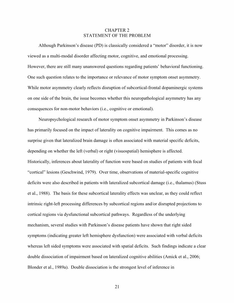
identified 5 such circuits, each influencing motor, cognitive, or emotional functioning (see
Figure 1-1 for motor and limbic circuitry). By the time initial motor symptoms are experienced
in PD, the SNc is approximately 70% inactive (Obeso et al., 2002).
In early stages of Parkinson disease, behavioral symptoms are most often lateralized to one
side of the body. With further progression, however, these symptoms appear bilaterally, but
usually remain worse on the presenting side (Lee et al., 1995). Imaging studies and post-
mortem cell counts suggest greater neuronal loss in the substantia nigra contralateral, or
opposite, the initially more affected body side (Kempster et al., 1989). It is currently unknown
whether asymmetrical neurodegeneration is due to inborn variations in dopaminergic neurons,
differential vulnerability to the disease, or by random occurrence (Djaldetti, Ziv, & Melamed,
2006).
The neuropathology associated with specific motor symptoms in PD has been elucidated
through neuroimaging studies. Positron emission tomography (PET) studies show that
bradykinesia and rigidity both arise from excessive inhibition of the thalamus and subsequent
motor cortices due to excitatory glutaminergic projections on the internal globus pallidus (Lozza
et al., 2002). The disease mechanism of tremor is not as clear, though some researchers postulate
13

that it involves hypoactivation of the basal ganglia along with hyperactivation of brain structures
outside the basal ganglia-thalamo-cortical loops, specifically in the cerebellum (Antonini et al.,
1998; Yu et al., 2007). Thus, loss of nigral dopaminergic cells leads to a gross imbalance of
chemical signals which results in motor impairment and, ultimately, cognitive and emotional
deficits. Medications (levodopa and dopamine agonists) and surgical interventions, including
deep brain stimulation (DBS) and damaging hyperactive areas of the basal ganglia or thalamus,
have been successfully employed to quell motor symptoms by temporarily reestablishing
neurochemical balance. However, neither medication nor surgical approaches are able to stop or
reverse neurodegeneration associated with Parkinson’s disease.
Motor Circuit Limbic Circuit
Motor Cortex
Putamen
Lateral Pallidum
Thalamus (medial dorsal nucleus)
-
+
-
+
Anterior Cingular Cortex
Ventral Striatum
Ventral Pallidum
Thalamus (dorsolateral nucleus)
-
+
-
SNc +
+
Figure 1-1: Simplified pathophysiological model of motor and emotional dysfunction in
Parkinson’s disease. Dopaminergic cells in the substantia nigra pars compacta die, leading to an imbalance of excitatory and inhibitory signals passed from higher cortical structures to the striatum via the thalamus. There are 5 segregated parallel basal ganglia loops, each of which involve different cortical and subcortical structures and modulate different areas of functioning. Illustrated here are the motor and limbic circuits, influencing physical and emotional functioning, respectively.
14

Lateralization of Emotional Processing
The limbic system has long been viewed as the primary neuroanatomic substrate for
emotion (Papez, 1995). It includes several key structures (amygdala, hypothalamus, septum,
cingulate, nucleus accumbens) that have been linked to appetitive/reward and avoidance/attack
behaviors. Basal ganglia systems that overlap with limbic circuitry (i.e., nucleus accumbens,
amygdale, cingulate) have also been implicated in emotion. Specifically, neuroimaging studies
have revealed activation in the medial prefrontal cortex and anterior cingulate during general
emotional processing, regardless of emotion elicited or induction method (Phan et al., 2002).
These particular regions are part of a basal ganglia circuit known as the “limbic loop,” and are
impacted by the subcortical dysfunction as seen in PD.
While involvement of the limbic system and basal ganglia modulation is apparent in
emotional behavior, the role of the cortical hemispheres has long been debated. One overarching
view is that cortical regions serve to provide higher order modulation of underlying limbic
regions. What is less clear is how the two hemispheres play differential roles in this cortical
modulation. There are two competing theories as to how emotion is processed in higher cortical
areas— the right hemisphere model and the bivalent model. According to the right hemisphere
model, the right cortical hemisphere is dominant for mediating all emotional behavior, regardless
of affect. This view is supported by lesion studies that demonstrate damage to the right versus
left hemisphere leads to significant deficits in the perception and expression of emotional stimuli
including facial expressions, emotional prosody, and lexical affective expression and experience
(Borod, 1992; Borod et al., 1998; Bowers, Bauer, & Heilman, 1993; Heilman & Bowers, 1990;
Liotti and Tucker, 1995). Similarly, patients with right hemisphere lesions show greater
impairment in recognizing facial expressions of emotion or emotional prosody (Bowers et al.,
1985; Bowers et al., 1987). Regarding emotional experience, a neuroimaging study of apathetic
15

stroke patients revealed greater hyperintensities in the right fronto-subcortical pathway than in
the left, despite stroke severity (Brodaty et al., 2005). Furthermore, studies conducted on facial
expressivity have shown that normal adults express their emotions more intensely on the left side
of the face, which is contralaterally controlled by the right hemisphere, and that patients with
right hemisphere damage are less facially expressive in general (Borod and Caron, 1980; Borod,
1992).
In terms of potential mechanisms underlying the right hemisphere hypothesis of emotion,
some have speculated that the functional organization of the right hemisphere has made it more
suitable for emotional processing. The right hemisphere has been shown to be specialized in
integration and representation of information, which is necessary in nonverbal emotional
communication. This explanation of right hemisphere dominance in emotion stems from
findings that focal left hemisphere lesions result in discrete cognitive deficits whereas focal
damage in the right hemisphere produce diffuse deficits in several domains (Semmes, 1968).
Thus, the right hemisphere may be crucial in the cognitive mediation of emotional experience
(Liotti & Tucker, 1995). Another line of evidence suggests greater connection density between
regions in the right hemisphere, which may result in more varied communication pathways with
the limbic system (Tucker, 1991). Moreover, a recent study featuring diffusion tensor
tractography confirmed asymmetrical white matter organization in the brain (Barrick et al.,
2006).
Another view as to hemispheric laterality of emotional processing is the bivalent
hypothesis. According to this hypothesis, the right hemisphere is more specialized in processing
negatively-valenced emotions while the left hemisphere is more active in positive emotions.
Historically, this view derived from observations of differing emotional changes experienced by
16

patients who suffered strokes of the left or right hemisphere. Goldstein (1939) noted that
individuals with left hemisphere strokes had “catastrophic” reactions, whereas those with right
hemisphere strokes were emotionally flattened and apathetic. Furthermore, post-stroke
depression following left hemisphere strokes has been shown to be particularly severe when
subcortical regions were involved (Starkstein, Robinson, & Price, 1987). These clinical
findings, in conjunction with subsequent research on mood-related EEG asymmetries in normals
(Davidson, 1995) led to the hypothesis that both hemispheres were involved in emotional
processing, though each differed in the specific type of emotions, i.e., positive versus negative.
Although intriguing, the bivalent model has not been consistently supported in empirical studies
of emotional perception or production (Borod, 2000; Heilman et al., 2003). Moreover, there has
been minimal support for the view that the left hemisphere mediates positive emotions (see
Harciarek et al., 2006).
In summary, two major models have been proposed regarding the role of the two
hemispheres in mediating emotional behavior. In one model, the right hemisphere takes on
primary responsibility for mediating all aspects of emotional behavior including perception,
expression, and experience. In the second (bivalent) model, the left hemisphere mediates
pleasant/approach related emotions whereas the right hemisphere is more involved in
negative/avoidance related emotions.
Emotion in Parkinson’s Disease
Emotional behavior is often researched in Parkinson’s disease as mood disorders are
highly comorbid with the disease. Depression is commonly observed with prevalence rates
ranging from 4% to 70% among patients (Cubo et al., 2002). The basis for depression in
Parkinson’s disease is unknown, although it involves both biological and psychological factors.
Psychological factors include reactions to receiving a diagnosis of a neurodegenerative illness,
17

difficulties in adjusting to lifestyle changes, and alterations in family dynamics and spousal roles.
Biologically, dopamine loss in the limbic circuit of the basal ganglia as well as disruption of
serotonin and other neurotransmitter systems are considered key components in PD-related
depression (Lieberman, 2006). While antidepressants such as SSRI’s are often prescribed in this
population to treat depression, there is a lack of evidence for antidepressant efficacy among PD
patients (Weintraub et al., 2003).
Another common mood disorder in Parkinson disease is apathy, which is a relatively new
area of research in this population. Apathy refers to a loss of motivation in affective, cognitive,
and behavioral domains (Marin, 1991). Symptoms of apathy have traditionally been
misdiagnosed as being symptoms of depression, thereby possibly inflating its rates. The
depressive symptom of anhedonia, or loss of interest, is especially similar to the characteristic
features of apathy. However, the two are dissociable in PD patients according to the Kirsch-
Darrow et al. (2006) finding that apathy can occur in the absence of depression and vice versa.
The neurobiological substrates of apathy are unknown , although apathy has been associated with
dysfunction in mesial frontal and anterior cingulate regions (for review, see Levy & Dubois,
2006).
Facial Expressivity and Parkinson’s Disease
Emotional facial expressivity is a fundamental form of communication, and, when
impaired as it often is in Parkinson’s disease, can have far-reaching consequences in the patient’s
quality of life and interpersonal relationships. For example, the seemingly ubiquitous symptom
of depression among PD patients may be overdiagnosed due to misattribution of negative
emotional states by healthcare workers (Pentland et al., 1988). Additionally, diminished facial
expressivity can impair communication between patients and loved ones, leading to confusion
18

and frustration on both ends. Paying greater attention to this debilitating yet underrepresented
symptom of PD may ultimately open the doors for more comprehensive treatment options. Thus,
it is important to understand the mechanical aspects of facial expressivity and how it is disrupted
in Parkinson’s disease. Greater elucidation of the process of diminished facial expressivity may
lead to better research techniques, and, ultimately, more comprehensive treatment options.
Facial expressions are created through projections from the motor cortex to the brain stem
via the corticobulbar system, a white matter pathway which controls the muscles of the face,
head, and neck. Corticobulbar projections to cell body groups controlling movements in the
lower face come exclusively from the contralateral hemisphere while bilateral projections
stimulate the upper face (Rinn, 1984). This system describes production of posed or voluntary
expressions that do not involve emotionality. Spontaneous emotional expressivity is more
subcortically modulated and was originally thought to involve only an extrapyramidal motor
system separate from the corticobulbar pathway. The extrapyramidal system, which includes the
basal ganglia and the nigrostriatal pathway, modulates motor activity without directly
innervating motor neurons (Rinn, 1984). More recent research states that both the corticobulbar
and extrapyramidal systems are implemented to create fluid, effective posed and spontaneous
facial expressions (Simons et al., 2004).
The mechanism underlying diminished facial expressivity in Parkinson’s disease is not
fully understood. One view indicates that dopamine depletion in the basal ganglia disrupts
healthy functioning of the extrapyramidal pathway, thereby greatly affecting spontaneous
expressivity (Turner et al., 2003; Rinn, 1984). However, Bowers et al. (2006) demonstrated that
voluntary expressions are also diminished in PD patients, as impairment in frontostriatal circuitry
may have a dampening effect on expression intensity, regardless of intention. Another view
19

20
speculates that depressive symptoms may lead to reduced emotional expressivity, although non-
depressed patients also present with masked faces. Thus, diminished facial expressivity is not
simply a result of affective dysfunction (McDonald et al., 2003; Bowers et al., 2006).
Furthermore, Bowers et al. (2006) found that PD patients were less expressive than healthy
controls regardless of emotional valence (i.e., happy vs. sad). The loss of muscle tone in the face
resulting from overall motoric disability in PD may also cause problems in facial expressivity
(De Letter et al., 2003). In sum, speculations as to the physical and emotional substrates of the
masked face in PD have been inconclusive, reflecting the elusive nature of this symptom and the
relative dearth of research conducted in this area.
CHAPTER 2 STATEMENT OF THE PROBLEM
Although Parkinson’s disease (PD) is classically considered a “motor” disorder, it is now
viewed as a multi-modal disorder affecting motor, cognitive, and emotional processing.
However, there are still many unanswered questions regarding patients’ behavioral functioning.
One such question relates to the importance or relevance of motor symptom onset asymmetry.
While motor asymmetry clearly reflects disruption of subcortical-frontal dopaminergic systems
on one side of the brain, the issue becomes whether this neuropathological asymmetry has any
consequences for non-motor behaviors (i.e., cognitive or emotional).
Neuropsychological research of motor symptom onset asymmetry in Parkinson’s disease
has primarily focused on the impact of laterality on cognitive impairment. This comes as no
surprise given that lateralized brain damage is often associated with material specific deficits,
depending on whether the left (verbal) or right (visuospatial) hemisphere is affected.
Historically, inferences about laterality of function were based on studies of patients with focal
“cortical” lesions (Geschwind, 1979). Over time, observations of material-specific cognitive
deficits were also described in patients with lateralized subcortical damage (i.e., thalamus) (Stuss
et al., 1988). The basis for these subcortical laterality effects was unclear, as they could reflect
intrinsic right-left processing differences by subcortical regions and/or disrupted projections to
cortical regions via dysfunctional subcortical pathways. Regardless of the underlying
mechanism, several studies with Parkinson’s disease patients have shown that right sided
symptoms (indicating greater left hemisphere dysfunction) were associated with verbal deficits
whereas left sided symptoms were associated with spatial deficits. Such findings indicate a clear
double dissociation of impairment based on lateralized cognitive abilities (Amick et al., 2006;
Blonder et al., 1989a). Double dissociation is the strongest level of inference in
21

neuropsychology and behavioral neurology (Teuber, 1955), thus providing substantial weight to
these findings. However, it should be noted that not all studies have found a significant
association between laterality and various cognitive deficits in patients with PD, and, thereby,
continues to call into question the importance of laterality (Cubo et al, 2000; St. Clair et al.,
1998).
In contrast to cognitive studies, the relationship between motor asymmetry and emotional
symptoms in PD has largely gone untouched. This hole in the literature is quite remarkable
given that there are dozens of replicated studies showing hemispheric specialization in
processing emotion as well as separate studies conducted on emotional behavior in PD.
Although several studies have focused on the effect of asymmetrical symptomatology on
depression in PD and yielded inconclusive and conflicting results (Fleminger, 1991; Spicer et al.,
1988; Starkstein et al., 1992), minimal attention has been paid to facial expressivity in relation to
laterality of symptom onset.
Thus, the overall goal of the present study was to examine the influence of motor
symptom laterality in Parkinson’s disease on different aspects of emotional behavior, particularly
facial expressivity and mood. The underlying basis for such an endeavor derives from two lines
of evidence: a) the tightly coupled functional relationship between subcortical regions within
each hemisphere, namely the basal ganglia and thalamus, and the cortical regions to which they
project and influence (Alexander et al., 1986), and b) hemispheric lateralization and
specialization of function for emotional behavior (Liotti and Tucker, 1995).
The current study addressed several issues pertinent to behavioral sequelae of Parkinson’s
disease. First, this study examined lateralized subcortical contributions to expressivity. As
subcortical structures have been shown to be influential in cognitive processing in asymmetrical
22

PD presentation, it may affect emotional processing as well. Thus, we investigated whether the
right cerebral hemisphere hypothesis of emotional behavior is still supported when examining
facial expressivity in PD. Second, the current study examined mood symptomatology via self-
report measures of depression and apathy in PD patients as a separate additional measure of
emotional processing, which presents a more global perspective of patients’ emotional profile
and may help discriminate emotional experience from expressivity. Finally, one of the unique
aspects of the current study was the manner in which facial expressivity was measured and
quantified. In contrast to most studies which involve subjective ratings of facial emotion by
blinded judges (St. Clair et al., 1998; Blonder et al., 1989b), the present study used a computer-
based imaging approach for quantifying facial expressions. This approach involved videotaping
participants while they posed different facial expressions in response to tone cues. Semi-
automated computer software developed in Dr. Bowers’ laboratory was then used to digitize
each video frame of the dynamic expression. This method allowed us to capture both total
movement of the face as well as the time it took to initiate the expression and the temporal
trajectory of the entire expression. The time variables in this experiment were thought to be
particularly important since “slowness” or bradykinesia is one of the prominent features of
Parkinson’s disease.
The current study had two specific aims. The primary aim was to learn whether onset
side of Parkinson’s disease motor symptoms was related to changes in posed facial expressivity.
To address this aim, PD patients with lateralized symptom onset were evaluated in terms of
quantitative aspects of their facial expressions. Based on the right hemisphere model of
emotional behavior, it was hypothesized that PD patients with left-sided motor symptom onset,
indicating greater right hemisphere neuropathology, would display greater facial expressivity
23

24
impairment than patients with right-sided motor symptom onset. Therefore, it was predicted that
facial expressions posed by left-onset PD patients would entail overall less movement and would
be slower to form than those posed by right-onset PD patients, regardless of the particular
emotion posed.
The secondary aim of the present study was to examine the relationship between mood
and disease-specific motor variables and PD patients’ facial expressivity. Given previous
research indicating the effect of nigrostriatal dopamine loss on emotional and motor functioning,
it was hypothesized that depression and apathy as well as greater motor disturbance would be
associated with greater impairment in facial expressivity. Thus, it was predicted that higher
scores on the self-report mood measures such as the Beck Depression Inventory (BDI) and the
Apathy Evaluation Scale (AES) would correspond to slower and slighter facial movement.
Similarly, it was predicted that increased severity of motor symptoms, particularly bradykinesia
and rigidity, on the standard Unified Parkinson Disease Rating Scale (UPDRS) would also be
associated with reduced facial expressivity.

CHAPTER 3 METHODS
Participants
Participants included 26 patients with idiopathic Parkinson’s disease and a healthy control
group of 13 participants. Parkinson’s disease patients were assigned to right or left symptom
onset groups based on their self-report. Patients who described bilateral onset of motor
symptoms or could not recall their initial side of onset were not included. Patients were divided
into groups based on onset side rather than current motor symptom presentation because studies
indicate that the classification of current side predominance according to the UPDRS score
oversimplifies the clinical picture and may be subjectively evaluated (Katzen et al., 2006; Tomer
et al., 1993). The final PD sample included 13 patients with right-sided motor symptom onset
(RSO-PD) and 13 patients with left-sided motor symptom onset (LSO-PD). Both PD patients and
healthy control participants were recruited through a larger NINDS-funded study entitled
Masked Faces in Parkinson’s Disease: Mechanisms and Treatment being conducted in the
Cognitive Neuroscience Laboratory at the University of Florida (Director: Dr. Dawn Bowers),
and participated in the complete or partial protocol designed for the parent study. Additionally,
several patients were recruited from the University of Florida Movement Disorders Center,
which maintains an extensive IRB and HIPAA-compliant database of current PD patients
(Directors: Dr. Michael Okun, Dr. Hubert Fernandez, Dr. Kelley Foote).
Patients included in the present study had to be between 45 and 80 years old and,
according to the UK Brain Bank criteria for diagnosis of idiopathic Parkinson’s disease, had to
display the presence of bradykinesia and at least one other motor sign (rigidity, resting tremor, or
gait disturbance) during their UPDRS Motor Examination (Hughes et al., 1992). Additionally, a
positive response to dopaminergic therapy was required to affirmatively diagnose idiopathic PD.
25

Diagnosis of idiopathic PD was ruled out if patients had a history of traumatic brain injury,
definite encephalitis, supranuclear gaze palsy, cerebellar signs, or displayed signs of Parkinson’s
plus syndromes, such as Lewy body disease.
Exclusion criteria for PD patients were as follows: (1) bilateral onset of Parkinson’s
disease (motor symptoms may be present on both sides of the body after initial unilateral onset);
(2) evidence of dementia or significant cognitive impairment; (3) evidence of a current or
chronic major psychiatric or psychological disturbance; (4) excessive oro-facial dyskinesias that
interfere with their facial expressivity; (5) unwillingness to shave facial hair or remove facial
jewelry that would compromise the quality of the image capture, (6) history of deep brain
stimulation or other surgical procedures designed to treat PD motor symptoms; (7) evidence of
Parkinson’s plus syndromes (i.e., Lewy body disease, corticobasal degeneration, multiple
systems atrophy). Exclusionary criteria for healthy control participants were the same as those
for PD patients with the additional exclusion criterion of diagnosed unilateral or bilateral
Parkinson’s disease or parkinsonian symptoms. Healthy control participants were between the
ages of 45-80 years old.
As shown in Table 3-1, the two PD patient groups (RSO-PD and LSO-PD) and healthy
controls were carefully selected from the parent study to be very well matched on demographic
variables such as age, education, sex, and handedness. Additionally, a review of patients’
medical records was performed in order to confirm unilateral onset side. A one-way ANOVA
was conducted to ensure well-matched groups. Results of this ANOVA did not reveal statistical
difference between the groups in age [F(2,38)= 0.41, p= 0.70] or education [F(2,38)= 0.52, p=
0.60]. The age range of the right-sided onset PD group (RSO-PD) was 52-76 years old (M=
67.69, SD= 7.10) and the age of left-sided onset PD patients (LSO-PD) ranged from 56-80 years
26

old (M= 70.08 , SD= 7.84). The age of healthy control participants ranged from 53-80 years old
(M= 67.60, SD= 8.74). Each group had 8 males and 5 females, reflecting established sex-based
differences in incidence of PD (Wooten et al., 2004). The RSO-PD group and healthy controls
each had 11 right-handed and 2 left-handed individuals while the LSO-PD had 10 right-handed
and 3 left-handed participants. The groups, on average, were highly educated and did not present
evidence of dementia as assessed by the Mattis Dementia Rating Scale-II.
The PD groups were also selected to be comparable on duration of symptoms and total
UPDRS motor score, as evidenced by insignificant differences in two-tailed independent samples
t-tests conducted on these disease-specific variables [duration of PD symptoms, t(24)= 0.66, p=
0.52; total UPDRS score, t(24)= -0.52, p= 0.48]. The average duration of symptoms in the RSO-
PD group was 8.23 years (SD= 4.88 years) and the group’s average UPDRS Motor score was
21.08 (SD= 8.25); the average duration of symptoms in the LSO-PD group was 7.08 years (SD=
4.01) with an average UPDRS Motor score of 24.15 (SD=10.00).
Table 3-1. Participant Demographics RSO-PD LSO-PD Controls Significance Demographic N=13 N=13 N=13 Mean (SD) Mean (SD) Mean (SD) Age 67.69 (7.10) 70.08 (7.84) 67.62 (8.74) NS Education 16.46 (2.82) 16.23 (2.31) 15.46 (2.67) NS Sex Ratio (Male: Female)
8:5 8:5 8:5 NS
Handedness (Right: Left)
11:2 10:3 11:2 NS
DRS Score 140.77 (2.95) 137.62 (5.64) 139.85 (4.58) NS Duration of Symptoms (in years)
8.23 (4.88) 7.08 (4.01) * NS
UPDRS Total 21.08 (8.25) 24.15 (10.00) * NS
27

Measures
Prior to enrollment, all PD patients underwent a thorough neurological examination to
verify their diagnosis and rate the severity of their motor symptoms. All participants were also
administered neurocognitive and psychiatric measures by a trained doctoral student to screen for
dementia, cognitive impairment, significant psychopathology or other factors that would exclude
them from the study. These measures are listed below.
Unified Parkinson’s Disease Rating Scale-III Motor Examination (UPDRS-III) (Lang &
Fahn, 1987): The UPDRS-III is a tool used by neurologists to assess the severity of various
motor symptoms and impairment of daily living activities in PD patients. Symptoms on the right
and left side of the body are rated separately in areas of resting and acting tremor, bradykinesia,
and rigidity on a 0-4 scale with higher values indicating greater motor impairment. The range of
possible scores on the UPDRS-III Motor Examination is 0 to 56.
Mattis Dementia Rating Scale-II (DRS-II) (Mattis, 2001): The DRS-II is a screening
measure for dementia in older adults with well-established psychometric properties that covers
the domains of memory, attention, initiation, language, and visuoconstruction. There are 144
possible points; a score of 125 or below suggests significant evidence for dementia.
Mental Health Screening Form-III (MHSF-III) (Carroll & McGinley, 2001): The MHSF-
III is a brief structured psychiatric interview measure used to screen for mental health problems
and refer identified cases for further diagnosis.
Beck Depression Inventory-II (BDI) (Beck et al., 1996): The BDI-II is a self-report
measure in which individuals are asked to rate the severity of symptoms associated with
depression, such as sleep, appetite, worthlessness, and anhedonia on a 0-3 Likert scale. There are
21 items on the BDI-II, yielding scores that range from 0-63 with higher scores indicating greater
28

depressive symptoms and a score ≥ 14 suggesting mild to moderate depression. Levin et al.
(1988) demonstrated that the BDI-II is a highly reliable and valid measure to assess depression,
particularly in PD patients.
Apathy Evaluation Scale-modified (AES) (Starkstein et al., 1992): Modified from the
original 18-item AES created by Marin (1991), the modified AES is a 14-item scale assessing
cognitive, emotional, and behavioral aspects of apathy on a 0-3 Likert scale. Scores range from
0-42 with higher scores indicating higher apathy and a score ≥ 14 suggesting significant signs of
apathy. The modified AES is shown to have excellent psychometric properties in Parkinson’s
disease (Kirsch-Darrow et al., 2006).
Procedures
All participants were asked to read and sign an informed consent agreement consistent
with the University of Florida Institutional Review Board and Federal HIPAA regulations at the
commencement of the study. Following informed consent, all participants underwent the
cognitive and psychiatric screening procedures and UPDRS-III motor scores were obtained for
the PD patients. All PD patients were “on medication” during testing, meaning they were
instructed to take their anti-parkinsonian medication (i.e., Levodopa, dopamine agonists)
according to their normal regimen on the day of testing.
Videotaping of facial expressions. Subjects were videotaped with a black and white
Pulnix camera (TM-TCN) and a Sony video recorder (SLV R 1000). The camera was positioned
approximately five feet in front of the patient. Indirect lighting was produced by reflecting two
150-watt tungsten light bulbs onto white photography umbrellas positioned approximately 3 ½
feet from the face. Lighting on each side of the face was balanced within one lux of brightness
using a Polaris light meter. To reduce extraneous facial movement, subjects were reminded to
29

not blink during the expression. To minimize head movement, an adjustable head restraining
device, the Vac-Loc Head Stabilization system, was employed. The Vac-Loc system consists of
a pliable plastic pillow filled with polystyrene beads. The pillow was molded around the
subject’s head, and then air was vacuumed from the bag to form a rigid mold to the individual’s
head, thereby effectively minimizing the impact of tremor or shifting.
During testing, participants were asked to pose three emotional expressions, “angry,”
“fearful, and “happy” at the onset of a tonal cue. These three particular facial expressions were
chosen for two reasons: a) they all have a strong, distinguishable affective quality, and b) they
incorporate both positively and negatively-valenced emotions. Each expression trial consisted of
the experimenter informing the participant of the target facial emotion. Participants were told to
make the target emotion as soon as the tone cue was presented, and to make the expressions as
distinctly and clearly as possible so that others would recognize the emotions they were trying to
convey. Tone onset was controlled by the experimenter and was created by a hand-held buzzer
synchronized to a light emitting diode (LED) placed within the video frame. The LED was
subsequently used during the face digitizing process to precisely measure the initiation of the
expression from the onset of the tonal cue. Participants were asked to produce each expression
twice to increase the chances of obtaining a trial free of extraneous motion or artifact.
Data processing of videotaped facial expressions. First, videotaped facial expressions
were viewed by two trained raters who independently viewed each expression and selected those
with the least motion artifact. Next, individual expressions were digitized using a Sony video
player, personal computer with Iscan-PCI video card, and EYEVIEW software (Imaging
Technology). Beginning with the onset of the trial (e.g., after the tone cue sounds), 90 video
frames (3 seconds or 30 frames/second) were captured for each expression and saved on the
30

computer to be analyzed. Finally, the digitized video facial expression images were quantified in
terms of movement changes using custom computer software, previously developed by Didem
Gokcay in the Cognitive Neuroscience Laboratory at the University of Florida. This software,
known as Computer Human Expression Evaluation System (CHEES), enables semi-automatic
processing and quantification of facial expression data (Bowers et al., 2006). In order to process
the expression, sixteen anatomic landmarks were first identified on the target face and denoted
on the first frame of an expression sequence. CHEES then uses these landmarks to automatically
compute nine geographic boundaries or regions of interest (ROIs) that are applied to all
subsequent frames of a particular expression (See Figure 3-1). These ROIs are compared over
consecutive frames to establish movement changes via an entropy calculation. Entropy is defined
as a measure of pixel intensity changes that occur during a dynamic expression. It is calculated
by subtracting the values of corresponding pixel intensities between adjacent frames, summing
their differences, and dividing that amount by the number of pixels used. This computation is
repeated over each pair of successive frames, yielding 89 difference images over 90 frames.
Entropy is a valid and reliable facial expression qualifier that indicates a normalized value with
respect to individual differences in faces (Bowers et al., 2006).
In the present study, only the lower 2/3 of the face (from beneath the eye to the bottom of
the chin) was analyzed. The decision to restrict analyses to the lower 2/3 of the face was based
on theoretical reasons and practical concerns. Theoretically, movement in the lower 2/3 of the
face is controlled primarily by contralateral hemispheric projections while the upper face is
controlled bilaterally. Thus, expressivity in the lower 2/3 of the face is more influenced by
asymmetrical neuropathophysiology, which is of primary interest in the current study.
31

Practically speaking, focusing on the lower 2/3 of the face also eliminated extraneous movement
caused by occasional eyeblinks in some participants.
Figure 3-1. Digitizing the Moving Face. The face is first landmarked using 20 anatomical points. Then, these marks are used to partition the face into 9 regions of interest based on muscular boundaries, which will be used to derive entropy and temporal variables of dynamic movement.
Dependent variables. Five major outcome variables were derived from the CHEES
process: 2 entropy variables and 3 temporal variables. The two entropy variables included total
entropy (total amount of movement change during a dynamic expression), and peak entropy (the
most rapid movement change that occurs when the expression can first be identified for its
emotion). The three temporal variables, measured in seconds, included latency, or initiation time
between the tonal cue and facial movement, rise time to peak entropy, and duration of
movement. The entropy curve and temporal points along the curve are depicted in Figure 3-2.
Statistical Analyses
To evaluate the Aim 1 prediction that LSO-PD patients would display greater overall
facial expressivity impairment than RSO-PD patients and controls, 5 separate Repeated
Measures ANOVAs were conducted, one for each of the 5 dependent variables described
previously. For each ANOVA, the within-subjects factor was Emotion expressed (angry, fearful,
and happy) and the between-subjects factor was Group (RSO-PD, LSO-PD, and controls).
32

33
To test the Aim 2 prediction that greater mood and motor disturbance would be associated
with decreased facial expressivity, a series of linear regression analyses were conducted on each
of the 5 facial expression dependent variables. First, in order to evaluate the association
between mood (i.e., depression and apathy) and expressivity, scores on the Beck Depression
Inventory (BDI-II) and Apathy Evaluation Scale- modified (AES) were examined in relation to
the facial expressivity variables. A second set of linear regressions looked at the relationship
between motor disturbance in PD patients and facial expressivity. The motor disturbance
variables included composite scores derived from the Unified Parkinson Disease Rating Scale
(UPDRS), specifically the bradykinesia/rigidity score and a tremor score.
R1
<<<<<<<<<<<<<
<<<<<<<<<<<<<<<<<<<<<<<
<<<<<<<<<<<<<<<<<<<<<<<<<<<<
<<<<<<<<<<<<<<<<<<<<<<<<<<<<<<<
<<<<<<<<<<<<<<<<<<<<<<<<<<<<<<<<<<<
<<<<<<<<<<<<<<<<<<<<<<<<<<<<<<<<<<<<<<<<
<<<<<<<<<<<<<<<<<<<<<<<<<<<<<<<<<<<<<<<<<<<<<<<<<<<
E1 E2
Peak Entropy
Baseline
R2
Total Entropy (E1 + E2)
End of movementExpression appears
most intenseBeginningMovement
P
Time
Cue
R1
<<<<<<<<<<<<<
<<<<<<<<<<<<<<<<<<<<<<<
<<<<<<<<<<<<<<<<<<<<<<<<<<<<
<<<<<<<<<<<<<<<<<<<<<<<<<<<<<<<
<<<<<<<<<<<<<<<<<<<<<<<<<<<<<<<<<<<
<<<<<<<<<<<<<<<<<<<<<<<<<<<<<<<<<<<<<<<<
<<<<<<<<<<<<<<<<<<<<<<<<<<<<<<<<<<<<<<<<<<<<<<<<<<<
<<<<<<<<<<<<<
<<<<<<<<<<<<<<<<<<<<<<<
<<<<<<<<<<<<<<<<<<<<<<<<<<<<
<<<<<<<<<<<<<<<<<<<<<<<<<<<<<<<
<<<<<<<<<<<<<<<<<<<<<<<<<<<<<<<<<<<
<<<<<<<<<<<<<<<<<<<<<<<<<<<<<<<<<<<<<<<<
<<<<<<<<<<<<<<<<<<<<<<<<<<<<<<<<<<<<<<<<<<<<<<<<<<<
E1 E2
Peak Entropy
Baseline
R2
Total Entropy (E1 + E2)
End of movementExpression appears
most intenseBeginningMovement
P
Time
Cue
Figure 3-2. Entropy Curve. The outcome variables in the current study are as follows: (1) Total Entropy (E1 + E2) = total amount of movement change during a dynamic expression; (2) Peak Entropy (E1) = most rapid movement change (corresponding to the point of initial emotion categorization by observers; (3) Latency (time between R1 and cue) = initiation time to expression; (4) Rise Time ( P – R1) = time to peak movement change; (5) Duration (R2-R1) = total time of dynamic movement change.

CHAPTER 4 RESULTS
The Effect of Laterality on Facial Expressivity
The primary focus of this study was to examine differences in facial expressivity between
Parkinson’s disease patients with right-sided motor symptom onset (RSO-PD) and those with
left-sided onset (LSO-PD) as well as healthy controls with the prediction that LSO-PD would be
more impaired in performing facial expressions than the other groups. To accomplish this, the
groups were compared on 5 aspects of facial expressivity: (1) total entropy, (2) peak entropy, (3)
latency to expression, (4) rise time to peak entropy, and (5) duration of movement. Five
independent Repeated Measures ANOVAs were conducted with Group (RSO-PD, LSO-PD, and
controls) as the between-subjects variable and Expression (Happy, Angry, Fearful) as the within-
subjects variable. SPSS computer software was used to complete all analyses. The assumption
of sphericity was successfully met in all 5 analyses; therefore, no corrections were necessary in
omnibus reports.
Regarding entropy variables, ANOVA results indicate no significant main effects of
Group in either total entropy, F(2,36)= 0.28, p= 0.75, ηp2 = 0.02, or peak entropy, F(2,36)= 0.36,
p= 0.70, ηp2 = 0.02. In other words, the three groups did not significantly differ in the total
amount of dynamic movement during the facial expression or in the peak movement change.
Entropy means and standard deviations for the three groups are shown in the Appendix, Table A-
1. However, there was a significant main effect of Emotion for total entropy [F(2,72)= 11.24, p<
0.05, ηp2 = 0.24]. Bonferroni-corrected post hoc comparisons indicated that posing Happy
expressions (M= 1.58, SD= 1.21) involved significantly greater total entropy (i.e., movement)
than posing Angry (M= 0.82, SD= 0.83) or Fearful expressions (M= 0.85, SD= 0.92) at alpha =
0.05. Similarly, there was also a main effect of Expression when examining peak entropy,
34

F(2,72)= 7.38, p< 0.05, ηp2 = 0.17. Bonferroni-corrected post hoc comparisons revealed that,
again, the Happy expression (M= 11.86, SD= 7.35) had a higher peak entropy, or rate of change,
than the other two expressions at alpha = 0.05 (Angry: M= 6.42, SD= 6.93; Fearful: M= 7.99,
SD= 8.63). The Group x Expression interaction was not significant for either total entropy or
peak entropy; thus, the groups did not differ in total amount of movement [F(4,72)= 1.13, p=
0.35, ηp2 = 0.06] or peak rate of change [F(4,72)= 1.34, p= 0.26, ηp
2 = 0.07], regardless of
emotion posed. A summary of the results from these ANOVAs is shown in the Appendix, Table
A-2.
In analyzing the three temporal variables, the only significant main effect of Group was
latency to expression, F(2,36)= 8.21, p= 0.001, ηp2 = 0.31. Bonferroni-corrected post hoc
analyses revealed that the LSO-PD group (M= 3.62 s, SD= 2.86 s) was significantly slower in
initiating movement following the tonal cue than the RSO-PD (M= 1.85 s, SD= 1.45 s) or
Control groups (M= 1.59 s, SD= 1.63 s) at alpha = 0.05. Thus, the LSO-PD group initiated their
expressions approximately 2 seconds later on average than the other two groups. There was also
a significant main effect of Expression [F(2,72)= 3.56, p= 0.03, ηp2 = 0.09]; post-hoc analyses
(Bonferroni-corrected) revealed that the Angry expression (M= 3.04 s, SD= 2.72 s) was
significantly slower to initiate than the Fearful expression (M= 2.04 s, SD= 2.26 s, p< 0.05) or
the Happy expression (M= 1.98 s, SD= 1.79 s, p< 0.05). The Group x Expression interaction
was nonsignificant [ F(4,72) = 0.82, p= 0.52, ηp2 = 0.04.] In examining the rise time to peak
expression, none of the main effects nor the interaction reached significance [Group: F(2,36)=
1.32, p= 0.28, ηp2 = 0.07; Expression: F(2,72)= 0.18, p= 0.84, ηp
2 = 0.01; Group x Expression:
F(4,72)= 0.61, p= 0.66, ηp2 = 0.03]. Similarly, an analysis of expression duration revealed no
significant main effects or interactions [Group: F(2,36)= 0.35, p= 0.71, ηp2 = 0.02; Expression:
35

F(2,72)= 2.31, p= 0.11, ηp2 = 0.06; Group x Expression: F(4,72)= 1.11, p= 0.36, ηp
2 = 0.06]. In
other words, the three groups were comparable in terms of rise time and duration of expression,
regardless of affect posed. Descriptive statistics as well as ANOVA results for temporal
variables measured are presented in the Appendix, Tables A-3 and A-4, respectively.
To sum the results from the primary aim, only one dependent variable, time to initiate a
facial expression, proved to be a significant difference among the groups in the direction
predicted. Namely, PD patients with left symptom onset were slower to initiate facial
expressions than the other two groups (See Figure 4-1). The groups did not differ in entropy or
other temporal variables. Family-wise, 1 of the 5 outcome variables, or 20% of the analyses
conducted, reached significance, demonstrating a small to moderate effect as opposed to a
chance finding. These results are somewhat surprising given past research showing a significant
difference in total amount of facial entropy between PD patients and healthy controls (Bowers et
al., 2006); possible reasons for this discrepancy will be discussed in the following section.
0
1
2
3
4
5
6
7
RSO-PD LSO-PD Control
Group
Late
ncy
to E
xpre
ssio
n (in
sec
onds
)
RSO-PDLSO-PDControl
Figure 4-1. Latency to Initiate Facial Expression in right-sided motor symptom onset PD patients, left-sided motor symptom onset PD patients, and healthy controls. Latency refers to time, in seconds, between tone cue and expression onset.
36

Mood and Motor Correlates of Expressivity
A secondary aim of this study was to examine the relationship among mood variables
(depression, apathy), PD-specific disease variables (motor symptoms) and facial expressivity.
Initial one-way ANOVAs and independent samples t-tests were conducted to learn whether the
participant groups differed in terms of depression (BDI), apathy (AES), or severity of motor
symptoms. Motor symptoms included composite scores for bradykinesia/rigidity and tremor
from the Unified Parkinson Disease Rating Scale (UPDRS). Table 4-1 depicts the means and
standard deviations of mood and motor symptom scores for the 3 subject groups. Results of the
ANOVAs revealed no significant group differences for the BDI [F(2,36)= 0.50, p= 0.61] or the
AES [F(2,36)= 0.91, p= 0.41]. Similarly, the t-tests indicated that there were no significant
differences between the two PD groups in severity of bradykinesia/rigidity [t(24)= -0.72, p=
0.48] or tremor scores [t(24) =0.91, p= 0.24].
Table 4-1. Descriptive Statistics for Mood and Motor Variables RSO-PD LSO-PD Controls Significance Variable N=13 N=13 N=13 Mean (SD) Mean (SD) Mean (SD) Mood BDI 5.46 (3.60) 4.85 (4.16) 3.85 (4.69) NS AES 9.00 (5.03) 10.54 (6.06) 7.92 (3.50) NS Motor Tremor 1.69 (1.49) 2.38 (1.45) * NS Bradykinesia/ Rigidity
12.23 (5.00) 13.92 (6.85) * NS
Relationship between Mood and Facial Expressivity
Linear regression analyses were conducted to examine the effect of mood (BDI-II, AES)
on the five facial expression variables (total entropy, peak entropy, latency, rise time, total
duration). All participants were grouped together and the outcome variables were averaged
across the expression type posed. It was predicted that higher scores on the Beck Depression
37

Inventory (BDI-II) and Apathy Evaluation Scale- modified (AES) would be associated with
reduced facial movement (total entropy, peak entropy) and longer/slower movement times (i.e.,
initiation time, rise time, duration). The results of the linear regression analyses are depicted in
Table 4-2. As shown, the mood variables of depression and apathy did not significantly explain
variance in any of the 5 facial expressivity variables. There was a trend towards significance in
the effect of the BDI-II on duration of expression [β= -0.59, t(2,38)= -1.92, p= 0.06]; however,
the overall model did not reach significance, R2 = 0.10, F(2,38)= 2.03, p= 0.15.
Because motor symptom onset side significantly affected the latency variable in facial
expressivity, a hierarchical regression was conducted with onset side in Block 1 and the mood
variables in Block 2 to see whether depression and apathy contributed to this facial expressivity
variable. Results indicate that the overall model was significant [R2 = 0.32, F(4,34)= 4.01, p=
0.01], but that only left-sided onset uniquely contributed to the relationship (β= 0.53, t(4,34)=
0.53, p< 0.01). Neither mood score effectively explained variance in the model [BDI-II: β=
0.09, t(4,34)= 0.61, p= 0.55; AES: β= -0.03, t(4,34)= -0.17, p= 0.86]. Thus, change statistics did
not reveal an additional effect of mood on the relationship between onset side and latency as
evidenced by the negligible change in R² from 0.31 to 0.32 in the hierarchical regression [F
change test: R2 = 0.01, F(2,34)= 0.18, p= 0.83]. However, this analysis confirmed the impact of
left-sided motor symptom onset on latency, as this independent variable accounted for 31% of
the variance. These results should be interpreted with caution, however, as the relatively low
number of participants per regressor in this analysis may severely limit power.
38

Table 4-2: Linear Regression Analysis of Mood Variables on Facial Expressivity M (SD) R2 β p-value
Total Entropy (N=39) 1.09 (0.75) 0.003 BDI-II -0.05 0.78 AES 0.05 0.79 Peak Entropy (N=39) 8.76 (5.57) 0.03 BDI-II -0.16 0.36 AES 0.16 0.37 Latency (N=39) 2.36 (1.63) 0.02 BDI-II 0.07 0.70 AES 0.10 0.58 Rise Time (N=39) 2.43 (1.54) 0.02 BDI-II -0.02 0.91 AES 0.13 0.48 Duration (N=39) 2.54 (2.48) 0.10 BDI-II -0.59 0.06T AES 0.03 0.91 T= Trend towards significance.
The Relationship between Motor Symptoms and Expressivity
To assess whether PD motor symptoms had an effect on facial expressivity, motor
symptoms of bradykinesia/rigidity and tremor were regressed on the 5 facial expressivity
variables. All PD patients were combined into a common group of 26 participants; healthy
controls were not included in this analysis. Collinearity diagnostics did not reveal significant
multicollinearity issues with these two regressors. The results of the linear regression analyses
are depicted in Table 4-3. As shown, neither bradykinesia/rigidity nor tremor scores were
positively associated with any of the facial expressivity variables at alpha = .05.
An additional hierarchical regression analysis was conducted specifically on the latency
variable as it was significantly affected by left-onset group status in the primary aim. The PD
patient groups (RSO-PD and LSO-PD) were included in Block 1 and UPDRS motor symptoms
(bradykinesia/rigidity and tremor) were placed in Block 2 to see whether patients’ motor
symptomatology explained additional variance in facial expressivity. Results demonstrated that
while the overall model was significant [R2 = 0.35, F(3,22)= 3.95, p= 0.02], the motor symptoms
39

40
did not uniquely contribute to the relationship [bradykinesia/rigidity: β = 0.08, t(3,22)= 0.42, p=
0.68; tremor: β= 0.19, t(3,22)= 0.97, p= 0.34]. Left-sided onset remained the only significant
regressor [β= 0.49, t(3,22)= 2.77, p= 0.01]. The F-change test confirmed the negligible
contribution of motor symptomatology to the relationship between left-onset side and latency to
expression, revealing an R2 change from 0.30 to 0.35 with the added regressors in Block 2 [F-
change test: R2 = 0.05, F(2,22) = 0.84, p= 0.44]. As was the case with regressing both mood and
onset side in the previously mentioned analysis, these results must be interpreted with caution,
for the low number of subjects per regressor may affect power.
Table 4-3: Linear Regression Analysis of Motor Variables on Facial Expressivity M (SD) R2 β p-value
Total Entropy (N=26) 1.04 (0.79) 0.002 Bradykinesia & Rigidity -0.03 0.91
Tremor 0.04 0.85 Peak Entropy (N=26) 9.06 (6.10) 0.01
Bradykinesia & Rigidity -0.09 0.70 Tremor 0.10 0.67 Latency (N=26) 2.74 (1.65) 0.12
Bradykinesia & Rigidity 0.11 0.62 Tremor 0.29 0.19 Rise Time (N=26) 2.36 (2.20) 0.10
Bradykinesia & Rigidity -0.23 0.30 Tremor 0.13 0.55 Duration (N=26) 6.85 (4.37) 0.02
Bradykinesia & Rigidity 0.12 0.61 Tremor -0.13 0.56

CHAPTER 5 DISCUSSION
The primary aim of the present study was to investigate the effect of asymmetric motor
symptom onset in Parkinson’s disease (PD) on emotional behavior as defined by facial
expressivity. The “masked face” of PD can sharply impact effective communication with family
and caregivers, yet is relatively understudied among the cardinal motor symptoms in PD. Even
more rarely examined is the effect of motor symptom onset laterality on emotional expression.
Thus, this project was undertaken to examine the relationship between hemispheric laterality and
different aspects of facial expressivity. It was predicted that PD patients with left-sided motor
symptom onset would have reduced facial expressivity, both in degree and timing, than patients
with right-sided motor symptom onset. This prediction was based upon the well-supported right
hemisphere hypothesis of emotion, which states that the right cerebral hemisphere is dominant in
the perception and expression of emotional behavior. Drawing from this model, it was posited
that right subcortical dysfunction, as is seen in PD patients with left body side motor symptoms,
would result in similar emotional deficits. A secondary aim was to discover the impact of mood
and motor symptoms on facial expressivity with the prediction that worse symptoms would be
positively correlated with expressivity impairment. This idea was based upon research
suggesting that diminished facial expressivity in PD may be related to both emotional and motor
disturbances (Simons et al., 2004). The overall purpose of this study was to examine the effect
of asymmetric neuropathology upon the masked face in Parkinson’s disease.
Summary and Interpretation of the Findings
The hypothesis that left-sided motor symptom onset PD patients (LSO-PD) would be less
facially expressive than those with right-sided onset (RSO-PD) was not entirely supported. The
groups differed only in latency to initiate target facial expressions. Namely, PD patients whose
41

motor symptoms initially presented on the left body side (i.e., right brain involvement) were
significantly slower to initiate their expressions, regardless of valence, than PD patients with
right body symptoms and healthy control participants. The other aspects of facial expressivity,
including entropy, rise time to peak movement, and movement duration, were not influenced by
laterality of symptom onset. Neither mood symptoms, such as depression and apathy, nor motor
symptom variables of bradykinesia, rigidity, and tremor contributed to changes in facial
movement.
These findings do not robustly support the right hemisphere hypothesis of emotion, upon
which the predictions from this study were based. Only one measure, initiation time, was
influenced by laterality of PD symptoms. Thus, greater damage to the basal ganglia-thalamo-
cortical loops in the right hemisphere, as is the norm in PD patients who present with left-sided
symptoms, did not negatively impact facial emotional behavior as defined by overall movement
and duration during posed facial expressions. The data also do not support the bivalent
hemisphere emotion mode (i.e., nonsignificant Group X Emotion interaction). According to this
model, negative emotions such as fear and anger are mediated more so by the right hemisphere,
and positive emotions, such as happiness, by the left hemisphere. Therefore, it seems that
neither model of hemispheric lateralization of emotion appropriately accounted for the results of
the present study.
There are several possible factors that may contribute to the failure to support either model.
One possibility relates to the nature of the facial expressions that were posed by the participants.
For instance, it may be argued that, because participants were asked to “look” happy, frightened,
or angry, they were not expressing true emotion. Thus, the expressions occurred in response to a
command rather than arising from particular affective states. Although a variety of studies have
42

found that posing an emotion on the face can actually induce psychophysiological changes
associated with true emotions (Lee et al., 2006; Ekman, 1992; Adelmann & Zajonc, 1989), we
have no independent verification that participants in the present study experienced emotions
associated with the expressions they were asked to produce. Moreover, a large neuroanatomic
literature has drawn a distinction between voluntary (posed) and spontaneous facial expressions,
with each being mediated by somewhat distinct neural circuitries (Rinn, 1984). The former
(posed) is primarily mediated by frontal pyramidal systems of the brain, whereas the latter
(spontaneous) involves more limbic and extrapyramidal regions. Following from this argument,
emotional processing centers in the brain may not have been involved in the present study. One
way to better address this would be to examine whether side of PD symptom onset influences
spontaneous facial expressivity.
Another possible reason why the results did not support either model of hemispheric
lateralization of emotion is that motor symptom onset is not necessarily indicative of the current
state of neurodegeneration. While motor symptoms usually remain worse on the body side of
initial presentation, this is not always the case. In the current sample, motor scores from the
Unified Parkinson Disease Rating Scale (UPDRS) recorded directly prior to study participation
revealed a discrepancy between laterality of symptom onset and current symptom laterality in 6
of the 26 PD patients. Two PD patients had slightly higher values (e.g., greater pathology) on
the body side opposite of their onset presentation, whereas 4 patients had equally bilateral motor
symptoms at the time of testing. Although these scores do not indicate that those specific
patients have “switched” motor symptom side as the difference between right and left motor
symptoms in these cases were very slight, it does show that motor asymmetry in PD is not
always a stable characteristic. Additionally, bilateral dysfunction may be present when motor
43

symptoms are first apparent despite unilateral presentation. Neurophysiological studies have
confirmed that even in hemi-parkinsonism, there is almost always some degree of bilateral
subcortical damage, which must be taken into account when assessing functional lateralization in
PD (St. Clair et al., 1998). Thus, perhaps the current study could not accurately portray
activation of parallel emotional pathways in the two hemispheres as the damage to the basal
ganglia circuits may have already been bilateral. Despite this caveat, most studies suggest that
even with bilateral dysfunction, there is some asymmetry, as evidenced by the plethora of studies
examining structural and functional correlates of lateralized motor symptoms in PD.
Latency to Expression as an Action-Intentional Deficit
The primary finding of the present study was that the left-onset PD patients were slower in
initiating facial expressions than the other two comparison groups. This slower response by the
left-onset group was present across all expressions and appeared to represent a more global
defect in rapidly initiating movement in the face. Bowers et al. (2006) also reported facial
bradykinesia in a small group of more severely affected PD patients who posed facial
expressions. However, these authors did not disentangle initiation time from overall movement
time and were therefore unable to determine which of these two processes, initiation or
execution, accounted for the slowing. The findings of the present study, however, seem to imply
that it is the time to initiate facial movement that is slowed in left-onset PD patients, and that,
once commenced, the expression occurs within a normal temporal window.
Turning to the literature, there are few studies that have associated slowed motor
processing with laterality of symptom onset. Some investigators (Zetusken & Jankovic, 1985)
have posited that PD patients with greater left-sided disease are more likely to exhibit overall
slowness and rigidity than patients who initially present with right-sided symptoms. These
observations have not been described in other reports, and currently there is very little
44

information as to whether greater left-sided symptoms are associated with a slowed response
time to external stimuli.
One key question concerns the basis for the slowed initiation of facial movements by the
left symptom onset PD patients in the present study. What might account for this laterality effect
in initiation time? In reviewing the neuropsychology and behavioral neurology literature, a
variety of studies over the past 30 years have reported that focal lesions of the right hemisphere
induce slowed reaction times on various cognitive and motor tasks, whereas this effect is not
seen in patients with left hemisphere lesions (Coslett and Heilman, 1989; De Renzi & Faglioni,
1965; Heilman and Boller, 1975). These findings, coupled with a vast literature on hemispatial
neglect following right hemisphere lesions (Meador et al., 1989; Gainotti et al., 1972) and
various attentional studies in those without brain damage (Petit et al., 2007; Stevens et al., 2005)
have led some to posit that the right hemisphere plays a special role in mediating intention, or the
physiological readiness to respond (Heilman, Watson, & Valenstein, 2003). This has been
identified as the right hemisphere attention/intention hypothesis.
Extending this line of reasoning to the present study, perhaps the slowed facial expression
initiation time by the left-symptom onset PD patients reflects a dominant role of the right
hemisphere in mediating readiness to respond. In other words, the significant finding may
actually stem from an action-intention deficit, associated with right hemisphere dysfunction,
rather than defective emotional processing, as originally predicted. In order to fully test the
hypothesis that left-sided onset PD patients are slower to initiate their expressions due to a
greater deficit in intentional behavior, it would be necessary to more broadly examine the
performance of PD patients across a series of timed tasks with signal/warning cues. Thus, more
45

studies must be conducted prior to further speculation on this post-hoc explanation of the finding
in the present study.
The second aim of the present study examined mood and motor correlates of facial
expressivity, with the prediction that greater severity of depression and apathy as well as more
severe symptoms such as bradykinesia and rigidity would negatively impact facial movement
during emotional expressions. However, regression analyses did not reveal a significant
contribution of mood or specific motor symptoms to changes in facial expressivity; moreover,
they did not affect the relationship between left-sided onset and latency to expression in
additional analyses. Several factors could account for the insignificant findings. As mentioned
earlier, the posed facial expressions may not have elicited the experience of emotion, and,
therefore, emotional centers in the brain may not have been activated. If this is the case, the
participants’ mood would have very little to do with their ability to pose the particular facial
expressions. Thus, an examination of the effect of mood on spontaneous facial expression
derived through known emotional experience may be more revealing. Another factor is that the
range of mood and motor symptom scores may not have been sufficiently large enough to allow
for adequate correlation with facial expression variables. As a group, the participants were not
depressed or apathetic and the severity of PD symptoms was moderate. Finally, the facial
variables themselves represented quantitative indices of facial movement, rather than subjective
ratings of emotion by independent judges. It is possible that subjective ratings, which involve
more of a gestalt impression, might relate more so to mood and ratings of motor severity.
Limitation of the Present Study
The current study had several limitations. First, the number of participants in each group
was less than originally desired, and may have considerably limited the power of the
relationships tested, thereby potentially precluding significant findings in several analyses.
46

However, given the weak effects of group on facial expressivity observed in 4 out of the 5
outcome variables in the primary aim and the minimal unique contributions of mood and motor
variables reported in the secondary aim, it is doubtful that a true relationship exists in these
analyses using the current sample. Additionally, the lack of ethnic and racial diversity in the
current sample may have significantly reduced the external validity of the results. Amongst 39
participants, there were only 2 African-Americans tested (1 RSO-PD and 1 healthy control) and
the rest were identified as Non-Hispanic Caucasian. Although there is currently no firm data to
suggest racial or ethnic differences in the prevalence, presentation, or course of Parkinson’s
disease, the results of this study are nonetheless limited in terms of generalizability to a broader
population.
Another limitation of the current study is that there may have been a selection bias in our
sample as participants were recruited from a larger study also examining facial expressivity in
PD, but not specifically the influence of laterality. Because of this crucial feature in the current
study, several participants were excluded from analyses for various reasons (i.e., bilateral or
unknown onset) while others were selected to create equal, matched groups. Furthermore,
because evidence of a major psychiatric disturbance was an exclusion criterion in the larger
study, participants with depression as measured by the BDI were also excluded in the present
study. Depression in the PD groups may have been especially interesting to examine given the
emotional focus of the study and the hypotheses concerning hemispheric roles in emotion. Thus,
there may be a chance that the current sample and subsequent results may not have accurately
represented the greater population given the goals of the present study.
There were also concerns regarding the methodology of our study. The decision to use
posed expressions rather than those induced by actual emotion may have limited the activation in
47

the extrapyramidal pathways described earlier. In other words, by potentially reducing the
involvement of emotional centers in the brain, the differences in true emotional expression
between groups may not have been captured. Of note, Bowers et al. (2006) had found that PD
patients exhibited less entropy than healthy controls in posed expressions, thereby providing
support for the idea that both cortical and subcortical enervations of the face are affected by PD.
However, because the goal of the present study was to examine the effect of laterality on
emotional behavior rather than to understand the nature of diminished facial expressivity, an
analysis of spontaneous expressivity may still have been more indicative of asymmetrical
emotional dysfunction. Another methodological concern in the present study was the decision to
only examine PD patients when fully on their levodopa therapy. This decision was made
because we wished to reveal any noticeable differences in expressivity between the PD patients
and controls in their normal, daily state of functioning. While this look at facial expressivity in
PD is highly ecologically valid, it may have negated some differences between groups by placing
the patients’ levels of motor functioning closer to that of healthy controls.
Another notable limitation in the current study was its failure to replicate the results in the
Bowers et al. 2006 study, which used the same face digitizing procedure in a group of 12 PD
patients and 12 controls and found that patients exhibited significantly less entropy during their
expressions than the control group. Following this finding, it was expected that the two patient
groups in the current study, regardless of how they compared with each other, would at least
display reduced facial expressivity compared to the control group. There are several
methodological differences that may account for this disparity. First, the sample of patients in
this study presented with mild to moderate PD, and some only had unilateral symptoms,
qualifying them as Stage 1 in the disease according to the Hoehn and Yahr Rating Scale.
48

Because diminished facial expressivity is more noticeable in later stages of PD, the current study
may not have captured the differences in facial expressivity as the Bowers et al. (2006) study
was able to do through inclusion of more severely affected patients. Secondly, the present study
only examined the lower 2/3 of the face while Bowers et al.’s results were based on the whole
face. Excluding emotional expressivity around the eyes most likely significantly reduced
entropy values. Thus, by controlling for extraneous movement, the results of the current may not
be telling the whole story regarding differences between groups in facial movement during an
emotional expression.
Directions for Future Research
To address some of the limitations of the present study, several changes in methodology
may be made in future paradigms. First, spontaneous expressions, which are also collected from
participants in the Cognitive Neuroscience Laboratory, may be used instead of posed expressions
to assess this aspect of emotional behavior. To induce involuntary emotional expression,
participants are shown clips from humorous or disgusting television shows (i.e., America’s
Funniest Home Videos, Fear Factor, etc.) and isolated expressions are selected to be digitized.
While spontaneous expressions are more difficult to capture as there is often uncontrollable
extraneous movement such as blinking or head shaking during laughs, they would afford a better
look at emotional experience and subsequent expression in the three groups. Another change
that could be made for future studies would be to compare the emotional expressions of right and
left motor symptom onset PD patients when not on their levodopa medication. This analysis is
perfectly feasible in the near future as PD patients are tested both on and off their medication in
our laboratory using the same protocol. Given the results of the current study, it is predicted that
there would be an even greater difference between right-sided and left-sided onset PD patients in
49

their expression initiation time and perhaps in other temporal variables that require intentional
movement, such as rise time to peak expressivity.
Conclusion
While the results of the present study did not support the right hemisphere hypothesis of
emotional behavior as originally predicted, they may be interpreted in terms of hemispheric
differences in controlling intentional behavior. The highly significant finding that patients with
left-sided motor symptom onset displayed greater latency to expression is thought to reflect
greater dysfunction in the right hemisphere via nigrostriatal dopamine depletion characteristic of
Parkinson’s disease. As the experimental design requires participants to respond to a sounded
cue with a posed facial expression, it can be viewed as a reaction time task in which one must
perform an intentional act. Because intentional deficits are more commonly associated with right
hemisphere damage, it is believed that the patients who presented with left-sided motor
symptoms in our study demonstrated defective activational behavior as opposed to dysfunctional
emotional processing.
The findings from this study are both clinically and scientifically relevant. The ability to
communicate through facial expressions is often taken for granted as it is an automatic process
for most individuals. Timing is an essential part of facial expressivity for it lets others know that
you are present and engaged. If the timing of a facial expression is delayed, even by a few
seconds as seen in the left-onset patients in this study, it can be very disconcerting to those on the
receiving end. It has been mentioned previously that depression may be overdiagnosed in PD
patients as clinicians may misattribute diminished facial expressivity and prosody for disturbed
mood. A dissonant reaction during a conversation may lead health care workers to assume
emotional problems in patients as well. To conclude, the current study may generate more
research on action-intention disorders in PD and how they differ from the cardinal motor
50

51
symptoms of the disease. Also, our study may stimulate interest in the effect of asymmetrical
symptomatology in Parkinson’s disease in domains other than cognition.

APPENDIX DESCRIPTIVE STATISTICS AND ANOVA TABLES FOR PRIMARY AIM
Table A-1. Descriptive Statistics for Entropy Variables Outcome Variable (Entropy)
RSO-PD LSO-PD Control
M (SD) M (SD) M (SD) Total Entropy Group 0.96 (1.07) 1.13 (0.92) 1.17 (0.90) Expression
Angry 0.95 (0.95) 0.86 (0.94) 0.66 (0.58) Fearful 0.54 (0.64) 0.80 (0.86) 1.22 (1.12) Happy 1.39 (1.61) 1.73 (0.96) 1.63 (1.01)
Peak Entropy Group 6.13 (7.45) 9.84 (8.32) 8.14 (6.89) Expression
Angry 8.21 (8.49) 7.28 (6.93) 3.78 (4.47) Fearful 6.46 (6.37) 8.82 (10.73) 8.69 (8.73) Happy 10.17 (7.49) 13.43 (7.31) 11.97 (7.48)
Table A-2. ANOVA Summary: Entropy Variables Outcome Variable (Entropy)
SS MS F p ηp2 Observed
Power Total Entropy Group 0.99 0.49 0.28 0.75 0.02 0.09 Error (Group) 62.63 1.74 Expression 14.45 7.22 11.24 <0.001* 0.24 0.99 Error (Expression) 46.29 0.64 Group x Expression 3.44 0.86 1.34 0.26 0.07 0.40 Peak Entropy Group 69.55 34.77 0.36 0.70 0.02 0.10 Error (Group) 3472.54 96.46 Expression 610.19 305.10 7.38 .001* 0.17 0.93 Error (Expression) 2978.50 41.37 Group x Expression 187.65 46.91 1.34 0.26 0.07 0.34 *= significant finding at alpha level 0.05.
52

53
Table A-3. Descriptive Statistics for Temporal Variables Outcome Variable (Temporal)
RSO-PD LSO-PD Control
M (SD) M (SD) M (SD) Latency Group 1.85 (1.45) 3.62 (2.86) 1.59 (1.63) Expression
Angry 2.51 (1.72) 4.79 (3.42) 1.82 (1.89) Fearful 1.85 (1.70) 2.79 (3.26) 1.49 (1.29) Happy 1.21 (0.93) 3.28 (1.89) 1.46 (1.70)
Rise Time Group 1.74 (1.42) 3.25 (4.01) 2.91 (4.52) Expression
Angry 2.31 (1.82) 2.49 (2.41) 3.59 (5.99) Fearful 1.33 (1.15) 3.85 (5.78) 3.03 (5.33) Happy 1.56 (1.28) 3.41 (3.83) 2.10 (2.25)
Duration Group 6.83 (6.62) 6.88 (4.93) 8.19 (6.92) Expression
Angry 7.23 (5.65) 7.21 (5.34) 11.31 (8.87)Fearful 6.46 (8.24) 7.69 (5.82) 7.95 (8.58) Happy 6.79 (5.99) 5.74 (3.65) 5.31 (3.29)
Table A-4. ANOVA Summary: Temporal Variables Outcome Variable (Temporal)
SS MS F p ηp2 Observed
Power Latency Group 858.58 429.29 8.21 0.001* 0.31 0.95 Error (Group) 1882.87 52.30 Expression 248.84 124.42 3.56 0.03* 0.09 0.64 Error (Expression) 2520.05 35.00 Group x Expression 115.11 28.78 0.82 0.52 0.04 0.25 Rise Time Group 441.86 220.93 1.32 0.28 0.07 0.27 Error (Group) 6027.03 167.42 Expression 39.20 19.60 0.18 0.84 0.01 0.08 Error (Expression) 7965.44 110.63 Group x Expression 266.03 66.51 Duration Group 416.46 208.23 0.35 0.71 0.02 0.10 Error (Group) 21469.64 596.38 Expression 1218.67 609.33 2.31 0.11 0.06 0.46 Error (Expression) 18977.13 263.57 Group x Expression 1172.87 293.22 1.11 0.36 0.06 0.33

LIST OF REFERENCES
Adelmann, P.K., & Zajonc, R.B. (1989). Facial efference and the experience of emotion. Annual Review of Psychology, 40, 249–80.
Alexander, G.E., DeLong, M.R., & Strick, P.L. (1986). Parallel organization of functionally
segregated circuits linking basal ganglia and cortex. Annual Review of Neuroscience, 9, 357-381.
Amick, M.M., Grace, J., & Chou, K.L. (2006). Body side of motor symptom onset in Parkinson's
disease is associated with memory performance. Journal of the International Neuropsychological Society, 12(5), 736-740.
Antonini, A., Moeller, J.R., Nakamura, T., Spetsieris, P., Dhawan, V., & Eidelberg, D. (1998).
The metabolic anatomy of tremor in Parkinson's disease. Neurology, 51(3), 803-810. Barrick, T.R., Lawes, N., Mackay, C.E., & Clark, C.A. (2005). White matter pathway asymmetry
underlies functional lateralization. Cerebral Cortex, 17(3), 591-598. Beck, A.T. (1978). Beck Depression Inventory. San Antonio, TX: The Psychological
Corporation. Blonder, L.X., Gur, R.E., & Gur, R.C. (1989a). The effects of right and left hemiparkinsonism
on prosody. Brain and Language, 36(2), 193-207. Blonder, L.X., Gur, R.E., Gur, R.C., Saykin, A.J., & Hurtig H.I. (1989b). Neuropsychological
functioning in hemiparkinsonism. Brain and Cognition, 9(2), 244-257. Borod, J.C. (Ed.). (2000). Neuropsychology of emotion. New York: Oxford University Press. Borod, J.C. (1992). Interhemispheric and intrahemispheric control of emotion: A focus on
unilateral brain damage. Journal of Consulting and Clinical Psychology, 60, 339-348. Borod, J.C., & Caron, H.S. (1980). Facedness and emotion related to lateral dominance, sex, and
expression type. Neuropsychologia, 18(2), 237-241. Borod, J., Cicero, B., Obler, L., Welkowitz, J., Erhan, H., Santschi, C., Grunwald, I., Agosti, R.,
& Whalen, J. (1998). Right hemisphere emotional perception: Evidence across multiple channels. Neuropsychology, 12, 446-458.
Bowers, D., Bauer, RM, & Heilman, K. (1993). The nonverbal affect lexicon: Theoretical
perspectives from neuropsychological studies of affect perception. Neuropsychology, 7(4), 433-444.
Bowers, D., Miller, K., Bosch, W., Gokcay, D., Pedraza, O., Springer, U., & Okun, M. (2006).
Faces of emotion in Parkinson’s disease: Micro-expressivity and bradykinesia during
54

voluntary facial expressions. Journal of the International Neuropsychological Society, 12(6), 765-773.
Bowers, D., Bauer, D.M., Coslett, H.B., & Heilman, K.M. (1985). Processing of faces by
patients with unilateral hemispheric lesions. I. Dissociation between judgments of facial affect and facial identity. Brain and Cognition, 4, 258-272.
Bowers, D., Coslett, H.B., Bauer, R.M., Speedie, L.J., & Heilman, K.M. (1987). Comprehension
of emotional prosody following unilateral hemispheric lesions: processing defect versus distraction defect. Neuropsychologia, 25(2), 317-328.
Brodaty, H., Sachdev, P.S., Withall, A., Altendorf, A., Valenzuela, M.J., & Lorentz, L. (2005).
Frequency and clinical, neuropsychological, and neuroimaging correlates of apathy following stroke--The Sydney Stroke Study. Psychological Medicine, 35(12), 1707-1716.
Carroll, J., & McGinley, J. (2001). A screening form for identifying mental health problems in
alcohol/other drug dependent persons. Alcoholism Treatment Quarterly, 19(4), 33–47. Coslett, H.B., & Heilman, K.M. (1989). Hemihypokinesia after right hemisphere stroke. Brain
and Cognition, 9(2), 267-278. Cubo, E., Bernard, B., Leurgans, S., & Raman, R. (2000). Cognitive and motor function in
patients with Parkinson’s disease with and without depression. Clinical Neuropharmacology, 23(6), 331-334.
Davidson, R.J. (1995). Cerebral asymmetry, emotion, and affective style. In R. J. Davidson & K.
Hugdahl (Eds.), Brain asymmetry, (pp. 361–387). Cambridge, MA: MIT Press. Davidson, R.J. (2004). What does the prefrontal cortex ‘do’ in affect: Perspectives on frontal
EEG asymmetry research. Biological Psychology, 67(1-2), 219-233. De Letter, M., Santens, P., & Van Borsel, J. (2003). The effects of levodopa on tongue strength
and endurance in patients with Parkinson's disease. Acta Neurologica Belgica, 103(1), 35-38.
De Renzi, E., & Faglioni, P. (1965). The comparative efficiency of intelligence and vigilance
tests in detecting hemispheric cerebral damage. Cortex, 1, 410-433. Djaldetti, R., Ziv, I., & Melamed, E. (2006). The mystery of motor asymmetry in Parkinson's
disease. Lancet Neurology, 5(9), 796-802. Ekman, P. (1992). Facial expressions of emotion: An old controversy and new findings.
Philosophical Transactions of the Royal Society of London Series B: Biological Sciences. 335, 63–69.
55

Fleminger, S. (1991). Left-sided Parkinson's disease is associated with greater anxiety and depression. Psychological Medicine, 21(3), 629-638.
Geschwind, N. (1979). Specialization of the human brain. Scientific American, 241(3),180-199. Gainotti, G., Messerli, P., & Tissot, R. (1972). Qualitative analysis of unilateral spatial neglect in
relation to laterality of cerebral lesions. Journal of Neurology, Neurosurgery, and Psychiatry, 35(4), 545-550.
Goldstein, K. (1939). The organism: A holistic approach to biology derived from pathological
data in man. New York: American Books. Harciarek, M., Heilman, K.M., & Jodzio K. (2006). Defective comprehension of emotional faces
and prosody as a result of right hemisphere stroke: Modality versus emotion-type specificity. Journal of the International Neuropsycholgocial Society, 12(6), 774-81.
Heilman, K.M., Blonder, L.X., Bowers, D., & Valenstein, E. (2003). Emotional disorders
associated with neurological diseases. In K.M. Heilman & E. Valenstein (Eds.), Clinical neuropsychology (4th ed., pp. 447-477). New York: Oxford University Press.
Heilman, K.M., & Bowers, D. (1990). Neuropsychological studies of emotional changes induced
by right and left hemispheric studies. In N.L. Stein, B. Leventhal, & T. Trabasso (Eds.), Psychological and biological approaches to emotion (pp. 97-113). Hillsdale, NJ: Erlbaum.
Heilman, K.M., Watson, R.T., & Valenstein, E. (2003). Neglect and related disorders. In K.M.
Heilman & E. Valenstein (Eds.), Clinical neuropsychology (4th ed., pp. 296-342). New York: Oxford University Press.
Howes, D., & Boller, F. (1975). Simple reaction time: evidence for focal impairment from
lesions of the right hemisphere. Brain, 98(2), 317-332. Hughes, A.J., Daniel, S.E., Kilford, L., & Lees, A.J., (1992). Accuracy of clinical diagnosis of
idiopathic Parkinson’s disease: A clinico-pathological study of 100 cases. Journal of Neurology, Neurosurgery, and Psychiatry, 72(6), 701-707.
Katzen, H.L., Levin, B.E., & Weiner, W. (2006). Side and type of motor symptoms influence
cognition in Parkinson’s disease. Movement Disorders, 21(11), 1947-1953. Kempster, P.A., Gibb, W.R., Stern, G.M., & Lees, A.J. (1989). Asymmetry of substantia nigra
neuronal loss in Parkinson's disease and its relevance to the mechanism of levodopa related motor fluctuations. Journal of Neurology, Neurosurgery, and Psychiatry, 52(1), 72-76.
Kirsch-Darrow, L., Fernandez, H.F., Marsiske, M., Okun, M.S., & Bowers, D. (2006).
Dissociating apathy and depression in Parkinson’s disease. Neurology, 67, 33-38.
56

Lang, A.E., & Fahn, S. (1989). Assessment of Parkinson’s disease. In T.L. Munsat (Ed.),
Quantification of neurologic deficit (pp. 285-309). Boston, MA: Buttersworths. Lee, C.S., Schulzer, M., Mak, E., Hammerstad, J.P., Calne, S., & Calne, D.B. (1995). Patterns of
asymmetry do not change over the course of idiopathic parkinsonism: Implications for pathogenesis. Neurology, 45(3), 435-439.
Lee, T.W., Josephs, O., Dolan, R.J. & Critchley, H.D. (2006). Imitating expressions: Emotion-
specific neural substrates in facial mimicry. Social Cognitive and Affective Neuroscience, 1(2), 122-135.
Levy, R., & Dubois, B. (2006). Apathy and the functional anatomy of the prefrontal cortex-basal
ganglia circuits. Cerebral Cortex, 16(7), 916-928. Lieberman, A. (2006). Depression in Parkinson's disease--A review. Acta Neurologica
Scandinavica, 113(1), 1-8. Liotti, M., & Tucker, D. M. (1995). Emotion in asymmetric cortico-limbic networks. In R. J.
Davidson & K. Hugdahl (Eds.), Brain Asymmetry (pp. 389–424). Cambridge, MA: MIT Press.
Lozza, C., Marié, R.M., & Baron, J.C. (2002). The metabolic substrates of bradykinesia and
tremor in uncomplicated Parkinson’s disease. Neuroimage, 17(2), 688-699. Marin, R.S. (1991). Apathy: A neuropsychiatric syndrome. Journal of Neuropsychiatry and
Clinical Neuroscience, 3, 243-254. Mark, M.A., & Mark, M.H. (1994). Parkinson’s disease and depression: The relationship to
disability and personality. Journal of Neuropsychiatry and Clinical Neuroscience, 6(2), 165-169.
Mattis, S. (2001). Dementia Rating Scale-2. Odessa, FL: Psychological Assessment Resources. McDonald, W.M., Richard, I.H., & DeLong, M.R. (2003). Prevalence, etiology, and treatment of
depression in Parkinson’s disease. Biological Psychiatry, 54(3), 363-375. Meador, K.J., Loring, D.W., Lee, G.P., Brooks, B.S., Nichols, F.T., Thompson, E.E., Thompson,
W.O., & Heilman, K.M. (1989). Hemisphere asymmetry for eye gaze mechanisms. Brain, 112(1), 103-111.
Obeso, J.A., Rodriguez-Oroz, M.C., Rodriguez, M., Arbizu, J., & Gimenez-Amaya, J.M. (2002).
The basal ganglia and disorders of movement: Pathophysiological mechanisms. News in Physiological Sciences, 17, 51-55.
57

Papez, J.W. (1995). A proposed mechanism of emotion. 1937. Journal of Neuropsychiatry and Clinical Neuroscience, 7(1), 103-112.
Pentland, B., Gray, J.M., Riddle, W.J., & Pitcairn, T.K. (1988). The effects of reduced non-
verbal communication in Parkinson's disease. The British Journal of Disorders of Communication, 23(1), 31-34.
Petit, L., Simon, G., Joliot, M., Andersson, F., Bertin, T., Zago, L., Mellet, E., & Tzourio-
Mazoyer, N. (2007). Right hemisphere dominance for auditory attention and its modulation by eye position: An event related fMRI study. Restorative Neurology and Neuroscience, 25(3-4), 211-225.
Phan, K.L., Wager, T., Taylor, S.F., & Liberzon, I. (2002). Functional neuroanatomy of emotion:
A meta-analysis of emotion activation studies in PET and fMRI. Neuroimage, 16, 331-348.
Rabinstein, A.A., & Shulman, L.M. (2001). Management of behavioral and psychiatric problems
in Parkinson’s disease. Parkinsonism and Related Disorders, 7, 41-50. Rinn, W.E. (1984). The neuropsychology of facial expression: A review of the neurological and
psychological mechanisms for producing facial expressions. Psychological Bulletin, 95(1), 52-77.
Spicer, K.B., Roberts, R.J., & LeWitt, P.A. (1988). Neuropsychological performance in
lateralized parkinsonism. Archives of Neurology, 45, 429-32. St. Clair, J., Borod, J.C., Sliwinski, M., Cote, L.J., & Stern, Y. (1998). Cognitive and affective
functioning in Parkinson’s disease patients with lateralized motor signs. Journal of Clinical and Experimental Neuropsychology, 20(3), 320-327.
Semmes, J. (1968). Hemispheric specialization: A possible clue to mechanism.
Neuropsychologia, 6, 11-26. Simons, G., Smith Pasqualini, M.C., Reddy, V., & Wood, J. (2004). Emotional and
nonemotional facial expressions in people with Parkinson’s disease. Journal of the International Neuropsychological Society, 10, 521-535.
Starkstein, S.E., Mayberg, H.S., Preziosi, T.J., Andrezejewski, P., Leiguarda, R., & Robinson,
R.G. (1992). Reliability, validity, and clinical correlates of apathy in Parkinson's disease. The Journal of Neuropsychiatry and Clinical Neurosciences, 4(2), 134-139.
Starkstein, S.E., Robinson, R.G., & Price, T.R. (1987). Comparison of cortical and subcortical
lesions in the production of poststroke mood disorders. Brain, 110(4), 1045-1059. Stevens, M.C., Calhoun, V.D., & Kiehl, K.A. (2005). Hemispheric differences in hemodynamics
elicited by auditory oddball stimuli. Neuroimage, 26(3), 782-792.
58
59
Stuss, D.T., Guberman, A., Nelson, R., & Larochelle S. (1988). The neuropsychology of
paramedian thalamic infarction. Brain and Cognition, 8(3), 348-378. Teuber, H.L. (1955). Physiological psychology. Annual Review of Psychology, 6, 267-296. Tucker, D.M. (1991). Development of emotion and cortical networks. In M. Gunnar & C. Nelson
(Eds.), Minnesota symposium on child development: Developmental neuroscience. New York, NY: Oxford University Press.
Tomer, R., Levin, B.E., & Weiner, W.J. (1993). Side of onset of motor symptoms influences
cognition in Parkinson's disease. Annals of Neurology, 34(4), 579-84. Turner, R.S., Grafton, S.T., McIntosh, A.R., DeLong, M.R., & Hoffman J.M. (2003). The
functional anatomy of parkinsonian bradykinesia. Neuroimage, 19(1), 163-179. Weintraub, D., Moberg, P.J., & Duda, J.E. (2004). Effect of psychiatric and other nonmotor
symptoms on disability in Parkinson's disease. Journal of the American Geriatrics Society, 52(5), 784-788.
Weintraub, D., Moberg, P.J., Duda, J.E., Katz, I.R., & Stern, M.B. (2003). Recognition and
treatment of depression in Parkinson's disease. Journal of Geriatric Psychiatry and Neurology, 16(3), 178-183.
Wooten, G.F., Currie, L.J., Bovbjerg, V.E., Lee, J.K., & Patrie, J. (2004). Are men at greater risk
for Parkinson's disease than women? Journal of Neurology Neurosurgery and Psychiatry, 75(4), 637-639.
Yu, H., Sternad, D., Corcos, D.M., & Vaillancourt, D.E. (2007). Role of hyperactive cerebellum
and motor cortex in Parkinson's disease. Neuroimage, 35(1), 222-233. Zetusky, W.J., & Jankovic, J. (1985). Laterality and symptom association in Parkinson's disease.
Archives of Neurology, 42(12), 1132-1133.

BIOGRAPHICAL SKETCH
Anne Noelle Nisenzon is originally from Voorhees, NJ, and graduated Summa Cum Laude
from Boston University with a Bachelor of Arts in psychology. Prior to entering graduate
school, she obtained functional neuroimaging research experience at Massachusetts General
Hospital in Boston, MA. She is currently pursuing her doctoral degree in clinical psychology at
the University of Florida. Current research and clinical interests include the emotional aspects of
Parkinson’s disease with a focus on patient-centered outcomes related to deep brain stimulation
treatment.
60
