the effect of hypertonic solution on the wet weight and
TRANSCRIPT

The Effect of Hypertonic Solution on
the Wet Weight and Contractions
of Rat Uterus and Vas Deferens*
P. M. C A R R O L L
From the Department of Pharmacology, University of Alberta, Edmonton, Alberta, Canada
ABSTRACT The role of propagated activity in the responses to agonist drugs was studied for the rat uterus and vas deferens. Hypertonic solutions were used to inhibit propagation of activity by shrinking cells. Tissue weight was used to indicate cell volume. Hypertonic solutions after 10 min caused weight loss and reduced the size of contractions in response to submaximal doses of drugs, to KC1, and to external electrical stimulation. Contractions in response to KCI and drugs were diminished to a similar degree in the vas deferens, but in the uterus, drug contractions were depressed much more. Prolonged action of hy- pertonic solution also differed for the two tissues. In the uterus, weight changes
correlated with changes in size of the drug-induced contractions. Uterine con-
tractions reduced in hypertonic solution could be increased by using supra-
maximal doses of drug. When stimulation was applied to one end of the uterus
in a three compartment bath, propagation of spontaneous drug- and KCl-in-
duced contraction occurred, but it was prevented by placing hypertonic solu-
tion in the center compartment. An increase of the KC1 to 44 mu in the hyper-
tonic solution restored propagation. These experiments yielded no evidence
of propagated responses in the rat vas deferens. I t was concluded that propa-
gated activity plays a role in drug-induced contractions in the rat uterus but not in the rat vas deferens. Hyperpolarization of shrunken cells might be in-
volved in inhibition of propagation by hypertonic solutions.
I N T R O D U C T I O N
W h e n a d rug causes the max ima l cont rac t ion of a smooth muscle it m a y act
in one of two ways, either on each individual muscle cell, or on a few sensitive
* Dr. P. M. Carroll died suddenly on 22 October 1968, after submission of this paper. The final revision which appears here was made by Dr. Mollie Holman, Department of Physiology, Monash University, Melbourne, Australia, and Dr. E. E. Daniel, Department of Pharmacology, University of Alberta, Edmonton, Canada. Requests for reprints should be sent to Dr. E. E. Daniel.
59 °
The Journal of General Physiology
on April 10, 2019jgp.rupress.org Downloaded from http://doi.org/10.1085/jgp.53.5.590Published Online: 1 May, 1969 | Supp Info:

P. M. CARROLL Hypertonic Solution Effect on Rat Uterus and Vas Deferens 591
cells which may be considered to initiate cell-to-cell excitation (Daniel, 1964 b). Evidence as to which is the mode of action for any particular drug on any particular smooth muscle is frequently lacking. When potassium chloride causes the contraction of a smooth muscle, it is generally believed to act on each individual muscle cell because any cell investigated by microelectrodes has been found to be depolarized. The investigations reported here were designed to determine whether propagation is involved in the responses of the rat uterus and the rat vas deferens to drugs. Hypertonic solutions were used to inhibit propagation of activity since Barr et al. (1968) and Johansson and Ljung (1967) have shown that such solutions inhibit conduction in the guinea pig taenia coli and the rat portal vein.
Since it has been suggested that blockage of conduction by hypertonic solutions is due to shrinkage of smooth muscle cells and the rupture of inter- cellular connections (nexuses) (Barr et al. 1968), tissue wet weights were determined as an indication of cell volume. The spontaneous contractions of the uterus, its response to electrical stimulation and drug action were initially depressed by hypertonic solutions and the degree of depression was correlated with the decrease in tissue wet weight. Responses of the vas deferens to drug action, and responses of both smooth muscles to KCI were also depressed but to a much smaller extent. A triple compar tment bath was used to demonstrate first that conduction of responses to drugs can occur in the uterus but not in the vas deferens, and second that hypertonic solutions block conduction in the uterus.
These results are in accordance with the hypothesis that drugs act directly on only a few sensitive ceils in the uterus and the activity then propagates from cell-to-cell, whereas in the rat vas deferens drugs act directly on each individual muscle cell.
M E T H O D S
Solutions
The physiological salt solution (PSS) contained NaGl ( 118 raM), KCI (4.5 mM), CaCI~ (1.4 rnM), MgCI~ (1.16 mM), NaH~PO4 (1.16 m~), NaHCO3 (25 m~), and glucose (11.1 raM).
The potassium-rich solution for inducing contraetures (122.5 mM KC1) was pre- pared by replacing the sodium chloride of the PSS by equimolar potassium chloride. For some experiments weaker potassium solutions (44 and 24 m.~) were obtained by mixing appropriate volumes of the 122.5 mM KCI salt solution and PSS. Solutions were made hypertonic by the inclusion of sucrose, choline chloride, or mannitol, without altering the concentrations of the other constituents. I0 % sucrose, 5 % man- nitol, and 2 % choline chloride are approximately osmotically equivalent to PSS. Carbogen ® (95 % O~ + 5 % CO~) was bubbled through all solutions.
592 T H E J O U R N A L OF G E N E R A L P H Y S I O L O G Y • V O L U M E 5 3 ' I969
Tissues
Wistar rats were killed by a blow on the head. The vasa deferentia were freed of ad- hering tissue and washed through to remove epididymal secretions. Uterine horns from 180 g rats, pretreated for 6 days with 50 q, diethylstilbestrol per day, were cut open longitudinally. After setting up a tissue, at least 1 hr was allowed for recovery before commencing tests.
Drugs
Unless otherwise stated, submaximal doses of drugs were used which gave two-thirds to three-quarters of the maximal response. The dose was determined for each tissue because of variation from animal to animal but was usually 200 #g/ml of acetylcholine chloride for the vas deferens and 0.1 #g/inl of acetylcholine chloride, 4 ng/rnl of sero- tonin creatinine sulfate, and 0.12 ng/ml of angiotensin for the uterus.
Single Compartment Baths
Baths had a capacity of 50 ird and recordings were made isotonically, using smoked drums and lightly loaded levers ( X 10).
Triple Compartment Baths
Baths were constructed, so that approximately 1 cm of the central portion of the tissue was in the center compartment, and the two ends were in the large end compart- ments, capacity 40 ml. Thin rubber diaphragms separated the compartments, with small holes to accommodate the tissues without strangling them. Leakage between the compartments was prevented by applying a little silicone grease at the diaphragms, and at the end of each experiment, absence of leakage was checked by adding methyl- ene blue to the center compartment. In some experiments, tissues were fixed at the middle of the center compar tment by pinning with a fixed needle, to prevent contrac- tion of one end of the tissue mechanically moving the other end, and the two halves of the tissue were put at right angles to each other to completely eliminate the possi- bility of mechanical influence. The two ends of the tissue were connected to Grass FT03 strain gauges and isometric contractions were recorded on a Grass Model 5 polygraph. In experiments in which potassium chloride was placed in the center compartment, some uteri were fixed twice by pinning with two needles, close to the diaphragms but in the outer compartments. Both ends of the tissue were then placed at right angles to the center portion so that any contractions in the center compart- ment could not mechanically affect the strain gauges.
Hypertonic sucrose-PSS was placed only in the center compartment; drug and KCI depolarizing solution used to contract the muscles, were placed only in one or the other of the end compartments.
Temperature
Experiments in single baths were all at 20-21 °C, because the uterus has rhythmical spontaneous contractions at 37°C. Experiments in triple baths were at 37°C for the vas deferens and at 37°C for the uterus when spontaneous rhythmical contractions

P. M. CARROLL Hypertonic Solution Effect on Rat Uterus and Vas Deferens 593
were being investigated, but at 25°C and 20°C for the uterus when drug-induced con- tractions were being investigated.
Electrical Stimulation
Platinum electrodes (each of tuning fork shape) were placed 4 mm apart at one end of the tissue, or 2.5 cm apart at opposite ends of the tissue. The stimulus was 15 v for 15 see at a frequency of 60 cps Ae.
Weight Changes in Hypertonic Solutions
All tissues were first incubated in physiological salt solution for 1-2 hr, initially at room temperature and then cooled to 20°C. Then the tissues were gently blotted between sheets of filter paper, weighed rapidly, and incubated either in hypertonic solution or in physiological salt solution as a control. One horn of each uterus or one vas deferens of each pair was used for the control. At intervals tissues were removed, blotted, weighed, and returned to the incubating baths, which process took 60 see.
Variations in Hypertonicity, Single Baths
Each contraction in hypertonic PSS (drug, potassium chloride, or electrical stimula- tion) alternated with a similar test in normal PSS. The average of the contractions in PSS (before and after that in hypertonic PSS) was designated 100, and the smaller contraction in the hypertonic PSS was calculated as a percentage of that value. Ex- actly 10 rain elapsed between the addition of the hypertonic PSS and the test with the drug, potassium chloride, or the first of two electrical stimulations. The potassium chloride solution was introduced into the bath by upward displacement and had the same tonicity as the solution used for the preceding 10 rain incubation. Electrical stimulation occurred 10 and 15 rain after the addition of the hypertonic solution, and the heights of the two contractions were averaged. After removal of the hypertonic solutions, the baths were refilled twice with normal PSS. Ample time was allowed for the tissue to relax before the next test.
Prolonged Exposure to Hypertonic Solution
The drug- or KCl-induced contraction was first recorded in solution of normal tonieity and designated as 100 %. The bath was refilled with hypertonie PSS and the tissue tested at intervals with drug or KCI solution (made hypertonic to the same degree). The drug or KC1 was washed out and the bath replenished with hypertonic
PSS each time.
R E S U L T S
Weight Changes in Hypertonic Solutions
Figs. 1 A and 1 B show the effect of solutions m a d e hyper ton ic wi th 5 % and 10 % sucrose, 5 % manni to l , and 2 % chol ine chlor ide on the wet weight of the uterus and Fig, 2 shows the effect of solutions m a d e hyper ton ic wi th 10 % sucrose and 5 % manni to l on the wet weight of the vas deferens. In all experi-

594 THE JOURNAL OF GENERAL PHYSIOLOGY • VOLUME 53 " z969
ments, the control tissue incubated in PSS lost a little weight, but in hy- pertonic PSS the loss was always greater. The uterus lost approximately 16 % wet weight in 10 % sucrose-PSS and 5 % mannitol-PSS, and 11% wet weight in 5 % sucrose-PSS but it gradually regained weight after 25 to 30 min.
0 . . . . .
._.=.=_ >¢.......-.-~ x
X ~ X _ _ X ~ X _ _ X,... - x / x '
- k,,. o ~ . Z I _ . ~ Z - ~ ' - " -,o [ /
o _,s [ -20
2'0 , ; ~o ~'o ,~o ,z'o MINUTES
FIOURE l A. The change in wet weight of uterine horns in hypertonic 5o'/0 sucrosc- PSS (0) and hypcrtonic 10% sucrosc-PSS (4) compared with control horns in PSS (×). There wcrc four horns in each group.
-5
-I0 z
z
- 2 0
/ 0 / o
o o ~ ° ~ ° " ' - - - - - ° . ~ - - ' . ~ ' ~ ~O--0 --O~0 -"'" / & / - ~ & /
[ \ , ,. , _ ,_ ,__ . , i , j , I
, t I i J i 0 40 60 80 K)O 120
MINUTES
Fxoum~ 1 B. The change in wet weight of uterine horns in hypertonic 5% mannitol- PSS (e) compared with control horns in PSS (o); also in hypertonic 2 % choline chlo- ride-PSS (4) compared with control horns in PSS (A). There were four horns in each group.
The vas deferens did not regain weight, even after 2 hr, by which time the loss was approximately 24 % in 10 % sucrose-PSS or 5 % mannitol-PSS.
Contractions in Hypertonic Solutions, Single Baths
Fig. 3 shows that for the uterus, exposure to hypertonic sucrose-PSS for 10 min reduced the size of the contraction induced by submaximal doses of drug

P. M. CARROLL Hypertonic Solution Effect on Rat Uterus and Vas Deferens 595
irrespective of the drug used. Thus the sucrose was not interfering with any particular drug receptor. The effect on the contractions in response to the drug was clearly greater than that on the contractions in response to the potassium. Tha t the effect of sucrose was due to its osmotic action and not to chemical action, was shown by experiments in which mannitol at concentra- tions osmotically equivalent to those of sucrose had a very similar effect on the uterine contractions in response to drugs.
In the vas deferens (Fig. 4) 10 rain exposure to hypertonic sucrose-PSS
- 5
I - - "1-
O
- 1 0
z_ _ ~5 W
ct I -
u -20
- 2 5
o o ° o o o o
~ 0 A ~ k
A
i
IZO , I I I I I
20 40 60 80 I00
MINUTES
FIOUI~ 2. The change in wet weight of vas deferens in hypertonic I 0 % sucrose-PSS (A) compared with that of control tubes in PSS (a,). There were eight vasa deferenfia in each group. The change in wet weight of vas deferens in hypertonic 5% mannitol- PSS (e) compared with that of control tubes in PSS (o). There were five vasa deferentia
in each group.
had an identical effect on the drug-induced contractions (submaximal doses) and on the contractions in response to KCI in contrast with the results from the uterus. In Table I the actions of 7.5 % sucrose-PSS on the uterus and vas deferens are compared. For the uterus there was a significant difference be- tween the effect on drug- and KCl-induced contractions, whereas there was no significant difference in the vas deferens.
Figs. 3 and 5 show that electrically stimulated and drug-induced uterine contractions were diminished by hypertonic sucrose-PSS to a similar degree. The results with the electrodes 4 m m apart at one end of the tissue were the same as with the electrodes 25 mm apart at opposite ends. Thus propagation

596
Ioo
b. O
< 6 0
I--
Io O h O
tt)
2O
T H E J O U R N A L O F G E N E R A L P H Y S I O L O G Y • V O L U M E 5 3 " I969
~ zx SEROTONIN
i I I I
2.5 5.0 7.5 IO.O % SUCROSE
FIGURE 3. The effect of hy- pertonic sucrose-PSS on the drug-induced contractions of rat uterus compared with its effect on the KCMnduced contractures. 4 experiments were performed with each drug (to- tal of 12 horns) and 8 experi- ments were performed with KC1.
_1
O Z
b . O W N
FIGURE 4.
I00
80
60
40
20
0
A C E T Y L C H ~ o
I I I I
2.5 5.0 7.5 I0.0 % SUCROSE
The effect of hypertonic sucrose-PSS on the drug-induced contractions of rat vas deferens compared with its effect on the KCl-induced contractures. Five ex- periments were performed with KC1 and five with acetylcholine.
597
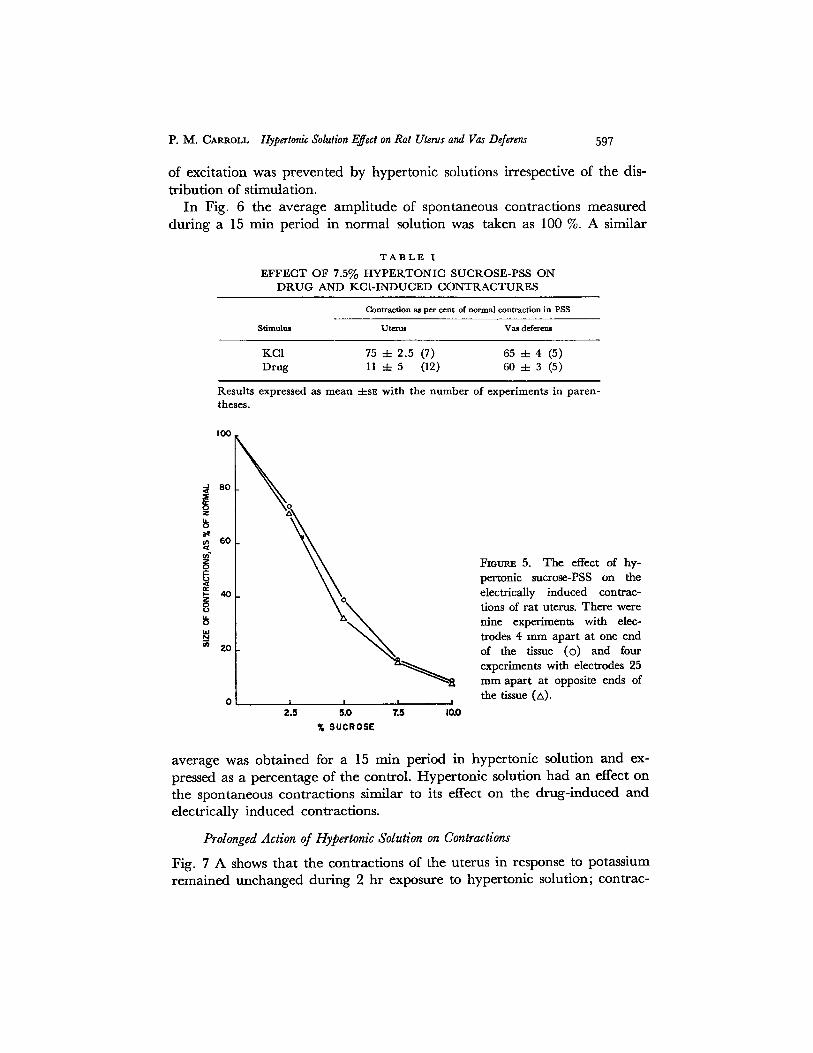
100
of exci ta t ion was p reven ted by hyper ton ic solutions irrespective of the dis-
t r ibu t ion of s t imulat ion. In Fig. 6 the average ampl i t ude of spontaneous cont rac t ions measured
dur ing a 15 rain per iod in no rma l solution was taken as 100 %. A similar
T A B L E I
EFFECT OF 7.5% HYPERTONIC SUCROSE-PSS ON DRUG AND KCI-INDUCED CONTRACTURES
C o n t r a c t i o n a t p e r cent o f n o r m a l c o n t r a c t i o n i n P S S
Stimulu= Uterus Vat deferem
K C I 75 4- 2 .5 (7) 65 4- 4 (5) D r u g 11 4- 5 (12) 60 ± 3 (5)
R e s u l t s expressed as m e a n 4-s~. w i t h t h e n u m b e r o f e x p e r i m e n t s in p a r e n - theses .
I I I I
2.5 5.0 7.5 I0.0
% SUCROSE
80
$ z
so
z _(2
¢r ~- 40 z
8
20
P. M. CARROLL Hypertonic Solution Effect on Rat Uterus and Vas Deferens
Fiom~ 5. The effect of hy- pertonic sucrose-PSS on the electrically induced contrac- tions of rat uterus. There were nine experiments with elec- trodes 4 mm apart at one end of the tissue (o) and four experiments with electrodes 25 ram apart at opposite ends of the tissue (z,).
average was ob ta ined for a 15 min per iod in hyper ton ic solution and ex- pressed as a pe rcen tage of the control . H y p e r t o n i c solution had an effect on the spontaneous cont rac t ions similar to its effect on the d rug - induced and electr ical ly induced contract ions.
Prolonged Action of Hypertonic Solution on Contractions
Fig. 7 A shows tha t the cont rac t ions of the uterus in response to potass ium r ema ined unchanged dur ing 2 h r exposure to hyper ton ic solut ion; cont rac-

598 T H E J O U R N A L O F G E N E R A L P H Y S I O L O G Y • V O L U M E 5 3 " x969
tions in response to drugs were markedly reduced but then gradually in- creased in size, increasing more rapidly in hypertonic choline chloride--PSS than in hypertonic sucrose-PSS. Additional experiments showed that 10 rain exposure to hypertonic choline chloride-PSS caused reductions of serotonin- and acetylcholine-induced contractions which were similar to those reductions caused by hypertonic sucrose-PSS (Fig. 3) and by hypertonic mannitol-PSS. Thus it is reasonable to assume that the action of choline chloride was due to its osmotic activity and not to chemical activity. Hence it is possible that the
80
N
~ 60
X
u t 2O
g 0 I
2.5
\ O
O
\ I I I
5.0 7.5 I0.0 % SUCROSE
Fxomuz 6. The effect of hy- pertonic sucrosc-PSS on the spontaneous contractions of rat uterus at 37°C. 12 horns were used.
more rapid increase in size of the contractions in hypertonic choline chloride- PSS compared with those in hypertonic sucrose-PSS (Fig. 7 A) was due to faster intracellular penetration of choline chloride compared with sucrose.
Fig. 7 B shows that the contractions of the vas deferens caused by potassium and drugs were very similar during the first 80 min and thus again there is a marked difference between the uterus and the vas deferens.
Contractions in Response to Supramaximal Dose of Drug
TaMe II shows that while 7.5 % sucrose-PSS considerably reduced the con- traction of the uterus in response to a dose of serotonin, which caused a maxi- mal contraction in normal PSS, the contraction in hypertonic solution could be increased by increasing the drug concentration to supramaximal levels. The response to submaximal doses of angiotensin was more affected by hyper- tonic sucrose-PSS than was the response to maximal doses, but because the contractions in response to low levels of drug plus high levels of sucrose were

P. M. CAaROXJ- Hypertonic Solution Effect on Rat Uterus and Vas Deferens 599
I O0
~ 60
"' 0 N I I I I I I
0 ZO 40 60 80 I00 120
MINUTES IN HYPERTONtC SOLUTION
FtGUR~ 7 A. The prolonged action of hypertonic 5 % sucrose-PSS on the KCl-induced contractures (×) (two horns) and on the contractions induced by acetylcholine (e) (two horns) of rat uterus. Also the prolonged action of hypertonie 2 % choline chloride- PSS on the KCl-induced contractures (×) (two horus) and on the contractions induced by serotonin (A) (two horns) of rat uterus.
~00
i , o
o I 0 I I I I I , , 40 60 8O I00 120
MINUTES IN 5% SUCROSE-PSS
The prolonged action of hypertonic 5 % sucrose-PSS on the KCl-induced FIOURE 7 B. contractures (x) and on the acetylcholine-induced contractions (e) of rat vas deferens. Two experiments are shown for both acetylcholine and KC1.
so tiny, fu r the r exper iments were no t per formed. Addi t iona l d a t a suggest tha t the results wi th angiotensin were no t compl ica ted by tachyphylaxis .
Contractions in Triple Baths
1. UTERUS
Fig. 8 A shows the spontaneous cont rac t ions of the uterus a t 37°C wi th the two ends of the ho rn in comple te synchrony. W h e n hyper ton ic 10 % or 15 % sucrose-PSS was p laced in the center ba th , in some exper iments the cont rac-

600 THE JOURNAL OF GENERAL PHYSIOLOGY • VOLUME 53 " X969
tions of the two ends became desynchronized, the cervical end contracting at a slower rate than the ovarian end (Fig. 8 B), and in some experiments the cervical end ceased contracting during the 10 min that the hypertonic PSS was in the center bath. The replacement of PSS in the center bath restored the synchrony of the two ends (Fig. 8).
When spontaneous contractions were eliminated by cooling (20 or 25°C), addition of serotonin to an end bath caused contraction of that end of the tissue and also a contraction at the end distant from the drug (Fig. 9). It was immaterial to which end the drug was added, excitation could travel in either direction. When hypertonic 10% or 15 % sucrose-PSS was placed in the center bath and 10 min elapsed before stimulating one end of the horn with
T A B L E I I
T H E E F F E C T OF S U P R A - AND S U B M A X I M A L D O S E S O F D R U G S O N T H E S I Z E OF C O N T R A C T I O N
IN H Y P E R T O N I C S U C R O S E - P S S
Contraction Contraction in ~cro~e:[: Serotonln 7.5 % Angiotendn
Supramaximal d o s e s racrose-PSS * Submaximal doae~ 5 % 7.5 %
1 X max. (4) 3 1 X max. (5) 43 21 4 X max. (4) 17 1/4 X max. (4) 34 9
25 X max. (4) 37 1/16 X max. (1) 9 0 50 X max. (4) 37
100 X max. (4) 53 250 X max. (4) 54
T h e n u m b e r s in paren theses refer to the n u m b e r of exper iments . * 100 was assigned to the con t rac t ion in response to the m a x i m u m dose of serotonin in the ab- sence of sucrose. T h e m a x i m u m dose for some tissues was 0.05/~g/50 ml.
100 was assigned to the cont rac t ion elicited by the test dose of angiotens in in the absence of sucrose. T h e m a x i m u m dose for most tissues was 0.025 gg/50 ml.
serotonin, then the far end of the tissue did not contract. After replacing PSS in the center bath and allowing 10 rain to elapse, restimulation of one end of the horn with the drug again caused a contraction of the far end (Fig. 9).
Likewise addition of KC1 depolarizing solution to an end bath caused a contraction of that end of the tissue and also a contracture at the end distant from the stimulus. This spread of excitation could be prevented by placing hypertonic sucrose-PSS in the center bath and could be restored by replacing PSS in the center bath (Fig. 10).
2. VAS DEFERENS
The vas deferens differed from the uterus. Addition of acetylcholine, adren- alin or KCI depolarizing solution to one end compartment while causing a contraction of that end of the tissue, failed to cause a contraction of the far end

A
°-TV-v-V-v-
B
c
C
°
Imin
FIoue~ 8. Spontaneous contractions of the ovarian end (0) and the cervical end (c) of the rat uterine horn in a three compartment bath. The top record of each pair has the base line at the top with contraction downwards; the bottom record of each pair has the base line at the bottom wlth contraction upwards. The central portion of the horn was in normal PSS in records A and C; the central portion was in hypertonic 10% sucrose-PSS in record B. Maximum contractions in these records were approximately
2g.
SEROTONIN
PSS
'20 sec'
SEROTONtN
I0 % SUCR(E:~-PSS PSS li-
IO%SUCROSE-PSS
I ~ SEROTONIN I' SEROTONIN
FIGURE 9. Serotonin-induced contractions of rat uterine horn in a three compartment bath. The top tracing has the base line at the top; the bottom tracing has the base line at the bottom. The drug was applied to either end of the horn, as indicated, and con- tractions recorded from both ends. The composition of the solution in the central com- partment is indicated for each test.
601

6o2 THE JOURNAL OF GENERAL PHYSIOLOGY • VOLUME 53 " t969
(Fig. 11). Tests of n ine vasa defe ren t ia gave the same result; thus I h a v e been u n a b l e to d e m o n s t r a t e the spread of exci ta t ion across a g a p of I c m in the r a t vas deferens b u t c a n n o t exc lude the possibili ty tha t in all expe r imen t s d a m a g e
~,KCI ~KCI
g wf
P-- _ _ ~ . , . z
PSS 10% SUCROSE- PSS /
J KCI
20 sec /
FZOURE 10. KCl-induced contractures of rat uterine horn in three compartment bath. The top tracing has the base line at the top; the bottom tracing has the base line at the bottom. The KC1 was applied to either end of the horn, as indicated and contractions recorded from both ends. The composition of the solution in the central compartment is indicated for each test.
ACETYL-
I 0 mgA~,Ornl 2 mg/4Oml
0.4. PSS PSS PSS PSS PSS PSS
ACETYL- T K CA T CHOLINE ~ ADRENALIN
2mg/4Oml ' ' IOmo~4Ornl 40 sec
FIGU~ ] 1. Contractions of rat vas deferens in three compartment bath. The top tracing has the base line at the top; the bottom tracing has the base line at the bottom. The stimulus (drug or KC1) was applied to the end indicated and contractions recorded from both the testicular end (top) and the bladder end (bottom). Normal PSS was in the central compartment in all cases.
to the tissue had occurred . Howeve r , the vas deferens was hand l ed in the same w a y as the u te rus in wh ich p r o p a g a t i o n of act ivi ty could be demons t r a t ed .
3. THE EFFECT OF POTASSIUM CHLORIDE IN THE CENTER COMPARTMENT ON
THE ACTION OF HYPERTONIC PSS
~¢Vhen hypertonic solutions (with I0 or 15 % sucrose) were placed in the center bath, the activity of uteri showing spontaneous synchronous contractions was

P. M. C~RROU. Hypertonic Solution Effect on Rat Utaus and Vas De/erens 6o 3
disrupted. Asynchrony of contractions at each end or cessation of the contrac- tions at the cervical end occurred. After 10 min, replacement by hypertonic sucrose-44 rn~ KCl-sal t solution in the center bath caused immediate restora- tion of synchronous contractions (Fig. 12). When uteri were contracting spontaneously and synchronously in PSS, the placement of hypertonic sucrose (10 or 15 %)-44 naxi KCl - sah solution in the center bath did not disrupt the
I O'B g wt
A c o o 1o 01t
C c 0
I min
FIOURE 12. Spontaneous con- tractions of ovarian end (o) and cervical end (c) of rat uterine horn. For each pair, the top record has the base line at the bottom, contrac- tion upwards, and the bottom record has the base line at the top, contraction downwards. The central portion of the horn was in normal PSS in record A; in 15% sucrose-PSS in record B; in 15% sucrose-44 mM KCl-salt solution in C.
synchronous contractions. 44 m_M KC1 either restored or prevented the dis- ruption of synchronous contractions by hypertonic sucrose-salt solution in all eight tests on five tissues.
Similarly 24 mM KC1 was used in the hypertonic sucrose-salt solution for four tests on two tissues; on three occasions it did not restore synchrony or prevent disruption of synchrony but on one occasion it did restore synchrony.
Thus 44 rnM KCI can overcome the effect of the hypertonic solution but 24 m u KC1 is hardly able to do so.
Weight Changes in the Response to 44 m~ KCl
A high level of potassium chloride did not alter the pattern of uterine wet weight loss due to hypertonic sucrose-salt solution when it was added 10 min

604 T H E J O U R N A L O F G E N E R A L P H Y S I O L O G Y * V O L U M E 53 • z969
after the onset of hypertonicity (Table I I I ) and thus the ability of potassium chloride to restore synchronous spontaneous contractions is apparently not due to it causing a change of cell volume.
T A B L E I I I
LACK OF A C T I O N OF 44 r r~ KCI ON W E I G H T LOSS OF U T E R I IN H Y P E R T O N I C S O L U T I O N
Per cent loss of tissue wet weight
Time in hypertonic solution 10% =ucrose-PSS 10% sucr~e-PSS
rain
5 10
15 20
7.4 7.2 lO .7 10.6
10% sucrose-44 ra~ KCl-salt solution
12.5 13.3 13.6 13.8
Results are the average of two experiments in which there were four horns in each group.
,0o j
8 o
x *t
60
i-
ill:
o
~ z 0
5,15 IOqO 15,5 20~0 VOLUME RATIO (122.5mM KCl SALT SOLUTION, PSS]
FlOURx 13. T h e dose-response curve of the uterus (O) (six horns) an d vas deferens (zx) (four tubes) to an increasing concentra t ion of KC1, obta ined by mixing 122.5 mM KC1 salt solution and PSS as indicated.
Dose-Response Curves for Potassium Chloride
Fig. 13 shows that the uterus contracts in response to a lower concentration of potassium than does the vas deferens, and the curve for the uterus is steeper than that for the vas deferens.

P. M. CAI~ROLL Hypertonk Solution Effect on Rat Uterus and Vas Deferens 605
D I S C U S S I O N
One of the aims of this work was to evaluate the contribution of conduction of excitation to the action of stimulant drugs on the rat uterus and vas deferens. I t is often assumed that responses of smooth muscle to drugs are determined exclusively and directly by the interaction between drug and receptors, and no account is taken of conduction. On the other hand, Daniel (1960, 1964 a) found evidence for conducted electrical activity during drug-induced and spontaneous activity in the rat uterus.
In the experiments reported here, a triple compar tment bath similar to that described by Johansson and Ljung (1967) was used to demonstrate that con- duction can occur in the uterus whether activity was initiated spontaneously or in response to stimulant drugs or KCI. Similar experiments on the vas deferens gave no indication of conduction, at least over a distance of 1 cm.
Barr et al. (1968) have shown that hypertonic solutions block conduction in guinea pig taenia coli, and Johansson and Ljung (1967) came to the same conclusion as the result of their experiments on the rat portal vein. I t was therefore decided to study the action of hypertonic solutions on the rat uterus and the vas deferens. Barr et al. (1968) reported that hypertonic solu- tions caused shrinkage of smooth muscle cells and the disruption of intercellu- lar connections (nexuses). Such connections have been observed in the uterus (Laguens and Lagrutta, 1964; Silva, 1967; Bergman, 1968). In the experiments reported here, tissue wet weight was taken as a qualitative mea- sure of cell volume. I t was found that the wet weight of both the uterus and vas deferens decreased during the first 20-30 min immersion in hypertonic solution. After reaching a min imum value (16 % reduction for solutions of approximately twice normal tonicity) the wet weight of the uterus began to increase. The wet weight of the vas deferens, however, continued to decrease for periods of immersion up to 120 rain.
The simplest explanation for a decrease in tissue wet weight in hypertonic solution would seem to be a decrease in cell volume. Replacement of extra- cellular or cellular fluid with a denser hypertonic solution would increase tissue weight. Therefore a decrease in weight must indicate a decrease in volume of some tissue compar tment and the decrease in wet weight observed in these experiments is probably an underestimate of tissue shrinkage. There is no reason why hypertonic solutions should cause a decrease in extracellular volume, whereas such solutions would be expected to extract water from cells.
The simplest explanation for the changes in gains in weights of the uterus during prolonged immersion in hypertonic solutions might be the slow penetration of sucrose and mannitol and the somewhat faster initial penetra- tion of choline chloride. After 120 min, however, tissues made hypertonic with choline chloride had not regained a higher proportion of their initial
606 T H E J O U R N A L O F G E N E R A L P H Y S I O L O G Y • V O L U M E 5 3 * ~969
weight loss than tissues in the other media (Fig. 1). Penetration of sucrose into a portion of the cellular water of frog stomach muscle has been demon- strated (Bozler, 1967). If this explanation is correct, then the permeability of the vas deferens to these agents must be considerably less than that of the uterus.
When hypertonic solutions were added to the central compartment of the triple bath, conduction of excitation in the uterus was blocked. If cell volume, as evidenced by changes in tissue wet weight, can be taken as an indication of the ability of the uterus to conduct a response to stimulation, then the magnitude of conducted responses should be depressed in proportion to the decrease in tissue wet weight. This was indeed the case for responses to electrical stimulation and for spontaneous contractions in single bath experi- ments. Furthermore, there was a close correlation between tissue wet weight and the magnitude of the response of the uterus to stimulant drugs. This result supports the idea that conduction of excitation may make a significant con- tribution to the drug-induced responses of this organ.
Contractions in response to KC1 were slightly depressed by hypertonic solutions in both preparations. In the vas deferens this decrease was paralleled by the decrease in response to acetylcholine. Since conduction could not be demonstrated in the vas deferens, and since KC1 probably acts on all smooth muscle cells, this result is also in accordance with the idea that the much more marked effect of hypertonic solutions on drug responses of the uterus was due to blockade of conduction. This result also implies that the large concentrations of acetylcholine required to stimulate the vas deferens were acting on all smooth muscle cells. An increase in drug concentration could partially antagonize the effects of hypertonic solutions in the uterus--presumably by activating a larger number of smooth muscle cells directly and so counter- acting the loss or depression of conduction.
Depression of responses to KC1 in both smooth muscles, and depression of responses to acetylcholine in the vas deferens may have been due to the effect of hypertonic solutions on excitation-contraction coupling (see Hodgkin and Horowicz, 1957; Howarth, 1958) or to the possibility that KCI caused a smaller depolarization in hypertonic solution due to an increase in the con- centration of intracellular K.
The mechanism by which hypertonic solutions block conduction remains to be studied. In the triple compartment bath experiments conduction block by hypertonic solutions could be reversed by the addition of KCI, as previously observed in the rat portal vein by Johansson and Ljung (1967) and Mellander et al. (1967). Johansson and Jonsson (1968) have discussed the mechanism of these effects. This result suggests that cell shrinkage may have caused hyper- polarization due to an increase in intracellular K concentration which could be counteracted by an increase in external K (see Tomita, 1966). Electron

P. M. CARROLL Hypertonic Solution Effect on Rat Uterus and Vas Deferens 607
m i c r o s c o p e s tud ie s a n d m e a s u r e m e n t o f t h e e l e c t r i c a l r e s i s t a n c e o f t h e u t e r u s
for l o n g i t u d i n a l c u r r e n t w i l l b e n e e d e d to d e c i d e w h e t h e r o r n o t t h e d i s r u p -
t i on o f i n t e r c e l l u l a r c o n n e c t i o n s is a l so i n v o l v e d .
This work was supported by a Life Insurance Medical Research Grant to Dr. Carroll.
Received for publication 20 May 1968.
REFERENCES
BARR, L., W. BERGER, and M. M. DEWRY. 1968. Electrical transmission at nexus between smooth muscle cells. J. Gen. Physiol. 51:347.
BERGMAN, R. A. 1968. Uterine smooth muscle fibers in castrate and estrogen-treated rats. J. Cell Biol. 36:639.
BOZLER, E. 1967. Determination of extracellular space in amphibian muscle. J. Gen. Physiol. 50:1459.
DANmL, E. E. 1960. The activation of various types of uterine muscle during stretch-induced contraction. Can. J. Biochem. Physiol. 38:1327.
DANIEL, E. E. 1964 a. The interconnecdon between active transport and contracture in uterine tissues. Can. J. Physiol. Pharmacol. 42:453.
DArnEL, E. E. 1964 b. Effect of drugs on contractions of vertebrate smooth muscle. Annu. Rev. Pharmacol. 4:189.
HODGKm, A. L., and P. HoRowIcz. 1957. The differential action of hypertonic solutions on the twitch and action potential of a muscle fibre. J. Physiol. (London). 136:17P.
HOWARTH, J. V. 1958. The behavior of frog muscle in hypertonic solutions. J. Physiol. (London). 144:167.
JOHANSSON, B., and O. JONSSON. 1968. Cell volume as a factor influencing electrical and me- chanical activity of vascular smooth muscle. Acta Physiol. Scand. 72:456.
JOHANSSON, B., and B. LJUNG. 1967. Spread of excitation in the smooth muscle of rat portal vein. Acta Physiol. Scan& 70:312.
LAG~NS, R., and J. LAORUTTA. 1964. Fine structure of human uterine muscle in pregnancy. Am. J. Obstet. Gynecol. 89:1040.
MELLANDER, S., B. JOI~NSSON, S. GRAY, O. JONSSON, J. L~DVAI.L, and B. LjUNa. 1967. The effects of hyperosmolarity on intact and isolated vascular smooth muscle. Possible role in exercise hyperemia. Angiologica. 4:310.
SILVA, D. G. 1967. The ultrastructure of the myometrium of the rat with special reference to innervation. Anat. Rec. 158:21.
TOMITA, T. 1966. Electrical responses of smooth muscle to external stimulation in hypertonic solutions. J. Physiol. (London). 183:450.