the ecophysiology of nitrite-oxidizing bacteria in the...
TRANSCRIPT
DISSERTATION
Titel der Dissertation
The ecophysiology of nitrite-oxidizing bacteria in the genus Nitrospira:
Novel aspects and unique features
angestrebter akademischer Grad
Doktor der Naturwissenschaften (Dr. rer. nat.) Verfasser: Frank Michael Maixner
Matrikel-Nummer: 0409617
Dissertationsgebiet (lt. Studienblatt):
444 Ökologie
Betreuer: Univ.-Prof. Dr. Michael Wagner
Wien, am 13. April 2009


meiner Familie

Contents Chapter I General Introduction and Outline 1 Chapter II Nitrite concentration influences the population
structure of Nitrospira-like bacteria 27 Chapter III Environmental genomics reveals a functional chlorite
dismutase in the nitrite-oxidizing bacterium ‘Candidatus Nitrospira defluvii’ 39
Chapter IV Nitrite oxidoreductase (Nxr) - the metabolic key enzyme
of nitrite-oxidizing Nitrospira: Characterization of an unusual Nxr and its application as a novel functional and phylogenetic marker gene 67
Chapter V Summary/Zusammenfassung 123 Appendix 131
Supplement chapter I – paper 1 Selective enrichment and molecular characterization of a previously uncultured Nitrospira-like bacterium from activated sludge 133 Supplement chapter I – paper 2 Physiological and phylogenetic characterization of a novel lithoautotrophic nitrite-oxidizing bacterium, ‘Candidatus Nitrospira bockiana’ 145 Supplement chapter I – figure 1 155 Supplement chapter I – table 1 161 List of publications 163 List of oral & poster presentations 165 Danksagung 169 Curriculum vitae 171

Abbreviations
A adenine
AmoA ammonia monooxygenase subunit A
ARB Arbor (software package comprising various tools for sequence and
phylogenetic analysis)
AOA ammonia-oxidizing archaea
AOB ammonia-oxidizing bacteria
C cytosin
CLD chlorite dismutase
CLSM confocal laser scanning microscope;
confocal laser scanning miocroscopy
DNA deoxyribonucleic acid
et al. et alii
Fig. figure
FISH fluorescence in situ hybridization
G guanine
Hao hydroxylamine oxidoreductase
N nitrogen
N. defluvii “Candidatus Nitrospira defluvii”
NOB nitrite-oxidizing bacteria
Nxr nitrite oxdioreductase
PCR polymerase chain reaction
RNA ribonucleic acid
rRNA ribosomal ribonucleic acid
T thymine
t ton
U uracile
wwtp wastewater treatment plant


Chapter I
General Introduction and Outline
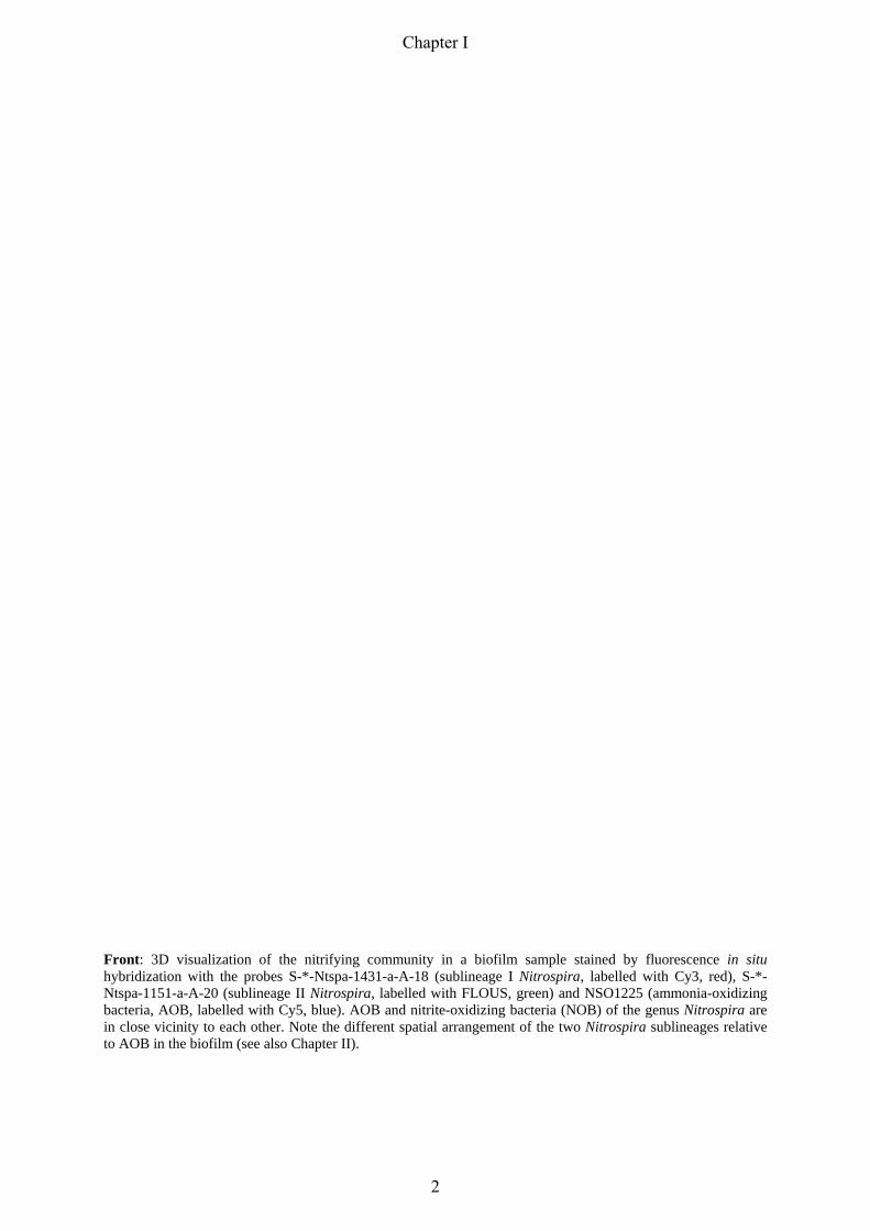
Front: 3D visualization of the nitrifying community in a biofilm sample stained by fluorescence in situ hybridization with the probes S-*-Ntspa-1431-a-A-18 (sublineage I Nitrospira, labelled with Cy3, red), S-*-Ntspa-1151-a-A-20 (sublineage II Nitrospira, labelled with FLOUS, green) and NSO1225 (ammonia-oxidizing bacteria, AOB, labelled with Cy5, blue). AOB and nitrite-oxidizing bacteria (NOB) of the genus Nitrospira are in close vicinity to each other. Note the different spatial arrangement of the two Nitrospira sublineages relative to AOB in the biofilm (see also Chapter II).
Chapter I
2
General Introduction
1. The Global Nitrogen Cycle
100 years after Fritz Haber patented the process to “synthesize ammonia from its elements”,
we nowadays live in a world which is highly dependent upon, and transformed by man-made
fixed nitrogen (Erisman et al., 2008). At first the “Haber-Bosch” nitrogen allowed large scale
production of explosives resulting in millions of war victims. Second the ammonia is a
starting product in several sectors in chemical industries producing plastics and synthetics.
Most of the fixed nitrogen, however, is used to produce fertilizer in the forms of ammonium
nitrate, calcium nitrate, ammonium bicarbonate, and several mixtures of nitrogen and
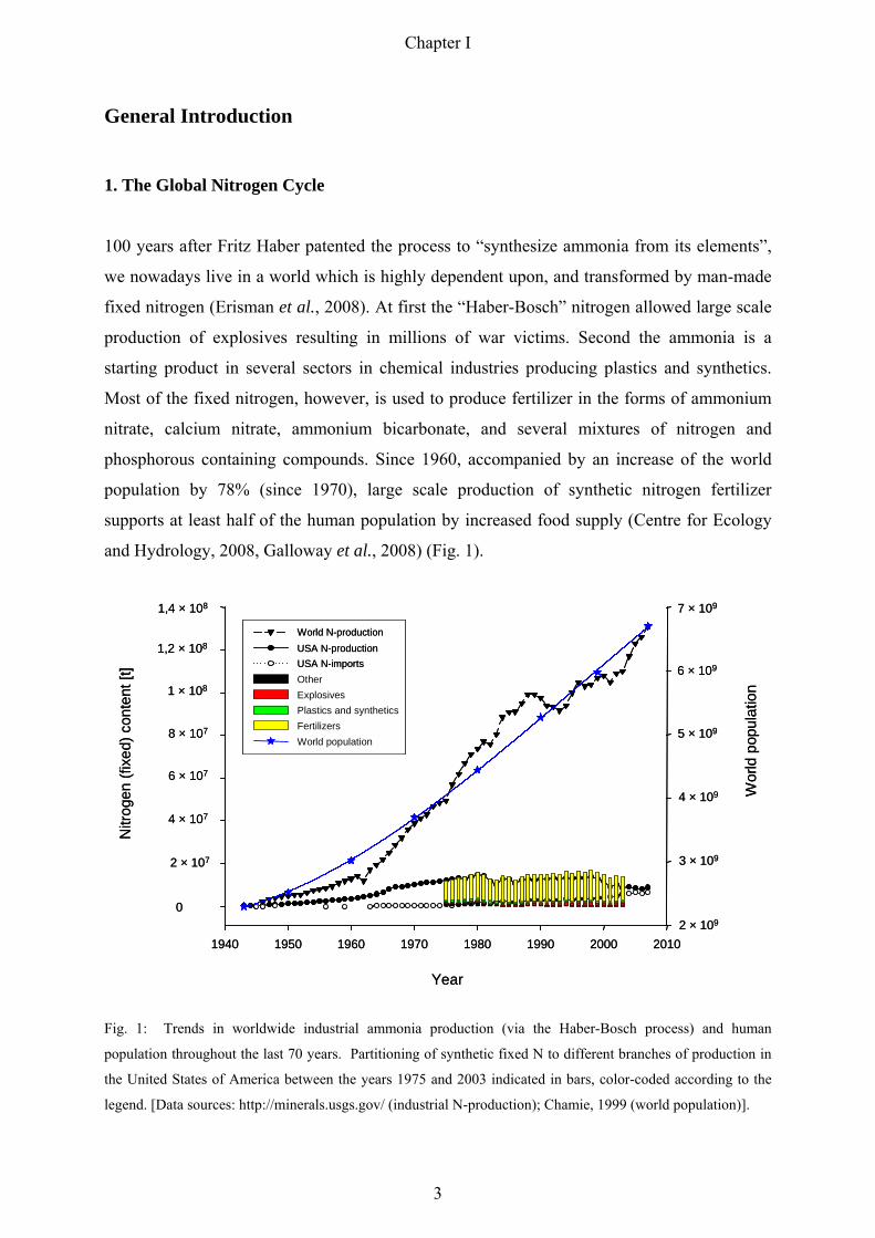
phosphorous containing compounds. Since 1960, accompanied by an increase of the world
population by 78% (since 1970), large scale production of synthetic nitrogen fertilizer
supports at least half of the human population by increased food supply (Centre for Ecology
and Hydrology, 2008, Galloway et al., 2008) (Fig. 1).
Fig. 1: Trends in worldwide industrial ammonia production (via the Haber-Bosch process) and human
population throughout the last 70 years. Partitioning of synthetic fixed N to different branches of production in
the United States of America between the years 1975 and 2003 indicated in bars, color-coded according to the
legend. [Data sources: http://minerals.usgs.gov/ (industrial N-production); Chamie, 1999 (world population)].
Year
1940 1950 1960 1970 1980 1990 2000 2010
Nitr
ogen
(fixe
d) c
onte
nt[t]
0
1,4 × 10⁸
Wor
ld p
opul
atio
n
7 × 10⁹
USA N-productionUSA N-imports
World N-production
OtherExplosives Plastics and syntheticsFertilizersWorld population
1,2 × 10⁸
1 × 10⁸
8 × 10⁷
6 × 10⁷
4 × 10⁷
2 × 10⁷
6 × 10⁹
5 × 10⁹
4 × 10⁹
3 × 10⁹
2 × 10⁹
Year
1940 1950 1960 1970 1980 1990 2000 2010
Nitr
ogen
(fixe
d) c
onte
nt[t]
0
1,4 × 10⁸
Wor
ld p
opul
atio
n
7 × 10⁹
USA N-productionUSA N-imports
World N-production
OtherExplosives Plastics and syntheticsFertilizersWorld population
USA N-productionUSA N-productionUSA N-importsUSA N-imports
World N-productionWorld N-production
OtherExplosives Plastics and syntheticsFertilizersWorld population
1,2 × 10⁸
1 × 10⁸
8 × 10⁷
6 × 10⁷
4 × 10⁷
2 × 10⁷
6 × 10⁹
5 × 10⁹
4 × 10⁹
3 × 10⁹
2 × 10⁹
Chapter I
3

Beside the nitrogen emitted to the atmosphere during fossil fuel burning the two major
sources of anthropogenic nitrogen released into the environment are fertilizers and
wastewater. Less than 30% of the synthetic nitrogen in fertilizers reaches the end- products
(Smil, 2001). The remaining nitrogen is either lost via emission of elemental nitrogen, nitric
oxides and ammonia or through leaching of nitrate. To counteract this loss often more
fertilizer is used in agriculture than needed. A recent study addressed the problem of
excessive nitrogen fertilization in agricultural areas in China (Ju et al., 2009). Ju and
coworkers tracked the fate of fertilizer nitrogen and could show that with a higher load of
fertilizer, plants are less efficient at taking up synthetic nitrogen. As a result most of the
nitrogen leached into the ground and surface water. A reduction of nitrogen fertilizer by two
thirds and better fertilization timing would reduce environmental nitrogen contamination
without compromising crop yields (Qiu, 2009).
The extraordinary population growth in the twentieth century resulted in a dramatically
increased nitrogen deposition into the environment. On the one hand there is the
aforementioned excess of fertilizer load due to the increased demand for food. Furthermore,
the increasing population causes a higher wastewater deposition and mainly in less developed
regions this wastewater ends up untreated in the environment. Therefore, beside better
nitrogen fertilizer management the efficient elimination of nitrogen in modern wastewater
treatment plants is a crucial process to reduce the import of nitrogen compounds into the
environment.
The increased anthropogenic nitrogen deposition destabilized ecosystems in most parts of the
world and resulted in major human health effects and unintended environmental consequences
(Vitousek et al., 1997, Galloway et al., 2008). On the one hand, atmospheric reactions of
emitted ammonia, nitric oxides, and sulfur oxides result in fine particle formation (Sharma,
2007), which increase the risk of cardiovascular and pulmonary diseases (Dominici et al.,
2006, Neuberger et al., 2007). Additionally, nitrogen-compounds like ammonia and nitrite are
highly toxic to aquatic life (Arthur et al., 1987) and elevated nitrite and nitrate levels in
drinking water can have severe health consequences to humans (Schneider & Selenka, 1974,
Ward et al., 2005). Beside these direct toxic effects of several nitrogen-compounds on living
organisms, raised deposition of anthropogenic nitrogen compounds into natural habitats
causes fatal ecological damages. Some environmental effects particularly noteworthy are
coastal eutrophication due to import of significant amounts of nitrogen through rivers
(Howarth & Marino, 2006) resulting in anoxia and hypoxia of the sea bottom (Rabalais, 2002,
Rabalais et al., 2002) or elevated nitrogen deposition linked to eutrophication of different
Chapter I
4
natural areas resulting in a considerable loss of floral biodiversity (Phoenix et al., 2006).
Beside the eutrophication of terrestrial and aquatic systems the interaction of the nitrogen and
carbon cycle and the resulting global acidification has already and will further have severe
consequences on earth climate (Gruber & Galloway, 2008). Recently, for example, aquatic
invertebrates in nitrate-rich environments have been shown to emit the potent greenhouse gas
nitrous oxide in quantitatively important amounts (Stief et al., 2009).
In summary, this manmade nitrogen deposition caused an ecological imbalance into many
ecosystems and the effects would have been even more dramatic without the high buffering
capacity of nitrogen cycling microorganisms. However, to effectively monitor ecological
changes due to anthropogenic pressure a better understanding of the mechanisms behind the
global nitrogen cycle is urgently needed. It is important to understand the nitrogen cycle at a
range of scales of biological organizations, beginning with the vast microbial diversity
involved in nitrogen conversion and their interactions (Horner-Devine & Martiny, 2008).
Especially during the last decade, groundbreaking and surprising findings in microbial
ecology such as bacteria capable of anaerobic ammonia oxidation (ANAMMOX) (Mulder et
al., 1995) or the existence of ammonia oxidizing archaea (Treusch et al., 2005, Könneke et
al., 2005) have shown that our knowledge of the nitrogen cycle and the involved microbial
key players is still scarce.
The following sections will focus on nitrite-oxidizing bacteria (NOB) of the genus Nitrospira,
one of the aforementioned key players in the nitrogen cycle but yet less intensively studied
groups of NOB.
Chapter I
5
2. The Biological Nitrogen Cycle: Microorganisms keep the wheel turning
All forms of life require nitrogen as integral part of proteins and nucleic acids, and thus every
living organism participates in the nitrogen-cycle. Microorganisms, however, catalyse
essential steps in the global biochemical nitrogen-cycle that other organisms are not able to
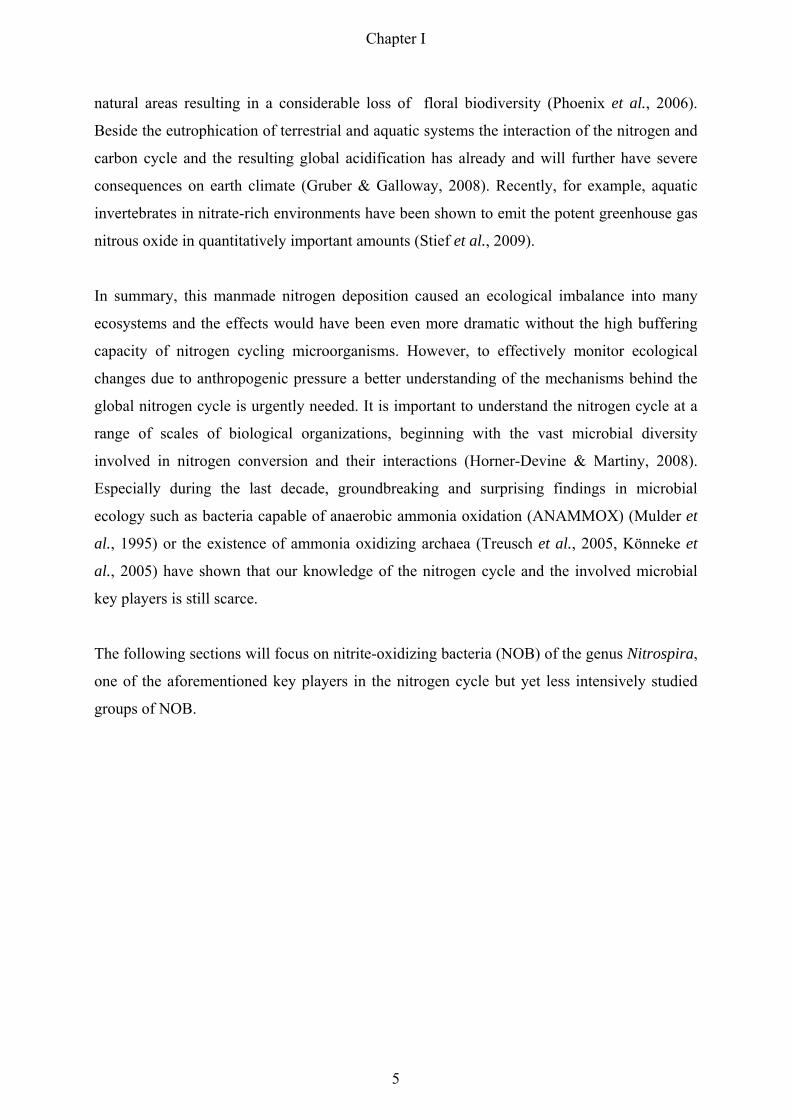
accomplish (Fig. 2). In microbial nitrogen fixation, gaseous dinitrogen is reduced by the
enzyme nitrogenase to ammonia and subsequently assimilated into cell material (Zehr et al.,
2003). Ammonia is released during microbial decomposition of organic substances, a process
termed ammonification or mineralization (McLain & Martens, 2005).
Fig. 2: Schematic representation of the N-cycle. Functional key enzymes involved in important redox-reactions
of oxidized and reduced N-compounds (yellow boxes) are displayed in white with the following abbreviations:
Amo, ammonia monooxygenase; Hao, hydroxylamine oxidoreductase; Nxr, nitrite oxidoreductase; Nar,
membrane-bound nitrate reductase; Nap, periplasmic nitrate reductase; NirK/NirS, nitrite reductase; Nor, nitric
oxide reductase; Nos; nitrous oxide reductase; Nif, nitrogenase; Hzo, hydrazine oxidoreductase.
At neutral pH most of the ammonia is available in form of ammonium. One part of the
released ammonium is rapidly recycled and converted into amino acids in microorganisms
and plants. However, especially in alkaline soils ammonia is lost via evaporation. The
remaining ammonia/ammonium can be catabolized either aerobically or anaerobically in two
distinct microbial key processes in the nitrogen-cycle. Under oxic conditions, microorganisms
gain energy from oxidizing ammonia to nitrite and subsequently nitrite to nitrate, a process
Chapter I
6

termed nitrification catalysed by two distinct functional groups of chemolithotrophic
prokaryotes: the ammonia-oxidizing bacteria (AOB) and archaea (AOA), and the nitrite-
oxidizing bacteria (NOB) (Prosser, 1989, Bock & Wagner, 2006, Könneke et al., 2005). In
anoxic habitats, however, ammonium can be catabolized by physiologically specialized
planctomycetes in a process called ANAMMOX (Kuenen, 2008). This reaction couples
ammonium oxidation to nitrite reduction with dinitrogen as final product.
Whereas the gaseous product of the ANAMMOX process is released into the atmosphere, the
nitrate produced via nitrification is either readily assimilated by plants and microorganisms or
it is used by facultatively anaerobic organisms as the respiratory electron acceptor under
anaerobic conditions (denitrification) (Zumft, 1997, Philippot & Hallin, 2005). Denitrification
is widespread among different bacterial genera. Furthermore, the distribution of
denitrification extends beyond the bacteria to the archaea and fungi, and surprisingly,
complete denitrification was also discovered in a benthic foraminifer (Risgaard-Petersen et
al., 2006). In the first step of denitrification nitrate is reduced to nitrite, followed by the
subsequent reduction of nitrite to nitric oxide. Furthermore, the gaseous nitric oxide serves in
lieu of dioxygen as terminal electron acceptor and is reduced to nitrous oxide, which is
subsequently reduced to dinitrogen. Hence, the denitrification and ANAMMOX processes
return dinitrogen to the atmosphere from terrestrial and aquatic habitats, thereby completing
the nitrogen-cycle. In addition to these anaerobic nitrogen-cycle processes, a recent study
describes a previously unknown reaction in which anoxygenic phototrophic bacteria closely
related to Thiocapsa roseopersicina use nitrite as an electron donor for photosynthesis
(Griffin et al., 2007). However, the ecological importance of these nitrite-oxidizing
phototrophs and their influence on the global biochemical nitrogen-cycle remain to be
determined.
The next sections deal with the aerobic key process of the nitrogen-cycle, nitrification. The
main focus is on microorganisms involved in nitrite-oxidation, their phylogenetic assignment
and their major biochemical properties.
Chapter I
7

NH4+
NO2-
NO3-
3. Nitrification – the oxidation of ammonia to nitrate
Different inorganic substances can serve for a highly specialized group of microorganisms,
the chemolihotrophs, as source of energy and reductants used for cell biosynthesis and
maintenance (Peck, 1968, Kelly & Wood, 2006). Chemolithotrophs can be classified
according to their substrate preference in functional groups such as iron oxidizers or
hydrogen-oxidizing bacteria.
This chapter focuses on nitrification, the aerobic, sequential oxidation of ammonia to nitrite
and nitrite to nitrate (Fig. 3). Nitrification is carried out by two spezialized groups of
chemolithotrophic microorganisms: ammonia-oxidizing bacteria (AOB) and archaea (AOA),
and nitrite-oxidizing bacteria (NOB). Since their discovery more than 100 years ago
(Winogradsky, 1890) lithotrophic bacterial nitrifiers conventionally have been classified as
one family, the Nitrobacteriaceae (Buchanan, 1917, Watson, 1971).
Fig. 3: Ammonia-oxidizing bacteria (AOB, purple) and nitrite-oxidizing bacteria (NOB, Nitrospira sublineage I,
yellow, Nitrospira sublineage II, turquoise) detected by FISH in a nitrifying biofilm sampled from a sequencing
batch biofilm reactor. Indicated in the image is the sequential oxidation of ammonia to nitrate during
nitrification. Note the close co-aggregation of AOB and NOB in the biofilm. Bar = 20 µm.
Chapter I
8

However, by comparative analysis of small subunit rRNA gene sequences, as initially
elaborated by Carl Woese and coworkers (Fox et al., 1977, Woese, 1987), the nitrifier
classification was totally revised. Comparative 16S rRNA sequence analysis revealed that
most known AOB and NOB belong to different subclasses of Proteobacteria (Fig. 4) (Teske
et al., 1994, Purkhold et al., 2000, Koops et al., 2003, Alawi et al., 2007). The only exception,
however, are NOB of the genus Nitrospira that form a distinct phylum within the domain
Bacteria (Ehrich et al., 1995). Furthermore, it is tempting to speculate, that Nitrospina-like
NOB, provisionally assigned to the delta-subclass of Proteobacteria (Teske et al., 1994),
represent in addition to Nitrospira an independent line of descent within the Bacteria (Fig 4).
In comparison to the 16S rRNA data, sequence analysis of the amoA gene, which encodes the
alpha subunit of ammonia monooxygenase, suggested a similar evolutionary relationship of
AOB (Purkhold et al., 2000). Based on the phylogeny of both marker genes so far all isolated
aerobic lithoautotrophic ammonia-oxidizer can be assigned to two monophyletic groups in the
beta-subclass and gamma-subclass of the Proteobacteria. Recent studies, however, suggested
amo gene homologues on metagenomic sequences derived from mesophilic crenarchaea
(Venter et al., 2004, Treusch et al., 2005). These groundbraking findings could be confirmed
by the successful enrichment and isolation of crenarcheal ammonia oxidizers from a marine
aquarium and terrestrial hot springs (Könneke et al., 2005, Hatzenpichler et al., 2008, de la
Torre et al., 2008). The detection of AOA revised the long-lasting assumption that ammonia-
oxidation would be restricted to few lineages of the Proteobacteria (Fig. 4).
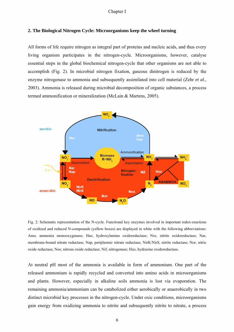
Fig. 4: 16S rRNA-based Maximum Likelihood trees displaying the phylogenetic affiliation of nitrifying
microorganisms. Nitrite-oxidizing bacteria (NOB) are highlighted in red, ammonia-oxidizing bacteria (AOB)
and archaea (AOA) are highlighted in blue and green, respectively. The tree in Fig. 4/A gives a general overview
of the distribution of nitrifiers in the “tree of life”, whereas the tree in Fig. 4/B illustrates a close-up view of Fig.
4/A representing all currently known nitrifying bacterial and archaeal genera and species. Both trees were
calculated using the Maximum Likelihood algorithm implemented in the ARB software tool (Ludwig et al.,
2004). The partially filled circles at the tree nodes in Fig. 4/B represent quartet puzzling reliability values ≥ 70 %
and filled circles symbolize additional high parsimony bootstrap support (≥ 90 %) based on 100 iterations. The
bar indicates 10 % estimated sequence divergence.
Chapter I
9
Archaea
Eukarya
Aquificae Thermotogae
Chloroflexi
DeinococcusThermusFibrobacteres
FusobacteriaFirmicutes
Actinobacteria
Cyanobacteria
Nitrospirae
Spirochaetes
BacteriodetesChlorobi
Planctomycetes
VerrucomicrobiaChlamydiae
Nitrospina
Acidobacteria
DeltaproteobacteriaEpsilonproteobacteria
Gammaproteobacteria
Betaproteobacteria
Alphaproteobacteria
0.10
AOA
NOB
AOBA
B Nitrosomonas europaea / Nitrosococcus mobilis
Nitrososmonas communis (AJ298732, AJ298740)
Nitrosomonas marina (AJ298734, Z46990)
Nitrososmonas oligotropha (AJ298736,AJ298730)Nitrosomonas cryotolerans (AJ298738)Nitrosospira spp. (M96396, AJ298746, CP000103)Siderooxidans lithoautotrophicus (DQ386264)“Candidatus Nitrotoga arctica“ (DQ839562)Gallionella ferruginea (L07897)Nitrosococcus spp. (CP000127, M96398, AF287298)Methylohalobius crimeensis (AJ581837)
Nitrococcus mobilis (L35510)Arhodomonas aquaeolei (M26631)
Thiocapsa roseopersicina (EF581005)Chromatium okenii (Y12376)Thiocystis violacea (Y11315)
Nitrobacter spp. (AF069956, CP000115, L11663, AM114522)Bradyrhizobium japonicum (X87272)
Namibian upwelling system clone (EF646147)Nitrospina gracilis (L35504)Napoli mud volcano sediment clone,1920m depth (AY592588)Nitrospira sp. (DQ059545, X82558, EU084879, X82559)4
Leptospirillum ferrooxidans (X86776)Halorubrum lacusprofundi (X82170)
Thermoplasma acidophilum (M38637)Methanosarcina acetivorans (M59137)
Archaeoglobus fulgidus DSM 4304 (AE000782)Caldivirga maquilingensis (AB013926)
“Candidatus Nitrosocaldus yellowstonii“ (EU239960)Nitrosopumilus maritimus (DQ085097)
“Candidatus Nitrososphaera gargensis“ (EU281334)0.10
(AB070982, AY123795, AF272413, AF037105)
Proteobacteria
beta
gamma
alpha
Nitrospina
Nitrospirae
ArchaeaKorarchaeota
Crenarchaeota
Chapter I
10
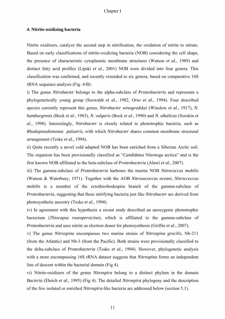
4. Nitrite-oxidizing bacteria
Nitrite oxidizers, catalyze the second step in nitrification, the oxidation of nitrite to nitrate.
Based on early classifications of nitrite-oxidizing bacteria (NOB) considering the cell shape,
the presence of characteristic cytoplasmic membrane structures (Watson et al., 1989) and
distinct fatty acid profiles (Lipski et al., 2001) NOB were divided into four genera. This
classification was confirmed, and recently extended to six genera, based on comparative 16S
rRNA sequence analysis (Fig. 4/B):
i) The genus Nitrobacter belongs to the alpha-subclass of Proteobacteria and represents a
phylogenetically young group (Seewaldt et al., 1982, Orso et al., 1994). Four described
species currently represent this genus, Nitrobacter winogradskyi (Winslow et al., 1917), N.
hamburgensis (Bock et al., 1983), N. vulgaris (Bock et al., 1990) and N. alkalicus (Sorokin et
al., 1998). Interestingly, Nitrobacter is closely related to phototrophic bacteria, such as
Rhodopseudomonas palustris, with which Nitrobacter shares common membrane structural
arrangement (Teske et al., 1994).
ii) Quite recently a novel cold adapted NOB has been enriched from a Siberian Arctic soil.
The organism has been provisionally classified as “Candidatus Nitrotoga arctica” and is the
first known NOB affiliated to the beta-subclass of Proteobacteria (Alawi et al., 2007).
iii) The gamma-subclass of Proteobacteria harbours the marine NOB Nitrococcus mobilis
(Watson & Waterbury, 1971). Together with the AOB Nitrosococcus oceani, Nitroccoccus
mobilis is a member of the ectothiorhodospira branch of the gamma-subclass of
Proteobacteria, suggesting that these nitrifying bacteria just like Nitrobacter are derived from
photosynthetic ancestry (Teske et al., 1994).
iv) In agreement with this hypothesis a recent study described an anoxygenic phototrophic
bacterium (Thiocapsa roseopersicina), which is affiliated to the gamma-subclass of
Proteobacteria and uses nitrite as electron donor for photosynthesis (Griffin et al., 2007).
v) The genus Nitrospina encompasses two marine strains of Nitrospina gracilis, Nb-211
(from the Atlantic) and Nb-3 (from the Pacific). Both strains were provisionally classified to
the delta-subclass of Proteobacteria (Teske et al., 1994). However, phylogenetic analysis
with a more encompassing 16S rRNA dataset suggests that Nitrospina forms an independent
line of descent within the bacterial domain (Fig 4).
vi) Nitrite-oxidizers of the genus Nitrospira belong to a distinct phylum in the domain
Bacteria (Ehrich et al., 1995) (Fig 4). The detailed Nitrospira phylogeny and the description
of the few isolated or enriched Nitrospira-like bacteria are addressed below (section 5.1).
Chapter I
11

Most work on the physiology and biochemistry of NOB was done with Nitrobacter species
and our knowledge about the biochemical properties of all other known nitrite-oxidizing
genera is only scarce. Therefore, it is important to keep in mind that most of the presented
data cannot be generalized for all NOB.
Two electrons are abstracted when nitrite is oxidized to nitrate. The formation of nitrate is
catalysed by the key enzyme of NOB, nitrite oxidoreductase (Nxr), according to the following
equation:
Nxr: NO2- + H2O ↔ NO3
- + 2H+ + 2e-
The additional oxygen atom is derived from water (Aleem et al., 1965). Most Nxr-related
studies have been performed with different Nitrobacter strains (Tanaka et al., 1983,
Sundermeyer-Klinger et al., 1984, Meincke et al., 1992). In the absence of oxygen, the Nxr of
Nitrobacter can reduce nitrate to nitrite. Therefore, the oxidation of nitrite is a reversible
reaction (Sundermeyer-Klinger et al., 1984). The Nxr of Nitrobacter is a integral membrane
bound enzyme complex, which consists of at least two subunits, the large subunit NxrA and
the small subunit NxrB (Sundermeyer-Klinger et al., 1984, Meincke et al., 1992). The mature
holoenzyme contains molybdopterin as cofactor and several iron-sulfur centers (Ingledew &
Halling, 1976, Sundermeyer-Klinger et al., 1984, Meincke et al., 1992). Immunocytochemical
studies revealed that the Nxr of Nitrobacter is localized at the cytoplasmic face of the cell
membrane and at the intracytoplasmic membranes (Spieck et al., 1996).
Only a few studies about the Nxr of Nitrospira exist (Spieck et al., 1998, Bartosch et al.,
1999, Chapter IV, this thesis). The results, however, indicated that the Nxr of Nitrospira
differs remarkably from the Nxr of Nitrobacter. In contrast to the Nxr of Nitrobacter, the Nxr
of Nitrospira was found to be membrane-associated in the periplasmic space (Spieck et al.,
1998). Furthermore, immunoblotting experiments and sequence analysis of Nxr subunits of
Nitrobacter and Nitrospira revealed major differences in their apparent molecular masses and
their phylogenetic affiliation (Bartosch et al., 1999, Chapter IV, this thesis). Taken together,
these results suggest a convergent evolution of the Nxr enzymatic function in two different
nitrite-oxidizing systems.
Lithoautotrophic NOB fix carbon dioxide (CO2) and about 80% of the energy generated by
nitrite oxidation is used for CO2 fixation (Spieck & Bock, 2005). Furthermore, the redox
Chapter I
12
potential of the NO2-/ NO3
- couple is extraordinarily high (+420 mV). Thus, similar to
AOB/AOA, nitrite-oxidizing bacteria are extremely slow-growing organism with minimal
generation times ranging from 10 h for Nitrobacter vulgaris (Bock et al., 1990) up to 90 h for
Nitrospira marina (Watson et al., 1986) under lithoautotrophic growth conditions.
Apart from aerobic nitrite oxidation several other metabolic properties are known for NOB.
Nitrobacter, for example, thrives by denitrification in anoxic habitats (Freitag et al., 1987,
Bock et al., 1988). Also Nitrospira moscoviensis can use nitrate as electron acceptor under
anoxic conditions with hydrogen as electron donor (Ehrich et al., 1995). Furthermore,
heterotrophic growth using a variety of different carbon compounds has been described for
Nitrobacter for many years. This organotrophic growth, however, is inefficient and slow
(Smith & Hoare, 1968, Bock, 1976). In contrast, Nitrospira marina grew best mixotrophically
in a medium containing nitrite, pyruvate, yeast extract, and peptone (Ehrich et al., 1995).
In summary, most available biochemical data of NOB comes from the easily culturable
Nitrobacter species. Our knowledge about other NOB is still limited, mainly due to the lack
of media to culture these fastidious and slow-growing organisms.
Chapter I
13

5. Nitrite-oxidizing bacteria of the genus Nitrospira
The next section includes a more detailed phylogenetic description of NOB of the
phylum/genus Nitrospira, one of the aforementioned less intensively studied groups of NOB.
Furthermore, the recent state of knowledge about their diversity and ecophysiology will be
summarized. Finally, from this current status major ecological questions arose, which have
been the basic framework for this thesis.
5.1 Phylogeny and Diversity of Nitrospira
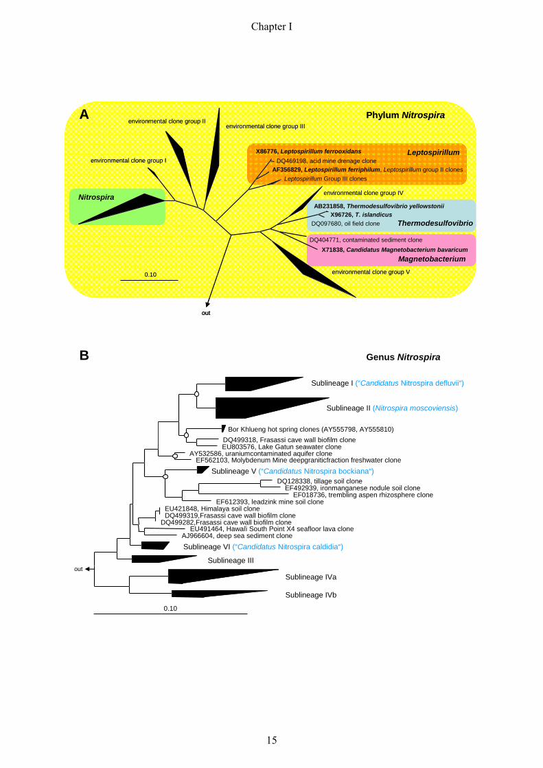
Nitrite oxidizers of the genus Nitrospira form a distinct phylum in the domain Bacteria. The
phylum Nitrospira comprises the genera Nitrospira, Leptospirillum, Thermodesulfovibrio and
Magnetobacterium (Ehrich et al., 1995), and several environmental clone groups, which are
not yet assigned to any of these genera (Fig. 5/A).
The genus Nitrospira represents a large and highly diverse group, with only five isolated/
enriched representatives so far (Fig 5/B). The first two described species are Nitrospira
marina, which was obtained from Atlantic Ocean samples (Watson et al., 1986), and
Nitrospira moscoviensis isolated from an urban heating system in Moscow (Ehrich et al.,
1995). However, several other attempts to selectively enrich Nitrospira-like bacteria from
different environmental samples failed, mainly because Nitrospira was outcompeted by
Nitrobacter when standard isolation procedures were used (Bartosch et al., 1999). One
determining factor for the outcome of the enrichment of NOB seems to be the initial nitrite
concentration in the medium. Whereas Nitrobacter overgrew Nitrospira-like bacteria in media
Fig. 5: 16S rRNA-based trees displaying the phylogeny of different genera (highlighted with different colours)
within the phylum Nitrospira (A, highlighted yellow) and the phylogenetic affiliation of different sublineages
within the genus Nitrospira (B). Sequences belonging to one sublineage have been clustered according to the
sublineage definition of Daims and colleagues (Daims et al., 2001) (Fig. 5/B). The so far isolated or enriched
Nitrospira-like bacteria are displayed in brackets in blue behind the respective sublineage. For the un-clustered
trees displaying the phylogeny within Nitrospira sublineages please refer to figure 1 in the Appendix. All trees
were calculated using the Maximum Likelihood algorithm implemented in the ARB software tool (Ludwig et al.,
2004). The partially filled circles at the tree nodes in Fig. 5/B represent quartet puzzling reliability values ≥ 70
% and filled circles symbolize additional high parsimony bootstrap support (≥ 90 %) based on 100 iterations.
The bar indicates 10 % estimated sequence divergence.
Chapter I
14
Nitrospira
environmental clone group IX86776, Leptospirillum ferrooxidans
DQ469198, acid mine drenage cloneAF356829, Leptospirillum ferriphilum, Leptospirillum group II clones
Leptospirillum Group III clones
AB231858, Thermodesulfovibrio yellowstoniiX96726, T. islandicus
DQ097680, oil field clone
DQ404771, contaminated sediment cloneX71838, Candidatus Magnetobacterium bavaricum
0.10
environmental clone group IIenvironmental clone group III
Leptospirillum
environmental clone group IV
environmental clone group V
out
Thermodesulfovibrio
Phylum Nitrospira
Magnetobacterium
A
Sublineage I (“Candidatus Nitrospira defluvii“)33
Sublineage II (Nitrospira moscoviensis)65
Bor Khlueng hot spring clones (AY555798, AY555810)DQ499318, Frasassi cave wall biofilm cloneEU803576, Lake Gatun seawater clone
AY532586, uraniumcontaminated aquifer cloneEF562103, Molybdenum Mine deepgraniticfraction freshwater clone
Sublineage V (“Candidatus Nitrospira bockiana“)5DQ128338, tillage soil clone
EF492939, ironmanganese nodule soil cloneEF018736, trembling aspen rhizosphere clone
EF612393, leadzink mine soil cloneEU421848, Himalaya soil cloneDQ499319,Frasassi cave wall biofilm clone
DQ499282,Frasassi cave wall biofilm cloneEU491464, Hawai’i South Point X4 seafloor lava clone
AJ966604, deep sea sediment cloneSublineage VI (“Candidatus Nitrospira caldidia“)6
Sublineage III5
Sublineage IVa32
Sublineage IVb10
0.10
out
B Genus Nitrospira
Chapter I
15

with higher nitrite concentration (2 g NaNO2 per liter), Nitrospira-like bacteria could be
selectively cultivated in mixotrophic media containing 0.2 g NaNO2 per liter (Bartosch et al.,
1999, Bartosch et al., 2002). Subsequently, a combined approach using low nitrite
concentration, percoll density gradient centrifugation for further purification, and serial
dilutions led to a high enrichment (86%) of previously uncultured Nitrospira-like bacteria
from activated sludge. The NOB has been classified as “Candidatus Nitrospira defluvii”
(Spieck et al., 2006) (Appendix: paper 1). Additionally, this elaborate strategy resulted in the
cultivation of “Candidatus Nitrospira bockiana” from the internal corrosion deposits of a steel
pipeline of the Moscow heating system (Lebedeva et al., 2008) (Appendix: paper 2) and to the
enrichment of the first thermophilic Nitrospira-like bacterium (“Candidatus Nitrospira
caldida”) from a microbial mat of the Gorjachinsk hot spring in the Baikal rift zone (Elena
Lebedeva, manuscript in preparation). The isolated and enriched Nitrospira-like bacteria
differ quite remarkably in their cell size and morphology. Furthermore, their optimal growth
temperature ranges from 28°C up to 50°C and the organisms display a different tolerance
against nitrite (Appendix: table 1). Taken together, these data reflect the various lifestyles
and isolation sources of the cultured Nitrospira-like bacteria, which are members of different
sublineages within the genus Nitrospira (Fig. 5/B). The small ribosomal subunit sequences of
the five isolated or enriched Nitrospira-like bacteria, however, represent only a minor
proportion of the 16S rRNA gene sequences that belong to the genus Nitrospira.
Extended phylogenetic analyses of the genus Nitrospira were mainly based on environmental
clone sequences obtained from various habitats, including bioreactors (Juretschko et al., 1998,
Schramm et al., 1998, Kindaichi et al., 2004), different freshwater habitats (Hovanec et al.,
1998, Altmann et al., 2003, Martiny et al., 2003), soil, rhizosphere (Kim et al., 2008,
Lesaulnier et al., 2008), hot springs (Lebedeva et al., 2005), marine habitats (Foesel et al.,
2008, Santelli et al., 2009) and sponges (Taylor et al., 2007) (Appendix: figure 1). These
comprehensive phylogenetic analyses extended the number of sublineages in the genus
Nitrospira from four, as previously proposed by Daims and colleagues (Daims et al., 2001),
to six monophyletic sublineages (Fig. 5/B). Interestingly, some of these sublineages seem to
represent a habitat-specific clustering of sequences, such as sublineage one, which almost
exclusively consists of bioreactor clones, or sublineage four where all clones and cultures
were derived from marine habitats or sponges (Appendix: figure 1).
In summary, cultivation and, moreover, 16S rRNA-based screenings have shown that NOB of
the genus Nitrospira are a highly diverse group of organisms, and that these bacteria are
Chapter I
16
ubiquitously distributed in a wide range of aquatic and terrestrial ecosystems. However, it
remains to be determined, whether this enormous phylogenetic diversity reflects differing
physiological properties within the genus Nitrospira.
5.2 Ecopysiology of Nitrospira
Laboratory-based studies on the few cultivated Nitrospira-like bacteria revealed first insights
into the physiology of Nitrospira. The abundance and in situ ecophysiology of Nitrospira-like
bacteria in different habitats, however, remained for a long time largely unknown. The
understanding of these NOB greatly increased with the introduction of different cultivation-
independent techniques into microbial ecology (Wagner et al., 2003). Mainly the application
of FISH in combination with microautoradiography (MAR-FISH) or with microelectrodes
uncovered novel aspects of their structural, functional and physiological properties.
First FISH analyses, aimed towards quantifying natural populations of nitrifiers, revealed that
uncultured bacteria related to the genus Nitrospira and not Nitrobacter are the dominant NOB
in aquaria, nitrifying laboratory-scale reactors, and full-scale wastewater treatment plants
(Hovanec et al., 1998, Juretschko et al., 1998, Schramm et al., 1998). Additionally, the in situ
visualization could show that AOB and Nitrospira-like bacteria form within activated sludge
flocs and biofilms compact microcolonies (consisting of hundreds to thousands of cells),
which are often in close vicinity to each other (Fig. 3). Interestingly, larger Nitrospira cell
aggregates in bioreactor samples contain multiple water-permeable channels, most likely
facilitating nutrient- and gas-supply to the cells inside a colony (Daims et al., 2001).
Moreover, by combining FISH and microautoradiograhpy, Daims and colleagues could show
that these Nitrospira-like bacteria took up under oxic conditions radiolabeled inorganic
carbon (in forms of HCO3- or CO2) and pyruvate, suggesting a mixotrophic growth in the
presence of pyruvate. In contrast, the Nitrospira-like bacteria incorporated the tested
radioactive carbon sources neither under anoxic nor anaerobic conditions (Daims et al., 2001).
Another MAR-FISH study additionally revealed tight ecophysiological interactions between
nitrifiers and heterotrophic bacteria in a carbon-limited autotrophic nitrifying biofilm with
ammonium as sole energy source. The results indicated that heterotrophic bacteria thrive on
the released soluble microbial products of nitrifiers, such as Nitrospira spp., and that there is
an efficient carbon food web in the biofilm community (Kindaichi et al., 2004). Nevertheless,
these two studies represent so far the only ecophysiological characterizations of Nitrospira-
like bacteria using the MAR-FISH approach. A further major advancement in microbial
Chapter I
17
ecology, beside MAR-FISH, was the combined application of FISH with microelectrodes.
This approach revealed not only the spatial localisation of nitrifiers within a biofilm along
different gas- and nutrient-gradients. Moreover, it was for the first time possible to record the
in situ activity of AOB and NOB and to subsequently estimate, for example, the cell-specific
activity of uncultured Nitrospira-like bacteria (Schramm et al., 1998, Schramm et al., 1999,
Schramm et al., 2000). The results obtained in these studies suggested that NOB of the genus
Nitrospira are K strategists adapted to low oxygen and nitrite concentration, whereas
Nitrobacter-like organism are r strategists, which overgrow Nitrospira spp. under conditions
of elevated nitrite and oxygen concentrations.
Taken together, Nitrospira spp. exhibit in comparison to Nitrobacter spp. a higher affinity for
nitrite and oxygen (Downing & Nerenberg, 2008). Considering the huge diversity within the
genus Nitrospira, these low Km values might be advantageous in certain environments,
enabling Nitrospira-like bacteria to thrive in ecological niches with constantly low substrate
concentrations. One remarkable finding beside these ecophysiologal differences between
Nitrospira and Nitrobacter was the co-existence of two Nitrospira populations in biofilm
samples of nitrifying bioreactors (Schramm et al., 1998, Schramm et al., 1999). The two
populations displayed a distinct spatial distribution pattern within the biofilm, suggesting a
niche differentiation within the genus Nitrospira due to different physiological adaptations.
Nevertheless, beside these few depicted fundamental studies describing the in situ
ecophysiology of Nitrospira-like bacteria, so far only a few studies indirectly referred to
physiological properties of these NOB. In one of these studies, for example, Freitag and
colleagues demonstrated the presence of different sublineage II Nitrospira-ecotypes in long-
term fertilized and unfertilized agricultural grassland soils, also indicating a physiological
diversification within the genus Nitrospira (Freitag et al., 2005). Interestingly, a recent study
showed that Nitrospira-like bacteria are more abundant in the rhizosphere compared to the
bulk soil (DeAngelis et al., 2009). However, it remains to be determined, whether these
sublineage II Nitrospira metabolize the freshly introduced root exudates in the rhizosphere
and therefore strongly respond to the root shooting. Finally, a study investigating temperature
thresholds for bacterial symbiosis revealed a quick response of Nitrospira-like bacteria in
sponges to elevated temperatures. A temperature shift from 27 °C to 33°C resulted in the loss
of microbial sponge symbionts, such as sublineage IVb Nitrospira (Webster et al., 2008).
Even though these few studies only touched on the question of the ecophysiology of
Nitrospira in different habitats, the results already indicate various physiological adaptations
within the genus Nitrospira. Whereas the theory behind the ecological differentiation of
Chapter I
18

Nitrobacter spp. and Nitrospira spp. is already well accepted (Schramm et al., 2000), our
knowledge about the physiological differentiation within the genus Nitrospira is still limited.
For example, a yet unresolved ecological question in this context is:
Does the coexistence of different Nitrospira populations in the same habitat reflect the
physiological adaptations to different environmental factors, such as nutrient
concentration, oxygen availability, or temperature?
Furthermore, first Nitrospira genome sequences, which became available during this thesis,
extended the question to:
i) Does the Nitrospira genome encode any resistance genes or genes indicating
metabolic versatility, which could explain physiological adaptations to certain
environments?
ii) Does the comparison of metabolic key enzymes (e.g. Nxr) of Nitrospira and
Nitrobacter reveal any indications for different enzymatic properties supporting
the distinct ecophysiological adaptations of these NOB?
These major ecological questions provided the theoretical framework for this thesis.
Chapter I
19

Aims of this thesis
One major goal of this thesis was to gain more insight into the ecophysiology of Nitrospira-
like bacteria by applying novel molecular in situ techniques and via in depth analysis of
metagenomic data. Furthermore, in the course of this thesis another yet unresolved question
was addressed, whether the Nxr subunits might be suitable as functional and also as
phylogenetic markers for the detection of Nitrospira-like bacteria in environmental samples.
The niche differentiation between two coexisting uncultured populations of Nitrospira-like
bacteria in nitrifying biofilm and activated sludge samples is described in Chapter II.
Distinct spatial distribution patterns of the two Nitrospira sublineages relative to AOB
suggested a niche differentiation with respect to their preferred concentrations of nitrite. A
long term experiment revealed that representatives of the two sublineages indeed showed
different response to nitrite concentration shifts. Population dynamics of Nitrospira-like
bacteria demonstrated significant differences in the ecophysiological properties of closely
related nitrifiers, a major requirement for niche differentiation in diverse habitats.
Chapter III offers insight into the first metagenome fragment of a Nitrospira-like bacterium
(“Candidatus Nitrospira defluvii”). Surprisingly, a gene similar to genes encoding chlorite
dismutases (CLD) was found on the Nitrospira contig. This enzyme, which has only been
known from (per)chlorate reducing Proteobacteria, transforms toxic chlorite to chloride and
oxygen. The biochemical activity of the non-proteobacterial CLD of Nitrospira was
confirmed by heterologous expression in E. coli followed by enzymatic tests. The catalytic
parameters of the recombinant CLD indicated that the gene encodes a highly active CLD.
Furthermore, Western blot analysis revealed that the gene is expressed in situ by Nitrospira.
The evolutionary origin of this enzyme family and the possible role of a CLD in a distinct
physiological adaptation of this Nitrospira-like organism have been discussed in detail.
The study described in Chapter IV emerged as a spin-off from a larger environmental
genomics project, which aimed at sequencing the whole genome of “Candidatus Nitrospira
defluvii”. Here, the goal was to identify and characterize in detail the genes comprising the
key enzyme nitrite oxidoreductase in the genome of N. defluvii. The Nxr of Nitrospira
consists of three subunits, which differ quite remarkably in sequence from the Nxr subunits of
Chapter I
20
Nitrobacter and Nitrococcus. Additionally, the Nxr beta subunit turned out to be a suitable
functional and phylogenetic marker for the genus Nitrospira.
A summary of the presented studies is given in Chapter V.
References
Alawi, M., A. Lipski, T. Sanders, E. M. Pfeiffer & E. Spieck, (2007) Cultivation of a novel cold-adapted nitrite oxidizing betaproteobacterium from the Siberian Arctic. ISME J 1: 256-264.
Aleem, M. I., G. E. Hoch & J. E. Varner, (1965) Water as the source of oxidant and reductant in bacterial chemosynthesis. Proc Natl Acad Sci U S A 54: 869-873.
Altmann, D., P. Stief, R. Amann, D. De Beer & A. Schramm, (2003) In situ distribution and activity of nitrifying bacteria in freshwater sediment. Environ Microbiol 5: 798-803.
Arthur, J. W., C. W. West, K. N. Allen & S. F. Hedtke, (1987) Seasonal toxicity of ammonia to five fish and nine invertebrate species. Bull Environ Contam Toxicol 38: 324-331.
Bartosch, S., C. Hartwig, E. Spieck & E. Bock, (2002) Immunological detection of Nitrospira-like bacteria in various soils. Microb Ecol 43: 26-33.
Bartosch, S., I. Wolgast, E. Spieck & E. Bock, (1999) Identification of nitrite-oxidizing bacteria with monoclonal antibodies recognizing the nitrite oxidoreductase. Appl Environ Microbiol 65: 4126-4133.
Bock, E., H. P. Koops, U. C. Möller & M. Rudert, (1990) A new facultative nitrite oxidizing bacterium Nitrobacter vulgaris. Arch Microbiol 153: 105-110.
Bock, E., Sundermeyer-Klinger H. & E. Stackebrandt, (1983) New facultative lithoautotrophic nitrite-oxidizing bacteria. Arch Microbiol 136: 281-284.
Bock, E. & M. Wagner, (2006) Oxidation of inorganic nitrogen compounds as energy source. The Prokaryotes 2: 457-495.
Bock, E., P. A. Wilderer & A. Freitag, (1988) Growth of Nitrobacter in the absence of dissolved oxygen. Water Res 22: 245-250.
Buchanan, R. E., (1917) Studies on the Nomenclature and Classification of the Bacteria: III. The Families of the Eubacteriales. J Bacteriol 2: 347-350.
Centre for Ecology and Hydrology, (2008) 100 years of ammonia synthesis: How a single patent changed the world. ScienceDaily http://www.sciencedaily.com/releases/2008/09/080929095708.htm.
Chamie, J.,(1999) The World at Six Billion UN report http://www.un.org/esa/population/publications/sixbillion/sixbillion.htm.
Daims, H., J. L. Nielsen, P. H. Nielsen, K. H. Schleifer & M. Wagner, (2001) In situ characterization of Nitrospira-like nitrite-oxidizing bacteria active in wastewater treatment plants. Appl Environ Microbiol 67: 5273-5284.
de la Torre, J. R., C. B. Walker, A. E. Ingalls, M. Konneke & D. A. Stahl, (2008) Cultivation of a thermophilic ammonia oxidizing archaeon synthesizing crenarchaeol. Environ Microbiol 10: 810-818.
Chapter I
21

DeAngelis, K. M., E. L. Brodie, T. Z. DeSantis, G. L. Andersen, S. E. Lindow & M. K. Firestone, (2009) Selective progressive response of soil microbial community to wild oat roots. ISME J 3: 168-178.
Dominici, F., R. D. Peng, M. L. Bell, L. Pham, A. McDermott, S. L. Zeger & J. M. Samet, (2006) Fine particulate air pollution and hospital admission for cardiovascular and respiratory diseases. JAMA 295: 1127-1134.
Downing, L. S. & R. Nerenberg, (2008) Effect of oxygen gradients on the activity and microbial community structure of a nitrifying, membrane-aerated biofilm. Biotechnol Bioeng 101: 1193-1204.
Ehrich, S., D. Behrens, E. Lebedeva, W. Ludwig & E. Bock, (1995) A new obligately chemolithoautotrophic, nitrite-oxidizing bacterium, Nitrospira moscoviensis sp. nov. and its phylogenetic relationship. Arch Microbiol 164: 16-23.
Erisman, J. W., M. A. Sutton, J. Galloway, Z. Klimont & W. Winiwarter, (2008) How a century of ammonia synthesis changed the world. Nature Geoscience 1: 636-639.
Foesel, B. U., A. Gieseke, C. Schwermer, P. Stief, L. Koch, E. Cytryn, J. R. de la Torre, J. van Rijn, D. Minz, H. L. Drake & A. Schramm, (2008) Nitrosomonas Nm143-like ammonia oxidizers and Nitrospira marina-like nitrite oxidizers dominate the nitrifier community in a marine aquaculture biofilm. FEMS Microbiol Ecol 63: 192-204.
Fox, G. E., K. R. Pechman & C. R. Woese, (1977) Comparative cataloging of 16S ribosomal ribonucleic acid: molecular approach to prokaryotic systematic. Int J Syst Bacteriol 27: 44-57.
Freitag, A., M. Rudert & E. Bock, (1987) Growth of Nitrobacter by dissimilatoric nitrate reduction. FEMS Microbiol Lett 48: 105-109.
Freitag, T. E., L. Chang, C. D. Clegg & J. I. Prosser, (2005) Influence of inorganic nitrogen management regime on the diversity of nitrite-oxidizing bacteria in agricultural grassland soils. Appl Environ Microbiol 71: 8323-8334.
Griffin, B. M., J. Schott & B. Schink, (2007) Nitrite, an electron donor for anoxygenic photosynthesis. Science 316: 1870.
Gruber, N. & J. N. Galloway, (2008) An Earth-system perspective of the global nitrogen cycle. Nature 451: 293-296.
Hatzenpichler, R., E. V. Lebedeva, E. Spieck, K. Stoecker, A. Richter, H. Daims & M. Wagner, (2008) A moderately thermophilic ammonia-oxidizing crenarchaeote from a hot spring. Proc Natl Acad Sci U S A 105: 2134-2139.
Horner-Devine, M. C. & A. C. Martiny, (2008) Biogeochemistry. News about nitrogen. Science 320: 757-758.
Hovanec, T. A., L. T. Taylor, A. Blakis & E. F. Delong, (1998) Nitrospira-Like Bacteria Associated with Nitrite Oxidation in Freshwater Aquaria. Appl Environ Microbiol 64: 258-264.
Howarth, R. W. & R. Marino, (2006) Nitrogen as the limiting nutrient for eutrophication in coastal marine ecosystems: Evolving views over three decades. Limnol. Oceanogr. 51: 364-376.
Ju, X. T., G. X. Xing, X. P. Chen, S. L. Zhang, L. J. Zhang, X. J. Liu, Z. L. Cui, B. Yin, P. Christie, Z. L. Zhu & F. S. Zhang, (2009) Reducing environmental risk by improving N management in intensive Chinese agricultural systems. Proc Natl Acad Sci U S A.
Juretschko, S., G. Timmermann, M. Schmid, K. H. Schleifer, A. Pommerening-Roser, H. P. Koops & M. Wagner, (1998) Combined molecular and conventional analyses of nitrifying bacterium diversity in activated sludge: Nitrosococcus mobilis and Nitrospira- like bacteria as dominant populations. Appl Environ Microbiol 64: 3042-3051.
Chapter I
22

Kim, J.-S., R. S. Dungan & D. Crowely, (2008) Microarray analysis of bacterial diversity and distribution in aggregates from a desert agricultural soil. Biol Fertil Soils 44: 1003-1011.
Kindaichi, T., T. Ito & S. Okabe, (2004) Ecophysiological interaction between nitrifying bacteria and heterotrophic bacteria in autotrophic nitrifying biofilms as determined by microautoradiography-fluorescence in situ hybridization. Appl Environ Microbiol 70: 1641-1650.
Könneke, M., A. E. Bernhard, J. R. de la Torre, C. B. Walker, J. B. Waterbury & D. A. Stahl, (2005) Isolation of an autotrophic ammonia-oxidizing marine archaeon. Nature 437: 543-546.
Koops, H. P., U. Purkhold, A. Pommerening-Roser, G. Timmermann & M. Wagner, (2003) The lithoautotrophic ammonia-oxdizing bacteria. in Dworkin et al., eds., The Prokaryotes: An Evolving Electronic Resource for the Microbiological Community.
Kuenen, J. G., (2008) Anammox bacteria: from discovery to application. Nat Rev Microbiol 6: 320-326.
Lebedeva, E. V., M. Alawi, C. Fiencke, B. Namsaraev, E. Bock & E. Spieck, (2005) Moderately thermophilic nitrifying bacteria from a hot spring of the Baikal rift zone. FEMS Microbiol Ecol 54: 297-306.
Lesaulnier, C., D. Papamichail, S. McCorkle, B. Ollivier, S. Skiena, S. Taghavi, D. Zak & D. van der Lelie, (2008) Elevated atmospheric CO2 affects soil microbial diversity associated with trembling aspen. Environ Microbiol 10: 926-941.
Lipski, A., E. Spieck, A. Makolla & K. Altendorf, (2001) Fatty acid profiles of nitrite-oxidizing bacteria reflect their phylogenetic heterogeneity. Syst Appl Microbiol 24: 377-384.
Martiny, A. C., T. M. Jorgensen, H. J. Albrechtsen, E. Arvin & S. Molin, (2003) Long-term succession of structure and diversity of a biofilm formed in a model drinking water distribution system. Appl Environ Microbiol 69: 6899-6907.
McLain, J. E. T. & D. A. Martens, (2005) Nitrous oxide flux from soil and amino acid mineralization. Soil Biol Biochem 37: 289-299.
Meincke, M., E. Bock, D. Kastrau & P. M. H. Kroneck, (1992) Nitrite oxidoreductase from Nitrobacter hamburgensis: redox centers and their catalytic role. Arch Microbiol 158: 127-131.
Mulder, A., A. A. van de Graaf, L. A. Robertson & J. G. Kuenen, (1995) Anaerobic ammonium oxidation discovered in a denitrifying fluidized bed reactor. FEMS Microbiol Ecol 16: 177-184.
Neuberger, M., D. Rabczenko & H. Moshammer, (2007) Extended effects of air pollution on cardiopulmonary mortality in Vienna. Atmosheric Environment 41: 8549-8556.
Orso, S., M. Gouy, E. Navarro & P. Normand, (1994) Molecular phylogenetic analysis of Nitrobacter spp. Int J Syst Bacteriol 44: 83-86.
Phoenix, G., K. Hicks, S. Cinderby, J. Kuylenstierna, W. Stocks, F. Dentener, K. Giller, A. Austin, R. Lefroy, B. Gimeno, M. Ashmore & P. Ineson, (2006) Atmospheric nitrogen deposition in world biodiversity hotspots: the need for a greater global perspective in assessing N deposition impacts. Global Change Biology 12: 470-476.
Purkhold, U., A. Pommerening-Roser, S. Juretschko, M. C. Schmid, H. P. Koops & M. Wagner, (2000) Phylogeny of all recognized species of ammonia oxidizers based on comparative 16S rRNA and amoA sequence analysis: implications for molecular diversity surveys. Appl Environ Microbiol 66: 5368-5382.
Qiu, J., (2009) Nitrogen fertilizer warning for China. Nature doi:10.1038/news.2009.105. Risgaard-Petersen, N., A. M. Langezaal, S. Ingvardsen, M. C. Schmid, M. S. Jetten, H.
J. Op den Camp, J. W. Derksen, E. Pina-Ochoa, S. P. Eriksson, L. P. Nielsen, N.
Chapter I
23

P. Revsbech, T. Cedhagen & G. J. van der Zwaan, (2006) Evidence for complete denitrification in a benthic foraminifer. Nature 443: 93-96.
Santelli, C. M., V. P. Edgcomb, W. Bach & K. J. Edwards, (2009) The diversity and abundance of bacteria inhabiting seafloor lavas positively correlate with rock alteration. Environ Microbiol 11: 86-98.
Schramm, A., D. De Beer, A. Gieseke & R. Amann, (2000) Microenvironments and distribution of nitrifying bacteria in a membrane-bound biofilm. Environ Microbiol 2: 680-686.
Schramm, A., D. de Beer, J. C. van den Heuvel, S. Ottengraf & R. Amann, (1999) Microscale distribution of populations and activities of Nitrosospira and Nitrospira spp. along a macroscale gradient in a nitrifying bioreactor: quantification by in situ hybridization and the use of microsensors. Appl Environ Microbiol 65: 3690-3696.
Schramm, A., D. De Beer, M. Wagner & R. Amann, (1998) Identification and activities in situ of Nitrosospira and Nitrospira spp. as dominant populations in a nitrifying fluidized bed reactor. Appl Environ Microbiol 64: 3480-3485.
Seewaldt, E., K. H. Schleifer, E. Bock & E. Stackebrandt, (1982) The close phylogenetic relationship of Nitrobacter and Rhodopseudomonas palustris. Acta Microbiol 131: 287-290.
Sharma, M., Kishore, S., Tripathi, S.N., Behera, S.N.,, (2007) Role of atmospheric ammonia in the formation of inorganic secondary particulate matter: A study at Kanpur, India. J. Atmos. Chem. 58: 1-17.
Smil, V., (2001) Enriching the earth: Fritz Haber, Carl Bosch and the Transformation of World Food Production MIT Press: 411.
Spieck, E., J. Aamand, S. Bartosch & E. Bock, (1996) Immunocytochemical detection and localization of the membrane-bound nitrite oxidoreductase in cells of Nitrobacter and Nitrospira. FEMS Microbiol Lett 139: 71-76.
Spieck, E. & E. Bock, (2005) The lithoautotrophic nitrite-oxidizing bacteria. In Garrity et al. (ed.), Bergey´s manual of systematic bacteriology. Springer Science+Business Media, New York 2: 149-153.
Spieck, E., S. Ehrich, J. Aamand & E. Bock, (1998) Isolation and immunocytochemical location of the nitrite-oxidizing system in Nitrospira moscoviensis. Arch Microbiol 169: 225-230.
Stief, P., M. Poulsen, L. P. Nielsen, H. Brix & A. Schramm, (2009) Nitrous oxide emission by aquatic macrofauna. Proc Natl Acad Sci U S A.
Sundermeyer-Klinger, H., W. Meyer, B. Waringhoff & E. Bock, (1984) Membrane-bound nitrite oxidoreductase of Nitrobacter: evidence for a nitrate reductase system. Arch Microbiol 140: 153-158.
Taylor, M. W., R. Radax, D. Steger & M. Wagner, (2007) Sponge-associated microorganisms: evolution, ecology, and biotechnological potential. Microbiol Mol Biol Rev 71: 295-347.
Teske, A., E. Alm, J. M. Regan, S. Toze, B. E. Rittmann & D. A. Stahl, (1994) Evolutionary relationships among ammonia- and nitrite-oxidizing bacteria. J Bacteriol 176: 6623-6630.
Wagner, M., M. Horn & H. Daims, (2003) Fluorescence in situ hybridisation for the identification and characterisation of prokaryotes. Curr Opin Microbiol 6: 302-309.
Watson, S. W., (1971) Taxonomic considerations of the family Nitrobacteraceae Buchanan: Request for opinions. Int J Syst Bacteriol 21: 254-270.
Watson, S. W., E. Bock, H. Harms, H. P. Koops & A. B. Hooper, (1989) Nirtifying bacteria. In Murray et al. (ed.), Bergey´s manual of systematic bacteriology. The Williams & Wilkins Co., Baltimore.: 1808-1834.
Chapter I
24
Watson, S. W., F. W. Valois, J. B. Waterbury & U. Schlosser, (1986) Nitrospira marina gen. nov. sp. nov.: a chemolithotrophic nitrite-oxidizing bacterium. Arch Microbiol 144: 1-7.
Watson, S. W. & J. B. Waterbury, (1971) Characteristics of two marine nitrite-oxidizing bacteria, Nitrospina gracilis nov. gen. nov. sp. and Nitrococcus mobilis nov. gen. nov. sp.. Arch Microbiol 77: 203-230.
Webster, N. S., R. E. Cobb & A. P. Negri, (2008) Temperature thresholds for bacterial symbiosis with a sponge. ISME J 2: 830-842.
Winogradsky, S. N., (1890) Sur les organismes de la nitrification. Compt Rend Acad Sci 110: 1013-1016.
Winslow, C. E., J. Broadhurst, R. E. Buchanan, C. Krumwiede, L. A. Rogers & G. H. Smith, (1917) The Families and Genera of the Bacteria: Preliminary Report of the Committee of the Society of American Bacteriologists on Characterization and Classification of Bacterial Types. J Bacteriol 2: 505-566.
Woese, C. R., (1987) Bacterial evolution. Microbiol. Rev. 51: 221-271. Zehr, J. P., B. D. Jenkins, S. M. Short & G. F. Steward, (2003) Nitrogenase gene diversity
and microbial community structure: a cross-system comparison. Environ Microbiol 5: 539-554.
Chapter I
25

Chapter I
26

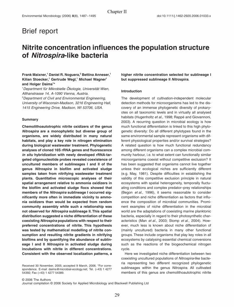
Chapter II
Nitrite concentration influences the population structure
of Nitrospira-like bacteria
Published in Environmental Microbiology (2006): 8 (8), 1487-1495

Chapter II
28
Environmental Microbiology (2006)
8
(8), 1487–1495 doi:10.1111/j.1462-2920.2006.01033.x
© 2006 The AuthorsJournal compilation © 2006 Society for Applied Microbiology and Blackwell Publishing Ltd
tion
Niche differentiation of NitrospiraF. Maixner
et al.
Received 30 November, 2005; accepted 9 March, 2006. *For corre-spondence. E-mail [email protected]; Tel. (
+
43) 1 427754392; Fax (
+
43) 1 4277 54389.
Brief report
Nitrite concentration influences the population structure of
Nitrospira
-like bacteria
Frank Maixner,
1
Daniel R. Noguera,
2
Bettina Anneser,
1
Kilian Stoecker,
1
Gertrude Wegl,
1
Michael Wagner
1
and Holger Daims
1
*
1
Department für Mikrobielle Ökologie, Universität Wien, Althanstrasse 14, A-1090 Vienna, Austria.
2
Department of Civil and Environmental Engineering, University of Wisconsin-Madison, 3216 Engineering Hall, 1415 Engineering Drive, Madison, WI 53706, USA.
Summary
Chemolithoautotrophic nitrite oxidizers of the genus
Nitrospira
are a monophyletic but diverse group oforganisms, are widely distributed in many naturalhabitats, and play a key role in nitrogen eliminationduring biological wastewater treatment. Phylogeneticanalyses of cloned 16S rRNA genes and fluorescence
in situ
hybridization with newly developed rRNA-tar-geted oligonucleotide probes revealed coexistence ofuncultured members of sublineages I and II of thegenus
Nitrospira
in biofilm and activated sludgesamples taken from nitrifying wastewater treatmentplants. Quantitative microscopic analyses of theirspatial arrangement relative to ammonia oxidizers inthe biofilm and activated sludge flocs showed thatmembers of the
Nitrospira
sublineage I occurred sig-nificantly more often in immediate vicinity to ammo-nia oxidizers than would be expected from randomcommunity assembly while such a relationship wasnot observed for
Nitrospira
sublineage II. This spatialdistribution suggested a niche differentiation of thesecoexisting
Nitrospira
populations with respect to theirpreferred concentrations of nitrite. This hypothesiswas tested by mathematical modelling of nitrite con-sumption and resulting nitrite gradients in nitrifyingbiofilms and by quantifying the abundance of sublin-eage I and II
Nitrospira
in activated sludge duringincubations with nitrite in different concentrations.Consistent with the observed localization patterns, a
higher nitrite concentration selected for sublineage Ibut suppressed sublineage II
Nitrospira
.
Introduction
The development of cultivation-independent moleculardetection methods for microorganisms has led to the dis-covery of an immense phylogenetic diversity of prokary-otes on all taxonomic levels and in virtually all analysedhabitats (Hugenholtz
et al
., 1998; Rappé and Giovannoni,2003). A recurring question in microbial ecology is howmuch functional differentiation is linked to this high phylo-genetic diversity: Do all different phylotypes found in thesame environmental sample represent organisms with dif-ferent physiological properties and/or survival strategies?A related question is how much functional redundancyamong different organisms can a complex microbial com-munity harbour, i.e. to what extent can functionally similarmicroorganisms coexist without competitive exclusion? Ithas been suggested that organisms cannot live togetherunless their ecological niches are sufficiently different(e.g. May, 1981). Despite difficulties in establishing thevalidity of this competitive exclusion principle in naturalecosystems with spatial heterogeneity, temporally fluctu-ating conditions and complex predator–prey relationships(Begon
et al
., 1996), it seems reasonable to considercompetition and niche differentiation as factors that influ-ence the composition of microbial communities. Promi-nent examples of niche differentiation in the microbialworld are the adaptations of coexisting marine planktonicbacteria, especially in regard to their photosynthetic char-acteristics (Man
et al
., 2003; Stomp
et al
., 2004). How-ever, much less is known about niche differentiation of(mainly uncultured) bacteria in many other functionalgroups. These include organisms that play key roles in allecosystems by catalysing essential chemical conversionssuch as the reactions of the biogeochemical nitrogencycle.
Here we investigated niche differentiation between twocoexisting uncultured populations of
Nitrospira
-like bacte-ria representing two different recognized phylogeneticsublineages within the genus
Nitrospira
. All cultivatedmembers of this genus are chemolithoautotrophic nitrite
Chapter II
29
1488
F. Maixner
et al.
© 2006 The AuthorsJournal compilation © 2006 Society for Applied Microbiology and Blackwell Publishing Ltd,
Environmental Microbiology
,
8
, 1487–1495
oxidizers (Watson
et al
., 1986; Ehrich
et al
., 1995; Spieck
et al
., 2006).
Nitrospira
-like bacteria occur in a large num-ber of different natural habitats (Daims
et al
., 2001) andare also the key nitrite oxidizers in biological wastewatertreatment (Burrell
et al
., 1998; Juretschko
et al
., 1998;Daims
et al
., 2001). Supporting evidence for niche parti-tioning was obtained by analysing, in biofilm and activatedsludge, the spatial arrangement of the two
Nitrospira
pop-ulations relative to ammonia oxidizer microcolonies andby monitoring the population dynamics of these
Nitrospira
-like bacteria during a long-term competition experiment.
Results and discussion
Detection and spatial arrangement of
Nitrospira
Nitrifying biofilm was taken from a continuously operatedpilot-scale biofilm reactor (‘Biofor 2’) (Arnold
et al
., 2000)receiving municipal wastewater, and nitrifying activatedsludge was sampled on a full-scale municipal wastewatertreatment plant (WWTP) near Gleisdorf, Austria (nitrifying-denitrifying stage with intermittent aeration; average influ-ent concentrations: 90 mg l
−
1
NH
4
+
-N, 0.5 mg l
−
1
NO
3–
-N;average effluent concentrations: 0.4 mg l
−
1
NH
4
+
-N,15 mg l
−
1
NO
3–
-N). The 16S rRNA genes of
Nitrospira
inthese samples were amplified by specific polymerasechain reaction (PCR) with a universal forward and a newlydeveloped reverse primer (Table 1). The amplicons werecloned and sequenced as described by Juretschko and
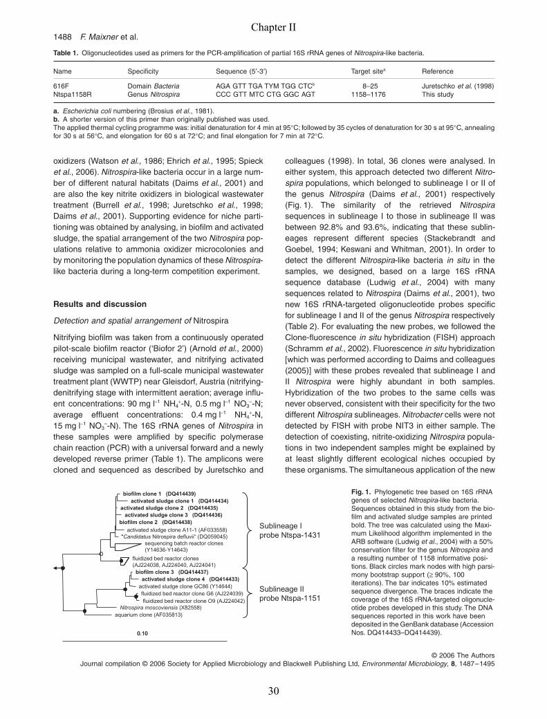
colleagues (1998). In total, 36 clones were analysed. Ineither system, this approach detected two different
Nitro-spira
populations, which belonged to sublineage I or II ofthe genus
Nitrospira
(Daims
et al
., 2001) respectively(Fig. 1). The similarity of the retrieved
Nitrospira
sequences in sublineage I to those in sublineage II wasbetween 92.8% and 93.6%, indicating that these sublin-eages represent different species (Stackebrandt andGoebel, 1994; Keswani and Whitman, 2001). In order todetect the different
Nitrospira
-like bacteria
in situ
in thesamples, we designed, based on a large 16S rRNAsequence database (Ludwig
et al
., 2004) with manysequences related to
Nitrospira
(Daims
et al
., 2001), twonew 16S rRNA-targeted oligonucleotide probes specificfor sublineage I and II of the genus
Nitrospira
respectively(Table 2). For evaluating the new probes, we followed theClone-fluorescence
in situ
hybridization (FISH) approach(Schramm
et al
., 2002). Fluorescence
in situ
hybridization[which was performed according to Daims and colleagues(2005)] with these probes revealed that sublineage I andII
Nitrospira
were highly abundant in both samples.Hybridization of the two probes to the same cells wasnever observed, consistent with their specificity for the twodifferent
Nitrospira
sublineages.
Nitrobacter
cells were notdetected by FISH with probe NIT3 in either sample. Thedetection of coexisting, nitrite-oxidizing
Nitrospira
popula-tions in two independent samples might be explained byat least slightly different ecological niches occupied bythese organisms. The simultaneous application of the new
Table 1.
Oligonucleotides used as primers for the PCR-amplification of partial 16S rRNA genes of
Nitrospira
-like bacteria.
Name Specificity Sequence (5
′
-3
′
) Target site
a
Reference
616F Domain
Bacteria
AGA GTT TGA TYM TGG CTC
b
8–25 Juretschko
et al
. (1998)Ntspa1158R Genus
Nitrospira
CCC GTT MTC CTG GGC AGT 1158–1176 This study
a.
Escherichia coli
numbering (Brosius
et al
., 1981).
b.
A shorter version of this primer than originally published was used.The applied thermal cycling programme was: initial denaturation for 4 min at 95
°
C; followed by 35 cycles of denaturation for 30 s at 95
°
C, annealingfor 30 s at 56
°
C, and elongation for 60 s at 72
°
C; and final elongation for 7 min at 72
°
C.
biofilm clone 1 (DQ414439)activated sludge clone 1 (DQ414434)
activated sludge clone 2 (DQ414435)activated sludge clone 3 (DQ414436)
biofilm clone 2 (DQ414438)activated sludge clone A11-1 (AF033558)
“Candidatus Nitrospira defluvii“ (DQ059045)sequencing batch reactor clones(Y14636-Y14643)
fluidized bed reactor clones(AJ224038, AJ224040, AJ224041)
biofilm clone 3 (DQ414437)activated sludge clone 4 (DQ414433)
activated sludge clone GC86 (Y14644)fluidized bed reactor clone G6 (AJ224039)fluidized bed reactor clone O9 (AJ224042)
Nitrospira moscoviensis (X82558)aquarium clone (AF035813)
0.10
Sublineage I probe Ntspa-1431
Sublineage II probe Ntspa-1151
“
0.10
Fig. 1.
Phylogenetic tree based on 16S rRNA genes of selected
Nitrospira
-like bacteria. Sequences obtained in this study from the bio-film and activated sludge samples are printed bold. The tree was calculated using the Maxi-mum Likelihood algorithm implemented in the ARB software (Ludwig
et al
., 2004) with a 50% conservation filter for the genus
Nitrospira
and a resulting number of 1158 informative posi-tions. Black circles mark nodes with high parsi-mony bootstrap support (
≥
90%, 100 iterations). The bar indicates 10% estimated sequence divergence. The braces indicate the coverage of the 16S rRNA-targeted oligonucle-otide probes developed in this study. The DNA sequences reported in this work have been deposited in the GenBank database (Accession Nos. DQ414433–DQ414439).
Chapter II
30
Niche differentiation of
Nitrospira 1489
© 2006 The AuthorsJournal compilation © 2006 Society for Applied Microbiology and Blackwell Publishing Ltd,
Environmental Microbiology
,
8
, 1487–1495
Nitrospira
-specific probes and probe Nso1225 targetingbetaproteobacterial ammonia-oxidizing bacteria (AOB;Table 2) led to the impression that in either sample, thetwo
Nitrospira
populations and the AOB were localizedaccording to specific spatial arrangement patterns. Sub-lineage I and II
Nitrospira
apparently clustered togetherwith the AOB, but in the direct vicinity of AOB the densityof sublineage I appeared to be higher than that of sublin-eage II
Nitrospira
, which seemed to maintain a certaindistance to the AOB and were sometimes almost shieldedfrom the AOB by surrounding sublineage I
Nitrospira
(Fig. 2A). We applied FISH, confocal laser scanningmicroscopy, and the recently developed image analysissoftware
daime
(Daims
et al
., 2006) to quantitativelyanalyse the spatial arrangement of sublineage I and II
Nitrospira
relative to AOB in the two samples. Thesequantitative analyses confirmed the visual impression thatthe arrangement of sublineage I and II
Nitrospira
wasdifferent. In the biofilm sample, pronounced co-aggrega-tion of AOB and sublineage I
Nitrospira
was observed atdistances below 50
µ
m relative to AOB with a maximumat 5
µ
m (Fig. 2D). Co-aggregation of sublineage II
Nitro-spira
and AOB was also detected, but only at distancesbetween 6 and 50
µ
m relative to AOB with a maximum at30
µ
m (Fig. 2D). Between 0 and 6
µ
m, no co-aggregationof AOB and sublineage II
Nitrospira
was detected. In thisdistance range, the density of sublineage II
Nitrospira
waseven below the density that would be expected if thepopulations were randomly distributed in space (Fig. 2D).The observed co-aggregation of AOB and
Nitrospira
-likebacteria is consistent with the hypothesis that AOB and
Nitrospira
are involved in a mutualistic relationship where
AOB deliver the substrate for growth of
Nitrospira
, whichin turn remove the nitrite that could have toxic effects onAOB (Stein and Arp, 1998). Beyond 50
µ
m distance toAOB, the localization pattern switched to random distribu-tion of both
Nitrospira
populations (Fig. 2D), probablybecause biological interactions between these bacteriadid not influence their localization over larger distances.In the activated sludge sample, the differences betweenthe localization patterns of sublineage I and II
Nitrospira
were less pronounced (Fig. 2E). However, the probabilityof encountering
Nitrospira sublineage I within a distanceof 0–6 µm away from AOB was higher than for sublineageII (Fig. 2E). Statistical support for this rather subtle differ-ence is provided by the non-overlapping confidence inter-vals over this distance range. The transition to randomdistribution of both Nitrospira populations was at a dis-tance of c. 35 µm to AOB (Fig. 2E).
The observed arrangement of sublineage I and II Nitro-spira relative to AOB in the biofilm might be explained bypreference of the two Nitrospira populations for differentnitrite concentrations. Ammonia-oxidizing bacteria convertammonia to nitrite, which diffuses into the surroundingspace and is consumed by Nitrospira colonies in the localneighbourhood of the AOB. It is tempting to speculate thatthe consumption of nitrite by Nitrospira may create in thebiofilm, on a small local scale, nitrite gradients with ahigher nitrite concentration close to the AOB colonies andlower concentrations at larger distances to the AOB. Inthe early stages of biofilm development, such gradientscould develop because of the activity of any Nitrospiracells that grow in the vicinity of AOB. Once established,the gradients could influence the further outcome of a
Table 2. 16S rRNA-targeted oligonucleotide probes used for FISH.
Probe namea Specificity Sequence (5′-3′) Target siteb FAc (%) Reference
S-*-Ntspa-1431-a-A-18
Sublineage I of the genus Nitrospira
TTG GCT TGG GCG ACT TCA 1431 35 This study
S-*-Ntspa-1151-a-A-20
Sublineage II of the genus Nitrospira
TTC TCC TGG GCA GTC TCT CC 1151 35–40 This study
S-G-Ntspa-0662-a-A-18d
Genus Nitrospira GGA ATT CCG CGC TCC TCT 662 35 Daims et al. (2001)
S-G-Nbac-1035-a-A-18(NIT3)e
Genus Nitrobacter CCT GTG CTC CAT GCT CCG 1035 40 Wagner et al. (1996)
S-F-bAOB-1224-a-A-20(Nso1225)
Betaproteobacterialammonia oxidizers
CGC CAT TGT ATT ACG TGT GA 1224 35 Mobarry et al. (1996);Mobarry et al. (1997)
EUB-Mix Most known Bacteria 1. GCT GCC TCC CGT AGG AGT2. GCA GCC ACC CGT AGG TGT3. GCT GCC ACC CGT AGG TGT
338 0–50 Amann et al. (1990);Daims et al. (1999)
a. According to the scheme suggested by Alm and colleagues (1996). Trivial names of widely used probes are given in parentheses.b. Escherichia coli numbering (Brosius et al., 1981).c. The formamide concentration in the hybridization buffer required for specific hybridization. FA, formamide.d. This probe was used in equimolar amounts together with the unlabelled competitor Comp-Ntspa-0662 (Daims et al., 2001).e. This probe was used in equimolar amounts together with the unlabelled competitor CNIT3 (Wagner et al., 1996).For further details about the probes, please refer to the probeBase database (Loy et al., 2003).
Chapter II
31

1490 F. Maixner et al.
© 2006 The AuthorsJournal compilation © 2006 Society for Applied Microbiology and Blackwell Publishing Ltd, Environmental Microbiology, 8, 1487–1495
Distance (µm)
0 10 20 30 40 50 60 70 80 90 100 110 120 130 140 150
Pai
r cr
oss-
corr
elat
ion
0
1
2
3
4
5
Pai
r cr
oss-
corr
elat
ion
0
1
2
3
4
5
Distance (µm)
0 10 20 30 40 50 60 70 80 90 100
Pai
r cr
oss-
corr
elat
ion
0
10
20
30
40
Distance (µm)
0 10 20 30 40 50 60 70 80 90 100
Pai
r cr
oss-
corr
elat
ion
0
10
20
30
40
ED
GF
A
highlow lowhigh
0 20 40 60 80 100 120 140 160 180
Vol
ume
frac
tion
(%)
0
20
40
60
80
100
8 ± 1
58 ± 3
0 20 40 60 80 100 120 140 160 180
Vol
ume
frac
tion
(%)
0
20
40
60
80
100
Time (day)
0 20 40 60 80 100 120 140 160 180
Vol
ume
frac
tion
(%)
0
20
40
60
80
100
8 ± 1
58 ± 3
72 ± 472 ± 4
0 20 40 60 80 100 120 140 160 180
0
20
40
60
80
100
Vol
ume
frac
tion
(%)
7 ± 2
0 20 40 60 80 100 120 140 160 180
0
20
40
60
80
100
Vol
ume
frac
tion
(%)
0 20 40 60 80 100 120 140 160 180
0
20
40
60
80
100
Time (day)
Vol
ume
frac
tion
(%)
8 ± 18 ± 1
38 ± 338 ± 3
7 ± 2
µm
0 10 20 30 40 50 60 70 80
µm
0
10
20
30
40
50
0.00
0.05
0.10
0.15
0.20
0.25
10 20 30 40 50 60 70 80
10
20
30
4050
Nit
rite
co
nce
ntr
atio
n (
mg
N l-1
)
µm
µm
0.000.050.100.150.200.25
B
C
Chapter II
32

Niche differentiation of Nitrospira 1491
© 2006 The AuthorsJournal compilation © 2006 Society for Applied Microbiology and Blackwell Publishing Ltd, Environmental Microbiology, 8, 1487–1495
competition between different Nitrospira populations.Those Nitrospira-like bacteria, which grow faster at highernitrite concentrations, could finally become dominant inclose spatial neighbourhood to AOB. The competitivestrength of these Nitrospira would decrease with increas-ing distance to the AOB. Thus, at larger distances, otherNitrospira populations adapted to lower nitrite concentra-tions could become established or even dominant, too.Based on the observed spatial distribution (Fig. 2D), sub-lineage I Nitrospira could have a selective advantage oversublineage II at higher nitrite concentrations. This hypoth-esis clearly depends on whether nitrite gradients couldform on such a small spatial scale in a biofilm. Therefore,the consumption of nitrite by Nitrospira at different dis-tances away from AOB was simulated based on a math-ematical model of mass transfer and conversion inbiofilms (Noguera and Picioreanu, 2004; Noguera et al.,2004). In our model, the left and right edges of a 2-D80 × 50 µm domain (Fig. 2C) were treated as the bordersof AOB colonies. The experimentally observed spatial dis-tribution of sublineage I and II Nitrospira in the biofilm wasmapped to this virtual 2-D domain (Fig. 2C). For this pur-pose, the total amount of the Nitrospira populations andtheir local abundance at different distances relative toAOB had to be considered. The mean densities (= areafractions) of sublineage I and II Nitrospira (DI and DII) inconfocal FISH-images of the biofilm were determined. Thedensity in one image is the ratio of the area covered by aNitrospira population to the total image area. Based onthe observed distribution in the biofilm (Fig. 2D), DI andDII were divided into local densities in 10 × 50 µm regions
at increasing distances to the left and right edges of the80 × 50 µm domain (Fig. 2C). Thus, area fractions of the2-D domain covered by each Nitrospira population wereobtained in 10 µm intervals away from these edges. Abso-lute cell numbers, which were needed for the simulation,were estimated by dividing these area fractions by theaverage area of single cells, which were measured forsublineage I or II Nitrospira in separate images recordedat a higher microscope magnification (data not shown).The obtained numbers of Nitrospira cells were randomlyarranged in the respective 10 × 50 µm parts of the 2-Ddomain (Fig. 2C). The empty space in Fig. 2C representsother organisms within the biofilm (not consuming nitrite),and the biomass density of the overall biofilm wasassumed to be 50 000 mg l−1. The nitrite concentration atthe left and right edges was set to a low value (0.25 mgnitrite-N l−1), and the diffusion coefficient for nitrite withinthe biofilm was 1.23 × 10⁻⁵ cm2 s−1 (Satoh et al., 2003).Estimated kinetic parameters of the two uncultured Nitro-spira populations used in the simulation are shown inTable 3. The precision of the model is limited as the exactkinetic properties of the Nitrospira-like bacteria areunknown and structural details of the real biofilm, includ-ing channels that could modify nutrient fluxes, were notsimulated. Nevertheless, the simulation results clearlyindicate that nitrite gradients can form within a fewmicrometres away from AOB colonies if physiologicallyactive nitrite oxidizers are present (Fig. 2B). In addition,the nitrite gradients in the modelling results are in agree-ment with the aforementioned hypothesis and the quanti-fied distribution of Nitrospira-like bacteria in the real
Fig. 2. Localization and competition of Nitrospira-like bacteria detected in the biofilm and activated sludge samples.A. Single confocal image of nitrifying bacteria in the biofilm sample stained by FISH with probes S-*-Ntspa-1431-a-A-18 (sublineage I Nitrospira, labelled with Cy3, red), S-*-Ntspa-1151-a-A-20 (sublineage II Nitrospira, labelled with FLUOS, green) and Nso1225 (AOB, labelled with Cy5, blue). The fluorescence signals are shown superimposed on a differential interference contrast image of the biofilm. Bar = 20 µm.B. Modelled nitrite concentration gradients caused by nitrite consumption by sublineage I and II Nitrospira distributed between two AOB colonies. Please refer to the text and C for details of the model. The x–y plane corresponds to the 2-D domain depicted in C. The nitrite concentration is plotted on the vertical z-axis.C. 2-D domain used as biofilm model for simulating nitrite consumption by Nitrospira. Cells of sublineage I and II Nitrospira are depicted by red and green dots respectively. The left and right edges mark the positions of virtual AOB colonies. Vertical dashed lines delimit 10 × 50 µm regions used for mapping the distribution of Nitrospira in the biofilm (D) to the 2-D domain (for details, please refer to the text).D and E. Statistical analyses of the spatial arrangement of Nitrospira-like bacteria relative to AOB in the biofilm (D) and activated sludge (E) samples. Solid lines show the mean pair cross-correlation for AOB and sublineage I Nitrospira (red) or sublineage II Nitrospira (green), respectively, determined by analysing 21–25 confocal FISH-images per Nitrospira population and per sample. Values > 1 indicate co-aggregation, values < 1 indicate repulsion, and value of 1 (horizontal blue line) indicates random distribution of AOB and the respective Nitrospira population at the corresponding distances. Dashed lines delimit 95% confidence intervals based on the evaluated 21–25 images per sample.F and G. Quantification of Nitrospira-like bacteria in activated sludge during the competition experiment (F, flasks A and B; G, flasks C and D). Markers indicate the mean biovolume fraction of sublineage I and sublineage II Nitrospira relative to the total biovolume of all Nitrospira-like bacteria, which was determined by FISH with probe S-G-Ntspa-0662-a-A-18. Red markers in F mean sublineage I, flask A (�) and B (�); green markers in F mean sublineage II, flask A (�) and B (�). Red markers in G mean sublineage I, flask C (�) and D (�); green markers in G mean sublineage II, flask C (�) and D (�). Each value was determined by analysing at least 30 image pairs; the error bars show the standard error of the mean for each measurement. Flasks B and D were accidentally not sampled between days 0 and 13, and due to failure of a pump no data are available for flask D after day 117. The values obtained for sublineages I and II do not sum up to 100% at all time points. This indicates that a yet unknown third Nitrospira population may have occurred in low abundance in the sample. Base mismatches at the primer and probe binding sites might explain why this population was not found in the clone libraries and was not detected by any probe below the genus level. The vertical blue lines indicate the days when the nitrite concentration was switched from low to high (F) and from high to low (G). Numbers with black arrows are the combined biovolume (in per cent) of all Nitrospira-like bacteria relative to the total bacterial biomass, which was determined in flasks A and C on the 3 days indicated by the arrows.
Chapter II
33
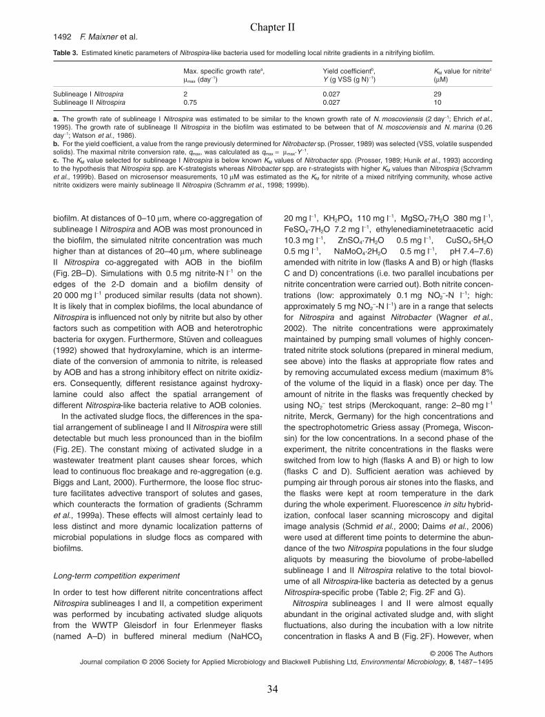
1492 F. Maixner et al.
© 2006 The AuthorsJournal compilation © 2006 Society for Applied Microbiology and Blackwell Publishing Ltd, Environmental Microbiology, 8, 1487–1495
biofilm. At distances of 0–10 µm, where co-aggregation ofsublineage I Nitrospira and AOB was most pronounced inthe biofilm, the simulated nitrite concentration was muchhigher than at distances of 20–40 µm, where sublineageII Nitrospira co-aggregated with AOB in the biofilm(Fig. 2B–D). Simulations with 0.5 mg nitrite-N l−1 on theedges of the 2-D domain and a biofilm density of20 000 mg l−1 produced similar results (data not shown).It is likely that in complex biofilms, the local abundance ofNitrospira is influenced not only by nitrite but also by otherfactors such as competition with AOB and heterotrophicbacteria for oxygen. Furthermore, Stüven and colleagues(1992) showed that hydroxylamine, which is an interme-diate of the conversion of ammonia to nitrite, is releasedby AOB and has a strong inhibitory effect on nitrite oxidiz-ers. Consequently, different resistance against hydroxy-lamine could also affect the spatial arrangement ofdifferent Nitrospira-like bacteria relative to AOB colonies.
In the activated sludge flocs, the differences in the spa-tial arrangement of sublineage I and II Nitrospira were stilldetectable but much less pronounced than in the biofilm(Fig. 2E). The constant mixing of activated sludge in awastewater treatment plant causes shear forces, whichlead to continuous floc breakage and re-aggregation (e.g.Biggs and Lant, 2000). Furthermore, the loose floc struc-ture facilitates advective transport of solutes and gases,which counteracts the formation of gradients (Schrammet al., 1999a). These effects will almost certainly lead toless distinct and more dynamic localization patterns ofmicrobial populations in sludge flocs as compared withbiofilms.
Long-term competition experiment
In order to test how different nitrite concentrations affectNitrospira sublineages I and II, a competition experimentwas performed by incubating activated sludge aliquotsfrom the WWTP Gleisdorf in four Erlenmeyer flasks(named A–D) in buffered mineral medium (NaHCO3
20 mg l−1, KH2PO4 110 mg l−1, MgSO4·7H2O 380 mg l−1,FeSO4·7H2O 7.2 mg l−1, ethylenediaminetetraacetic acid10.3 mg l−1, ZnSO4·7H2O 0.5 mg l−1, CuSO4·5H2O0.5 mg l−1, NaMoO4·2H2O 0.5 mg l−1, pH 7.4–7.6)amended with nitrite in low (flasks A and B) or high (flasksC and D) concentrations (i.e. two parallel incubations pernitrite concentration were carried out). Both nitrite concen-trations (low: approximately 0.1 mg NO2
–-N l−1; high:approximately 5 mg NO2
–-N l−1) are in a range that selectsfor Nitrospira and against Nitrobacter (Wagner et al.,2002). The nitrite concentrations were approximatelymaintained by pumping small volumes of highly concen-trated nitrite stock solutions (prepared in mineral medium,see above) into the flasks at appropriate flow rates andby removing accumulated excess medium (maximum 8%of the volume of the liquid in a flask) once per day. Theamount of nitrite in the flasks was frequently checked byusing NO2
– test strips (Merckoquant, range: 2–80 mg l−1
nitrite, Merck, Germany) for the high concentrations andthe spectrophotometric Griess assay (Promega, Wiscon-sin) for the low concentrations. In a second phase of theexperiment, the nitrite concentrations in the flasks wereswitched from low to high (flasks A and B) or high to low(flasks C and D). Sufficient aeration was achieved bypumping air through porous air stones into the flasks, andthe flasks were kept at room temperature in the darkduring the whole experiment. Fluorescence in situ hybrid-ization, confocal laser scanning microscopy and digitalimage analysis (Schmid et al., 2000; Daims et al., 2006)were used at different time points to determine the abun-dance of the two Nitrospira populations in the four sludgealiquots by measuring the biovolume of probe-labelledsublineage I and II Nitrospira relative to the total biovol-ume of all Nitrospira-like bacteria as detected by a genusNitrospira-specific probe (Table 2; Fig. 2F and G).
Nitrospira sublineages I and II were almost equallyabundant in the original activated sludge and, with slightfluctuations, also during the incubation with a low nitriteconcentration in flasks A and B (Fig. 2F). However, when
Table 3. Estimated kinetic parameters of Nitrospira-like bacteria used for modelling local nitrite gradients in a nitrifying biofilm.
Max. specific growth ratea,µmax (day−1)
Yield coefficientb,Y (g VSS (g N)−1)
KM value for nitritec
(µM)
Sublineage I Nitrospira 2 0.027 29Sublineage II Nitrospira 0.75 0.027 10
a. The growth rate of sublineage I Nitrospira was estimated to be similar to the known growth rate of N. moscoviensis (2 day−1; Ehrich et al.,1995). The growth rate of sublineage II Nitrospira in the biofilm was estimated to be between that of N. moscoviensis and N. marina (0.26day−1; Watson et al., 1986).b. For the yield coefficient, a value from the range previously determined for Nitrobacter sp. (Prosser, 1989) was selected (VSS, volatile suspendedsolids). The maximal nitrite conversion rate, qmax, was calculated as qmax = µmax·Y −1.c. The KM value selected for sublineage I Nitrospira is below known KM values of Nitrobacter spp. (Prosser, 1989; Hunik et al., 1993) accordingto the hypothesis that Nitrospira spp. are K-strategists whereas Nitrobacter spp. are r-strategists with higher KM values than Nitrospira (Schrammet al., 1999b). Based on microsensor measurements, 10 µM was estimated as the KM for nitrite of a mixed nitrifying community, whose activenitrite oxidizers were mainly sublineage II Nitrospira (Schramm et al., 1998; 1999b).
Chapter II
34
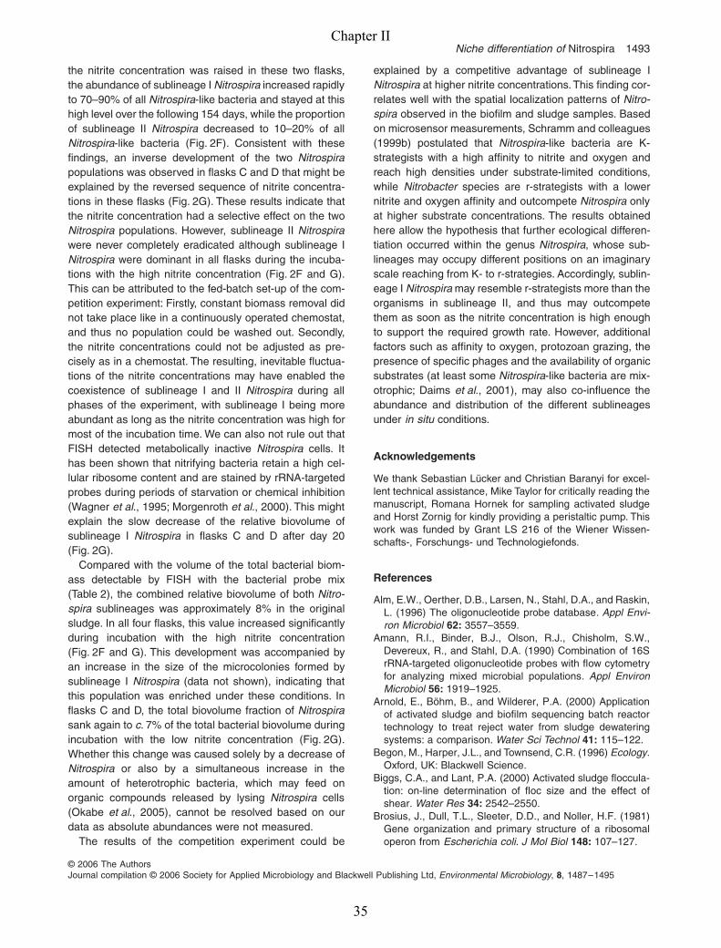
Niche differentiation of Nitrospira 1493
© 2006 The AuthorsJournal compilation © 2006 Society for Applied Microbiology and Blackwell Publishing Ltd, Environmental Microbiology, 8, 1487–1495
the nitrite concentration was raised in these two flasks,the abundance of sublineage I Nitrospira increased rapidlyto 70–90% of all Nitrospira-like bacteria and stayed at thishigh level over the following 154 days, while the proportionof sublineage II Nitrospira decreased to 10–20% of allNitrospira-like bacteria (Fig. 2F). Consistent with thesefindings, an inverse development of the two Nitrospirapopulations was observed in flasks C and D that might beexplained by the reversed sequence of nitrite concentra-tions in these flasks (Fig. 2G). These results indicate thatthe nitrite concentration had a selective effect on the twoNitrospira populations. However, sublineage II Nitrospirawere never completely eradicated although sublineage INitrospira were dominant in all flasks during the incuba-tions with the high nitrite concentration (Fig. 2F and G).This can be attributed to the fed-batch set-up of the com-petition experiment: Firstly, constant biomass removal didnot take place like in a continuously operated chemostat,and thus no population could be washed out. Secondly,the nitrite concentrations could not be adjusted as pre-cisely as in a chemostat. The resulting, inevitable fluctua-tions of the nitrite concentrations may have enabled thecoexistence of sublineage I and II Nitrospira during allphases of the experiment, with sublineage I being moreabundant as long as the nitrite concentration was high formost of the incubation time. We can also not rule out thatFISH detected metabolically inactive Nitrospira cells. Ithas been shown that nitrifying bacteria retain a high cel-lular ribosome content and are stained by rRNA-targetedprobes during periods of starvation or chemical inhibition(Wagner et al., 1995; Morgenroth et al., 2000). This mightexplain the slow decrease of the relative biovolume ofsublineage I Nitrospira in flasks C and D after day 20(Fig. 2G).
Compared with the volume of the total bacterial biom-ass detectable by FISH with the bacterial probe mix(Table 2), the combined relative biovolume of both Nitro-spira sublineages was approximately 8% in the originalsludge. In all four flasks, this value increased significantlyduring incubation with the high nitrite concentration(Fig. 2F and G). This development was accompanied byan increase in the size of the microcolonies formed bysublineage I Nitrospira (data not shown), indicating thatthis population was enriched under these conditions. Inflasks C and D, the total biovolume fraction of Nitrospirasank again to c. 7% of the total bacterial biovolume duringincubation with the low nitrite concentration (Fig. 2G).Whether this change was caused solely by a decrease ofNitrospira or also by a simultaneous increase in theamount of heterotrophic bacteria, which may feed onorganic compounds released by lysing Nitrospira cells(Okabe et al., 2005), cannot be resolved based on ourdata as absolute abundances were not measured.
The results of the competition experiment could be
explained by a competitive advantage of sublineage INitrospira at higher nitrite concentrations. This finding cor-relates well with the spatial localization patterns of Nitro-spira observed in the biofilm and sludge samples. Basedon microsensor measurements, Schramm and colleagues(1999b) postulated that Nitrospira-like bacteria are K-strategists with a high affinity to nitrite and oxygen andreach high densities under substrate-limited conditions,while Nitrobacter species are r-strategists with a lowernitrite and oxygen affinity and outcompete Nitrospira onlyat higher substrate concentrations. The results obtainedhere allow the hypothesis that further ecological differen-tiation occurred within the genus Nitrospira, whose sub-lineages may occupy different positions on an imaginaryscale reaching from K- to r-strategies. Accordingly, sublin-eage I Nitrospira may resemble r-strategists more than theorganisms in sublineage II, and thus may outcompetethem as soon as the nitrite concentration is high enoughto support the required growth rate. However, additionalfactors such as affinity to oxygen, protozoan grazing, thepresence of specific phages and the availability of organicsubstrates (at least some Nitrospira-like bacteria are mix-otrophic; Daims et al., 2001), may also co-influence theabundance and distribution of the different sublineagesunder in situ conditions.
Acknowledgements
We thank Sebastian Lücker and Christian Baranyi for excel-lent technical assistance, Mike Taylor for critically reading themanuscript, Romana Hornek for sampling activated sludgeand Horst Zornig for kindly providing a peristaltic pump. Thiswork was funded by Grant LS 216 of the Wiener Wissen-schafts-, Forschungs- und Technologiefonds.
References
Alm, E.W., Oerther, D.B., Larsen, N., Stahl, D.A., and Raskin,L. (1996) The oligonucleotide probe database. Appl Envi-ron Microbiol 62: 3557–3559.
Amann, R.I., Binder, B.J., Olson, R.J., Chisholm, S.W.,Devereux, R., and Stahl, D.A. (1990) Combination of 16SrRNA-targeted oligonucleotide probes with flow cytometryfor analyzing mixed microbial populations. Appl EnvironMicrobiol 56: 1919–1925.
Arnold, E., Böhm, B., and Wilderer, P.A. (2000) Applicationof activated sludge and biofilm sequencing batch reactortechnology to treat reject water from sludge dewateringsystems: a comparison. Water Sci Technol 41: 115–122.
Begon, M., Harper, J.L., and Townsend, C.R. (1996) Ecology.Oxford, UK: Blackwell Science.
Biggs, C.A., and Lant, P.A. (2000) Activated sludge floccula-tion: on-line determination of floc size and the effect ofshear. Water Res 34: 2542–2550.
Brosius, J., Dull, T.L., Sleeter, D.D., and Noller, H.F. (1981)Gene organization and primary structure of a ribosomaloperon from Escherichia coli. J Mol Biol 148: 107–127.
Chapter II
35

1494 F. Maixner et al.
© 2006 The AuthorsJournal compilation © 2006 Society for Applied Microbiology and Blackwell Publishing Ltd, Environmental Microbiology, 8, 1487–1495
Burrell, P.C., Keller, J., and Blackall, L.L. (1998) Microbiologyof a nitrite-oxidizing bioreactor. Appl Environ Microbiol 64:1878–1883.
Daims, H., Brühl, A., Amann, R., Schleifer, K.-H., and Wag-ner, M. (1999) The domain-specific probe EUB338 is insuf-ficient for the detection of all Bacteria: development andevaluation of a more comprehensive probe set. Syst ApplMicrobiol 22: 434–444.
Daims, H., Nielsen, J.L., Nielsen, P.H., Schleifer, K.H., andWagner, M. (2001) In situ characterization of Nitrospira-likenitrite-oxidizing bacteria active in wastewater treatmentplants. Appl Environ Microbiol 67: 5273–5284.
Daims, H., Stoecker, K., and Wagner, M. (2005) Fluores-cence in situ hybridisation for the detection of prokaryotes.In Molecular Microbial Ecology. Osborn, A.M., and Smith,C.J. (eds). Abingdon, UK: Bios-Garland, pp. 213–239.
Daims, H., Lücker, S., and Wagner, M. (2006) daime, a novelimage analysis program for microbial ecology and biofilmresearch. Environ Microbiol 8: 200–213.
Ehrich, S., Behrens, D., Lebedeva, E., Ludwig, W., and Bock,E. (1995) A new obligately chemolithoautotrophic, nitrite-oxidizing bacterium, Nitrospira moscoviensis sp. nov. andits phylogenetic relationship. Arch Microbiol 164: 16–23.
Hugenholtz, P., Goebel, B.M., and Pace, N.R. (1998) Impactof culture-independent studies on the emerging phyloge-netic view of bacterial diversity. J Bacteriol 180: 4765–4774.
Hunik, J.H., Meijer, H.J.G., and Tramper, J. (1993) Kineticsof Nitrobacter agilis at extreme substrate, product and saltconcentrations. Appl Microbiol Biotechn 40: 442–448.
Juretschko, S., Timmermann, G., Schmid, M., Schleifer, K.-H., Pommerening-Röser, A., Koops, H.-P., and Wagner, M.(1998) Combined molecular and conventional analyses ofnitrifying bacterium diversity in activated sludge: Nitroso-coccus mobilis and Nitrospira-like bacteria as dominantpopulations. Appl Environ Microbiol 64: 3042–3051.
Keswani, J., and Whitman, W.B. (2001) Relationship of 16SrRNA sequence similarity to DNA hybridization in prokary-otes. Int J Syst Evol Microbiol 51: 667–678.
Loy, A., Horn, M., and Wagner, M. (2003) probeBase: anonline resource for rRNA-targeted oligonucleotide probes.Nucleic Acids Res 31: 514–516.
Ludwig, W., Strunk, O., Westram, R., Richter, L., Meier, H.,Yadhu, K., et al. (2004) ARB: a software environment forsequence data. Nucleic Acids Res 32: 1363–1371.
Man, D., Wang, W., Sabehi, G., Aravind, L., Post, A.F., Mas-sana, R., et al. (2003) Diversification and spectral tuningin marine proteorhodopsins. EMBO J 22: 1725–1731.
May, R.M. (ed.) (1981) Theoretical Ecology. Principles andApplications. Boston, MA, USA: Blackwell ScientificPublications.
Mobarry, B.K., Wagner, M., Urbain, V., Rittmann, B.E., andStahl, D.A. (1996) Phylogenetic probes for analyzing abun-dance and spatial organization of nitrifying bacteria. ApplEnviron Microbiol 62: 2156–2162.
Mobarry, B.K., Wagner, M., Urbain, V., Rittmann, B.E., andStahl, D.A. (1997) Phylogenetic probes for analyzing abun-dance and spatial organization of nitrifying bacteria (vol 62,p. 2157, 1996). Appl Environ Microbiol 63: 815.
Morgenroth, E., Obermayer, A., Arnold, E., Brühl, A., Wag-ner, M., and Wilderer, P.A. (2000) Effect of long-term idle
periods on the performance of sequencing batch reactors.Water Sci Technol 41: 105–113.
Noguera, D.R., and Picioreanu, C. (2004) Results from themulti-species benchmark problem 3 (BM3) using two-dimensional models. Water Sci Technol 49: 169–176.
Noguera, D.R., Pizarro, G.E., and Regan, J.M. (2004) Mod-eling biofilms. In Microbial Biofilms. Ghannoum, M., andO’Toole, G.A. (eds). Washington, DC, USA: AmericanSociety for Microbiology Press, pp. 222–249.
Okabe, S., Kindaichi, T., and Ito, T. (2005) Fate of 14C-labeledmicrobial products derived from nitrifying bacteria inautotrophic nitrifying biofilms. Appl Environ Microbiol 71:3987–3994.
Prosser, J.I. (1989) Autotrophic nitrification in bacteria. AdvMicrob Physiol 30: 125–181.
Rappé, M.S., and Giovannoni, S.J. (2003) The unculturedmicrobial majority. Annu Rev Microbiol 57: 369–394.
Satoh, H., Okabe, S., Yamaguchi, Y., and Watanabe, Y.(2003) Evaluation of the impact of bioaugmentation andbiostimulation by in situ hybridization and microelectrode.Water Res 37: 2206–2216.
Schmid, M., Twachtmann, U., Klein, M., Strous, M.,Juretschko, S., Jetten, M., et al. (2000) Molecular evidencefor a genus-level diversity of bacteria capable of catalyzinganaerobic ammonium oxidation. Syst Appl Microbiol 23:93–106.
Schramm, A., de Beer, D., Wagner, M., and Amann, R.(1998) Identification and activities in situ of Nitrosospiraand Nitrospira spp. as dominant populations in a nitrifyingfluidized bed reactor. Appl Environ Microbiol 64: 3480–3485.
Schramm, A., Santegoeds, C.M., Nielsen, H.K., Ploug, H.,Wagner, M., Pribyl, M., et al. (1999a) On the occurrenceof anoxic microniches, denitrification, and sulfate reductionin aerated activated sludge. Appl Environ Microbiol 65:4189–4196.
Schramm, A., de Beer, D., van den Heuvel, J.C., Ottengraf,S., and Amann, R. (1999b) Microscale distribution of pop-ulations and activities of Nitrosospira and Nitrospira spp.along a macroscale gradient in a nitrifying bioreactor:quantification by in situ hybridization and the use ofmicrosensors. Appl Environ Microbiol 65: 3690–3696.
Schramm, A., Fuchs, B.M., Nielsen, J.L., Tonolla, M., andStahl, D.A. (2002) Fluorescence in situ hybridization of16S rRNA gene clones (Clone-FISH) for probe validationand screening of clone libraries. Environ Microbiol 4:713–720.
Spieck, E., Hartwig, C., McCormack, I., Maixner, F., Wagner,M., Lipski, A., and Daims, H. (2006) Selective enrichmentand molecular characterization of a previously unculturedNitrospira-like bacterium from activated sludge. EnvironMicrobiol 8: 405–415.
Stackebrandt, E., and Goebel, B.M. (1994) Taxonomic note:a place for DNA–DNA reassociation and 16S rRNAsequence analysis in the present species definition in bac-teriology. Int J Syst Bacteriol 44: 846–849.
Stein, L.Y., and Arp, D.J. (1998) Loss of ammonia monoox-ygenase activity in Nitrosomonas europaea upon exposureto nitrite. Appl Environ Microbiol 64: 4098–4102.
Stomp, M., Huisman, J., De Jongh, F., Veraart, A.J., Gerla,D., Rijkeboer, M., et al. (2004) Adaptive divergence in
Chapter II
36
Niche differentiation of Nitrospira 1495
© 2006 The AuthorsJournal compilation © 2006 Society for Applied Microbiology and Blackwell Publishing Ltd, Environmental Microbiology, 8, 1487–1495
pigment composition promotes phytoplankton biodiversity.Nature 432: 104–107.
Stüven, R., Vollmer, M., and Bock, E. (1992) The impact oforganic matter on nitric oxide formation by Nitrosomonaseuropaea. Arch Microbiol 158: 439–443.
Wagner, M., Rath, G., Amann, R., Koops, H.-P., and Schle-ifer, K.-H. (1995) In situ identification of ammonia-oxidizingbacteria. Syst Appl Microbiol 18: 251–264.
Wagner, M., Rath, G., Koops, H.-P., Flood, J., and Amann,R. (1996) In situ analysis of nitrifying bacteria in sewagetreatment plants. Water Sci Technol 34: 237–244.
Wagner, M., Loy, A., Nogueira, R., Purkhold, U., Lee, N., andDaims, H. (2002) Microbial community composition andfunction in wastewater treatment plants. Antonie Van Leeu-wenhoek 81: 665–680.
Watson, S.W., Bock, E., Valois, F.W., Waterbury, J.B., andSchlosser, U. (1986) Nitrospira marina gen. nov. sp. nov.:a chemolithotrophic nitrite-oxidizing bacterium. Arch Micro-biol 144: 1–7.
Chapter II
37

Chapter II
38
Chapter III
Environmental genomics reveals a functional
chlorite dismutase in the nitrite-oxidizing bacterium
‘Candidatus Nitrospira defluvii’
Published in Environmental Microbiology (2008): 10 (11), 3043-3056

Chapter III
40

Environmental genomics reveals a functional chloritedismutase in the nitrite-oxidizing bacterium‘Candidatus Nitrospira defluvii’
Frank Maixner,1 Michael Wagner,1*Sebastian Lücker,1 Eric Pelletier,2
Stephan Schmitz-Esser,1 Karin Hace,1 Eva Spieck,3
Robert Konrat,4 Denis Le Paslier2 and Holger Daims1
1Department für Mikrobielle Ökologie, Universität Wien,Althanstrasse 14, A-1090 Vienna, Austria.2CEA/Genoscope CNRS-UMR 8030, 2 rue GastonCrémieux CP 5706, 91057 Evry, France.3Universität Hamburg, Biozentrum Klein Flottbek,Mikrobiologie, Ohnhorststr. 18, D-22609 Hamburg,Germany.4Department für Biomolekulare Strukturchemie,Universität Wien, Campus Vienna Biocenter 5, A-1030Vienna, Austria.
Summary
Nitrite-oxidizing bacteria of the genus Nitrospira areubiquitous in natural ecosystems and also in waste-water treatment plants. Nitrospira are members of adistinct phylum, not closely related to other nitrifiers,and no genomic sequences from this genus havebeen available so far. Here we applied an environmen-tal genomics approach to sequence and assemble a137 kbp-long genome fragment of ‘Candidatus Nitro-spira defluvii ’, which had been enriched from acti-vated sludge and belongs to Nitrospira sublineageI without isolated representatives. The annotationof this contig, which carried the 16S rRNA gene ofN. defluvii, offered first insight into the genome ofNitrospira. Surprisingly, we found a gene similar togenes encoding chlorite dismutase (CLD), an enzymedegrading chlorite (ClO2
-) to Cl- and O2. To date, CLDswith high catalytic activity have been found only inperchlorate- and chlorate-reducing bacteria but not innitrifiers. Heterologous expression in E. coli followedby enzymatic tests confirmed that this gene of Nitro-spira encodes a highly active CLD, which is alsoexpressed in situ by Nitrospira, indicating that thisnitrite oxidizer might be involved in the bioremedia-
tion of perchlorate and chlorite. Phylogenetic analy-ses showed that CLD and related proteins are widelydistributed among the Bacteria and Archaea, and indi-cated that this enzyme family appeared relativelyearly in evolution, has been subject to functionaldiversification and might play yet unknown roles inmicrobial metabolism.
Introduction
Nitrification, the microbially catalysed oxidations ofammonia to nitrite and of nitrite to nitrate, is a key processof the biogeochemical nitrogen cycle and of biologicalwastewater treatment. The two steps of nitrification arecarried out by two distinct functional groups of chem-olithotrophic prokaryotes: the ammonia-oxidizing bacteria(AOB) and archaea (AOA) (Koops et al., 2003; Könnekeet al., 2005), and the nitrite-oxidizing bacteria (NOB) (Bockand Wagner, 2001; Alawi et al., 2007). As all known nitri-fying prokaryotes are fastidious and slow-growing organ-isms, their enrichment and isolation from environmentalsamples are difficult and most physiological studies havebeen performed with pure cultures of a few ‘model’ nitrifi-ers, in particular AOB related to the genus Nitrosomonasand NOB of the genus Nitrobacter. The recently publishedgenome sequences of cultured bacterial nitrifiers (Chainet al., 2003; Klotz et al., 2006; Starkenburg et al., 2006;Stein et al., 2007) offer yet unmatched insight intothe biology of these organisms. However, studies usingcultivation-independent techniques have revealed a largediversity of yet uncultured and so far poorly characterizedAOB, NOB and AOA in the environment (e.g. Daims et al.,2001a;Smith et al., 2001; Francis et al., 2005; Leiningeret al., 2006). Our understanding of nitrification and nitro-gen cycling in nature remains incomplete without moredata about the ecophysiology and genomics of theseuncultured nitrifiers.
The genus Nitrospira is one of the less intensivelystudied groups of NOB. This genus, which belongs to thedistinct bacterial phylum Nitrospirae (Ehrich et al., 1995), isunrelated to all other known nitrifiers. Except two describedspecies (Watson et al., 1986; Ehrich et al., 1995) andone recently isolated candidate species (Lebedevaet al., 2008), no members of this genus are available in
Received 7 December, 2007; accepted 22 March, 2008. *Forcorrespondence. E-mail [email protected]; Tel. (+43)14277 54390; Fax (+43) 14277 54389.
Environmental Microbiology (2008) 10(11), 3043–3056 doi:10.1111/j.1462-2920.2008.01646.x
© 2008 The AuthorsJournal compilation © 2008 Society for Applied Microbiology and Blackwell Publishing Ltd
Chapter III
41

pure culture. 16S rRNA sequence analyses showed thatthe genus Nitrospira contains several phylogenetic sublin-eages (Daims et al., 2001b), whose uncultured represen-tatives are widely distributed in many kinds of aquatic andterrestrial habitats. Moreover, Nitrospira are the dominantnitrite oxidizers in wastewater treatment plants (Juretschkoet al., 1998). Nevertheless, our understanding of thebiology of Nitrospira is very limited compared withmore easily culturable nitrifiers such as Nitrobacter andNitrosomonas species.
During the last few years, environmental genomicsapproaches have dramatically influenced the way we thinkabout the nitrogen cycle (Schleper et al., 2005; Hallamet al., 2006; Strous et al., 2006). In this study, we appliedan environmental genomics approach to sequence a largegenome fragment of the nitrite oxidizer ‘Candidatus Nitro-spira defluvii’ (Spieck et al., 2006). The analysis providedfirst insight into the genome of Nitrospira and led to thediscovery of an unexpected function, which has previouslynot been documented in any nitrifying organism.
Results and discussion
Analysis of a large genome piece of Nitrospira
Bacterial artificial chromosome (BAC) and plasmidshotgun libraries were established from genomic DNA,which had been extracted from an enrichment of ‘Can-didatus Nitrospira defluvii ’. This organism was enrichedfrom a domestic wastewater treatment plant and belongsto the phylogenetic sublineage I of the genus Nitrospira(Spieck et al., 2006), whose representatives are thedominant NOB in many wastewater treatment systems(Daims et al., 2001b). At the time when the metagenomelibraries for this study were created, the enrichment con-sisted of approximately 40% of N. defluvii as determinedby quantitative fluorescence in situ hybridization (FISH).No Nitrobacter cells were detectable by FISH in theenrichment. Despite the relatively low degree of enrich-ment of N. defluvii, it was possible to reconstruct fromthe obtained metagenomic sequences a 137 336 bp-long contig, which contained the complete rRNA operonof N. defluvii and thus clearly belonged to this bacterium(Fig. 1). The contig had an average GC-content of58.45% and contained 153 coding sequences. No func-
tions could be assigned to 74 open reading frames(ORFs) that shared sequence similarities only withORFs of unknown function in other organisms (13ORFs), or contained only short conserved motifs orstructural features (two ORFs), or showed no similarityat all to any sequence in the public databases (59ORFs). The large number of ORFs without any databasematches reflects that N. defluvii belongs to a bacterialphylum without genome-sequenced representativesexcept some Leptospirillum strains (Tyson et al., 2004),which are acidophilic iron oxidizers with a different physi-ology than Nitrospira.
Functions were assigned with a relatively high degreeof confidence to 59 of the remaining 79 coding sequencesbased on strong similarities to functionally characterizedgenes in other organisms. Among them was the rrnoperon with the genes of the 16S, 23S and 5S ribosomalRNAs. The spacer region between the 16S and 23S rRNAgenes contained two tRNA genes for isoleucine andalanine respectively (Fig. 1). A description of the otherannotated genes is provided as supplementary onlinematerial together with a complete gene list (Table S1) anda figure showing the localization of all genes on the contig(Fig. S1).
N. defluvii has a gene highly similar tochlorite dismutase
A surprising discovery on the analysed genome piece ofN. defluvii was the gene cld, whose predicted product issimilar in length and amino acid sequence to the enzymechlorite dismutase (CLD) of Dechloromonas aromatica,D. agitata and Ideonella dechloratans (Table 1). Theseheterotrophic Betaproteobacteria are able to respire,
purB cld purC
purS purQ purL purF rRNA_16S rRNA_23S
rRN
A_5
S
tRN
A_I
letR
NA
_Ala
Fig. 1. Schematic representation of a part of the analysed genome region of N. defluvii. Arrows show the locations of the gene encodingchlorite dismutase (cld), the pur purine biosynthesis genes in close vicinity to cld and the rrn operon (including the spacer regions with twotRNA genes). The function of the ORF between purC and purS (white arrow) is unknown. This figure is a downsized version of Fig. S1 thatshows the complete contig with all identified coding sequences.
Table 1. Amino acid sequence identities between the CLD ofN. defluvii (264 amino acids) and the CLDs of known (per)chlorate-reducing bacteria.
OrganismAmino acidresidues Identity (%)a
Dechloromonas aromatica RCB 282 40.5Dechloromonas agitata 277 39.6Ideonella dechloratans 285 38.0
a. Identity to the CLD of N. defluvii.
3044 F. Maixner et al.
© 2008 The AuthorsJournal compilation © 2008 Society for Applied Microbiology and Blackwell Publishing Ltd, Environmental Microbiology, 10, 3043–3056
Chapter III
42
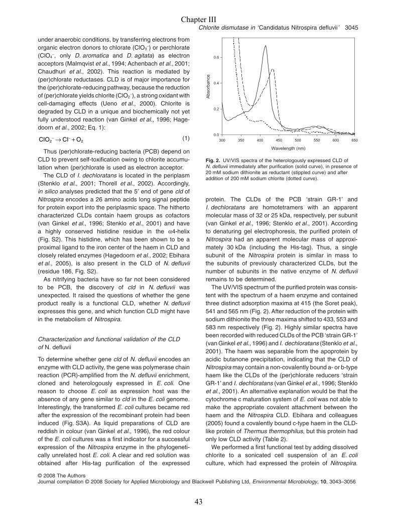
under anaerobic conditions, by transferring electrons fromorganic electron donors to chlorate (ClO3
-) or perchlorate(ClO4
-, only D. aromatica and D. agitata) as electronacceptors (Malmqvist et al., 1994; Achenbach et al., 2001;Chaudhuri et al., 2002). This reaction is mediated by(per)chlorate reductases. CLD is of major importance forthe (per)chlorate-reducing pathway, because the reductionof (per)chlorate yields chlorite (ClO2
-), a strong oxidant withcell-damaging effects (Ueno et al., 2000). Chlorite isdegraded by CLD in a unique and biochemically not yetfully understood reaction (van Ginkel et al., 1996; Hage-doorn et al., 2002; Eq. 1):
CIO CI O2 2− −→ + (1)
Thus (per)chlorate-reducing bacteria (PCB) depend onCLD to prevent self-toxification owing to chlorite accumu-lation when (per)chlorate is used as electron acceptor.
The CLD of I. dechloratans is located in the periplasm(Stenklo et al., 2001; Thorell et al., 2002). Accordingly,in silico analyses predicted that the 5′ end of gene cld ofNitrospira encodes a 26 amino acids long signal peptidefor protein export into the periplasmic space. The hithertocharacterized CLDs contain haem groups as cofactors(van Ginkel et al., 1996; Stenklo et al., 2001) and havea highly conserved histidine residue in the a4-helix(Fig. S2). This histidine, which has been shown to be aproximal ligand to the iron center of the haem in CLD andclosely related enzymes (Hagedoorn et al., 2002; Ebiharaet al., 2005), is also present in the CLD of N. defluvii(residue 186, Fig. S2).
As nitrifying bacteria have so far not been consideredto be PCB, the discovery of cld in N. defluvii wasunexpected. It raised the questions of whether the geneproduct really is a functional CLD, whether N. defluviiexpresses this gene, and which function CLD might havein the metabolism of Nitrospira.
Characterization and functional validation of the CLDof N. defluvii
To determine whether gene cld of N. defluvii encodes anenzyme with CLD activity, the gene was polymerase chainreaction (PCR)-amplified from the N. defluvii enrichment,cloned and heterologously expressed in E. coli. Onereason to choose E. coli as expression host was theabsence of any gene similar to cld in the E. coli genome.Interestingly, the transformed E. coli cultures became redafter the expression of the recombinant protein had beeninduced (Fig. S3A). As liquid preparations of CLD arereddish in colour (van Ginkel et al., 1996), the red colourof the E. coli cultures was a first indicator for a successfulexpression of the Nitrospira enzyme in the phylogeneti-cally unrelated host E. coli. A clear and red solution wasobtained after His-tag purification of the expressed
protein. The CLDs of the PCB ‘strain GR-1’ andI. dechloratans are homotetramers with an apparentmolecular mass of 32 or 25 kDa, respectively, per subunit(van Ginkel et al., 1996; Stenklo et al., 2001). Accordingto denaturing gel electrophoresis, the purified protein ofNitrospira had an apparent molecular mass of approxi-mately 30 kDa (including the His-tag). Thus, a singlesubunit of the Nitrospira protein is similar in mass tothe subunits of previously characterized CLDs, but thenumber of subunits in the native enzyme of N. defluviiremains to be determined.
The UV/VIS spectrum of the purified protein was consis-tent with the spectrum of a haem enzyme and containedthree distinct adsorption maxima at 415 (the Soret peak),541 and 565 nm (Fig. 2). After reduction of the protein withsodium dithionite the three maxima shifted to 433, 553 and583 nm respectively (Fig. 2). Highly similar spectra havebeen recorded with reduced CLDs of the PCB ‘strain GR-1’(van Ginkel et al., 1996) and I. dechloratans (Stenklo et al.,2001). The haem was separable from the apoprotein byacidic butanone precipitation, indicating that the CLD ofNitrospira may contain a non-covalently bound a- or b-typehaem like the CLDs of the (per)chlorate reducers ‘strainGR-1’ and I. dechloratans (van Ginkel et al., 1996; Stenkloet al., 2001). An alternative explanation would be that thecytochrome c maturation system of E. coli was not able tomake the appropriate covalent attachment between thehaem and the Nitrospira CLD. Ebihara and colleagues(2005) found a covalently bound c-type haem in the CLD-like protein of Thermus thermophilus, but this protein hadonly low CLD activity (Table 2).
We performed a first functional test by adding dissolvedchlorite to a sonicated cell suspension of an E. coliculture, which had expressed the protein of Nitrospira.
Wavelength (nm)
300 350 400 450 500 550 600 650
Abs
orba
nce
0.0
0.2
0.4
0.6
Fig. 2. UV/VIS spectra of the heterologously expressed CLD ofN. defluvii immediately after purification (solid curve), in presence of20 mM sodium dithionite as reductant (stippled curve) and afteraddition of 200 mM sodium chlorite (dotted curve).
Chlorite dismutase in ‘Candidatus Nitrospira defluvii’ 3045
© 2008 The AuthorsJournal compilation © 2008 Society for Applied Microbiology and Blackwell Publishing Ltd, Environmental Microbiology, 10, 3043–3056
Chapter III
43

According to Eq. 1, production of gas (presumably O2)was observed instantly (Fig. S3B). The subsequent addi-tion of silver nitrate confirmed that Cl- had been formed,too (Fig. S3C). In contrast, no visible gas productionoccurred and no Cl- was formed after addition of chloriteto a sonicated E. coli culture that had not expressed genecld of Nitrospira (Fig. S3). Production of gas and Cl- werealso observed when ClO2
- was added to a solution of thepurified recombinant enzyme (data not shown). Thus, theheterologously expressed enzyme of Nitrospira clearlyhad the activity of CLD. In these experiments, the redcolour of CLD diminished during the reaction (not shown),indicating that the enzyme was degraded owing to oxida-tion by excess chlorite. Similar observations were madeby van Ginkel and colleagues (1996) with the CLD of thePCB ‘strain GR-1’. The degradation of CLD by excesssubstrate also led to a dramatic reduction of the threeadsorption maxima in the UV/VIS spectrum (Fig. 2) andcaused the specific activity of CLD to decline in presenceof higher chlorite concentrations (Fig. 3A). The tempera-ture and pH optima of the enzyme were 25°C and pH 6.0(Fig. S4). These optima are similar to those of the CLDs of‘strain GR-1’ (30°C, pH 6.0; van Ginkel et al., 1996) andDechlorosoma sp. strain KJ (pH 6.0; Xu and Logan,2003). We determined the Km value of the CLD ofN. defluvii to be 15.8 � 1.1 mmol l-1 of chlorite and Vmax tobe 1939 � 47 U (mg protein)-1 under optimal reactionconditions (Fig. 3B). Although Vmax and the turnovernumber (kcat) of the Nitrospira enzyme are similar to therespective values of chlorite dismutases of PCB, its Km ismuch higher (Table 2). The high Km might be explained bysuboptimal folding or missing post-translational modifica-tions (e.g. a missing covalent attachment of the haem) ofthe recombinant Nitrospira CLD in the heterologousexpression host E. coli, and the native enzyme may havedifferent catalytic properties in Nitrospira. Alternatively, therelatively low affinity for chlorite could have evolutionaryor physiological causes. The ratio kcat/Km, which is referredto as the performance constant, is an indicator for thereaction efficiency of an enzyme with a given substrate(Koshland, 2002). Mutations that increase the catalyticrate (i.e. increase kcat) or the substrate affinity (i.e.
decrease Km) result in a better catalytic performance. Itslower performance constant indicates that the CLD ofNitrospira may not be adapted as perfectly as the CLDs ofknown PCB to the substrate chlorite (Table 2). It could stillbe an evolving CLD or it might have an unknown primaryfunction that is not chlorite degradation. Ebihara and col-leagues (2005) determined the 3D structure of a CLD-likeprotein, which has a very low performance constant withchlorite (Table 2). Because this enzyme of Thermus ther-mophilus also showed a low catalase activity, they hypoth-esized that it might protect the cell from damage owing toreactive oxygen intermediates (ROI) by catalysing elec-tron transfers via its haem groups. At present we cannotexclude that the enzyme of Nitrospira functions in vivo asa scavenger of ROI, reactive nitrogen intermediates, orother toxic substances different from chlorite. Preliminaryexperiments were performed with H2O2, hydroxylamine,nitrite and NO as candidate substrates, but no functionalclue was obtained (data not shown).
Expression and function of CLD in Nitrospira
The cld genes and the genes coding for (per)chloratereductases are located in close vicinity in the genomesof I. dechloratans, D. agitata and D. aromatica (Thorellet al., 2003; Bender et al., 2005), forming a functional unitfor (per)chlorate reduction in these PCB. In contrast, genecld of N. defluvii is part of a cluster that contains six othergenes, which are involved in the de novo biosynthesis ofpurines, and one ORF of unknown function (Fig. 1). Thus,based on the genomic context of cld the functional role ofCLD in N. defluvii is not as obvious as in the aforemen-tioned PCB.
To determine whether N. defluvii actually expressesCLD, Western blot experiments were performed withCLD-specific antibodies and cell extracts from an aerobi-cally grown nitrite-oxidizing enrichment of N. defluvii. Thisnewer enrichment, which was obtained after the metage-nome library construction, contained as much as 86%of N. defluvii (Spieck et al., 2006). The Western blotrevealed that N. defluvii expressed CLD under the appliedaerobic incubation conditions (Fig. 4). The high specificity
Table 2. Catalytic parameters of characterized enzymes with CLD activity and chlorite as substrate.
OrganismVmax
[U(mg protein)-1]kcat
(s-1)Km
(mM)kcat/Km
(M-1s-1) Reference
Nitrospira defluvii 1.9 ¥ 103 0.96 ¥ 103 15.8 6.1 ¥ 104 This study‘Strain GR1’ 2.2 ¥ 103 1.2 ¥ 103 0.17 7.1 ¥ 106 van Ginkel et al. (1996)Ideonella dechloratans 4.3 ¥ 103 1.8 ¥ 103 0.26 6.9 ¥ 106 Stenklo et al. (2001)Thermus thermophilusa 1.6 0.77 13 59 Ebihara et al. (2005)
a. Vmax was not reported in the original publication and is estimated based on kcat and the molar mass, which was derived from the amino acidsequence.Ideonella dechloratans and ‘strain GR1’ are known PCB.
3046 F. Maixner et al.
© 2008 The AuthorsJournal compilation © 2008 Society for Applied Microbiology and Blackwell Publishing Ltd, Environmental Microbiology, 10, 3043–3056
Chapter III
44
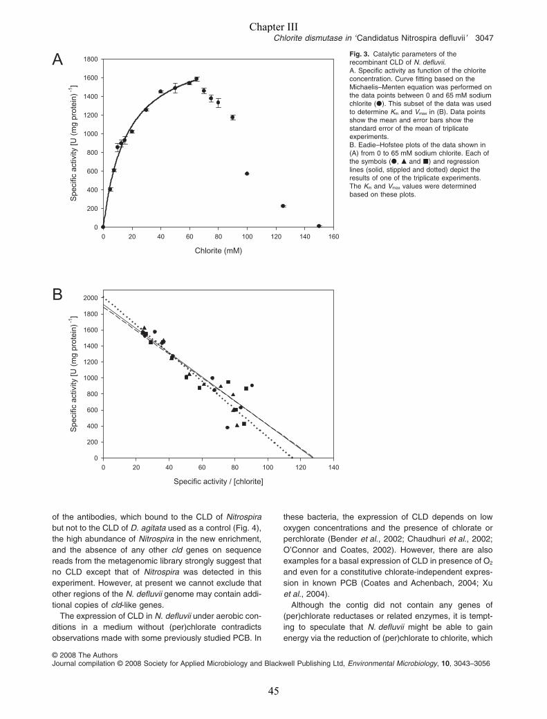
of the antibodies, which bound to the CLD of Nitrospirabut not to the CLD of D. agitata used as a control (Fig. 4),the high abundance of Nitrospira in the new enrichment,and the absence of any other cld genes on sequencereads from the metagenomic library strongly suggest thatno CLD except that of Nitrospira was detected in thisexperiment. However, at present we cannot exclude thatother regions of the N. defluvii genome may contain addi-tional copies of cld-like genes.
The expression of CLD in N. defluvii under aerobic con-ditions in a medium without (per)chlorate contradictsobservations made with some previously studied PCB. In
these bacteria, the expression of CLD depends on lowoxygen concentrations and the presence of chlorate orperchlorate (Bender et al., 2002; Chaudhuri et al., 2002;O’Connor and Coates, 2002). However, there are alsoexamples for a basal expression of CLD in presence of O2
and even for a constitutive chlorate-independent expres-sion in known PCB (Coates and Achenbach, 2004; Xuet al., 2004).
Although the contig did not contain any genes of(per)chlorate reductases or related enzymes, it is tempt-ing to speculate that N. defluvii might be able to gainenergy via the reduction of (per)chlorate to chlorite, which
Fig. 3. Catalytic parameters of therecombinant CLD of N. defluvii.A. Specific activity as function of the chloriteconcentration. Curve fitting based on theMichaelis–Menten equation was performed onthe data points between 0 and 65 mM sodiumchlorite (�). This subset of the data was usedto determine Km and Vmax in (B). Data pointsshow the mean and error bars show thestandard error of the mean of triplicateexperiments.B. Eadie–Hofstee plots of the data shown in(A) from 0 to 65 mM sodium chlorite. Each ofthe symbols (�, � and �) and regressionlines (solid, stippled and dotted) depict theresults of one of the triplicate experiments.The Km and Vmax values were determinedbased on these plots.
A
B
Chlorite (mM)
0 20 40 60 80 100 120 140 160
Spe
cific
act
ivity
[U (
mg
prot
ein)
-1]
0
200
400
600
800
1000
1200
1400
1600
1800
Specific activity / [chlorite]
0 20 40 60 80 100 120 140
Spe
cific
act
ivity
[U (
mg
prot
ein)
-1]
0
200
400
600
800
1000
1200
1400
1600
1800
2000
Chlorite dismutase in ‘Candidatus Nitrospira defluvii’ 3047
© 2008 The AuthorsJournal compilation © 2008 Society for Applied Microbiology and Blackwell Publishing Ltd, Environmental Microbiology, 10, 3043–3056
Chapter III
45
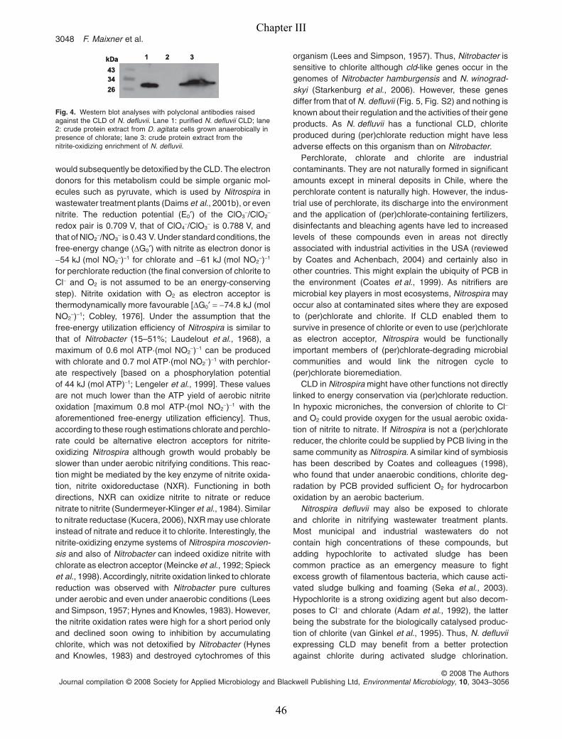
would subsequently be detoxified by the CLD. The electrondonors for this metabolism could be simple organic mol-ecules such as pyruvate, which is used by Nitrospira inwastewater treatment plants (Daims et al., 2001b), or evennitrite. The reduction potential (E0′) of the ClO3
-/ClO2-
redox pair is 0.709 V, that of ClO4-/ClO3
- is 0.788 V, andthat of NlO2
-/NO3- is 0.43 V. Under standard conditions, the
free-energy change (DG0′) with nitrite as electron donor is-54 kJ (mol NO2
-)-1 for chlorate and -61 kJ (mol NO2-)-1
for perchlorate reduction (the final conversion of chlorite toCl- and O2 is not assumed to be an energy-conservingstep). Nitrite oxidation with O2 as electron acceptor isthermodynamically more favourable [DG0′ = -74.8 kJ (molNO2
-)-1; Cobley, 1976]. Under the assumption that thefree-energy utilization efficiency of Nitrospira is similar tothat of Nitrobacter (15–51%; Laudelout et al., 1968), amaximum of 0.6 mol ATP·(mol NO2
-)-1 can be producedwith chlorate and 0.7 mol ATP·(mol NO2
-)-1 with perchlor-ate respectively [based on a phosphorylation potentialof 44 kJ (mol ATP)-1; Lengeler et al., 1999]. These valuesare not much lower than the ATP yield of aerobic nitriteoxidation [maximum 0.8 mol ATP·(mol NO2
-)-1 with theaforementioned free-energy utilization efficiency]. Thus,according to these rough estimations chlorate and perchlo-rate could be alternative electron acceptors for nitrite-oxidizing Nitrospira although growth would probably beslower than under aerobic nitrifying conditions. This reac-tion might be mediated by the key enzyme of nitrite oxida-tion, nitrite oxidoreductase (NXR). Functioning in bothdirections, NXR can oxidize nitrite to nitrate or reducenitrate to nitrite (Sundermeyer-Klinger et al., 1984). Similarto nitrate reductase (Kucera, 2006), NXR may use chlorateinstead of nitrate and reduce it to chlorite. Interestingly, thenitrite-oxidizing enzyme systems of Nitrospira moscovien-sis and also of Nitrobacter can indeed oxidize nitrite withchlorate as electron acceptor (Meincke et al., 1992; Spiecket al., 1998).Accordingly, nitrite oxidation linked to chloratereduction was observed with Nitrobacter pure culturesunder aerobic and even under anaerobic conditions (Leesand Simpson, 1957; Hynes and Knowles, 1983). However,the nitrite oxidation rates were high for a short period onlyand declined soon owing to inhibition by accumulatingchlorite, which was not detoxified by Nitrobacter (Hynesand Knowles, 1983) and destroyed cytochromes of this
organism (Lees and Simpson, 1957). Thus, Nitrobacter issensitive to chlorite although cld-like genes occur in thegenomes of Nitrobacter hamburgensis and N. winograd-skyi (Starkenburg et al., 2006). However, these genesdiffer from that of N. defluvii (Fig. 5, Fig. S2) and nothing isknown about their regulation and the activities of their geneproducts. As N. defluvii has a functional CLD, chloriteproduced during (per)chlorate reduction might have lessadverse effects on this organism than on Nitrobacter.
Perchlorate, chlorate and chlorite are industrialcontaminants. They are not naturally formed in significantamounts except in mineral deposits in Chile, where theperchlorate content is naturally high. However, the indus-trial use of perchlorate, its discharge into the environmentand the application of (per)chlorate-containing fertilizers,disinfectants and bleaching agents have led to increasedlevels of these compounds even in areas not directlyassociated with industrial activities in the USA (reviewedby Coates and Achenbach, 2004) and certainly also inother countries. This might explain the ubiquity of PCB inthe environment (Coates et al., 1999). As nitrifiers aremicrobial key players in most ecosystems, Nitrospira mayoccur also at contaminated sites where they are exposedto (per)chlorate and chlorite. If CLD enabled them tosurvive in presence of chlorite or even to use (per)chlorateas electron acceptor, Nitrospira would be functionallyimportant members of (per)chlorate-degrading microbialcommunities and would link the nitrogen cycle to(per)chlorate bioremediation.
CLD in Nitrospira might have other functions not directlylinked to energy conservation via (per)chlorate reduction.In hypoxic microniches, the conversion of chlorite to Cl-
and O2 could provide oxygen for the usual aerobic oxida-tion of nitrite to nitrate. If Nitrospira is not a (per)chloratereducer, the chlorite could be supplied by PCB living in thesame community as Nitrospira. A similar kind of symbiosishas been described by Coates and colleagues (1998),who found that under anaerobic conditions, chlorite deg-radation by PCB provided sufficient O2 for hydrocarbonoxidation by an aerobic bacterium.
Nitrospira defluvii may also be exposed to chlorateand chlorite in nitrifying wastewater treatment plants.Most municipal and industrial wastewaters do notcontain high concentrations of these compounds, butadding hypochlorite to activated sludge has beencommon practice as an emergency measure to fightexcess growth of filamentous bacteria, which cause acti-vated sludge bulking and foaming (Seka et al., 2003).Hypochlorite is a strong oxidizing agent but also decom-poses to Cl- and chlorate (Adam et al., 1992), the latterbeing the substrate for the biologically catalysed produc-tion of chlorite (van Ginkel et al., 1995). Thus, N. defluviiexpressing CLD may benefit from a better protectionagainst chlorite during activated sludge chlorination.
26
3443
1 2 3kDa
26
3443
1 2 3kDa
Fig. 4. Western blot analyses with polyclonal antibodies raisedagainst the CLD of N. defluvii. Lane 1: purified N. defluvii CLD; lane2: crude protein extract from D. agitata cells grown anaerobically inpresence of chlorate; lane 3: crude protein extract from thenitrite-oxidizing enrichment of N. defluvii.
3048 F. Maixner et al.
© 2008 The AuthorsJournal compilation © 2008 Society for Applied Microbiology and Blackwell Publishing Ltd, Environmental Microbiology, 10, 3043–3056
Chapter III
46
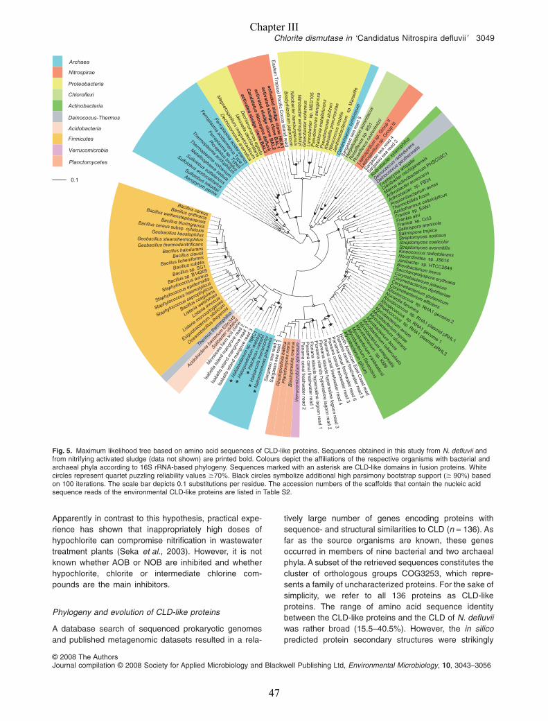
Apparently in contrast to this hypothesis, practical expe-rience has shown that inappropriately high doses ofhypochlorite can compromise nitrification in wastewatertreatment plants (Seka et al., 2003). However, it is notknown whether AOB or NOB are inhibited and whetherhypochlorite, chlorite or intermediate chlorine com-pounds are the main inhibitors.
Phylogeny and evolution of CLD-like proteins
A database search of sequenced prokaryotic genomesand published metagenomic datasets resulted in a rela-
tively large number of genes encoding proteins withsequence- and structural similarities to CLD (n = 136). Asfar as the source organisms are known, these genesoccurred in members of nine bacterial and two archaealphyla. A subset of the retrieved sequences constitutes thecluster of orthologous groups COG3253, which repre-sents a family of uncharacterized proteins. For the sake ofsimplicity, we refer to all 136 proteins as CLD-likeproteins. The range of amino acid sequence identitybetween the CLD-like proteins and the CLD of N. defluviiwas rather broad (15.5–40.5%). However, the in silicopredicted protein secondary structures were strikingly
Sulfolobus tokodaii
Sulfolobus acidocaldarius
Sulfolobus solfataricus
Metallosphaera sedula
Thermoplasma volcanium
Thermoplasm
a acidophilum
Picrophilustorridus
Ferroplasma
sp. TypeII
Ferroplasma
acidarmanus
TypeI
Ferroplasma
acidarmanus
Aeropyrum pernix
activatedsludge
cloneB
AL3
activatedslu
dg
eclo
ne
BA
L2
activatedslu
dg
eclo
ne
BA
L1
activatedslu
dg
eclo
ne
HK
A1
Candidatus
Nitro
spirad
efluvii
Dechlorom
onasarom
atica
Magnetospirillum
magnetotacticum
Ideonelladechloratans
Dechlorom
onasagitata
Ral
ston
iam
etal
lidur
ans
Pse
udom
onas
stut
zeri
Kle
bsie
llapn
eum
onia
eN
itroc
occu
sm
obili
s
Jant
hino
bact
eriu
msp
. Mar
seill
e
Glo
eoba
cter
viol
aceu
sLi
mno
bact
ersp
. ME
D10
5P
seud
omon
asae
rugi
nosa
Eastern
Tropical P
acificC
ocosisland
readN
itrobacterham
burgiensisN
itrobacterw
inogradskyi
Bradyrhizobium
japonicum
Cen
arch
aeum
sym
bios
um
Sar
gass
ose
are
ad5
Ros
eifle
xus
sp. R
S1
Ros
eifle
xus
cast
enho
lzii
Her
peto
siph
onau
rant
iacu
sSar
gass
ose
are
ad4
Sarga
sso
sea
read
3
Rubro
bacte
r xylan
ophil
us
Deinoc
occu
s radio
duran
s
Deinococc
us geothermalis
Lept
ospi
rillu
msp
. Gro
upII
Lept
ospi
rillu
msp
. Gro
upIII
Clavibacte
r michiganensis
Marine actinobacteriu
mPHSC20C1
Arthrobacter aurescens
Arthrobacter sp. FB24Tropherym
a whipplei
Thermobifida fusca
Acidothermus cellulolyticus
Frankia sp. EAN1
Frankia alni
Frankia sp. CcI3
Propionibacterium acnes
Salinispora arenicola
Salinispora tropica
Streptomyces coelicolorStreptomyces avermitilis
Streptomyces nodosus
Kineococcus radiotoleransNocardioides sp. JS614Janibacter sp. HTCC2649Brevibacterium linens
Rhodococcus sp. RHA1 genome 2
Nocardia farcinica
Rhodococcussp. RHA1 genome 1
Rhodococcussp. RHA1 plasmid pRHL3
Rhodococcus sp. RHA1 plasmid pRHL1
Mycobacteriumavium
Mycobacterium
bovis
Mycobacterium
tuberculosis
Mycobacterium
ulcerans
Mycobacteriumleprae
Mycobacterium
smegm
atis
Mycobacterium
sp. KMS
Mycobacterium
flavescens
Mycobacterium
gilvum
Mycobacterium
vanbaalenii
Saccharopolyspora erythraea
Corynebacterium jeikeium
Corynebacterium diphtheriae
Corynebacterium glutamicum
Corynebacterium efficiens
Nat
rono
mon
asph
arao
nis
Hal
oarc
ula
mar
ism
ortu
i
Hal
ofer
axvo
lcan
ii
Hal
oqua
drat
umw
alsb
yi
Hal
obac
teriu
msp
. NR
C1
Isab
ella
isla
ndm
angr
ove
read
1
Isab
ella
isla
ndm
angr
ove
read
2
Isab
ella
isla
ndm
ango
vere
ad3
Min
neso
tafa
rmso
ilcl
one
Solib
acte
r usit
atus
Acidob
acte
riaba
cteriu
mEllin
345
Therm
usth
erm
ophil
us
Ocean
obacil
lusihey
ensis
Exiguobacte
rium
sibiric
um
Listeria
innocua
Listeriawelshimeri
Listeria
monocytogenes
Bacillus coagulans
Staphylococcus saprophyticus
Staphylococcus haemolyticus
Staphylococcus epidermidis
Staphylococcus aureus
Bacillus sp. B14905Bacillus sp. SG1Bacillus subtilisBacillus licheniformisBacillus clausiiBacillus halodurans
Bacillus cereus subsp. cytotoxis
Bacillus thuringiensis
Bacillus weihenstephanensis
Bacillus anthracis
Bacillus cereus
Geobacillus thermodenitrificansGeobacillus stearothermophilus
Geobacillus kaustophilus
North
Am
ericanE
ast Coast read
Panam
a canal freshwater read
5
Panam
acanal freshw
aterread
6
Panam
acanal freshw
aterread
3
Panam
acanal freshw
aterread
4
Floreana
islandshypersaline
lagoonread
3
Floreana
islandshypersaline
lagoonread
2
Panam
acanal freshw
aterread
1
Panam
acanalfreshw
aterread
2
Floriana
islandshypersaline
lagoonread
1
Ver
r uco
mic
robi
umsp
inos
um
Bla
stop
irellu
lam
arin
a
Pla
ncto
myc
esm
aris
Sar
gass
ose
are
ad1
Sar
gass
ose
are
ad2
Rho
dopi
rellu
laba
ltica
Archaea
Nitrospirae
Proteobacteria
Chloroflexi
Actinobacteria
Deinococus-Thermus
Acidobacteria
Firmicutes
Verrucomicrobia
Planctomycetes
0.1
**
***
Fig. 5. Maximum likelihood tree based on amino acid sequences of CLD-like proteins. Sequences obtained in this study from N. defluvii andfrom nitrifying activated sludge (data not shown) are printed bold. Colours depict the affiliations of the respective organisms with bacterial andarchaeal phyla according to 16S rRNA-based phylogeny. Sequences marked with an asterisk are CLD-like domains in fusion proteins. Whitecircles represent quartet puzzling reliability values �70%. Black circles symbolize additional high parsimony bootstrap support (� 90%) basedon 100 iterations. The scale bar depicts 0.1 substitutions per residue. The accession numbers of the scaffolds that contain the nucleic acidsequence reads of the environmental CLD-like proteins are listed in Table S2.
Chlorite dismutase in ‘Candidatus Nitrospira defluvii’ 3049
© 2008 The AuthorsJournal compilation © 2008 Society for Applied Microbiology and Blackwell Publishing Ltd, Environmental Microbiology, 10, 3043–3056
Chapter III
47

similar for most of the analysed sequences and theconsensus structure consisted of nine helices andnine b-strands arranged in a conserved order (Fig. S2).The crystal structure of the CLD-like protein ofT. thermophilus consists of seven a-helices, two 310-helices and 10 b-strands (Ebihara et al., 2005) and closelyresembles the in silico-predicted structure (Fig. S2).Notable structural differences among the proteins seem toexist in the region between b-strands b4 and b5 (number-ing according to Ebihara et al., 2005), where the CLD-likeproteins of Planctomycetes contain a large insertion, andaround b-strands b8 and b9. Strand b8 is replaced by apredicted helix in the CLD-like proteins of Mycobacteriaand Nitrobacter, while both b8 and b9 are replaced byputative a-helices in the CLDs of N. defluvii and knownPCB (Fig. S2). According to Ebihara and colleagues(2005), b8 and b9 are localized close to the active centreof the CLD-like protein of T. thermophilus, which has avery low chlorite dismutation activity (Table 2). Hence, thereplacement of these b-strands by helices might be astructural hallmark of more efficient CLDs like those ofN. defluvii and I. dechloratans (Fig. S2). Four residuesconserved in all analysed CLD-like proteins (Fig. S2) arelikely to play important roles in the catalytic mechanismsof this enzyme family. For example, the highly conservedhistidine residue His-172 (homologous to His-186 in theCLD of N. defluvii) is the haem ligand in the enzyme ofT. thermophilus and probably also in the CLD ofI. dechloratans (Ebihara et al., 2005).
The topology of CLD-based phylogenetic trees (Fig. 5)was consistent for all treeing methods applied. It sug-gests that lateral transfer of cld-like genes has occurrednot only among phyla within the Bacteria and Archaea,respectively, but also on the domain level between Bac-teria and Archaea. For example, the CLD-like proteins ofhalophilic Euryarchaeota (e.g. Halobacterium) and ofCenarchaeum symbiosum, which belongs to the Crenar-chaeota, are closer related to bacterial CLD-like proteinsthan to those of other Archaea (Fig. 5). The CLD-likeproteins from the phylum Nitrospirae formed two non-monophyletic groups. One group contained the CLD ofN. defluvii and was closely related to the CLDs of pro-teobacterial PCB, whereas the other group containedCLD-like proteins of Leptospirillum-related bacteria(Fig. 5). Previous phylogenetic analyses of CLDs of(per)chlorate-reducing Proteobacteria revealed incongru-ent topologies of CLD- and 16S-rRNA based trees(Bender et al., 2004). Our analysis includes validatedCLDs and CLD-like proteins with unknown enzymaticactivities and confirms that their evolutionary historymost likely included lateral gene transfer events. Never-theless, some larger groups of related organisms(according to 16S rRNA-based phylogeny) also clustertogether in the CLD tree. For example, all CLD
sequences of Firmicutes and most of Actinobacteriaformed distinct large clusters (Fig. 5). Monophyly wasobserved for the CLD-like proteins of the Firmicutes, Aci-dobacteria, Planctomycetes and Chloroflexi, but thisremains uncertain as long as the CLD-like sequencesfrom metagenomic datasets cannot be assigned toorganisms of known phylogenetic affiliation. The com-mon ancestry of the Planctomycetes and Verrucomicro-bium spinosum in the CLD-based tree corroborates therecent hypothesis that the Planctomycetes and the Ver-rucomicrobia belong to the same bacterial superphylum(Wagner and Horn, 2006).
Functional diversification within families of relatedproteins is a well-known phenomenon. A prominentexample are the rhodopsins, whose functional class-ification into sensory rhodopsins, bacteriorhodopsins,halorhodopsins and proteorhodopsins is mirrored in phy-logenetic trees (Sabehi et al., 2003). The main branchesof the CLD-based tree could also represent groups ofstructurally similar enzymes with different catalytic prop-erties. For example, the CLDs of N. defluvii and ofknown (per)chlorate-reducing Proteobacteria, which effi-ciently degrade chlorite, cluster together although Nitro-spira and Proteobacteria are phylogenetically not closelyrelated (Fig. 5). The enzyme of T. thermophilus, whichhas a low CLD activity (Table 2), falls into a differentlineage in the CLD-based tree (Fig. 5). Recently, a(per)chlorate-reducing member of the Firmicutes wasisolated, but the sequence and thus the phylogeneticaffiliation of its CLD are not yet known (Balk et al.,2008). Interestingly, the sequences from halophilicarchaea actually represent CLD-like domains fusedto another protein similar to antibiotic biosynthesismonooxygenase (ABM). Bab-Dinitz and colleagues(2006) suggested several functions for these fusions, forexample that the CLD-like domain might process chloriteor other substrates and thereby supply the ABM domainwith O2 in hypoxic environments.
The wide distribution of CLD-like proteins in the Bac-teria and Archaea might imply that this enzyme familyappeared relatively early in evolution and was propa-gated within and across phyla and domains by verticalinheritance and lateral gene transfer. However, theindustrial contaminant chlorite is unlikely to have beenthe main substrate of the first evolved CLD-like proteins.In contrast, microbes have always been exposed to toxicbyproducts of their own metabolism (e.g. free radicals,ROI), which may be targets of CLD-like proteins (seeabove and Ebihara et al., 2005). Their function in the(per)chlorate-reducing pathway may be a relativelyrecent specialization that impressively demonstrateshow rapidly microbes can adapt to new conditionssuch as increasing (per)chlorate concentrations in theenvironment.
3050 F. Maixner et al.
© 2008 The AuthorsJournal compilation © 2008 Society for Applied Microbiology and Blackwell Publishing Ltd, Environmental Microbiology, 10, 3043–3056
Chapter III
48

Experimental procedures
Characterization of the N. defluvii enrichment
The enrichment of N. defluvii was maintained as described bySpieck and colleagues (2006). Two stages of the enrichmentwere used in this study. The earlier stage, which containedabout 40% of N. defluvii, was the starting material for envi-ronmental genomics. The expression of CLD was demon-strated with a later stage that contained 86% of N. defluvii buthad not been available when metagenome sequencing wasstarted. The relative abundance of N. defluvii in the enrich-ments was quantified, by FISH with probes S-G-Ntspa-0662-a-A-18 (Daims et al., 2001b) and EUB338I-III (Amann et al.,1990; Daims et al., 1999), as described elsewhere (Schmidet al., 2000), and by using the image analysis software daime(Daims et al., 2006). Probe NIT3 (Wagner et al., 1996) wasused to check for the presence of Nitrobacter.
Environmental genomics and in silicosequence analyses
High molecular weight DNA was extracted in agarose plugsfrom a N. defluvii enrichment biomass pellet according to theprotocol described by Strous and colleagues (2006). Follow-ing partial HindIII digestion of the DNA, a BAC library wasestablished using vector pBeloBAC5 (Epicentre, Madison,USA) as outlined previously (Le Paslier et al., 2000). In addi-tion, a shotgun randomly sheared DNA plasmid library wasconstructed in the low copy cloning vector pCDNA2 (Invitro-gen, Carlsbad, USA). Clone picking from all libraries andbi-directional sequencing were carried out according to stan-dard protocols. A preliminary assembly of sequence readsfrom the shotgun library was then performed using Phrap.Two small contigs, which contained parts of the 16S rRNAgene of N. defluvii, were obtained. Subsequently, BAC cloneswith end sequences that overlapped with one of these contigswere identified. These BAC clones were further characterizedby restriction fingerprinting with HindIII to verify that theyoverlapped with each other and with the two small contigs.Selected overlapping BAC clones were then shotgunsequenced and assembled individually. Eventually, a newglobal assembly was performed to obtain the final contig.
The prediction of coding sequences on the contig and theannotation of genes were carried out by using the PEDANT(Frishman et al., 2001) and MaGe (Vallenet et al., 2006) soft-ware systems. The data collected for each identified ORF byautomated searches in sequence databases and motif anddomain libraries were refined by careful manual annotation.Gene functional categories were assigned according to theTIGR FAM database classification (Haft et al., 2001). Thesequence of the contig has been deposited in the GenBankdatabase (Accession No. EU559167).
Cloning and heterologous expression of CLD
The cld gene of N. defluvii was PCR-amplified and clonedinto the expression vector pET21b(+), which contains apromoter for T7 RNA polymerase and a C-terminal His-tag (Novagen, Heidelberg, Germany). Instead of extracted
genomic DNA, 2 ml of the N. defluvii enrichment was addeddirectly to the PCR reaction mix. Polymerase chain reactionwas performed by using the Extensor Hi-Fidelity PCRenzyme mix (ABgene, Epsom, UK) and according to theprotocol recommended by the manufacturer. As the gene hasbase mismatches to previously published primers targetingthe cld genes of PCB (Bender et al., 2004), a new primer setspecific for cld of N. defluvii was designed: the forward primerCLDF (5′-CGA GCG CAT ATG GCC GAT CGC GAG AAGTTA-3′) with a NdeI restriction site and the reverse primerCLDR (5′-GCG CGA GGA TCC CCC TGT GCG AAC TTTTCC AG-3′) with a BamHI restriction site. Primer CLDF binds80 bases downstream of the 5′ end, and expression of theamplified partial gene leads to a recombinant protein thatlacks the putative signal peptide. Preliminary experimentshad shown that heterologous expression of the complete cldgene (including the signal peptide) did not yield a functionalrecombinant CLD. The obtained PCR-amplicon was purifiedwith the peq GOLD Cycle-Pure-Kit (peqLab Biotechnology,Erlangen, Germany). The purified amplicon and the expres-sion vector were digested with the restriction endonucleasesNdeI and BamHI and were then ligated with T4 DNA Ligase(Invitrogen, Carlsbad, USA) according to the manufacturer’sinstructions. The expression vector with the cld gene wastransformed by electroporation into E. coli XL1 Blue cells(Stratagene, Heidelberg, Germany). Sanger sequencing of10 cloned inserts confirmed that they were identical to genecld on the contig except for four single-nucleotide differences,which were not found in two or more sequences and weremost likely PCR or sequencing errors (data not shown). Thesequence of the cloned insert used for heterologous expres-sion was identical to the sequence on the contig. For theheterologous expression of CLD, the expression vector withcld was transformed into E. coli strain BL21 (DE3) (Strat-agene, Heidelberg, Germany). The recombinant cells weregrown under agitation (225 r.p.m.) at 37°C in liquid Luria–Bertani medium (30 l of total culture volume). Followinggrowth up to an optical density (600 nm) of 0.8, the expres-sion of CLD was induced by adding isopropyl-b-D-thiogalactopyranoside (IPTG) to a final concentration of1 mM. Cells were harvested approximately 4 h later by cen-trifugation (5000 g, 10 min) and the reddish coloured cellpellets were stored at -20°C.
Purification and characterization of recombinant CLD
Frozen cell pellets were re-suspended in 20 ml of phosphate-buffered saline (10 mM sodium phosphate, 130 mM NaCl,pH 7.2) and lysed by incubation with 10 mg ml-1 of lysozyme(Sigma-Aldrich, St Gallen, Switzerland; 107 648 U mg-1) for60 min at 37°C. Cell debris was removed by two sequentialcentrifugation steps at 4°C (5000 g for 5 min and 18 000 g for20 min respectively). The red supernatant was sterilized byfiltration (0.2 mm pore size) and stored at 4°C until furtherprocessing. The following chromatographic purification stepswere carried out by using a ÄKTA explorer chromatographysystem (GE Healthcare Life Sciences, Little Chalfont, UK).Cell-free protein extract was loaded onto a HisTrap HP 5 mlcolumn (GE Healthcare Life Sciences, Little Chalfont, UK),which had been equilibrated with 10 mM sodium phosphate(pH 7.2) binding buffer. Subsequently, the column was equili-
Chlorite dismutase in ‘Candidatus Nitrospira defluvii’ 3051
© 2008 The AuthorsJournal compilation © 2008 Society for Applied Microbiology and Blackwell Publishing Ltd, Environmental Microbiology, 10, 3043–3056
Chapter III
49
brated with 10 mM sodium phosphate buffer containing20 mM imidazol (pH 7.2), washed with the same buffer plus1 M NaCl, and equilibrated again with the imidazol bufferwithout NaCl. The final addition of 10 mM sodium phosphatebuffer with 500 mM imidazol eluted the recombinant CLD,which formed a sharp red band in the column. Imidazol wasremoved by loading the eluate onto a HiPrep 26/10 DesaltingColumn (GE Healthcare Life Sciences, Little Chalfont, UK)and eluting with 10 mM sodium phosphate buffer (pH 7.2). Tomonitor the success of the purification process and to deter-mine the apparent molecular mass of the CLD monomer,denaturing sodium dodecylsulfate-polyacrylamide gelelectrophoresis (SDS-PAGE) was performed according toLaemmli (1970) with a 12.5% polyacrylamide (PAA) gel andmolecular weight markers (PageRuler Unstained ProteinLadder #SM0661, Fermentas, St Leon-Rot, Germany). Poly-acrylamide gels were stained with Coomassie brilliantblueR250. The concentration of the purified CLD was determinedby using the 2-D-Quant-Kit (GE Healthcare Life Sciences,Little Chalfont, UK) according to the manufacturer’s in-structions. Absorption spectra of the purified CLD wererecorded by using a NanoDrop UV/VIS spectrophotometerND-1000 (NanoDrop Technologies, Wilmington, USA).
Validation and reaction kinetics of CLD
To test whether the recombinant CLD converted chlorite toCl- and O2, an E. coli culture expressing the CLD of N. defluviiwas sonicated on ice to lyse the cells and 500 ml of a 200 mMNaClO2 solution was added to the sonicated cell suspension.After the reaction (visible as large amounts of produced gasbubbles and foam) had stopped, 1 ml of a 200 mM AgNO3
solution was added to confirm that Cl- was produced. Controlexperiments were performed with an E. coli culture that didnot express the CLD of N. defluvii.
The rate of Cl- production by CLD was determined bymeasuring the concentration of Cl- with a chloride electrode(DX235-Cl connected to a Seven Multi station; Mettler-Toledo, Greifensee, Switzerland). For each measurementseries, 50 ml of reaction buffer (10 mM sodium phosphate,adjusted to the respective pH) containing the respective con-centration of NaClO2 was stirred in a 100 ml beaker in aheated water bath at the respective temperature. After theenzymatic reaction had been started by adding 2.58 mg(250 ml) of purified CLD, the Cl- concentration was recordedevery second during 1 min. Subsequently, the measured Cl-
concentrations were plotted against time and the rate of Cl-
formation was calculated based on the linear part of thecurve. Specific enzyme activity was expressed in U (mg pro-tein)-1, where one unit (U) is defined as the amount of Cl-
produced per minute (mmol min-1; van Ginkel et al., 1996).In a preliminary experiment, the NaClO2 concentration that
brought about the highest specific activity of CLD was deter-mined by using the optimal reaction conditions of the CLD ofPCB ‘strain GR-1’ as reported by van Ginkel and colleagues(1996) (30°C, pH 6) and NaClO2 concentrations ranging from1 to 100 mM. This experiment was required, because CLDwas quickly degraded in presence of excess NaClO2. Theoptimal NaClO2 concentration (65 mM) was then used todetermine the pH and temperature optima of the enzyme.Under these optimal conditions (25°C, pH 6), another series
of measurements was performed with 0–150 mM NaClO2.The resulting specific activities were plotted against theNaClO2 concentration, and curve fitting based on theMichaelis–Menten equation was performed with SigmaPlotversion 8.0 to determine which range of NaClO2 concentra-tions was suitable to determine Km and Vmax under optimalreaction conditions (Fig. 3A). Subsequently, Eadie–Hofsteeplots over this concentration range (0–65 mM NaClO2) wereused to determine the catalytic parameters (Fig. 3B). Allexperiments were performed in triplicates. The turnovernumber (kcat) was calculated by multiplying Vmax with themolar mass of one monomer of the enzyme. For this purpose,the molar mass was calculated from the amino acidsequence by using the ProtParam tool (Gasteiger et al.,2005).
Immunological detection of CLD expressed byN. defluvii
To determine whether N. defluvii expresses the cld gene,crude cell extracts were subjected to Western blot analyseswith CLD-specific polyclonal antibodies. Biomass from anitrite-oxidizing enrichment of N. defluvii was harvested bycentrifugation (10 000 g; 20 min) and washed once in PBS.Subsequently, the biomass pellet was re-suspended in 4¥SDS-PAGE loading buffer [200 mM TRIS/HCl pH 6.8, 8%(w/v) SDS, 40% (w/v) glycerol, 0.2% (w/v) bromphenol blue,12.5% (v/v) b-mercaptoethanol], heated for 3 min at 90°Cand loaded onto a SDS-PAGE gel (12.5% PAA) withmolecular weight markers (PageRuler Prestained ProteinLadder #SM0671, Fermentas, St Leon-Rot, Germany). Fol-lowing gel electrophoresis, the separated proteins weretransferred to a polyvinylidene fluoride (PVDF) membraneby using the Western blot TE77 semidry-transfer unit (GEHealthcare life siences, Little Chalfont, UK) and Towbintransfer buffer according to manufacturer’s instructions. Thefollowing incubations were carried out at room temperature.First, the PVDF membrane was incubated for 1 h in 5%non-fat dry milk in TBS buffer (20 mM TRIS, 150 mM NaCl,pH 7.5) to block-free binding sites. Second, the membranewas incubated for 30 min with polyclonal antibodies directedagainst purified CLD of N. defluvii (Eurogentec, Seraing,Belgium), which were diluted 1:500 in TBS plus 0.1% (v/v)Tween 20. After three washing steps in TBS-Tween buffer,the membrane was incubated for 30 min with the secon-dary antibody (peroxidase-conjugated goat antirabbit IgG;dianova, Hamburg, Germany), which was diluted 1:500 000in TBS-Tween buffer. After a final washing step in TBS-Tween buffer, bound antibodies were detected by using theWestern Lightning Chemoluminescence Reagent Plus Kit(Perkin Elmer Life Sciences, Milano, Italy). In all Westernblot experiments, purified CLD of N. defluvii was used aspositive control. Crude cell extracts from the PCB D. agitatawere used as negative control to verify that the polyclonalantibodies did not bind to a closely related albeit differentCLD from another bacterium than N. defluvii. Dechloromo-nas agitata was grown anaerobically in the medium of Bruceand colleagues (1999) containing chlorate, and the pres-ence of active CLD was confirmed by addition of NaClO2 toan aliquot of the culture, which was followed by visible O2
production.
3052 F. Maixner et al.
© 2008 The AuthorsJournal compilation © 2008 Society for Applied Microbiology and Blackwell Publishing Ltd, Environmental Microbiology, 10, 3043–3056
Chapter III
50
Phylogenetic analyses of CLD-like proteins
Nucleic acid and amino acid sequences of CLD-like proteinswere retrieved from public databases by using Pfam (Finnet al., 2008) and the BLAST (Altschul et al., 1990) search toolsNCBI blastp and tblastn, IMG/M BLAST, and Camera BLAST
Wizard (Markowitz et al., 2006; Seshadri et al., 2007). At firsta core dataset of sequences containing the Pfam chloritedismutase domain was collected. Subsequently, BLAST
searches were performed with each of these sequences andhits were added to the dataset if (i) their length was similar, (ii)they also contained the Pfam chlorite dismutase domain, and(iii) the predicted protein secondary structure (see below)was consistent with the other CLD-like proteins. Whererequired, the tool ORF Finder (Wheeler et al., 2003) wassubsequently applied to extract cld-like genes from sequencereads or contigs from metagenomic databases. DNAsequences were translated into amino acids by using therespective tools of the ARB software (Ludwig et al., 2004).Partial sequences of less than 167 amino acid residues wereexcluded from all following steps and analyses. A multiplealignment of the protein sequences, which was based onamino acid similarities and predicted protein secondary struc-tures, was obtained by using PROMALS (Pei and Grishin,2007). The automatically inferred alignment was manuallyrefined by using the ARB sequence editor. Phylogeneticanalyses were performed by applying distance-matrix,maximum-parsimony and maximum-likelihood methods:neighbour-joining (using the Dayhoff PAM 001 matrix asamino acid substitution model and the implementation in theARB software package), FITCH (PHYLIP version 3.66 with theJTT substitution model), protein parsimony (PHYLIP version3.66 with 100 bootstrap iterations) and protein maximum-likelihood [PHYLIP version 3.66 and PhyML (Guindon andGascuel, 2003), both with the JTT substitution model, andTREE-PUZZLE (Strimmer and von Haeseler, 1996) with1000 puzzling steps and the HKY substitution model]. In total,210 alignment columns were used for phylogenetic analysis.The circular tree shown in Fig. 5 was displayed by using theiTOL online tool (Letunic and Bork, 2007).
Acknowledgements
We thank Joseph Strauss for fruitful discussions on the enzy-matic properties of CLD, Christian Baranyi for excellent tech-nical assistance and Agnes Kövari for help with proteinpurification. Christian Heinz is acknowledged for advice oninorganic and protein chemistry. This work was funded bygrant LS 216 of the Vienna Science and Technology Fund(WWTF), by the University Research Focus ‘Symbiosisresearch and molecular principles of recognition’ of the Uni-versity of Vienna (project ‘Molecular interactions betweenintracellular bacteria and their eukaryotic host cells’) and bygrant SP667/3-1 of the Deutsche Farschungs-gemeinschaft(DFG).
References
Achenbach, L.A., Michaelidou, U., Bruce, R.A., Fryman, J.,and Coates, J.D. (2001) Dechloromonas agitata gen. nov.,
sp. nov. and Dechlorosoma suillum gen. nov., sp. nov.,two novel environmentally dominant (per)chlorate-reducingbacteria and their phylogenetic position. Int J Syst EvolMicrobiol 51: 527–533.
Adam, L.C., Fábián, I., Suzuki, K., and Gordon, G. (1992)Hypochlorous acid decomposition in the pH 5–8 region.Inorg Chem 31: 3534–3541.
Alawi, M., Lipski, A., Sanders, T., Pfeiffer, E.-M., and Spieck,E. (2007) Cultivation of a novel cold-adapted nitrite oxidiz-ing betaproteobacterium from the Siberian Arctic. ISME J1: 256–264.
Altschul, S.F., Gish, W., Miller, W., Myers, E.W., and Lipman,D.J. (1990) Basic local alignment search tool. J Mol Biol215: 403–410.
Amann, R.I., Binder, B.J., Olson, R.J., Chisholm, S.W.,Devereux, R., and Stahl, D.A. (1990) Combination of 16SrRNA-targeted oligonucleotide probes with flow cytometryfor analyzing mixed microbial populations. Appl EnvironMicrobiol 56: 1919–1925.
Bab-Dinitz, E., Shmuely, H., Maupin-Furlow, J., Eichler, J.,and Shaanan, B. (2006) Haloferax volcanii PitA: anexample of functional interaction between the Pfam chloritedismutase and antibiotic biosynthesis monooxygenasefamilies? Bioinformatics 22: 671–675.
Balk, M., van Gelder, T., Weelink, S.A., and Stams, A.J.M.(2008) (Per)chlorate reduction by the thermophilic bacte-rium, Moorella perchloratireducens sp. nov., isolated froman underground gas storage. Appl Environ Microbiol 74:403–409.
Bender, K.S., O’Connor, S.M., Chakraborty, R., Coates, J.D.,and Achenbach, L.A. (2002) Sequencing and transcrip-tional analysis of the chlorite dismutase gene of Dechlo-romonas agitata and its use as a metabolic probe. ApplEnviron Microbiol 68: 4820–4826.
Bender, K.S., Rice, M.R., Fugate, W.H., Coates, J.D., andAchenbach, L.A. (2004) Metabolic primers for detection of(per)chlorate-reducing bacteria in the environment andphylogenetic analysis of cld gene sequences. Appl EnvironMicrobiol 70: 5651–5658.
Bender, K.S., Shang, C., Chakraborty, R., Belchik, S.M.,Coates, J.D., and Achenbach, L.A. (2005) Identification,characterization, and classification of genes encoding per-chlorate reductase. J Bacteriol 187: 5090–5096.
Bock, E., and Wagner, M. (2001) Oxidation of inorganic nitro-gen compounds as an energy source. In The Prokaryotes:An Evolving Electronic Resource for the MicrobiologicalCommunity (Volume 2). Dworkin, M., Falkow, S., Rosen-berg, E., Schleifer, U.-H., Stackebrandt, E. et al. (eds).New York, USA: Springer Verlag, pp. 457–495.
Bruce, R.A., Achenbach, L.A., and Coates, J.D. (1999)Reduction of (per)chlorate by a novel organism isolatedfrom paper mill waste. Environ Microbiol 1: 319–329.
Chain, P., Lamerdin, J., Larimer, F., Regala, W., Lao, V.,Land, M., et al. (2003) Complete genome sequence of theammonia-oxidizing bacterium and obligate chemolithoau-totroph Nitrosomonas europaea. J Bacteriol 185: 2759–2773.
Chaudhuri, S.K., O’Connor, S.M., Gustavson, R.L., Achen-bach, L.A., and Coates, J.D. (2002) Environmental factorsthat control microbial perchlorate reduction. Appl EnvironMicrobiol 68: 4425–4430.
Chlorite dismutase in ‘Candidatus Nitrospira defluvii’ 3053
© 2008 The AuthorsJournal compilation © 2008 Society for Applied Microbiology and Blackwell Publishing Ltd, Environmental Microbiology, 10, 3043–3056
Chapter III
51

Coates, J.D., and Achenbach, L.A. (2004) Microbial perchlo-rate reduction: rocket-fueled metabolism. Nat Rev Micro-biol 2: 569–580.
Coates, J.D., Bruce, R.A., and Haddock, J.D. (1998) Anoxicbioremediation of hydrocarbons. Nature 396: 730.
Coates, J.D., Michaelidou, U., Bruce, R.A., O’Connor, S.M.,Crespi, J.N., and Achenbach, L.A. (1999) Ubiquity anddiversity of dissimilatory (per)chlorate-reducing bacteria.Appl Environ Microbiol 65: 5234–5241.
Cobley, J.G. (1976) Energy-conserving reactions in phospho-rylating electron-transport particles from Nitrobacter-winogradskyi – activation of nitrite oxidation by electricalcomponent of protonmotive force. Biochem J 156: 481–491.
Daims, H., Brühl, A., Amann, R., Schleifer, K.-H., andWagner, M. (1999) The domain-specific probe EUB338 isinsufficient for the detection of all Bacteria: developmentand evaluation of a more comprehensive probe set.System Appl Microbiol 22: 434–444.
Daims, H., Purkhold, U., Bjerrum, L., Arnold, E., Wilderer,P.A., and Wagner, M. (2001a) Nitrification in sequencingbiofilm batch reactors: lessons from molecular approaches.Water Sci Technol 43: 9–18.
Daims, H., Nielsen, J.L., Nielsen, P.H., Schleifer, K.H., andWagner, M. (2001b) In situ characterization of Nitrospira-like nitrite-oxidizing bacteria active in wastewater treatmentplants. Appl Environ Microbiol 67: 5273–5284.
Daims, H., Lücker, S., and Wagner, M. (2006) daime, a novelimage analysis program for microbial ecology and biofilmresearch. Environ Microbiol 8: 200–213.
Ebihara, A., Okamoto, A., Kousumi, Y., Yamamoto, H., Masui,R., Ueyama, N., et al. (2005) Structure-based functionalidentification of a novel heme-binding protein fromThermus thermophilus HB8. J Struct Funct Genomics 6:21–32.
Ehrich, S., Behrens, D., Lebedeva, E., Ludwig, W., and Bock,E. (1995) A new obligately chemolithoautotrophic, nitrite-oxidizing bacterium, Nitrospira moscoviensis sp. nov. andits phylogenetic relationship. Arch Microbiol 164: 16–23.
Finn, R.D., Tate, J., Mistry, J., Coggill, P.C., Sammut, S.J.,Hotz, H.R., et al. (2008) The Pfam protein familiesdatabase. Nucleic Acids Res 36: D281–D288.
Francis, C.A., Roberts, K.J., Beman, J.M., Santoro, A.E., andOakley, B.B. (2005) Ubiquity and diversity of ammonia-oxidizing archaea in water columns and sediments of theocean. Proc Natl Acad Sci USA 102: 14683–14688.
Frishman, D., Albermann, K., Hani, J., Heumann, K., Metan-omski, A., Zollner, A., and Mewes, H.W. (2001) Functionaland structural genomics using PEDANT. Bioinformatics17: 44–57.
Gasteiger, E., Hoogland, C., Gattiker, A., Duvaud, S.,Wilkins, M.R., Appel, R.D., and Bairoch, A. (2005) Proteinidentification and analysis tools on the ExPASy server. InThe Proteomics Protocols Handbook. Walker, J.M. (ed.).New Jersey, USA: Humana Press, pp. 571–608.
van Ginkel, C.G., Plugge, C.M., and Stroo, C.A. (1995)Reduction of chlorate with various energy substrates andinocula under anaerobic conditions. Chemosphere 31:4057–4066.
van Ginkel, C.G., Rikken, G.B., Kroon, A.G., and Kengen,
S.W. (1996) Purification and characterization of chloritedismutase: a novel oxygen-generating enzyme. ArchMicrobiol 166: 321–326.
Guindon, S., and Gascuel, O. (2003) A simple, fast, andaccurate algorithm to estimate large phylogenies bymaximum likelihood. Syst Biol 52: 696–704.
Haft, D.H., Loftus, B.J., Richardson, D.L., Yang, F., Eisen,J.A., Paulsen, I.T., and White, O. (2001) TIGRFAMs: aprotein family resource for the functional identification ofproteins. Nucleic Acids Res 29: 41–43.
Hagedoorn, P.L., De Geus, D.C., and Hagen, W.R. (2002)Spectroscopic characterization and ligand-binding proper-ties of chlorite dismutase from the chlorate respiring bac-terial strain GR-1. Eur J Biochem 269: 4905–4911.
Hallam, S.J., Konstantinidis, K.T., Putnam, N., Schleper, C.,Watanabe, Y., Sugahara, J., et al. (2006) Genomic analysisof the uncultivated marine crenarchaeote Cenarchaeumsymbiosum. Proc Natl Acad Sci USA 103: 18296–18301.
Hynes, R.K., and Knowles, R. (1983) Inhibition of chemoau-totrophic nitrification by sodium chlorate and sodiumchlorite: a reexamination. Appl Environ Microbiol 45: 1178–1182.
Juretschko, S., Timmermann, G., Schmid, M., Schleifer,K.-H., Pommerening-Röser, A., Koops, H.-P., and Wagner,M. (1998) Combined molecular and conventional analysesof nitrifying bacterium diversity in activated sludge: Nitroso-coccus mobilis and Nitrospira-like bacteria as dominantpopulations. Appl Environ Microbiol 64: 3042–3051.
Klotz, M.G., Arp, D.J., Chain, P.S., El-Sheikh, A.F., Hauser,L.J., Hommes, N.G., et al. (2006) Complete genomesequence of the marine, chemolithoautotrophic, ammonia-oxidizing bacterium Nitrosococcus oceani ATCC 19707.Appl Environ Microbiol 72: 6299–6315.
Könneke, M., Bernhard, A.E., de la Torre, J.R., Walker, C.B.,Waterbury, J.B., and Stahl, D.A. (2005) Isolation of anautotrophic ammonia-oxidizing marine archaeon. Nature437: 543–546.
Koops, H.P., Purkhold, U., Pommerening-Röser, A., Timmer-mann, G., and Wagner, M. (2003) The lithoautotrophicammonia-oxidizing bacteria. In The Prokaryotes:An Evolving Electronic Resource for the MicrobiologicalCommunity (Volume 5). Dworkin, M., Falkow, S., Rosen-berg, E., Schleifer, K.-H., Stackebrandt, E. (eds). NewYork, USA: Springer Verlag, pp. 778–811.
Koshland, D.E. (2002) The application and usefulness of theratio k(cat)/K(M). Bioorg Chem 30: 211–213.
Kucera, I. (2006) Interference of chlorate and chloritewith nitrate reduction in resting cells of Paracoccusdenitrificans. Microbiology 152: 3529–3534.
Laemmli, U.K. (1970) Cleavage of structural proteins duringthe assembly of the head of bacteriophage T4. Nature 227:680–685.
Laudelout, H., Simonart, P.C., and van Droogenbroeck, R.(1968) Calorimetric measurement of free energy utilizationby Nitrosomonas and Nitrobacter. Arch Mikrobiol 63: 256–277.
Le Paslier, M.C., Pierce, R.J., Merlin, F., Hirai, H., Wu, W.,Williams, D.L., et al. (2000) Construction and characteriza-tion of a Schistosoma mansoni bacterial artificial chromo-some library. Genomics 65: 87–94.
Lebedeva, E.V., Alawi, M., Maixner, F., Jozsa, P.G., Daims,
3054 F. Maixner et al.
© 2008 The AuthorsJournal compilation © 2008 Society for Applied Microbiology and Blackwell Publishing Ltd, Environmental Microbiology, 10, 3043–3056
Chapter III
52
H., and Spieck, E. (2008) Physiological and phylogeneticalcharacterization of a new lithoautotrophic nitrite-oxidizingbacterium ‘Candidatus Nitrospira bockiana’ sp. nov. Int JSyst Evol Microbiol 58: 242–250.
Lees, H., and Simpson, J.R. (1957) The biochemistry of thenitrifying organisms. V. Nitrite oxidation by Nitrobacter.Biochem J 65: 297–305.
Leininger, S., Urich, T., Schloter, M., Schwark, L., Qi, J., Nicol,G.W., et al. (2006) Archaea predominate among ammonia-oxidizing prokaryotes in soils. Nature 442: 806–809.
Lengeler, J.W., Drews, G., and Schlegel, H.G. (eds) (1999)Biology of the Prokaryotes. Stuttgart, Germany: GeorgThieme Verlag.
Letunic, I., and Bork, P. (2007) Interactive Tree of Life (iTOL):an online tool for phylogenetic tree display and annotation.Bioinformatics 23: 127–128.
Ludwig, W., Strunk, O., Westram, R., Richter, L., Meier, H.,Yadhu, K., et al. (2004) ARB: a software environment forsequence data. Nucleic Acids Res 32: 1363–1371.
Malmqvist, A., Welander, T., Moore, E., Ternstrom, A., Molin,G., and Stenstrom, I.M. (1994) Ideonella dechloratans gen.nov., sp. nov., a new bacterium capable of growing anaero-bically with chlorate as an electron acceptor. Syst ApplMicrobiol 17: 58–64.
Markowitz, V.M., Ivanova, N., Palaniappan, K., Szeto, E.,Korzeniewski, F., Lykidis, A., et al. (2006) An experimentalmetagenome data management and analysis system. Bio-informatics 22: e359–367.
Meincke, M., Bock, E., Kastrau, D., and Kroneck, P.M.H.(1992) Nitrite oxidoreductase from Nitrobacter hamburgen-sis: redox centers and their catalytic role. Arch Microbiol158: 127–131.
O’Connor, S.M., and Coates, J.D. (2002) Universal immuno-probe for (per)chlorate-reducing bacteria. Appl EnvironMicrobiol 68: 3108–3113.
Pei, J., and Grishin, N.V. (2007) PROMALS: towards accu-rate multiple sequence alignments of distantly relatedproteins. Bioinformatics 23: 802–808.
Sabehi, G., Massana, R., Bielawski, J.P., Rosenberg, M.,Delong, E.F., and Beja, O. (2003) Novel Proteorhodopsinvariants from the Mediterranean and Red Seas. EnvironMicrobiol 5: 842–849.
Schleper, C., Jurgens, G., and Jonuscheit, M. (2005)Genomic studies of uncultivated archaea. Nat Rev Micro-biol 3: 479–488.
Schmid, M., Twachtmann, U., Klein, M., Strous, M.,Juretschko, S., Jetten, M., et al. (2000) Molecular evidencefor genus-level diversity of bacteria capable of catalyzinganaerobic ammonium oxidation. Syst Appl Microbiol 23:93–106.
Seka, M.A., Hammes, F., and Verstraete, W. (2003) Predict-ing the effects of chlorine on the micro-organisms offilamentous bulking activated sludges. Appl Microbiol Bio-technol 61: 562–568.
Seshadri, R., Kravitz, S.A., Smarr, L., Gilna, P., andFrazier, M. (2007) CAMERA: a community resource formetagenomics. PLoS Biol 5: e75.
Smith, Z., McCaig, A.E., Stephen, J.R., Embley, T.M., andProsser, J.I. (2001) Species diversity of uncultured andcultured populations of soil and marine ammonia oxidizingbacteria. Microb Ecol 42: 228–237.
Spieck, E., Ehrich, S., Aamand, J., and Bock, E. (1998)Isolation and immunocytochemical location of the nitrite-oxidizing system in Nitrospira moscoviensis. Arch Microbiol169: 225–230.
Spieck, E., Hartwig, C., McCormack, I., Maixner, F., Wagner,M., Lipski, A., and Daims, H. (2006) Selective enrichmentand molecular characterization of a previously unculturedNitrospira-like bacterium from activated sludge. EnvironMicrobiol 8: 405–415.
Starkenburg, S.R., Chain, P.S., Sayavedra-Soto, L.A.,Hauser, L., Land, M.L., Larimer, F.W., et al. (2006)Genome sequence of the chemolithoautotrophic nitrite-oxidizing bacterium Nitrobacter winogradskyi Nb-255. ApplEnviron Microbiol 72: 2050–2063.
Stein, L.Y., Arp, D.J., Berube, P.M., Chain, P.S.G., Hauser, L.,Jetten, M.S.M., et al. (2007) Whole-genome analysis of theammonia-oxidizing bacterium, Nitrosomonas eutrophaC91: implications for niche adaptation. Environ Microbiol9: 2993–3007.
Stenklo, K., Thorell, H.D., Bergius, H., Aasa, R., and Nilsson,T. (2001) Chlorite dismutase from Ideonella dechloratans.J Biol Inorg Chem 6: 601–607.
Strimmer, K., and von Haeseler, A. (1996) Quartet puzzling:a quartet maximum-likelihood method for reconstructingtree topologies. Mol Biol Evol 13: 964–969.
Strous, M., Pelletier, E., Mangenot, S., Rattei, T., Lehner, A.,Taylor, M.W., et al. (2006) Deciphering the evolution andmetabolism of an anammox bacterium from a communitygenome. Nature 440: 790–794.
Sundermeyer-Klinger, H., Meyer, W., Warninghoff, B., andBock, E. (1984) Membrane-bound nitrite oxidoreductase ofNitrobacter: evidence for a nitrate reductase system. ArchMicrobiol 140: 153–158.
Thorell, H.D., Karlsson, J., Portelius, E., and Nilsson, T.(2002) Cloning, characterisation, and expression of anovel gene encoding chlorite dismutase from Ideonelladechloratans. BBA 1577: 445–451.
Thorell, H.D., Stenklo, K., Karlsson, J., and Nilsson, T. (2003)A gene cluster for chlorate metabolism in Ideonelladechloratans. Appl Environ Microbiol 69: 5585–5592.
Tyson, G.W., Chapman, J., Hugenholtz, P., Allen, E.E., Ram,R.J., Richardson, P.M., et al. (2004) Community structureand metabolism through reconstruction of microbialgenomes from the environment. Nature 428: 37–43.
Ueno, H., Oishi, K., Sayato, Y., and Nakamuro, K. (2000)Oxidative cell damage in Kat-sod assay of oxyhalides asinorganic disinfection by-products and their occurrence byozonation. Arch Environ Contam Toxicol 38: 1–6.
Vallenet, D., Labarre, L., Rouy, Z., Barbe, V., Bocs, S.,Cruveiller, S., et al. (2006) MaGe: a microbial genomeannotation system supported by synteny results. NucleicAcids Res 34: 53–65.
Wagner, M., and Horn, M. (2006) The Planctomycetes, Ver-rucomicrobia, Chlamydiae and sister phyla comprise asuperphylum with biotechnological and medical relevance.Curr Opin Biotechnol 17: 241–249.
Wagner, M., Rath, G., Koops, H.-P., Flood, J., and Amann, R.(1996) In situ analysis of nitrifying bacteria in sewage treat-ment plants. Wat Sci Techn 34: 237–244.
Watson, S.W., Bock, E., Valois, F.W., Waterbury, J.B., andSchlosser, U. (1986) Nitrospira marina gen. nov. sp. nov.: a
Chlorite dismutase in ‘Candidatus Nitrospira defluvii’ 3055
© 2008 The AuthorsJournal compilation © 2008 Society for Applied Microbiology and Blackwell Publishing Ltd, Environmental Microbiology, 10, 3043–3056
Chapter III
53

chemolithotrophic nitrite-oxidizing bacterium. Arch Micro-biol 144: 1–7.
Wheeler, D.L., Church, D.M., Federhen, S., Lash, A.E.,Madden, T.L., Pontius, J.U., et al. (2003) Databaseresources of the National Center for Biotechnology.Nucleic Acids Res 31: 28–33.
Xu, J., and Logan, B.E. (2003) Measurement of chlorite dis-mutase activities in perchlorate respiring bacteria.J Microbiol Methods 54: 239–247.
Xu, J., Trimble, J.J., Steinberg, L., and Logan, B.E. (2004)Chlorate and nitrate reduction pathways are separatelyinduced in the perchlorate-respiring bacterium Dechlo-rosoma sp. KJ and the chlorate-respiring bacteriumPseudomonas sp. PDA. Water Res 38: 673–680.
Supplementary material
The following supplementary material is available for thisarticle online:Fig. S1. Illustration of the genome region of N. defluvii thatwas sequenced and analysed in this study.Arrows indicate theposition and orientation of coding sequences (CDS). Coloursindicate the assignment of CDS to TIGR role categories, whichare listed in the bottom part of the figure. Black arrows indicatethat the respective CDS shared sequence similarities only withORFs of unknown function in other organisms, or containedonly short conserved motifs or structural features. Whitearrows indicate that the respective CDS showed no similarityat all to any sequence in public databases. Scale bars andnumbers indicate nucleotide positions.Fig. S2. Alignment of selected chlorite dismutase (CLD)-likeprotein sequences based on conserved secondary structuremotifs. Positions that are conserved in all analysedsequences of CLD-like proteins (n = 136) are shaded in grey.Vertical arrows point at the histidine residue that is likely to bethe haem ligand in these enzymes. Each sequence iscoloured according to PSIPRED (Jones, 1999) secondarystructure prediction (red: a-helix, blue: b-strand). Thecoloured bars above the alignment show secondary structure
motifs (red: a-helix, yellow: 310-helix, blue: b-strand) asdetected in the crystal structure of the CLD-like protein ofThermus thermophilus (Ebihara et al., 2005). The last columnin the last row (Id. %) shows amino acid sequence identitiesto the CLD of N. defluvii.Fig. S3. Functional validation of the CLD of N. defluvii.A. The red-coloured E. coli culture, which expressed therecombinant CLD of N. defluvii (left test tube), and the controlE. coli culture, which did not contain this gene (right testtube), after cell lysis by sonication but prior to addition ofNaClO2.B. The same test tubes as in (A) immediately after addition of200 mM NaClO2. Formation of gas (presumably O2) is visibleonly in the left tube, which contained the E. coli cultureexpressing the CLD of N. defluvii.C. The same test tubes as in (B) after subsequent addition of200 mM AgNO3. Ag+ forms a white precipitate with Cl-, but ayellow precipitate with ClO2
- (Holleman and Wiberg, 1985).The results shown in (B) and (C) clearly demonstrate that theCLD of N. defluvii has chlorite dismutase activity.Fig. S4. Effects of pH (A) and temperature (B) on the spe-cific activity of the CLD of N. defluvii. The pH optimum wasdetermined in 10 mM sodium phosphate buffer solutions at30°C. The temperature optimum was determined in 10 mMsodium phosphate buffer (pH 6.0). All experiments were per-formed with 65 mM chlorite. Error bars indicate the standarderror of the mean (n = 3).Table S1. List of all coding sequences identified on theanalysed genome fragment of ‘Candidatus Nitrospira defluvii’and the corresponding annotations.Table S2. Sources of environmental sequences of CLD-likeproteins that were used for protein phylogeny (see Fig. 5).
This material is available as part of the online article fromhttp://www.blackwell-synergy.com
Please note: Blackwell Publishing is not responsible for thecontent or functionality of any supplementary materials sup-plied by the authors. Any queries (other than missing mate-rial) should be directed to the corresponding author for thearticle.
3056 F. Maixner et al.
© 2008 The AuthorsJournal compilation © 2008 Society for Applied Microbiology and Blackwell Publishing Ltd, Environmental Microbiology, 10, 3043–3056
Chapter III
54

117
167
ribH
nusB
purB
cld
purC
purS
purQ
purL
purF
ORF006
ORF011
ORF012
ORF013
ORF014
ORF015
ORF016
ORF017
ORF018
ORF019
ORF020
ORF02117
168
34 3
34
recG
gppA
scpA
scpB
ORF022
ORF023
ORF025
ORF027
ORF028
ORF029
ORF030
ORF031
ORF032
ORF035
ORF036
34 3
3551
501
guaB
guaA
rluB
aspS
aroC
rnhA
abcX
ORF047
rluE
OR
F040
ORF045
ORF046
ORF048
ORF049
ORF051
ORF052
ORF053
ORF054
51 5
0268
668
ung
cah
yadR
forB1
istA
istB
ORF055
ORF056
ORF057
rRNA_16S
rRNA_IlerRNA_Ala
rRNA_23S
rRNA_5S
ORF060
ORF061
ORF062
ORF063ORF064ORF065
ORF066
ORF067ORF068ORF069
68 6
6985
835
forB1
forB2
yjbJ
rhlE
nifA
dpm1
ORF077
ORF078
ORF079
ORF080
ORF081
ORF083
ORF084
ORF086
ORF087
ORF088
ORF090
85 8
3610
3 00
2
mltC
gacA
yaeJ
rbpA
htrB
ORF092
ORF093
ORF094
ORF095
ORF096
ORF097
ORF099
ORF100
ORF101
ORF104
scdA
ORF106
ORF107
ORF109
ORF110
103
003
120
169
gspE
proS
ecfE
dxr
cdsA
ispU
tldD
pmbA
udgA
ORF111
ORF112
ORF113
OR
F114
ORF115
ORF124
ORF125
120
170
137
336
crtB
pph1
cas3
ORF127
ORF129
ORF130
ORF131
ORF132
ORF133
ORF134
ORF135
ORF137
trpH
ORF139
ORF140
ORF141ORF142
ORF143
ORF144
ORF145
ORF146
ORF147
Am
ino
acid
bio
synt
hesi
sB
iosy
nthe
sis
of c
ofac
tors
, pro
sthe
tic g
roup
s, a
nd c
arrie
rs
Cen
tral i
nter
med
iary
met
abol
ism
DN
A m
etab
olis
m
Cel
l env
elop
eC
ellu
lar p
roce
sses
Pur
ines
, pyr
imid
ines
, nuc
leos
ides
, and
nuc
leot
ides
Tran
spor
t and
bin
ding
pro
tein
sTr
ansc
riptio
n
Reg
ulat
ory
func
tions
Sig
nal t
rans
duct
ion
Unk
now
n fu
nctio
n
Fatty
aci
d an
d ph
osph
olip
idm
etab
olis
mE
nerg
y m
etab
olis
m
Pro
tein
syn
thes
isP
rote
in fa
teM
obile
and
ext
rach
rom
osom
alel
emen
t fun
ctio
nsH
ypot
hetic
al p
rote
ins
117
167
ribH
nusB
purB
cld
purC
purS
purQ
purL
purF
ORF006
ORF011
ORF012
ORF013
ORF014
ORF015
ORF016
ORF017
ORF018
ORF019
ORF020
ORF02117
168
34 3
34
recG
gppA
scpA
scpB
ORF022
ORF023
ORF025
ORF027
ORF028
ORF029
ORF030
ORF031
ORF032
ORF035
ORF036
34 3
3551
501
guaB
guaA
rluB
aspS
aroC
rnhA
abcX
ORF047
rluE
OR
F040
ORF045
ORF046
ORF048
ORF049
ORF051
ORF052
ORF053
ORF054
51 5
0268
668
ung
cah
yadR
forB1
istA
istB
ORF055
ORF056
ORF057
rRNA_16S
rRNA_IlerRNA_Ala
rRNA_23S
rRNA_5S
ORF060
ORF061
ORF062
ORF063ORF064ORF065
ORF066
ORF067ORF068ORF069
68 6
6985
835
forB1
forB2
yjbJ
rhlE
nifA
dpm1
ORF077
ORF078
ORF079
ORF080
ORF081
ORF083
ORF084
ORF086
ORF087
ORF088
ORF090
85 8
3610
3 00
2
mltC
gacA
yaeJ
rbpA
htrB
ORF092
ORF093
ORF094
ORF095
ORF096
ORF097
ORF099
ORF100
ORF101
ORF104
scdA
ORF106
ORF107
ORF109
ORF110
103
003
120
169
gspE
proS
ecfE
dxr
cdsA
ispU
tldD
pmbA
udgA
ORF111
ORF112
ORF113
OR
F114
ORF115
ORF124
ORF125
120
170
137
336
crtB
pph1
cas3
ORF127
ORF129
ORF130
ORF131
ORF132
ORF133
ORF134
ORF135
ORF137
trpH
ORF139
ORF140
ORF141ORF142
ORF143
ORF144
ORF145
ORF146
ORF147
117
167
ribH
nusB
purB
cld
purC
purS
purQ
purL
purF
ORF006
ORF011
ORF012
ORF013
ORF014
ORF015
ORF016
ORF017
ORF018
ORF019
ORF020
ORF021
117
167
ribH
nusB
purB
cld
purC
purS
purQ
purL
purF
ORF006
ORF011
ORF012
ORF013
ORF014
ORF015
ORF016
ORF017
ORF018
ORF019
ORF020
ORF02117
168
34 3
34
recG
gppA
scpA
scpB
ORF022
ORF023
ORF025
ORF027
ORF028
ORF029
ORF030
ORF031
ORF032
ORF035
ORF036
17 1
6834
334
recG
gppA
scpA
scpB
ORF022
ORF023
ORF025
ORF027
ORF028
ORF029
ORF030
ORF031
ORF032
ORF035
ORF036
34 3
3551
501
guaB
guaA
rluB
aspS
aroC
rnhA
abcX
ORF047
rluE
OR
F040
ORF045
ORF046
ORF048
ORF049
ORF051
ORF052
ORF053
ORF054
34 3
3551
501
guaB
guaA
rluB
aspS
aroC
rnhA
abcX
ORF047
rluE
OR
F040
ORF045
ORF046
ORF048
ORF049
ORF051
ORF052
ORF053
ORF054
51 5
0268
668
ung
cah
yadR
forB1
istA
istB
ORF055
ORF056
ORF057
rRNA_16S
rRNA_IlerRNA_Ala
rRNA_23S
rRNA_5S
ORF060
ORF061
ORF062
ORF063ORF064ORF065
ORF066
ORF067ORF068ORF069
51 5
0268
668
ung
cah
yadR
forB1
istA
istB
ORF055
ORF056
ORF057
rRNA_16S
rRNA_IlerRNA_Ala
rRNA_23S
rRNA_5S
ORF060
ORF061
ORF062
ORF063ORF064ORF065
ORF066
ORF067ORF068ORF069
68 6
6985
835
forB1
forB2
yjbJ
rhlE
nifA
dpm1
ORF077
ORF078
ORF079
ORF080
ORF081
ORF083
ORF084
ORF086
ORF087
ORF088
ORF090
68 6
6985
835
forB1
forB2
yjbJ
rhlE
nifA
dpm1
ORF077
ORF078
ORF079
ORF080
ORF081
ORF083
ORF084
ORF086
ORF087
ORF088
ORF090
85 8
3610
3 00
2
mltC
gacA
yaeJ
rbpA
htrB
ORF092
ORF093
ORF094
ORF095
ORF096
ORF097
ORF099
ORF100
ORF101
ORF104
scdA
ORF106
ORF107
ORF109
ORF110
85 8
3610
3 00
2
mltC
gacA
yaeJ
rbpA
htrB
ORF092
ORF093
ORF094
ORF095
ORF096
ORF097
ORF099
ORF100
ORF101
ORF104
scdA
ORF106
ORF107
ORF109
ORF110
103
003
120
169
gspE
proS
ecfE
dxr
cdsA
ispU
tldD
pmbA
udgA
ORF111
ORF112
ORF113
OR
F114
ORF115
ORF124
ORF125
103
003
120
169
gspE
proS
ecfE
dxr
cdsA
ispU
tldD
pmbA
udgA
ORF111
ORF112
ORF113
OR
F114
ORF115
ORF124
ORF125
120
170
137
336
crtB
pph1
cas3
ORF127
ORF129
ORF130
ORF131
ORF132
ORF133
ORF134
ORF135
ORF137
trpH
ORF139
ORF140
ORF141ORF142
ORF143
ORF144
ORF145
ORF146
ORF147
120
170
137
336
crtB
pph1
cas3
ORF127
ORF129
ORF130
ORF131
ORF132
ORF133
ORF134
ORF135
ORF137
trpH
ORF139
ORF140
ORF141ORF142
ORF143
ORF144
ORF145
ORF146
ORF147
Am
ino
acid
bio
synt
hesi
sB
iosy
nthe
sis
of c
ofac
tors
, pro
sthe
tic g
roup
s, a
nd c
arrie
rs
Cen
tral i
nter
med
iary
met
abol
ism
DN
A m
etab
olis
m
Cel
l env
elop
eC
ellu
lar p
roce
sses
Pur
ines
, pyr
imid
ines
, nuc
leos
ides
, and
nuc
leot
ides
Tran
spor
t and
bin
ding
pro
tein
sTr
ansc
riptio
n
Reg
ulat
ory
func
tions
Sig
nal t
rans
duct
ion
Unk
now
n fu
nctio
n
Fatty
aci
d an
d ph
osph
olip
idm
etab
olis
mE
nerg
y m
etab
olis
m
Pro
tein
syn
thes
isP
rote
in fa
teM
obile
and
ext
rach
rom
osom
alel
emen
t fun
ctio
nsH
ypot
hetic
al p
rote
ins
Am
ino
acid
bio
synt
hesi
sB
iosy
nthe
sis
of c
ofac
tors
, pro
sthe
tic g
roup
s, a
nd c
arrie
rs
Cen
tral i
nter
med
iary
met
abol
ism
DN
A m
etab
olis
m
Cel
l env
elop
eC
ellu
lar p
roce
sses
Am
ino
acid
bio
synt
hesi
sA
min
o ac
id b
iosy
nthe
sis
Bio
synt
hesi
s of
cof
acto
rs, p
rost
hetic
gro
ups,
and
car
riers
Bio
synt
hesi
s of
cof
acto
rs, p
rost
hetic
gro
ups,
and
car
riers
Cen
tral i
nter
med
iary
met
abol
ism
Cen
tral i
nter
med
iary
met
abol
ism
DN
A m
etab
olis
mD
NA
met
abol
ism
Cel
l env
elop
eC
ell e
nvel
ope
Cel
lula
r pro
cess
esC
ellu
lar p
roce
sses
Pur
ines
, pyr
imid
ines
, nuc
leos
ides
, and
nuc
leot
ides
Tran
spor
t and
bin
ding
pro
tein
sTr
ansc
riptio
n
Reg
ulat
ory
func
tions
Sig
nal t
rans
duct
ion
Unk
now
n fu
nctio
n
Pur
ines
, pyr
imid
ines
, nuc
leos
ides
, and
nuc
leot
ides
Pur
ines
, pyr
imid
ines
, nuc
leos
ides
, and
nuc
leot
ides
Tran
spor
t and
bin
ding
pro
tein
sTr
ansp
ort a
nd b
indi
ng p
rote
ins
Tran
scrip
tion
Tran
scrip
tion
Reg
ulat
ory
func
tions
Reg
ulat
ory
func
tions
Sig
nal t
rans
duct
ion
Sig
nal t
rans
duct
ion
Unk
now
n fu
nctio
nU
nkno
wn
func
tion
Fatty
aci
d an
d ph
osph
olip
idm
etab
olis
mE
nerg
y m
etab
olis
m
Pro
tein
syn
thes
isP
rote
in fa
teM
obile
and
ext
rach
rom
osom
alel
emen
t fun
ctio
nsH
ypot
hetic
al p
rote
ins
Fatty
aci
d an
d ph
osph
olip
idm
etab
olis
mFa
tty a
cid
and
phos
phol
ipid
met
abol
ism
Ene
rgy
met
abol
ism
Ene
rgy
met
abol
ism
Pro
tein
syn
thes
isP
rote
in s
ynth
esis
Pro
tein
fate
Pro
tein
fate
Mob
ile a
nd e
xtra
chro
mos
omal
elem
ent f
unct
ions
Mob
ile a
nd e
xtra
chro
mos
omal
elem
ent f
unct
ions
Hyp
othe
tical
pro
tein
sH
ypot
hetic
al p
rote
ins
Chapter III - Supplementary material
55

Fig. S1. Illustration of the genome region of N. defluvii that was sequenced and analyzed in
this study. Arrows indicate the position and orientation of coding sequences (CDS). Colours
indicate the assignment of CDS to TIGR role categories, which are listed in the bottom part of
the figure. Black arrows indicate that the respective CDS shared sequence similarities only
with ORFs of unknown function in other organisms, or contained only short conserved motifs
or structural features. White arrows indicate that the respective CDS showed no similarity at
all to any sequence in public databases. Scale bars and numbers indicate nucleotide positions.
Chapter III - Supplementary material
56
Description of the annotated genes on the contig
Two tRNA synthetase genes were identified on the genome fragment of N. defluvii analyzed
in this study: aspS for aspartyl-tRNA and proS for prolyl-tRNA synthetase. The contig
contained genes encoding 6 of the 12 enzymes required for the de novo purine biosynthesis of
inosine monophosphate (IMP) from 5-phosphoribosylpyrophosphate (purB, purC, purF,
purL, purQ and purS) as well as both genes needed to form guanosine monophosphate from
IMP (guaA and guaB). The products of three genes (ung, udgA and recG) are involved in
DNA repair.
The gene cah codes for an alpha-type carbonic anhydrase (CA) that catalyzes the
interconversion of CO2 and HCO3-. Among the proposed roles of CA in prokaryotes are (i)
the transport and concentration of CO2 or HCO3- inside the cell prior to autotrophic carbon
fixation and also (ii) the removal of CO2, which may increase the efficiency of
decarboxylation reactions (reviewed by Smith and Ferry, 2000). As N. defluvii is an autotroph
(Daims et al., 2001; Spieck et al., 2006), CA is likely to play a central role in its metabolism
as it does in other autotrophic bacteria such as Synechococcus (Fukuzawa et al., 1992) and
Nitrobacter (Starkenburg et al., 2006). The contig contained also two copies of gene forB
(designated forB1 and forB2), which seems to encode the beta subunit of a ferredoxin
oxidoreductase similar to the pyruvate:ferredoxin oxidoreductase (POR) and 2-
oxoglutarate:ferredoxin oxidoreductase (OGOR) of Hydrogenobacter thermophilus.
Interestingly, forB1 was interrupted by two genes, istA and istB, whose homologues in other
organisms are part of IS21-like insertion elements and are essential for their transposition
(Reimmann and Haas, 1990). The presence and location of istA and istB in N. defluvii show
that insertion elements of the widespread IS21 family occur in the phylum Nitrospirae and
can cause gene inactivation in Nitrospira. The other copy of forB (forB2) seems to be intact.
Although no other subunits of POR or OGOR were found on the contig, the forB2 gene
indicates that in Nitrospira one of these enzymes might be involved in pyruvate metabolism
or in carbon fixation via the reductive citric acid cycle as in H. thermophilus (Yoon et al.,
1997; Yamamoto et al., 2006). POR and OGOR are commonly considered to be oxygen
sensitive and are associated with anaerobic metabolism (Campbell et al., 2006), but nitrite
oxidation is an aerobic process. However, Nitrospira-like bacteria have been found in hypoxic
regions of nitrifying biofilms (Okabe et al., 1999; Gieseke et al., 2003). Given that their other
subunits are encoded elsewhere in the genome, POR or OGOR might be part of an enzymatic
inventory that could help Nitrospira to persist under fully aerobic as well as microaerobic or
even anaerobic conditions. This is the case in the facultatively anaerobic bacterium H.
Chapter III - Supplementary material
57
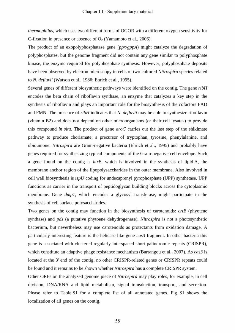
thermophilus, which uses two different forms of OGOR with a different oxygen sensitivity for
C-fixation in presence or absence of O2 (Yamamoto et al., 2006).
The product of an exopolyphosphatase gene (ppx/gppA) might catalyze the degradation of
polyphosphates, but the genome fragment did not contain any gene similar to polyphosphate
kinase, the enzyme required for polyphosphate synthesis. However, polyphosphate deposits
have been observed by electron microscopy in cells of two cultured Nitrospira species related
to N. defluvii (Watson et al., 1986; Ehrich et al., 1995).
Several genes of different biosynthetic pathways were identified on the contig. The gene ribH
encodes the beta chain of riboflavin synthase, an enzyme that catalyzes a key step in the
synthesis of riboflavin and plays an important role for the biosynthesis of the cofactors FAD
and FMN. The presence of ribH indicates that N. defluvii may be able to synthesize riboflavin
(vitamin B2) and does not depend on other microorganisms (or their cell lysates) to provide
this compound in situ. The product of gene aroC carries out the last step of the shikimate
pathway to produce chorismate, a precursor of tryptophan, tyrosine, phenylalanine, and
ubiquinone. Nitrospira are Gram-negative bacteria (Ehrich et al., 1995) and probably have
genes required for synthesizing typical components of the Gram-negative cell envelope. Such
a gene found on the contig is htrB, which is involved in the synthesis of lipid A, the
membrane anchor region of the lipopolysaccharides in the outer membrane. Also involved in
cell wall biosynthesis is ispU coding for undecaprenyl pyrophosphate (UPP) synthetase. UPP
functions as carrier in the transport of peptidoglycan building blocks across the cytoplasmic
membrane. Gene dmp1, which encodes a glycosyl transferase, might participate in the
synthesis of cell surface polysaccharides.
Two genes on the contig may function in the biosynthesis of carotenoids: crtB (phyotene
synthase) and pds (a putative phytoene dehydrogenase). Nitrospira is not a photosynthetic
bacterium, but nevertheless may use carotenoids as protectants from oxidation damage. A
particularly interesting feature is the helicase-like gene cas3 fragment. In other bacteria this
gene is associated with clustered regularly interspaced short palindromic repeats (CRISPR),
which constitute an adaptive phage resistance mechanism (Barrangou et al., 2007). As cas3 is
located at the 3' end of the contig, no other CRISPR-related genes or CRISPR repeats could
be found and it remains to be shown whether Nitrospira has a complete CRISPR system.
Other ORFs on the analyzed genome piece of Nitrospira may play roles, for example, in cell
division, DNA/RNA and lipid metabolism, signal transduction, transport, and secretion.
Please refer to Table S1 for a complete list of all annotated genes. Fig. S1 shows the
localization of all genes on the contig.
Chapter III - Supplementary material
58

β1 α1 α2 Thermus thermophilus ----------------------------------------MERHVPEPTHTLEGWHVLHDFRLLDFARWFSAPLEAREDAWEELKG 46 Bacillus anthracis --------------------------------------------MSEATTTLDGWYCLHDLRSIDWAAWKTLSSDERGQAVSEFLN 42 Mycobacterium tuberculosis ---------------------------------------------MARLDYDALNATLRYLMFSVFSVSPGALGDQRDAIIDDAST 41 Thermoplasma acidophilum ---------------------------------------------------MTEIYTSVLSYRLLE--GKAYSDADTRSLDRMMRS 33 Blastopirellula marina ---------------------------------MSHGRPQQTPPPPPSIIPTEGWHCAHYYYSFDRALLNAFDAEEIGAGRAELSQ 53 Nitrobacter winogradskyi -------------------------------------------------------------------------------------- Ideonella dechloratans MKVRCVSLVAAGLLTIAGSAIGQPAPAPMPAMAPAAKPAMNTPVDRAKILSAPGVFVAFSTYKIRP-DYFKVALAERKGAADEVMA 85 Candidatus Nitrospira defluvii -----------------MNFRSAGRIAVLAGLLVLVAVWPAPAADREKLLTESGVYGTFATFQMDH-DWWDLPGESRVISVAEVKG 68 β2 β3 α3 β4 Thermus thermophilus LVREWRELEEAGQGSYGIYQVVGHKA--DLLFLNLRPGLDPLLEAEARLSRSAF-ARYLGRSYSFYSVVELGSQEKPL-------- 121 Bacillus anthracis VVEKWNDVAAAKKGSHAMYTVVGQKA--DIMLMILRPTMEELNEIETELNKTTL-AEYMVPAYSYVSVVELSNYLPA--------- 116 Mycobacterium tuberculosis FFKQQEE-RGV--VVRGLYDVAGLRADADFMVWTHAERVEALQATYADFRRTTTLGRACTPVWSGVGLHRPAEFNKS--------- 116 Thermoplasma acidophilum IDEFFS--ANP--GYINFHIYRSYRTDSDVIFWYSSRNPDLMILAKERVQASM--RPIAVSSFSSISIYDESPYNAM--------- 104 Blastopirellula marina ILSGEGE-GAP--ERIQVSIVSGHKA--DFGVMLMDPNPLVVDSVHQSIMASTL-GPAIQPTYSFVSLSEVSEYVPSVEQYGERLI 133 Nitrobacter winogradskyi --------------MTFTVFTGGDSG--AWSILSVAPVIGESLMAASHLAIAPSLSLGDTSATTPWQLRGVASHARYVER------ 64 Ideonella dechloratans VLEKHKEK-----VIVDAYLTRGYEAKSDYFLRVHAYDAVAAQAFLVDFRATRF-GMYSDVTESLVGITKALNYISK--------- 156 Candidatus Nitrospira defluvii LVEQWSGK-----ILVESYLLRGLSDHADLMFRVHARTLSDTQQFLSAFMGTRL-GRHLTSGGLLHGVSKKPTYVAG--------- 139 β5 β6 β7 α4 ↓ Thermus thermophilus -------------------DPESPYVKPRLT-------PRVPKSGYVCFYPMNKRRQGQDNWYMLPAKERASLMKA---HGETGRK 178 Bacillus anthracis ----------------DEDPYQNPQILARLY-------PELPKANHICFYPMDKRRQGDDNWYMLPMEERKKMMYS---HSKIGRQ 176 Mycobacterium tuberculosis ------------------------HIPAFL--------AGEEPGAYICVYPFVRS----YEWYLLPDEERRRMLAE---HGMAARG 163 Thermoplasma acidophilum ------------------------NKKLEDS-------LRLPPLRYFVAYPMSKT----PDWYLLDFDTRKEIMHE---HIKMALN 152 Blastopirellula marina RDGEEAGSETFEAKLNAYRQREPMMRKQRLT-------PEFPQWPATCFYPMNKKRKVGENWFTMPFAERNSLMAE---HAQSGMQ 209 Nitrobacter winogradskyi ------------------------AEKIALTSV--QAGLGRNEATRAALIPIRKS----AAWWEMTQDERRAIFEDKSHHIAASLK 120 Ideonella dechloratans ------------------------DKSPDLNKGLSGATYAGDAPRFAFMIPVKKN----ADWWNLTDEQRLKEMET---HTLPTLP 211 Candidatus Nitrospira defluvii ------------------------FPESMKTEL--QVNGESGSRPYAIVIPIKKD----AEWWALDQEARTALMQE---HTQAALP 192 ↑ β8 β9 α5 α6 β10a β10b α7 Id.% Thermus thermophilus YQG--EVMQVISGAQGLD-DWEWGVDLFSED--PVQFKKIVYEMRFDEVSARYGEF-GPFFVGKYLDEEALRAFLGL-- 249 21 Bacillus anthracis YAG--KVRQVISGSVGFD-DFEWGVTLFADD--VLQFKKLIYEMRFDEVSARYGEF-GTFFVGNILPDEKVEKFLHI-- 247 18 Mycobacterium tuberculosis YK---DVRANTVPAFALG-DYEWILAFEAPE--LDRIVDLMRELRATDARRHTRAE-TPFFTGPRVPVEQLVHSLP--- 231 23 Thermoplasma acidophilum HPDEKGIRSYTTYSFGIG-DQEFVVLYEIPD--IAAWSRVTEKLREARARKWIIKE-TPILLGRLVDAGDIAGFLL--- 224 16 Blastopirellula marina FAG--RVSQLITVSVGLD-DWEWGVTLWARN--PEYLKEIVYKMRFDEASARYAEF-GPFYTSYISTAAEMLDHCRIQ- 281 18 Nitrobacter winogradskyi YLP--AIARQLYHCRDIGEPFDFLTWFEYAPEHATMFEDLVGVLRATEEWTYVERE----------VDIRLARAI---- 183 24 Ideonella dechloratans FLV--NVKRKLYHSTGLD-DTDFITYFETND--LGAFNNLMLSLAKVPENKYHVRWGNPTVLGTIQPIENLVKTLSMGN 285 37 Candidatus Nitrospira defluvii YLK--TVKRKLYHSTGLD-DVDFITYFETER—LEDFHNLVRALQQVKEFRHNRRFGHPTLLGTMSPLDEILEKFAQ--- 264 100
Fig. S2. Alignment of selected chlorite dismutase (CLD)-like protein sequences based on
conserved secondary structure motifs. Positions that are conserved in all analyzed sequences
of CLD-like proteins (n=136) are shaded in grey. Vertical arrows point at the histidine residue
that is likely to be the heme ligand in these enzymes. Each sequence is colored according to
PSIPRED (Jones, 1999) secondary structure prediction (red: alpha helix, blue: beta strand).
The colored bars above the alignment show secondary structure motifs (red: alpha helix,
yellow: 310-helix, blue: beta strand) as detected in the crystal structure of the CLD-like
protein of Thermus thermophilus (Ebihara et al., 2005). The last column in the last row
(Id. %) shows amino acid sequence identities to the CLD of N. defluvii.
Chapter III - Supplementary material
59
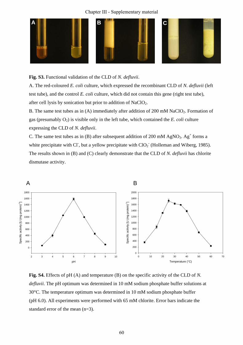
Fig. S3. Functional validation of the CLD of N. defluvii.
A. The red-coloured E. coli culture, which expressed the recombinant CLD of N. defluvii (left
test tube), and the control E. coli culture, which did not contain this gene (right test tube),
after cell lysis by sonication but prior to addition of NaClO2.
B. The same test tubes as in (A) immediately after addition of 200 mM NaClO2. Formation of
gas (presumably O2) is visible only in the left tube, which contained the E. coli culture
expressing the CLD of N. defluvii.
C. The same test tubes as in (B) after subsequent addition of 200 mM AgNO3. Ag+ forms a
white precipitate with Cl-, but a yellow precipitate with ClO2- (Holleman and Wiberg, 1985).
The results shown in (B) and (C) clearly demonstrate that the CLD of N. defluvii has chlorite
dismutase activity.
Fig. S4. Effects of pH (A) and temperature (B) on the specific activity of the CLD of N.
defluvii. The pH optimum was determined in 10 mM sodium phosphate buffer solutions at
30°C. The temperature optimum was determined in 10 mM sodium phosphate buffer
(pH 6.0). All experiments were performed with 65 mM chlorite. Error bars indicate the
standard error of the mean (n=3).
A B CA B C
pH
2 3 4 5 6 7 8 9 10
Spec
ific
activ
ity [U
(mg
prot
ein)
-1]
0
200
400
600
800
1000
1200
1400
1600
1800
Temperature (°C)
0 10 20 30 40 50 60 70
Spec
ific
activ
ity [U
(mg
prot
ein)
-1]
0
200
400
600
800
1000
1200
1400
1600
1800
2000
A B
pH
2 3 4 5 6 7 8 9 10
Spec
ific
activ
ity [U
(mg
prot
ein)
-1]
0
200
400
600
800
1000
1200
1400
1600
1800
Temperature (°C)
0 10 20 30 40 50 60 70
Spec
ific
activ
ity [U
(mg
prot
ein)
-1]
0
200
400
600
800
1000
1200
1400
1600
1800
2000
A B
Chapter III - Supplementary material
60

Table S1. List of all coding sequences identified on the analyzed genome fragment of "Candidatus Nitrospira defluvii" and the corresponding annotations.
Label Begin End Gene Product ECnumber Class ProductTypeORF001 1 539 ribH Riboflavin synthase, beta chain 2.5.1.9 2a : Function of homologous gene experimentally demonstrated in an other
organisme : enzyme
ORF002 553 1026 nusB transcription antitermination protein 2b : Function of strongly homologous gene f : factorORF003 1103 2437 purB Adenylosuccinate lyase 4.3.2.2 2a : Function of homologous gene experimentally demonstrated in an other
organisme : enzyme
ORF004 2434 3228 cld Chlorite dismutase 1.13.11.49 1 : Function experimentally demonstrated in the studied organism e : enzymeORF005 3244 3957 purC Phosphoribosylaminoimidazole-succinocarboxamide synthetase6.3.2.6 2a : Function of homologous gene experimentally demonstrated in an other
organisme : enzyme
ORF006 3965 4192 exported protein of unknown function 5 : No homology to any previously reported sequences o : ORF of unknown functionORF007 4189 4428 purS Phosphoribosylformylglycinamidine synthase, PurS subunit
(FGAM component)6.3.5.3 2a : Function of homologous gene experimentally demonstrated in an other
organisme : enzyme
ORF008 4487 5245 purQ Phosphoribosylformylglycinamidine synthase, PurQ subunit (FGAM synthase I)
6.3.5.3 2a : Function of homologous gene experimentally demonstrated in an other organism
e : enzyme
ORF009 5314 7560 purL Phosphoribosylformylglycinamidine synthase, PurL subunit (FGAM synthase II)
6.3.5.3 2a : Function of homologous gene experimentally demonstrated in an other organism
e : enzyme
ORF010 7573 9003 purF Amidophosphoribosyltransferase 2.4.2.14 2a : Function of homologous gene experimentally demonstrated in an other organism
e : enzyme
ORF011 9071 9511 protein of unknown function 5 : No homology to any previously reported sequences o : ORF of unknown functionORF012 9836 10153 protein of unknown function 5 : No homology to any previously reported sequences o : ORF of unknown functionORF013 10388 11428 protein of unknown function 5 : No homology to any previously reported sequences o : ORF of unknown functionORF014 11430 12725 protein of unknown function 5 : No homology to any previously reported sequences o : ORF of unknown functionORF015 12917 13462 putative thiol-disulfide oxidoreductase 3 : Function proposed based on presence of conserved amino acid motif,
structural feature or limited homologype : putative enzyme
ORF016 13482 13721 protein of unknown function 5 : No homology to any previously reported sequences o : ORF of unknown functionORF017 13746 14342 conserved protein of unknown function with Metallo-
hydrolase/oxidoreductase domain4 : Homologs of previously reported genes of unknown function o : ORF of unknown function
ORF018 14349 15173 protein of unknown function 5 : No homology to any previously reported sequences o : ORF of unknown functionORF019 15170 15607 protein of unknown function 5 : No homology to any previously reported sequences o : ORF of unknown functionORF020 15739 16098 protein of unknown function 5 : No homology to any previously reported sequences o : ORF of unknown functionORF021 16004 16492 membrane protein of unknown function 5 : No homology to any previously reported sequences o : ORF of unknown functionORF022 16651 17607 exported protein of unknown function 5 : No homology to any previously reported sequences o : ORF of unknown functionORF023 17611 19053 putative TPR repeat: Peptidase M, neutral zinc
metallopeptidases, zinc-binding site3 : Function proposed based on presence of conserved amino acid motif, structural feature or limited homology
pe : putative enzyme
ORF024 19215 21755 recG ATP-dependent DNA helicase 3.6.1.- 2a : Function of homologous gene experimentally demonstrated in an other organism
e : enzyme
ORF025 21794 22120 protein of unknown function 5 : No homology to any previously reported sequences o : ORF of unknown functionORF026 22131 23081 gppA Exopolyphosphatase 3.6.1.11 2b : Function of strongly homologous gene e : enzymeORF027 23162 24655 protein of unknown function 5 : No homology to any previously reported sequences o : ORF of unknown functionORF028 24737 25021 putative copper binding protein, Plastocyanin family 3 : Function proposed based on presence of conserved amino acid motif,
structural feature or limited homologypc : putative carrier
ORF029 25363 25950 conserved protein of unknown function 4 : Homologs of previously reported genes of unknown function o : ORF of unknown functionORF030 26162 26614 protein of unknown function 5 : No homology to any previously reported sequences o : ORF of unknown functionORF031 26643 27323 exported protein of unknown function 5 : No homology to any previously reported sequences o : ORF of unknown functionORF032 27460 28515 protein of unknown function 5 : No homology to any previously reported sequences o : ORF of unknown functionORF033 28614 29393 scpA Segregation and condensation protein A 2a : Function of homologous gene experimentally demonstrated in an other
organismcp : cell process
ORF034 29390 30295 scpB putative chromosome segregation and condensation protein B 3 : Function proposed based on presence of conserved amino acid motif, structural feature or limited homology
cp : cell process
ORF035 30445 31011 exported protein of unknown function 5 : No homology to any previously reported sequences o : ORF of unknown functionORF036 31336 34305 putative NHL repeat protein 3 : Function proposed based on presence of conserved amino acid motif,
structural feature or limited homologype : putative enzyme
ORF037 34344 35810 guaB Inosine-5'-monophosphate dehydrogenase (IMP) 1.1.1.205 2a : Function of homologous gene experimentally demonstrated in an other organism
e : enzyme
ORF038 35895 37451 guaA GMP synthetase (glutamine aminotransferase) 6.3.5.2 2a : Function of homologous gene experimentally demonstrated in an other organism
e : enzyme
ORF039 37531 38793 rluB ribosomal large subunit pseudouridine synthase B (modular protein?)
4.2.1.70 3 : Function proposed based on presence of conserved amino acid motif, structural feature or limited homology
e : enzyme
ORF040 38474 38914 protein of unknown function 5 : No homology to any previously reported sequences o : ORF of unknown functionORF041 38790 40553 aspS aspartyl-tRNA synthetase 6.1.1.12 2a : Function of homologous gene experimentally demonstrated in an other
organisme : enzyme
ORF042 40572 41708 aroC chorismate synthase 4.2.3.5 2a : Function of homologous gene experimentally demonstrated in an other organism
e : enzyme
ORF043 41823 42293 rnhA Ribonuclease H 3.1.26.4 2a : Function of homologous gene experimentally demonstrated in an other organism
e : enzyme
ORF044 42336 43133 abcX ABC transporter, ATPase component 2b : Function of strongly homologous gene t : transporterORF045 43143 44177 putative Trypsin-like serine protease 3 : Function proposed based on presence of conserved amino acid motif,
structural feature or limited homologype : putative enzyme
ORF046 44334 44888 putative outer membrane lipoprotein precursor yeaY 3 : Function proposed based on presence of conserved amino acid motif, structural feature or limited homology
pm : putative membrane component
ORF047 45105 45971 conserved protein of unknown function, putative neutral zinc metallopeptidase
4 : Homologs of previously reported genes of unknown function o : ORF of unknown function
ORF048 46050 46232 protein of unknown function 5 : No homology to any previously reported sequences o : ORF of unknown functionORF049 46320 46892 exported protein of unknown function 5 : No homology to any previously reported sequences o : ORF of unknown functionORF050 47022 47609 rluE Ribosomal large subunit pseudouridine synthase E 5.4.99.12,
4.2.1.702a : Function of homologous gene experimentally demonstrated in an other organism
e : enzyme
ORF051 47768 48229 conserved exported protein of unknown function 4 : Homologs of previously reported genes of unknown function o : ORF of unknown functionORF052 48409 48951 exported protein of unknown function 5 : No homology to any previously reported sequences o : ORF of unknown functionORF053 49155 49634 membrane protein of unknown function 5 : No homology to any previously reported sequences o : ORF of unknown functionORF054 49631 50506 conserved membrane protein of unknown function 4 : Homologs of previously reported genes of unknown function o : ORF of unknown functionORF055 50515 52152 conserved protein of unknown function (containing GH3 auxin-
responsive domain)4 : Homologs of previously reported genes of unknown function o : ORF of unknown function
ORF056 52152 53246 putative Muconate cycloisomerase 5.5.1.- 3 : Function proposed based on presence of conserved amino acid motif, structural feature or limited homology
pe : putative enzyme
ORF057 53304 54332 conserved exported protein of unknown function 4 : Homologs of previously reported genes of unknown function o : ORF of unknown functionrRNA_16S 54662 56193 16S
rRNA16S ribosomal RNA 2a : Function of homologous gene experimentally demonstrated in an other
organismn : RNA
tRNA_Ile 56366 56439 Ile tRNA Isoleucine tRNA 2a : Function of homologous gene experimentally demonstrated in an other organism
tRNA_Ala 56507 56579 Ala tRNA
Alanine tRNA 2a : Function of homologous gene experimentally demonstrated in an other organism
rRNA_23S 56703 59712 23S rRNA
23S ribosomal RNA 2a : Function of homologous gene experimentally demonstrated in an other organism
n : RNA
rRNA_5S 59836 59952 5S rRNA
5S ribosomal RNA 2a : Function of homologous gene experimentally demonstrated in an other organism
n : RNA
ORF058 60223 60903 ung Uracil-DNA-glycosylase 3.2.2.- 2a : Function of homologous gene experimentally demonstrated in an other organism
e : enzyme
ORF059 61041 61829 cah Carbonic anhydrase 4.2.1.1 2a : Function of homologous gene experimentally demonstrated in an other organism
e : enzyme
ORF060 61978 62160 conserved protein of unknown function 4 : Homologs of previously reported genes of unknown function o : ORF of unknown functionORF061 62196 62537 protein of unknown function 5 : No homology to any previously reported sequences o : ORF of unknown functionORF062 62872 63327 protein of unknown function 5 : No homology to any previously reported sequences o : ORF of unknown functionORF063 63410 63571 protein of unknown function 5 : No homology to any previously reported sequences o : ORF of unknown functionORF064 63674 63841 exported protein of unknown function 5 : No homology to any previously reported sequences o : ORF of unknown functionORF065 63856 64080 exported protein of unknown function 5 : No homology to any previously reported sequences o : ORF of unknown functionORF066 64105 64446 protein of unknown function 5 : No homology to any previously reported sequences o : ORF of unknown functionORF067 64538 64726 protein of unknown function 5 : No homology to any previously reported sequences o : ORF of unknown functionORF068 64735 64914 protein of unknown function 5 : No homology to any previously reported sequences o : ORF of unknown functionORF069 64911 65108 protein of unknown function 5 : No homology to any previously reported sequences o : ORF of unknown functionORF070 65155 65475 yadR Iron-sulfur cluster assembly accessory protein 2b : Function of strongly homologous gene f : factorORF071 65744 65938 forB1-N partial 2-oxoacid:ferredoxin oxidoreductase beta subunit
(fragment N-terminal)1.2.7.1 2b : Function of strongly homologous gene e : enzyme
ORF072 65973 67310 istA Integrase 2a : Function of homologous gene experimentally demonstrated in an other organism
e : enzyme
ORF073 67307 68086 istB Insertion sequence IS21 putative ATP-binding protein 2a : Function of homologous gene experimentally demonstrated in an other organism
e : enzyme
ORF074 68167 68889 forB1-C partial 2-oxoacid:ferredoxin oxidoreductase beta subunit (fragment C-terminal)
1.2.7.1 2a : Function of homologous gene experimentally demonstrated in an other organism
e : enzyme
ORF075 68942 69835 forB2 2-oxoacid:ferredoxin oxidoreductase beta subunit 1.2.7.1 2a : Function of homologous gene experimentally demonstrated in an other organism
e : enzyme
ORF076 70279 70521 yjbJ putative stress response protein 3 : Function proposed based on presence of conserved amino acid motif, structural feature or limited homology
pf : putative factor
ORF077 70774 70944 protein of unknown function 5 : No homology to any previously reported sequences o : ORF of unknown functionORF078 71447 73111 exported protein of unknown function 5 : No homology to any previously reported sequences o : ORF of unknown function
Chapter III - Supplementary material
61

Table S1. List of all coding sequences identified on the analyzed genome fragment of "Candidatus Nitrospira defluvii" and the corresponding annotations.ORF079 73318 74406 exported protein of unknown function 5 : No homology to any previously reported sequences o : ORF of unknown functionORF080 74429 74740 protein of unknown function 5 : No homology to any previously reported sequences o : ORF of unknown functionORF081 74897 75190 exported protein of unknown function 5 : No homology to any previously reported sequences o : ORF of unknown functionORF082 75497 76792 rhlE ATP-dependent RNA helicase 3.6.1.- 2a : Function of homologous gene experimentally demonstrated in an other
organisme : enzyme
ORF083 76822 77169 protein of unknown function 5 : No homology to any previously reported sequences o : ORF of unknown functionORF084 77451 77732 protein of unknown function 5 : No homology to any previously reported sequences o : ORF of unknown functionORF085 78100 79653 nifA Nif-specific regulatory protein 2b : Function of strongly homologous gene r : regulatorORF086 79660 81360 exported protein of unknown function 5 : No homology to any previously reported sequences o : ORF of unknown functionORF087 81478 81969 exported protein of unknown function 5 : No homology to any previously reported sequences o : ORF of unknown functionORF088 82498 83223 putative sterol desaturase 3 : Function proposed based on presence of conserved amino acid motif,
structural feature or limited homologype : putative enzyme
ORF089 83360 84454 dpm1 Dolichyl-phosphate beta-D-mannosyltransferase 2.4.1.83 2b : Function of strongly homologous gene e : enzymeORF090 84660 86231 conserved membrane protein of unknown function 4 : Homologs of previously reported genes of unknown function o : ORF of unknown functionORF091 86334 87569 mltC Membrane-bound lytic murein transglycosylase C precursor 3.2.1.- 2b : Function of strongly homologous gene e : enzymeORF092 87874 88437 putative Toluene tolerance precursor 3 : Function proposed based on presence of conserved amino acid motif,
structural feature or limited homologypt : putative transporter
ORF093 88593 89342 putative Two component transcriptional regulator, LuxR family, response regulator receiver
3 : Function proposed based on presence of conserved amino acid motif, structural feature or limited homology
pr : putative regulator
ORF094 89364 89654 protein of unknown function 5 : No homology to any previously reported sequences o : ORF of unknown functionORF095 89824 90135 protein of unknown function 5 : No homology to any previously reported sequences o : ORF of unknown functionORF096 90191 90622 protein of unknown function 5 : No homology to any previously reported sequences o : ORF of unknown functionORF097 90638 90982 protein of unknown function 5 : No homology to any previously reported sequences o : ORF of unknown functionORF098 91198 91842 gacA Response regulator (Global activator) 2a : Function of homologous gene experimentally demonstrated in an other
organismr : regulator
ORF099 91911 94907 Response regulator receiver and signal transduction histidine kinase (modular protein)
3 : Function proposed based on presence of conserved amino acid motif, structural feature or limited homology
pr : putative regulator
ORF100 94904 95365 Response regulator receiver (CheY-like) 2b : Function of strongly homologous gene r : regulatorORF101 95389 97161 Signal transduction histidine kinase (modular protein) 2.7.13.3 3 : Function proposed based on presence of conserved amino acid motif,
structural feature or limited homologype : putative enzyme
ORF102 97362 97778 yaeJ Peptidyl-tRNA hydrolase domain protein 2b : Function of strongly homologous gene f : factorORF103 97860 98183 rbpA RNA-binding protein 2b : Function of strongly homologous gene f : factorORF104 98126 98365 protein of unknown function 5 : No homology to any previously reported sequences o : ORF of unknown functionORF105 98545 99414 scdA Stearoyl-CoA 9-desaturase (fragment) 1.14.19.1 2b : Function of strongly homologous gene e : enzymeORF106 99691 100014 exported protein of unknown function 5 : No homology to any previously reported sequences o : ORF of unknown functionORF107 100186 100560 conserved protein of unknown function 4 : Homologs of previously reported genes of unknown function o : ORF of unknown functionORF108 100761 101699 htrB Lipid A biosynthesis lauroyl (or palmitoleoyl) acyltransferase 2.3.1.- 2a : Function of homologous gene experimentally demonstrated in an other
organisme : enzyme
ORF109 101847 102221 protein of unknown function 5 : No homology to any previously reported sequences o : ORF of unknown functionORF110 102273 102605 conserved protein of unknown function 4 : Homologs of previously reported genes of unknown function o : ORF of unknown functionORF111 102826 103605 ABC transporter permease protein 2b : Function of strongly homologous gene t : transporterORF112 103602 104333 ABC-type transport system, ATPase component (fragment) 3.6.3.- 2b : Function of strongly homologous gene t : transporterORF113 104381 105370 conserved protein of unknown function 4 : Homologs of previously reported genes of unknown function o : ORF of unknown functionORF114 105478 105981 protein of unknown function 6 : Doubtful CDS o : ORF of unknown functionORF115 105523 106323 TPR-repeat protein 3 : Function proposed based on presence of conserved amino acid motif,
structural feature or limited homologyo : ORF of unknown function
ORF116 106352 108664 gspE Type II secretion system protein E 2b : Function of strongly homologous gene t : transporterORF117 108878 110509 proS Prolyl-tRNA synthetase 6.1.1.15 2a : Function of homologous gene experimentally demonstrated in an other
organisme : enzyme
ORF118 110591 111982 ecfE Peptidase M50, putative membrane-associated zinc metallopeptidase
3.4.24.- 2b : Function of strongly homologous gene e : enzyme
ORF119 112008 113168 dxr 1-deoxy-D-xylulose-5-phosphate reductoisomerase 1.1.1.267 2a : Function of homologous gene experimentally demonstrated in an other organism
e : enzyme
ORF120 113201 114049 cdsA Phosphatidate cytidylyltransferase 2.7.7.41 2a : Function of homologous gene experimentally demonstrated in an other organism
e : enzyme
ORF121 114093 114878 ispU Undecaprenyl pyrophosphate synthase 2.5.1.31 2a : Function of homologous gene experimentally demonstrated in an other organism
e : enzyme
ORF122 115071 116507 tldD Peptidase U62 2b : Function of strongly homologous gene e : enzymeORF123 116510 117868 pmbA Peptidase U62 2b : Function of strongly homologous gene e : enzymeORF124 118159 118713 Phosphatidylethanolamine-binding protein 3 : Function proposed based on presence of conserved amino acid motif,
structural feature or limited homologyo : ORF of unknown function
ORF125 118750 119205 protein of unknown function 5 : No homology to any previously reported sequences o : ORF of unknown functionORF126 119270 119941 udgA Uracil-DNA glycosylase, family 4 3.2.2.- 2a : Function of homologous gene experimentally demonstrated in an other
organismcp : cell process
ORF127 120032 121342 putative Carotene 7,8-desaturase 1.14.99.30 3 : Function proposed based on presence of conserved amino acid motif, structural feature or limited homology
pe : putative enzyme
ORF128 121339 122250 crtB Phytoene synthase 2.5.1.- 2b : Function of strongly homologous gene e : enzymeORF129 122222 123154 ABC-type transport system, periplasmic binding protein 2b : Function of strongly homologous gene t : transporterORF130 123151 123858 putative beta-phosphoglucomutase 5.4.2.6 3 : Function proposed based on presence of conserved amino acid motif,
structural feature or limited homologype : putative enzyme
ORF131 124017 124781 protein of unknown function 5 : No homology to any previously reported sequences o : ORF of unknown functionORF132 125074 125487 UPF0047 protein 4 : Homologs of previously reported genes of unknown function o : ORF of unknown functionORF133 125650 126663 protein of unknown function 5 : No homology to any previously reported sequences o : ORF of unknown functionORF134 126816 127397 exported protein of unknown function 5 : No homology to any previously reported sequences o : ORF of unknown functionORF135 127390 128124 FHA domain containing protein 3 : Function proposed based on presence of conserved amino acid motif,
structural feature or limited homologypr : putative regulator
ORF136 128134 128970 pph1 Serine/threonine protein phosphatase 2C 3.1.3.16 2a : Function of homologous gene experimentally demonstrated in an other organism
e : enzyme
ORF137 128979 130388 putative serine/threonine protein kinase 2.7.11.- 3 : Function proposed based on presence of conserved amino acid motif, structural feature or limited homology
pe : putative enzyme
ORF138 130535 131383 trpH Phosphoesterase (PHP family) 2b : Function of strongly homologous gene e : enzymeORF139 131380 132309 Modular protein: putative Dihydroneopterin aldolase and SAM-
dependent methyltransferases4.1.2.25 3 : Function proposed based on presence of conserved amino acid motif,
structural feature or limited homologype : putative enzyme
ORF140 132367 132837 protein of unknown function 5 : No homology to any previously reported sequences o : ORF of unknown functionORF141 133060 133266 protein of unknown function 5 : No homology to any previously reported sequences o : ORF of unknown functionORF142 133274 133513 protein of unknown function 5 : No homology to any previously reported sequences o : ORF of unknown functionORF143 133827 134345 exported protein of unknown function 5 : No homology to any previously reported sequences o : ORF of unknown functionORF144 134374 135285 Peptidase C14, Caspase-1 catalytic subunit p20 3.4.22.- 2b : Function of strongly homologous gene e : enzymeORF145 135376 135606 protein of unknown function 5 : No homology to any previously reported sequences o : ORF of unknown functionORF146 135816 136100 protein of unknown function 5 : No homology to any previously reported sequences o : ORF of unknown functionORF147 136129 136392 protein of unknown function 5 : No homology to any previously reported sequences o : ORF of unknown functionORF148 136492 137336 cas3 CRISPR-associated helicase Cas3 2b : Function of strongly homologous gene cp : cell process
Chapter III - Supplementary material
62

Table S2: Sources of environmental sequences of CLD-like proteins that were used for
protein phylogeny (see Fig. 5).
Label in Fig. 5 Accesion no.1 Reference Eastern tropical pacific Cocos island read EM972568 Rusch et al. (2007) North American East cost read EN082124 ditto Isabela island mangrove read 1 EP183024 ditto Isabela island mangrove read 2 EP183024 ditto Isabela island mangrove read 3 EP183024 ditto Floreana island hypersaline lagoon read 1 EP836070 ditto Floreana island hypersaline lagoon read 2 EP835918 ditto Floreana island hypersaline lagoon read 3 EQ050993 ditto Panama canal freshwater read 1 EM410546 ditto Panama canal freshwater read 2 EM410546 ditto Panama canal freshwater read 3 EM490579 ditto Panama canal freshwater read 4 EM490579 ditto Panama canal freshwater read 5 EP707854 ditto Panama canal freshwater read 6 EP707854 ditto Sargasso sea read 1 EM426291 Venter et al. (2004) Sargasso sea read 2 AACY01204540 ditto Sargasso sea read 3 EP943272 ditto Sargasso sea read 4 EN484981 ditto Sargasso sea read 5 EP924593 ditto Minnesota farm soil clone AAFX01003328 Tringe et al. (2005) 1 Accession numbers refer to published scaffolds that contain the respective nucleic acid sequence reads of CLD-like proteins. References Barrangou, R., Fremaux, C., Deveau, H., Richards, M., Boyaval, P., Moineau, S. et al. (2007)
CRISPR provides acquired resistance against viruses in prokaryotes. Science 315:
1709-1712.
Campbell, B.J., Engel, A.S., Porter, M.L., and Takai, K. (2006) The versatile epsilon-
proteobacteria: key players in sulphidic habitats. Nat. Rev. Microbiol. 4: 458-468.
Daims, H., Nielsen, J.L., Nielsen, P.H., Schleifer, K.H., and Wagner, M. (2001) In situ
characterization of Nitrospira-like nitrite-oxidizing bacteria active in wastewater
treatment plants. Appl. Environ. Microbiol. 67: 5273-5284.
Ebihara, A., Okamoto, A., Kousumi, Y., Yamamoto, H., Masui, R., Ueyama, N. et al. (2005)
Structure-based functional identification of a novel heme-binding protein from
Thermus thermophilus HB8. J. Struct. Funct. Genomics 6: 21-32.
Ehrich, S., Behrens, D., Lebedeva, E., Ludwig, W., and Bock, E. (1995) A new obligately
chemolithoautotrophic, nitrite-oxidizing bacterium, Nitrospira moscoviensis sp. nov.
and its phylogenetic relationship. Arch. Microbiol. 164: 16-23.
Chapter III - Supplementary material
63

Fukuzawa, H., Suzuki, E., Komukai, Y., and Miyachi, S. (1992) A gene homologous to
chloroplast carbonic anhydrase (icfA) is essential to photosynthetic carbon dioxide
fixation by Synechococcus PCC7942. Proc. Natl. Acad. Sci. U. S. A. 89: 4437-4441.
Gieseke, A., Bjerrum, L., Wagner, M., and Amann, R. (2003) Structure and activity of
multiple nitrifying bacterial populations co-existing in a biofilm. Environ. Microbiol.
5: 355-369.
Holleman, A.F., and Wiberg, E. (1985) Lehrbuch der Anorganischen Chemie. Berlin: Walter
de Gruyter.
Jones, D.T. (1999) Protein secondary structure prediction based on position-specific scoring
matrices. J. Mol. Biol. 292: 195-202.
Okabe, S., Satoh, H., and Watanabe, Y. (1999) In situ analysis of nitrifying biofilms as
determined by in situ hybridization and the use of microelectrodes. Appl. Environ.
Microbiol. 65: 3182-3191.
Reimmann, C., and Haas, D. (1990) The istA gene of insertion sequence IS21 is essential for
cleavage at the inner 3' ends of tandemly repeated IS21 elements in vitro. Embo J. 9:
4055-4063.
Rusch, D.B., Halpern, A.L., Sutton, G., Heidelberg, K.B., Williamson, S., Yooseph, S. et al.
(2007) The Sorcerer II Global Ocean Sampling expedition: northwest Atlantic through
eastern tropical Pacific. PLoS Biol. 5: e77.
Smith, K.S., and Ferry, J.G. (2000) Prokaryotic carbonic anhydrases. FEMS Microbiol. Rev.
24: 335-366.
Spieck, E., Hartwig, C., McCormack, I., Maixner, F., Wagner, M., Lipski, A., and Daims, H.
(2006) Selective enrichment and molecular characterization of a previously uncultured
Nitrospira-like bacterium from activated sludge. Environ. Microbiol. 8: 405-415.
Starkenburg, S.R., Chain, P.S., Sayavedra-Soto, L.A., Hauser, L., Land, M.L., Larimer, F.W.
et al. (2006) Genome sequence of the chemolithoautotrophic nitrite-oxidizing
bacterium Nitrobacter winogradskyi Nb-255. Appl. Environ. Microbiol. 72: 2050-
2063.
Tringe, S.G., von Mering, C., Kobayashi, A., Salamov, A.A., Chen, K., Chang, H.W. et al.
(2005) Comparative metagenomics of microbial communities. Science 308: 554-557.
Venter, J.C., Remington, K., Heidelberg, J.F., Halpern, A.L., Rusch, D., Eisen, J.A. et al.
(2004) Environmental genome shotgun sequencing of the Sargasso Sea. Science 304:
66-74.
Chapter III - Supplementary material
64
Watson, S.W., Bock, E., Valois, F.W., Waterbury, J.B., and Schlosser, U. (1986) Nitrospira
marina gen. nov. sp. nov.: a chemolithotrophic nitrite-oxidizing bacterium. Arch.
Microbiol. 144: 1-7.
Yamamoto, M., Arai, H., Ishii, M., and Igarashi, Y. (2006) Role of two 2-
oxoglutarate:ferredoxin oxidoreductases in Hydrogenobacter thermophilus under
aerobic and anaerobic conditions. FEMS Microbiol. Lett. 263: 189-193.
Yoon, K.S., Ishii, M., Kodama, T., and Igarashi, Y. (1997) Purification and characterization
of pyruvate:ferredoxin oxidoreductase from Hydrogenobacter thermophilus TK-6.
Arch. Microbiol. 167: 275-279.
Chapter III - Supplementary material
65

Chapter III - Supplementary material
66

Chapter IV
Nitrite oxidoreductase (Nxr) -
the metabolic key enzyme of nitrite-oxidizing Nitrospira:
Characterization of an unusual Nxr and its application as
a novel functional and phylogenetic marker gene
To be submitted

Chapter IV
68
Nitrite oxidoreductase (Nxr) -
the metabolic key enzyme of nitrite-oxidizing Nitrospira:
Characterization of an unusual Nxr and its application as a novel functional
and phylogenetic marker gene
Frank Maixner1, Hanna Koch1, Sandra Hauzmayer1, Eva Spieck2, Denis Le Paslier3, Michael
Wagner1, Holger Daims1
Draft was not corrected by the co-authors.
1 Department für Mikrobielle Ökologie, Universität Wien, Althanstrasse 14, A-1090 Vienna,
Austria. 2 Universität Hamburg, Biozentrum Klein Flottbek, Mikrobiologie, Ohnhorststr. 18, D-22609
Hamburg, Germany. 3CEA/Genoscope CNRS-UMR 8030, 2 rue Gaston Crémieux CP 5706, 91057 Evry, France.
Summary
The Nxr genes of ”Candidatus Nitrospira defluvii” were identified based on protein sequence
comparisons with already known Nxr of other nitrite-oxidizing bacteria (NOB) and related
nitrate/nitrite-binding enzymes of other microorganisms. The Nxr of Nitrospira consists of
three subunits (alpha to gamma) and is present in two similar copies in the genome of N.
defluvii. Like the Nxr forms of other characterized NOB, the Nitrospira enzyme belongs to
the superfamily of molybdopterin-binding proteins. However, phylogenetic analyses revealed
that the Nxr of N. defluvii differs remarkably in sequence from the Nxr of Nitrobacter and
Nitrococcus, possibly reflecting key differences in enzymatic properties such as different
affinities for nitrite. Also in contrast to the Nxr of Nitrobacter, which is localized in the
cytoplasm, the Nitrospira Nxr seems to be located in the periplasm but attached to the
cytoplasmic membrane via its gamma subunit. To prove the functionality of the detected
Nitrospira Nxr, the transcription of the nxrB gene, encoding the Nxr beta subunit, was
monitored in a highly enriched N. defluvii culture under starvation and nitrite-oxidizing
conditions, respectively. In addition, the translation of Nxr was followed by the use of Nxr
beta subunit-specific antibodies. Both experiments clearly showed that Nxr was expressed,
and that the level of expression was much higher in presence of nitrite than during starvation.
Chapter IV
69
Interestingly, N. defluvii cultures still contained detectable amounts of Nxr even after very
long starvation periods. Thus, a stable Nxr-content may enable the organism to respond
quickly to increasing nitrite concentrations without the need of energy-consuming de novo
synthesis of Nxr.
Furthermore, a set of Nitrospira nxrB-specific PCR primers was designed based on the nxrB
sequences of N. defluvii and evaluated with enrichments and pure cultures of other Nitrospira
strains. These primers were used to amplify the nxrB genes of the cultured Nitrospira, which
belonged to different sublineages of the genus Nitrospira, and of uncultured Nitrospira in
different environmental samples. In-depth sequence analyses comparing the nxrB- and 16S
rRNA-based phylogenies of the different sublineages of the genus Nitrospira showed
congruent results, thus enabling the use of nxrB as novel phylogenetic marker. Subsequently,
this new phylogenetic framework allowed the assignment of metagenomic reads to
Nitrospira-like nxrB sequences.
Introduction
In all three domains of life one driving force for evolution is the exploitation of unused
niches. Particularly microbes harbor a diverse repertoire of biochemical “machineries” to take
advantage of chemical gradients and different nutrients for energy generation. Prominent
examples for functional diversification are certain phototrophs, which use different types of
proteorhodopsin adapted to various spectral properties within their environment for light-
driven energy generation (Sabehi et al., 2007). The very fact that microbes are key players in
all biogeochemical nutrient cycles and that they are found essentially anywhere is a testament
to their metabolic innovation. Nevertheless, our current knowledge about enzymatic key
enzymes involved in dissimilatory substrate turnovers is still scarce. Here we report about a
novel nitrite oxdiodreductase, which catalyzes the oxidation of nitrite to nitrate, an essential
step in the aerobic nitrification pathway. Nitrite-oxidizing bacteria (NOB) play a crucial role
in the global biogeochemical N-cycle and also in nitrification in biological wastewater
treatment. As the oxidation of nitrite to nitrate yields little free energy, all hitherto identified
NOB are highly specialized, slow-growing organisms that are also very difficult to culture in
artificial nutrient media. The key enzyme of nitrite oxidation is the nitrite oxidoreductase
(Nxr), a complex heteromer that consists of at least two different subunits in Nitrobacter
(Meincke et al., 1992). In contrast to other key functional proteins involved in nitrification
and denitrification (such as ammonia monooxygenase and nitrate reductases), our current
Chapter IV
70
knowledge of Nxr is still limited. For example, yet unresolved questions are whether all
known NOB harbor similar forms of Nxr and whether the Nxr subunits might be suitable as
functional and also as phylogenetic markers for the detection of NOB in environmental
samples. Recently, the latter question was addressed for Nitrobacter, whose Nxr beta subunit
(NxrB) was used to reconstruct phylogenetic trees that resembled 16S rRNA-based
phylogenies (Vanparys et al., 2007).
Most of the few Nxr-related studies were carried out with pure cultures of Nitrobacter
(Tanaka et al., 1983, Sundermeyer-Klinger et al., 1984, Meincke et al., 1992, Spieck et al.,
1996b). However, cultivation-independent approaches have revealed that different NOB,
which belong to the genus Nitrospira, are the key NOB in wastewater treatment plants
(wwtps) (Juretschko et al., 1998). Moreover, 16S rRNA-based screenings have shown that
Nitrospira are the most diverse known NOB in nature, and that these bacteria are ubiquitously
distributed in a wide range of aquatic and terrestrial ecosystems (Nakamura et al., 2006,
Foesel et al., 2008, Urich et al., 2008). Thus, molecular tools to detect and identify these
mostly uncultured NOB are essential for studies of nitrification in natural and engineered
habitats. Ideally, such tools would allow one to simultaneously identify Nitrospira and
monitor their nitrite-oxidizing activities in situ. The 16S rRNA approach combined with
FISH-microautoradiography meets these requirements (Daims et al., 2001), but a less time-
consuming and non-radioactive approach, which would be applicable to larger numbers of
samples, is still lacking.
This study emerged as a spin-off from a larger environmental genomics project, which aimed
at sequencing the whole genome of a Nitrospira strain (N. defluvii) enriched from a nitrifying
wwtp. Here, the goal was to identify and characterize in detail the nxr genes in the N. defluvii
genome and to determine whether these genes are suitable as functional and phylogenetic
markers of Nitrospira. In this case, nxr could be applied in microbial diversity surveys in a
similar manner as amoA has been used to detect and identify ammonia oxidizers.
Results and Discussion
Novel nitrite-oxidizing system in N. defluvii
An environmental genome approach applied on a high enrichment (86% biovolume) of N.
defluvii resulted in the first closed genome of a Nitrospira-like organism (for details on N.
defluvii enrichment please refer to Spieck et al., 2006). First genomic analyses supported the
results of former immunological and enzymatic studies (Spieck et al., 1998, Bartosch et al.,
Chapter IV
71
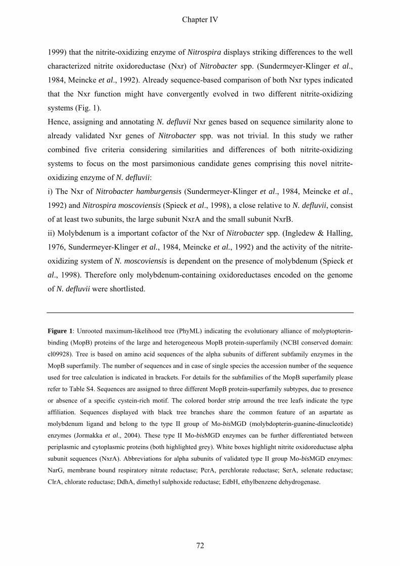
1999) that the nitrite-oxidizing enzyme of Nitrospira displays striking differences to the well
characterized nitrite oxidoreductase (Nxr) of Nitrobacter spp. (Sundermeyer-Klinger et al.,
1984, Meincke et al., 1992). Already sequence-based comparison of both Nxr types indicated
that the Nxr function might have convergently evolved in two different nitrite-oxidizing
systems (Fig. 1).
Hence, assigning and annotating N. defluvii Nxr genes based on sequence similarity alone to
already validated Nxr genes of Nitrobacter spp. was not trivial. In this study we rather
combined five criteria considering similarities and differences of both nitrite-oxidizing
systems to focus on the most parsimonious candidate genes comprising this novel nitrite-
oxidizing enzyme of N. defluvii:
i) The Nxr of Nitrobacter hamburgensis (Sundermeyer-Klinger et al., 1984, Meincke et al.,
1992) and Nitrospira moscoviensis (Spieck et al., 1998), a close relative to N. defluvii, consist
of at least two subunits, the large subunit NxrA and the small subunit NxrB.
ii) Molybdenum is a important cofactor of the Nxr of Nitrobacter spp. (Ingledew & Halling,
1976, Sundermeyer-Klinger et al., 1984, Meincke et al., 1992) and the activity of the nitrite-
oxidizing system of N. moscoviensis is dependent on the presence of molybdenum (Spieck et
al., 1998). Therefore only molybdenum-containing oxidoreductases encoded on the genome
of N. defluvii were shortlisted.
Figure 1: Unrooted maximum-likelihood tree (PhyML) indicating the evolutionary alliance of molyptopterin-
binding (MopB) proteins of the large and heterogeneous MopB protein-superfamily (NCBI conserved domain:
cl09928). Tree is based on amino acid sequences of the alpha subunits of different subfamily enzymes in the
MopB superfamily. The number of sequences and in case of single species the accession number of the sequence
used for tree calculation is indicated in brackets. For details for the subfamilies of the MopB superfamily please
refer to Table S4. Sequences are assigned to three different MopB protein-superfamily subtypes, due to presence
or absence of a specific cystein-rich motif. The colored border strip arround the tree leafs indicate the type
affiliation. Sequences displayed with black tree branches share the common feature of an aspartate as
molybdenum ligand and belong to the type II group of Mo-bisMGD (molybdopterin-guanine-dinucleotide)
enzymes (Jormakka et al., 2004). These type II Mo-bisMGD enzymes can be further differentiated between
periplasmic and cytoplasmic proteins (both highlighted grey). White boxes highlight nitrite oxidoreductase alpha
subunit sequences (NxrA). Abbreviations for alpha subunits of validated type II group Mo-bisMGD enzymes:
NarG, membrane bound respiratory nitrate reductase; PcrA, perchlorate reductase; SerA, selenate reductase;
ClrA, chlorate reductase; DdhA, dimethyl sulphoxide reductase; EdbH, ethylbenzene dehydrogenase.
Chapter IV
72
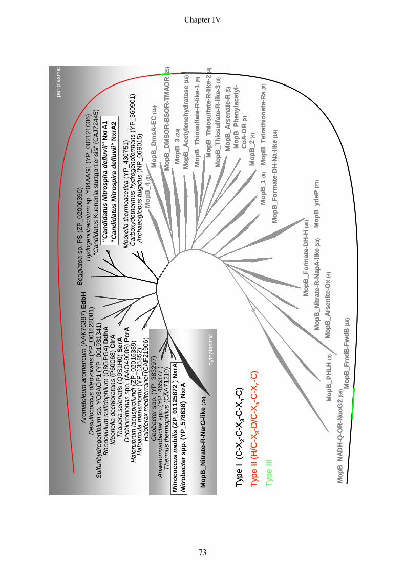
Mop
B_N
itrat
e-R
-Nar
G-li
ke(7
8)
Nitr
obac
ters
pp. (
YP_5
7863
8)N
xrA
Nitr
ococ
cus
mob
ilis
(ZP_
0112
5872
) Nxr
A
Ana
erom
yxob
acte
rspp
. (YP
_465
377
)Th
erm
usth
erm
ophi
lus
(CA
A712
10)
Geo
bact
ersp
p. (Y
P_38
3297
)H
alof
erax
med
iterr
anei
(CAF
2190
6)H
aloa
rcul
am
aris
mor
tui(
YP_1
3585
2)H
alor
ubru
mla
cusp
rofu
ndi(
ZP_0
2016
389)
Dec
hlor
omon
assp
p. (A
AO49
008)
Pcr
ATh
auer
ase
lena
tis(Q
9S1H
0) S
erA
Ideo
nella
dech
lora
tans
(P60
068)
ClrA
Rho
dovu
lum
sulfi
doph
ilum
(Q8G
PG4)
Ddh
AS
ulfu
rihyd
roge
nibi
umsp
. YO
3AO
P1 (Y
P_00
1931
341)
Des
ulfo
cocc
usol
eovo
rans
(YP_
0015
2808
1)
Begg
iato
asp
. PS
(ZP_
0200
0390
)H
ydog
enob
acul
umsp
. Y04
AAS1
(YP_
0021
2100
6)“C
andi
datu
sKu
enen
iast
uttg
artie
nsis
“(C
AJ72
445)
“Can
dida
tus
Nitr
ospi
rade
fluvi
i“N
xrA2
Moo
rella
ther
moa
cetic
a(Y
P_43
0751
)C
arbo
xydo
ther
mus
hydr
ogen
ofor
man
s(Y
P_36
0901
)A
rcha
eogl
obus
fulg
idus
(NP_
0690
15)
Mop
B_4
(8)
Mop
B_D
msA
-EC
(15)
Mop
B_D
MSO
R-B
SOR
-TM
AOR
(25)
Mop
B_3
(24)
Mop
B_A
cety
lene
hydr
atas
e(1
5)
Mop
B_T
hios
ulfa
te-R
-like
-1 (9
)
Mop
B_T
hios
ulfa
te-R
-like
-2 (4
)
Mop
B_T
hios
ulfa
te-R
-like
-3 (3
)
Mop
B_A
rsen
ate-
R(5
)
Mop
B_P
heny
lace
tyl-
CoA
-OR
(2)
Mop
B_2
(4)
Mop
B_T
etra
thio
nate
-Ra
(8)
Mop
B_1
(9)
Mop
B_F
orm
ate-
DH
-Na-
like
(14)
Mop
B_y
deP
(21)
Mop
B_F
orm
ate-
DH
-H(3
0)
Mop
B_N
itrat
e-R
-Nap
A-lik
e(3
3)
Mop
B_A
rsen
ite-O
x(4
)
Mop
B_F
mdB
-Fw
dB(1
8)
Mop
B_P
HLH
(4)
Mop
B_N
AD
H-Q
-OR
-Nuo
G2
(59)
“Can
dida
tus
Nitr
ospi
rade
fluvi
i“N
xrA1
Aro
mat
oleu
mar
omat
icum
(AAK
7638
7)Ed
bH
cyto
plas
mic
perip
lasm
ic
Type
I (C
-X2-
C-X
3-C
-Xn-
C)
Type
II (H
/C-X
3-D
/C-X
n-C
-Xn-
C)
Type
III
Mop
B_N
itrat
e-R
-Nar
G-li
ke(7
8)
Nitr
obac
ters
pp. (
YP_5
7863
8)N
xrA
Nitr
ococ
cus
mob
ilis
(ZP_
0112
5872
) Nxr
A
Ana
erom
yxob
acte
rspp
. (YP
_465
377
)Th
erm
usth
erm
ophi
lus
(CA
A712
10)
Geo
bact
ersp
p. (Y
P_38
3297
)H
alof
erax
med
iterr
anei
(CAF
2190
6)H
aloa
rcul
am
aris
mor
tui(
YP_1
3585
2)H
alor
ubru
mla
cusp
rofu
ndi(
ZP_0
2016
389)
Dec
hlor
omon
assp
p. (A
AO49
008)
Pcr
ATh
auer
ase
lena
tis(Q
9S1H
0) S
erA
Ideo
nella
dech
lora
tans
(P60
068)
ClrA
Rho
dovu
lum
sulfi
doph
ilum
(Q8G
PG4)
Ddh
AS
ulfu
rihyd
roge
nibi
umsp
. YO
3AO
P1 (Y
P_00
1931
341)
Des
ulfo
cocc
usol
eovo
rans
(YP_
0015
2808
1)
Begg
iato
asp
. PS
(ZP_
0200
0390
)H
ydog
enob
acul
umsp
. Y04
AAS1
(YP_
0021
2100
6)“C
andi
datu
sKu
enen
iast
uttg
artie
nsis
“(C
AJ72
445)
“Can
dida
tus
Nitr
ospi
rade
fluvi
i“N
xrA2
Moo
rella
ther
moa
cetic
a(Y
P_43
0751
)C
arbo
xydo
ther
mus
hydr
ogen
ofor
man
s(Y
P_36
0901
)A
rcha
eogl
obus
fulg
idus
(NP_
0690
15)
Mop
B_4
(8)
Mop
B_D
msA
-EC
(15)
Mop
B_D
MSO
R-B
SOR
-TM
AOR
(25)
Mop
B_3
(24)
Mop
B_A
cety
lene
hydr
atas
e(1
5)
Mop
B_T
hios
ulfa
te-R
-like
-1 (9
)
Mop
B_T
hios
ulfa
te-R
-like
-2 (4
)
Mop
B_T
hios
ulfa
te-R
-like
-3 (3
)
Mop
B_A
rsen
ate-
R(5
)
Mop
B_P
heny
lace
tyl-
CoA
-OR
(2)
Mop
B_2
(4)
Mop
B_T
etra
thio
nate
-Ra
(8)
Mop
B_1
(9)
Mop
B_F
orm
ate-
DH
-Na-
like
(14)
Mop
B_y
deP
(21)
Mop
B_F
orm
ate-
DH
-H(3
0)
Mop
B_N
itrat
e-R
-Nap
A-lik
e(3
3)
Mop
B_A
rsen
ite-O
x(4
)
Mop
B_F
mdB
-Fw
dB(1
8)
Mop
B_P
HLH
(4)
Mop
B_N
AD
H-Q
-OR
-Nuo
G2
(59)
“Can
dida
tus
Nitr
ospi
rade
fluvi
i“N
xrA1
Aro
mat
oleu
mar
omat
icum
(AAK
7638
7)Ed
bH
cyto
plas
mic
perip
lasm
ic
Type
I (C
-X2-
C-X
3-C
-Xn-
C)
Type
II (H
/C-X
3-D
/C-X
n-C
-Xn-
C)
Type
III
Chapter IV
73

iii) In contrast to all other molybdenum-binding oxidoreductases found on the genome of N.
defluvii the predicted products of the candidate genes, the large subunits NxrA1 and NxrA2,
are most similar in length and amino acid sequence to Nxr large subunits of Nitrobacter
winogradskyi Nb-255 (Starkenburg et al., 2006), N. hamburgensis X14 (Starkenburg et al.,
2008) and Nitrococcus mobilis Nb-231 (Table S1).
iv) Immunocytochemical studies on the localization of Nxr in the cell revealed one major
difference in the nitrite-oxidizing systems of Nitrobacter spp. and Nitrospira spp.. Whereas
the Nxr of Nitrobacter is localized at the cytoplasmic face of the cell membrane and at the
intracytoplasmic membranes (Spieck et al., 1996a), the Nxr of Nitrospira is found to be
membrane-associated in the periplasmic space (Spieck et al., 1998). This latter observation is
in agreement with the in silico predicted twin-arginine signal peptide cleavage sites (TatP 1.0
server; Bendtsen et al., 2005) in both large subunits of Nxr (NxrA1, NxrA2) of N. defluvii.
The twin-arginine signal peptide strongly suggests an export of the Nxr complex into the
periplasm via the Tat translocase system, which transports folded proteins across the
cytoplasmic membrane (Lee et al., 2006).
v). An additional characteristic mark distinguishing Nxr of Nitrobacter and Nitrospira has
been observed in immunoblotting experiments with monoclonal antibodies raised against the
alpha and beta subunit of Nxr of N. hamburgensis, respectively (Aamand et al., 1996, Spieck
et al., 1998, Bartosch et al., 1999). While the anti-alpha subunit antibody Hyb 153.2 gave a
positive signal only with the NxrA of Nitrobacter spp. (Aamand et al., 1996, Bartosch et al.,
1999), the NxrB of Nitrobacter and Nitrospira seem to share a common binding motif for the
anti-beta subunit antibody Hyb 153.3 (Bartosch et al., 1999). The NxrB of both nitrite-
oxidizing organisms could be detected by Hyb 153.3 (Spieck et al., 1998, Bartosch et al.,
1999), although the apparent molecular masses of the recognized proteins differed in the
respective organism (Nitrobacter: 65 kDa, Nitrospira: 50 kDa). The latter result is in
agreement with the in silico predicted molar masses of the NxrB amino acid sequences using
the ProtParam tool (Nitrobacter: 58.04 kDa, N. defluvii: 49.98 kDa) (Gasteiger et al., 2005).
In summary, the aforementioned similarities and differences between the Nxr of Nitrobacter
and Nitrospira enabled us to pinpoint the most parsimonious candidate genes for this unusual
Nxr on the genome of N. defluvii.
Nitrite oxidoreductase genes of N. defluvii: the genomic environment
Two gene copies encoding the alpha and beta subunits of Nxr, respectively, are located in
immediate vicinity on the genome of N. defluvii. Both nxrAB clusters appeared to be
Chapter IV
74

organized in an operon (Fig. 2). Also other already sequenced genomes of nitrite-oxidizing
organisms carry multiple nitrite oxidoreductase gene copies (Table S2). However, in contrast
to N. defluvii, only one set of nxrA and nxrB of Nitrobacter or Nitrococcus is organised in an
operon. All other copies are segregated and scattered in the respective genomes. Nevertheless,
the central Nxr gene cassette is conserved in all three Nitrobacter genomes and encompasses
in contrast to the Nxr operon of N. defluvii not only genes for NxrA and NxrB but also genes
for several other putative accessory proteins (Starkenburg et al., 2006, Starkenburg et al.,
2008). A unique feature of the nxr operons of N. defluvii are the transcriptional regulators
(nifA, NIDE3254) located upstream of both operons (Fig. 2). The regulators possess a helix-
turn-helix DNA-binding domain in the C-terminal section and a Sigma-54 interaction domain
and play presumably an important role in the transcriptional activation of the respective
downstream encoded Nxr genes by a conserved mechanism common to members of the
enhancer binding protein family (Buck et al., 2000). Beside these two similar domains in the
C-terminal section, however, both regulators share no similarity at all in the N-terminal part
of the protein. The gene product of NIDE3254 has a common CheY-like response regulator
receiver region at the N-terminus interacting with a yet unknown histidine kinase in a two-
component signal transduction system (Stock et al., 2000). The amino terminus of the nifA-
like gene product, however, comprises a GAF domain, a motif found in signalling and sensory
proteins from all domains of life (Aravind & Ponting, 1997, Ho et al., 2000).
Figure 2: Metagenomic stretch containing two gene copies encoding the nitrite oxidoreductase alpha (nxrA1,
nxrA2) and beta (nxrB1, nxrB2) subunits of N. defluvii. The oxidoreductase genes are displayed in red, genes for
transcriptional regulators and transporters/porins are highlighted in yellow and blue, respectively. The scale bar
indicates 5 kilobase pairs genomic length. For further information on the displayed annotated open reading
frames please refer to table S3.
nxrB2
nxrA2
nifA
nxrA1
nxrB1
yeaO
NIDE32
26
NIDE32
30
NIDE32
31
NIDE32
27
czcA
sugE
chaA
Oxidoreductases
Transcriptional regulators
wrbA relA
Transporter/porins
5 kb
NIDE32
28
NIDE32
29
NIDE32
34
NIDE32
35
NIDE32
39
NIDE32
40
NIDE32
41
NIDE32
42
NIDE32
43
NIDE32
44
NIDE32
45
NIDE32
46
NIDE32
47
NIDE32
48
NIDE32
49
NIDE32
50
NIDE32
54
NIDE32
57
NIDE32
60
NIDE32
61
NIDE32
62
Chapter IV
75
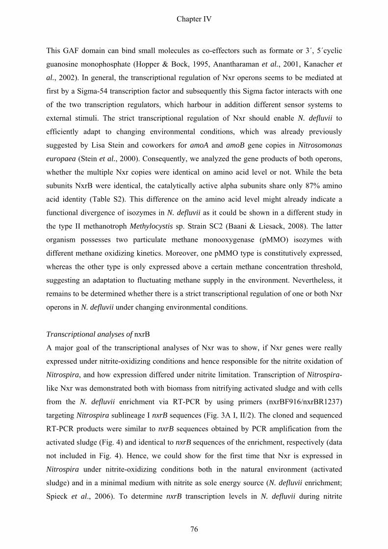
This GAF domain can bind small molecules as co-effectors such as formate or 3´, 5´cyclic
guanosine monophosphate (Hopper & Bock, 1995, Anantharaman et al., 2001, Kanacher et
al., 2002). In general, the transcriptional regulation of Nxr operons seems to be mediated at
first by a Sigma-54 transcription factor and subsequently this Sigma factor interacts with one
of the two transcription regulators, which harbour in addition different sensor systems to
external stimuli. The strict transcriptional regulation of Nxr should enable N. defluvii to
efficiently adapt to changing environmental conditions, which was already previously
suggested by Lisa Stein and coworkers for amoA and amoB gene copies in Nitrosomonas
europaea (Stein et al., 2000). Consequently, we analyzed the gene products of both operons,
whether the multiple Nxr copies were identical on amino acid level or not. While the beta
subunits NxrB were identical, the catalytically active alpha subunits share only 87% amino
acid identity (Table S2). This difference on the amino acid level might already indicate a
functional divergence of isozymes in N. defluvii as it could be shown in a different study in
the type II methanotroph Methylocystis sp. Strain SC2 (Baani & Liesack, 2008). The latter
organism possesses two particulate methane monooxygenase (pMMO) isozymes with
different methane oxidizing kinetics. Moreover, one pMMO type is constitutively expressed,
whereas the other type is only expressed above a certain methane concentration threshold,
suggesting an adaptation to fluctuating methane supply in the environment. Nevertheless, it
remains to be determined whether there is a strict transcriptional regulation of one or both Nxr
operons in N. defluvii under changing environmental conditions.
Transcriptional analyses of nxrB
A major goal of the transcriptional analyses of Nxr was to show, if Nxr genes were really
expressed under nitrite-oxidizing conditions and hence responsible for the nitrite oxidation of
Nitrospira, and how expression differed under nitrite limitation. Transcription of Nitrospira-
like Nxr was demonstrated both with biomass from nitrifying activated sludge and with cells
from the N. defluvii enrichment via RT-PCR by using primers (nxrBF916/nxrBR1237)
targeting Nitrospira sublineage I nxrB sequences (Fig. 3A I, II/2). The cloned and sequenced
RT-PCR products were similar to nxrB sequences obtained by PCR amplification from the
activated sludge (Fig. 4) and identical to nxrB sequences of the enrichment, respectively (data
not included in Fig. 4). Hence, we could show for the first time that Nxr is expressed in
Nitrospira under nitrite-oxidizing conditions both in the natural environment (activated
sludge) and in a minimal medium with nitrite as sole energy source (N. defluvii enrichment;
Spieck et al., 2006). To determine nxrB transcription levels in N. defluvii during nitrite
Chapter IV
76
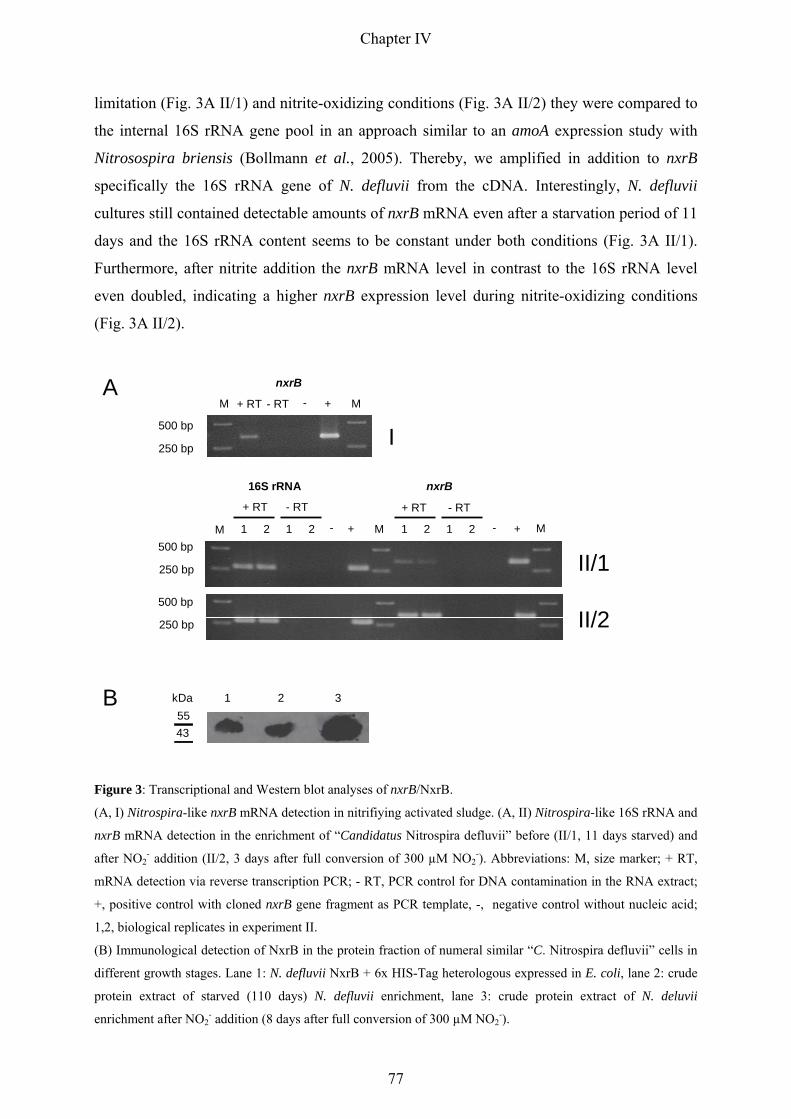
limitation (Fig. 3A II/1) and nitrite-oxidizing conditions (Fig. 3A II/2) they were compared to
the internal 16S rRNA gene pool in an approach similar to an amoA expression study with
Nitrosospira briensis (Bollmann et al., 2005). Thereby, we amplified in addition to nxrB
specifically the 16S rRNA gene of N. defluvii from the cDNA. Interestingly, N. defluvii
cultures still contained detectable amounts of nxrB mRNA even after a starvation period of 11
days and the 16S rRNA content seems to be constant under both conditions (Fig. 3A II/1).
Furthermore, after nitrite addition the nxrB mRNA level in contrast to the 16S rRNA level
even doubled, indicating a higher nxrB expression level during nitrite-oxidizing conditions
(Fig. 3A II/2).
5543
kDa 1 2 3B
A
I250 bp
500 bp
II/1
II/2250 bp
500 bp
250 bp
500 bpM - + M M- +
16S rRNA nxrB
1 12 2 1 12 2
+ RT + RT - RT- RT
nxrB
M M+ RT - RT - +
Figure 3: Transcriptional and Western blot analyses of nxrB/NxrB.
(A, I) Nitrospira-like nxrB mRNA detection in nitrifiying activated sludge. (A, II) Nitrospira-like 16S rRNA and
nxrB mRNA detection in the enrichment of “Candidatus Nitrospira defluvii” before (II/1, 11 days starved) and
after NO2- addition (II/2, 3 days after full conversion of 300 µM NO2
-). Abbreviations: M, size marker; + RT,
mRNA detection via reverse transcription PCR; - RT, PCR control for DNA contamination in the RNA extract;
+, positive control with cloned nxrB gene fragment as PCR template, -, negative control without nucleic acid;
1,2, biological replicates in experiment II.
(B) Immunological detection of NxrB in the protein fraction of numeral similar “C. Nitrospira defluvii” cells in
different growth stages. Lane 1: N. defluvii NxrB + 6x HIS-Tag heterologous expressed in E. coli, lane 2: crude
protein extract of starved (110 days) N. defluvii enrichment, lane 3: crude protein extract of N. deluvii
enrichment after NO2- addition (8 days after full conversion of 300 µM NO2
-).
Chapter IV
77

It remains to be determined, whether the nxrB presence during starvation is due to increased
mRNA stability or constant mRNA supply on a low expression level. In heterotrophic
bacteria it has been shown, that gene expression can be regulated at the level of mRNA
stability (Mackie, 1998). The physiological state of the cell as well as the transcript secondary
structure can be important determinants of variations in mRNA decay (Meyer & Schottel,
1991, Thorne & Williams, 1997, Coburn & Mackie, 1999). Nevertheless, the half-life of
mRNA in heterotrophic bacteria is on average only 3 min (Takayama & Kjelleberg, 2000,
Bernstein et al., 2002), indicating that rather constitutive and regulated mRNA expression
than mRNA stability influences the mRNA level during starvation.
Also other autotrophic nitrifiers keep a certain cell-internal RNA level during starvation, but
in contrast to heterotrophs only some selected RNAs appear to be associated with the
adaptation to starvation. Several studies with different ammonia-oxdizing bacteria revealed
that relatively high levels of amoCAB mRNA, comprising the ammonia monooxygenase
enzyme, and high levels of 16S rRNA were maintained during complete deprivation of
ammonia (Sayavedra-Soto et al., 1998, Morgenroth et al., 2000, Wei et al., 2004, Bollmann et
al., 2005, Wei et al., 2006, El Sheikh & Klotz, 2008). These previous results with AOB are in
agreement with the nxrB mRNA and 16S rRNA maintenance in Nitrospira observed in this
study. Hence, it might be a common strategy in nitrifiers to sustain starvation periods by
keeping the cell-internal content of certain RNAs high to quickly respond when substrate
again becomes available.
In summary, beside this quite high mRNA level even after 11 days of starvation the
comparison of the both growth phases via RT-PCR indicated a higher nxrB expression level
under nitrite-oxidizing conditions (Fig. 3A II). Even the obvious limitations of this semi-
quantitative approach (RNA extraction and cDNA synthesis efficiencies, different primer
efficiencies, etc.) considered, these differences in the expression level were significant and
reproducibly. In addition to the five criteria outlined in the first chapter, these results
corroborate the hypothesis that the in silico depicted Nxr candidate genes comprise the nitrite-
oxidizing system of N. defluvii.
Western blot analyses of NxrB
Already in the year 1996 Aamand and colleagues presented in their study three monoclonal
antibodies (Hyb 153.1, 153.2, 153.3) raised against the Nxr of Nitrobacter hamburgensis
(Aamand et al., 1996). In follow-up studies one (Hyb 153.3) out of these three antibodies
turned out to bind also specifically to a protein of Nitrospira-like bacteria (Spieck et al., 1998,
Chapter IV
78

Bartosch et al., 1999). Even though the apparent molecular mass of the detected protein in
Nitrospira was different to the mass of the recognised Nxr beta subunit of Nitrobacter, it was
obvious that both proteins share a common binding motif for the monoclonal antibody Hyb
153.3. Thus, it was assumed that this protein represents the Nxr beta subunit of Nitrospira.
More than one decade after the first description of this antibody we could clearly link in our
study the antibody specificity with sequence data of the recognised protein in the genome of
N. defluvii. Western blot analyses with the heterologous expressed nxrB gene revealed a
specific binding of the antibody Hyb 153.3 with the recombinant protein (Fig. 3B).
With the reevaluated antibody Hyb 153.3 we had a closer look into the cell-internal NxrB
content of starved and nitrite-oxidizing N. defluvii cells (Fig 3B). For this approach we used
cell extracts of numerical similar N. defluvii cells of both growth phases. Interestingly, the
Western blot results revealed that even after a starvation period of 110 days a high cell-
internal NxrB content remained in N. defluvii. Furthermore, after reactivation of the starved
cells with nitrite the per-cell NxrB content was even doubled. N. defluvii seems to have a
certain core set of proteins, including Nxr, which enables the organism to restart its nitrite-
oxidizing system relatively fast after starvation. Part of the thereby gained energy seems to be
immediately used for the assembly of new Nxr.
This survival strategy is similar to the starvation adaptation employed by ammonium-
oxidizing bacteria (AOB). During starvation of AOB the cell-internal AmoA content
remained stable (Stein & Arp, 1998) and AmoB could even be detected after one year of
ammonia starvation (Pinck et al., 2001). Moreover, the potential activity of the Amo enzyme
was not affected over prolonged starvation periods (Wilhelm et al., 1998). In general, it can
be concluded that nitrifiers posses several physiological traits such as low maintenance energy
demand (Tappe et al., 1999), quorum sensing (Batchelor et al., 1997, Laanbroek et al., 2002)
or as illustrated above stable amount of both, enzymes and specific RNAs, to withstand
unfavourable environmental conditions and moreover to regain their metabolic activity within
a short timeframe (Geets et al., 2006). Thus, in natural and engineered environments with
fluctuating nutrient supply, nitrifiers such as N. defluvii perfectly adapted their “cellular
machineries” to these changing conditions.
In the following three sections the gene products comprising the Nxr alpha, beta and gamma
subunit will be described in detail, to gain first insights into the biochemical features of this
unusual enzyme.
Chapter IV
79
The alpha subunit NxrA
The large subunit of Nxr (NxrA1/NxrA2) of N. defluvii has two characteristic domains, an N-
terminal iron-sulphur cluster [Fe-S]-binding motif and molybdopterin cofactor-binding sites.
The latter domain defines NxrA according to the NCBI Conserved domain database as
member of the molybdopterin-binding superfamily of proteins (cl09928) (Marchler-Bauer et
al., 2007), which contains all known proteins formely assigned to the DMSO reductase family
(Hille, 1996). Here the term DMSO reductase family will be used in order to avoid confusions
with previous studies about proteins of this family (McDevitt et al., 2002, Bender et al.,
2005). The DMSO reductase family is a large and heterogeneous enzyme family and shares a
common form of molybdenum cofactor, the bis(molybdopterin guanine dinucleotide)Mo
(Mo-bisMGD) (Kisker et al., 1997). Phylogenetic analyses with selected alpha subunits of
DMSO reductase family enzymes revealed a unique and unexpected position of N. defluvii
NxrA in the MopB superfamily tree (Fig. 1). The NxrA sequences of Nitrospira cluster
together with membrane-bound nitrate reductase (NarG)-like sequences of `Candidatus
Kuenenia stuttgartiensis´, Hydrogenobaculum sp. Y04AAS1 and Beggiatoa sp. PS
comprising a monophyletic clade in the tree. Most apparent is the separate position of this
latter described branch from the Nxr alpha subunits of Nitrobacter spp.. The results indicate a
convergent evolution of similar enzymatic functions in two different nitrite-oxidizing systems.
The Nitrobacter NxrAs cluster together with the NxrA of Nitrococcus mobilis and are closely
related to the large subunit of the membrane-bound nitrate reductases (NarG) (Kirstein K.,
1993). Within a few exceptions the overall tree topology is in most cases consistent with the
functional affiliation of the respective sequences. Beside this functional clustering (Table S4)
an additional classification based on structural variations can be applied on members of the
DMSO reductase family. Three distinct DMSO reductase family types with differences in the
[Fe-S]-binding motif in the alpha subunit can be distinguished (Fig. 1) (Trieber et al., 1996).
The majority of hitherto known DMSO reductase family enzymes, such as the perplasmic
nitrate reductase (Nap), the formate dehydrogenase (Fdh) or the arsenite reductase (Aso) are
members of type I enzymes defined by an cystein-rich [Fe-S]-binding motif (C-X2-C-X3-C-
Xn-C) (McDevitt et al., 2002). In almost all type I enzymes the motif is located at the N-
terminal part of the alpha subunit except for the large subunit (NuoG) of NADH-ubiquinone
oxidoreductases (cd02768). In this latter enzyme family an internal domain of NuoG contains
the afromentioned cystein-rich motif and shares homology to Fdh sequences supporting a
common ancestor for both enzyme families (Friedrich, 1998). Similar to type I enzymes type
II enzymes have also N-terminal cystein residues for [Fe-S]-binding in the alpha subunit, but
Chapter IV
80

in contrast to type I enzymes three amino acid residues instead of two separate the first and
the second cystein residue (Trieber et al., 1996). Furthermore, the first cystein residue can be
replaced to a histidine ligand in some type II enzymes, such as the membrane-bound nitrate
reductase (Nar) of E. coli (Bertero et al., 2003). Furthermore, Jormarkka and co-workers
proposed based on a NarG crystal structure of E. coli that most type II enzymes seem to share
a conserved aspartate residue in the active centre acting as molybdenum ion ligand (Jormakka
et al., 2004). In contrast to the first two DMSO reductase family types, type III enzymes lack
a cystein-rich motif. Two MopB families belong to the type III, the MopB_PHLH (cd02764)
family with uncharacterized hydrogenase-like enzymes and the MopB_DMSOR_
BSOR_TMAOR (cd02769) family including the biotine sulfoxide reductase of Rhodobacter
sphaeroides (Pollock & Barber, 1997) and the trimethylamine N-oxide reductase of E. coli
(Weiner et al., 1992).
Based on these aforementioned classifications we propose that the Nxr of N. defluvii is a
member of type II enzymes of the DMSA reductase family considering two observations: (i)
NxrA shares the molybdenum ion-binding Asp residue with most alpha subunits of type II
enzyme family members (Fig S1B); (ii) The first two cysteine residues of the [Fe-S]-binding
motif of NxrA of N. defluvii are separated by three amino acids, which is characteristic for
type II enzymes (Fig. S1A). One major difference in NxrA of Nitrospira and related
sequences to all other type II enzymes studied so far is the replacement of the second cystein
residue in the binding motif with an aspartate (Fig. S1A). Since aspartate has been previously
shown in a ferredoxin of the hyperthermophilic Pyrocccus furiosus to be a proximal ligand of
the [Fe-S] (Calzolai et al., 1995), we suggest to expand the type II signature motif proposed
by Jormakka and colleagues (Jormakka et al., 2004) to the following coordination pattern:
H/C-X3-D/C-Xn-C-Xn-C. Beside these common characteristics, type II DMSO reductase
family enzymes can be distinguished between cytoplasmic localized proteins and enzymes
with a twin-arginine signal peptide transported via Tat translocase system into the periplasm
(Fig. 1) (Martinez-Espinosa et al., 2007).
In summary, the Nxr of N. defluvii is a periplasmic NarG-like type II DMSO reductase family
enzyme, closely related to the NarG-like enzyme of “C. Kuenenia stuttgartiensis” and with
only weak similarity to previously described cytoplasmic Nxr of Nitrobacter spp..
The beta subunit NxrB
NxrB (NxrB1/NxrB2) of N. defluvii is a member of the bacterial oxidoreductase electron
transfer subunits (Berks et al., 1995). Although there is no indication in the NxrB sequences
Chapter IV
81

of N. defluvii for a signal peptide, it is very likely that NxrA and NxrB are translocated
together as folded protein complex via the Tat translocase system by using a “hitchhiker”
mechanism proposed by Rodrigue et al. (1999). Comparison of NxrA and NxrB phylogeny
indicated a shared evolutionary history for the alpha and beta subunit, suggesting a tight
functional alliance of both subunits (Fig. S2). Crystal structures of beta subunits closely
related to the NxrB of N. defluvii, such as the nitrate reductase beta subunit NarH of E. coli
(Bertero et al., 2003) or the beta subunit of the ethylbenzene dehydrogenase of Aromatoleum
aromaticum (Kloer et al., 2006) confirmed the presence of three [4Fe-4S] cluster (I, III, IV)
and one [3Fe-4S] cluster (II) (Fig. S3B). These [Fe-S] play an important role in the
intramolecular electron transfer (Blasco et al., 2001). Comparative sequence analysis of NxrB
and selected beta subunits of type II DMSO reductase enzymes revealed the presence of four
cysteine-rich motifs for [FeS]-binding (Fig. S3A). These four [FeS] have been characterized
in detail in the nitrate reductase beta subunit (NarH) of E. coli. EPR spectroscopy and site-
directed mutagenesis (Guigliarelli et al., 1996, Blasco et al., 2001) as well as structural
analysis (Bertero et al., 2003, Jormakka et al., 2004) of NarH revealed that the [Fe-S] are
organized in two pairs, each with one low- and one high-potential cluster (IV/I and III/II)
(Fig. S3B). A unique feature of the NxrB of N. defluvii compared to all other known beta
subunit sequences is the replacement of one cysteine to an aspartate residue binding
presumably the low-potential [4Fe-4S] cluster IV (Fig. S3). Furthermore, two out of three
cysteine residues, which additionally bind [4Fe-4S] cluster IV, are separated by an insertion
of eight amino acids specific for the beta subunit sequences of N. defluvii and “C. Kuenenia
stuttgartiensis” (Fig. S3). It remains to be determined whether these differences to other
characterized beta subunits result in a unique mode of electron transfer.
Gamma subunit
The mature holoenzymes of most type II DMSO reductase family enzymes studied so far
contain beside the catalytic active alpha subunit and the electron transferring beta subunit an
additional gamma subunit. These gamma subunits can either have membrane-spanning helices
as it is known from the nitrate reductase gamma subunit NarI from E. coli anchoring the alpha
and beta subunit to the cytoplasma membrane (Martinez-Espinosa et al., 2007) or the gamma
subunits can lack trans-membrane helices as seen in the soluble periplasmic ethylbenzene
dehydrogenase EBDH of A. aromaticum (Kloer et al., 2006). Despite these differences in
their localisation, structural and spectroscopic analysis demonstrated that all gamma subunits
of type II DMSO reductase family enzymes seem to contain at least one heme b cofactor.
Chapter IV
82
Whereas NarI of E. coli contains two heme b, only a single heme b is found in the periplasmic
EBDH (Kloer et al., 2006), in the selenate reductase SerC of Thauera selenatis (Schroder et
al., 1997) and in the chlorate reductase ClrC of Ideonella dechloratans (Thorell et al., 2003).
Three gene products (NxrC1/C2/C3) encoded on the genome of N. defluvii displayed highest
similarity to the aforementioned gamma subunits and to heme b binding subunits of “C.
Kuenenia stuttgartiensis” (CAJ72454, CAJ72449), Hydrogenobaculum sp. Y04AAS1
(YP_002121004) and Pyrobaculum calidifontis (YP_001056787) (Fig.S4).
In contrast to other DMSO reductase family enzymes such as the perchlorate reductase of
Dechloromonas agitata (Bender et al., 2005) or the Nxr of Nitrobacter spp. (Starkenburg et
al., 2006), where the subunit encoding genes are organized in a operon structure, the three
nxrC gene copies are separated from nxrA and nxrB and scattered in the genome of N.
defluvii. Although all three NxrC copies are quite dissimilar to each other on the amino acid
level (24-39%), together they share with other type II gamma subunits two conserved residues
(Fig. S4), which were previously identified in the crystallized EBDH gamma subunit as
proximal heme b ligands (Kloer et al., 2006). Thus, it is likely that NxrC contains at least one
heme b cofactor and is part of an electron transport system involving the iron-sulphur
complexes of NxrA and NxrB and the heme b in NxrC. In addition, the in silico predicted
signal peptides in all three NxrC copies suggest a periplasmic localisation of the Nxr gamma
subunit (Fig. S4).
One unique feature of NxrC3 compared to the two other NxrC copies of N. defluvii is the C-
terminal transmembrane spanning domain (Fig. S4). Assuming that Nitrospira moscoviensis
and N. defluvii have similar nitrite-oxidizing systems especially this latter finding is in
aggreement with former studies with the Nxr of N. moscoviensis. Spieck and colleagues
(1998) isolated the nitrite-oxidizing system of N. moscoviensis by density gradient
centrifugation and separated the proteins by SDS-PAGE. The Nxr complex seems to consist
of at least four major proteins with apparent molecular masses of approximately 130, 62, 46
and 29 kDa. The 130 and 46 kDa protein band might represent the alpha and beta subunit of
Nxr, which have in N. defluvii the calculated molar masses (ProtParam) of 129 kDa and 50
kDa, respectively. Until know the 62 kDa protein band could not be asigned to any gene
product of N. defluvii, but the apparent molecular mass of 29 kDa of the last prominent
protein band of the Nxr of N. moscoviensis is similar to calculated molar masses of the three
NxrC copies of N. defluvii (NxrC1: 27 kDa, NxrC2: 30 kdA, NxrC3: 26 kDa). Furthermore,
the Nxr of N. moscoviensis exhibited its highest enzymatic activity in the membrane fraction,
suggesting that the enzyme is membrane bound (Spieck et al., 1998). Therefore, there has to
Chapter IV
83

be an additional membrane anchoring subunit in Nxr of Nitrospira. Thus, NxrC3 in N.
defluvii with the C-terminal transmembrane helix might take over this part in the nitrite-
oxidizing system of N. defluvii (Fig S4).
Design and evaluation of nxrB primer sets targeting the nitrite oxidoreductase of Nitrospira-
like organisms
One major goal of this study beside the characterization of the candidate genes encoding the
Nxr of N. defluvii was the investigation whether one of these genes could serve as functional
and phylogenetic marker for the genus Nitrospira. The most conserved of the Nxr genes,
nxrB, was selected for primer design and evaluation (Table S2). Based on the alignment of the
two nearly identical nxrB copies (99.9 % identity on DNA level) of N. defluvii (nxrB1, nxrB2)
and several shotgun reads of N. moscoviensis, with high sequence identity to the nxrB of N.
defluvii, a first nxrB primer pair (nxrBF14/nxrBR1239, Table 1) was designed. The sequence
CAJ72448 of “C. Kuenenia stuttgartiensis”, the closest related sequence to the Nitrospira
nxrB in the public database, served as control to test in silico for unspecific primer binding.
Table 1: Specifications of different primer pairs developed in this study targeting Nitrospira-like nxrB.
1 For detailed specificity please refer to SOMS Fig. S5 2 R = A + G 3 Positions relative to nxrB (NIDE3256) gene of N. defluvii 4 See Experimental procedures for detailed description of PCR protocols
Name Specificity 1 Sequence 5´-3´ 2 Target-site 3 Fragment
size (bp)
PCR
protocol 4
nxrBF14 ATA ACT GGC AAC TGG GAC
GG 14 - 33
nxrBR1239
Sublineage I and
II of the genus
Nitrospira TGT AGA TCG GCT CTT CGA
CC 1239 - 1258
1245 1
nxrBF19 TGG CAA CTG GGA CGG AAG
ATG 19 - 39
nxrBR1237
Beryl spring
enrichment,
N. bockiana,
N. marina
GTA GAT CGG CTC TTC GAC
CTG 1237 - 1257
1239 2
nxrBF169 TAC ATG TGG TGG AAC A 169 - 184
nxrBR638
All known
Nitrospira-like
nxrB
CGG TTC TGG TCR ATC A 638 - 653
485 3
Chapter IV
84
The first primer pair successfully amplified with the evaluated PCR conditions nxrB-like
sequences (1245 bp in length) of N. defluvii, N. moscoviensis and two environmental samples
(activated sludge, beech forest soil) but no amplification product was retrieved from other
Nitrospira species (Fig. S5). For a broader coverage we designed a second primer pair
(nxrBF19/nxrB1237, Table1) based on this extended dataset. Indeed, additional nxrB-like
sequences (1239 bp in length) of N. marina, “C. N. bockiana” and from a nitrifying hot spring
enrichment (Beryl spring) could be obtained (Fig. S5). Finally, the primer combination
nxrBF169/nxrB638 (Table 1), designed on internal conserved regions of thitherto retrieved
Nitrospira-like nxrB sequences, gave nxrB amplificates (485 bp in length) with all the before
tested Nitrospira-like organisms and environmental samples. Furthermore, this latter primer
pair amplified in contrast to all other primer combinations nxrB-like sequences of a marine
sponge sample (Fig. S5). Due to its broad coverage we suggest to use the primer pair
nxrBF169/nxrB638 (Table1) in environmental surveys to get a first insight into the diversity
and abundance of nxrB genes in complex microbial communities. For in-depth phylogenetic
analysis, where a higher informative content is needed additional nxrB amplification with the
two other primer pairs (nxrBF14/nxrBR1239, nxrBF19/nxrB1237, Table1) is recommended.
Phylogenetic analysis of Nitrospira-like nxrB
A prerequisite for a suitable functional and phylogenetic maker gene is a congruent
evolutionary history with the 16S rRNA gene of the respective organism. Direct comparison
between 16S rRNA gene and marker gene trees indicates whether the marker gene has
undergone a lateral gene transfer (LGT) among the studied organism or not. Some studies
showed similar evolutionary relationships between 16S rRNA genes and marker genes, such
as the alpha subunit of the ammonia monooxygenase (amoA) of proteobacterial ammonia
oxidizers (Purkhold et al., 2000) or the Nxr genes of Nitrobacter spp. (Vanparys et al., 2007,
Poly et al., 2008). The dissimilatory sulfite reductase genes (dsrAB) of sulphate reducers
(Klein et al., 2001) or the nitrogenase reductase gene nifH of diazotrophs (Zehr et al., 2003)
on the contrary have been subjected to rare LGT, therefore aggravating the inference of
phylogenetic relationships between these organisms based on their marker genes. Finally,
LGT and gene duplication/divergence events in denitrification genes (nar, nir, nor, nos)
actually resulted in a disagreement between marker gene phylogeny and 16S rRNA-based
organism classification (Gregory et al., 2003, Dandie et al., 2007, Heylen et al., 2007, Jones
et al., 2008). The two explicit assumptions in all these studies and also in our analysis are that
Chapter IV
85
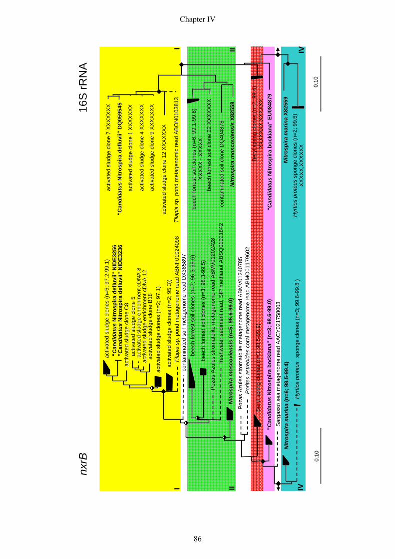
activ
ated
slud
gecl
ones
(n=5
; 97.
2-99
.1)
"Can
dida
tus
Nitr
ospi
rade
fluvi
i" N
IDE3
256
"Can
dida
tus
Nitr
ospi
rade
fluvi
i" N
IDE3
236
activ
ated
slud
gecl
one
C8
activ
ated
slud
gecl
one
5ac
tivat
edsl
udge
enric
hmen
tcD
NA
8ac
tivat
edsl
udge
enric
hmen
tcD
NA
12ac
tivat
edsl
udge
clon
eB
18
Tila
pia
sp. p
ond
met
agen
ome
read
AB
NF0
1024
098
cont
amin
ated
soil
met
agen
ome
read
DX3
8589
7be
ech
forr
ests
oilc
lone
s(n
=7; 9
6.3-
99.6
)
Poz
asA
zule
sst
rom
atol
item
etag
enom
ere
adAB
MV
0120
2428
Nitr
ospi
ram
osco
vien
sis
(n=5
; 96.
6-99
.0)
Ber
yl s
prin
g cl
ones
(n=3
; 98.
5-99
.9)
"Can
dida
tus
Nitr
ospi
rabo
ckia
na"
(n=3
; 98.
6-99
.0)
Sar
gass
ose
am
etag
enom
ere
adA
ACY0
2175
8003
Nitr
ospi
ram
arin
a(n
=6; 9
8.5-
99.4
)
Hyr
tios
prot
eus
spon
gecl
ones
(n=3
; 99.
6-99
.8 )
0.10
activ
ated
slud
gecl
ones
(n=2
; 97.
1)
activ
ated
slud
gecl
ones
(n=2
; 95.
3))
beec
hfo
rres
tsoi
lclo
nes
(n=3
; 98.
3-99
.5)
fresh
wat
erse
dim
entr
ead,
SIP
met
hano
lAB
SQ
0102
1842
Poza
sAz
ules
stro
mat
olite
met
agen
ome
read
AB
MV
0124
0785
Por
ites
astre
oide
sco
ralm
etag
enom
ere
adA
BM
O01
1796
02
activ
ated
slud
gecl
one
7 XX
XX
XX
X
"Can
dida
tus
Nitr
ospi
rade
fluvi
i" D
Q05
9545
activ
ated
slud
gecl
one
1 XX
XXX
XX
activ
ated
slud
gecl
one
4 XX
XX
XX
X
activ
ated
slud
gecl
one
9 XX
XX
XX
X
activ
ated
slud
gecl
one
12 X
XX
XX
XX
Tila
pia
sp. p
ond
met
agen
omic
read
AB
ON
0103
813
beec
hfo
rres
tsoi
lclo
ne22
XX
XX
XX
X
cont
amin
ated
soil
clon
eD
Q40
4878
Nitr
ospi
ram
osco
vien
sis
X825
58
Ber
yl s
prin
g cl
ones
(n=2
; 99.
4)X
XX
XX
XX,X
XX
XX
X"C
andi
datu
sN
itros
pira
bock
iana
" EU
0848
79
Nitr
ospi
ram
arin
aX8
2559
Hyr
tios
prot
eus
spon
gecl
ones
(n=2
; 99.
6)X
XX
XX,
XX
XX
XX
0.10
beec
hfo
rres
tsoi
lclo
nes
(n=6
; 99.
1-99
.8)
XX
XX
X -X
XX
XX
nxrB
16S
rRN
A
I II IV
I II IV
Chapter IV
86

Figure 4: Comparison of nxrB and 16S rRNA gene phylogeny of nitrite-oxidizing Nitrospira-like bacteria. Basic
tree topology is based on maximum-likelihood (fastDNAml) analysis of the respective full length nucleic acid
sequences. A total of 1158 and 1197 informative positions were used in 16S rRNA gene and nxrB analysis,
respectively. Shorter sequences were added by using the ARB Parsimony tool implemented in the ARB software
package (Ludwig et al., 2004) without changing the overall tree topologies. Branches leading to shorter
sequences are indicated in dotted lines, highlighting their unsupported affiliation. Filled and open diamonds
symbolize >90% and 80-90% Parsimony bootstrap support (100 iterations) respectively. The scale bar indicates
0.1 estimated change per nucleotide. Sequences that belong to isolated or enriched Nitrospira-like bacteria are in
boldface. In case of sequence clusters the number of sequences in the cluster and the maximal and minimal
sequence similarity is indicated in brackets. Accession numbers for 16S rRNA and environmental sequences are
at the end of the sequence headers. The remaining nxrB accession numbers are listed in the tree in Fig. S5.
Consistent monophyletic groups in the nxrB and 16S rRNA tree are highlighted in the same color and three
groups were assigned to the Nitrospira sublineages I, II and IV according to Daims et al. (2001).
the 16S rRNA phylogeny reflects the organismal phylogeny and the 16S rRNA gene is not
influenced by LGT (Woese, 1987). Comparison of trees based on Nitrospira-like 16S rRNA
and nxrB sequences revealed a highly similar topology (Fig. 4). Within the few available
isolated or highly enriched Nitrospira-like bacteria no evidence for LGT of nxrB could be
ascertained. Nevertheless, future analysis of more Nitrospira enrichments or isolates is
necessary to exclude LGT-events of nxrB in the genus Nitrospira. Among all analyzed
Nitrospira only N. defluvii has two nearly identical nxrB copies. All other Nitrospira seem to
have more than two nxrB copies encoded on their genome with a minimal similarity of 96.6 %
in N. moscoviensis (Fig. 4). Excluding PCR-artefacts or sequencing errors the analyzed
Nitrospira enrichments and isolates might consist of various strains with differences between
their nxrB sequences and varying nxrB copy number. This could possibly result in false nxrB
copy number estimates for one organism (strain). Variation in gene copy number in different
strains of one species is a phenomenon already observed by comparative genomics of two
Shigella flexneri strains (Wei et al., 2003). Consequently, it could not be concluded from
sequence clustering or similarity thresholds whether the nxrB sequences belong to one or
more Nitrospira strains. Only other completed Nitrospira genomes would eventually make
the nxrB copy number of other Nitrospira than N. defluvii assessible. This would facilitate the
assignment of nxrB sequences to specific 16S rRNA sequences.
Beside the nxrB sequence analyses with the few isolated and enriched Nitrospira-like
bacteria, we additionally screened different environmental samples for Nitrospira-like
bacteria on the basis of the 16S rRNA gene and the nxrB gene, respectively. Of each analyzed
sample Nitrospira-like 16S rRNA sequences and nxrB sequences could be obtained.
Chapter IV
87

Phylogenetic analysis revealed a congruent positioning of these genes derived from the same
sample within their phylogenetic trees at least on the sublineage level (Fig. 4).
Despite the similar overall topology some minor inconsistencies can be observed in the
bifurcations within Nitrospira sublineage I and in branching nodes to sublineage II (Fig. 4). In
sublineage I the nxrB sequences showed a more pronounced organisation in sub-cluster as
expected by observing the 16S rRNA phylogeny. The reasons for this could be the
underestimated 16S rRNA diversity of Nitrospira-like bacteria in the analysed activated
sludge samples or the yet unknown nxrB copy number in other Nitrospira than N. defluvii. In
sublineage II the 16S rRNA sequences cluster in a monophyletic group, whereas sublineage II
nxrB sequences split in all applied treeing methods into two groups (N. moscoviensis and
beech forest clones). It remains to be seen if detailed analysis with more available sequences
influences the correlation of sublineage II topology of 16S rRNA gene and nxrB based trees.
Beside these differences all other branching nodes in the trees were congruent and supported
by high bootstrap values, once more highlighting the suitability of nxrB as functional and
phylogenetic marker for the genus Nitrospira allowing at least an assignment of nxrB
sequences to different Nitrospira sublineages (Fig. 4).
Metagenomic nxrB-like sequences
Database search of different publicly available metagenomic libraries revealed that a number
of reads from the Sargasso sea (Venter et al., 2004), the coral Porites astreoides (Wegley et
al., 2007), the sediment from Lake Washington (Kalyuzhnaya et al., 2008), the microbial
fraction of a freshwater pond and a freshwater stromatolite (Dinsdale et al., 2008), and a
metagenomic survey of contaminated sediments (Abulencia et al., 2006) (Fig. 4) showed
high sequence similarities to nxrB of Nitrospira. These nxrB-like sequences formed distinct
branches within the nxrB phylogenetic tree (Fig. 4). Although their short read length
hampered their phylogenetic positioning some reads clustered with Nitrospira sublineages
whereas other reads formed new branches, which lacked their 16S rRNA gene counterpart.
Two metagenomes contained in addition to nxrB reads Nitrospira-like 16S rRNA sequences.
Abulencia and coworkers (2006) found a sublineage II Nitrospira-like 16S rRNA sequence,
while comparing a conventional PCR-based SSU rRNA library with a metagenomic survey of
contaminated sediments. However, the nxrB sequence found in this metagenomic library
forms a destinct branch located between sublineage I and II (Fig. 4). The most parsimonious
explanations for the different positions of both sequences in the respective trees could be
either the short read length of the nxrB read, which hampers a coherent phylogenetic analysis,
Chapter IV
88

or that the sequences originate from different organism. Nitrospira-like nxrB and 16S rRNA
metagenomic reads of a freshwater pond (Dinsdale et al., 2008), however, cluster both in
sublineage I with exact the same positioning in the respective phylogenetic tree (Fig. 4)
Interestingly, this pond stocked with tilapia (cichlids) is used for waste water treatment, which
corresponds with the fact that so far sublineage I almost exclusively consists of nitrifying
bioreactor clones (Daims et al., 2001).
Summing up, nxrB is beyond its role as functional and phylogenetic marker in PCR-based
studies a valuable informational unit for gene-centric analysis in metagenomic datasets. Gene-
centric analysis describes a community from the viewpoint of its component genes
(Hugenholtz & Tyson, 2008) indicating certain biochemical reactions or nutrient fluxes in the
analysed environment.
Conclusions
Genomic and phylogenetic evidence showed that the Nxr of N. defluvii differs considerably
from the Nxr of Nitrobacter spp. and Nitrococcus mobilis. Nevertheless, all hitherto known
Nxr belong to a large and heterogeneous molybdopterin-binding enzyme superfamily and the
mature holoenzyme is composed of at least three subunits (alpha to gamma).
While the alpha and beta subunit of Nxr were each encoded by two nearly identical gene
copies, three genes of different gamma subunits could be allocated on the genome of N.
defluvii. Sequence analysis with the alpha and gamma subunits indicated a periplasmatic
localisation of Nxr anchored via a gamma subunit to the cytoplasmic membrane. The strict
transcriptional regulation of the two nxrAB operons suggested a controlled gene expression
dependent on different environmental conditions.
Results obtained in transcriptional and Western blot analyses indicated an Nxr up-regulation
during nitrite-oxidizing conditions. Moreover, these studies revealed similar mechanisms in
N. defluvii to cope with starvation as seen in AOB.
In-depth phylogenetic analysis of nxrB sequences encoding the Nxr beta subunit suggested a
16S rRNA-conform phylogeny without evidence of LGT. Hence, nxrB is a suitable functional
and phylogentic marker for the genus Nitrospira allowing an assignment of nxrB at least on
the Nitrospira sublineage level. This new marker gene extends the functional gene toolbox for
monitoring activity and abundance of nitrifiers.
Chapter IV
89
Experimental procedures
Isolates and enrichments of Nitrospira-like bacteria
Different isolates and enrichments of Nitrospira-like bacteria were used in this study to
evaluate the developed nxrB primer pairs. The enrichment of “Candidatus Nitrospira defluvii”
was maintained as described by Spieck and colleagues (Spieck et al., 2006), the isolate
Nitrospira moscoviensis was grown under the conditions used by Ehrich and colleagues
(Ehrich et al., 1995). Biomass from Nitrospira marina (Watson et al., 1986), “Candidatus
Nitrospira bockiana” (Lebedeva et al., 2008) and of a nitrite-oxidizing enrichment from Beryl
Spring (44°,40`N 110°, 44`W; Gibbon Geyser Basin, Yellowstone National Park, USA) was
kindly provided from our collaboration partner Eva Spieck in Hamburg. All isolates and
enrichments were checked by flourescence in situ hybridization (FISH) for purity as described
elsewhere (Maixner et al., 2008)
Environmental samples: site description and nucleic acid isolation
PCR-based screening for nxrB genes was performed in different environmental samples:
Nitrifying activated sludge was sampled on the main full-scale wastewater treatment plant
(WWTP) in Vienna, Austria, receiving municipal and industrial wastewater (aerated nitrifying
circulation tank, BB2_M_35). For DNA isolation samples were pelleted by centrifugation
(10000g; 5min) and stored at -20°C. DNA was isolated according to Griffiths and colleagues
(Griffiths et al., 2000). A soil sample was collected from a beech forest site next to Klausen-
Leopoldsdorf, Austria. For detailed site description please refer to Kitzler and colleagues
(Kitzler B., 2006). DNA was isolated with the MOBIO Power SoilTM DNA Kit (MOBIO Lab.
Inc., Salana Beach, CA, USA) according to manufactures instructions. Hyrtios proteus sponge
material was collected by scuba diving at 3m depth close to the mangrove island Twin Cays
(16°48`N, 88°05`W, Belize Barrier Reef, Caribbian Sea) in January 2008. The DNA
extraction protocol described by Webster and colleagues (Webster et al., 2008) was applied.
(Meta)genomic data
Nitrite oxidoreductase gene (nxrB) sequences of Nitrobacter winogradskyi (Starkenburg et
al., 2006) and Nitrococcus mobilis Nb-255 (unpublished, NCBI genome project 13475), were
used as query sequences to blast against the metagenomic sequence dataset of N. defluvii. For
details to the environmental genome approach applied on an enrichment of N. defluvii please
refer to Maixner and colleagues (Maixner et al., 2008). A low coverage shotgun read dataset
Chapter IV
90
(unpublished data) of the genome of N. moscoviensis turned out to be an additional valuable
resource for small nxrB fragments facilitating the nxrB primer design.
Amplification, cloning and phylogenetic anaylsis of 16S rRNA and nxrB genes
PCR-amplification of partial 16S rRNA genes of Nitrospira-like bacteria was carried out
according to Maixner et al. (2006). Three different PCR protocols evaluated for each primer
pair (see Table 1, Fig. S5) were used to amplify partial nxrB fragments of Nitrospira-like
bacteria from 5-100 ng of DNA isolated from environmental samples. In case of isolated or
enriched Nitrospira 2 µl of suspended and pre-cooked (95°C, 5min) biomass was directly
added to the PCR reaction mix. For all nxrB primer combinations the reaction mixture with
each primer at 1 pM was prepared according the manufacturer’s recommendations in a total
volume of 50 µl by using 8 mM MgCl2 reaction buffer, 10 nmol of each deoxynucleoside
triphosphate and 1.25 U of Taq polymerase (Fermentas, St. Leon-Rot, Germany). The
programs for protocol 1 and 2 differ only in the annealing temperature and were carried out
by an initial denaturation step at 95°C for 5 min, followed by 35 cycles of denaturation at
95°C for 40 s, annealing at a temperature gradient ranging from 68 to 60°C
(nxrBF14/nxrBR1239, protocol 1) or 59°C and 51.5 to 48°C (nxrBF19/nxrBR1237, protocol
2) for 40 s and elongation at 72°C for 90 s. Cycling was completed by a final elongation step
at 72°C for 10 min. The third protocol was adapted to the shorter amplification product size
and was carried out by an initial denaturation step at 95°C for 4 min, followed by 35 cycles of
denaturation at 95°C for 40 s, annealing at 56.2°C (nxrBF169/nxrBR638, protocol 3) for 30 s
and elongation at 72°C for 60 s. Cycling was completed by a final elongation step at 72°C for
10 min. All PCR protocols were run on an iCycler thermal cycler (Bio-Rad, Hercules, CA,
USA). Presence and size of the amplification products were determined by agarose (1.5 %)
gel electrophoreses of the reaction product. For cloning the PCR products were loaded on a
low-melting preparative gel (1.5 %, Biozym Sieve GeneticPure Agarose, Biozym, Hess.
Oldendorf, Germany) and amplification products with the expected size were cut out with a
micro haematocrit tube (BRAND, Wertheim, Germany). Thereby retrieved gel pieces were
melted at 70°C , diluted with 100 µl double-distilled water and subsequently used for cloning.
Cloning, cloned sequence diversity screening with RFLP and sequencing were done as
described elsewhere (Hatzenpichler et al., 2008). The resulting sequence data were further
phylogenetically analyzed by using the ARB software package (Ludwig et al., 2004) with a
comprehensive 16SrRNA and nxrB dataset, respectively. 16S rRNA gene sequence alignment
and phylogenetic analysis was carried out according to Daims et al. (Daims et al., 2001).
Chapter IV
91

Nucleic acid and amino acid sequences of NxrB-like proteins were collected from public
databases using the BLAST (Altschul et al., 1990) search tools NCBI blastp with the non-
redundant protein sequence database and tblastn with the nucleotide collection, patent
sequences and environmental samples databases. In addition to the NCBI sequence database
the metagenome sequence collection databases IMG/M, CAMERA and MG-Rast (Markowitz
et al., 2006, Seshadri et al., 2007, Meyer et al., 2008) were screened for nxrB-like sequences.
A core dataset of full-length sequences (> 975 bp with start and stop-codon) containing the
NarY conserved domain in CDD (Marchler-Bauer et al., 2007) was initially used for sequence
alignment. DNA sequences were translated into amino acids by using the Perform Translation
tool in the ARB software (Ludwig et al., 2004). The NxrB sequence alignment was
automatically inferred with the ClustalW protein alignment program implemented in the ARB
software package and afterwards manually refined by using the ARB sequence editor. The
four conserved FeS-cluster-binding sites characteristic for the beta subunits of type II Mo-
bisMGD enzymes were used as starting points for the manual refinement of the sequence
alignment. Finally, partial environmental sequences highly similar to nxrB sequences of
Nitrospira-like organism were aligned against the core alignment of full-length sequences. All
phylogenetic analyses were performed with nucleic acid sequences. Therefore the nucleic acid
sequences were realigned according to the corresponding amino acid sequences with the
respective tool in the ARB software. Phylogenetic analysis were performed by applying
distance-matrix, maximum-parsimony and maximum-likelihood methods implemented in the
ARB software package: neighbour-joining (using the Felsenstein algorithm for nucleic acid
correction), DNA parsimony (PHYLIP version 3.66 with 100 bootstrap iterations) and DNA
maximum-likelihood [fastDNAml, PhyML (Guindon & Gascuel, 2003) with the HKY
substitution model and TREE-PUZZLE (Strimmer & von Haeseler, 1996) with 1000 puzzling
steps and the HKY substitution model]. In total 1197 alignment columns were used for
phylogenetic analysis. To determine the basic tree topology all full length sequences were
processed by the maximum-likelihood method fastDNAml and all shorter sequences were
added subsequently by use of the ARB_PARSIMONY tool implemented in the ARB program
(Ludwig et al., 2004) without changing the overall tree topology.
RT-PCR of nxrB mRNA of Nitrospira-like bacteria
Biomass from a nitrifying activated sludge and cells from the N. defluvii enrichment (both
starved and induced) were harvested by centrifugation and stored at -80°C until further
processing. Total RNA was isolated using TRIzol (Invitrogen, Carlsbad, USA) according to
Chapter IV
92

the protocol recommended by the manufacturer and with the same modifications described in
Hatzenpichler et al. (2008). After DNA digestion with DNase (Fermentas, St. Leon-Rot,
Germany), reverse transcription of 3µg total RNA of each treatment was carried out by using
the RevertAID first strand cDNA synthesis kit (Fermentas, St. Leon-Rot, Germany)
according to the manufacturer`s instructions. The reverse primer Ntspa1158R (Maixner et al.,
2006), specific for the 16S rRNA gene of the genus Nitrospira and nxrBR1237 (see Table 1)
targeting the nxrB gene were used for cDNA synthesis. For cDNA amplification the reaction
mixture with the primer combinations 616F/Ntspa1158R (Maixner et al., 2006) for the 16S
rRNA gene and nxrBF916 (5`-GAG CAG GTG GCG CTC CCG C-3`) /nxrB1237R (see
Table 1) for the nxrB gene, respectively was prepared according the manufacturer’s
recommendations in a total volume of 50 µl by using 8 mM MgCl2 reaction buffer and 1.25 U
of Taq polymerase (Fermentas, St. Leon-Rot, Germany). Thermal cycling for both primer
combinations was carried out by an initial denaturation step at 95°C for 4 min, followed by 40
cycles of denaturation at 95°C for 40 s, annealing at 58°C for 40 s and elongation at 72°C for
60 s. Cycling was completed by an final elongation step at 72°C for 10 min.
Cloning and heterologous expression of NxrB
The nxrB gene of N. defluvii was amplified by using the High Fidelity PCR enzyme mix
(Fermentas, St. Leon-Roth, Germany) according to the protocol recommended by the
manufacturer and cloned into the expression vector pET21b(+), which contains a promoter for
T7 RNA polymerase and a C-terminal His-tag (Novagen, Heidelberg, Germany). Instead of
extracted genomic DNA, 2 µl of precooked N. defluvii enrichment were added directly to the
PCR reaction mix. The forward primer NXRV2 (5`- CGA GCG CAT ATG CCA GAA GTC
TAT AAC TGG -3`) containing an NdeI restriction site upstream of the start codon and the
reverse primer NXRR (5`- TTA CGA GAA TTC CCC AGC CAG TTC ACG CGC TC -3`)
with an EcoRI restriction site were used in the reaction. Thermal cycling was carried out by
an initial denaturation step at 94°C for 5 min, followed by 35 cycles of denaturation at 94°C
for 30 s, annealing at 56.4°C for 40 s and elongation at 68°C for 90 s. Cycling was completed
by an final elongation step at 68°C for 10 min. The resulting amplification product was cloned
into the cloning vector pCR-XL-TOPO by using the TOPO XL cloning kit (Invitrogen,
Carlsbad, USA). The cloned amplicon and the expression vector pET 21b(+) were digested
with the restriction endonucleases NdeI and EcoRI and were then ligated with T4 DNA
Ligase (Invitrogen, Carlsbad, USA) according to the manufacturer`s protocol. The expression
vector with the nxrB gene was transformed by electroporation into E. coli XL1 blue cells
Chapter IV
93
(Stratagene, Heidelberg, Germany). Sanger sequencing confirmed that the cloned insert used
for heterologous expression was identical to the sequence of the genome. For heterologous
expression of NxrB, the expression vector pET21b(+) with the nxrB gene was transformed
into E. coli BL21 (DE3) cells (Stratagene, Heidelberg, Germany). The recombinant cells were
grown at 37°C under agitation (225 r.p.m.) in liquid Luria-Bertani media. After growth up to
an optical density (600 nm) of 0.8, the expression of NxrB was induced by adding isopropyl-
β-D-thiogalactopyranosidase (IPTG) to a final concentration of 1mM. Cells were harvested
approximately 4h later by centrifugation (5000 g, 10 min) and the cell pellets were stored at -
20°C.
Immunological detection of NxrB of N. defluvii
Western blot analysis was applied on crude cell extracts of a defined number of N. defluvii
cells with the NxrB-specific monoclonal antibodies Hyb 153-3 (Aamand et al., 1996) to
monitor the per cell amount of NxrB in N. defluvii after nitrite limitation and induction with
nitrite, respectively. Biomass of N. defluvii enrichment was collected in the aforementioned
growth stages (limited, induced). In order to disintegrate the tight cell assembly of N. defluvii
for quantification of absolute cell numbers, the biomass was bead beaten with the Fastprep
Bead-beater (BIO 101, Vista; using only the big sphere) at level 4 for 5 seconds. The disjoint
biomass was harvested by centrifugation (10000 g, 20 min) and the pellet was re-suspended in
1x phosphate-buffered saline (1xPBS). The cell suspension was used one half each for
immunological detection (stored at 4°C) and to determine the cell concentration of N. defluvii
in the suspension by direct visual cell counting. For this purpose the latter cell fraction was
fixed with ice cold 4% paraformaldehyde (3 vol. PFA solution, 1vol. sample) for 1h at RT and
filtered onto a polycarbonate filter (pore size 0.2 µm; diameter 47 mm; type GTTP; Millipore,
Bedford, USA). Subsequently, the filters were washed two times with 1xPBS and double-
distilled water; air dried and stored at -20°C prior to fluorescence in situ hybridization (FISH).
FISH of fixed cells on membrane filters was performed according to the protocol of Glöckner
and colleagues (Glöckner et al., 1996). Altogether three Cy3-labelled probes [Ntspa1431
(Maixner et al., 2006), Ntspa662 and Ntspa712 (Daims et al., 2001)] targeting all N. defluvii
in the enrichment were used simultaneously in one FISH experiment to increase the signal to
noise ratio. In each experiment 28 pictures were recorded with a CLSM (LSM 510 Meta,
Zeiss, Oberkochen, Germany) and the fluorescent cells in each image were counted
(approximately 100 cells per image). To account in the calculation of the initial cell
concentration for the cell-loss during FISH the median biovolume area before and after FISH
Chapter IV
94
needed to be determined. Therefore, filter pieces before and after FISH were embedded in a
1:500 dilution of Sybr Green II (Cambrex, Rockland, USA) in Citiflour (Citifluor Ltd.,
Leicester, United Kingdom). 17 pictures of the stained biomass were recorded for each
experiment and the median biovolume area was obtained by using the digital image analysis
program daime (Daims et al., 2006). The ratio of the median biovolume area before and after
FISH resulted in the so called FISH-loss factor. Finally, the cell concentration in the original
suspension was calculated from the average number of cells per image, the known area of
each image (in µm2), the known area of the filter, the FISH-loss factor, the filtered volume of
the diluted suspension, and the dilution factor. In regard to the determined cell concentration a
defined volume of the original suspension was taken to end up with equal amounts of starved
and induced N. defluvii cells in the biomass. The biomass of N. defluvii was harvested by
centrifugation (10000g; 20 min), the biomass pellet was re-suspended in 5 x Lysis buffer [7 M
urea, 2 M thiourea, 20 mg/ml amberlite, 4% (w/v) CHAPS, 40 mM TRIS, 2% (v/v) IPG-
buffer, 0.2% (w/v) bromphenol blue, 1% (w/v) DDT, 10% (w/v) glycerol], heated for 3 min at
90°C and loaded onto a SDS-PAGE gel (12.5% PAA) with molecular weight marker
(PageRuler Prestained Protein Ladder #SM0671, Fermentas, St. Leon-Rot, Germany). All
subsequent steps for the immunological detection of NxrB were performed as described by
Maixner and colleagues (Maixner et al., 2008) with the following modifications. The
polyvinylidene fluoride (PVDF) membrane was incubated for 30 min with the monoclonal
antibody Hyb 153.3, (Aamand et al., 1996) which was diluted 1:1000 in TBS buffer (20 mM
TRIS, 150 mM NaCl, pH 7,5) plus 0.1% (v/v) Tween 20. The secondary antibody
(peroxidase-conjugated goat antimouse IgG; dianova, Hamburg, Germany) was diluted
1:5000 in TBS-Tween buffer. In the Western blot experiments crude cell extracts of
recombinant E.coli BL21 (DE3) cells after heterologuos expression of NxrB were used as
positive control. There was no unspecific binding of Hyb 153-3 against crude cell extract of
non-recombinant E. coli BL21 (DE3) observed (data not shown).
Phylogenetic analysis of the large nitrite oxidoreductase subunit (NxrA) of N. defuvii in a
molybdopterin-binding (MopB) protein-superfamily tree
By using the query tool in the Conserved Domain database (Marchler-Bauer et al., 2007), the
NxrA sequences of N.defluvii could be assigned to the Molybdopterin-Binding (MopB)
superfamily of proteins, due to their similar domain architecture. Sequences of all subfamilies
of the large MopB enzyme superfamily (cl09928) were collected by using the protein domain
hierarchy viewer and editor CDTree (CDTree 3.1, (Marchler-Bauer et al., 2007). Dependent
Chapter IV
95

on the sequence number in one subfamily at least 2 sequences (see table S4) per subfamily
were selected for phylogenetic analysis to cover the subfamily sequence diversity. Additional
NxrA-like sequences closely related to the NxrA sequences of N. defluvii, Nitrobacter
hamburgiensis X14 and Nitrococcus mobilis Nb-255 were retrieved from public databases
using the BLAST (Altschul et al., 1990) search tool NCBI blastp with the non-redundant
protein sequence database. The alignment for in total 432 sequences was automatically
inferred with the ClustalW protein alignment program implemented in the ARB software
package and afterwards manually refined using the ARB sequence editor. Phylogenetic
analyses were performed by applying distance-matrix, maximum-parsimony and maximum-
likelihood methods implemented in the ARB software package: neighbour-joining (using the
Dayhoff PAM 001 matrix as amino acid substitution model), protein parsimony (PHYLIP
version 3.66 with 100 bootstrap iterations) and DNA maximum-likelihood [PhyML (Guindon
& Gascuel, 2003) with the JTT substitution model]. In total 680 alignment columns were
used for phylogenetic analysis.
Acknowledgment
For Erika, Franziska and Maria. We thank Christian Baranyi for technical assistance. Special
thanks go to Kilian Stoecker and Markus Schmid for their excellent advices.
References
Aamand, J., T. Ahl & E. Spieck, (1996) Monoclonal antibodies recognizing nitrite oxidoreductase of Nitrobacter hamburgensis, N. winogradskyi, and N. vulgaris. Appl Environ Microbiol 62: 2352-2355.
Abulencia, C. B., D. L. Wyborski, J. A. Garcia, M. Podar, W. Chen, S. H. Chang, H. W. Chang, D. Watson, E. L. Brodie, T. C. Hazen & M. Keller, (2006) Environmental whole-genome amplification to access microbial populations in contaminated sediments. Appl Environ Microbiol 72: 3291-3301.
Altschul, S. F., W. Gish, W. Miller, E. W. Myers & D. J. Lipman, (1990) Basic local alignment search tool. J Mol Biol 215: 403-410.
Anantharaman, V., E. V. Koonin & L. Aravind, (2001) Regulatory potential, phyletic distribution and evolution of ancient, intracellular small-molecule-binding domains. J Mol Biol 307: 1271-1292.
Aravind, L. & C. P. Ponting, (1997) The GAF domain: an evolutionary link between diverse phototransducing proteins. Trends Biochem Sci 22: 458-459.
Baani, M. & W. Liesack, (2008) Two isozymes of particulate methane monooxygenase with different methane oxidation kinetics are found in Methylocystis sp. strain SC2. Proc Natl Acad Sci U S A 105: 10203-10208.
Bartosch, S., I. Wolgast, E. Spieck & E. Bock, (1999) Identification of nitrite-oxidizing bacteria with monoclonal antibodies recognizing the nitrite oxidoreductase. Appl Environ Microbiol 65: 4126-4133.
Chapter IV
96

Batchelor, S. E., M. Cooper, S. R. Chhabra, L. A. Glover, G. S. Stewart, P. Williams & J. I. Prosser, (1997) Cell density-regulated recovery of starved biofilm populations of ammonia-oxidizing bacteria. Appl Environ Microbiol 63: 2281-2286.
Bender, K. S., C. Shang, R. Chakraborty, S. M. Belchik, J. D. Coates & L. A. Achenbach, (2005) Identification, characterization, and classification of genes encoding perchlorate reductase. J Bacteriol 187: 5090-5096.
Bendtsen, J. D., H. Nielsen, D. Widdick, T. Palmer & S. Brunak, (2005) Prediction of twin-arginine signal peptides. BMC Bioinformatics 6: 167.
Berks, B. C., M. D. Page, D. J. Richardson, A. Reilly, A. Cavill, F. Outen & S. J. Ferguson, (1995) Sequence analysis of subunits of the membrane-bound nitrate reductase from a denitrifying bacterium: the integral membrane subunit provides a prototype for the dihaem electron-carrying arm of a redox loop. Mol Microbiol 15: 319-331.
Bernstein, J. A., A. B. Khodursky, P. H. Lin, S. Lin-Chao & S. N. Cohen, (2002) Global analysis of mRNA decay and abundance in Escherichia coli at single-gene resolution using two-color fluorescent DNA microarrays. Proc Natl Acad Sci U S A 99: 9697-9702.
Bertero, M. G., R. A. Rothery, M. Palak, C. Hou, D. Lim, F. Blasco, J. H. Weiner & N. C. Strynadka, (2003) Insights into the respiratory electron transfer pathway from the structure of nitrate reductase A. Nat Struct Biol 10: 681-687.
Blasco, F., B. Guigliarelli, A. Magalon, M. Asso, G. Giordano & R. A. Rothery, (2001) The coordination and function of the redox centres of the membrane-bound nitrate reductases. Cell Mol Life Sci 58: 179-193.
Bollmann, A., I. Schmidt, A. M. Saunders & M. H. Nicolaisen, (2005) Influence of starvation on potential ammonia-oxidizing activity and amoA mRNA levels of Nitrosospira briensis. Appl Environ Microbiol 71: 1276-1282.
Buck, M., M. T. Gallegos, D. J. Studholme, Y. Guo & J. D. Gralla, (2000) The bacterial enhancer-dependent sigma(54) (sigma(N)) transcription factor. J Bacteriol 182: 4129-4136.
Calzolai, L., C. M. Gorst, Z. H. Zhao, Q. Teng, M. W. Adams & G. N. La Mar, (1995) 1H NMR investigation of the electronic and molecular structure of the four-iron cluster ferredoxin from the hyperthermophile Pyrococcus furiosus. Identification of Asp 14 as a cluster ligand in each of the four redox states. Biochemistry 34: 11373-11384.
Coburn, G. A. & G. A. Mackie, (1999) Degradation of mRNA in Escherichia coli: an old problem with some new twists. Prog Nucleic Acid Res Mol Biol 62: 55-108.
Daims, H., S. Lucker & M. Wagner, (2006) daime, a novel image analysis program for microbial ecology and biofilm research. Environ Microbiol 8: 200-213.
Daims, H., J. L. Nielsen, P. H. Nielsen, K. H. Schleifer & M. Wagner, (2001) In situ characterization of Nitrospira-like nitrite-oxidizing bacteria active in wastewater treatment plants. Appl Environ Microbiol 67: 5273-5284.
Dandie, C. E., D. L. Burton, B. J. Zebarth, J. T. Trevors & C. Goyer, (2007) Analysis of denitrification genes and comparison of nosZ, cnorB and 16S rDNA from culturable denitrifying bacteria in potato cropping systems. Syst Appl Microbiol 30: 128-138.
Dinsdale, E. A., R. A. Edwards, D. Hall, F. Angly, M. Breitbart, J. M. Brulc, M. Furlan, C. Desnues, M. Haynes, L. Li, L. McDaniel, M. A. Moran, K. E. Nelson, C. Nilsson, R. Olson, J. Paul, B. R. Brito, Y. Ruan, B. K. Swan, R. Stevens, D. L. Valentine, R. V. Thurber, L. Wegley, B. A. White & F. Rohwer, (2008) Functional metagenomic profiling of nine biomes. Nature 452: 629-632.
Chapter IV
97

Ehrich, S., D. Behrens, E. Lebedeva, W. Ludwig & E. Bock, (1995) A new obligately chemolithoautotrophic, nitrite-oxidizing bacterium, Nitrospira moscoviensis sp. nov. and its phylogenetic relationship. Arch Microbiol 164: 16-23.
El Sheikh, A. F. & M. G. Klotz, (2008) Ammonia-dependent differential regulation of the gene cluster that encodes ammonia monooxygenase in Nitrosococcus oceani ATCC 19707. Environ Microbiol 10: 3026-3035.
Foesel, B. U., A. Gieseke, C. Schwermer, P. Stief, L. Koch, E. Cytryn, J. R. de la Torre, J. van Rijn, D. Minz, H. L. Drake & A. Schramm, (2008) Nitrosomonas Nm143-like ammonia oxidizers and Nitrospira marina-like nitrite oxidizers dominate the nitrifier community in a marine aquaculture biofilm. FEMS Microbiol Ecol 63: 192-204.
Friedrich, T., (1998) The NADH:ubiquinone oxidoreductase (complex I) from Escherichia coli. Biochim Biophys Acta 1364: 134-146.
Gasteiger, E., C. Hoogland, A. Gattiker, S. Duvaud, M. R. Wilkins, R. D. Appel & A. Bairoch, (2005) Protein identification and analysis tools on the ExPASy server. In The proteomics protocols handbook. Walker, J.M. (ed): Humana Press.
Geets, J., N. Boon & W. Verstraete, (2006) Strategies of aerobic ammonia-oxidizing bacteria for coping with nutrient and oxygen fluctuations. FEMS Microbiol Ecol 58: 1-13.
Glöckner, F. O., R. Amann, A. Alfreider, P. J., R. Psenner, K. Trebesius & K.-H. Schleifer, (1996) An in situ hybridization protocol for detection and identification of planktonic bacteria. Syst. Appl. Microbiol. 19: 403-406.
Gregory, L. G., P. L. Bond, D. J. Richardson & S. Spiro, (2003) Characterization of a nitrate-respiring bacterial community using the nitrate reductase gene (narG) as a functional marker. Microbiology 149: 229-237.
Griffiths, R. I., A. S. Whiteley, A. G. O'Donnell & M. J. Bailey, (2000) Rapid method for coextraction of DNA and RNA from natural environments for analysis of ribosomal DNA- and rRNA-based microbial community composition. Appl Environ Microbiol 66: 5488-5491.
Guigliarelli, B., A. Magalon, M. Asso, P. Bertrand, C. Frixon, G. Giordano & F. Blasco, (1996) Complete coordination of the four Fe-S centers of the beta subunit from Escherichia coli nitrate reductase. Physiological, biochemical, and EPR characterization of site-directed mutants lacking the highest or lowest potential [4Fe-4S] clusters. Biochemistry 35: 4828-4836.
Guindon, S. & O. Gascuel, (2003) A simple, fast, and accurate algorithm to estimate large phylogenies by maximum likelihood. Syst Biol 52: 696-704.
Hatzenpichler, R., E. V. Lebedeva, E. Spieck, K. Stoecker, A. Richter, H. Daims & M. Wagner, (2008) A moderately thermophilic ammonia-oxidizing crenarchaeote from a hot spring. Proc Natl Acad Sci U S A 105: 2134-2139.
Heylen, K., B. Vanparys, D. Gevers, L. Wittebolle, N. Boon & P. De Vos, (2007) Nitric oxide reductase (norB) gene sequence analysis reveals discrepancies with nitrite reductase (nir) gene phylogeny in cultivated denitrifiers. Environ Microbiol 9: 1072-1077.
Hille, R., (1996) The Mononuclear Molybdenum Enzymes. Chem Rev 96: 2757-2816. Ho, Y. S., L. M. Burden & J. H. Hurley, (2000) Structure of the GAF domain, a ubiquitous
signaling motif and a new class of cyclic GMP receptor. EMBO J 19: 5288-5299. Hopper, S. & A. Bock, (1995) Effector-mediated stimulation of ATPase activity by the
sigma 54-dependent transcriptional activator FHLA from Escherichia coli. J Bacteriol 177: 2798-2803.
Hugenholtz, P. & G. W. Tyson, (2008) Microbiology: metagenomics. Nature 455: 481-483.
Chapter IV
98
Ingledew, W. J. & P. J. Halling, (1976) Paramagnetic Centers of the nitrite oxidizing bacterium Nitrobacter. FEBS Lett 67: 90-93.
Jones, C. M., B. Stres, M. Rosenquist & S. Hallin, (2008) Phylogenetic analysis of nitrite, nitric oxide, and nitrous oxide respiratory enzymes reveal a complex evolutionary history for denitrification. Mol Biol Evol 25: 1955-1966.
Jormakka, M., D. Richardson, B. Byrne & S. Iwata, (2004) Architecture of NarGH reveals a structural classification of Mo-bisMGD enzymes. Structure 12: 95-104.
Juretschko, S., G. Timmermann, M. Schmid, K. H. Schleifer, A. Pommerening-Roser, H. P. Koops & M. Wagner, (1998) Combined molecular and conventional analyses of nitrifying bacterium diversity in activated sludge: Nitrosococcus mobilis and Nitrospira-like bacteria as dominant populations. Appl Environ Microbiol 64: 3042-3051.
Kalyuzhnaya, M. G., A. Lapidus, N. Ivanova, A. C. Copeland, A. C. McHardy, E. Szeto, A. Salamov, I. V. Grigoriev, D. Suciu, S. R. Levine, V. M. Markowitz, I. Rigoutsos, S. G. Tringe, D. C. Bruce, P. M. Richardson, M. E. Lidstrom & L. Chistoserdova, (2008) High-resolution metagenomics targets specific functional types in complex microbial communities. Nat Biotechnol 26: 1029-1034.
Kanacher, T., A. Schultz, J. U. Linder & J. E. Schultz, (2002) A GAF-domain-regulated adenylyl cyclase from Anabaena is a self-activating cAMP switch. EMBO J 21: 3672-3680.
Kirstein K., B. E., (1993) Close genetic relationship between Nitrobacter hamburgensis nitrite oxidoreductase and Escherichia coli nitrate reductase. Arch Microbiol 160: 447-453.
Kisker, C., H. Schindelin & D. C. Rees, (1997) Molybdenum-cofactor-containing enzymes: structure and mechanism. Annu Rev Biochem 66: 233-267.
Kitzler B., Z.-B. S., Holtermann C., Skiba U., Butterbach-Bahl K., (2006) Nitrogen oxides emission from two beech forests subjected to different nitrogen loads. Biogeosciences 3: 293-310.
Klein, M., M. Friedrich, A. J. Roger, P. Hugenholtz, S. Fishbain, H. Abicht, L. L. Blackall, D. A. Stahl & M. Wagner, (2001) Multiple lateral transfers of dissimilatory sulfite reductase genes between major lineages of sulfate-reducing prokaryotes. J Bacteriol 183: 6028-6035.
Kloer, D. P., C. Hagel, J. Heider & G. E. Schulz, (2006) Crystal structure of ethylbenzene dehydrogenase from Aromatoleum aromaticum. Structure 14: 1377-1388.
Laanbroek, H. J., M. J. Bar-Gilissen & H. L. Hoogveld, (2002) Nitrite as a stimulus for ammonia-starved Nitrosomonas europaea. Appl Environ Microbiol 68: 1454-1457.
Lebedeva, E. V., M. Alawi, F. Maixner, P. G. Jozsa, H. Daims & E. Spieck, (2008) Physiological and phylogenetic characterization of a novel lithoautotrophic nitrite-oxidizing bacterium, 'Candidatus Nitrospira bockiana'. Int J Syst Evol Microbiol 58: 242-250.
Lee, P. A., D. Tullman-Ercek & G. Georgiou, (2006) The bacterial twin-arginine translocation pathway. Annu Rev Microbiol 60: 373-395.
Ludwig, W., O. Strunk, R. Westram, L. Richter, H. Meier, Yadhukumar, A. Buchner, T. Lai, S. Steppi, G. Jobb, W. Forster, I. Brettske, S. Gerber, A. W. Ginhart, O. Gross, S. Grumann, S. Hermann, R. Jost, A. Konig, T. Liss, R. Lussmann, M. May, B. Nonhoff, B. Reichel, R. Strehlow, A. Stamatakis, N. Stuckmann, A. Vilbig, M. Lenke, T. Ludwig, A. Bode & K. H. Schleifer, (2004) ARB: a software environment for sequence data. Nucleic Acids Res 32: 1363-1371.
Mackie, G. A., (1998) Ribonuclease E is a 5'-end-dependent endonuclease. Nature 395: 720-723.
Chapter IV
99

Maixner, F., D. R. Noguera, B. Anneser, K. Stoecker, G. Wegl, M. Wagner & H. Daims, (2006) Nitrite concentration influences the population structure of Nitrospira-like bacteria. Environ Microbiol 8: 1487-1495.
Maixner, F., M. Wagner, S. Lucker, E. Pelletier, S. Schmitz-Esser, K. Hace, E. Spieck, R. Konrat, D. Le Paslier & H. Daims, (2008) Environmental genomics reveals a functional chlorite dismutase in the nitrite-oxidizing bacterium 'Candidatus Nitrospira defluvii'. Environ Microbiol 10: 3043-3056.
Marchler-Bauer, A., J. B. Anderson, M. K. Derbyshire, C. DeWeese-Scott, N. R. Gonzales, M. Gwadz, L. Hao, S. He, D. I. Hurwitz, J. D. Jackson, Z. Ke, D. Krylov, C. J. Lanczycki, C. A. Liebert, C. Liu, F. Lu, S. Lu, G. H. Marchler, M. Mullokandov, J. S. Song, N. Thanki, R. A. Yamashita, J. J. Yin, D. Zhang & S. H. Bryant, (2007) CDD: a conserved domain database for interactive domain family analysis. Nucleic Acids Res 35: D237-240.
Markowitz, V. M., N. Ivanova, K. Palaniappan, E. Szeto, F. Korzeniewski, A. Lykidis, I. Anderson, K. Mavromatis, V. Kunin, H. Garcia Martin, I. Dubchak, P. Hugenholtz & N. C. Kyrpides, (2006) An experimental metagenome data management and analysis system. Bioinformatics 22: e359-367.
Martinez-Espinosa, R. M., E. J. Dridge, M. J. Bonete, J. N. Butt, C. S. Butler, F. Sargent & D. J. Richardson, (2007) Look on the positive side! The orientation, identification and bioenergetics of 'Archaeal' membrane-bound nitrate reductases. FEMS Microbiol Lett 276: 129-139.
McDevitt, C. A., P. Hugenholtz, G. R. Hanson & A. G. McEwan, (2002) Molecular analysis of dimethyl sulphide dehydrogenase from Rhodovulum sulfidophilum: its place in the dimethyl sulphoxide reductase family of microbial molybdopterin-containing enzymes. Mol Microbiol 44: 1575-1587.
Meincke, M., E. Bock, D. Kastrau & P. M. H. Kroneck, (1992) Nitrite oxidoreductase from Nitrobacter hamburgensis: redox centers and their catalytic role. Arch Microbiol 158: 127-131.
Meyer, B. J. & J. L. Schottel, (1991) A novel transcriptional response by the cat gene during slow growth of Escherichia coli. J Bacteriol 173: 3523-3530.
Meyer, F., D. Paarmann, M. D'Souza, R. Olson, E. M. Glass, M. Kubal, T. Paczian, A. Rodriguez, R. Stevens, A. Wilke, J. Wilkening & R. A. Edwards, (2008) The metagenomics RAST server - a public resource for the automatic phylogenetic and functional analysis of metagenomes. BMC Bioinformatics 9: 386.
Morgenroth, E., A. Obermayer, E. Arnold, A. Brühl, M. Wagner & P. A. Wilderer, (2000) Effect of long-term idle periods on the performance of sequencing batch reactors. Water Sci Technol 41: 105-113.
Nakamura, Y., H. Satoh, T. Kindaichi & S. Okabe, (2006) Community structure, abundance, and in situ activity of nitrifying bacteria in river sediments as determined by the combined use of molecular techniques and microelectrodes. Environ Sci Technol 40: 1532-1539.
Pinck, C., C. Coeur, P. Potier & E. Bock, (2001) Polyclonal antibodies recognizing the AmoB protein of ammonia oxidizers of the beta-subclass of the class Proteobacteria. Appl Environ Microbiol 67: 118-124.
Pollock, V. V. & M. J. Barber, (1997) Biotin sulfoxide reductase. Heterologous expression and characterization of a functional molybdopterin guanine dinucleotide-containing enzyme. J Biol Chem 272: 3355-3362.
Poly, F., S. Wertz, E. Brothier & V. Degrange, (2008) First exploration of Nitrobacter diversity in soils by a PCR cloning-sequencing approach targeting functional gene nxrA. FEMS Microbiol Ecol 63: 132-140.
Chapter IV
100

Purkhold, U., A. Pommerening-Roser, S. Juretschko, M. C. Schmid, H. P. Koops & M. Wagner, (2000) Phylogeny of all recognized species of ammonia oxidizers based on comparative 16S rRNA and amoA sequence analysis: implications for molecular diversity surveys. Appl Environ Microbiol 66: 5368-5382.
Rodrigue, A., A. Chanal, K. Beck, M. Muller & L. F. Wu, (1999) Co-translocation of a periplasmic enzyme complex by a hitchhiker mechanism through the bacterial tat pathway. J Biol Chem 274: 13223-13228.
Sabehi, G., B. C. Kirkup, M. Rozenberg, N. Stambler, M. F. Polz & O. Beja, (2007) Adaptation and spectral tuning in divergent marine proteorhodopsins from the eastern Mediterranean and the Sargasso Seas. ISME J 1: 48-55.
Sayavedra-Soto, L. A., N. G. Hommes, J. J. Alzerreca, D. J. Arp, J. M. Norton & M. G. Klotz, (1998) Transcription of the amoC, amoA and amoB genes in Nitrosomonas europaea and Nitrosospira sp. NpAV. FEMS Microbiol Lett 167: 81-88.
Schroder, I., S. Rech, T. Krafft & J. M. Macy, (1997) Purification and characterization of the selenate reductase from Thauera selenatis. J Biol Chem 272: 23765-23768.
Seshadri, R., S. A. Kravitz, L. Smarr, P. Gilna & M. Frazier, (2007) CAMERA: a community resource for metagenomics. PLoS Biol 5: e75.
Spieck, E., J. Aamand, S. Bartosch & E. Bock, (1996a) Immunocytochemical detection and localization of the membrane-bound nitrite oxidoreductase in cells of Nitrobacter and Nitrospira. FEMS Microbiol Lett 139: 71-76.
Spieck, E., S. Ehrich, J. Aamand & E. Bock, (1998) Isolation and immunocytochemical location of the nitrite-oxidizing system in Nitrospira moscoviensis. Arch Microbiol 169: 225-230.
Spieck, E., C. Hartwig, I. McCormack, F. Maixner, M. Wagner, A. Lipski & H. Daims, (2006) Selective enrichment and molecular characterization of a previously uncultured Nitrospira-like bacterium from activated sludge. Environ Microbiol 8: 405-415.
Spieck, E., S. Müller, A. Engel, E. Mandelkow, H. Patel & E. Bock, (1996b) Two-dimensional structure of membrane bound nitrite oxidoreductase from Nitrobacter hamburgiensis Journal of Structural Biology 117: 117-123.
Starkenburg, S. R., P. S. Chain, L. A. Sayavedra-Soto, L. Hauser, M. L. Land, F. W. Larimer, S. A. Malfatti, M. G. Klotz, P. J. Bottomley, D. J. Arp & W. J. Hickey, (2006) Genome sequence of the chemolithoautotrophic nitrite-oxidizing bacterium Nitrobacter winogradskyi Nb-255. Appl Environ Microbiol 72: 2050-2063.
Starkenburg, S. R., F. W. Larimer, L. Y. Stein, M. G. Klotz, P. S. Chain, L. A. Sayavedra-Soto, A. T. Poret-Peterson, M. E. Gentry, D. J. Arp, B. Ward & P. J. Bottomley, (2008) Complete genome sequence of Nitrobacter hamburgensis X14 and comparative genomic analysis of species within the genus Nitrobacter. Appl Environ Microbiol 74: 2852-2863.
Stein, L. Y. & D. J. Arp, (1998) Ammonium Limitation Results in the Loss of Ammonia-Oxidizing Activity in Nitrosomonas europaea. Appl Environ Microbiol 64: 1514-1521.
Stein, L. Y., L. A. Sayavedra-Soto, N. G. Hommes & D. J. Arp, (2000) Differential regulation of amoA and amoB gene copies in Nitrosomonas europaea. FEMS Microbiol Lett 192: 163-168.
Stock, A. M., V. L. Robinson & P. N. Goudreau, (2000) Two-component signal transduction. Annu Rev Biochem 69: 183-215.
Strimmer, K. & A. von Haeseler, (1996) Quartet puzzling: a quartet maximum-likelihood method for reconstructing tree topologies. Mol Biol Evol 13: 964-969.
Sundermeyer-Klinger, H., W. Meyer, B. Waringhoff & E. Bock, (1984) Membrane-bound nitrite oxidoreductase of Nitrobacter: evidence for a nitrate reductase system. Arch Microbiol 140: 153-158.
Chapter IV
101
Takayama, K. & S. Kjelleberg, (2000) The role of RNA stability during bacterial stress responses and starvation. Environ Microbiol 2: 355-365.
Tanaka, Y., Y. Fukumori & T. Yamanaka, (1983) Purification of cytochrome a1c1 from Nitrobacter agilis and characterization of nitrite oxidation system of the bacterium. Arch Microbiol 135.
Tappe, W., A. Laverman, M. Bohland, M. Braster, S. Rittershaus, J. Groeneweg & H. W. van Verseveld, (1999) Maintenance energy demand and starvation recovery dynamics of Nitrosomonas europaea and Nitrobacter winogradskyi cultivated in a retentostat with complete biomass retention. Appl Environ Microbiol 65: 2471-2477.
Thorell, H. D., K. Stenklo, J. Karlsson & T. Nilsson, (2003) A gene cluster for chlorate metabolism in Ideonella dechloratans. Appl Environ Microbiol 69: 5585-5592.
Thorne, S. H. & H. D. Williams, (1997) Adaptation to nutrient starvation in Rhizobium leguminosarum bv. phaseoli: analysis of survival, stress resistance, and changes in macromolecular synthesis during entry to and exit from stationary phase. J Bacteriol 179: 6894-6901.
Trieber, C. A., R. A. Rothery & J. H. Weiner, (1996) Engineering a novel iron-sulfur cluster into the catalytic subunit of Escherichia coli dimethyl-sulfoxide reductase. J Biol Chem 271: 4620-4626.
Urich, T., A. Lanzen, J. Qi, D. H. Huson, C. Schleper & S. C. Schuster, (2008) Simultaneous assessment of soil microbial community structure and function through analysis of the meta-transcriptome. PLoS ONE 3: e2527.
Vanparys, B., E. Spieck, K. Heylen, L. Wittebolle, J. Geets, N. Boon & P. De Vos, (2007) The phylogeny of the genus Nitrobacter based on comparative rep-PCR, 16S rRNA and nitrite oxidoreductase gene sequence analysis. Syst Appl Microbiol 30: 297-308.
Venter, J. C., K. Remington, J. F. Heidelberg, A. L. Halpern, D. Rusch, J. A. Eisen, D. Wu, I. Paulsen, K. E. Nelson, W. Nelson, D. E. Fouts, S. Levy, A. H. Knap, M. W. Lomas, K. Nealson, O. White, J. Peterson, J. Hoffman, R. Parsons, H. Baden-Tillson, C. Pfannkoch, Y. H. Rogers & H. O. Smith, (2004) Environmental genome shotgun sequencing of the Sargasso Sea. Science 304: 66-74.
Watson, S. W., F. W. Valois, J. B. Waterbury & U. Schlosser, (1986) Nitrospira marina gen. nov. sp. nov.: a chemolithotrophic nitrite-oxidizing bacterium. Arch Microbiol 144: 1-7.
Webster, N. S., R. E. Cobb & A. P. Negri, (2008) Temperature thresholds for bacterial symbiosis with a sponge. ISME J 2: 830-842.
Wegley, L., R. Edwards, B. Rodriguez-Brito, H. Liu & F. Rohwer, (2007) Metagenomic analysis of the microbial community associated with the coral Porites astreoides. Environ Microbiol 9: 2707-2719.
Wei, J., M. B. Goldberg, V. Burland, M. M. Venkatesan, W. Deng, G. Fournier, G. F. Mayhew, G. Plunkett, 3rd, D. J. Rose, A. Darling, B. Mau, N. T. Perna, S. M. Payne, L. J. Runyen-Janecky, S. Zhou, D. C. Schwartz & F. R. Blattner, (2003) Complete genome sequence and comparative genomics of Shigella flexneri serotype 2a strain 2457T. Infect Immun 71: 2775-2786.
Wei, X., L. A. Sayavedra-Soto & D. J. Arp, (2004) The transcription of the cbb operon in Nitrosomonas europaea. Microbiology 150: 1869-1879.
Wei, X., T. Yan, N. G. Hommes, X. Liu, L. Wu, C. McAlvin, M. G. Klotz, L. A. Sayavedra-Soto, J. Zhou & D. J. Arp, (2006) Transcript profiles of Nitrosomonas europaea during growth and upon deprivation of ammonia and carbonate. FEMS Microbiol Lett 257: 76-83.
Weiner, J. H., R. A. Rothery, D. Sambasivarao & C. A. Trieber, (1992) Molecular analysis of dimethylsulfoxide reductase: a complex iron-sulfur molybdoenzyme of Escherichia coli. Biochim Biophys Acta 1102: 1-18.
Chapter IV
102
Wilhelm, R., A. Abeliovich & A. Nejidat, (1998) Effect of long-term ammonia starvation on the oxidation of ammonia and hydroxylamine by Nitrosomonas europaea. J Biochem 124: 811-815.
Woese, C. R., (1987) Bacterial evolution. Microbiol. Rev. 51: 221-271. Zehr, J. P., B. D. Jenkins, S. M. Short & G. F. Steward, (2003) Nitrogenase gene diversity
and microbial community structure: a cross-system comparison. Environ Microbiol 5: 539-554.
Chapter IV
103

Chapter IV
104
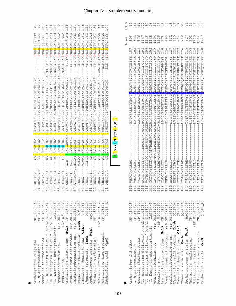
A
Archeoglobus fulgidus (NP_069015) 37 GEIKTVR-----SVCSPNCTGA--CGFDALVYNGRIETLTQAADYPEPEYN----------PRGCLRGQSMM 91
C. hydrogenoformans (YP_360901) 41 REVKFVR-----TTCSPNCTSA--CGIKAMVVDGQIKALFPTNDYPDPEYN----------PRGCLRGISFI 96
Moorella thermoacetica (YP_430751) 68 KEVKYVR-----TTCSPNCTLA--CGIRAMVVDGQIKALLPSNDYPEPEYG----------PRGCLRGLSFI 122
“C. Nitrospira defluvii“ NxrA2(NIDE3256) 59 RYDSSFT-----WVCSPNDTHA--CRIRAFVRNGVVMRVEQNYDHQTYEDLYGNRGTFAHNPRMCLKGFTFH 123
“C. Nitrospira defluvii“ NxrA1(NIDE3237) 60 RYDSSFT-----WCCSPNDTHG--CRVRAFVRNGVVMRVEQNYDHQTYEDLYGNRGTFAHNPRMCLKGFTFH 124
“C. Kuenenia stuttgartiensis (CAJ72445) 58 QYDRTFT-----YCCSPNDTHA--CRIRAFVRNNVMMRVEQNYDHQNYSDLYGNKATRNWNPRMCLKGYTFH 122
Hydrogenobaculum sp. (YP_002121006) 55 KPDETTV-----ILCNPNDTHG--CYLNAYVKNGIITRLEPTYKYGDATDIYGIKASHRWEPRCCNKGLALV 119
Beggiatoa sp. (ZP_02000390) 48 HYDSSFT-----FLCAPNDTHN--CLLRGYVKNGVVTRIAPTYGYQKAKDLDGNQSTQRWDPRCCQKGLALV 112
Aromatoleum aromaticum EdbH (YP_158333) 73 KWDKVN---WGS--HLNICWPQGSCKFYVYVRNGIVWREEQAAQTPACNVD-----YVDYNPLGCQKGSAFN 134
Desulfococcus oleovorans (YP_001528081) 60 TWDKVT---YGT--HLVDCYPGN-CLWRVYSKDGVVFREEQAAKYPVIDPS-----GPDFNPRGCQKGASYS 120
Sulfurihydrogenibium sp. (YP_001931341) 57 AYTYGKKEEHGTAYHCVNCQGN--CAWDVWVQNGIVTRENQAANYPQINPK-----IPDANPRGCNKGVQHS 121
Rhodovulum sulfidophilum DdhA (Q8GPG4) 57 TWDY-----VGKAAHCINCLGN--CAFDIYVKDGIVIREEQLAKYPQISPD-----IPDANPRGCQKGAIHS 116
Ideonella dechloratans ClrA (P60068) 60 TWDS-----VGVMTHSNGCVAG--CAWNVFVKNGIPMREEQISKYPQL-PG-----IPDMNPRGCQKGAVYC 118
Thauera selenatis SerA (Q9S1H0) 64 TWDS-----TGFITHSNGCVAG--CAWRVFVKNGVPMREEQVSEYPQL-PG-----VPDMNPRGCQKGAVYC 122
Dechloromonas agitata PcrA (AAO49008) 51 SWDKKTR-----GAHLINCTGA--CPHFVYTKDGVVIREEQSKDIPPMPN------IPELNPRGCNKGECAH 109
Haloarcula marismortui (YP_135852) 70 DWDSVAR-----STHSVNCTGS--CSWNVYVKDGQVWREEQAGDYPTFDES-----LPDPNPRGCQKGACYT 129
Nitrococcus mobilis (ZP_01125872) 24 QHDNIFR-----STHGVNCTGG--CSWAIYVKDGIITWEMQQTDYPLLGRGEGGRGIPPYEPRGCQRGISAS 88
Nitrobacter hamburgiensis (YP_578638) 24 SHDNVFR-----STHGVNCTGG--CSWAIYVKDGIITWEMQQTDYPLLER-----SLPPYEPRGCQRGISAS 83
Escherichia coli NarG (1Q16_A) 41 QHDKIVR-----STHGVNCTGS--CSWKIYVKNGLVTWETQQTDYPRTRP-----DLPNHEPRGCPRGASYS 100
H/C
xxxD
/Cx(n
)Cx(n
)C
B
L.aa Id.%
Archeoglobus fulgidus (NP_069015) 155 VHKGAMMRLAS-----------------MFGWSALHGYTMNGDLPAFWSQTFGVQTEEFE 197 896 22
C. hydrogenoformans (YP_360901) 161 VHKGAMVALAT-----------------LAGWTLHHAYDLNGDLPMFWPQTFGVQTEELE 203 853 21
Moorella thermoacetica (YP_430751) 186 VQKGAWIALAT-----------------MAGWSLIHPYDQNGDLPMFWPQTFGVQTEELE 228 876 21
“C. Nitrospira defluvii“ NxrA2(NIDE3256) 235 MGKHANTRFNNCVLPLLDSWIRKVNPDQAQGGRYWNNYTWHGDQDPSQPWWNGTQNCDVD 294 1147 100
“C. Nitrospira defluvii“ NxrA1(NIDE3237) 236 VGKMMNTRFNGGCLPLLDSWIRKVDAEKAQGGKYYSNYTWHGDQDPSHPFWNGTQNCDVD 295 1146 87
“C. Kuenenia stuttgartiensis (CAJ72445) 234 IGKYGMYRFNN-CLAIVDAHNRGVGPDQALGGRNWSNYTWHGDQAPGHPFSHGLQTSDVD 292 1148 58
Hydrogenobaculum sp. (YP_002121006) 230 LRYVGQYRMSN-MMALLDSNIRKVEPDKALGGVGWDNYSEHTDLPPAHTLVTGQQTVDFD 289 1154 37
Beggiatoa sp. (ZP_02000390) 223 TRVFAQYREAN-AMALLDDKIRGKGTD-SLGGRGFDNYSWHTDLPPGHPMVTGQQTVDFD 281 1138 39
Aromatoleum aromaticum EdbH (YP_158333) 198 IAWGAGFRMTY-----------------LMDGVSPDINVDIGDTYMGAFHTFGKMHMGYS 240 976 19
Desulfococcus oleovorans (YP_001528081) 184 VHVMAVHRLMV-----------------SLGATVLDLDSTIGDFNRGIYETFGKFMFMDS 226 993 17
Sulfurihydrogenibium sp. (YP_001931341) 185 ARAASGKRLGT-----------------LLGAVRPYIASYVGDMFPGVSLVYGEGNIGFS 227 962 19
Rhodovulum sulfidophilum DdhA (Q8GPG4) 180 MRMAAPYRFAS-----------------LVGGVQLDIFTDVGDLNTGAHLAYGNALESFT 222 910 17
Ideonella dechloratans ClrA (P60068) 180 ITNTAYTRMTK-----------------LLGAISPDATSMTGDLYTGIQTVRVPASTVST 222 914 19
Thauera selenatis SerA (Q9S1H0) 184 ITSAGYSRLAN-----------------LIGAIKPDVSSMTGDLYPGIQTVRMPARTVST 226 918 20
Dechloromonas agitata PcrA (AAO49008) 173 VSFSAGHRFAH-----------------YIGAHTHTFFDWYSDHPTGQTQTCGVQGDSAE 215 927 21
Haloarcula marismortui (YP_135852) 193 VSFASGSRLVN-----------------LLGGVSHSFYDWYSDLPPGQPITWGTQTDNAE 235 952 21
Nitrococcus mobilis (ZP_01125872) 186 LSYAAGSRFLQ-----------------LFGGVNMSFYDWYADLPNSFPEIWGDQTDVCE 228 1218 17
Nitrobacter hamburgiensis (YP_578638) 181 LSFAAGTRFLS-----------------LMGGSLMSFYDWYADLPTSFPEIWGDQTDVCE 223 1214 16
Escherichia coli NarG (1Q16_A) 198 VSYASGARYLS-----------------LIGGTCLSFYDWYCDLPPASPQTWGEQTDVPE 240 1247 16
Chapter IV - Supplementary material
105

Figure S1: Alignments of metal-coordinating regions of alpha subunits of molybdopterin binding (MopB)
proteins which belong to the type II group of Mo-bisMGD (molybdopterin-guanine-dinucleotide) enzymes
(Jormarkka et al., 2004). Accession numbers of the sequences are indicated in brackets. Positions
conserved in all analyzed sequences have red characters. (A) Iron-sulfur [FeS]-binding center. [FeS]-
binding residues are highlighted. A new signature motif for type II Mo-bisMGD enzymes is displayed in
the box below the alignment. (B) Molybdenum (Mo) ligand binding site. The conserved aspartate residue,
highlighted in blue, coordinates the Mo ion in the active center. The last two columns show the overall
length of the sequences in amino acids (L. aa) and amino acid sequence identities (Id. %) to the NxrA2 of
N. defluvii.
Chapter IV - Supplementary material
106
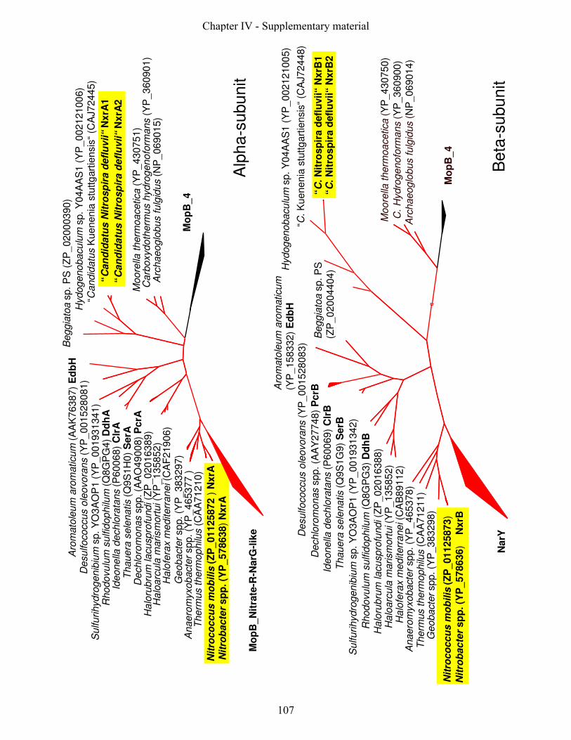
Mo
pB
_N
itra
te-R
-NarG
-lik
e
Nit
rob
acte
rsp
p. (Y
P_578638)
NxrA
Nit
roco
ccu
sm
ob
ilis
(ZP
_01125872
) N
xrA
Anaero
myxobacte
rspp. (Y
P_465377 )
Therm
us
therm
ophilu
s(C
AA
71210)
Geobacte
rspp. (Y
P_383297)
Ha
lofe
rax
mediterr
anei(C
AF
219
06)
Ha
loarc
ula
marism
ort
ui(Y
P_135852)
Ha
loru
bru
mla
cuspro
fundi(Z
P_02016389)
Dechlo
rom
onas
spp. (A
AO
49008)
PcrA
Thauera
sele
natis
(Q9S
1H
0)
SerA
Ideonella
dechlo
rata
ns
(P60068)
Clr
AR
hodovu
lum
sulfid
ophilu
m(Q
8G
PG
4)
Dd
hA
Sulfurihydro
genib
ium
sp. Y
O3A
OP
1 (
YP
_001931
341)
Desulfococcus
ole
ovora
ns
(YP
_001528081)
Beggia
toa
sp. P
S (
ZP
_02000390)
Hydogenobacu
lum
sp. Y
04A
AS
1 (
YP
_00212100
6)
“Candid
atu
sK
uenenia
stu
ttg
art
iensis
“(C
AJ72445)
“C
an
did
atu
sN
itro
sp
ira
defl
uvii“
NxrA
2
Moore
llath
erm
oacetica
(YP
_430751)
Carb
oxydoth
erm
us
hydro
genofo
rmans
(YP
_360901)
Arc
haeoglo
bus
fulg
idus
(NP
_069015)
Mo
pB
_4
“C
an
did
atu
sN
itro
sp
ira
defl
uvii“
NxrA
1
Aro
mato
leum
aro
maticum
(AA
K76387)
Ed
bH
Alp
ha-s
ubu
nit
Beta
-subu
nit
“C. K
uenenia
stu
ttg
art
iensis
“(C
AJ72448)
“C
.N
itro
sp
ira
defl
uvii“
NxrB
1
NarY
Geobacte
rspp. (Y
P_383298)
“C
. N
itro
sp
ira
defl
uvii“
NxrB
2
Moore
llath
erm
oacetica
(YP
_430750)
C.
Hydro
genofo
rmans
(YP
_360900)
Arc
haeoglo
bus
fulg
idus
(NP
_069014)
Mo
pB
_4
Hydogenobacu
lum
sp. Y
04A
AS
1 (
YP
_00212100
5)
Desulfococcus
ole
ovora
ns
(YP
_001528083)
Aro
mato
leum
aro
maticum
(YP
_158332)
Ed
bH
Beggia
toa
sp. P
S
(ZP
_02004404)
Dechlo
rom
onas
spp. (A
AY
27748)
PcrB
Ideonella
dechlo
rata
ns
(P60069)
Clr
B
Thauera
sele
natis
(Q9S
1G
9)
SerB
Sulfurihydro
genib
ium
sp. Y
O3A
OP
1 (
YP
_001931
342)
Rhodovu
lum
sulfid
ophilu
m(Q
8G
PG
3)
Dd
hB
Ha
loru
bru
mla
cuspro
fundi(Z
P_02016388)
Ha
loarc
ula
marism
ort
ui(Y
P_135852)
Ha
lofe
rax
mediterr
anei(C
AB
89112)
Nit
rob
acte
rsp
p. (Y
P_578636)
N
xrB
Nit
roco
ccu
sm
ob
ilis
(ZP
_01125873)
Therm
us
therm
ophilu
s(C
AA
71211)
Anaero
myxobacte
rspp. (Y
P_465378)
Chapter IV - Supplementary material
107

Figure S2: Comparison of amino acid tree topologies of the alpha- and beta-subunit of type II
group of selected Mo-bisMGD (molybdopterin-guanine-dinucleotide) enzymes. Sequences of
the alpha-subunit displayed with red tree branches share the common feature of an aspartate
as molybdenum ligand. Corresponding beta-subunit sequences are also highlighted with red
tree branches. Yellow boxes highlight nitrite oxidoreductases (NxrA). The MopB_4 enzyme
subfamily acts as outgroup. The tree calculations were performed using the maximum-
likelyhood algorithm (PhyML) implemented in the ARB software package (Ludwig et al.
2004). A total of 480 and 429 informative positions were used in the alpha- and beta-subunit
analysis, respectively. The accession numbers of the sequences used for tree calculation are
indicated in brackets. For the abbreviations please refer to Fig. 1. The beta subunit
abbreviations only differ in the last character from the listed alpha subunits.
Chapter IV - Supplementary material
108
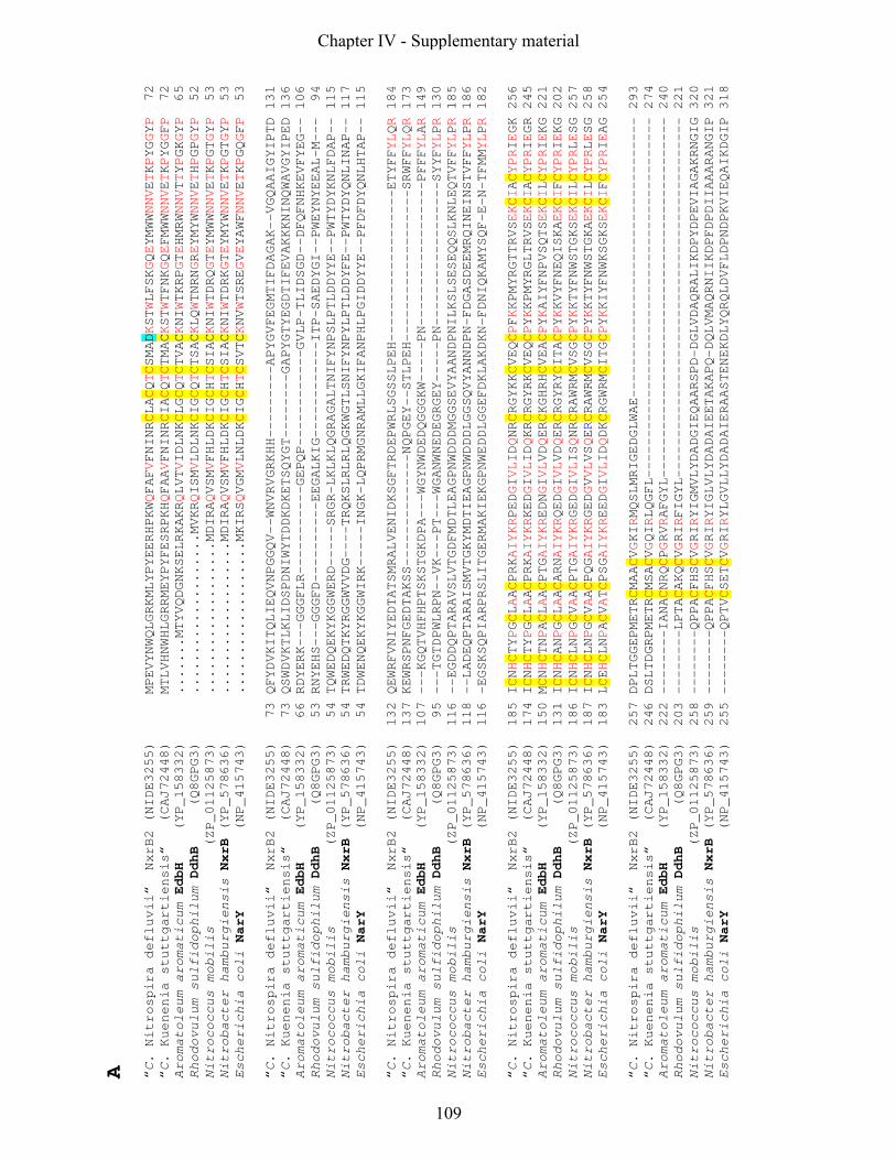
A
“C. Nitrospira defluvii“ NxrB2 (NIDE3255) MPEVYNWQLGRKMLYPYEERHPKWQFAFVFNINRCLACQTCSMADKSTWLFSKGQEYMWWNNVETKPYGGYP 72
“C. Kuenenia stuttgartiensis“ (CAJ72448) MTLVHNWHLGRRMEYPYFESRPKHQFAAVFNINRCIACQTCTMACKSTWTFNKGQEFMWWNNVETKPYGGFP 72
Aromatoleum aromaticum EdbH (YP_158332) .......MTYVQDGNKSELRKAKRQLVTVIDLNKCLGCQTCTVACKNIWTKRPGTEHMRWNNVTTYPGKGYP 65
Rhodovulum sulfidophilum DdhB (Q8GPG3) ....................MVKRQISMVLDLNKCIGCQTCTSACKLQWTNRNGREYMYWNNVETHPGPGYP 52
Nitrococcus mobilis (ZP_01125873) ...................MDIRAQVSMVFHLDKCIGCHTCSIACKNIWTDRQGTEYMWWNNVETKPGTGYP 53
Nitrobacter hamburgiensis NxrB (YP_578636) ...................MDIRAQVSMVFHLDKCIGCHTCSIACKNIWTDRKGTEYMYWNNVETKPGTGYP 53
Escherichia coli NarY (NP_415743) ...................MKIRSQVGMVLNLDKCIGCHTCSVTCKNVWTSREGVEYAWFNNVETKPGQGFP 53
“C. Nitrospira defluvii“ NxrB2 (NIDE3255) 73 QFYDVKITQLIEQVNPGGQV--WNVRVGRKHH---------APYGVFEGMTIFDAGAK--VGQAAIGYIPTD 131
“C. Kuenenia stuttgartiensis“ (CAJ72448) 73 QSWDVKTLKLIDSPDNIWYTDDKDKETSQYGT--------GAPYGTYEGDTIFEVAKKKNINQWAVGYIPED 136
Aromatoleum aromaticum EdbH (YP_158332) 66 RDYERK---GGGFLR-----------GEPQP------------GVLP-TLIDSGD--DFQFNHKEVFYEG-- 106
Rhodovulum sulfidophilum DdhB (Q8GPG3) 53 RNYEHS---GGGFD----------EEGALKIG------------ITP-SAEDYGI--PWEYNYEEAL-M--- 94
Nitrococcus mobilis (ZP_01125873) 54 TQWEDQEKYKGGWERD-----SRGR-LKLKLQGRAGALTNIFYNPSLPTLDDYYE--PWTYDYKNLFDAP-- 115
Nitrobacter hamburgiensis NxrB (YP_578636) 54 TRWEDQTKYRGGWVVDG----TRQKSLRLRLQGKWGTLSNIFYNPYLPTLDDYFE--PWTYDYQNLINAP-- 117
Escherichia coli NarY (NP_415743) 54 TDWENQEKYKGGWIRK-----INGK-LQPRMGNRAMLLGKIFANPHLPGIDDYYE--PFDFDYQNLHTAP-- 115
“C. Nitrospira defluvii“ NxrB2 (NIDE3255) 132 QEWRFVNIYEDTATSMRALVENIDKSGFTRDEPWRLSGSSLPEH-------------------ETYFFYLQR 184
“C. Kuenenia stuttgartiensis“ (CAJ72448) 137 KEWRSPNFGEDTAKSS--------------NQPGEY--STLPEH-------------------SRWFFYLQR 173
Aromatoleum aromaticum EdbH (YP_158332) 107 ---KGQTVHFHPTSKSTGKDPA---WGYNWDEDQGGGKW-----PN------------------PFFFYLAR 149
Rhodovulum sulfidophilum DdhB (Q8GPG3) 95 ---TGTDPWLRPN--VK---PT---WGANWNEDEGRGEY-----PN------------------SYYFYLPR 130
Nitrococcus mobilis (ZP_01125873) 116 --EGDDQPTARAVSLVTGDFMDTLEAGPNWDDDMGGSEVYAANDPNILKSLSESEQQSLKNLEQTVFFYLPR 185
Nitrobacter hamburgiensis NxrB (YP_578636) 118 --LADEQPTARAISMVTGKYMDTIEAGPNWDDDLGGSQVYANNDPN-FDGASDEEMRQINEINSTVFFYLPR 186
Escherichia coli NarY (NP_415743) 116 -EGSKSQPIARPRSLITGERMAKIEKGPNWEDDLGGEFDKLAKDKN-FDNIQKAMYSQF-E-N-TFMMYLPR 182
“C. Nitrospira defluvii“ NxrB2 (NIDE3255) 185 ICNHCTYPGCLAACPRKAIYKRPEDGIVLIDQNRCRGYKKCVEQCPFKKPMYRGTTRVSEKCIACYPRIEGK 256
“C. Kuenenia stuttgartiensis“ (CAJ72448) 174 ICNHCTYPGCLAACPRKAIYKRKEDGIVLIDQKRCRGYRKCVEQCPYKKPMYRGLTRVSEKCIACYPRIEGR 245
Aromatoleum aromaticum EdbH (YP_158332) 150 MCNHCTNPACLAACPTGAIYKREDNGIVLVDQERCKGHRHCVEACPYKAIYFNPVSQTSEKCILCYPRIEKG 221
Rhodovulum sulfidophilum DdhB (Q8GPG3) 131 ICNHCANPGCLAACARNAIYKRQEDGIVLVDQERCRGYRYCITACPYKKVYFNEQISKAEKCIFCYPRIEKG 202
Nitrococcus mobilis (ZP_01125873) 186 ICNHCLNPGCVAACPTGAIYKRGEDGIVLISQNRCRAWRMCVSGCPYKKTYFNWSTGKSEKCILCYPRLESG 257
Nitrobacter hamburgiensis NxrB (YP_578636) 187 ICNHCLNPGCVAACPQGAIYKRGEDGVVLVSQERCRAWRMCVSGCPYKKTYFNWSTGKAEKCILCYPRLESG 258
Escherichia coli NarY (NP_415743) 183 LCEHCLNPACVATCPSGAIYKREEDGIVLIDQDKCRGWRMCITGCPYKKIYFNWKSGKSEKCIFCYPRIEAG 254
“C. Nitrospira defluvii“ NxrB2 (NIDE3255) 257 DPLTGGEPMETRCMAACVGKIRMQSLMRIGEDGLWAE----------------------------------- 293
“C. Kuenenia stuttgartiensis“ (CAJ72448) 246 DSLTDGRPMETRCMSACVGQIRLQGFL--------------------------------------------- 274
Aromatoleum aromaticum EdbH (YP_158332) 222 --------IANACNRQCPGRVRAFGYL--------------------------------------------- 240
Rhodovulum sulfidophilum DdhB (Q8GPG3) 203 --------LPTACAKQCVGRIRFIGYL--------------------------------------------- 221
Nitrococcus mobilis (ZP_01125873) 258 --------QPPACFHSCVGRIRYIGMVLYDADGIEQAARSPD-DGLVDAQRALIKDPYDPEVIAGAKRNGIG 320
Nitrobacter hamburgiensis NxrB (YP_578636) 259 --------QPPACFHSCVGRIRYIGLVLYDADAIEETAKAPQ-DQLVMAQRNIIKDPFDPDIIAAARANGIP 321
Escherichia coli NarY (NP_415743) 255 --------QPTVCSETCVGRIRYLGVLLYDADAIERAASTENEKDLYQRQLDVFLDPNDPKVIEQAIKDGIP 318
Chapter IV - Supplementary material
109
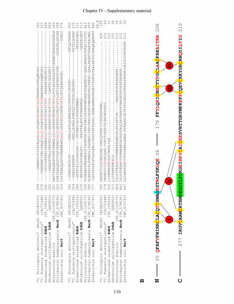
“C. Nitrospira defluvii“ NxrB2 (NIDE3255) 294 ------DRWHPLYYTIRVEQVALPLYPQWGTEPNGYYIPPRHAPRGYNRQMFGPG----------------- 342
“C. Kuenenia stuttgartiensis“ (CAJ72448) 275 ----DDNPKNPITWLIRHQKIALPLYPQFGTEPNIYYIPPRWAPRAYLRQMFGPG----------------- 323
Aromatoleum aromaticum EdbH (YP_158332) 241 -----DDTTSHVHKLVKKWKVALPLHAEYGTGPNIYYVPP--MGARGFGEDGEIT----------------- 288
Rhodovulum sulfidophilum DdhB (Q8GPG3) 222 -----DDEAGPVHLLVERYKVAIPLHPEWGTKPSVFYVPP--LAPPRIGDDGEPT----------------- 269
Nitrococcus mobilis (ZP_01125873) 321 DAVVTAAQNSPVYKFVKRWGLALPLHPEFRTMPMLFYVPPLLPVIAAIRDGQYETTAGMETAPADETGPNLN 392
Nitrobacter hamburgiensis NxrB (YP_578636) 322 DSKIEAAQKSPVYQFVKKWGIALPLHPEFRTLPMLFYVPPLGPVLAKVENGVYDN-----IASESRLGPLMS 388
Escherichia coli NarY (NP_415743) 319 LSVIEAAQQSPVYKMAMEWKLALPLHPEYRTLPMVWYVPPLSPIQSAADAGEL--------------GSNGI 376
“C. Nitrospira defluvii“ NxrB2 (NIDE3255) 343 --VDNAIEKYLVP-------SRELLAVLQLWRASQQIVFRYDVIPGPKV-------------FETQIHGKRF 392
“C. Kuenenia stuttgartiensis“ (CAJ72448) 324 --VDEAIEKFMVP-------SRELLAVMSLFRMTQTIVYEYKIEEGPKV-------------FETEIHGKKF 373
Aromatoleum aromaticum EdbH (YP_158332) 289 --DKTRIPLDVLEGLFG----PEVKRVLAVLHTERENMRA-----------------------GRGSELMDL 331
Rhodovulum sulfidophilum DdhB (Q8GPG3) 270 --EETRVPLAYLKELFG----EAVVPALETLKTERAKKQS-----------------------GAESELMDT 312
Nitrococcus mobilis (ZP_01125873) 393 TVDKARIPMQYMASLFTAGNVDKIKHVYHKLVAVRMYMRSEQVGDQKEEDV-QEALRRADLTPEQCISIFEL 463
Nitrobacter hamburgiensis NxrB (YP_578636) 389 SLERSRIPLRYMASLLSGGNEDIIRDVYKKLVAVRVYMRSKKVKDIPEEEV-QRALAEGKTTAAEVEAIWRL 459
Escherichia coli NarY (NP_415743) 377 -LPDVESLRIPVQYLANLLTAGDTKPVLRAL-KRMLAMRHYKRAETVDGKVDTRALEEVGLTEAQAQEMYRY 446
Id.%
“C. Nitrospira defluvii“ NxrB2 (NIDE3255) 393 DMYNDTVLGFNKSGKE-VARIQVEEPIYIRPAERVNWL............................. 429 100
“C. Kuenenia stuttgartiensis“ (CAJ72448) 374 TMYNDTVIGFGEDGKE-VVRTTVEEPIHIRPDKHYNSI............................. 410 69
Aromatoleum aromaticum EdbH (YP_158332) 332 LISKKWSDRFGGFTNDPLTQS.............................................. 352 31
Rhodovulum sulfidophilum DdhB (Q8GPG3) 313 LIGYRHPEMFKLS...................................................... 325 36
Nitrococcus mobilis (ZP_01125873) 464 TSLPTFEERFVVPPMERERAMDGELPPLDPYSR-----KAAGGFGFRDAPARRF............. 512 32
Nitrobacter hamburgiensis NxrB (YP_578636) 460 TSLPTFEERFVVPPMERETAVDSLFPQLDPVSRNYPVRKGEVGVGFHTDPARGP............. 513 30
Escherichia coli NarY (NP_415743) 447 LAIANYEDRFVVPSSHRELAREA-FPEKNGCGFTFGDGCHGSDTKFNLFNSRRIDAIDVTSKTEPHP 512 28
B
N 25 QFAFVFNINRCLACQTCSMADKSTWLFSKGQE 56 179 FFYLQRICNHCTYPGCLAACPRKAIYKR 206
C 277 IKGVCAAMCRTEMPEGGTLPDKGEIRPYCAICKESVRTTGRYMPKKFPCQEVCKKYGRCRNQDILVIG 210
I II
II
I IV
Chapter IV - Supplementary material
110

Figure S3: (A) Alignment of selected beta subunits of type II Mo-bisMGD enzymes. Accession numbers of
the sequences are indicated in brackets. Residues conserved in all displayed sequences appear red. [FeS]
coordinating residues are highlighted in yellow. The last column in the last row shows amino acid sequence
identities (Id. %) to NxrB2 of N. defluvii. (B) Sketch of the possible [FeS] cluster coordination in NxrB of
N. defluvii. Sequence stretches were taken from (A) and stringed together from the N- to the C-terminus.
[FeS] coordinating residues are highlighted in yellow and blue. A sequence specific insertion is highlighted
in green. The red filled circles represent the four [FeS] cluster.
Chapter IV - Supplementary material
111
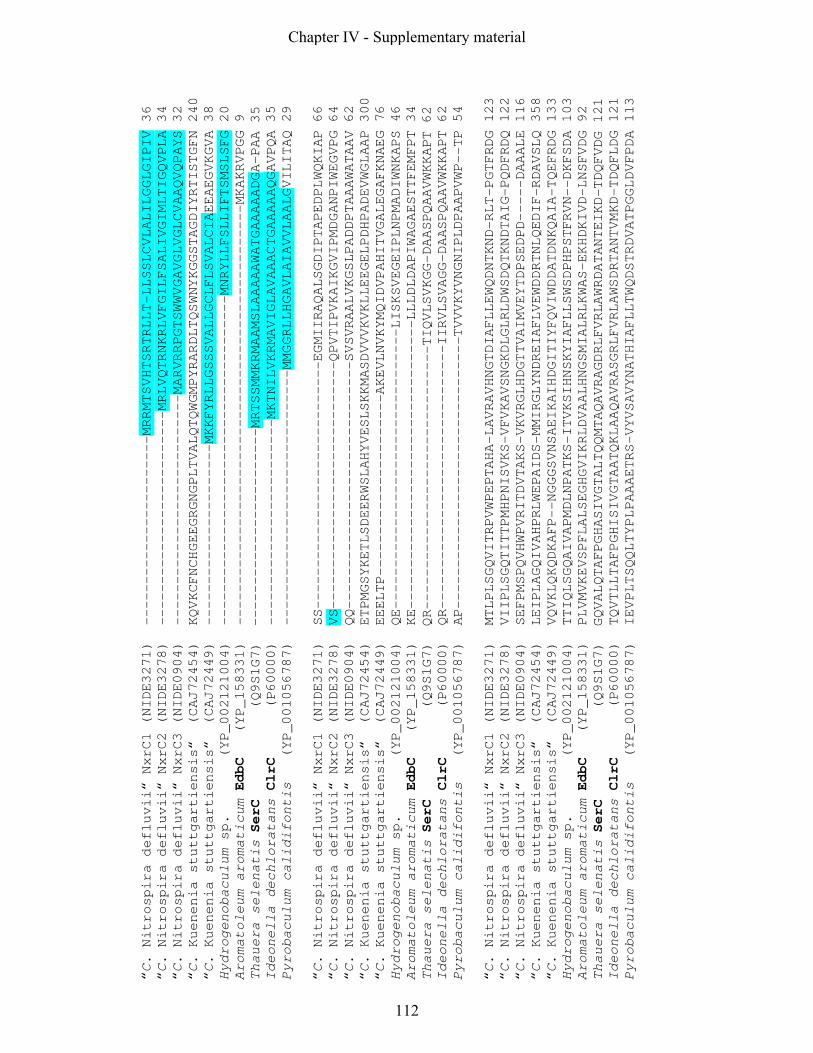
“C. Nitrospira defluvii“ NxrC1 (NIDE3271) -----------------------MRRMTSVHTSRTRLLT-LLSSLCVLALILGGLGIPIV 36
“C. Nitrospira defluvii“ NxrC2 (NIDE3278) --------------------------MRLVQTRNKRLVFGILFSALIVGIMLTIGQVPLA 34
“C. Nitrospira defluvii“ NxrC3 (NIDE0904) ----------------------------MARVRRPGTSWWVGAVGLVGLCVAAQVQPAYS 32
“C. Kuenenia stuttgartiensis“ (CAJ72454) KQVKCFNCHGEEGRGNGPLTVALQTQWGMPYRARDLTQSWNYKGGSTAGDIYRTISTGFN 240
“C. Kuenenia stuttgartiensis“ (CAJ72449) ----------------------MKKFYRLLGSSSVALLGCLFLSVALCIAEEAEGVKGVA 38
Hydrogenobaculum sp. (YP_002121004) ----------------------------------------MNRYLLFSLLIFTSMSLSFG 20
Aromatoleum aromaticum EdbC (YP_158331) ---------------------------------------------------MKAKRVPGG 9
Thauera selenatis SerC (Q9S1G7) ------------------------MRTSSMMKRMAAMSLAAAAAWATGAAAAADGA-PAA 35
Ideonella dechloratans ClrC (P60000) -------------------------MKTNILVKRMAVIGLAVAAACTGAAAAAQGAVPQA 35
Pyrobaculum calidifontis (YP_001056787) -------------------------------MMGGRLLHGAVLAIAVVLAALGVILITAQ 29
“C. Nitrospira defluvii“ NxrC1 (NIDE3271) SS------------------------------EGMIIRAQALSGDIPTAPEDPLWQKIAP 66
“C. Nitrospira defluvii“ NxrC2 (NIDE3278) VS------------------------------QPVTIPVKAIKGVIPMDGANPIWEGVPG 64
“C. Nitrospira defluvii“ NxrC3 (NIDE0904) QQ------------------------------SVSVRAALVKGSLPADDPTAAAWATAAV 62
“C. Kuenenia stuttgartiensis“ (CAJ72454) ETPMGSYKETLSDEERWSLAHYVESLSKKMASDVVVKVKLLEEGELPDHPADEVWGLAAP 300
“C. Kuenenia stuttgartiensis“ (CAJ72449) EEELTP----------------------AKEVLNVKYMQIDVPAHITVGALEGAFKNAEG 76
Hydrogenobaculum sp. (YP_002121004) QE----------------------------------LISKSVEGEIPLNPMADIWNKAPS 46
Aromatoleum aromaticum EdbC (YP_158331) KE-----------------------------------LLLDLDAPIWAGAESTTFEMFPT 34
Thauera selenatis SerC (Q9S1G7) QR--------------------------------TIQVLSVKGG-DAASPQAAVWKKAPT 62
Ideonella dechloratans ClrC (P60000) QR--------------------------------IIRVLSVAGG-DAASPQAAVWKKAPT 62
Pyrobaculum calidifontis (YP_001056787) AP---------------------------------TVVVKYVNGNIPLDPAAPVWP--TP 54
“C. Nitrospira defluvii“ NxrC1 (NIDE3271) MTLPLSGQVITRPVWPEPTAHA-LAVRAVHNGTDIAFLLEWQDNTKND-RLT-PGTFRDG 123
“C. Nitrospira defluvii“ NxrC2 (NIDE3278) VIIPLSGQTITTPMHPNISVKS-VFVKAVSNGKDLGLRLDWSDQTKNDTAIG-PQDFRDQ 122
“C. Nitrospira defluvii“ NxrC3 (NIDE0904) SEFPMSPQVHWPVRITDVTAKS-VKVRGLHDGTTVAIMVEYTDPSEDPD-----DAAALE 116
“C. Kuenenia stuttgartiensis“ (CAJ72454) LEIPLAGQIVAHPRLWEPAIDS-MMIRGLYNDREIAFLVEWDDRTNLQEDIF-RDAVSLQ 358
“C. Kuenenia stuttgartiensis“ (CAJ72449) VQVKLQKQDKAFP--NGGGSVNSAEIKAIHDGITIYFQVIWDDATDNKQAIA-TQEFRDG 133
Hydrogenobaculum sp. (YP_002121004) TTIQLSGQAIVAPMDLNPATKS-ITVKSIHNSKYIAFLLSWSDPHPSTFRVN--DKFSDA 103
Aromatoleum aromaticum EdbC (YP_158331) PLVMVKEVSPFLALSEGHGVIKRLDVAALHNGSMIALRLKWAS-EKHDKIVD-LNSFVDG 92
Thauera selenatis SerC (Q9S1G7) GQVALQTAFPGHASIVGTALTQQMTAQAVRAGDRLFVRLAWRDATANTEIKD-TDQFVDG 121
Ideonella dechloratans ClrC (P60000) TQVTLLTAFPGHISIVGTAATQKLAAQAVRASGRLFVRLAWSDRTANTVMKD-TDQFLDG 121
Pyrobaculum calidifontis (YP_001056787) IEVPLTSQQLTYPLPAAAETRS-VYVSAVYNATHIAFLLTWQDSTRDVATPGGLDVFPDA 113
Chapter IV - Supplementary material
112

“C. Nitrospira defluvii“ NxrC1 (NIDE3271) VALGLPLG------DAPAFFCMGQLDHYINIWHWKADWQSDIDRRASRTTEKDKAAS--- 174
“C. Nitrospira defluvii“ NxrC2 (NIDE3278) AAVMFPVNT----AGAPPFQCMGQSGGTVNIWRWNAEWQKDLGKDSAGMWDVDDQYPGIF 178
“C. Nitrospira defluvii“ NxrC3 (NIDE0904) FMVGDTKA-----HFAH-GQPMAQEGGPVNIWYWK------------------------- 146
“C. Kuenenia stuttgartiensis“ (CAJ72454) FPVKIPES-----LKKP-YFGMGDSGGVVNLWQWKACWKEGFGSIAE------------- 399
“C. Kuenenia stuttgartiensis“ (CAJ72449) AALMFPLGKITISPEEPFSPRMGDRQKPVNLWHWKADWEADLLATGGIEECPARYPN--- 190
Hydrogenobaculum sp. (YP_002121004) VALEVPLK-----PDNTTPITMGGNGKEVVILQWAAYRQYNITKGFADIPKIEPYYYTG- 157
Aromatoleum aromaticum EdbC (YP_158331) VGAMFPVA------RGAQAVTMGATGRPVNAWYWKAN----------------------- 123
Thauera selenatis SerC (Q9S1G7) AAVQFPVN-----GKDTTLAFMGDPDNPVNVWHWR------------------------- 151
Ideonella dechloratans ClrC (P60000) AAVEFPVN-----GKVATLPFMGDPVNVVNVWHWR------------------------- 151
Pyrobaculum calidifontis (YP_001056787) VAIQFPVS-----RAQLPYICMGTVDNPVNIIYWKAGVGVENLVAGAGYG---------- 158
“C. Nitrospira defluvii“ NxrC1 (NIDE3271) -----GEPRRFEVIPR------------------------RASSVEDLIGGGFSTLTTKE 205
“C. Nitrospira defluvii“ NxrC2 (NIDE3278) WDYYFEEPAGGVTYPDRIGRSLGPFNSGIWSGNIMSDPTLRVSSVEDLNANGFSTLTTQA 238
“C. Nitrospira defluvii“ NxrC3 (NIDE0904) ---------------------------------------NKDGKGADLGAKGFGTLKPHG 167
“C. Kuenenia stuttgartiensis“ (CAJ72454) ------------------------------------MPANEPGEIVELNAKGFKKISIQS 423
“C. Kuenenia stuttgartiensis“ (CAJ72449) --MHDDFSTNPHSVNYHKGVIQSAAELSGGYAAHNLLSLPRGRAVEDLNAEGFGTLTSQD 248
Hydrogenobaculum sp. (YP_002121004) -----YIYPQAKPPYAYPKDWNNKYAQDYVGGKVPYWKNTRDTSVIEFIADGYTTTTWRK 212
Aromatoleum aromaticum EdbC (YP_158331) -----------------------------------------ANEPMEIVAEGFSAVRRMK 142
Thauera selenatis SerC (Q9S1G7) ----------------------------------------ADGRTRNLVAKGFGTATPVP 171
Ideonella dechloratans ClrC (P60000) ----------------------------------------ADGRTLNLLAKGFGTSTPVP 171
Pyrobaculum calidifontis (YP_001056787) -------------------------------------LSPQQREALGLQATPTSPVELLP 181
“C. Nitrospira defluvii“ NxrC1 (NIDE3271) KQGRVQG-----KATWKDG-----TWRVVMRRPLSSEEQENEAKLIPGRIQAVSFAVWNG 255
“C. Nitrospira defluvii“ NxrC2 (NIDE3278) HQDVIGNGVWEPSGSLKGGGYTGPTWRVVVKRSLETS-DANDTQFKAGASVPIAFAIWDG 297
“C. Nitrospira defluvii“ NxrC3 (NIDE0904) -----------HQDVKAKGVYQGGVWKVVFSRTLNTEHVAEDTQFQPGQFASIAFAVWDG 216
“C. Kuenenia stuttgartiensis“ (CAJ72454) PE---------NQDVMGEGLHDNGRWKVVFKRPLKTEDGKKDIQFEIGKLIPIAFAVWDG 474
“C. Kuenenia stuttgartiensis“ (CAJ72449) -----------HQDVDGCSKFENKKWTVVFCRSLNTG-DPLDVQFVPGESTYFNMAVWNG 296
Hydrogenobaculum sp. (YP_002121004) -----------HQEAKGVGVYKNGKWYVEIIRPFNIK-GALNPEWGPNSKTFIDFAVWQG 260
Aromatoleum aromaticum EdbC (YP_158331) DKAG--------SDLKAVAQHRNGEWNVILCRSMATG--DGLAKLQAGGSSKIAFAVWSG 192
Thauera selenatis SerC (Q9S1G7) -----------AEGLRSTATRTRDGWEVVISRPLRVK-AEEGADLQGRRTMPIAFAAWDG 219
Ideonella dechloratans ClrC (P60000) -----------TEDLRSASVRTGDGWEVVLSRPLRVK-AEEGANLQGRRTMPIGFAAWDG 219
Pyrobaculum calidifontis (YP_001056787) QN---------AQVVQAYTTYAGGRWQVVIVRPLASV---HPLMSSLASDFSAAFATWDG 229
Chapter IV - Supplementary material
113

“C. Nitrospira defluvii“ NxrC1 (NIDE3271) ENKERN------GQKAIA-PWFQLALDPVTKT---------------------------- 280
“C. Nitrospira defluvii“ NxrC2 (NIDE3278) SNIERN------GMKALS-TWFTLKMP--------------------------------- 317
“C. Nitrospira defluvii“ NxrC3 (NIDE0904) KKMETGQPKEKGSEKAISSWWYFRADAPPDYSPYMYALLAVALALGFELVLVRRLKKGS- 275
“C. Kuenenia stuttgartiensis“ (CAJ72454) SNGDAG------SQKSVS-SWYYILLEKPMPKTVFVYVLVAVVMGASVELWFLARLRRFP 528
“C. Kuenenia stuttgartiensis“ (CAJ72449) DREDRN------GQKNISIQWHPLSLERIAWQ---------------------------- 322
Hydrogenobaculum sp. (YP_002121004) YAKERD------SRKAISYAWIPLIIEGK------------------------------- 283
Aromatoleum aromaticum EdbC (YP_158331) GNAERS------GRKSYSGEFVDFEILK-------------------------------- 214
Thauera selenatis SerC (Q9S1G7) ENQERD------GLKAVTMEWWQLNF---------------------------------- 239
Ideonella dechloratans ClrC (P60000) ENQERD------GLKAVTMEWWQLRF---------------------------------- 239
Pyrobaculum calidifontis (YP_001056787) GKGERG------GLKA-TSGWISFTLERPAAPARVETVTQAAPTTVTATQIVETSPTWAW 283
Id.% Id.%
Id.%
NxrC1 NxrC2 NxrC3
“C. Nitrospira defluvii“ NxrC1 (NIDE3271) --------------------- 280 100
39
24
“C. Nitrospira defluvii“ NxrC2 (NIDE3278) --------------------- 317 39
100 24
“C. Nitrospira defluvii“ NxrC3 (NIDE0904) --------------------- 275 24
24
100
“C. Kuenenia stuttgartiensis“ (CAJ72454) PKSEEEDQ------------- 535 25
26
34
“C. Kuenenia stuttgartiensis“ (CAJ72449) --------------------- 322 26
28 22
Hydrogenobaculum sp. (YP_002121004) --------------------- 283 30
25
22
Aromatoleum aromaticum EdbC (YP_158331) --------------------- 214 23
25
19
Thauera selenatis SerC (Q9S1G7) --------------------- 239 27
27 24
Ideonella dechloratans ClrC (P60000) --------------------- 239 27
27 27
Pyrobaculum calidifontis (YP_001056787) VVIGVLIAVIAILLGLAFRRK 303 30
30 22
Chapter IV - Supplementary material
114

Figure S4: ClustalW alignment of selected gamma subunits of type II Mo-bisMGD enzymes.
Sequences display the first NCBI Blast hits for the three NxrC copies of N. defluvii.
Accession numbers of the sequences are indicated in brackets. Residues conserved in all
displayed sequences appear red. Heme b coordinating residues are highlighted with an
asterisk. Transmembrane topologies and signal peptides predicted with the Phobius server
(Kall et al., 2004) are highlighted with green and blue, respectively. The sequence CAJ72454
was truncated at the N-terminus. Therefore the predicted signal peptide is not displayed. The
last columns in the last row shows amino acid sequence identities (Id. %) to the three NxrC
copies of N. defluvii.
Chapter IV - Supplementary material
115

activated sludge clone C13 (XXXX)activated sludge clone C6
activated sludge clone B3activated sludge clone C10
activated sludge clone C17"Candidatus Nitrospira defluvii" NIDE3256"Candidatus Nitrospira defluvii" NIDE3236
activated sludge clone C8activated sludge clone5
activated sludge clone B18activated sludge clone B8activated sludge clone G9activated sludge clone C7
activated sludge clone E8beech forrest soil clone S22beech forrest soil clone S8beech forrest soil clone S17beech forrest soil clone S2
beech forrest soil clone S7beech forrest soil clone S9
beech forrest soil clone S5beech forrest soil clone S16beech forrest soil clone S20beech forrest soil clone S4
Nitrospira moscoviensis nxrB20Nitrospira moscoviensis nxrB17Nitrospira moscoviensis nxrB3Nitrospira moscoviensis nxrB11Nitrospira moscoviensis nxrB6
Beryl spring clone 11Beryl spring clone 14
Beryl spring clone7"Candidatus Nitrospira bockiana" nxrB12"Candidatus Nitrospira bockiana" nxrB15
"Candidatus Nitrospira bockiana" nxrB17Nitrospira marina nxrB17Nitrospira marina nxrB7Nitrospira marina nxrB9Nitrospira marina nxrBN4
Nitrospira marina nxrB11Nitrospira marina nxrB12
Hyrtios proteus sponge clone 25Hyrtios proteus sponge clone 26Hyrtios proteus sponge clone19
0.10
nxrB
F1
4/n
xrB
R1
23
9
nxrB
F1
9/n
xrB
R1
23
7
nxrB
F1
69
/nxrB
R6
38
68
°C -
62
°C; 1
56
.2°C
; 3
59
°C; 2
51
.5°C
-
48
°C; 2
Chapter IV - Supplementary material
116

Figure S5: Overview tree displaying all Nitrospira-like nxrB sequences derived in this study. The
maximum-likelyhood nxrB tree in Fig. 4 provided the basis tree. The displayed groups in Fig. 4 have been
ungrouped and all environmental metagenome and cDNA sequences were removed in the ARB software
package (Ludwig et al., 2004) without changing the overall tree topology. The scale bar indicates 0.1
estimated change per nucleotide. Sequences that belong to isolated or enriched Nitrospira-like bacteria are
in boldface. Accession numbers of the sequences in the tree are indicted in brackets.. Vertical bars on the
right display the specificity of the nxrB primer pairs listed in Table 1. Each bar is superimposed with the
primer pair names. Beneath the bar the optimal annealing temperatures and PCR protocol (for details see
Experimental Procedures) is listed.
Chapter IV - Supplementary material
117
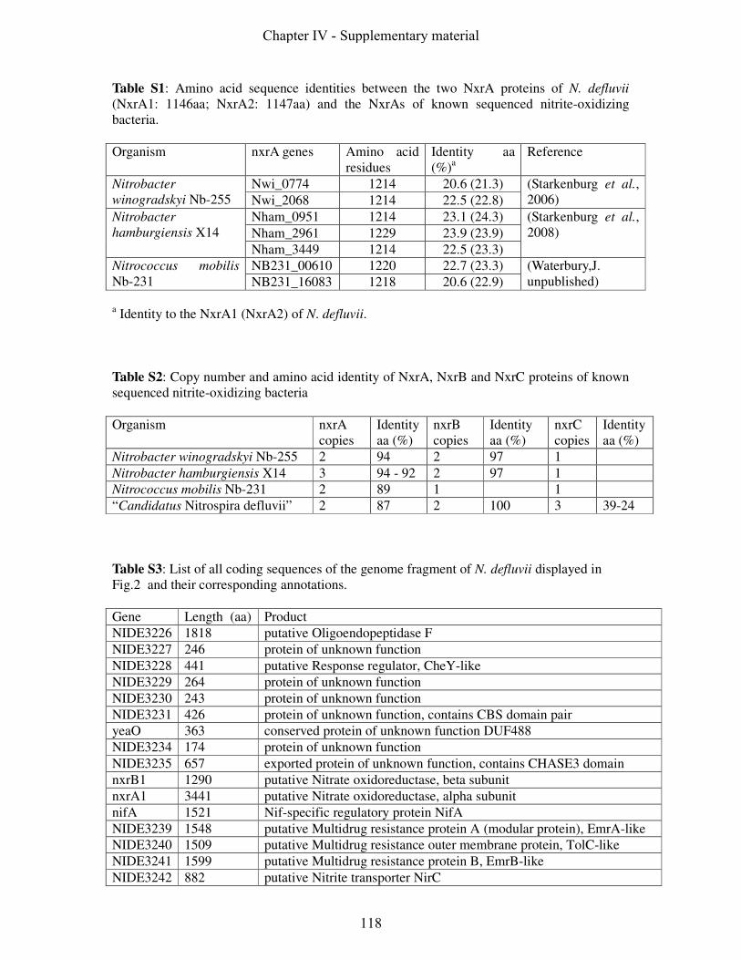
Table S1: Amino acid sequence identities between the two NxrA proteins of N. defluvii
(NxrA1: 1146aa; NxrA2: 1147aa) and the NxrAs of known sequenced nitrite-oxidizing
bacteria.
Organism nxrA genes Amino acid
residues
Identity aa
(%)a
Reference
Nwi_0774 1214 20.6 (21.3) Nitrobacter
winogradskyi Nb-255 Nwi_2068 1214 22.5 (22.8)
(Starkenburg et al.,
2006)
Nham_0951 1214 23.1 (24.3)
Nham_2961 1229 23.9 (23.9)
Nitrobacter
hamburgiensis X14
Nham_3449 1214 22.5 (23.3)
(Starkenburg et al.,
2008)
NB231_00610 1220 22.7 (23.3) Nitrococcus mobilis
Nb-231 NB231_16083 1218 20.6 (22.9)
(Waterbury,J.
unpublished)
a Identity to the NxrA1 (NxrA2) of N. defluvii.
Table S2: Copy number and amino acid identity of NxrA, NxrB and NxrC proteins of known
sequenced nitrite-oxidizing bacteria
Organism nxrA
copies
Identity
aa (%)
nxrB
copies
Identity
aa (%)
nxrC
copies
Identity
aa (%)
Nitrobacter winogradskyi Nb-255 2 94 2 97 1
Nitrobacter hamburgiensis X14 3 94 - 92 2 97 1
Nitrococcus mobilis Nb-231 2 89 1 1
“Candidatus Nitrospira defluvii” 2 87 2 100 3 39-24
Table S3: List of all coding sequences of the genome fragment of N. defluvii displayed in
Fig.2 and their corresponding annotations.
Gene Length (aa) Product
NIDE3226 1818 putative Oligoendopeptidase F
NIDE3227 246 protein of unknown function
NIDE3228 441 putative Response regulator, CheY-like
NIDE3229 264 protein of unknown function
NIDE3230 243 protein of unknown function
NIDE3231 426 protein of unknown function, contains CBS domain pair
yeaO 363 conserved protein of unknown function DUF488
NIDE3234 174 protein of unknown function
NIDE3235 657 exported protein of unknown function, contains CHASE3 domain
nxrB1 1290 putative Nitrate oxidoreductase, beta subunit
nxrA1 3441 putative Nitrate oxidoreductase, alpha subunit
nifA 1521 Nif-specific regulatory protein NifA
NIDE3239 1548 putative Multidrug resistance protein A (modular protein), EmrA-like
NIDE3240 1509 putative Multidrug resistance outer membrane protein, TolC-like
NIDE3241 1599 putative Multidrug resistance protein B, EmrB-like
NIDE3242 882 putative Nitrite transporter NirC
Chapter IV - Supplementary material
118
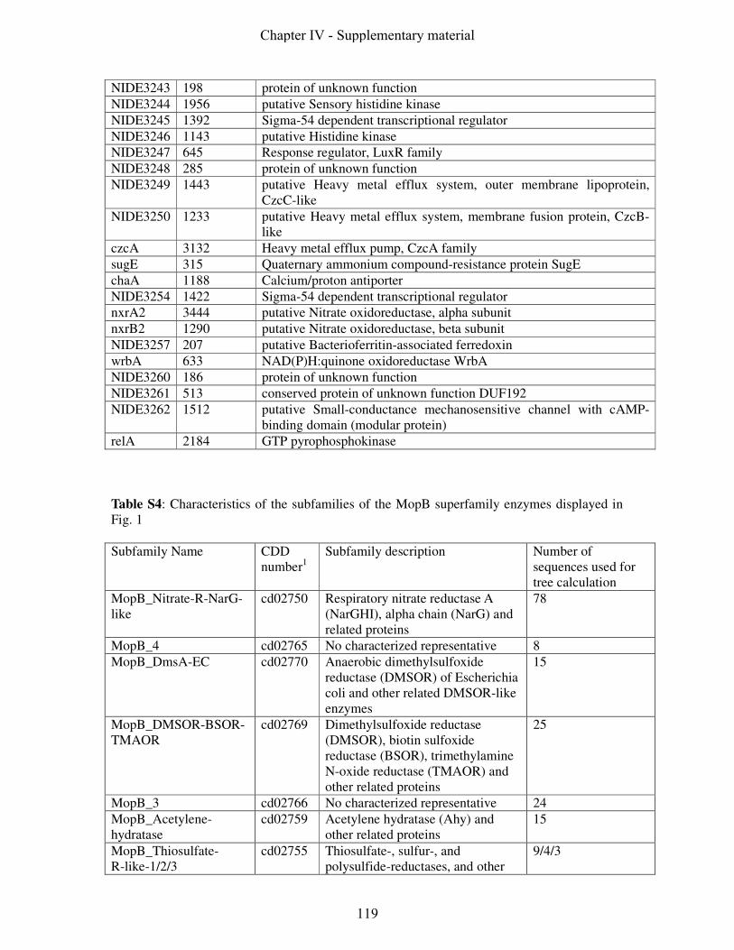
NIDE3243 198 protein of unknown function
NIDE3244 1956 putative Sensory histidine kinase
NIDE3245 1392 Sigma-54 dependent transcriptional regulator
NIDE3246 1143 putative Histidine kinase
NIDE3247 645 Response regulator, LuxR family
NIDE3248 285 protein of unknown function
NIDE3249 1443 putative Heavy metal efflux system, outer membrane lipoprotein,
CzcC-like
NIDE3250 1233 putative Heavy metal efflux system, membrane fusion protein, CzcB-
like
czcA 3132 Heavy metal efflux pump, CzcA family
sugE 315 Quaternary ammonium compound-resistance protein SugE
chaA 1188 Calcium/proton antiporter
NIDE3254 1422 Sigma-54 dependent transcriptional regulator
nxrA2 3444 putative Nitrate oxidoreductase, alpha subunit
nxrB2 1290 putative Nitrate oxidoreductase, beta subunit
NIDE3257 207 putative Bacterioferritin-associated ferredoxin
wrbA 633 NAD(P)H:quinone oxidoreductase WrbA
NIDE3260 186 protein of unknown function
NIDE3261 513 conserved protein of unknown function DUF192
NIDE3262 1512 putative Small-conductance mechanosensitive channel with cAMP-
binding domain (modular protein)
relA 2184 GTP pyrophosphokinase
Table S4: Characteristics of the subfamilies of the MopB superfamily enzymes displayed in
Fig. 1
Subfamily Name CDD
number1
Subfamily description Number of
sequences used for
tree calculation
MopB_Nitrate-R-NarG-
like
cd02750
Respiratory nitrate reductase A
(NarGHI), alpha chain (NarG) and
related proteins
78
MopB_4 cd02765 No characterized representative 8
MopB_DmsA-EC cd02770
Anaerobic dimethylsulfoxide
reductase (DMSOR) of Escherichia
coli and other related DMSOR-like
enzymes
15
MopB_DMSOR-BSOR-
TMAOR
cd02769
Dimethylsulfoxide reductase
(DMSOR), biotin sulfoxide
reductase (BSOR), trimethylamine
N-oxide reductase (TMAOR) and
other related proteins
25
MopB_3 cd02766 No characterized representative 24
MopB_Acetylene-
hydratase
cd02759 Acetylene hydratase (Ahy) and
other related proteins
15
MopB_Thiosulfate-
R-like-1/2/3
cd02755 Thiosulfate-, sulfur-, and
polysulfide-reductases, and other
9/4/3
Chapter IV - Supplementary material
119

related proteins
MopB_Arsenate-R
cd02757 Respiratory arsenate reductase,
As(V), catalytic subunit (ArrA) and
other related proteins
5
MopB_Phenylacetyl-
CoA-OR
cd02760 Phenylacetyl-CoA:acceptor
oxidoreductase, large subunit
(PadB2), and other related proteins
2
MopB_2 cd02763 No characterized representative 4
MopB_Tetrathionate-Ra cd02758 Tetrathionate reductase, subunit A,
(TtrA) and other related proteins
8
MopB_1 cd02762 No characterized representative 9
MopB_Formate-DH-Na-
like
cd02752 Formate dehydrogenase N, alpha
subunit
14
MopB_ydeP cd02767 No characterized representative 21
MopB_Formate-DH-H cd02753 Formate dehydrogenase H 30
MopB_Nitrate-R-NapA-
like
cd02754 Nitrate reductases, NapA, NasA,
and NarB
33
MopB_Arsenite-Ox
cd02756
Arsenite oxidase 4
MopB_FmdB-FwdB cd02761
Molybdenum/tungsten
formylmethanofuran
dehydrogenases, subunit B
18
MopB_PHLH cd02764 uncharacterized putative
hydrogenase-like homologs
(PHLH) of molybdopterin binding
(MopB) proteins
4
MopB_NADH-Q-OR-
NuoG2
cd02768 The NuoG/Nad11/75-kDa subunit
(second domain) of the NADH-
quinone oxidoreductase (NADH-Q-
OR)/respiratory complex I/NADH
dehydrogenase-1 (NDH-1)
59
1 NCBI Conserved Domains (http://www.ncbi.nlm.nih.gov/Structure/cdd/cdd.shtml) number
References
Jormakka, M., D. Richardson, B. Byrne & S. Iwata, (2004) Architecture of NarGH reveals
a structural classification of Mo-bisMGD enzymes. Structure 12: 95-104.
Kall, L., A. Krogh & E. L. Sonnhammer, (2004) A combined transmembrane topology and
signal peptide prediction method. J Mol Biol 338: 1027-1036.
Ludwig, W., O. Strunk, R. Westram, L. Richter, H. Meier, Yadhukumar, A. Buchner, T.
Lai, S. Steppi, G. Jobb, W. Forster, I. Brettske, S. Gerber, A. W. Ginhart, O.
Gross, S. Grumann, S. Hermann, R. Jost, A. Konig, T. Liss, R. Lussmann, M.
May, B. Nonhoff, B. Reichel, R. Strehlow, A. Stamatakis, N. Stuckmann, A. Vilbig, M. Lenke, T. Ludwig, A. Bode & K. H. Schleifer, (2004) ARB: a software
environment for sequence data. Nucleic Acids Res 32: 1363-1371.
Chapter IV - Supplementary material
120

Starkenburg, S. R., P. S. Chain, L. A. Sayavedra-Soto, L. Hauser, M. L. Land, F. W. Larimer, S. A. Malfatti, M. G. Klotz, P. J. Bottomley, D. J. Arp & W. J. Hickey,
(2006) Genome sequence of the chemolithoautotrophic nitrite-oxidizing bacterium
Nitrobacter winogradskyi Nb-255. Appl Environ Microbiol 72: 2050-2063.
Starkenburg, S. R., F. W. Larimer, L. Y. Stein, M. G. Klotz, P. S. Chain, L. A. Sayavedra-Soto, A. T. Poret-Peterson, M. E. Gentry, D. J. Arp, B. Ward & P. J. Bottomley,
(2008) Complete genome sequence of Nitrobacter hamburgensis X14 and comparative
genomic analysis of species within the genus Nitrobacter. Appl Environ Microbiol 74:
2852-2863.
Chapter IV - Supplementary material
121
Chapter IV - Supplementary material
122

Chapter V
Summary/Zusammenfassung

Chapter V
124

Summary
Nitrification is a key process of the biogeochemical nitrogen cycle in natural and engineered
(e.g. wastewater treatment plants) habitats. The two steps of aerobic nitrification, the
oxidation of ammonia to nitrite and subsequently from nitrite to nitrate, are catalysed by two
distinct functional groups of chemolithotrophic prokaryotes: the ammonia-oxidizing bacteria
(AOB) and archaea (AOA), and the nitrite-oxidizing bacteria (NOB). So far, most genomic
and physiological data originate from a few “model” nitrifiers, in particular AOB related to
the genus Nitrosomonas and NOB of the genus Nitrobacter. Among the fastidious and slow-
growing nitrifiers these two organisms stand in the forefront of isolated microorganisms.
However, cultivation-independent approaches have revealed a huge diversity of poorly
characterized AOB, AOA and NOB in a variety of ecosystems. Moreover, most of these yet
uncultivated nitrifiers seem to represent nitrifying key players in different environments.
Hence, our current understanding of nitrification and the biogeochemical nitrogen cycle is far
from being complete.
This thesis mainly focused on the genus Nitrospira, one of the aforementioned important but
yet less intensively studied groups of NOB. Nitrospira-like bacteria are the dominant NOB in
wastewater treatment plants and the most diverse known NOB in nature, being widespread in
various aquatic and terrestrial habitats. However, ecophysiological and genomic data of these
mostly uncultured NOB are either scarce or lacking. Molecular and, for the first time
metagenomic data retrieved in the course of this thesis brought forward novel previously
unknown ecophysiological traits of these conspicuous NOB.
In the first part of this thesis, niche differentiation between two coexisting uncultured
populations of Nitrospira-like bacteria in nitrifying biofilm and activated sludge samples were
analyzed. Fluorescence in situ hybridization (FISH) and subsequent quantitative image
analysis provided indication of a different spatial arrangement of members of two phylogentic
sublineages within the genus Nitrospira relative to AOB microcolonies. The obtained spatial
distribution pattern suggested a niche differentiation of the coexisting Nitrospira populations
with respect to their preferred concentrations of nitrite. Supporting evidence for this
hypothesis was obtained by monitoring the population dynamics of these Nitrospira-like
bacteria during a long-term competition experiment: high nitrite concentration selected for
one sublineage whereas the other sublineage was suppressed under these conditions. In
addition mathematical modelling of nitrite consumption predicted steep nitrite gradients
within biofilms which would allow the co-occurrence of different Nitrospira populations
Chapter V
125
adapted to low and high nitrite concentrations, respectively. Thus, the results obtained in this
study confirm the hypothesis that ecological differentiation occurred within the genus
Nitrospira.
Within the second part of this thesis the successful enrichment of a Nitrospira-like bacterium
enabled us to launch an environmental genomic approach aimed towards sequencing large
genome fragments and ultimately the complete genome of this organism, which was
provisionally classified as “Candidatus Nitrospira defluvii”. The analysis of a 137 kbp-long
fragment, harbouring the ribosomal RNA of N. defluvii, revealed the presence of an open
reading frame with high sequence similarity to chlorite dismutase (Cld) genes. This enzyme,
which has yet been described only for (per)chlorate-reducing Proteobacteria, transforms toxic
chlorite to chloride and oxygen. The biochemical activity of the non-proteobacterial Cld of N.
defluvii was confirmed by heterologous expression of the intact enzyme in E. coli and
subsequent incubation with chlorite. The presence of Cld in the genome of a nitrifying
organism is surprising because these bacteria were not known to be involved in (per)chlorate
degradation. Nevertheless, given that (per)chlorate and chlorite are widely used as
disinfectants or bleaching agents and are by-products of various industrial processes, it is
tempting to speculate that N. defluvii might link the bioremediation of these compounds with
nitrogen turnover at contaminated sites.
Finally, the last chapter of this thesis describes for the first time in detail the metabolic key
enzyme of nitrite oxidizing Nitrospira, the nitrite oxidoreductase (Nxr). The goal was to
identify the nxr genes in the already closed N. defluvii genome and to determine whether these
genes are suitable as functional and phylogenetic markers of Nitrospira. The mature Nxr
holoenzyme is composed of at least three subunits (alpha to gamma) encoded on the genome
of N. defluvii. Interestingly, sequence analysis revealed remarkable differences between Nxr
subunits of N. defluvii and Nxr subunits of other known NOB, such as Nitrobacter and
Nitrococcus. Furthermore, in contrast to the Nxr of latter NOB, which are anchored to the
cytoplasmic face of the cell membrane, the Nitrospira Nxr is located in the periplasm.
Transcriptional analysis of the detected Nitrospira Nxr genes clearly indicated a higher
expression level in presence of nitrite than during starvation. Moreover, both nxr mRNA and
Nxr protein could be detected even after a long starvation period. During starvation slow-
growing nitrifiers possess stable enzymes and RNAs to quickly respond to nutrient pulses
without energy consuming de novo synthesis of their cellular “machineries”. Additionally,
comparative phylogenetic analysis between Nitrospira 16S rRNA and nxrB gene sequences,
the gene encoding the Nxr beta subunit, revealed a congruent evolutionary history without
Chapter V
126
any sign of lateral gene transfer. Thus, nxrB is the first functional and phylogenetic marker for
the genus Nitrospira and the new nxrB-specific primer set extends the molecular toolbox for
monitoring activity and abundance of NOB.
Taken together, all three chapters of this thesis deeply extend our current knowledge of the
ecology and physiology of nitrite-oxidizing Nitrospira-like bacteria. Combining novel
molecular methods with metagenomic data allowed in-depth characterization of this slow-
growing, mostly uncultured NOB. The presented results in this thesis provide a sound basis
for already ongoing new research topics, such as the crystallization of Cld and the
heterologous expression of Nxr.
Chapter V
127

Zusammenfassung
Ein Kernprozess des biogeochemischen Stickstoffzyklus in natürlichen Lebensräumen und
technischen Anlagen (z. B. Kläranlagen) ist die Nitrifikation. Dieser aerobe Prozess
untergliedert sich in zwei Stufen, der oxidativen Umwandlung von Ammoniak zu Nitrit bzw.
von Nitrit zu Nitrat. Zwei verschiedene funktionelle Gruppen chemolithotropher
Mikroorganismen sind dabei katalytisch an der Stoffumsetzung beteiligt: die Ammoniak
oxidierenden Bakterien (AOB) bzw. Archaeen (AOA) und die Nitrit oxidierenden Bakterien
(NOB). Unter den anspruchsvollen, langsam wachsenden nitrifizierenden Bakterien galten
bislang besonders AOB der Gattung Nitrosomonas und NOB der Gattung Nitrobacter als die
Modellorganismen, über die die meisten physiologischen Werte und Genomdaten existieren.
Kultivierungsunabhängige Ansätze jedoch haben ergeben dass in den unterschiedlichsten
Ökosystemen eine Vielzahl uncharakterisierter AOB, AOA und NOB vorkommen, die
darüber hinaus eine Schlüsselrolle in der Nitrifikation in diesen Habitaten einnehmen. Unser
bisheriges Verständnis über Nitrifikation und dem biogeochemischen Stickstoffzyklus ist
daher nur lückenhaft und es bedarf einer genaueren Untersuchung dieser nicht kultivierbaren
nitrifizierenden Mikroorganismen um tiefere Erkenntnisse über diesen Schlüsselprozess in der
Umwelt zu erlangen.
Ein Hauptaugenmerk in dieser Doktorarbeit wurde auf NOB der Gattung Nitrospira gelegt.
Diese wenig untersuchte Gruppe von Bakterien ist maßgeblich an der Stickstoffeleminierung
in Kläranlagen beteiligt und ist darüber hinaus weit verbreitet in den unterschiedlichsten
aquatischen und terrestrischen Lebensräumen. Bisher ist jedoch wenig bekannt über die
Ökophysiologie dieser meist unkultivierbaren NOB und es gab auch keine Genomdaten der
Gattung Nitrospira. In dieser Arbeit konnte zum ersten Mal auf Metagenomsequenzen dieser
Bakteriengruppe zurückgegriffen werden und zusammen mit dem Einsatz verschiedener
molekularbiologischer Techniken erschlossen sich bis dato unbekannte ökophysiologische
Charakteristika dieser besonderen NOB.
Der erste Teil dieser Arbeit handelt von zwei verschiedenen Nitrospira Populationen, die
gemeinsam in nitrifizierenden Biofilmen oder in Belebtschlamm in Kläranlagen auftreten,
dort aber unterschiedliche ökologische Nischen besetzen. Der gekoppelte Einsatz von
Fluoreszenz in situ Hybridisierung (FISH) und anschließender quantitativer Bildanalyse ergab
erste Hinweise für eine unterschiedliche räumliche Anordnung beider Populationen gegenüber
AOB Mikrokolonien. Dieses räumliche Verteilungsmuster lässt auf eine
Nischendifferenzierung beider Nitrospira Populationen bezüglich ihrer Präferenz für
Chapter V
128

verschiedene Nitritkonzentrationen schließen. Unterstützt wurde diese Hypothese zum einem
durch Erkenntnisse, die aus der Dynamik der Nitrospira Populationen in einen
Langzeitversuch gewonnen wurden: hohe Nitritkonzentrationen wirkten selektiv auf eine
Population wobei das Wachstum der anderen Population unter diesen Bedingungen
unterdrückt wurde. Zum anderem konnte ein mathematisches Modell zeigen, dass durch
konstanten Nitritverbrauch mittels NOB starke Nitritgradienten innerhalb von Biofilmen
auftreten können, was wiederum ein gemeinsames Auftreten verschiedener Nitrospira
Populationen mit unterschiedlicher Nitritaffinität ermöglichen würde. Ökologische
Differenzierung tritt folglich nicht nur bei verschiedenen NOB sondern auch bei
Mikroorganismen innerhalb der Gattung Nitrospira auf.
Im zweiten Abschnitt dieser Doktorarbeit ermöglichte eine hohe Anreicherung eines
Vertreters der Gattung Nitrospira, “Candidatus Nitrospira defluvii“, Metagenomanalysen mit
diesem NOB durchzuführen. Dabei wurde auf einem Sequenzfragment mit 137 kbp Länge
neben der ribosomalen RNS von N. defluvii ein „open reading frame“ mit hoher
Sequenzähnlichkeit zu Genen, die für Chloritdismutasen (Cld) kodieren, entdeckt. Es handelt
sich dabei um ein Enzym dass die Umwandlung von giftigem Chlorit zu Chlorid und
Sauerstoff katalysiert und bis dato nur in (Per)chlorat-reduzierenden Proteobakterien
beschrieben wurde. Die Cld von N. defluvii wurde erfolgreich in E. coli überexpremiert und
das gewonnene rekombinante Enzym zeigte hohe Aktivität nach Zugabe von Chlorit. Da
bisher NOB mit dem Abbau von (Per)chlorat nicht in Verbindung gebracht wurden ist es
einerseits erstaunlich das N. defluvii dieses Enzym besitzt. Andererseits werden gerade
(Per)chlorat und Chlorit häufig als Desinfektions- oder als Bleichmittel eingesetzt und sind
das Beiprodukt verschiedenster industrieller Prozesse. Somit ist es gut möglich, dass N.
defluvii den Abbau dieser Komponenten mit der Nitrifizierung an kontaminierten Standorten
koppelt.
Das letzte Kapitel dieser Arbeit beschreibt im Detail die Nitritoxidoreduktase (Nxr), das
metabolische Schlüsselenzym der Nitrit-oxidierenden Nitrospiren. Ziel war es zu einem Nxr
Gene im Genom von N. defluvii zu entschlüsseln und darüber hinaus festzustellen ob eines
dieser Gene als funktioneller und phylogenetischer Marker verwendet werden kann. Der Nxr
Enzymkomplex besteht aus mindestens drei Untereinheiten (Alpha, Beta, Gamma).
Interessanterweise besteht nur eine geringe Sequenzähnlichkeit zwischen den Nxr
Untereinheiten von N. defluvii und den Untereinheiten anderer bekannter NOBs (Nitrobacter
und Nitrococcus). Ein weiteres Unterscheidungsmerkmal stellt die zelluläre Lokalisation der
NXR bei den verschiedenen NOBs dar. Während sich die NXR von Nitrobacter und
Chapter V
129
Nitrococcus im Cytoplasma befindet, ragt die Nxr von N. defluvii in das Periplasma. Allen
NOBs ist aber gemeinsam, dass sie die Nxr über die Gamma-Untereinheit an der
Cytoplasmamembran verankern. Transkriptionsanalysen der Nitrospiren Nxr Gene haben des
Weiteren ergeben, dass der Expressionslevel der Nxr unter Nitrit-oxidierenden Bedingungen
deutlich über dem bei Hungerperioden liegt, wobei die Halbwertezeit der Nxr mRNS und des
Nxr Enzyms in den Zeiten ohne Energiezufuhr ungewöhnlich hoch ist. Langsam wachsende
nitrifizierende Mikroorganismen scheinen während Hungerperioden einen basalen Satz an
stabilen Enzymen und RNS zu behalten, der es ihnen ermöglicht ohne Proteinneusynthese
rasch auf Nährstoffeinträge zu reagieren. Neben der Expressionsanalyse lag ein Schwerpunkt
dieser Studie in der vergleichenden phylogenetischen Analyse von Nitrospiren 16S rRNS
Genen und Nxr Genen. Es stellte sich heraus, dass die nxrB-Phylogenie (Gen der Nxr Beta-
Untereinheit) der Phylogenie der 16S rRNS folgt und es bis jetzt keinen Hinweis für lateralen
Gentransfer für dieses Nxr Gen gibt. Unter dieser Voraussetzung bildet nxrB den ersten
funktionellen und phylogenetischen Marker für die Gattung Nitrospira. Die in dieser Arbeit
entwickelten nxrB spezifischen Primer erweitern somit die molekularen Möglichkeiten,
Aktivität und Vorkommen dieser NOB in unterschiedlichen Habitaten zu untersuchen.
Insgesamt erweitern alle drei Kapitel dieser Doktorarbeit unser bisheriges Verständnis über
die Ökophysiologie Nitrit-oxidierender Nitrospiren. Gerade erst durch die Kopplung neuer
molekularer Methoden mit ersten Metagenomdaten wurde eine detaillierte Beschreibung
dieser langsam wachsenden, meist unkultivierbaren NOB möglich. Darüber hinaus bilden die
in dieser Arbeit vorgelegten Ergebnisse bereits die Grundlage für aktuelle Forschungsthemen,
wie z.B. die Kristallisation der Cld oder die heterologe Expression der Nxr.
Chapter V
130

Appendix

Appendix
132
Environmental Microbiology (2006)
8
(3), 405–415 doi:10.1111/j.1462-2920.2005.00905.x
© 2005 Society for Applied Microbiology and Blackwell Publishing Ltd
Blackwell Science, LtdOxford, UKEMIEnvironmental Microbiology 1462-2912Society for Applied Microbiology and Blackwell Publishing Ltd, 2005
8
3405415
Original Article
Selective enrichment of Nitrospira-like bacteriaE. Spieck
et al.
Received 19 May, 2005; accepted 18 July, 2005. *For correspon-dence. E-mail [email protected]; Tel. (
+
49) 4042816 424; Fax (
+
49) 40 42816 254.
Selective enrichment and molecular characterization of a previously uncultured
Nitrospira
-like bacterium from activated sludge
Eva Spieck,
1
*
Christine Hartwig,
1
Iris McCormack,
1
Frank Maixner,
2
Michael Wagner,
2
André Lipski
3
and Holger Daims
2
1
Universität Hamburg, Biozentrum Klein Flottbek, Mikrobiologie, Ohnhorststr. 18, D-22609 Hamburg, Germany.
2
Department für Mikrobielle Ökologie, Universität Wien, Althanstr. 14, A-1090 Vienna, Austria.
3
Universität Osnabrück, Abteilung Mikrobiologie, Fachbereich Biologie/Chemie, Universität Osnabrück, 49069 Osnabrück, Germany.
Summary
Previously uncultured nitrite-oxidizing bacteria affili-ated to the genus
Nitrospira
have for the first timebeen successfully enriched from activated sludgefrom a municipal wastewater treatment plant. Duringthe enrichment procedure, the abundance of the
Nitro-spira
-like bacteria increased to approximately 86% ofthe total bacterial population. This high degree of puri-fication was achieved by a novel enrichment protocol,which exploits physiological features of
Nitrospira
-like bacteria and includes the selective repression ofcoexisting
Nitrobacter
cells and heterotrophic con-taminants by application of ampicillin in a final con-centration of 50
mmmm
g ml
----
1
. The enrichment process wasmonitored by electron microscopy, fluorescence
insitu
hybridization (FISH) with rRNA-targeted probesand fatty acid profiling. Phylogenetic analysis of 16SrRNA gene sequences revealed that the enrichedbacteria represent a novel
Nitrospira
species closelyrelated to uncultured
Nitrospira
-like bacteria previ-ously found in wastewater treatment plants and nitri-fying bioreactors. The enriched strain is provisionallyclassified as ‘
Candidatus
Nitrospira defluvii’.
Introduction
Aerobic chemolithoautotrophic nitrification is a two-stepprocess that involves two distinct functional groups of
bacteria: ammonia-oxidizing bacteria (AOB), which trans-form ammonia to nitrite, and nitrite-oxidizing bacteria(NOB), which further oxidize nitrite to nitrate. Based onmorphological characteristics, NOB have been dividedinto four genera. This classification also reflects the phy-logenetic diversity of NOB:
Nitrobacter
and
Nitrococcus
belong to the
Alpha
- and
Gammaproteobacteria
, respec-tively, whereas
Nitrospina
is provisionally assigned to the
Deltaproteobacteria
. The genus
Nitrospira
is more dis-tantly related to the other known NOB, because it is partof the deep-branching bacterial phylum
Nitrospira.
It so farcontains only two described and cultured species,
Nitro-spira moscoviensis
isolated from an urban heating systemin Moscow (Ehrich
et al
., 1995) and
Nitrospira marina
,which was obtained from Atlantic Ocean water samples(Watson
et al
., 1986). Besides morphological and phylo-genetic characterization, the fatty acid profiles of NOBoffer a high potential for differentiation of new isolates andenrichment cultures, because the phylogenetic heteroge-neity of these bacteria is reflected in genus-specific pro-files (Lipski
et al
., 2001).Until recently, most insight into the ecology and
biochemistry of NOB were based on experiments donewith pure
Nitrobacter
cultures. However, cultivation-independent molecular methods (Wagner
et al
., 1996;Burrell
et al
., 1998; Hovanec
et al
., 1998; Juretschko
et al
., 1998) and immunological techniques (Bartosch
et al
., 1999) have revealed that uncultured bacteriarelated to the genus
Nitrospira
and not
Nitrobacter
are thedominant NOB in aquaria, nitrifying laboratory-scale reac-tors, and full-scale wastewater treatment plants. In subse-quent studies, similar
Nitrospira
-like bacteria were alsodetected in different soils (Bartosch
et al
., 2002), marinesponge tissue (Hentschel
et al
., 2002), the AustralianNullarbor caves (Holmes
et al
., 2001) and recently in hotsprings (Lebedeva
et al
. 2005). Hence it has becomeclear that nitrification in natural and engineered ecosys-tems cannot be understood without a detailed insight intothe ecophysiology of these novel uncultured nitrite oxidiz-ers. The first insight into their physiological properties wasachieved by applying cultivation-independent molecularapproaches (Okabe
et al
., 1999; Schramm
et al
., 1999;Daims
et al
., 2001a; Wagner
et al
., 2002), but detailedbiochemical and genome-level analyses of
Nitrospira
-likebacteria would require high enrichments or pure cultures.
Supplement chapter I - paper 1
133
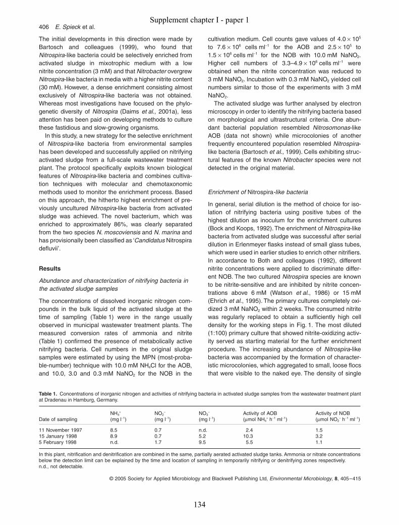
406
E. Spieck
et al.
© 2005 Society for Applied Microbiology and Blackwell Publishing Ltd,
Environmental Microbiology
,
8
, 405–415
The initial developments in this direction were made byBartosch and colleagues (1999), who found that
Nitrospira
-like bacteria could be selectively enriched fromactivated sludge in mixotrophic medium with a lownitrite concentration (3 mM) and that
Nitrobacter
overgrew
Nitrospira
-like bacteria in media with a higher nitrite content(30 mM). However, a dense enrichment consisting almostexclusively of
Nitrospira
-like bacteria was not obtained.Whereas most investigations have focused on the phylo-genetic diversity of
Nitrospira
(Daims
et al
., 2001a), lessattention has been paid on developing methods to culturethese fastidious and slow-growing organisms.
In this study, a new strategy for the selective enrichmentof
Nitrospira
-like bacteria from environmental sampleshas been developed and successfully applied on nitrifyingactivated sludge from a full-scale wastewater treatmentplant. The protocol specifically exploits known biologicalfeatures of
Nitrospira
-like bacteria and combines cultiva-tion techniques with molecular and chemotaxonomicmethods used to monitor the enrichment process. Basedon this approach, the hitherto highest enrichment of pre-viously uncultured
Nitrospira
-like bacteria from activatedsludge was achieved. The novel bacterium, which wasenriched to approximately 86%, was clearly separatedfrom the two species
N. moscoviensis
and
N. marina
andhas provisionally been classified as ‘
Candidatus
Nitrospiradefluvii’.
Results
Abundance and characterization of nitrifying bacteria in the activated sludge samples
The concentrations of dissolved inorganic nitrogen com-pounds in the bulk liquid of the activated sludge at thetime of sampling (Table 1) were in the range usuallyobserved in municipal wastewater treatment plants. Themeasured conversion rates of ammonia and nitrite(Table 1) confirmed the presence of metabolically activenitrifying bacteria. Cell numbers in the original sludgesamples were estimated by using the MPN (most-proba-ble-number) technique with 10.0 mM NH
4
Cl for the AOB,and 10.0, 3.0 and 0.3 mM NaNO
2
for the NOB in the
cultivation medium. Cell counts gave values of 4.0
¥
10
5
to 7.6
¥
10
6
cells ml
-
1
for the AOB and 2.5
¥
10
5
to1.5
¥
10
6
cells ml
-
1
for the NOB with 10.0 mM NaNO
2
.Higher cell numbers of 3.3–4.9
¥
10
6
cells ml
-
1
wereobtained when the nitrite concentration was reduced to3 mM NaNO
2
. Incubation with 0.3 mM NaNO
2
yielded cellnumbers similar to those of the experiments with 3 mMNaNO
2
.The activated sludge was further analysed by electron
microscopy in order to identify the nitrifying bacteria basedon morphological and ultrastructural criteria. One abun-dant bacterial population resembled
Nitrosomonas
-likeAOB (data not shown) while microcolonies of anotherfrequently encountered population resembled
Nitrospira
-like bacteria (Bartosch
et al
., 1999). Cells exhibiting struc-tural features of the known
Nitrobacter
species were notdetected in the original material.
Enrichment of
Nitrospira
-like bacteria
In general, serial dilution is the method of choice for iso-lation of nitrifying bacteria using positive tubes of thehighest dilution as inoculum for the enrichment cultures(Bock and Koops, 1992). The enrichment of
Nitrospira
-likebacteria from activated sludge was successful after serialdilution in Erlenmeyer flasks instead of small glass tubes,which were used in earlier studies to enrich other nitrifiers.In accordance to Both and colleagues (1992), differentnitrite concentrations were applied to discriminate differ-ent NOB. The two cultured
Nitrospira
species are knownto be nitrite-sensitive and are inhibited by nitrite concen-trations above 6 mM (Watson
et al
., 1986) or 15 mM(Ehrich
et al
., 1995). The primary cultures completely oxi-dized 3 mM NaNO
2
within 2 weeks. The consumed nitritewas regularly replaced to obtain a sufficiently high celldensity for the working steps in Fig. 1. The most diluted(1:100) primary culture that showed nitrite-oxidizing activ-ity served as starting material for the further enrichmentprocedure. The increasing abundance of
Nitrospira
-likebacteria was accompanied by the formation of character-istic microcolonies, which aggregated to small, loose flocsthat were visible to the naked eye. The density of single
Table 1.
Concentrations of inorganic nitrogen and activities of nitrifying bacteria in activated sludge samples from the wastewater treatment plantat Dradenau in Hamburg, Germany.
Date of samplingNH
4
+
(mg l
-
1
)NO
2–
(mg l
-
1
)NO
3–
(mg l
-
1
)Activity of AOB (
m
mol NH
4
+
h
-
1
ml
-
1
)Activity of NOB(
m
mol NO
2–
h
-
1
ml
-
1
)
11 November 1997 8.5 0.7 n.d. 2.4 1.515 January 1998 8.9 0.7 5.2 10.3 3.25 February 1998 n.d. 1.7 9.5 5.5 1.1
In this plant, nitrification and denitrification are combined in the same, partially aerated activated sludge tanks. Ammonia or nitrate concentrationsbelow the detection limit can be explained by the time and location of sampling in temporarily nitrifying or denitrifying zones respectively.n.d., not detectable.
Supplement chapter I - paper 1
134

Selective enrichment of
Nitrospira-
like bacteria
407
© 2005 Society for Applied Microbiology and Blackwell Publishing Ltd,
Environmental Microbiology
, 8, 405–415
planktonic cells and small cell aggregates never becamehigh enough to cause turbidity of the medium. The typicalbrownish colour of NOB became visible after concentra-ting the enriched biomass by centrifugation. In Nitrobacterthe brownish colour belongs to FeS proteins that are partof the b-subunit of the nitrite oxidoreductase (Kirstein andBock, 1993). In N. moscoviensis an unstable haem wasdetected in the b-subunit of the nitrite-oxidizing system(Spieck et al., 1998).
Inhibition of Nitrobacter and heterotrophic organisms
Various treatments with antibiotics were tested to deter-mine whether the growth of Nitrobacter and heterotrophicbacteria could be suppressed without compromisingNitrospira-like bacteria. Tetracycline and streptomycinwere not suitable as these antibiotic substances causeda decrease of Nitrospira-like bacteria. In contrast, the frac-
tion of Nitrospira-like bacteria in the enrichments becamelarger when 50 mg ml-1 ampicillin was added to themedium. The size of the largest, macroscopically visiblemicrocolonies increased approximately by a factor of 10(data not shown). Microscopic observation confirmed thatrepeated application of ampicillin also inhibited growth ofNitrobacter. However, higher concentrations of ampicillin(100 or 200 mg ml-1) suppressed the growth of Nitrospira-like bacteria, too.
Morphology of enriched Nitrospira-like bacteria
The structure of the large cell aggregates of Nitrospira-like bacteria detected by fluorescence in situ hybridization(FISH) was similar to that observed in activated sludge inearlier studies (Juretschko et al., 1998; Bartosch et al.,1999; Daims et al., 2001a). They consisted of smallgroups of cells with a spherical appearance, separated bycell-free channels and voids of different sizes (Fig. 2A).Electron microscopy confirmed that these Nitrospira clus-ters were relatively loose aggregates of short, slightlycurved cells that were frequently organized in tetradsembedded in extracellular polymeric substances (EPS)(Fig. 3A). Interestingly, the shape and arrangement of theNitrospira cells changed significantly after prolonged incu-bation (about 2 months) with repeated addition of freshsubstrate to the cultures. Following nitrate accumulationand increasing cell density, the microcolonies disinte-grated (Fig. 2B) and the length of the cells increased(Fig. 3B and C). Their irregular appearance now resem-bled that of N. moscoviensis, which tends to form star-likeextensions of the outer membrane (Ehrich et al., 1995).Likewise, longer periods of starvation led to the appear-ance of relatively long, curved rods with one turn, whichoccurred mainly as free-living planktonic cells (Fig. 3D).This morphological change to the characteristic spiral cellshape could be accelerated in stirred or aged cultures andwas reversible when starved cultures were transferred intofresh mineral medium containing nitrite.
Purification by Percoll density gradient centrifugation
The separation of Nitrospira-like bacteria from othermicroorganisms was further improved by Percoll densitygradient centrifugation of highly enriched cultures, wherea relatively large fraction of the target NOB occurred assingle planktonic cells. Fresh cultures were not suitablefor this technique because most Nitrospira cells wereembedded in large aggregates that included also othermicroorganisms (Fig. 2A) and thereby prevented physicalcell isolation. After centrifugation of a concentrated cul-ture, Nitrospira-like bacteria formed a brownish colouredband in the upper part of the centrifugation tube. Thisfraction was harvested and used as inoculum for new
Fig. 1. Flow chart of the procedure developed to enrich Nitrospira-like bacteria. Every step was performed in mineral medium with 3 mM NaNO2. All media, except in the 3 l bottles and the dilution series, were supplemented with ampicillin (50 mg ml-1). One per cent (v/v) inoculum was used to transfer cultures.
Incubate for 2-3 months.Check for nitrite oxidation weekly.If needed, replace consumed nitrite.
Transfer to fresh medium.
Transfer culture to 3 l flasks with 1.5l of medium, incubate2-3 months without stirring, feed 5-10x with fresh nitrite.
Percoll density gradient centrifugation
Make dilution series in tubes with 10 ml of medium.Incubate for 6 weeks.
Transfer most diluted nitrite-oxidizing culture to300 ml flasks. Check for nitrite consumption weekly.
If needed, replace consumed nitrite (3-5x, thentransfer culture to fresh medium)
Keep stock culture at 17C in the dark.Transfer to fresh medium every 4-6 months.
Mix inoculum [1% (v/v) activated sludge]with mineral medium.
Make dilution series (10-1 to 10-5) in 100 ml flaskswith 50 ml of mineral medium.
Incubate for 6-8 weeks at 28C without shaking.Check regularly for nitrite consumption.
If needed, replace consumed nitrite.
Transfer the most diluted nitrite-oxidizing cultureto 300 ml flasks with 150 ml of medium.
Rep
eat 3
x
Monitoring of enrichmentby FISH, fatty acid
analyses, and electron microscopy.
Supplement chapter I - paper 1
135
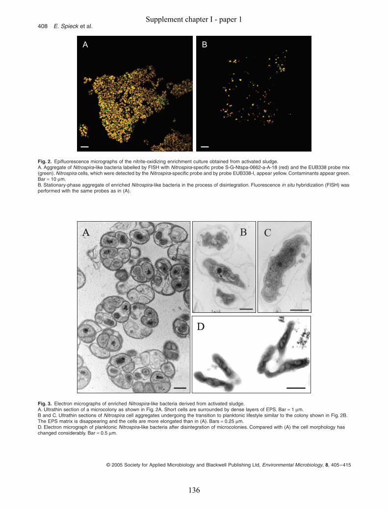
408 E. Spieck et al.
© 2005 Society for Applied Microbiology and Blackwell Publishing Ltd, Environmental Microbiology, 8, 405–415
Fig. 2. Epifluorescence micrographs of the nitrite-oxidizing enrichment culture obtained from activated sludge.A. Aggregate of Nitrospira-like bacteria labelled by FISH with Nitrospira-specific probe S-G-Ntspa-0662-a-A-18 (red) and the EUB338 probe mix (green). Nitrospira cells, which were detected by the Nitrospira-specific probe and by probe EUB338-I, appear yellow. Contaminants appear green. Bar = 10 mm.B. Stationary-phase aggregate of enriched Nitrospira-like bacteria in the process of disintegration. Fluorescence in situ hybridization (FISH) was performed with the same probes as in (A).
A BAAA BBB
Fig. 3. Electron micrographs of enriched Nitrospira-like bacteria derived from activated sludge.A. Ultrathin section of a microcolony as shown in Fig. 2A. Short cells are surrounded by dense layers of EPS. Bar = 1 mm.B and C. Ultrathin sections of Nitrospira cell aggregates undergoing the transition to planktonic lifestyle similar to the colony shown in Fig. 2B. The EPS matrix is disappearing and the cells are more elongated than in (A). Bars = 0.25 mm.D. Electron micrograph of planktonic Nitrospira-like bacteria after disintegration of microcolonies. Compared with (A) the cell morphology has changed considerably. Bar = 0.5 mm.
A
D
B CA
D
BB CC
Supplement chapter I - paper 1
136
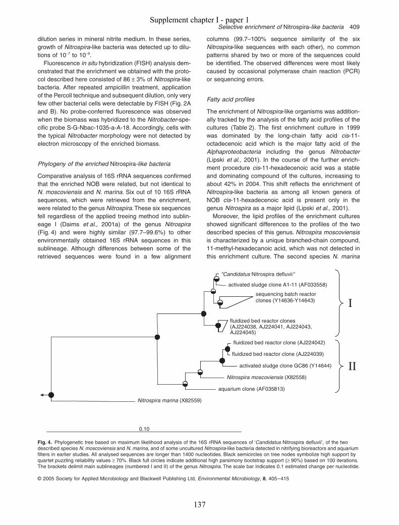
Selective enrichment of Nitrospira-like bacteria 409
© 2005 Society for Applied Microbiology and Blackwell Publishing Ltd, Environmental Microbiology, 8, 405–415
dilution series in mineral nitrite medium. In these series,growth of Nitrospira-like bacteria was detected up to dilu-tions of 10-7 to 10-9.
Fluorescence in situ hybridization (FISH) analysis dem-onstrated that the enrichment we obtained with the proto-col described here consisted of 86 ± 3% of Nitrospira-likebacteria. After repeated ampicillin treatment, applicationof the Percoll technique and subsequent dilution, only veryfew other bacterial cells were detectable by FISH (Fig. 2Aand B). No probe-conferred fluorescence was observedwhen the biomass was hybridized to the Nitrobacter-spe-cific probe S-G-Nbac-1035-a-A-18. Accordingly, cells withthe typical Nitrobacter morphology were not detected byelectron microscopy of the enriched biomass.
Phylogeny of the enriched Nitrospira-like bacteria
Comparative analysis of 16S rRNA sequences confirmedthat the enriched NOB were related, but not identical toN. moscoviensis and N. marina. Six out of 10 16S rRNAsequences, which were retrieved from the enrichment,were related to the genus Nitrospira. These six sequencesfell regardless of the applied treeing method into sublin-eage I (Daims et al., 2001a) of the genus Nitrospira(Fig. 4) and were highly similar (97.7–99.6%) to otherenvironmentally obtained 16S rRNA sequences in thissublineage. Although differences between some of theretrieved sequences were found in a few alignment
columns (99.7–100% sequence similarity of the sixNitrospira-like sequences with each other), no commonpatterns shared by two or more of the sequences couldbe identified. The observed differences were most likelycaused by occasional polymerase chain reaction (PCR)or sequencing errors.
Fatty acid profiles
The enrichment of Nitrospira-like organisms was addition-ally tracked by the analysis of the fatty acid profiles of thecultures (Table 2). The first enrichment culture in 1999was dominated by the long-chain fatty acid cis-11-octadecenoic acid which is the major fatty acid of theAlphaproteobacteria including the genus Nitrobacter(Lipski et al., 2001). In the course of the further enrich-ment procedure cis-11-hexadecenoic acid was a stableand dominating compound of the cultures, increasing toabout 42% in 2004. This shift reflects the enrichment ofNitrospira-like bacteria as among all known genera ofNOB cis-11-hexadecenoic acid is present only in thegenus Nitrospira as a major lipid (Lipski et al., 2001).
Moreover, the lipid profiles of the enrichment culturesshowed significant differences to the profiles of the twodescribed species of this genus. Nitrospira moscoviensisis characterized by a unique branched-chain compound,11-methyl-hexadecanoic acid, which was not detected inthis enrichment culture. The second species N. marina
Fig. 4. Phylogenetic tree based on maximum likelihood analysis of the 16S rRNA sequences of ‘Candidatus Nitrospira defluvii’, of the two described species N. moscoviensis and N. marina, and of some uncultured Nitrospira-like bacteria detected in nitrifying bioreactors and aquarium filters in earlier studies. All analysed sequences are longer than 1400 nucleotides. Black semicircles on tree nodes symbolize high support by quartet puzzling reliability values ≥ 70%. Black full circles indicate additional high parsimony bootstrap support (≥ 90%) based on 100 iterations. The brackets delimit main sublineages (numbered I and II) of the genus Nitrospira. The scale bar indicates 0.1 estimated change per nucleotide.
Nitrospira marina (X82559)
0.10
activated sludge clone A1-11 (AF033558)
(AJ224038, AJ224041, AJ224043,AJ224045)
fluidized bed reactor clone (AJ224042)
fluidized bed reactor clone (AJ224039)
activated sludge clone GC86 (Y14644)
Nitrospira moscoviensis (X82558)
aquarium clone (AF035813)
"Candidatus Nitrospira defluvii"
sequencing batch reactorclones (Y14636-Y14643)
fluidized bed reactor clones
I
II
Supplement chapter I - paper 1
137

410 E. Spieck et al.
© 2005 Society for Applied Microbiology and Blackwell Publishing Ltd, Environmental Microbiology, 8, 405–415
has major amounts of 7-cis-hexadecenoic acid, which waspresent only in small amounts in the enrichment culturesderived from activated sludge. These data confirmed theresults of the 16S rRNA sequence analyses that the Nitro-spira-like cells are related to but not identical with theknown species of the genus Nitrospira. Minor amounts offatty acids so far not assigned to the genus Nitrospiraindicated the presence of several groups of accompanyingmicroorganisms. These are iso/anteiso-branched-chainfatty acids and cyclopropyl lipids, which when summedtogether account for about 12% of probably non-Nitrospira-related lipids in the enrichment culture from2004.
Discussion
This work describes the first selective enrichment of amember of Nitrospira sublineage I, which is of majorimportance for nitrogen removal in wastewater treatmentplants but does not contain cultured representatives. Theabundance of these organisms in nitrifying wastewatertreatment plants usually amounts for 1–10% of the totalbacterial population (Daims et al., 2001b; Juretschkoet al., 2002).
The activated sludge biomass was diluted in Erlen-meyer flasks in order to reduce the density of het-erotrophic bacteria and organic material. Preliminaryexperiments have shown that growth of Nitrospira derivedfrom activated sludge was enhanced in Erlenmeyer flaskssupplying a large surface for the medium that facilitatesoxygen diffusion in comparison with tubes. Low nitriteconcentrations in the mineral medium and regular dilu-tions of the cultures effectively promoted Nitrospira duringthe enrichment process. This was important as the fattyacid profiles from the early stages of the enrichment sug-gested that Nitrobacter cells were also present. Schrammand colleagues (1999) postulated, based on microsensorand FISH data, that Nitrospira-like bacteria have a higheraffinity for nitrite than Nitrobacter. Reactor experimentsperformed with different nitrite concentrations in the influ-ent lend additional weight to this hypothesis (Wagneret al., 2002). Under the assumption that nitrite concentra-tion is the major factor controlling the competition betweenNitrospira and Nitrobacter and based on models of r- andK-selection in microbial ecology (Andrews and Harris,1986), Nitrospira-like bacteria can thus be considered asK-strategists while Nitrobacter species are r-strategists,which depend on relatively high nitrite concentrations andwill be outcompeted by Nitrospira-like bacteria if nitrite isthe growth-limiting factor.
A newly discovered property of Nitrospira-like bacteria,which was exploited during the enrichment procedure, istheir resistance to ampicillin applied in a concentrationapparently harmful to Nitrobacter and contaminating het-erotrophic bacteria. The higher resistance of Nitrospiraagainst ampicillin might be explained by the formation ofdense microcolonies embedded in EPS that serve asdiffusion barrier for dissolved substances. Furthermore,Foley and Gilbert (1996) suggested that observed resis-tance of microbial biofilms against antibiotics could be dueto a high binding capacity of charged molecules and deg-radation by extracellular enzymes.
The inhibition of heterotrophs was important for theenrichment even though the standard mineral mediumdid not contain any organic carbon source. Extracellularexopolymeric substances and soluble organic compoundsproduced by nitrifying bacteria are utilized by het-erotrophic organisms (Rittmann et al., 1994; Kindaichiet al., 2004) and most likely served as carbon and energysources for contaminating bacteria in the enrichments.Further separation of Nitrospira-like bacteria wasachieved by Percoll density gradient centrifugation of sta-tionary-phase cultures, which contained many planktonicNitrospira cells (Fig. 3D). It is tempting to speculate thatthe switch from growth in clusters while nutrients are plen-tiful (Figs 2A and 3A) to a planktonic lifestyle whenresources become limited (Fig. 3D) is an efficient strategyto escape adverse conditions and to colonize new habi-
Table 2. Fatty acid profiles of the enriched Nitrospira-like bacteria.
Date of cultureNovember1999
May2001
October2004
Fatty acids (%)8:0 0.512:0 2.5 1.0 0.814:0 8.5 12.1 7.4ECLa 14.429 1.015:0 iso 4.7 5.9 2.115:0 anteiso 2.3 0.815:0 0.5 0.414:0 3OH 0.716:0 iso 1.2 1.116:1 cis 7 2.9 3.016:1 cis 9 3.3 2.1 1.316:1 cis 11 14.2 33.6 42.216:0 18.1 16.9 15.815:0 iso 3OH 1.016:0 10-methyl 0.617:0 anteiso 0.5 0.417:0 cyclo 9–10 6.6 6.717:0 0.5ECLa 17.122 1.016:0 3OH 3.7 2.9 2.6ECLa 17.567 1.918:1 cis 9 0.5 1.418:1 cis 11 34.5 1.4 5.118:0 3.3 0.7 3.018:1 cis 11 11-methyl 2.517:0 iso 3OH 1.8ECLa 18.762 1.219:0 cyclo 11–12 4.8 0.9 1.8ECLa 19.685 1.4
a. For unknown fatty acids the equivalent chain length (ECL) is given.Culture volume, 1.5 l.
Supplement chapter I - paper 1
138

Selective enrichment of Nitrospira-like bacteria 411
© 2005 Society for Applied Microbiology and Blackwell Publishing Ltd, Environmental Microbiology, 8, 405–415
tats. Variable cell morphology in adaptation to changinggrowth conditions may be a common feature of the genusNitrospira as this phenomenon was also reported forN. moscoviensis (Ehrich et al., 1995) and N. marina (Wat-son et al., 1986).
The application of FISH combined with microautora-diography (FISH-MAR; Lee et al., 1999) on unculturedNitrospira-like bacteria in nitrifying biofilm provided evi-dence that these NOB take up and assimilate pyruvate(Daims et al., 2001a). According to Watson and col-leagues (1986) N. marina grows better in the presence ofpyruvate than in pure nitrite mineral medium. Therefore,in order to achieve faster growth and higher yield of Nitro-spira-like bacteria, some of the enrichments obtained inthis study were supplied with small amounts of pyruvate(0.5 mM). Although this substrate stimulated growth ofNitrospira, the enhanced growth of heterotrophs provedthe addition of pyruvate unsuitable for obtaining largeramounts of highly enriched Nitrospira biomass.
Nitrospira-like bacteria are fastidious and slow-growingorganisms, which are easily overgrown by other bacteriaunder suboptimal conditions. Therefore, the cultures weremonitored by molecular, chemotaxonomic and microscop-ical methods to determine which factors enhanced orinhibited the enrichment of Nitrospira. This strategyallowed selection of the best enrichments for the nextsteps of the procedure and led to the development of aprotocol that can be used in future to enrich or even isolateother Nitrospira-like bacteria or similarly recalcitrant nitri-fiers from environmental samples (Fig. 1). By the applica-tion of ampicillin further high enrichments of Nitrospira-likebacteria were obtained from several other habitats (E.Spieck, unpublished results). It is reasonable to believethat the accelerated collection of new members of theseformerly ‘uncultivated’ bacteria will disclose increasingphylogenetic, physiological and ecological diversity.
Based on phylogenetic analyses of 16S rRNA genesequences, the genus Nitrospira has been divided intofour sublineages (Daims et al., 2001a). The genus hosts
two described species, which belong to sublineage II(N. moscoviensis) and IV (N. marina) respectively. Incontrast, the Nitrospira-like bacterium enriched in thisstudy is a representative of sublineage I, which so farcontained only 16S rRNA sequences of uncultured Nitro-spira-like bacteria found in nitrifying wastewater treat-ment plants and bioreactors (Daims et al., 2001a). Thisorganism is clearly different from N. moscoviensis and N.marina (Table 3). The similarity between the 16S rRNAsequences of these species and the enriched organismis 94.1% (N. moscoviensis) and 88.3% (N. marina).According to Stackebrandt and Goebel (1994), it isunlikely that two organisms with 16S rRNA sequencesimilarity below 97% have more than 70% DNA similarityand belong to the same species. Furthermore, Keswaniand Whitman (2001) showed that even if two organismshave as much as 98.6% 16S rRNA sequence similaritythere is still a probability of 99% that they represent dif-ferent species. Phenotypically, the Nitrospira-like culturecan be differentiated from the two representatives of sub-lineage II and IV by its unique lipid pattern dominated by11-cis-hexadecenoic acid and not by 11-methyl-hexade-canoic acid and 7-cis-hexadecenoic acid respectively.Based on the results of this study we propose, accordingto Murray and Schleifer (1994) and Murray and Stacke-brandt (1995), provisional classification of the novelNitrospira-like bacterium as ‘Candidatus Nitrospira deflu-vii’. The short description of ‘Candidatus Nitrospira deflu-vii’ is as follows: phylogenetically related to the genusNitrospira; not isolated; Gram-negative; major fatty acidsare 11-cis-hexadecenoic acid and hexadeacanoic acid;short curved rods in microcolonies with a diameter of0.5–1.2 mm or long spiral-shaped rods with one to fourturns, 0.2–0.4 mm in width and length of 0.7–1.7 mm;basis of assignment, 16S rRNA gene sequences (Acces-sion No. DQ059545) and detection by the genus-specificoligonucleotide probe complementary to helix 23 of the16S rRNA S-G-Ntspa-0662-a-A-18 (5¢-AGAGGAGCGCGGAAUUCC-3¢); cell aggregates or free-living plank-
Table 3. Comparable characteristics of ‘Candidatus Nitrospira defluvii’ enriched in this study, N. moscoviensis and N. marina.
‘Candidatus Nitrospira defluvii’ N. moscoviensis N. marina
Cell morphology Short, slightly curved cells or spiral-shaped rods
Irregularly shaped cells or spiral-shaped rods
‘Comma-shaped’ cells or spiral-shaped rods
Size 0.2–0.4 mm ¥ 0.7–1.7 mm 0.2–0.4 mm ¥ 0.9–2.2 mm 0.3–0.4 mm ¥ 0.8–1.0 mmTurns 1–4 1–3 1–12Tendency to aggregate Strong Present WeakTemperature for cultivation 28∞C 37∞C 28∞CMedium Non-marine Non-marine MarinePhylogenetic affiliation in genus Nitrospira Sublineage I Sublineage II Sublineage IVDominant lipids 16:1 cis 11 16:1 cis 11 16:1 cis 7
16:0 16:0 16:1 cis 1116:0 11-methyla 16:0a
a. As reported by Lipski and colleagues (2001).
Supplement chapter I - paper 1
139
412 E. Spieck et al.
© 2005 Society for Applied Microbiology and Blackwell Publishing Ltd, Environmental Microbiology, 8, 405–415
tonic cells; aerobic chemolithoautotrophic nitrite oxidizer,mesophilic; utilizes pyruvate (this study).
Nitrospira-like bacteria, and not members of the genusNitrobacter, are mainly responsible for nitrite oxidation inmost wastewater treatment systems and also in manynatural habitats. As no pure cultures or high enrichmentsof the widespread sublineage I of this genus were avail-able, our knowledge of the physiology of these organismsis scarce. The enrichment obtained in this study will makeit possible to conduct encompassing physiological exper-iments that could not be performed with environmentalsamples containing lower amounts of Nitrospira-like bac-teria, and it provides the biomass for an environmentalgenomics approach launched recently in order to investi-gate the genomic make-up and biology of these andrelated Nitrospira-like bacteria.
Experimental procedures
Activated sludge samples
The samples used as primary inoculum for the enrichmentof Nitrospira-like bacteria were taken between November1997 and December 1998 from the nitrification stage of themunicipal wastewater treatment plant in Köhlbrandhöft/Dradenau, Hamburg, Germany (1.8 ¥ 106 population equiva-lents). The activated sludge had a pH of 7.7–8.1 and a tem-perature of 18–20∞C. The chemical demand for oxygen wasin the range of 295–325 mg ml-1.
Chemical analyses
Concentrations of ammonium were measured by high-perfor-mance liquid chromatography (HPLC) according to Corbin(1984). Nitrite and nitrate concentrations were determinedby ion-pair chromatography with a Hypersil ODS C18column (125 ¥ 4.6 mm) (Meincke et al., 1992) followed byUV detection in an automated system (Kontron, Eching,Germany).
Quantification of AOB and NOB
Most-probable-number (MPN) analyses were performed asdescribed by Mansch and Bock (1998) to estimate the abun-dance of culturable AOB and NOB in activated sludge sam-ples. Aliquots of sludge containing nitrifying bacteria weresonicated for 3 min and dilution series were prepared in 0.9%NaCl up to 10-7. Three replicates per dilution were inoculatedwith 0.1 ml aliquots using 10 ml glass tubes containing nitritemineral medium (Ehrich et al., 1995) with a final volume of5 ml per tube. The nitrite concentration in the medium wasadjusted to 10.0, 3.0 or 0.3 mM in parallel dilution series. ForAOB mineral medium according to Krümmel and Harms(1982) with 10 mM NH4Cl was prepared. The diluted sampleswere incubated for 6 weeks without agitation in the dark at28∞C. Formation or consumption of nitrite detected by theGriess–Ilosavay spot test (Schmidt and Belser, 1982) indi-cated growth of AOB and NOB respectively. Statistical calcu-
lations for the estimation of the MPN were performed inaccordance to Alexander (1982).
Determination of nitrification activity
In order to measure the turnover of ammonia and nitrite, 5 mlof activated sludge was transferred into 45 ml of mineralmedium amended with 5 mM NH4Cl or 5 mM NaNO2 respec-tively. The sludge samples were protected from light andincubated under agitation (120 r.p.m.) at 28∞C. The consump-tion of ammonia and nitrite was monitored during a period of6 h.
Enrichment of Nitrospira-like bacteria
In contrast to the protocol followed by Bartosch and col-leagues (1999), Nitrospira-like bacteria were enriched in puremineral medium (Ehrich et al., 1995), which contained only3 mM NaNO2 as energy source and no organic carbon inorder to suppress growth of heterotrophic bacteria. Theenrichment was started in 100 ml Erlenmeyer flasks filledwith 50 ml of the medium and 1% (v/v) activated sludge. Theflasks were incubated at 28∞C in the dark without agitation.Nitrite consumption was frequently measured to identify cul-tures containing actively growing NOB. These enrichmentswere supplied with sterile nitrite solution every 3–5 days to afinal concentration of 3 mM NaNO2 respectively. After approx-imately 6 weeks, 1.5 ml aliquots of the enrichments weretransferred into 300 ml flasks filled with 150 ml of fresh nitritemedium. Three different antibiotics were added to the enrich-ments in order to suppress growth of nitrite-oxidizing Nitro-bacter cells and heterotrophic bacteria. Filter-sterilized stocksolutions of ampicillin, tetracycline or streptomycin wereadded to the medium to final concentrations of 50–200 mgml-1 (ampicillin), 10–25 mg ml-1 (tetracycline) and 50–100 mgml-1 (streptomycin). Cultures with a high cell density weregrown in 3 l flasks and were used to separate Nitrospira-likebacteria from contaminants by Percoll density gradient cen-trifugation and subsequent serial dilutions as described byEhrich and colleagues (1995).
Electron microscopy
For observation by electron microscopy, cells were fixed with2.5% (v/v) glutaraldehyde and 2% (w/v) osmium tetroxide andembedded in Epon 812 (Serva) according to a previouslypublished protocol (Ehrich et al., 1995). The embedded sam-ples were sectioned, stained with uranyl acetate (Watson,1958) and lead citrate (Reynolds, 1963) and examined in atransmission electron microscope (Model 420, Philips). Cellmorphology was visualized by staining the bacteria with ura-nyl acetate as described by Spieck and colleagues (1996).
Fluorescence in situ hybridization
Aliquots of the enrichments were prepared for FISH byparaformaldehyde fixation as described by Amann (1995).Fixed biomass was spotted onto microscope slides and FISHwas performed according to the protocol detailed by Manz
Supplement chapter I - paper 1
140
Selective enrichment of Nitrospira-like bacteria 413
© 2005 Society for Applied Microbiology and Blackwell Publishing Ltd, Environmental Microbiology, 8, 405–415
and colleagues (1992) and Daims and colleagues (2005).The applied oligonucleotide probes were S-G-Ntspa-0662-a-A-18 specific for the genus Nitrospira (Daims et al., 2001a),S-*-Ntspa-0712-a-A-21 targeting the phylum Nitrospira(Daims et al., 2001a), S-G-Nbac-1035-a-A-18 specific for thegenus Nitrobacter (= NIT3; Wagner et al., 1996), and theEUB probe mix that detects most known Bacteria (Amannet al., 1990; Daims et al., 1999). The probes were 5¢ labelledwith either the dye FLUOS [5(6)-carboxyfluorescein-N-hydroxysuccinimide ester] or the sulfoindocyanine dye Cy3.The Nitrospira- and Nitrobacter-specific probes were usedtogether with their competitor oligonucleotides as describedby Daims and colleagues (2001a) and Wagner and col-leagues (1996) respectively. Labelled probes and unlabelledcompetitors were obtained from Thermo Hybaid (InteractivaDivision, Ulm, Germany). Probe-conferred fluorescence wasrecorded with a confocal laser scanning microscope (LSM510 Meta, Zeiss, Oberkochen, Germany) by using an Ar ionlaser to detect FLUOS (488 nm excitation wavelength) andan HeNe laser (543 nm) to detect Cy3 respectively.
Quantification of Nitrospira
The fraction of Nitrospira-like bacteria in the enrichments,based on the total bacterial population (autotrophs plus het-erotrophs), was determined as relative biovolume by quanti-tative FISH, confocal laser scanning microscopy and digitalimage analysis according to a protocol described elsewhere(Schmid et al., 2000) and by using DAIME, an image analysisprogram developed at the University of Vienna.
Cloning and phylogenetic analysis of 16S rRNA genes
Bacterial 16S rRNA genes were PCR amplified, cloned andsequenced as described by Juretschko and colleagues(1998) with the only modification that instead of extractedgenomic DNA, 1.5 ml of Nitrospira enrichment was addeddirectly to the PCR reaction mix for 16S rRNA gene amplifi-cation. The obtained sequences were aligned and phyloge-netically analysed by using the ARB program (Ludwig et al.,2004) according to Daims and colleagues (2001a).
Fatty acid profiles
Biomass for fatty acid extraction was obtained from large-scale enrichments grown in 1.5 l of medium in 3 l Erlenmeyerflasks. Cultures were incubated without stirring for 2–3 months and regularly fed with sterile nitrite solution. Whendense flocs of Nitrospira-like bacteria had developed, cellswere harvested by centrifugation, washed in 0.9% (w/v) NaCland stored at -20∞C. Saponification of the cell pellets with15% NaOH in 50% methanol, acid methylation with 6 N HClin 50% methanol and extraction of fatty acid methyl esters(FAMEs) were performed as described by Sasser (1990). TheFAME extracts were analysed using a Hewlett–Packardmodel 6890 gas chromatograph equipped with a 5% phenylmethyl silicone capillary column and a flame ionization detec-tor. The identity of fatty acids was verified by GC-MS with aHewlett–Packard model 5890 series II gas chromatographequipped with a 5% phenyl methyl silicone capillary column
and a model 5972 mass selective detector. The chromato-graphic conditions were used as described previously (Lipskiand Altendorf, 1997). The positions of hydroxy, methyl, cyclo-propene groups and double bonds were given from the car-boxyl group of the fatty acid molecule according to therecommendations of the IUPAC-IUB Commission on bio-chemical nomenclature (CBN) 1977.
Acknowledgements
This work was partly funded by Grant LS 216 of the WienerWissenschafts-, Forschungs-und Technologiefonds to H.D.and M.W. The excellent technical assistance of Irmhild Wach-holz and Christian Baranyi is acknowledged.
References
Alexander, M. (1982) Most probable number method formicrobial populations. In Methods of Soil Analysis Proper-ties. Miller, R.H., and Keeney, D.R. (eds). Madison, USA:American Society of Agronomy, pp. 815–820.
Amann, R.I. (1995) In situ identification of micro-organismsby whole cell hybridization with rRNA-targeted nucleic acidprobes. In Molecular Microbial Ecology Manual. Akkeman,A.D.C., van Elsas, J.D., and de Bruigin, F.J. (eds). Dor-drecht, the Netherlands: Kluwer Academic Publishers, pp.1–15.
Amann, R.I., Binder, B.J., Olson, R.J., Chisholm, S.W.,Devereux, R., and Stahl, D.A. (1990) Combination of 16SrRNA-targeted oligonucleotide probes with flow cytometryfor analyzing mixed microbial populations. Appl EnvironMicrobiol 56: 1919–1925.
Andrews, J.H., and Harris, R.F. (1986) r- and K-selection andmicrobial ecology. Adv Microb Ecol 9: 99–147.
Bartosch, S., Wolgast, I., Spieck, E., and Bock, E. (1999)Identification of nitrite-oxidizing bacteria with monoclonalantibodies recognizing the nitrite oxidoreductase. ApplEnviron Microbiol 65: 4126–4133.
Bartosch, S., Hartwig, C., Spieck, E., and Bock, E. (2002)Immunological detection of Nitrospira-like bacteria in vari-ous soils. Microb Ecol 43: 26–33.
Bock, E., and Koops, H.-P. (1992) The genus Nitrobacter andrelated genera. In The Prokaryotes. Balows, A., Trüper,H.G., Dworkin, M., Harder, W., and Schleifer, K.-H. (eds).New York, USA: Springer-Verlag, pp. 2302–2309.
Both, G.J., Gerards, S., and Laanbroek, H.J. (1992) Theoccurrence of chemolitho-autotrophic nitrifiers in water-saturated grassland soils. Microb Ecol 23: 15–26.
Burrell, P.C., Keller, J., and Blackall, L.L. (1998) Microbiologyof a nitrite-oxidizing bioreactor. Appl Environ Microbiol 64:1878–1883.
Corbin, J.L. (1984) Liquid chromatographic-fluorescencedetermination of ammonia from nitrogenase reactions.Appl Environ Microbiol 47: 1027–1030.
Daims, H., Brühl, A., Amann, R., Schleifer, K.-H., and Wag-ner, M. (1999) The domain-specific probe EUB338 is insuf-ficient for the detection of all Bacteria: development andevaluation of a more comprehensive probe set. Syst ApplMicrobiol 22: 434–444.
Daims, H., Nielsen, J.L., Nielsen, P.H., Schleifer, K.H., and
Supplement chapter I - paper 1
141
414 E. Spieck et al.
© 2005 Society for Applied Microbiology and Blackwell Publishing Ltd, Environmental Microbiology, 8, 405–415
Wagner, M. (2001a) In situ characterization of Nitrospira-like nitrite-oxidizing bacteria active in wastewater treatmentplants. Appl Environ Microbiol 67: 5273–5284.
Daims, H., Purkhold, U., Bjerrum, L., Arnold, E., Wilderer,P.A., and Wagner, M. (2001b) Nitrification in sequencingbiofilm batch reactors: lessons from molecular ap-proaches. Water Sci Tech 43: 9–18.
Daims, H., Stoecker, K., and Wagner, M. (2005) Fluores-cence in situ hybridisation for the detection of prokaryotes.In Molecular Microbial Ecology. Osborn, A.M., and Smith,C.J. (eds). Abingdon, UK: Bios-Garland (in press).
Ehrich, S., Behrens, D., Lebedeva, E., Ludwig, W., and Bock,E. (1995) A new obligately chemolithoautotrophic, nitrite-oxidizing bacterium, Nitrospira moscoviensis sp. nov. andits phylogenetic relationship. Arch Microbiol 164: 16–23.
Foley, I., and Gilbert, P. (1996) Antibiotic resistance of bio-films. Biofouling 10: 331–346.
Hentschel, U., Hopke, J., Horn, M., Friedrich, A.B., Wagner,M., Hacker, J., and Moore, B.S. (2002) Molecular evidencefor a uniform microbial community in sponges from differentoceans. Appl Environ Microbiol 68: 4431–4440.
Holmes, A.J., Tujula, N.A., Holley, M., Contos, A., James,J.M., Rogers, P., and Gillings, M.R. (2001) Phylogeneticstructure of unusual aquatic microbial formations in Nullar-bor caves, Australia. Environ Microbiol 3: 256–264.
Hovanec, T.A., Taylor, L.T., Blakis, A., and DeLong, E.F.(1998) Nitrospira-like bacteria associated with nitrite oxida-tion in freshwater aquaria. Appl Environ Microbiol 64: 258–264.
Juretschko, S., Timmermann, G., Schmid, M., Schleifer,K.-H., Pommerening-Röser, A., Koops, H.-P., and Wagner,M. (1998) Combined molecular and conventional analysesof nitrifying bacterium diversity in activated sludge: Nitroso-coccus mobilis and Nitrospira-like bacteria as dominantpopulations. Appl Environ Microbiol 64: 3042–3051.
Juretschko, S., Loy, A., Lehner, A., and Wagner, M. (2002)The microbial community composition of a nitrifying-denitrifying activated sludge from an industrial sewagetreatment plant analyzed by the full-cycle rRNA approach.Syst Appl Microbiol 25: 84–99.
Keswani, J., and Whitman, W.B. (2001) Relationship of 16SrRNA sequence similarity to DNA hybridization in prokary-otes. Int J Syst Evol Microbiol 51: 667–678.
Kindaichi, T., Ito, T., and Okabe, S. (2004) Ecophysiologicalinteraction between nitrifying bacteria and heterotrophicbacteria in autotrophic nitrifying biofilms as determined bymicroautoradiography-fluorescence in situ hybridization.Appl Environ Microbiol 70: 1641–1650.
Kirstein, K., and Bock, E. (1993) Close genetic relationshipbetween Nitrobacter hamburgensis nitrite oxidoreductaseand Escherichia coli nitrate reductases. Arch Microbiol160: 447–453.
Krümmel, A., and Harms, H. (1982) Effect of organic matteron growth and cell yield of ammonia-oxidizing bacteria.Arch Microbiol 133: 50–54.
Lebedeva, E.V., Alawi, M., Fiencke, C., Namsaraev, B., Bock,E., and Spieck, E. (2005) Moderately thermophilic nitrifyingbacteria from a hot spring of the Baikal rift zone. FEMSMicrobiol Ecol (in press).
Lee, N., Nielsen, P.H., Andreasen, K.H., Juretschko, S.,Nielsen, J.L., Schleifer, K.-H., and Wagner, M. (1999)
Combination of fluorescent in situ hybridization andmicroautoradiography – a new tool for structure-functionanalyses in microbial ecology. Appl Environ Microbiol 65:1289–1297.
Lipski, A., and Altendorf, K. (1997) Identification of het-erotrophic bacteria isolated from ammonia-supplied exper-imental biofilters. Syst Appl Microbiol 20: 448–457.
Lipski, A., Spieck, E., Makolla, A., and Altendorf, K. (2001)Fatty acid profiles of nitrite-oxidizing bacteria reflect theirphylogenetic heterogeneity. Syst Appl Microbiol 24: 377–384.
Ludwig, W., Strunk, O., Westram, R., Richter, L., Meier, H.,Yadhukumar, et al. (2004) ARB: a software environment forsequence data. Nucleic Acids Res 32: 1363–1371.
Mansch, R., and Bock, E. (1998) Biodeterioration of naturalstone with special reference to nitrifying bacteria. Biodeg-radation 9: 47–64.
Manz, W., Amann, R., Ludwig, W., Wagner, M., and Schlei-fer, K.-H. (1992) Phylogenetic oligodeoxynucleotide probesfor the major subclasses of proteobacteria: problems andsolutions. Syst Appl Microbiol 15: 593–600.
Meincke, M., Bock, E., Kastrau, D., and Kroneck, P.M.H.(1992) Nitrite oxidoreductase from Nitrobacter hamburgen-sis: redox centers and their catalytic role. Arch Microbiol158: 127–131.
Murray, R.G., and Schleifer, K.H. (1994) Taxonomic notes: aproposal for recording the properties of putative taxa ofprocaryotes. Int J Syst Bacteriol 44: 174–176.
Murray, R.G., and Stackebrandt, E. (1995) Taxonomic note:implementation of the provisional status Candidatus forincompletely described procaryotes. Int J Syst Bacteriol45: 186–187.
Okabe, S., Satoh, H., and Watanabe, Y. (1999) In situ anal-ysis of nitrifying biofilms as determined by in situ hybrid-ization and the use of microelectrodes. Appl EnvironMicrobiol 65: 3182–3191.
Reynolds, E.S. (1963) The use of lead citrate at high pH asan electron opaque stain in electron microscopy. Cell Biol18: 208–212.
Rittmann, B.E., Regan, J.M., and Stahl, D.A. (1994) Nitrifica-tion as a source of soluble organic substrate in biologicaltreatment. Water Sci Technol 30: 1–8.
Sasser, M. (1990) Identification of bacteria through fatty acidanalysis. In Methods in Phytobacteriology. Klement, Z.,Rudoplph, K., and Sands, D.C. (eds). Budapest, Hungary:Akademiai Kiado, pp. 199–204.
Schmid, M., Twachtmann, U., Klein, M., Strous, M.,Juretschko, S., Jetten, M., et al. (2000) Molecular evidencefor a genus-level diversity of bacteria capable of catalyzinganaerobic ammonium oxidation. Syst Appl Microbiol 23:93–106.
Schmidt, E.L., and Belser, L.W. (1982) Nitrifying bacteria. InMethods of Soil Analysis Properties. Miller, R.H., andKeeney, D.R. (eds). Madison, USA: American Society ofAgronomy, pp. 1027–1042.
Schramm, A., de Beer, D., van den Heuvel, J.C., Ottengraf,S., and Amann, R. (1999) Microscale distribution of popu-lations and activities of Nitrosospira and Nitrospira spp.along a macroscale gradient in a nitrifying bioreactor:quantification by in situ hybridization and the use ofmicrosensors. Appl Environ Microbiol 65: 3690–3696.
Supplement chapter I - paper 1
142

Selective enrichment of Nitrospira-like bacteria 415
© 2005 Society for Applied Microbiology and Blackwell Publishing Ltd, Environmental Microbiology, 8, 405–415
Spieck, E., Müller, S., Engel, A., Mandelkow, E., Patel, H.,and Bock, E. (1996) Two-dimensional structure ofmembrane-bound nitrite oxidoreductase from Nitrobacterhamburgensis. J Structural Biol 117: 117–123.
Spieck, E., Ehrich, S., Aamand, J., and Bock, E. (1998)Isolation and immunocytochemical location of the nitrite-oxidizing system in Nitrospira moscoviensis. Arch Microbiol169: 225–230.
Stackebrandt, E., and Goebel, B.M. (1994) Taxonomic note:a place for DNA–DNA reassociation and 16S rRNAsequence analysis in the present species definition in bac-teriology. Int J Syst Bacteriol 44: 846–849.
Wagner, M., Rath, G., Koops, H.-P., Flood, J., and Amann,
R. (1996) In situ analysis of nitrifying bacteria in sewagetreatment plants. Water Sci Techn 34: 237–244.
Wagner, M., Loy, A., Nogueira, R., Purkhold, U., Lee, N., andDaims, H. (2002) Microbial community composition andfunction in wastewater treatment plants. Antonie Van Leeu-wenhoek 81: 665–680.
Watson, M.L. (1958) Staining of tissue sections for electronmicroscopy with heavy metals. J Biophys Biochem Cytol4: 475–478.
Watson, S.W., Bock, E., Valois, F.W., Waterbury, J.B., andSchlosser, U. (1986) Nitrospira marina gen. nov. sp. nov.:a chemolithotrophic nitrite-oxidizing bacterium. Arch Micro-biol 144: 1–7.
Supplement chapter I - paper 1
143

Supplement chapter I - paper 1
144
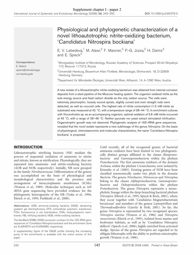
Physiological and phylogenetic characterization of anovel lithoautotrophic nitrite-oxidizing bacterium,‘Candidatus Nitrospira bockiana’
E. V. Lebedeva,1 M. Alawi,2 F. Maixner,3 P.-G. Jozsa,2 H. Daims3
and E. Spieck2
Correspondence
E. Spieck
spieck@mikrobiologie.
uni-hamburg.de
1Winogradsky Institute of Microbiology, Russian Academy of Sciences, Prospect 60-let Oktyabrya7/2, Moscow 117312, Russia
2Universitat Hamburg, Biozentrum Klein Flottbek, Mikrobiologie, Ohnhorststr. 18, D-22609Hamburg, Germany
3Department fur Mikrobielle Okologie, Universitat Wien, Althanstr. 14, A-1090 Wien, Austria
A new isolate of a lithoautotrophic nitrite-oxidizing bacterium was obtained from internal corrosion
deposits from a steel pipeline of the Moscow heating system. The organism oxidized nitrite as the
sole energy source and fixed carbon dioxide as the only carbon source. The cells were
extremely pleomorphic: loosely wound spirals, slightly curved and even straight rods were
detected, as well as coccoid cells. The highest rate of nitrite consumption (1.5 mM nitrite as
substrate) was measured at 42 6C, with a temperature range of 28–44 6C. In enrichment cultures
with Nocardioides sp. as an accompanying organism, optimal oxidation of 5.8 mM nitrite occurred
at 45 6C, with a range of 28–48 6C. Neither pyruvate nor yeast extract stimulated nitrification.
Organotrophic growth was not observed. Phylogenetic analysis of 16S rRNA gene sequences
revealed that the novel isolate represents a new sublineage of the genus Nitrospira. On the basis
of physiological, chemotaxonomic and molecular characteristics, the name ‘Candidatus Nitrospira
bockiana’ is proposed.
INTRODUCTION
Lithoautotrophic nitrifying bacteria (NB) mediate theprocess of sequential oxidation of ammonia to nitriteand nitrate, known as nitrification. Physiologically, they areseparated into ammonia- and nitrite-oxidizing bacteria(AOB and NOB, respectively). Initially, NB were groupedin the family Nitrobacteraceae. Differentiation of the generawas accomplished on the basis of physiological andmorphological characteristics and the presence andarrangement of intracytoplasmic membranes (ICMs)(Watson et al., 1989). Molecular techniques such as 16SrRNA gene sequencing have provided evidence for thephylogenetic heterogeneity of the NB (Teske et al., 1994;Ehrich et al., 1995; Purkhold et al., 2000).
Until recently, all of the recognized genera of bacterialammonia oxidizers have been limited to two phylogeneti-cally distinct groups affiliated to the classes Betaproteo-bacteria and Gammaproteobacteria within the phylumProteobacteria. The first ammonia oxidizers of the domainArchaea, within the phylum Crenarchaeota, were isolated byKonneke et al. (2005). Existing genera of NOB have beenclassified taxonomically under two phyla in the domainBacteria. The genera Nitrobacter, Nitrococcus and Nitrospinabelong to the classes Alphaproteobacteria, Gammaproteo-bacteria and Deltaproteobacteria within the phylumProteobacteria. The genus Nitrospira represents a mono-phyletic lineage within the deep-branching bacterial phylumNitrospira (Ehrich et al., 1995; Spieck & Bock, 2001), wherethey occur together with ‘Candidatus Magnetobacteriumbavaricum’ and members of the genera Leptospirillum andThermodesulfovibrio (Garrity & Holt, 2001). So far, thegenus Nitrospira is represented by two recognized species:Nitrospira marina (Watson et al., 1986) and Nitrospiramoscoviensis (Ehrich et al., 1995), isolated from marine andfreshwater habitats, as well as by ‘Candidatus Nitrospiradefluvii’ (Spieck et al., 2006), highly enriched from activatedsludge. Species of the genus Nitrospira are regarded to beobligate lithotrophs with the ability to perform mixotrophicgrowth (Watson et al., 1986).
Abbreviations: AOB, ammonia-oxidizing bacteria; DGGE, denaturinggradient gel electrophoresis; EPS, extracellular polymeric substances;FISH, fluorescence in situ hybridization; ICMs, intracytoplasmic mem-branes; NB, nitrifying bacteria; NOB, nitrite-oxidizing bacteria.
The GenBank/EMBL/DDBJ accession numbers for the 16S rRNA genesequences of ‘Candidatus Nitrospira bockiana’ and the enrichment cloneare EU084879 and EU084880, respectively.
A supplementary figure of the DGGE profile showing the increasingpurity of the enrichments is available with the online version of thispaper.
International Journal of Systematic and Evolutionary Microbiology (2008), 58, 242–250 DOI 10.1099/ijs.0.65379-0
242 65379 G 2008 IUMS Printed in Great Britain
Supplement chapter I - paper 2
145
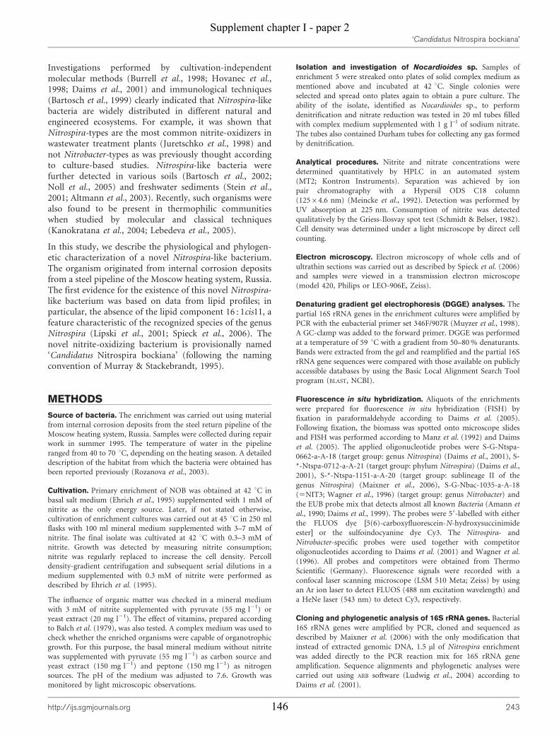
Investigations performed by cultivation-independentmolecular methods (Burrell et al., 1998; Hovanec et al.,1998; Daims et al., 2001) and immunological techniques(Bartosch et al., 1999) clearly indicated that Nitrospira-likebacteria are widely distributed in different natural andengineered ecosystems. For example, it was shown thatNitrospira-types are the most common nitrite-oxidizers inwastewater treatment plants (Juretschko et al., 1998) andnot Nitrobacter-types as was previously thought accordingto culture-based studies. Nitrospira-like bacteria werefurther detected in various soils (Bartosch et al., 2002;Noll et al., 2005) and freshwater sediments (Stein et al.,2001; Altmann et al., 2003). Recently, such organisms werealso found to be present in thermophilic communitieswhen studied by molecular and classical techniques(Kanokratana et al., 2004; Lebedeva et al., 2005).
In this study, we describe the physiological and phylogen-etic characterization of a novel Nitrospira-like bacterium.The organism originated from internal corrosion depositsfrom a steel pipeline of the Moscow heating system, Russia.The first evidence for the existence of this novel Nitrospira-like bacterium was based on data from lipid profiles; inparticular, the absence of the lipid component 16 : 1cis11, afeature characteristic of the recognized species of the genusNitrospira (Lipski et al., 2001; Spieck et al., 2006). Thenovel nitrite-oxidizing bacterium is provisionally named‘Candidatus Nitrospira bockiana’ (following the namingconvention of Murray & Stackebrandt, 1995).
METHODS
Source of bacteria. The enrichment was carried out using material
from internal corrosion deposits from the steel return pipeline of the
Moscow heating system, Russia. Samples were collected during repair
work in summer 1995. The temperature of water in the pipeline
ranged from 40 to 70 uC, depending on the heating season. A detailed
description of the habitat from which the bacteria were obtained has
been reported previously (Rozanova et al., 2003).
Cultivation. Primary enrichment of NOB was obtained at 42 uC in
basal salt medium (Ehrich et al., 1995) supplemented with 1 mM of
nitrite as the only energy source. Later, if not stated otherwise,
cultivation of enrichment cultures was carried out at 45 uC in 250 ml
flasks with 100 ml mineral medium supplemented with 3–7 mM of
nitrite. The final isolate was cultivated at 42 uC with 0.3–3 mM of
nitrite. Growth was detected by measuring nitrite consumption;
nitrite was regularly replaced to increase the cell density. Percoll
density-gradient centrifugation and subsequent serial dilutions in a
medium supplemented with 0.3 mM of nitrite were performed as
described by Ehrich et al. (1995).
The influence of organic matter was checked in a mineral medium
with 3 mM of nitrite supplemented with pyruvate (55 mg l21) or
yeast extract (20 mg l21). The effect of vitamins, prepared according
to Balch et al. (1979), was also tested. A complex medium was used to
check whether the enriched organisms were capable of organotrophic
growth. For this purpose, the basal mineral medium without nitrite
was supplemented with pyruvate (55 mg l21) as carbon source and
yeast extract (150 mg l21) and peptone (150 mg l21) as nitrogen
sources. The pH of the medium was adjusted to 7.6. Growth was
monitored by light microscopic observations.
Isolation and investigation of Nocardioides sp. Samples of
enrichment 5 were streaked onto plates of solid complex medium as
mentioned above and incubated at 42 uC. Single colonies were
selected and spread onto plates again to obtain a pure culture. The
ability of the isolate, identified as Nocardioides sp., to perform
denitrification and nitrate reduction was tested in 20 ml tubes filled
with complex medium supplemented with 1 g l–1 of sodium nitrate.
The tubes also contained Durham tubes for collecting any gas formed
by denitrification.
Analytical procedures. Nitrite and nitrate concentrations were
determined quantitatively by HPLC in an automated system
(MT2; Kontron Instruments). Separation was achieved by ion
pair chromatography with a Hypersil ODS C18 column
(12564.6 nm) (Meincke et al., 1992). Detection was performed by
UV absorption at 225 nm. Consumption of nitrite was detected
qualitatively by the Griess-Ilosvay spot test (Schmidt & Belser, 1982).
Cell density was determined under a light microscope by direct cell
counting.
Electron microscopy. Electron microscopy of whole cells and of
ultrathin sections was carried out as described by Spieck et al. (2006)
and samples were viewed in a transmission electron microscope
(model 420, Philips or LEO-906E, Zeiss).
Denaturing gradient gel electrophoresis (DGGE) analyses. The
partial 16S rRNA genes in the enrichment cultures were amplified by
PCR with the eubacterial primer set 346F/907R (Muyzer et al., 1998).
A GC-clamp was added to the forward primer. DGGE was performed
at a temperature of 59 uC with a gradient from 50–80 % denaturants.
Bands were extracted from the gel and reamplified and the partial 16S
rRNA gene sequences were compared with those available on publicly
accessible databases by using the Basic Local Alignment Search Tool
program (BLAST, NCBI).
Fluorescence in situ hybridization. Aliquots of the enrichments
were prepared for fluorescence in situ hybridization (FISH) by
fixation in paraformaldehyde according to Daims et al. (2005).
Following fixation, the biomass was spotted onto microscope slides
and FISH was performed according to Manz et al. (1992) and Daims
et al. (2005). The applied oligonucleotide probes were S-G-Ntspa-
0662-a-A-18 (target group: genus Nitrospira) (Daims et al., 2001), S-
*-Ntspa-0712-a-A-21 (target group: phylum Nitrospira) (Daims et al.,
2001), S-*-Ntspa-1151-a-A-20 (target group: sublineage II of the
genus Nitrospira) (Maixner et al., 2006), S-G-Nbac-1035-a-A-18
(5NIT3; Wagner et al., 1996) (target group: genus Nitrobacter) and
the EUB probe mix that detects almost all known Bacteria (Amann et
al., 1990; Daims et al., 1999). The probes were 59-labelled with either
the FLUOS dye [5(6)-carboxyfluorescein-N-hydroxysuccinimide
ester] or the sulfoindocyanine dye Cy3. The Nitrospira- and
Nitrobacter-specific probes were used together with competitor
oligonucleotides according to Daims et al. (2001) and Wagner et al.
(1996). All probes and competitors were obtained from Thermo
Scientific (Germany). Fluorescence signals were recorded with a
confocal laser scanning microscope (LSM 510 Meta; Zeiss) by using
an Ar ion laser to detect FLUOS (488 nm excitation wavelength) and
a HeNe laser (543 nm) to detect Cy3, respectively.
Cloning and phylogenetic analysis of 16S rRNA genes. Bacterial
16S rRNA genes were amplified by PCR, cloned and sequenced as
described by Maixner et al. (2006) with the only modification that
instead of extracted genomic DNA, 1.5 ml of Nitrospira enrichment
was added directly to the PCR reaction mix for 16S rRNA gene
amplification. Sequence alignments and phylogenetic analyses were
carried out using ARB software (Ludwig et al., 2004) according to
Daims et al. (2001).
‘Candidatus Nitrospira bockiana’
http://ijs.sgmjournals.org 243
Supplement chapter I - paper 2
146
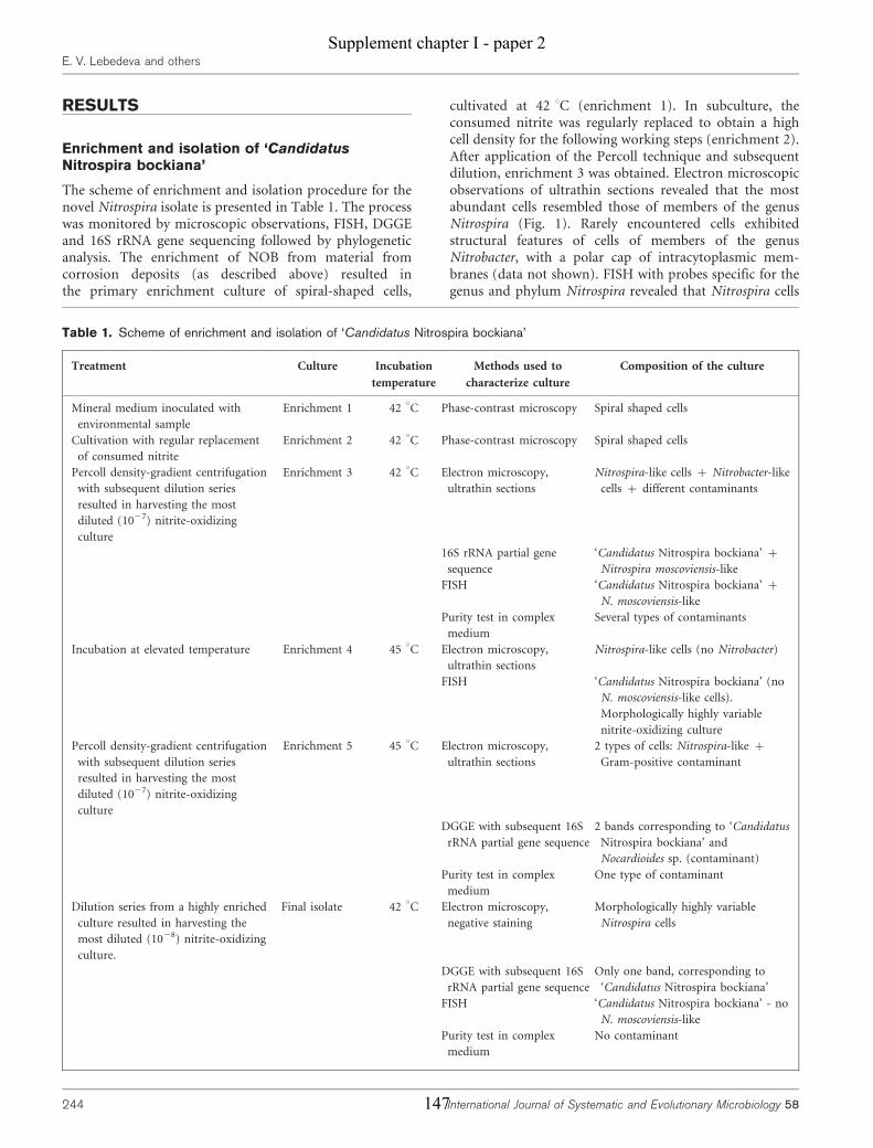
RESULTS
Enrichment and isolation of ‘CandidatusNitrospira bockiana’
The scheme of enrichment and isolation procedure for thenovel Nitrospira isolate is presented in Table 1. The processwas monitored by microscopic observations, FISH, DGGEand 16S rRNA gene sequencing followed by phylogeneticanalysis. The enrichment of NOB from material fromcorrosion deposits (as described above) resulted inthe primary enrichment culture of spiral-shaped cells,
cultivated at 42 uC (enrichment 1). In subculture, theconsumed nitrite was regularly replaced to obtain a highcell density for the following working steps (enrichment 2).After application of the Percoll technique and subsequentdilution, enrichment 3 was obtained. Electron microscopicobservations of ultrathin sections revealed that the mostabundant cells resembled those of members of the genusNitrospira (Fig. 1). Rarely encountered cells exhibitedstructural features of cells of members of the genusNitrobacter, with a polar cap of intracytoplasmic mem-branes (data not shown). FISH with probes specific for thegenus and phylum Nitrospira revealed that Nitrospira cells
Table 1. Scheme of enrichment and isolation of ‘Candidatus Nitrospira bockiana’
Treatment Culture Incubation
temperature
Methods used to
characterize culture
Composition of the culture
Mineral medium inoculated with
environmental sample
Enrichment 1 42 uC Phase-contrast microscopy Spiral shaped cells
Cultivation with regular replacement
of consumed nitrite
Enrichment 2 42 uC Phase-contrast microscopy Spiral shaped cells
Percoll density-gradient centrifugation
with subsequent dilution series
resulted in harvesting the most
diluted (1027) nitrite-oxidizing
culture
Enrichment 3 42 uC Electron microscopy,
ultrathin sections
Nitrospira-like cells + Nitrobacter-like
cells + different contaminants
16S rRNA partial gene
sequence
‘Candidatus Nitrospira bockiana’ +
Nitrospira moscoviensis-like
FISH ‘Candidatus Nitrospira bockiana’ +
N. moscoviensis-like
Purity test in complex
medium
Several types of contaminants
Incubation at elevated temperature Enrichment 4 45 uC Electron microscopy,
ultrathin sections
Nitrospira-like cells (no Nitrobacter)
FISH ‘Candidatus Nitrospira bockiana’ (no
N. moscoviensis-like cells).
Morphologically highly variable
nitrite-oxidizing culture
Percoll density-gradient centrifugation
with subsequent dilution series
resulted in harvesting the most
diluted (1027) nitrite-oxidizing
culture
Enrichment 5 45 uC Electron microscopy,
ultrathin sections
2 types of cells: Nitrospira-like +
Gram-positive contaminant
DGGE with subsequent 16S
rRNA partial gene sequence
2 bands corresponding to ‘Candidatus
Nitrospira bockiana’ and
Nocardioides sp. (contaminant)
Purity test in complex
medium
One type of contaminant
Dilution series from a highly enriched
culture resulted in harvesting the
most diluted (1028) nitrite-oxidizing
culture.
Final isolate 42 uC Electron microscopy,
negative staining
Morphologically highly variable
Nitrospira cells
DGGE with subsequent 16S
rRNA partial gene sequence
Only one band, corresponding to
‘Candidatus Nitrospira bockiana’
FISH ‘Candidatus Nitrospira bockiana’ - no
N. moscoviensis-like
Purity test in complex
medium
No contaminant
E. V. Lebedeva and others
244 International Journal of Systematic and Evolutionary Microbiology 58
Supplement chapter I - paper 2
147
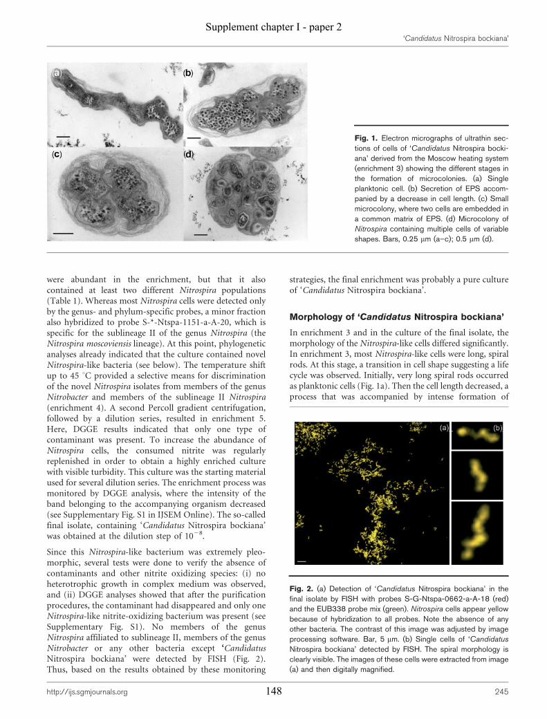
were abundant in the enrichment, but that it alsocontained at least two different Nitrospira populations(Table 1). Whereas most Nitrospira cells were detected onlyby the genus- and phylum-specific probes, a minor fractionalso hybridized to probe S-*-Ntspa-1151-a-A-20, which isspecific for the sublineage II of the genus Nitrospira (theNitrospira moscoviensis lineage). At this point, phylogeneticanalyses already indicated that the culture contained novelNitrospira-like bacteria (see below). The temperature shiftup to 45 uC provided a selective means for discriminationof the novel Nitrospira isolates from members of the genusNitrobacter and members of the sublineage II Nitrospira(enrichment 4). A second Percoll gradient centrifugation,followed by a dilution series, resulted in enrichment 5.Here, DGGE results indicated that only one type ofcontaminant was present. To increase the abundance ofNitrospira cells, the consumed nitrite was regularlyreplenished in order to obtain a highly enriched culturewith visible turbidity. This culture was the starting materialused for several dilution series. The enrichment process wasmonitored by DGGE analysis, where the intensity of theband belonging to the accompanying organism decreased(see Supplementary Fig. S1 in IJSEM Online). The so-calledfinal isolate, containing ‘Candidatus Nitrospira bockiana’was obtained at the dilution step of 1028.
Since this Nitrospira-like bacterium was extremely pleo-morphic, several tests were done to verify the absence ofcontaminants and other nitrite oxidizing species: (i) noheterotrophic growth in complex medium was observed,and (ii) DGGE analyses showed that after the purificationprocedures, the contaminant had disappeared and only oneNitrospira-like nitrite-oxidizing bacterium was present (seeSupplementary Fig. S1). No members of the genusNitrospira affiliated to sublineage II, members of the genusNitrobacter or any other bacteria except ‘CandidatusNitrospira bockiana’ were detected by FISH (Fig. 2).Thus, based on the results obtained by these monitoring
strategies, the final enrichment was probably a pure cultureof ‘Candidatus Nitrospira bockiana’.
Morphology of ‘Candidatus Nitrospira bockiana’
In enrichment 3 and in the culture of the final isolate, themorphology of the Nitrospira-like cells differed significantly.In enrichment 3, most Nitrospira-like cells were long, spiralrods. At this stage, a transition in cell shape suggesting a lifecycle was observed. Initially, very long spiral rods occurredas planktonic cells (Fig. 1a). Then the cell length decreased, aprocess that was accompanied by intense formation of
Fig. 1. Electron micrographs of ultrathin sec-tions of cells of ‘Candidatus Nitrospira bocki-ana’ derived from the Moscow heating system(enrichment 3) showing the different stages inthe formation of microcolonies. (a) Singleplanktonic cell. (b) Secretion of EPS accom-panied by a decrease in cell length. (c) Smallmicrocolony, where two cells are embedded ina common matrix of EPS. (d) Microcolony ofNitrospira containing multiple cells of variableshapes. Bars, 0.25 mm (a–c); 0.5 mm (d).
Fig. 2. (a) Detection of ‘Candidatus Nitrospira bockiana’ in thefinal isolate by FISH with probes S-G-Ntspa-0662-a-A-18 (red)and the EUB338 probe mix (green). Nitrospira cells appear yellowbecause of hybridization to all probes. Note the absence of anyother bacteria. The contrast of this image was adjusted by imageprocessing software. Bar, 5 mm. (b) Single cells of ‘Candidatus
Nitrospira bockiana’ detected by FISH. The spiral morphology isclearly visible. The images of these cells were extracted from image(a) and then digitally magnified.
‘Candidatus Nitrospira bockiana’
http://ijs.sgmjournals.org 245
Supplement chapter I - paper 2
148

extracellular polymeric substances (EPS) (Fig. 1b). Cellscontinued to divide inside small microcolonies (Fig. 1c) andfinally, numerous short cells were organized in a dense layerof a biofilm matrix, where the spiral cell shape could hardlybe detected anymore (Fig. 1d). In contrast, the cells of thefinal isolate occurred mainly as free-living planktonic cellsand demonstrated extremely high morphological variability.Their cell shape ranged from loosely wound spirals (Fig. 3a)with a variable number of coils to slightly curved and evenstraight rods (Fig. 3b) as well as coccoid cells with a diameterof 0.9 mm (Fig. 3d). The size of the spiral- and rod-shapedcells ranged from 0.3 to 0.6 mm in width and from 1.0 to2.5 mm in length. The absence of motility was characteristicfor all enrichment stages. Cells reproduced by binary or byunequal fission (Fig. 3c). As has already been shown forNitrospira moscoviensis (Spieck et al., 1998), coccoid cellswere found to contain membrane-bound particles (Fig. 3e),
as revealed by negative staining. The particulate nitriteoxidizing system was localized on the periplasmic side of thecytoplasmic membrane, shining through the outer mem-brane in partially lysed cells.
During all enrichment stages, the ultrastructure of the cellswas typical of that of members of the genus Nitrospira(Fig. 1a and b). In the observed ultrathin sections, theNitrospira-like bacteria possessed an enlarged periplasmicspace that is characteristic for this genus (Watson et al.,1986). No intracytoplasmic membranes or carboxysomeswere found. As shown by electron microscopy, cells oftencontained glycogen and polyphosphate-like deposits asstorage compounds.
Physiological properties
Physiological investigations started with enrichment 5,which was characterized as a co-culture of ‘CandidatusNitrospira bockiana’ and Nocardioides sp. (seeSupplementary Fig. S1). Enrichment 5 had the highestrate of nitrite consumption at around 45 uC (Fig. 4) at apH of 7.6–8.0. The oxidation of nitrite correlated with anincrease in the cell concentration (data not shown). Thedoubling time of the total cell number was between 8–9 hin a mineral medium with 6.8 mM nitrite, which wasoxidized within 4 days. Enrichment 5 was characterized bya wide tolerance of high nitrite concentrations, acharacteristic previously noted for ‘Candidatus Nitrospiradefluvii’ (Spieck et al., 2006). The substrate was oxidized atconcentrations up to 26–30 mM (Table 2).
In contrast to enrichment 5, the temperature optimum ofthe final isolate dropped to 42 uC (Fig. 4). The highesttolerated nitrite concentration was also reduced to 18 mM(Table 2). The rate of nitrite consumption of the finalisolate was significantly lower when compared withenrichment 5, e.g, 5 mM of nitrite was consumed within10 days. Optimal growth occurred in a mineral saltmedium with a relatively low nitrite concentration (0.3–1.5 mM). The addition of organic substances or vitaminsdid not enhance nitrite oxidation. Chemo-organotrophicgrowth was also not observed.
It was found that the temperature range of the nitrite-oxidizing activity largely depended on the amount ofnitrite. Starting with a low substrate concentration of0.3 mM, enrichment 5 and the final isolate were able tooxidize nitrite between 17 and 48 uC and 17 to 44 uC,respectively, while 1.5 mM nitrite was oxidized within atemperature range of 28–48 uC and 28–44 uC, respectively.Using a higher concentration of 6 mM nitrite, growth ofenrichment 5 was restricted to an even narrower temper-ature range of 37 to 48 uC.
Characterization of the contaminant
Based on phylogeny using partial 16S rRNA genesequences, the accompanying heterotrophic organism was
Fig. 3. Overall shape of ‘Candidatus Nitrospira bockiana’, stainedwith uranyl acetate. (a) Spiral rod; (b) long straight rod; (c) unequalfission; (d) coccoid cell; (e) section of (d) showing membrane-boundparticles in detail. Bars, 0.2 mm (a); 0.5 mm (b–d); 0.1 mm (e).
E. V. Lebedeva and others
246 International Journal of Systematic and Evolutionary Microbiology 58
Supplement chapter I - paper 2
149
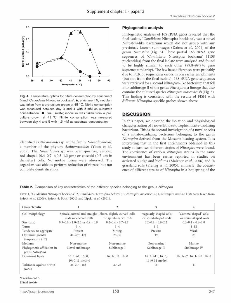
identified as Nocardioides sp. in the family Nocardiodaceae,a member of the phylum Actinomycetales (Yoon et al.,2005). The Nocardioides sp. was Gram-positive, aerobic,rod-shaped (0.4–0.7 60.5–1.3 mm) or coccoid (0.7 mm indiameter) cells. No motile forms were observed. Theorganism was able to perform reduction of nitrate, but notcomplete denitrification.
Phylogenetic analysis
Phylogenetic analyses of 16S rRNA genes revealed that thefinal isolate, ‘Candidatus Nitrospira bockiana’, was a novelNitrospira-like bacterium which did not group with anypreviously known sublineages (Daims et al., 2001) of thegenus Nitrospira (Fig. 5). Three partial 16S rRNA genesequences of ‘Candidatus Nitrospira bockiana’ (1158nucleotides) from the final isolate were analysed and foundto be highly similar to each other (99.8–99.9 % genesequence similarity). The few base differences were probablydue to PCR or sequencing errors. From earlier enrichments(but not from the final isolate), 16S rRNA gene sequenceswere retrieved for a second Nitrospira-like bacterium that fellinto sublineage II of the genus Nitrospira, a lineage that alsocontains the cultured species Nitrospira moscoviensis (Fig. 5).This finding is consistent with the results of FISH withdifferent Nitrospira-specific probes shown above.
DISCUSSION
In this paper, we describe the isolation and physiologicalcharacterization of a novel lithoautotrophic nitrite-oxidizingbacterium. This is the second investigation of a novel speciesof a nitrite-oxidizing bacterium belonging to the genusNitrospira derived from the Moscow heating system. It isinteresting that in the first enrichments obtained in thisstudy at least two different strains of Nitrospira were found.The coexistence of various Nitrospira strains in the sameenvironment has been earlier reported in studies onactivated sludge and biofilms (Maixner et al., 2006) and ingrassland soils (Freitag et al., 2005). Similarly, the coexist-ence of different strains of Nitrospira in a hot spring of the
Fig. 4. Temperature optima for nitrite consumption by enrichment5 and ‘Candidatus Nitrospira bockiana’. m, enrichment 5; inoculumwas taken from a pre-culture grown at 45 6C. Nitrite consumptionwas measured between day 3 and 4 with 5 mM as substrateconcentration. $, final isolate; inoculum was taken from a pre-culture grown at 42 6C. Nitrite consumption was measuredbetween day 4 and 5 with 1.5 mM as substrate concentration.
Table 2. Comparison of key characteristics of the different species belonging to the genus Nitrospira
Taxa: 1, ‘Candidatus Nitrospira bockiana’; 2, ‘Candidatus Nitrospira defluvii’; 3, Nitrospira moscoviensis; 4, Nitrospira marina. Data were taken from
Spieck et al. (2006), Spieck & Bock (2001) and Lipski et al. (2001).
Characteristic 1 2 3 4
Cell morphology Spirals, curved and straight
rods or coccoid cells
Short, slightly curved cells
or spiral-shaped rods
Irregularly shaped cells
or spiral-shaped rods
‘Comma-shaped’ cells
or spiral-shaped rods
Size (mm) 0.3–0.661.0–2.5 or 0.960.9 0.2–0.460.7–1.7 0.2–0.460.9–2.2 0.3–0.460.8–1.0
Turns 1–4 1–4 1–3 1–12
Tendency to aggregate Present Strong Present Weak
Optimum growth
temperature (uC)
44–46*, 42D 28–32 39 28
Medium Non-marine Non-marine Non-marine Marine
Phylogenetic affiliation in
genus Nitrospira
Novel sublineage Sublineage I Sublineage II Sublineage IV
Dominant lipids 16 : 1cis7, 16 : 0,
16 : 0 11 methyl
16 : 1cis11, 16 : 0 16 : 1cis11, 16 : 0,
16 : 0 11 methyl
16 : 1cis7, 16 : 1cis11, 16 : 0
Tolerance against nitrite
(mM)
26–30*, 18D 20–25 15 6
*Enrichment 5.
DFinal isolate.
‘Candidatus Nitrospira bockiana’
http://ijs.sgmjournals.org 247
Supplement chapter I - paper 2
150
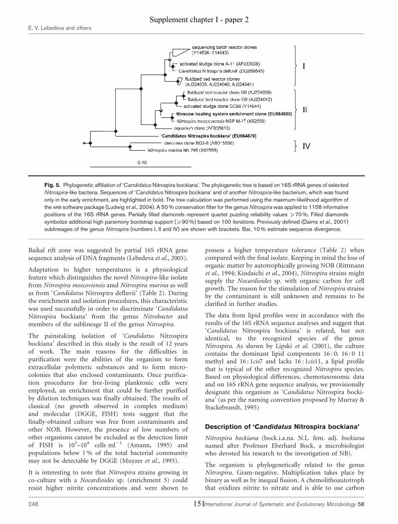
Baikal rift zone was suggested by partial 16S rRNA genesequence analysis of DNA fragments (Lebedeva et al., 2005).
Adaptation to higher temperatures is a physiologicalfeature which distinguishes the novel Nitrospira-like isolatefrom Nitrospira moscoviensis amd Nitrospira marina as wellas from ‘Candidatus Nitrospira defluvii’ (Table 2). Duringthe enrichment and isolation procedures, this characteristicwas used successfully in order to discriminate ‘CandidatusNitrospira bockiana’ from the genus Nitrobacter andmembers of the sublineage II of the genus Nitrospira.
The painstaking isolation of ‘Candidatus Nitrospirabockiana’ described in this study is the result of 12 yearsof work. The main reasons for the difficulties inpurification were the abilities of the organism to formextracellular polymeric substances and to form micro-colonies that also enclosed contaminants. Once purifica-tion procedures for free-living planktonic cells wereemployed, an enrichment that could be further purifiedby dilution techniques was finally obtained. The results ofclassical (no growth observed in complex medium)and molecular (DGGE, FISH) tests suggest that thefinally-obtained culture was free from contaminants andother NOB. However, the presence of low numbers ofother organisms cannot be excluded as the detection limitof FISH is 103–104 cells ml21 (Amann, 1995) andpopulations below 1 % of the total bacterial communitymay not be detectable by DGGE (Muyzer et al., 1993).
It is interesting to note that Nitrospira strains growing inco-culture with a Nocardioides sp. (enrichment 5) couldresist higher nitrite concentrations and were shown to
possess a higher temperature tolerance (Table 2) whencompared with the final isolate. Keeping in mind the loss oforganic matter by autotrophically growing NOB (Rittmannet al., 1994; Kindaichi et al., 2004), Nitrospira strains mightsupply the Nocardioides sp. with organic carbon for cellgrowth. The reason for the stimulation of Nitrospira strainsby the contaminant is still unknown and remains to beclarified in further studies.
The data from lipid profiles were in accordance with theresults of the 16S rRNA sequence analyses and suggest that‘Candidatus Nitrospira bockiana’ is related, but notidentical, to the recognized species of the genusNitrospira. As shown by Lipski et al. (2001), the culturecontains the dominant lipid components 16 : 0; 16 : 0 11methyl and 16 : 1cis7 and lacks 16 : 1cis11, a lipid profilethat is typical of the other recognized Nitrospira species.Based on physiological differences, chemotaxonomic dataand on 16S rRNA gene sequence analysis, we provisionallydesignate this organism as ‘Candidatus Nitrospira bocki-ana’ (as per the naming convention proposed by Murray &Stackebrandt, 1995)
Description of ‘Candidatus Nitrospira bockiana’
Nitrospira bockiana (bock.i.a.na. N.L. fem. adj. bockiananamed after Professor Eberhard Bock, a microbiologistwho devoted his research to the investigation of NB).
The organism is phylogenetically related to the genusNitrospira. Gram-negative. Multiplication takes place bybinary as well as by inequal fission. A chemolithoautotrophthat oxidizes nitrite to nitrate and is able to use carbon
Fig. 5. Phylogenetic affiliation of ‘Candidatus Nitrospira bockiana’. The phylogenetic tree is based on 16S rRNA genes of selectedNitrospira-like bacteria. Sequences of ‘Candidatus Nitrospira bockiana’ and of another Nitrospira-like bacterium, which was foundonly in the early enrichment, are highlighted in bold. The tree calculation was performed using the maximum-likelihood algorithm ofthe ARB software package (Ludwig et al., 2004). A 50 % conservation filter for the genus Nitrospira was applied to 1158 informativepositions of the 16S rRNA genes. Partially filled diamonds represent quartet puzzling reliability values ¢70 %. Filled diamondssymbolize additional high parsimony bootstrap support (¢90 %) based on 100 iterations. Previously defined (Daims et al., 2001)sublineages of the genus Nitrospira (numbers I, II and IV) are shown with brackets. Bar, 10 % estimate sequence divergence.
E. V. Lebedeva and others
248 International Journal of Systematic and Evolutionary Microbiology 58
Supplement chapter I - paper 2
151

dioxide as a sole carbon source. The highest rate of nitriteconsumption occurs at 42 uC, with a temperature rangebetween 28 and 44 uC. Pleomorphic cells range fromloosely wound spirals with a variable number of coils toslightly curved and even straight rods, as well as coccoidcells (0.9 mm diameter). The width of the spiral and rod-shaped cells ranges from 0.3 to 0.6 mm and the lengthranges from 1.0 to 2.5 mm. Major fatty acids are 16 : 0; 16 : 011 methyl and 16 : 1cis7. Neither pyruvate nor yeast extractstimulates nitrite oxidation. Organotrophic growth is notobserved.
ACKNOWLEDGEMENTS
The authors would like to thank Irmhild Wachholz and Elke Wolken
for excellent technical assistance in electron microscopy and Christian
Baranyi for DNA sequencing. Sabine Bartosch is acknowledged for
providing electron micrographs. This work was partly supported by
the Deutsche Forschungsgemeinschaft (667 to E. S., 436 RUS 17/112/
06 to E. V. L.) and by the Vienna Science and Technology Fund
(WWTF project LS 216 to H. D.).
REFERENCES
Altmann, D., Stief, P., Amann, R., De Beer, D. & Schramm, A. (2003).In situ distribution and activity of nitrifying bacteria in freshwater
sediment. Environ Microbiol 5, 798–803.
Amann, R. I. (1995). In situ identification of micro-organisms by
whole cell hybridization with rRNA-targeted nucleic acid probes. In
Molecular Microbial Ecology Manual, pp. 1–15. Edited by A. D. C.
Akkeman, J. D. van Elsas & F. J. de Bruigin. Dortrecht: Kluwer
Academic Publishers.
Amann, R. I., Binder, B. J., Olson, R. J., Chisholm, S. W., Devereux, R.& Stahl, D. A. (1990). Combination of 16S rRNA-targeted
oligonucleotide probes with flow cytometry for analyzing mixed
microbial populations. Appl Environ Microbiol 56, 1919–1925.
Balch, W. E., Fox, G. E., Magrum, L. J., Woese, C. R. & Wolfe, R. S.(1979). Methanogens: reevaluation of a unique biological group.
Microbiol Rev 43, 260–296.
Bartosch, S., Wolgast, I., Spieck, E. & Bock, E. (1999). Identification
of nitrite-oxidizing bacteria with monoclonal antibodies recognizing
the nitrite oxidoreductase. Appl Environ Microbiol 65, 4126–4133.
Bartosch, S., Hartwig, C., Spieck, E. & Bock, E. (2002).Immunological detection of Nitrospira-like bacteria in various soils.
Microb Ecol 43, 26–33.
Burrell, P. C., Keller, J. & Blackall, L. L. (1998). Microbiology of a
nitrite-oxidizing bioreactor. Appl Environ Microbiol 64, 1878–1883.
Daims, H., Bruhl, A., Amann, R., Schleifer, K.-H. & Wagner, M. (1999).
The domain-specific probe EUB338 is insufficient for the detection of
all Bacteria: development and evaluation of a more comprehensive
probe set. Syst Appl Microbiol 22, 434–444.
Daims, H., Nielsen, J. L., Nielsen, P. H., Schleifer, K. H. & Wagner, M.(2001). In situ characterization of Nitrospira-like nitrite-oxidizing
bacteria active in wastewater treatment plants. Appl Environ Microbiol
67, 5273–5284.
Daims, H., Stoecker, K. & Wagner, M. (2005). Fluorescence in situ
hybridization for the detection of prokaryotes. In Advanced Methods
in Molecular Microbial Ecology, pp. 213–239. Edited by A. M. Osborn
& C. J. Smith. Abingdon, UK: Bios-Garland.
Ehrich, S., Behrens, D., Lebedeva, E., Ludwig, W. & Bock, E. (1995).A new obligately chemolithoautotrophic, nitrite-oxidizing bacterium,Nitrospira moscoviensis sp. nov. and its phylogenetic relationship.Arch Microbiol 164, 16–23.
Freitag, T. E., Chang, L., Clegg, C. D. & Prosser, J. I. (2005). Influenceof inorganic nitrogen management regime on the diversity of nitrite-oxidizing bacteria in agricultural grassland soils. Appl EnvironMicrobiol 71, 8323–8334.
Garrity, G. M. & Holt, J. G. (2001). Phylum BVIII. Nitrospirae phy. nov.In Bergey’s Manual of Systematic Bacteriology: The Archaea and theDeeply Branching and Phototrophic Bacteria, pp. 451–464. Edited byD. R. Boone, R. W. Castenholz & G. M. Garrity. New York, NY:Springer-Verlag.
Hovanec, T. A., Taylor, L. T., Blakis, A. & DeLong, E. F. (1998).Nitrospira-like bacteria associated with nitrite oxidation in freshwateraquaria. Appl Environ Microbiol 64, 258–264.
Juretschko, S., Timmermann, G., Schmid, M., Schleifer, K.-H.,Pommerening-Roser, A., Koops, H.-P. & Wagner, M. (1998).Combined molecular and conventional analyses of nitrifyingbacterium diversity in activated sludge: Nitrosococcus mobilis andNitrospira-like bacteria as dominant populations. Appl EnvironMicrobiol 64, 3042–3051.
Kanokratana, P., Chanapan, S., Pootanakit, K. & Eurwilaichitr, L.(2004). Diversity and abundance of bacteria and archaea in the BorKhlueng hot spring in Thailand. J Basic Microbiol 44, 430–444.
Kindaichi, T., Ito, T. & Okabe, S. (2004). Ecophysiological interactionbetween nitrifying bacteria and heterotrophic bacteria in autotrophicnitrifying biofilms as determined by microautoradiography-fluor-escence in situ hybridization. Appl Environ Microbiol 70, 1641–1650.
Konneke, M., Bernhard, A. E., de la Torre, J. R., Wolker, C. B.,Waterbury, J. B. & Stahl, D. A. (2005). Isolation of an autotrophicammonia-oxidizing marine archaeon. Nature 437, 543–546.
Lebedeva, E. V., Alawi, M., Fiencke, C., Namsaraev, B., Bock, E. &Spieck, E. (2005). Moderately thermophilic nitrifying bacteria from ahot spring of the Baikal rift zone. FEMS Microbiol Ecol 54, 297–306.
Lipski, A., Spieck, E., Makolla, A. & Altendorf, K. (2001). Fatty acidprofiles of nitrite-oxidizing bacteria reflect their phylogeneticheterogeneity. Syst Appl Microbiol 24, 377–384.
Ludwig, W., Strunk, O., Westram, R., Richter, L., Meier, H., Yadhu, K.,Buchner, A., Lai, T., Steppi, S. & other authors (2004). ARB: a softwareenvironment for sequence data. Nucleic Acids Res 32, 1363–1371.
Maixner, F., Noguera, D. R., Anneser, B., Stoecker, K., Wegl, G.,Wagner, M. & Daims, H. (2006). Nitrite concentration influences thepopulation structure of Nitrospira-like bacteria. Environ Microbiol 8,1487–1495.
Manz, W., Amann, R. I., Ludwig, W., Wagner, M. & Schleifer, K. H.(1992). Phylogenetic oligodeoxynucleotide probes for the majorsubclasses of proteobacteria: problems and solutions. Syst ApplMicrobiol 15, 593–600.
Meincke, M., Bock, E., Kastrau, D. & Kroneck, P. M. H. (1992). Nitriteoxidoreductase from Nitrobacter hamburgensis: redox centers andtheir catalytic role. Arch Microbiol 158, 127–131.
Murray, R. G. & Stackebrandt, E. (1995). Taxonomic note:implementation of the provisional status Candidatus for incompletelydescribed procaryotes. Int J Syst Bacteriol 45, 186–187.
Muyzer, G., de Waal, E. C. & Uitterlinden, A. G. (1993). Profiling ofcomplex microbial populations by denaturing gradient gel electro-phoresis analysis of polymerase chain reaction-amplified genes codingfor 16S rRNA. Appl Environ Microbiol 59, 695–700.
Muyzer, G., Brinkhoff, T., Nubel, U., Santegoeds, C., Schafer, H. &Wawer, C. (1998). Denaturing gradient gel electrophoresis (DGGE) inmicrobial ecology. In Molecular Microbial Ecology Manual, vol. 3.4.4,
‘Candidatus Nitrospira bockiana’
http://ijs.sgmjournals.org 249
Supplement chapter I - paper 2
152
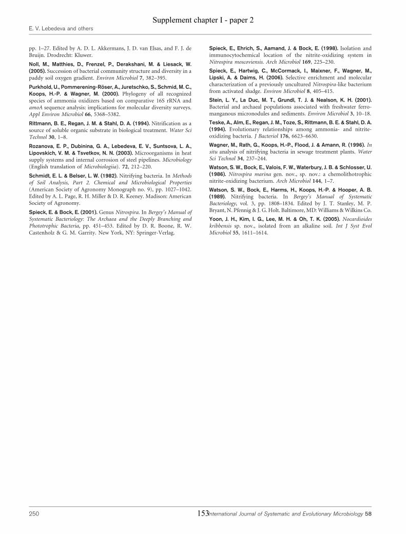
pp. 1–27. Edited by A. D. L. Akkermans, J. D. van Elsas, and F. J. de
Bruijn. Drodrecht: Kluwer.
Noll, M., Matthies, D., Frenzel, P., Derakshani, M. & Liesack, W.(2005). Succession of bacterial community structure and diversity in a
paddy soil oxygen gradient. Environ Microbiol 7, 382–395.
Purkhold, U., Pommerening-Roser, A., Juretschko, S., Schmid, M. C.,Koops, H.-P. & Wagner, M. (2000). Phylogeny of all recognized
species of ammonia oxidizers based on comparative 16S rRNA andamoA sequence analysis: implications for molecular diversity surveys.Appl Environ Microbiol 66, 5368–5382.
Rittmann, B. E., Regan, J. M. & Stahl, D. A. (1994). Nitrification as asource of soluble organic substrate in biological treatment. Water SciTechnol 30, 1–8.
Rozanova, E. P., Dubinina, G. A., Lebedeva, E. V., Suntsova, L. A.,Lipovskich, V. M. & Tsvetkov, N. N. (2003). Microorganisms in heatsupply systems and internal corrosion of steel pipelines. Microbiology
(English translation of Microbiologiia). 72, 212–220.
Schmidt, E. L. & Belser, L. W. (1982). Nitrifying bacteria. In Methodsof Soil Analysis, Part 2. Chemical and Microbiological Properties
(American Society of Agronomy Monograph no. 9), pp. 1027–1042.Edited by A. L. Page, R. H. Miller & D. R. Keeney. Madison: AmericanSociety of Agronomy.
Spieck, E. & Bock, E. (2001). Genus Nitrospira. In Bergey’s Manual ofSystematic Bacteriology: The Archaea and the Deeply Branching andPhototrophic Bacteria, pp. 451–453. Edited by D. R. Boone, R. W.
Castenholz & G. M. Garrity. New York, NY: Springer-Verlag.
Spieck, E., Ehrich, S., Aamand, J. & Bock, E. (1998). Isolation andimmunocytochemical location of the nitrite-oxidizing system inNitrospira moscoviensis. Arch Microbiol 169, 225–230.
Spieck, E., Hartwig, C., McCormack, I., Maixner, F., Wagner, M.,Lipski, A. & Daims, H. (2006). Selective enrichment and molecularcharacterization of a previously uncultured Nitrospira-like bacteriumfrom activated sludge. Environ Microbiol 8, 405–415.
Stein, L. Y., La Duc, M. T., Grundl, T. J. & Nealson, K. H. (2001).Bacterial and archaeal populations associated with freshwater ferro-manganous micronodules and sediments. Environ Microbiol 3, 10–18.
Teske, A., Alm, E., Regan, J. M., Toze, S., Rittmann, B. E. & Stahl, D. A.(1994). Evolutionary relationships among ammonia- and nitrite-oxidizing bacteria. J Bacteriol 176, 6623–6630.
Wagner, M., Rath, G., Koops, H.-P., Flood, J. & Amann, R. (1996). Insitu analysis of nitrifying bacteria in sewage treatment plants. WaterSci Technol 34, 237–244.
Watson, S. W., Bock, E., Valois, F. W., Waterbury, J. B. & Schlosser, U.(1986). Nitrospira marina gen. nov., sp. nov.: a chemolithotrophicnitrite-oxidizing bacterium. Arch Microbiol 144, 1–7.
Watson, S. W., Bock, E., Harms, H., Koops, H.-P. & Hooper, A. B.(1989). Nitrifying bacteria. In Bergey’s Manual of SystematicBacteriology, vol. 3, pp. 1808–1834. Edited by J. T. Stanley, M. P.Bryant, N. Pfennig & J. G. Holt. Baltimore, MD: Williams & Wilkins Co.
Yoon, J. H., Kim, I. G., Lee, M. H. & Oh, T. K. (2005). Nocardioideskribbensis sp. nov., isolated from an alkaline soil. Int J Syst EvolMicrobiol 55, 1611–1614.
E. V. Lebedeva and others
250 International Journal of Systematic and Evolutionary Microbiology 58
Supplement chapter I - paper 2
153

Supplement chapter I - paper 2
154
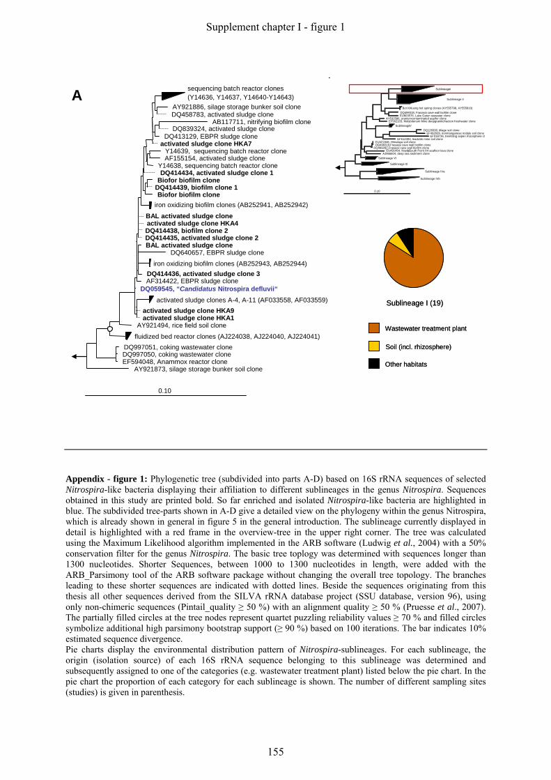
sequencing batch reactor clones(Y14636, Y14637, Y14640-Y14643)
AY921886, silage storage bunker soil cloneDQ458783, activated sludge clone
AB117711, nitrifying biofilm cloneDQ839324, activated sludge clone
DQ413129, EBPR sludge cloneactivated sludge clone HKA7
Y14639, sequencing batch reactor cloneAF155154, activated sludge clone
Y14638, sequencing batch reactor cloneDQ414434, activated sludge clone 1
Biofor biofilm cloneDQ414439, biofilm clone 1Biofor biofilm clone
iron oxidizing biofilm clones (AB252941, AB252942)
BAL activated sludge cloneactivated sludge clone HKA4 DQ414438, biofilm clone 2DQ414435, activated sludge clone 2BAL activated sludge clone
DQ640657, EBPR sludge cloneiron oxidizing biofilm clones (AB252943, AB252944)
DQ414436, activated sludge clone 3AF314422, EBPR sludge clone
DQ059545, “Candidatus Nitrospira defluvii“activated sludge clones A-4, A-11 (AF033558, AF033559)
activated sludge clone HKA9activated sludge clone HKA1
AY921494, rice field soil clone
fluidized bed reactor clones (AJ224038, AJ224040, AJ224041)DQ997051, coking wastewater cloneDQ997050, coking wastewater cloneEF594048, Anammox reactor clone
AY921873, silage storage bunker soil clone
0.10
A
Wastewater treatment plant
Soil (incl. rhizosphere)
Other habitats
Sublineage I (19)
Wastewater treatment plant
Soil (incl. rhizosphere)
Other habitats
Sublineage I (19)
SublineageI33
Sublineage II65
Bor Khlueng hot spring clones (AY555798, AY555810)DQ499318, Frasassi cave wall biofilm cloneEU803576, Lake Gatun seawater clone
AY532586, uraniumcontaminated aquifer cloneEF562103, Molybdenum Mine deepgraniticfraction freshwater clone
SublineageV5DQ128338, tillage soil clone
EF492939, ironmanganese nodule soil cloneEF018736, trembling aspen rhizosphere clo
EF612393, leadzink mine soil cloneEU421848, Himalaya soil cloneDQ499319,Frasassi cave wall biofilm clone
DQ499282,Frasassi cave wall biofilm cloneEU491464, Hawai’i South Point X4 seafloor lava clone
AJ966604, deep sea sediment cloneSublineage VI6
Sublineage III5
Sublineage IVa32
Sublineage IVb10
0.10
Appendix - figure 1: Phylogenetic tree (subdivided into parts A-D) based on 16S rRNA sequences of selected Nitrospira-like bacteria displaying their affiliation to different sublineages in the genus Nitrospira. Sequences obtained in this study are printed bold. So far enriched and isolated Nitrospira-like bacteria are highlighted in blue. The subdivided tree-parts shown in A-D give a detailed view on the phylogeny within the genus Nitrospira, which is already shown in general in figure 5 in the general introduction. The sublineage currently displayed in detail is highlighted with a red frame in the overview-tree in the upper right corner. The tree was calculated using the Maximum Likelihood algorithm implemented in the ARB software (Ludwig et al., 2004) with a 50% conservation filter for the genus Nitrospira. The basic tree toplogy was determined with sequences longer than 1300 nucleotides. Shorter Sequences, between 1000 to 1300 nucleotides in length, were added with the ARB_Parsimony tool of the ARB software package without changing the overall tree topology. The branches leading to these shorter sequences are indicated with dotted lines. Beside the sequences originating from this thesis all other sequences derived from the SILVA rRNA database project (SSU database, version 96), using only non-chimeric sequences (Pintail_quality ≥ 50 %) with an alignment quality ≥ 50 % (Pruesse et al., 2007). The partially filled circles at the tree nodes represent quartet puzzling reliability values ≥ 70 % and filled circles symbolize additional high parsimony bootstrap support (≥ 90 %) based on 100 iterations. The bar indicates 10% estimated sequence divergence. Pie charts display the environmental distribution pattern of Nitrospira-sublineages. For each sublineage, the origin (isolation source) of each 16S rRNA sequence belonging to this sublineage was determined and subsequently assigned to one of the categories (e.g. wastewater treatment plant) listed below the pie chart. In the pie chart the proportion of each category for each sublineage is shown. The number of different sampling sites (studies) is given in parenthesis.
Supplement chapter I - figure 1
155

AJ582041, uranium mining waste pile soil cloneAY493909, soil aggregate clone
EF643416, radish field soil cloneAY493910, soil aggregate clone
AY921743, silage storage bunker soil cloneAY921744, silage storage bunker soil cloneAB240280, Phragmites rhizosphere cloneAY326514, Amazon forest soil cloneAY326515, Amazon forest soil cloneEF492918, ironmanganese nodule soil clone
EU881325, maizesweet potato cropland soil cloneDQ839384, Chinese red soil clone
DQ811892, mangrove soil cloneEF445485, Himalaya soil clone
DQ814725, zebrafish digestive tract cloneDQ814918, zebrafish digestive tract clone
EF492941, ironmanganese nodule soil cloneDQ451477, Taiwan rain forest soil clone
AF407702, Artesian Basin cloneAF155155, Nitrospira cf. moscoviensis SBR2046AY328743, drinking water distribution system simulator clone
AF155152, Nitrospira cf. moscoviensis SBR1015EU097257, radish field soil clone
AB252940, iron oxidzing biofilm clonefluidized bed reactor clones (AJ224039, AJ224042)
DQ414437, biofilm clone 3DQ414433, activated sludge clone 4Y14644, activated sludge clone GC86
EU015101, bioreactor cloneAF155153, Nitrospira cf. moscoviensis SBR1024
AY741094, nitrifying reactor cloneAY328715, drinking water distribution system simulator cloneAY328760, drinking water distribution system simulator clone
X82558, Nitrospira moscoviensisEU084880, Moscow heating system enrichment clone
EU374034, Cretan margin sediment cloneAJ617920, flooded paddy soil clone
AJ617918, flooded paddy soil cloneAJ617919, flooded paddy soil clone
EU808230, nonchloraminated benchscale chemostat cloneEU808901, chloraminated distribution system simulator clone
EU809416, low chloramine dose distribution system simulator cloneEU808752, chloraminated distribution system Prospect Tank clone
EU808151, chloraminated benchscale chemostat cloneAY532585, uranium contaminated aquifer cloneAF035813, aquarium clone
AB252937, iron oxidizing biofilm cloneAB252938, iron oxidizing biofilm cloneDQ404878, contaminated soil clone
AB252939, iron oxidizing biofilm cloneAY493911, soil aggregate cloneAJ617917, flooded paddy soil clone
0.10
Sublineage II (44)
Wastewater treatment plant
Soil (incl. rhizosphere)
Other habitats
Freshwater environment(incl. sediment)
Marine environment(incl. sediment)
Hot springs
DQ298149, silkworm midgut cloneDQ067038, Lake Washington sediment clone
AY221079, Mammoth cave cloneAJ534638, uranium mining waste pile soil clone
EF580942, freshwater calcareous mat cloneDQ058674, drinking water distribution system clone
AF351228, groundwater cloneEF580927, freshwater calcareous mat clone
AF418966, freshwater reservoir clonedrinking water distribution system clones
EF580980, freshwater calcareous mat clone
EF019559, trembling aspen rhizosphere cloneAM991227, karst spring water clone
EU266788, taroil contaminated aquifer sediment cloneAM072426, uranium mining waste pile soil clone
(DQ058675, DQ058676, DQ058679)
B
DQ499307, Frasassi cave wall biofilm clone
beech forrest soil clonesbeech forest soil clone 22beech forrest soil clonesbeech forest soil clone 22
SublineageI33
Sublineage II65
Bor Khlueng hot spring clones (AY555798, AY555810)DQ499318, Frasassi cave wall biofilm cloneEU803576, Lake Gatun seawater clone
AY532586, uraniumcontaminated aquifer cloneEF562103, Molybdenum Mine deepgraniticfraction freshwater clone
SublineageV5DQ128338, tillage soil clone
EF492939, ironmanganese nodule soil cloneEF018736, trembling aspen rhizosphere clo
EF612393, leadzink mine soil cloneEU421848, Himalaya soil cloneDQ499319,Frasassi cave wall biofilm clone
DQ499282,Frasassi cave wall biofilm cloneEU491464, Hawai’i South Point X4 seafloor lava clone
AJ966604, deep sea sediment cloneSublineage VI6
Sublineage III5
Sublineage IVa32
Sublineage IVb10
0.10
Supplement chapter I - figure 1
156
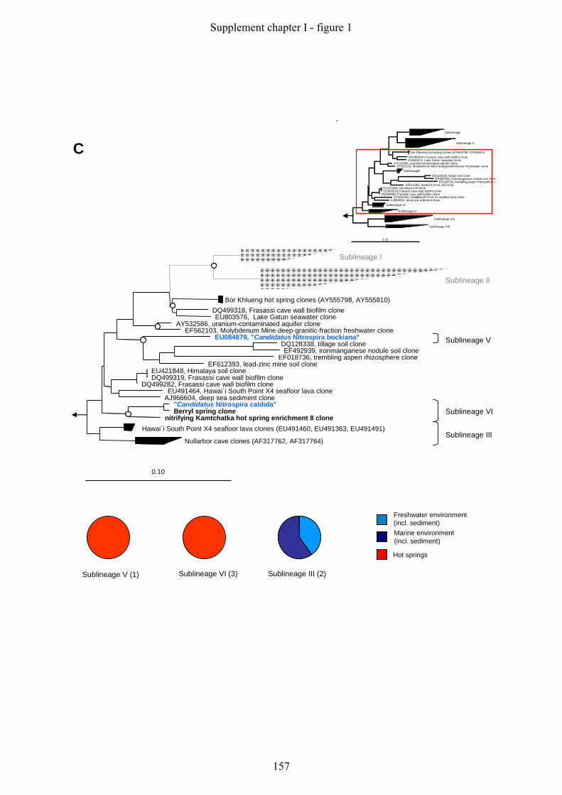
Sublineage I
Sublineage II
Bor Khlueng hot spring clones (AY555798, AY555810)DQ499318, Frasassi cave wall biofilm cloneEU803576, Lake Gatun seawater clone
AY532586, uranium-contaminated aquifer cloneEF562103, Molybdenum Mine deep-granitic-fraction freshwater clone
EU084879, "Candidatus Nitrospira bockiana"DQ128338, tillage soil cloneEF492939, ironmanganese nodule soil clone
EF018736, trembling aspen rhizosphere cloneEF612393, lead-zinc mine soil clone
EU421848, Himalaya soil cloneDQ499319, Frasassi cave wall biofilm clone
DQ499282, Frasassi cave wall biofilm cloneEU491464, Hawai`i South Point X4 seafloor lava clone
AJ966604, deep sea sediment clone"Candidatus Nitrospira caldida"Berryl spring clone
nitrifying Kamtchatka hot spring enrichment 8 cloneHawai`i South Point X4 seafloor lava clones (EU491460, EU491363, EU491491)
Nullarbor cave clones (AF317762, AF317764)
0.10
C
Sublineage V
Sublineage VI
Sublineage III
Sublineage V (1) Sublineage VI (3)
Freshwater environment(incl. sediment)Marine environment(incl. sediment)
Hot springs
Sublineage III (2)
SublineageI33
Sublineage II65
Bor Khlueng hot spring clones (AY555798, AY555810)DQ499318, Frasassi cave wall biofilm cloneEU803576, Lake Gatun seawater clone
AY532586, uraniumcontaminated aquifer cloneEF562103, Molybdenum Mine deepgraniticfraction freshwater clone
SublineageV5DQ128338, tillage soil clone
EF492939, ironmanganese nodule soil cloneEF018736, trembling aspen rhizosphere c
EF612393, leadzink mine soil cloneEU421848, Himalaya soil cloneDQ499319,Frasassi cave wall biofilm clone
DQ499282,Frasassi cave wall biofilm cloneEU491464, Hawai’i South Point X4 seafloor lava clone
AJ966604, deep sea sediment cloneSublineage VI6
Sublineage III5
Sublineage IVa32
Sublineage IVb10
0.10
Supplement chapter I - figure 1
157

D
AB015550, deep-sea sediment cloneAY375077, western Pacific deep sea sediment clone
EU287156, Pacific arctic surface sediment cloneEU236285, Pacific Ocean clone
EU491702, East Pacific Rise seafloor lava cloneDQ395470, deep-sea octacoral clone
EU491612, East Pacific Rise seafloor lavas cloneEU652664, Yellow Sea sediment clone
EU652663, Yellow Sea sediment cloneDQ351808, North sea sediment cloneEU491406, Hawai`i South Point X4 seafloor lava cloneEU491478, Hawai`i South Point X4 seafloor lava clone
EU491446, Hawai`i South Point X4 seafloor lava cloneEU236287, Pacific Ocean clone
AM295545, marine aquaculture biofilm cloneEF999362, Pearl River Estuary sediments clone
marine aquaculture biofilm clones (AM295537-AM295544)DQ058673, drinking water distribution system clone
X82559, Nitrospira marina Nb295EF157239, Rancho La Brea tar pit clone
EF157246, Rancho La Brea tar pit cloneAJ863251, flooded poplar tree microcosm soil clone
DQ917836, Muricea elongata coral cloneDQ917832, Muricea elongata coral clone
EU652665, Yellow Sea sediment cloneNorth sea nitrifying enrichment clone
Hyrtios proteus sponge cloneEF076109, Plakortis sp. sponge clone
Hyrtios proteus sponge cloneGeodia sp. metatranscriptome assembley clone
AJ347039, Aplysina aerophoba sponge cloneEF092255, Axinella corrugata sponge clone
Agelas dilatata (EF076129, EF076168)EU491260, Loi`hi Seamount Pisces Peak X2 seafloor lavas clone
Cymbastella concentrica sponge clones (AY942757, AY942775)EU491045, Loi`hi Seamount South Rift X3 seafloor lavas clone
EU287071, Pacific arctic surface sediment clone
0.10
Sublineage IVa
Sublineage IVb
Sublineage IVa (16) Sublineage IVb (9)
Soil (incl. rhizosphere)
Other habitats
Marine environment(incl. sediment)
Sponge symbionts
Sublineage IVa (16) Sublineage IVb (9)
Soil (incl. rhizosphere)
Other habitats
Marine environment(incl. sediment)
Sponge symbionts
SublineageI33
Sublineage II65
Bor Khlueng hot spring clones (AY555798, AY555810)DQ499318, Frasassi cave wall biofilm cloneEU803576, Lake Gatun seawater clone
AY532586, uraniumcontaminated aquifer cloneEF562103, Molybdenum Mine deepgraniticfraction freshwater clone
SublineageV5DQ128338, tillage soil clone
EF492939, ironmanganese nodule soil cloneEF018736, trembling aspen rhizosphere c
EF612393, leadzink mine soil cloneEU421848, Himalaya soil cloneDQ499319,Frasassi cave wall biofilm clone
DQ499282,Frasassi cave wall biofilm cloneEU491464, Hawai’i South Point X4 seafloor lava clone
AJ966604, deep sea sediment cloneSublineage VI6
Sublineage III5
Sublineage IVa32
Sublineage IVb10
0.10
Supplement chapter I - figure 1
158

References
Ludwig, W., O. Strunk, R. Westram, L. Richter, H. Meier, Yadhukumar, A. Buchner, T.
Lai, S. Steppi, G. Jobb, W. Forster, I. Brettske, S. Gerber, A. W. Ginhart, O.
Gross, S. Grumann, S. Hermann, R. Jost, A. Konig, T. Liss, R. Lussmann, M.
May, B. Nonhoff, B. Reichel, R. Strehlow, A. Stamatakis, N. Stuckmann, A. Vilbig, M. Lenke, T. Ludwig, A. Bode & K. H. Schleifer, (2004) ARB: a software
environment for sequence data. Nucleic Acids Res 32: 1363-1371.
Pruesse, E., C. Quast, K. Knittel, B. M. Fuchs, W. Ludwig, J. Peplies & F. O. Glockner,
(2007) SILVA: a comprehensive online resource for quality checked and aligned
ribosomal RNA sequence data compatible with ARB. Nucleic Acids Res 35: 7188-
7196.
Supplement chapter I - figure 1
159

Supplement chapter I - figure 1
160
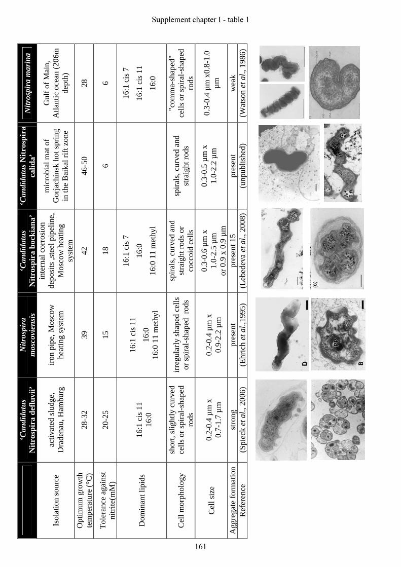
‘C
andi
datu
s N
itros
pira
def
luvi
i’ N
itros
pira
m
osco
vien
sis
‘Can
dida
tus
Nitr
ospi
ra b
ocki
ana’
‘C
andi
datu
s Nitr
ospi
ra
calid
a’
Nitr
ospi
ra m
arin
a
Isol
atio
n so
urce
ac
tivat
ed sl
udge
, D
rade
nau,
Ham
burg
iro
n pi
pe, M
osco
w
heat
ing
syst
em
inte
rnal
cor
rosi
on
depo
sits
,ste
el p
ipel
ine,
M
osco
w h
eatin
g sy
stem
mic
robi
al m
at o
f G
orja
chin
sk h
ot sp
ring
in th
e B
aika
l rift
zon
e
Gul
f of M
ain,
A
tlant
ic o
cean
(206
m
dept
h)
Opt
imum
gro
wth
te
mpe
ratu
re (°
C)
28-3
2 39
42
46
-50
28
Tole
ranc
e ag
ains
t ni
trite
(mM
) 20
-25
15
18
6 6
Dom
inan
t lip
ids
16:1
cis
11
16:0
16:1
cis
11
16:0
16
:0 1
1 m
ethy
l
16:1
cis
7
16:0
16:0
11
met
hyl
16:1
cis
7
16:1
cis
11
16:0
Cel
l mor
phol
ogy
shor
t, sl
ight
ly c
urve
d ce
lls o
r spi
ral-s
hape
d ro
ds
irreg
ular
ly sh
aped
cel
ls
or sp
iral-s
hape
d ro
ds
spira
ls, c
urve
d an
d st
raig
ht ro
ds o
r co
ccoi
d ce
lls
spira
ls, c
urve
d an
d st
raig
ht ro
ds
"com
ma-
shap
ed"
cells
or s
pira
l-sha
ped
rods
Cel
l siz
e 0.
2-0.
4 µm
x
0.7-
1.7
µm
0.2-
0.4
µm x
0.
9-2.
2 µm
0.3-
0.6
µm x
1.
0-2.
5 µm
or
0.9
x 0
.9 µ
m
0.3-
0.5
µm x
1.
0-2.
2 µm
0.3-
0.4
µm x
0.8-
1.0
µm
Agg
rega
te fo
rmat
ion
stro
ng
pres
ent
pres
ent 1
5 pr
esen
t w
eak
Ref
eren
ce
(Spi
eck
et a
l., 2
006)
(E
hric
h et
al.,
1995
) (L
ebed
eva
et a
l., 2
008)
(unp
ublis
hed)
(W
atso
n et
al.,
198
6)
Supplement chapter I - table 1
161
Appendix - table 1: Comparable characteristics of so far isolated and enriched Nitrospira-like bacteria. References Ehrich, S., D. Behrens, E. Lebedeva, W. Ludwig & E. Bock, (1995) A new obligately
chemolithoautotrophic, nitrite-oxidizing bacterium, Nitrospira moscoviensis sp. nov. and its phylogenetic relationship. Arch Microbiol 164: 16-23.
Lebedeva, E. V., M. Alawi, F. Maixner, P. G. Jozsa, H. Daims & E. Spieck, (2008) Physiological and phylogenetic characterization of a novel lithoautotrophic nitrite-oxidizing bacterium, 'Candidatus Nitrospira bockiana'. Int J Syst Evol Microbiol 58: 242-250.
Spieck, E., C. Hartwig, I. McCormack, F. Maixner, M. Wagner, A. Lipski & H. Daims, (2006) Selective enrichment and molecular characterization of a previously uncultured Nitrospira-like bacterium from activated sludge. Environ Microbiol 8: 405-415.
Watson, S. W., F. W. Valois, J. B. Waterbury & U. Schlosser, (1986) Nitrospira marina gen. nov. sp. nov.: a chemolithotrophic nitrite-oxidizing bacterium. Arch Microbiol 144: 1-7.
Supplement chapter I - table 1
162

List of publications
Spieck, E., Hartwig, C., McCormack, I., Maixner, F., Wagner, M., Lipski, A., Daims, H.
(2006). Selective enrichment and molecular characterization of a previously
uncultured Nitrospira-like bacterium from activated sludge. Environ. Microbiol. 8:
405-415.
Maixner, F., Noguera, D. R., Anneser, B., Stoecker, K., Wegl, G., Wagner, M., Daims, H.
(2006) Nitrite concentration influences the population structure of Nitrospira-like
bacteria. Environ. Microbiol. 8: 1487-1495.
Daims, H., Maixner, F., Lücker, S., Stoecker, K., Hace, K., Wagner, M. (2006)
Ecophysiology and niche differentiation of Nitrospira-like bacteria, the key nitrite
oxidizers in wastewater treatment plants. Wat. Sci. Tech. 54: 21-27.
Loy, A., Maixner, F., Wagner, M., Horn, M. (2007). probeBase - an online resource for
rRNA-targeted oligonucleotide probes: new features 2007. Nucleic Acids Res. 35:
D800-D804.
Lebedeva, E. V., Alawi, M., Maixner, F., Jozsa, P.G., Daims, H., Spieck, E. (2008)
Physiological and phylogenetical characterization of a new lithoautotrophic
nitrite-oxidizing bacterium 'Candidatus Nitrospira bockiana' sp. nov. Int. J. Syst. Evol.
Microbiol. 58: 242-250.
Maixner, F., Wagner, M., Lücker, S., Pelletier, E., Schmitz-Esser, S., Hace, K., Spieck,
E., Konrat, R., Le Paslier, D., Daims, H. (2008). Environmental genomics reveals a
functional chlorite dismutase in the nitrite-oxidizing bacterium "Candidatus Nitrospira
defluvii". Environ. Microbiol. 10: 3043-3056.
Kostan, J., Maixner, F., Sjoeblom, B., Wagner, M., Daims, H., Djinovic Carugo, K.
(2009) The christal structure of a chlorite dismutase of the nitrite-oxidizing bacterium
"Candidatus Nitrospira defluvii": one residue makes the difference. In preparation.
Publications
163

Hall, E., Maixner, F., Franklin, O., Battin, T., Daims, H., Richter, A. (2009) Ecological
stoichiometry: Linking microbial community composition to ecosystem processes
Biogeochemistry To be submitted.
Maixner, F., Koch, H., Hauzmayer, S., Spieck, E., Le Paslier, D., Wagner, M., Daims,
H., (2009) Nitrite oxidoreductase (Nxr) - the metabolic key enzyme of nitrite
oxidizing Nitrospira: Characterization of an unusual Nxr and its application as
anovel functional and phylogenetic marker gene. To be submitted.
Bookchapter:
Daims, H., Maixner, F., Schmid, M. C. (2009) The nitrifying microbes: Ammonia oxidizers,
nitrite oxidizers, and anaerobic ammonia oxidizers. In FISH Handbook for biological
wastewater treatment: Identification and quantification of microorganisms in activated
sludge and biofilms. (Nielson, P. H., Daims, H., Lemmer, H. ed.) IWA publishing. In
press.
Publications
164
List of oral presentations
Frank Maixner, Michael Wagner, Holger Daims (2004) Nitrospira spp. – Biodiversity and
Ecophysiology of important Nitrite-oxidizing bacteria.
IECB Wissenschaftstag, Vienna, Austria, April 2004
Frank Maixner, Bettina Anneser, Kilian Stöcker, Sebastian Lücker, Karin Hace
Michael Wagner, Holger Daims (2006) Nitrospira spp., key players in the biological N-
elimination in wastewater treatment plants.
Limnological Seminar, Department of Freshwater Ecology, University of Vienna,
December 2006
Frank Maixner, Sebastian Lücker, Karin Hace, Eva Spieck, Denis LePaslier, Michael
Wagner, Holger Daims (2007) Nitrite oxidoreductase Nxr, the metabolic key enzyme of
Nitrospira-like bacteria.
6th
International workshop on New Techniques in Microbial Ecology, Sæby, Denmark,
October 2007
Frank Maixner, Hanna Koch, Sandra Hauzmayer, Sebastian Lücker, Eva Spieck, Denis
LePaslier, Michael Wagner, Holger Daims (2007) “Candidatus Nitrospira defluvii” –
chlorite dismutase Cld – nitrite oxidoreductase Nxr.
Research visit at the Department of Microbiology Radboud University Nijmegen, The
Netherlands, November 2007
Frank Maixner, Eva Spieck, Denis Le Paslier, Michael Wagner, Holger Daims (2008)
Environmental genomics reveals the genes of nitrite oxidoreductase (Nxr) in Nitrospira:
Application of this metabolic key enzyme as functional and phylogenetic marker for nitrite-
oxidizing Nitrospira-like bacteria.
Annual Conference of the Vereinigung für Allgemeine und Angewandte Mikrobiologie
(VAAM), Frankfurt, Germany, March 2008
Oral presentations
165
Frank Maixner, Sandra Hauzmayer, Josef Strauss, Andreas Richter, Michael Wagner,
Holger Daims (2008) Nitrification linked to litter decay – Autotrophic – Heterotrophic -
Stratification.
Microbial diversity and Ecosystem function (MICDIF) Science day, Lunz, Austria,
June 2008
Tutor and lecturer of the “International FISH-course” (2004, 2006, 2007, 2008),
Department of Microbial Ecology, University of Vienna, Austria
Oral presentations
166

List of poster presentations
Holger Daims, Karin Hace, Frank Maixner, Bettina Anneser, Sebastian Lücker, Eva
Spieck, Kilian Stöcker, Michael Wagner (2005) Ecophysiology, Niche differentiation, and
Ecogenomics of Nitrospira-like bacteria. American Society for Microbiology (ASM), General
Meeting, Atlanta, USA, June 2005
Frank Maixner, Karin Hace, Denis Le Paslier, Eva Spieck, Michael Wagner, Holger
Daims (2005) Discovery and biochemical validation of the first non-proteobacterial chlorite
dismutase in a nitrite-oxidizing Nitrospira-like bacterium by an environmental genomics
approach. Annual Conference of the Vereinigung für Allgemeine und Angewandte
Mikrobiologie (VAAM), Göttingen, Germany, September 2005
Sebastian Lücker, Frank Maixner, Bettina Anneser, Gertrude Wegl, Kilian Stöcker,
Michael Wagner, Holger Daims (2005) Responses of closely related nitrite-oxidizing
Nitrospira-like bacteria to shifts of nitrite concentration: an example of niche differentiation
in the microbial world. Annual Conference of the Vereinigung für Allgemeine und
Angewandte Mikrobiologie (VAAM), Göttingen, Germany, September 2005
Karin Hace, Sebastian Lücker, Frank Maixner, Christian Baranyi, Kilian Stöcker,
Michael Wagner, Holger Daims (2005) Molecular screening of chemically diverse habitats
reveals a hitherto unexplored biodiversity of nitrite-oxidizing Nitrospira-like bacteria. Annual
Conference of the Vereinigung für Allgemeine und Angewandte Mikrobiologie (VAAM),
Göttingen, Germany, September 2005
Frank Maixner, Daniel R. Noguera, Karin Hace, Dennis Le Paslier, Eva Spieck,
Gertrude Wegl, Michael Wagner, Holger Daims (2006) Ecophysiology and genomics of
nitrite-oxidizing Nitrospira-like bacteria. Wiener Wissenschafts- und Technologiefond
(WWTF), Vienna, Austria, May 2006
Alexander Loy, Frank Maixner, Michael Wagner, Matthias Horn (2006) probeBase – a
widely used online database for ribosomal RNA-targeted oligonucleotide probes. 11th
International Symposium of Microbial Ecology (ISME), Vienna, Austria, August 2006
Poster presentations
167
Frank Maixner, Daniel R. Noguera, Dennis Le Paslier, Eric Pelletier, Stephanie
Füreder, Gertrude Wegl, Kilian Stoecker, Michael Wagner, Holger Daims (2006)
Ecophysiology of nitrite-oxidizing Nitrospira-like bacteria. 11th
International Symposium of
Microbial Ecology (ISME), Vienna, Austria, August 2006
Sebastian Lücker, Karin Hace, Frank Maixner, Christian Baranyi, Kilian Stöcker,
Michael Wagner, Holger Daims (2006) Molecular screening of chemically diverse habitats
reveals a hitherto unexplored biodiversity of Nitrospira. 11th
International Symposium of
Microbial Ecology (ISME), Vienna, Austria, August 2006
Alexander Loy, Roland Arnold, Patrick Tischler, Thomas Rattei, Frank Maixner,
Michael Wagner, Matthias Horn (2008) probeBase and probeCheck – a database for rRNA-
targeted probes and a central resources for evaluating oligo coverage and specificity. Annual
Conference of the Vereinigung für Allgemeine und Angewandte Mikrobiologie (VAAM),
Frankfurt, Germany, March 2008
Sebastian Lücker, Eric Pelletier, Frank Maixner, Karin Hace, Eva Spieck, Dennis Le
Paslier, Michael Wagner, Holger Daims (2008) The complete genome of the uncultured
nitrite oxidizer “Candidatus Nitrospira defluvii” as basis for comparative genomics with other
Nitrospira-like bacteria. Annual Conference of the Vereinigung für Allgemeine und
Angewandte Mikrobiologie (VAAM), Frankfurt, Germany, March 2008
Edward Hall, Ieda Hämmerle, Frank Maixner, Marvin Pölzl, Christian Schwarz, Tom
Battin, Holger Daims, Andreas Richter (2008) Looking inside the box: evaluating the
relationships between resource stoichiometry, biomass stoichiometry and physiology of the
model organism Verrucomicroium spinosum. 12th
International Symposium of Microbial
Ecology (ISME), Cairns, Australia, August 2008
Poster presentations
168
Danksagung
Nach 6 Jahren Wien scheint die Liste an Erinnerungen endlos und ich muss zugeben, dass
mich keine Zeit vorher so geprägt hat wie diese. Beruflich aber vor allem privat hat sich
einiges bei mir geändert und ich möchte mich nun bei den Menschen bedanken die mich dabei
begleitet haben.
Ausschlaggebend war dabei ein Spaziergang mit Michi am Campus in Freising, bei dem ich
mich entschlossen habe meine Diplomarbeit in Wien an seinem neuen Lehrstuhl anzufertigen.
Aus dem anfänglich geplanten Jahr im Ausland wurden nun sechs Jahre. Für die Möglichkeit
an diesem Lehrstuhl zu arbeiten, die Freiheiten in unserer Forschung und die guten Ideen im
entscheidenden Moment möchte ich mich hiermit bei Michi bedanken.
Die Diplomarbeit glich einer wissenschaftlichen Berg- und Talfahrt. Kilian hat es trotz allem
geschafft das schlingernde Schiff auf Kurs zu halten. Vielen Dank dafür. Während dieser Zeit
hat sich eine sehr gute Freundschaft entwickelt, die wir einmal im Jahr mit einem
Fliegenfischenausflug feiern müssen.
Nach meiner Diplomarbeit hat Holger mir die Chance gegeben weiter in seiner Arbeitsgruppe
zu arbeiten. Jemanden Vertrauen schenken ist etwas sehr Großes. Ich denke ich konnte es Dir
zurückgeben. Besonders hilfreich und auch nicht selbstverständlich waren Deine Zusage zur
freien Zeiteinteilung und die Weiterfinanzierung während meiner Doktorarbeit. Vielen Dank
dafür. Mit Dir habe ich während dieser Zeit gelernt, dass sich gerade im Detail die
interessanten wissenschaftlichen Fragestellungen verbergen.
Eine Begebenheit im Mai 2004, kurz nach Beginn meiner Doktorarbeit, sollte mein ganzes
Leben verändern. Mehr unbewusst als bewusst haben dabei Susi, Doris, Elena und Bettl
mitgewirkt, dass ich auf einem Konzert im Reigen meine Frau Erika kennen lernen durfte.
Im Laufe der Jahre seid ihr mir gute Freunde geworden.
Was wäre diese Zeit in Wien gewesen ohne die vielen Freunde: Stephan, bei dem ich viel
Lernen durfte; Matthias und Assi, danke für die schöne Zeit; Alex und Biene, danke für die
tollen Tage am Anfang; Christian B., dem flinken Kollegen der immer eine Säule im Labor
war; Basti und Eva, meinen guten Nachbarn; Vielen Dank auch meinen drei Diplomandinnen,
Gerti, Sandra und Hanna und der gesamten DOME Mannschaft.
Ein besonderer Gruß und Dank geht auch an meine Eltern, Brüder und an meine Großmutter,
die mich immer von der Ferne in allem was ich tue unterstützt haben und mir eine große Hilfe
sind.
Mein letzter Dank gilt den wichtigsten Menschen in meinem Leben, meiner Frau Erika.
Diese Arbeit ist nur durch Dich möglich geworden.
Unsere wundervollen Kinder sind das größte Geschenk auf Erden.
169

170

Curriculum vitae Name: Frank Michael Maixner Date of birth: 26.08.1977 Place of birth: Freising, Germany Citizenship: German Family status: Married to Erika Lercher, Two children, Franziska and Maria Education: 1984-1988 Primary school, Miesbach, Germany 1988-1997 Grammar school, Miesbach, Germany
Graduation with Abitur 1997 1997-1998 “Freiwilliges soziales Jahr”, m&I Fachklinik Bad Heilbrunn, Germany 1998-2004 Studies at the Technical University of München (TUM), Germany,
Main subject: Microbiology Minor subjects: Medical Virology, Limnology Diploma Thesis at the Department of Microbial Ecology, Faculty of Life Sciences, University of Vienna, Austria, with the title: “Diversität und Genomik ausgewählter stickstoffelemenierender Bakterien aus Abwasseranlagen.” Dipl.-Biol. Univ. graduation, April 2004.
2004-2009 Ph.D studies at the Department of Microbial Ecology, Faculty of Life
Sciences, University of Vienna, Austria, supervised by Prof. Dr. Michael Wagner
171

172