the drosophila melanogaster mutants ap and ap affect an ... · the drosophila melanogaster mutants...
TRANSCRIPT
INVESTIGATION
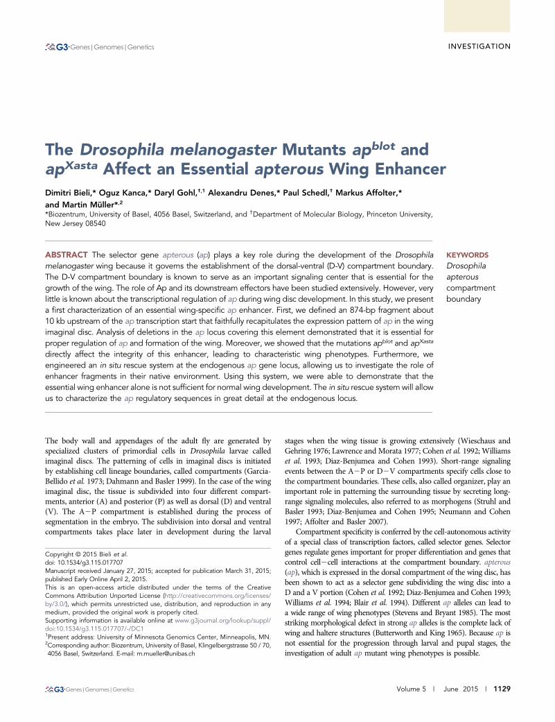
The Drosophila melanogaster Mutants apblot andapXasta Affect an Essential apterous Wing EnhancerDimitri Bieli,* Oguz Kanca,* Daryl Gohl,†,1 Alexandru Denes,* Paul Schedl,† Markus Affolter,*and Martin Müller*,2
*Biozentrum, University of Basel, 4056 Basel, Switzerland, and †Department of Molecular Biology, Princeton University,New Jersey 08540
ABSTRACT The selector gene apterous (ap) plays a key role during the development of the Drosophilamelanogaster wing because it governs the establishment of the dorsal-ventral (D-V) compartment boundary.The D-V compartment boundary is known to serve as an important signaling center that is essential for thegrowth of the wing. The role of Ap and its downstream effectors have been studied extensively. However, verylittle is known about the transcriptional regulation of ap during wing disc development. In this study, we presenta first characterization of an essential wing-specific ap enhancer. First, we defined an 874-bp fragment about10 kb upstream of the ap transcription start that faithfully recapitulates the expression pattern of ap in the wingimaginal disc. Analysis of deletions in the ap locus covering this element demonstrated that it is essential forproper regulation of ap and formation of the wing. Moreover, we showed that the mutations apblot and apXasta
directly affect the integrity of this enhancer, leading to characteristic wing phenotypes. Furthermore, weengineered an in situ rescue system at the endogenous ap gene locus, allowing us to investigate the role ofenhancer fragments in their native environment. Using this system, we were able to demonstrate that theessential wing enhancer alone is not sufficient for normal wing development. The in situ rescue system will allowus to characterize the ap regulatory sequences in great detail at the endogenous locus.
KEYWORDS
Drosophilaapterouscompartmentboundary
The body wall and appendages of the adult fly are generated byspecialized clusters of primordial cells in Drosophila larvae calledimaginal discs. The patterning of cells in imaginal discs is initiatedby establishing cell lineage boundaries, called compartments (Garcia-Bellido et al. 1973; Dahmann and Basler 1999). In the case of the wingimaginal disc, the tissue is subdivided into four different compart-ments, anterior (A) and posterior (P) as well as dorsal (D) and ventral(V). The A2P compartment is established during the process ofsegmentation in the embryo. The subdivision into dorsal and ventralcompartments takes place later in development during the larval
stages when the wing tissue is growing extensively (Wieschaus andGehring 1976; Lawrence and Morata 1977; Cohen et al. 1992; Williamset al. 1993; Diaz-Benjumea and Cohen 1993). Short-range signalingevents between the A2P or D2V compartments specify cells close tothe compartment boundaries. These cells, also called organizer, play animportant role in patterning the surrounding tissue by secreting long-range signaling molecules, also referred to as morphogens (Struhl andBasler 1993; Diaz-Benjumea and Cohen 1995; Neumann and Cohen1997; Affolter and Basler 2007).
Compartment specificity is conferred by the cell-autonomous activityof a special class of transcription factors, called selector genes. Selectorgenes regulate genes important for proper differentiation and genes thatcontrol cell2cell interactions at the compartment boundary. apterous(ap), which is expressed in the dorsal compartment of the wing disc, hasbeen shown to act as a selector gene subdividing the wing disc into aD and a V portion (Cohen et al. 1992; Diaz-Benjumea and Cohen 1993;Williams et al. 1994; Blair et al. 1994). Different ap alleles can lead toa wide range of wing phenotypes (Stevens and Bryant 1985). The moststriking morphological defect in strong ap alleles is the complete lack ofwing and haltere structures (Butterworth and King 1965). Because ap isnot essential for the progression through larval and pupal stages, theinvestigation of adult ap mutant wing phenotypes is possible.
Copyright © 2015 Bieli et al.doi: 10.1534/g3.115.017707Manuscript received January 27, 2015; accepted for publication March 31, 2015;published Early Online April 2, 2015.This is an open-access article distributed under the terms of the CreativeCommons Attribution Unported License (http://creativecommons.org/licenses/by/3.0/), which permits unrestricted use, distribution, and reproduction in anymedium, provided the original work is properly cited.Supporting information is available online at www.g3journal.org/lookup/suppl/doi:10.1534/g3.115.017707/-/DC11Present address: University of Minnesota Genomics Center, Minneapolis, MN.2Corresponding author: Biozentrum, University of Basel, Klingelbergstrasse 50 / 70,4056 Basel, Switzerland. E-mail: [email protected]
Volume 5 | June 2015 | 1129
The target genes of Ap and their downstream functions in thepatterning of the wing disc are relatively well understood. The activityof Ap initiates a bidirectional Notch signaling cascade at the D2Vcompartment boundary, which subsequently induces the expressionof wingless (wg) in a stripe along the compartment boundary (Diaz-Benjumea and Cohen 1993; Williams et al. 1994; Irvine and Wieschaus1994; Rulifson and Blair 1995; Kim et al. 1995; Couso et al. 1995). Wg,a ligand of the Wnt family, is responsible for the growth of the wingpouch and patterning along the D2V-axis, although its mode of actionas a classical morphogen currently is questioned (Neumann and Cohen1997; Alexandre et al. 2014).
Despite the rather detailed knowledge about the functions of Ap inwing disc development, our knowledge of the mechanisms regulatingap expression is still limited. It has been shown that activation of theepidermal growth factor receptor by its ligand Vein is necessary andsufficient to activate the expression of ap in the dorsal compartment ofthe wing disc (Zecca and Struhl 2002a,b). Moreover, early ventral wgexpression has been shown to restrict the expression of ap to the dorsalportion of the developing wing disc (Williams et al. 1994).
To identify the wing disc-specific cis-regulatory elements of ap, weused several genetic approaches. First, a classical LacZ enhancer reporterstudy was performed. Second, deletions with defined breakpoints in theap genomic locus were generated. Third, we have characterized twoclassical ap alleles, apblot and apXasta (apXa), at the molecular level andhave associated their respective molecular alterations to the minimalwing enhancer. Finally, we engineered a FC31-integrase-dependent insitu rescue system, which enabled us to dissect the role of these cis-regulatory elements in their native environment.
Using these assays, we have defined an essential, but notsufficient, minimal 874-bp ap wing enhancer fragment that drivesreporter gene expression in the dorsal compartment of the wingimaginal disc.
MATERIAL AND METHODS
Fly stocks and methodsFlies were grown on standard cornmeal agar at 25�, unless otherwisestated. ape01573 (PBac{RB}e01573), apf08090 (PBac{WH}f08090), apf00451
(PBac{WH}f00451), and apf00878 (PBac{WH}f00878) were purchasedfrom the Exelixis stock collection at Harvard Medical School. Df(2R)nap1 (BL#1006), apblot (BL#4190), w�; T(2;3)apXa, apXa/CyO; TM3, Sb1
(BL#2475), P{hsFLP}12, y1 w� (BL#1929), Df(2R)BSC696 (BL# 26548),w�; P{10XUAS-IVS-mCD8::GFP}attP40 (BL#32186), Df(3R)Exel6176(BL#7655), TM3, ryRK Sb1 Ser1 P{D2-3}99B (BL#1808), Bx-Gal4 (w1118
P{GawB}BxMS1096, BL#8860), y1 w�; Mi{y[+mDint2]=MIC}MI00964(BL#34133), y1 w�; Mi{y[+mDint2]=MIC}MI02330/SM6a (BL#33205),y1 w67c23; P{EPgy2}EY03046 (BL#15619), ptc-Gal4 (P{GawB}ptc559.1;BL#2017) were all obtained from the Bloomington Stock Center.fng-Gal4 (y� w�; P{w+mW.hs = GawB}NP5399 / TM6, P{w-=UAS-lacZ.UW23-1}UW23-1, DGCR#104990) was obtained from Kyoto Drosoph-ila Research Center. Dad4-Gal4 was established in our laboratory asdescribed in the sections to follow. actin-Gal4 (y w1118 ; P{actin5c::Gal4,w-}/CyO) and GMR-Gal4 (w1118 ; P{GMR::Gal4, w-}/CyO) were obtainedfrom Steven Henikoff (Ahmad and Henikoff 2001). salE-Gal4 wasobtained from the Basler lab via Fisun Hamarotoglu (Mosimannet al. 2006). dpp-Gal4 is described in Staehling-Hampton et al. (1994).UAS-ap was obtained from Marco Milán (Milán and Cohen 1999). y wM{vas-int.Dm}zh-2A, a stock producing FC31-integrase under thecontrol of the vasa promoter, and insertion platform M{3xP3-RFP.attP}zh-86Fb were obtained from Johannes Bischof (Bischof et al.2007). ap41F/T(2;3)apXa was obtained from John B. Thomas.
According to our genetic and molecular analysis, ap41F should notbe listed as an allele of ap. First, and contrary to a previous report(Bourgouin et al. 1992), hemizygous ap41F flies have normal wings andhalters. Second, although molecular analysis confirmed the presence ofa P-element insertion just proximal to vulcan on the ap41F chromo-some, polymerase chain reaction (PCR), and sequencing failed to pro-vide evidence for a ~200-bp deletion within 1.5 kb of the longest apcDNA (D. Bieli and M. Müller, unpublished data). The GFP knock-inallele ap::GFP is described in Caussinus et al. (2011) (BL#38423). apMM
has been described in Gohl et al. (2008). It contains an insertion ~400bp upstream of the longest ap cDNA. Dad4-GFP (P{Dad4::EGFPnuc,w+}) was obtained from Jorgos Pyrowolakis (Vuilleumier et al. 2010).Nuclear enhanced green fluorescence protein (GFP) is expressed underthe control of the Dad4 enhancer (Weiss et al. 2010). dadP1883D32 /TM3, Sb was obtained from Tetsuya Tabata. This deletion covers atleast 24 kb downstream of the DadP1883 insertion, including the com-plete Dad open-reading frame (ORF) and three neighboring genes(CG3983, CG5184, and CG3962; T . Tabata, personal communication;Tsuneizumi et al. 1997; Henderson et al. 1999). A recombinant be-tween apXa and P{y[+t7.7] w[+mC]=10XUAS-IVS-mCD8::GFP}attP40,inserted on 2R at 25C6 (Pfeiffer et al. 2010), was obtained by meioticrecombination and selection for the dominant Xasta and mini-whitemarkers. The generation of deficiencies apDG1, apDG3, apDG8, andapDG11 is described below in section FC31-integrase–mediated trans-genesis and generation of deletions.
Adult wings were dissected and mounted in Hoyer’s. Then, wingpreparations were baked at 58� for a few hours. Preparations wereallowed to harden at room temperature and flattened by applyinga 40-g metal cylinder on the cover slip. Pictures were taken with a NikonMicrophot-FXA microscope with a Sony NEX-5RK digital camera. Thenotums of adult flies were photographed with a Leica M125 binocularequipped with a Leica DFC420C camera.
Introduction of FC31-integrase targets into the aplocus by gene conversion at the site of apMM
A method known as direct gene conversion has previously beendeveloped to engineer a desired DNA fragment into the genomic site ofa P-element insertion (Gloor et al. 1991; Sipos et al. 2007). Uponexposure of a given P-element insertion to P-element transposase, thetransposon is excised and a double strand break is created. It is nor-mally repaired by the cellular machinery using the homologous chro-mosome as a template. However, the repair process may also use anexogenous plasmid containing the desired DNA fragment flanked byhomology arms derived from either side of the P-element insertion site.Such a gene conversion template plasmid containing homology armsflanking the site of apMM insertion, along with hsp70-GFP bracketed bya pair of inverted attB sites was constructed and named pLAPGPRA(see Figure 5A). The construction of this plasmid was a multi-stepprocedure. Details can be obtained upon request. In brief, left(899 bp long) and right (1981 bp long) ap-homology arms were am-plified by PCR. To minimize sequence polymorphism which coulddecrease the efficiency of gene conversion, apMM genomic DNA(gDNA; isolated as described in Ashburner 1989) was used as thetemplate for PCR. As primers we used apLA-R, apLA-FNotI, apRA-F,and apRA-R (for primer sequences, see Supporting Information, TableS1). Inserts in the proper orientation for subsequent cloning were iden-tified using diagnostic digests and sequencing.
pLAPGPRAwas injected (650 ng/mL) along with pTurbo (250 ng/mlL)into embryos derived from a cross of y w; apDG3{w+}/+; TM3, Sb D2-3/+males with y w; apMM{y+} ; + virgins. Surviving injectees were trans-ferred to fresh vials and carefully tended at 18�. Among the hatching
1130 | D. Bieli et al.

adults, males and virgins representing the two desired genotypes(apDG3{w+}/apMM{y+} and apDG3{w+}/apMM{y+}; TM3, Sb D2-3/+) wereselected for further work. apDG3/apMM flies have normal wings and hal-teres. The apDG3 chromosome was included because it lacks the DNAcorresponding to the homology arms of pLAPGPRA and hence cannotserve as a template for double strand gap repair. A total of 72 fertilecrosses involving virgins (in pairs) or single males mated with y w; alb c sp/SM6a flies could be set up. Originally, it was intended to screen thelarval progeny of these crosses for GFP expression. Unfortunately, thiselegant approach failed in practice. Therefore, the progeny was screenedfor y2 w2 males. This phenotype indicates loss of the y marker andtherefore most likely also of apMM and was, in the absence of the positiveGFP selection, the only selectable marker to identify putative conversioncandidates. A total of 105 y2 w2 males were selected from 32 (out of 72)crosses yielding such males. Balanced lines of potential gene conversionevents were established and screened for GFP fluorescence in larval wingdiscs. Five candidate gene conversion lines with weak GFP expression inwing imaginal discs in an ap-like pattern were obtained from two in-dependent dysgenic crosses (isolation numbers: c1.4a, c1.4b, c1.4d, andc1.4e; c1.13a).
To confirm that the five GFP-positive candidate gene conversionlines had the attP-flanked GFP construct integrated in the ap locus,gDNA was isolated and analyzed by PCR. PCR products were obtainedfor all five candidates between a primer (SV40out55) within the SV40trailer sequence (just downstream of GFP) and a primer (apLOF2) inthe ap gene outside of the left homology arm. Sequencing of all fivelines confirmed the integrity of the ap promoter region and the pres-ence of the ap proximal attP site (data not shown).
On the distal side, PCRs using primers in the hsp70 promoter (justupstream GFP) and several primers outside of the right homology arminitially failed to produce products (data not shown). Later, by use ofone of the lines obtained by RMCE (see below), the integrity of thejunction between the template plasmid and the right homology armcould be verified by PCR and sequencing using the Mcp-dir-y and ap-dir-3 as primers. Mcp-dir-y primes toward the end of the mini-yellowgene present on our Recombination-Mediated Cassette Exchange(RMCE) insertion cassette. We also tested whether the junction betweenthe right homology arm and the flanking ap sequence is intact by PCRusing a primer near the end of the right homology arm (apRAendF),and a primer in the flanking ap sequence (apROR2). A product of theexpected size was observed, indicating that the junction is intact.
apc1.4a, apc1.4b, apc1.4d, apc1.4e, and apc1.13a homozygotes all havewild-type wings, indicating that the function of the ap wing enhancerand promoter were not disrupted by the gene conversion event. Geneconversion events apc1.4a, apc1.4b, apc1.4d, apc1.4e also have a rough eyephenotype when homozygous, but not over apDG3. The rough eyephenotype can be separated from the ap locus by meiotic recombina-tion. Finally, only apc1.4b was chosen for further work. One of itsapplications is the targeted insertion of exogenous DNA into the aplocus by RMCE (Bateman et al. 2006).
FC31-integrase2mediated transgenesis andgeneration of deletionsConstructs for FC31-integrase2mediated transgenesis were generatedbased on plasmid piB-LLFY(BI) [details about the construction of piB-LLFY(BI) can be acquired upon request]. As required for RMCE, itcontains two inverted attB sites. Separating them are the followingthree genetic components: (1) two LoxP sites in direct orientation witha multiple cloning site in between them; (2) the LoxP cassette is fol-lowed by a single FRT site; (3) the mini-yellow transformation markercompletes piB-LLFY(BI). mini-yellow refers to a yellow reporter gene
lacking all of its characterized tissue specific enhancers. It consists ofthe yellow cDNA fused to ~330 bp of 59 genomic DNA, including theyellow promoter and extending up to a KpnI restriction site. The mini-yellow fragment was isolated from plasmid C4yellow (referred to asDint in Geyer and Corces 1987; Gohl et al. 2008). In the context of theap gene and in a y background, mini-yellow activity always manifestsitself in phenotypically yellow+ wings. Depending on the transgene andorientation of insert, thoracic bristles may also acquire yellow+ pig-mentation (D. Gohl and M. Müller, unpublished data).
Constructs were introduced into the ap locus by RMCE into twodocking sites, Mi{y[+mDint2]=MIC}MI02330 (Venken et al. 2011)and apc1.4b. DNA was injected at a concentration of 300 ng/mL in1· phosphate-buffered saline (PBS) into early embryos of the genotypey w M{vas-int.Dm}zh-2A; MI02330/CyO or y w M{vas-int.Dm}zh-2A;apc1.4b/CyO. The relevant transgenic lines obtained in this way areapDD35.34 and apD5f.1, respectively. Their position and the orientationof the FRT are depicted in Figure 1C together with four other FRTcontaining transposon insertions. Five of the six stocks are homozygousand hemizygous viable. Their wings and halteres are of wild-type ap-pearance. This is not the case for apf08090. The lethality of this chromo-some cannot be reverted by excision of the PBac{WH}, indicating that itis associated with a second site lethal. Rare homozygous revertant escap-ers as well as frequent hemizygous revertants have normal wings. There-fore, the PBac{WH} insert is responsible for the strong phenotype inhemizygous apf08090 flies. However, this phenotype is not dependent onthe gypsy insulator present in apf08090 because the wing phenotype is notsuppressed in a su(Hw)2 background (M. Müller, unpublished data).
We have noted that in the Drosophila literature, two divergentdefinitions for FRT orientation are in use! In this study, FRT orien-tation is indicated according to Thibault et al. (2004).
In Drosophila, the production of deletions by Flipase-catalyzed re-combination between two FRT sites either in cis or in trans has enabledthe community to obtain a huge collection of tailor-made deficiencies(Golic and Golic 1996; Ryder et al. 2007). We have previously appliedthis technology to generate a ~27-kb deletion named Df(2R)apDG be-tween two FRT sites in apMM and ape01573 (Gohl et al. 2008). Note thatin this study, Df(2R)apDG is referred to as apDG1. Applying analogousgenetic crossing schemes, we have generated three further deletions:
Df(2R)apDG3: An ~44-kb deletion between two FRT sites located inapf08090 and ape01573. It is referred to as apDG3. In this deficiency, a largepart of the ap transcription unit is lost together with ~27 kb of inter-genic DNA separating ap from l(2)09851. Although a considerable partof the ap ORF located proximal to the break in apDG3 remains in place,genetic observations are consistent with it being a true null allele withrespect to ap function in wing and haltere tissue. Flanking the new FRTjunction are three genetic elements: gypsy insulator and mini-white (ofapf08090) and a splice acceptor (of ape01573). Over several kilobases, theregion of the new fusion is identical to PBac{RB}e01573 and, hence, noadequate apDG3-specific PCR primers could be designed. Thus, fourPCR primer pairs distributed evenly over the ~44-kb interval missingon the apDG3 chromosome were tested on w1 and on apDG3/Df(2R)nap1 flies (Df(2R)nap1 being a cytologically visible deletion also uncov-ering ap). The absence of the corresponding DNA in the latter couldunambiguously be demonstrated (data not shown).
Df(2R)apDG8{w+}: An ~20-kb deletion between two FRT sites locatedin apf00878 and apD5f.1. It is referred to as apDG8. It corresponds toa rather clean deletion of the complete ap transcription unit. Its phe-notypes are indistinguishable from those observed for apDG3. Flankingthe new FRT junction are two genetic elements: a UAS-induciblepromoter (of apf00878) and mini-white (of apD5f.1). It was verified by
Volume 5 June 2015 | apterous Wing Enhancer | 1131
PCR and sequencing. Aprec-LA-AscI-F and WARIout#1 primerswere used for PCR. For sequencing, we used primers apEnhDel-Seq-PBrev and WARIout#2.
Df(2R)apDG11, al: An ~11-kb deletion between 2 FRT sites in alapMM sp and apDD35.34. It is referred to as apDG11. Because apDG1,homozygous apDG11 flies have no wings. Both deficiencies share thesame proximal break point. Previous transvection studies have sug-gested that the ap promoter immediately proximal to apDG1 (andhence also of apDG11) remains intact (Gohl et al. 2008). Flankingthe new FRT junction are two genetic elements: a LoxP site (of apMM)and mini-yellow (of apDD35.34). The new junction was verified bysequencing. For PCR amplification of the region, the apMM-200forand yellow59out primer pair was used. Part of the fragment wassequenced with yellow59out and Inverseappromfor.
Generation of a FC31-integrase based in situ rescuesystem at ap
Construction of pBSattBattPLoxFRTy: Two complementary oligos(attBPfor and attBPrev) containing attB and attP sites in tandem werepurchased from Sigma-Aldrich. These oligos were annealed and clonedbetween the XhoI and KpnI sites of pBSIIKS. The new plasmid’s nameis pBSIIKSattBattP. A XhoI-ClaI fragment containing LoxP, FRT,and mini-yellow was isolated from piB-LLFY(BI) and subcloned intopBSIIKSattBattP, thereby generating the pBSattBattPLoxFRTy vectorused for FC31 integrase mediated transgenesis (see Figure 5B). TheattB and attP sites on this vector are separated by only 6 bp. It wasassumed that therefore the two elements are too close for efficientintramolecular recombination. The fact the two desired insertions(one in each attP site present in apc1.4b, see Figure 5B) could be isolatedseems to support this assumption.
Generation of apattPDEnh, a platform for insertion of ap enhancerfragments: pBSattBattPLoxFRTy DNA was injected into y w M{vas-int.Dm}zh-2A; apc1.4b/CyO embryos. Yellow+ marked flies could beisolated and mated. The desired insert orientation could be identifiedby PCR using the apdown-forN and aptransch_yw_rev primers.A stock with isolation number 6.1 was selected for correct orientationof insert pBSattBattPLoxFRTy. It is referred to as apattBPFRTy2 (seeFigure 5C; the other insert orientation was also isolated and calledapattBPFRTy1). We wished to further modify this stock by introducingthe same ~27-kb deletion as in apDG1. Therefore, y w; apattBPFRTy2
males were mated with y w hsFlp; PBac{RB}e01573 virgins. Progenywas heat-shocked at three subsequent days during its larval develop-ment for 1 hr in a 37� water bath. Hatchlings were individuallycrossed to y w M{vas-int.Dm}zh-2A ; Sp Pin/CyO flies. A total of 7of 80 single crosses produced phenotypically yellow2 and white2 flies,indicating the loss of all DNA between the FRT sites of apattBPFRTy2
and PBac{RB}e01573, including mini-yellow and mini-white. Thenewly established deletion was named apattPDEnh (see Figure 4D)and kept as a y w M{vas-int.Dm}zh-2A ; apattPDEnh/CyO stock. Thedeletion was confirmed by PCR and sequencing with the primer pairapEnhDel_seq_dwnst_for and apEnhDel_seq_PB_rev.
The apattPDEnh chromosome contains a single functional attP siteready for FC31-integrase catalyzed insertion of pEnh-Reentry derivedplasmids (see Figure 5D). Insertion events can be further modified bysuitably placed LoxP and FRT sites, allowing for the deletion of theyellow marker or the enhancer fragment-yellow+ marker cassette, re-spectively (see Figure 5E).
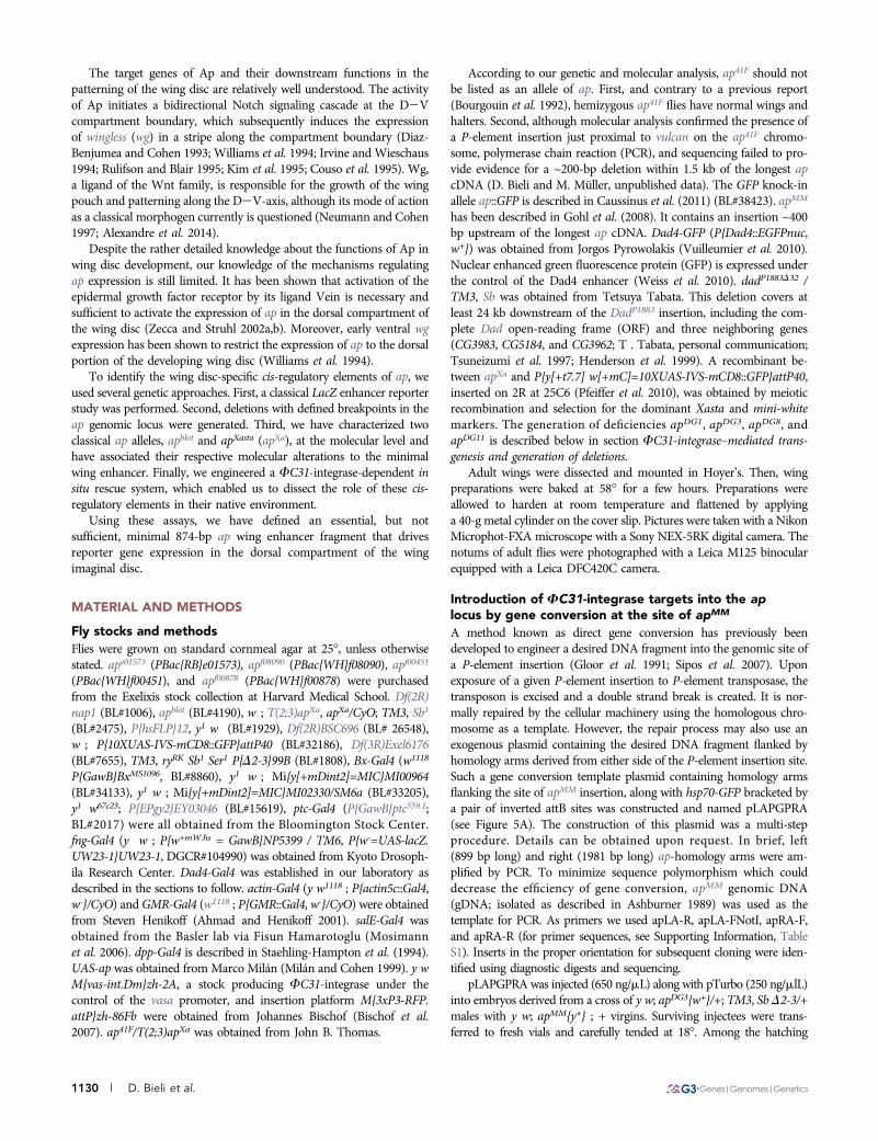
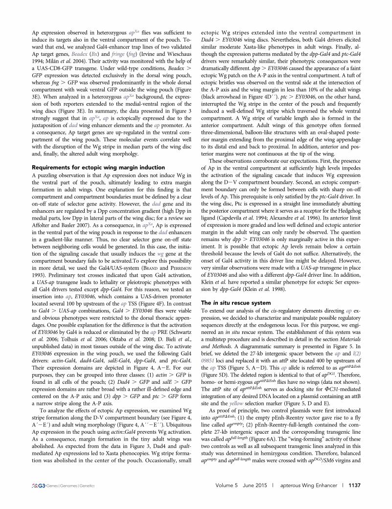
Figure 1 LacZ reporter assay and deletion analysis at the apterouslocus. (A) Diagrammatic representation of the ap locus. As drawn atthe top of the panel, it extends over roughly 50 kb. Its transcribed partis shown in green. ap is flanked by two genes indicated in blue: vulcanon the proximal and l(2)09851 on its distal side. Arrows above thegenomic interval specify the direction of transcription of the threegenes. Fragment apC, indicated in orange, has been reported to drivereporter expression in the dorsal compartment of the pouch, the hingeand the notum of the wing imaginal disc, where ap is normallyexpressed. Below, the relative positions and dimensions of nine frag-ments tested with our LacZ reporter assay are depicted. Fragmentscolored in orange (apO, apR, apOR, apOR3, and apRXa) elicit thesame expression pattern as apC. Fragments depicted in gray (apP,apQ, apS, apOR2) do not drive reporter gene expression in the wingdisc. (B) X-Gal staining in the wing disc of an apC-LacZ transgenic fly.Scale bar: 100 mm. (C) Deletions generated at the endogenous aplocus with FRT-containing inserts. At the top of the panel, trianglesalong the ap locus indicate the position of six different inserts. Pinkarrowheads within them mark the orientation of the FRT sites accord-ing to the definition of Thibault et al. (2004). The location of the apRXafragment is shown in orange. apDG3 deletes approximately 44 kb be-tween inserts apf08090 to ape01573, thereby removing most of ap ORFand upstream sequences. apDG8 is a 20-kb deficiency that deletes thecomplete ap ORF from apf00878 to apD5f.1. apDG1 removes the com-plete intergenic spacer between apMM to ape01573. apDG11 deletes an11-kb fragment from apMM to apDD35.34. Note that apD5f.1 and apMM
have exactly the same insertion site. (D) Notum pictures of a wild-typefly and trans-heterozygous ap mutants. In the wild type, the wing andthe haltere (arrowhead) are well formed and clearly visible. Df(2R)BSC696 is a large deletion at the base of 2R, deleting approximately360 kb, including the whole ap locus. When Df(2R)BSC696 is crossedto apDG3 all wing and haltere structures are lost. Only small stumps ofamorphic tissue remain at the actual attachment site of the wing (seearrow).Very similar phenotypes are observed in apDG8/apDG3, apDG1/apDG3 and apDG11/apDG3 flies. Scale bar: 25 mm.
1132 | D. Bieli et al.
Generation of pEnh-Reentry constructs: yellow+ coding sequenceand body cuticle enhancer were subcloned into pBSIIKS as a BglIIfragment from C4yellow, thereby generating plasmid pBSIIKS-yellow.Please note that the yellow wing enhancer is not part of the BglIIfragment! attB and FRT LoxP fragments were cloned by first anneal-ing and phosphorylating oligos attBtop and attBbottom as well asFRTLoxPtop and FRTLoxPbottom followed by three fragment liga-tion with pBSIIKS-yellow vector cut with SacI and XbaI. The resultingplasmid was called pEnh-Reentry and served as the backbone for allconstructs described below.
The 27-kb full-length enhancer was recombineered in pEnh-Reentryfrom BACR45O18 (purchased from the Berkeley Drosophila GenomeProject). The left homology arm was amplified with PCR with primerscontaining NotI and XhoI sites (primer pair: apenhrecLA_Not_for andapenhrecLA_XhoI_rev). The right homology arm was amplified withprimers containing XhoI and BglII sites (primer pair: apenhrecRA_XhoI_forand apenhrecRA_BglII_rev). Homology arms were cloned in pEnh-Reentry cut with NotI/BglII as 3 fragment ligation. Recombineeringwas performed according to Thomason et al. 2007. In brief, thepEnh-Reentry-homologyarms vector was linearized with XhoI andtransformed into bacterial strain DY380 (purchased from NCI at Fred-erick) pretransformed with BAC45O18 (purchased from BDGP), andpre-induced at 42� for 15 min. Recombinants were selected on ampi-cillin and screened by PCR. The correct recombineering product’sname is pEnh-Reentry-Full-length.
Dad enhancer fragments and apRXa were amplified from apXa
gDNA. First, fragments apRXaDadInt2, DadInt52, and Dad4 werecloned into a pBluescript II KS(+) vector, where the XbaI site wasmutated previously into a AvrII site. For apRXaDadInt52, primersapR_AvrII_for and dadint52_XmaI_SpeI_rev were used. For DadInt52,primers dadint52_XmaI_SpeI_rev andXa_brkpnt_AvrII_for wereused. To clone Dad4, we used the primer pair dad4_AvrII_for anddad4_XmaI_SpeI_rev. These fragments were combined via the respectiveSpeI or AvrII sites to produce apRXaDadInt52Dad4 and DadInt52Dad4fusion fragments. These were subcloned from pBluescript II KS(+) viaAvrII and XmaI sites into pEnh-Re-entry cut with AvrII and AgeI. apR,apRXa, apP, and apY were amplified from pEnh-Reentry-Full-lengthplasmid and cloned into pEnh-Re-entry via NotI, AvrII or AgeI sites.To clone apR, primers apR_AvrII_for and apR_XmaI_SpeI_rev were used.For apRXa, primer pair apR_AvrII_for and apRXa_AgeI_rev was used.To amplify apP, primers apP_NotI_for and apP_AvrII_rev were used. apYwas amplified using primer pair apY_NotI_for and apY_AgeI_rev.
All pEnh-Reentry derived constructs were brought into the ap locusby FC31-integrase mediated recombination (see Figure 4, D and E).DNAs were injected at a concentration of 300 ng/mL in 1·PBS into y wM{vas-int.Dm}zh-2A ; apattPDEnh/CyO embryos. Transgenic flies wereselected with the help of the yellow+ marker and balanced stocks weregenerated according to standard genetic procedure.
Generation of LacZ-reporter linesap regulatory DNA were amplified via PCR from y1 w67c23 gDNA withprimers containing restriction enzyme sites as overhangs, and subse-quently cloned into plasmid pAttBLaZ (Weiss et al. 2010) usingthe respective enzymes. apC was amplified with the primer pairapC_AscI_for and apC_BglII_rev. The apC fragment was defined byLundgren et al. 1995. The apO fragment was cloned with the primersapC_AscI_for and apO_BglII_rev. For apP, primers apC_BglII_revand apP_AscI_for were used. To clone apQ, primer pair apC_ BglII_revand apQ_AscI_for was used. For apR, the primers apR_AscI_for andapR_BglII_rev were used. apS was cloned with the primers apS_AscI_forand apS_BglII_rev. apOR was amplified with apR_AscI_for and
apO_BglII_rev. For apOR2, primers apR_AscI_for and apOR2_XbaI_revwere used. apOR3 was amplified with apR_AscI_for and apOR3_XbaI_rev.apRXa was cloned with apR_AscI_for and apRXa_XbaI_rev.
All the reporter transgenes were generated with the FC31-basedintegration system using the landing platform M{3xP3-RFP.attP}zh-86Fb (Bischof et al. 2007).
Molecular characterization of apblot
Complementation crosses with apblot over a set of overlapping ap dele-tions mapped the mutation to am ~11-kb interval upstream of apMM.Therefore, a set of PCR primer pairs was designed to screen for a lesionin that region of apblot gDNA. y1 w67c23 gDNA served as positive control.With one primer pair, a discontinuity could be identified on the apblot
chromosome. It could be best reconciled with the presence of a largerinsertion of DNA of unknown origin. Inverse PCR (iPCR) was sub-sequently used to obtain sequence information about the ends of theputative insertion. Toward that end, apblot gDNA was digested withBsaWI and ligated with T4 Ligase under conditions as previously de-scribed (Ochman et al. 1988). Primer pairs used for iPCR on theproximal side of the insertion were iPCR_for and iPCR_rev. Primerpairs used for iPCR on the distal side of the insertion were K_for andL_rev. Following this strategy, sequence information could be obtainedfor both ends of the inserted DNA. Sequence comparison identifiedthem as LTRs of the blood retrotransposon (Bingham and Chapman1986). To verify the insertion, primers out of blood 39 and 59 LTR(blood3prime and blood5prime, respectively) were used with primersbinding in adjacent ap regions (iPCR_for and L_rev, respectively). Se-quencing was performed by Microsynth AG, Switzerland.
Molecular characterization of apXa
The dominant Xasta allele was originally induced by X-ray mutagen-esis in a stock already containing two large inversions on 2R and 3R(Serebrovsky and Dubinin 1930; Waddington 1940; Lewis 1951;Hetherington et al. 1968). The new rearrangement was classified asa reciprocal translocation with breakpoints 41F9-41F11;89E8-89F1.Allelism with ap was inferred from noncomplementation with knownap alleles (Butterworth and King 1965; Stevens and Bryant 1985).Complementation crosses with a set of small overlapping ap deletionsfailed to narrow down the location of apXa. Hence, the whole ap locuswas screened by overlapping primer pairs. PCR products obtainedfrom amplification of apXa/+ and y1 w67c23 gDNA were compared.The analysis of these reactions identified a difference close to theinsertion break point found in apblot. Again, this region was probedby iPCR. apXa/+ gDNA was cut with NlaIII and religated under di-luted conditions. For iPCR, the primer pair iPCR_Xa_rev and 19_forwas used. Sequencing of the iPCR product revealed that the reciprocaltranslocation had fused DNA originating from dad locus on 3R to ap-specific sequences. The fusion was confirmed by PCR and sequencingwith 19_for and a primer in the dad region (primer dadint52out). Thebreakpoint associated with Xasta in ap was found to be identical in thetwo stocks ap41F/T(2;3)apXa and w�; T(2;3)apXa, apXa/CyO; TM3, Sb1.
Generation of Dad4-Gal4 fly lineThe minimal hsp70 promoter was amplified from the pUAST vectorwith the primer pair hsp70_XbaI_for and hsp70_BamHI_rev, thencloned into pBluescript II KS(+) via the XbaI and BamHI sites. TheDad4 fragment was amplified from gDNAwith the primers dad_NotI_forand dad_NheI_rev, followed by the insertion of the fragment into theNotI and XbaI digested pBS-hsp70 plasmid. Gal4 was amplified froma pCaSpeR4-Gal4 plasmid, obtained from the lab of Konrad Basler,with the primer pair Gal4_BglII_for and Gal4_HindIII_rev. The Gal4
Volume 5 June 2015 | apterous Wing Enhancer | 1133
fragment was subsequently cloned into the BamHI and HindIIIdigested pBS-Dad4-hsp70 plasmid. We amplified the SV40-PA termi-nator sequence from the pUAST vector, using the primer pairSV40_HindIII_for and SV40_BamHI_ApaI. The SV40_PA was sub-sequently inserted into the pBS-Dad4-hsp70-Gal4 plasmid using theHindIII and ApaI restriction sites. Finally, the Dad4-hsp70-Gal4-SV40_PA sequence was subcloned into the pCaSpeR4 vector, usingthe NotI and BamHI restriction sites. Transgenic flies were selected ina y1 w67c23 background with the help of the mini-white marker. TheDad4-Gal4 insert used in this study is linked to the X chromosome.
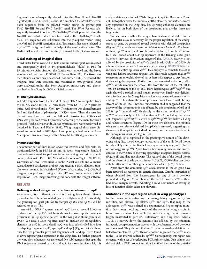
X-Gal staining of imaginal discsThird instar larvae were cut in half, and the anterior part was invertedand subsequently fixed in 1% glutaraldehyde (Fluka) in PBS for15 min on ice. After fixation, the fixative was removed and the larvaewere washed twice with PBST (0.1% Tween 20 in PBS). The tissue wasthen stained as previously described (Ashburner 1989). Afterward, theimaginal discs were dissected and mounted in 80% glycerol. Discswere analyzed under the Zeiss Axiophot microscope and photo-graphed with a Sony NEX-5RK digital camera.
In situ hybridizationA 1.5-kb fragment from the 39 end of the ap cDNA was amplified fromthe cDNA clone HL02012 (purchased from DGRC) with primersinsitu_SacI_for and insitu_KpnI_rev. The fragment was cloned betweenSacI and KpnI sites of pBluescript II KS(+) vector. Then, the resultingplasmid was linearized with Acc65I and digoxigenin-(DIG)-labeledRNA was produced from T7 promoter according to the manufacturer’sprotocol (Roche, Switzerland). In situ hybridizations were performed asdescribed in Tautz and Pfeifle (1989). Wing imaginal discs were dis-sected and mounted in 80% glycerol and photographed under a NikonMicrophot-FXA microscope with a Sony NEX-5RK digital camera.
ImmunostainingThe anterior part of third instar larvae was inverted and fixed with 4%paraformaldehyde in PBS for 25 min at room temperature. Standardprotocols were used to perform immunostaining. As primary anti-bodies, rabbit a-GFP (1:1000; Abcam) and mouse a-Wg (1:120, DSHB,University of Iowa) were used. a-rabbit AlexaFlour488 and a-mouseAlexaFlour568 (Molecular Probes) were used at a 1:750 dilution. Sam-ples were mounted in Vectashield (Vector Laboratories, Inc.). Confocalimaging was performed using a Leica SP5 microscope with a verticalstep size of 1 mm. Image processing was done with the ImageJ software.
RESULTS
Defining a short wing-specific enhancer element in apCAt apterous, four different transcripts starting from three differentpromoters have been annotated (see www.flybase.org). In this study,the transcription start site for transcripts ap-RA and ap-RC will bereferred to as ap TSS.
An ~8-kb DNA fragment named apC located several kilobasesupstream of the ap TSS had been shown to drive reporter gene ex-pression in an ap-specific pattern in the wing disc (Lundgren et al.1995). We used a LacZ reporter assay to analyze the cis-regulatoryelements in apC in more detail. apC was first sub-divided into fouroverlapping fragments, apO, apR, apP and apQ (Figure 1A). Of these,only the two promoter proximal fragments, apO and apR were foundto drive reporter gene expression in the wing disc. To further pinpointthe wing disc enhancers, we generated five subfragments that span theDNA sequences covered by apO and apR. As shown in Figure 1A, this
analysis defines a minimal 874-bp fragment, apRXa. Because apP andapOR2 together cover the minimal apRXa element, but neither showedany expression in the wing disc, key ap wing enhancer elements arelikely to be on both sides of the breakpoint that divides these twofragments.
To determine whether the wing enhancer element identified in theLacZ reporter assay is necessary for the proper regulation of the endog-enous ap gene, we generated several deletions with defined breakpoints(Figure 1C; for details see the sectionMaterials andMethods). The largestof these, apDG3, removes almost the entire ap locus, from the 4th intronto a site located about 500 bp upstream of the flanking distal gene,l(2)09851. Previous observations suggested that l(2)09851 activity is notaffected by the proximity of apDG3’s distal break (Gohl et al. 2008). Asa homozygote or when in trans to a large deficiency, Df(2R)BSC696, thatincludes the entire ap locus, apDG3 flies displayed a complete loss of allwing and haltere structures (Figure 1D). This result suggests that apDG3
represents an amorphic allele of ap, at least with respect to Ap functionduring wing development. Furthermore, we generated a deletion, calledapDG8, which removes the whole ORF from the end of the 39UTR to~400 bp upstream of the ap TSS. Trans-heterozygous apDG8/apDG3 fliesagain showed a typical ap-null mutant phenotype. Finally, two deficien-cies affecting only the 59 regulatory region were generated, namely apDG1
and apDG11. They share the same proximal break located ~400 bp up-stream of the ap TSS. Previous transvection studies suggested that theactivity of the ap promoter is not affected by this breakpoint (Gohl et al.2008). apDG1 extends ~27 kb distally to the same position as apDG3.apDG11 removes only ~11 kb of upstream DNA, including the wholeapC fragment. apDG1/apDG3 as well as apDG11/apDG3 flies lacked all wingand haltere structures (Figure 1D). In these two deletions, the minimalwing enhancer element defined by apRXa is removed, suggesting thatelements within apRXa are indeed necessary for the regulation of ap inthe endogenous locus (see Figure 1C).
Although ap is expressed in the presumptive notum of the devel-oping wing disc, the phenotypic appearance of the adult dorsal thoraxis only mildly affected in flies lacking any ap activity (e.g., apDG8/apDG3
or homozygous apDG8). Apart from a few missing macro- and micro-chaetae in the vicinity of the wing appendage, it appears largely normal(Figure 1D and data not shown). The reduced size of the dorsal thoraxand the aberrant bristle pattern in apDG3/Df(2R)BSC696 flies can prob-ably be attributed to other genetic loci deleted in Df(2R)BSC696.
Apart from the dominant apXa allele, lesions in the ap gene havebeen reported as recessive in genetic character. Careful inspection ofwings obtained from flies heterozygous for any of the 4 deletionspresented in Figure 1C corroborated this fact. However, ~2% of themhad small margin defects, indicating a mild dominance of strong aploss-of-function alleles (data not shown).
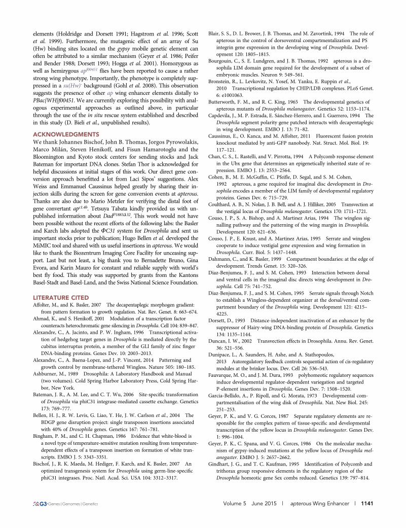
Mutations in the apR region result in wing phenotypesIn the course of investigating the cis-regulatory region of ap, weidentified two classical ap alleles, apblot and apXa, that map to theapR region. apblot was isolated as a spontaneous, hypomorphic muta-tion that causes notching mostly of the posterior wing margin inhomozygous mutant flies, while the anterior wing margin remainslargely unaffected (Figure 2A; Butterworth and King 1965; Whittle1979). To narrow down the genomic site affected by the mutation,intragenic complementation crosses with the aforementioned deletionswere analyzed. They showed that apDG11 was the smallest deletion thatfailed to complement apblot. This observation suggested that apblotmapsto the ~11-kb interval defined by apDG11. Consequently, this region wasscreened with a set of overlapping PCR primer pairs. One primer pairdid not yield a PCR product and thus identified the site of the putative
1134 | D. Bieli et al.

lesion on the apblot chromosome. Using iPCR, we identified the in-sertion of a retrotransposable element of the blood family in the apRXasequence (Figure 2B, see the sectionMaterials and Methods for details).This event caused the typical 4-bp duplication at the insertion sitecharacteristic for blood family transposons (Figure 2C; Bingham andChapman 1986; Wilanowski et al. 1995).
Phenotypes caused by blood insertions at other loci are sometimestemperature-sensitive (Bingham and Chapman 1986). To test this pos-sibility, we raised homozygous apblot flies at different temperatures andscored their wing phenotypes (Table 1). At 18�, only 28% of the wingsdisplayed minor defects. In most of these, the posterior cross vein failedto connect with the 4th wing vein (Figure 2A). At greater temperatures,more severe wing phenotypes were detected with a higher penetrance.At 25� and 29�, 52% and 70%, respectively, of the wings showedextensive notching within the posterior compartment and reducedwing size (Figure 2A).
The dominant apXa allele was generated by X-ray mutagenesis andis associated with a reciprocal translocation between chromosome arms2R and 3R. The breakpoints were mapped to 41F and 89EF, respec-tively (Serebrovsky and Dubinin 1930; Waddington 1940; Lewis 1951;Hetherington et al. 1968). When heterozygous, apXa flies show thecharacteristic dominant mitten-shaped wing phenotype, in which thedistal tip of the wing is missing leading to a deep notching of the wingblade. In hemizygous apXa flies, only long wing stumps with little or nowing margin and unstructured vein patterns are formed (Figure 2D).The break on 2R has long been known to affect the ap locus (Butterworthand King 1965; Stevens and Bryant 1985). However, our attempt tomap apXa by intragenic complementation was not successful, suggest-ing that the lesion in apterous prevents this type of genetic analysis(see also Figure 3D). Thus, we screened the entire ap locus withoverlapping PCR primer pairs. We identified a discontinuity in theapR region and determined the molecular nature of the breakpoint(Figures 2, E and F; for details see the sectionMaterials and Methods).It localized right at the edge of the apRXa fragment, 142 bp distal tothe insertion site of the blood transposon in apblot. Only the proximal874 bp of apR remain associated with the ap transcription unit (seeFigure 2E). The DNA on the other side of the breakpoint is from the
daughters against dpp (dad) locus located at 89E on 3R. As predictedfrom the cytological mapping of the rearranged apXa chromosomes,the dad locus is inverted compared to its wild-type orientation on 3R(for a comprehensive drawing of the apXa polytene chromosomes, seeHetherington et al. 1968). We were not able to determine the break-point at the reciprocal site of the translocation. Nevertheless, based onits reciprocal nature, it is conceivable that the dad locus is split withinits 4th intron and hence destroyed. Because dad is expressed in theimaginal wing disc, it is formally possible that the Xasta phenotype isdue to the loss of Dad activity. This possibility was addressed bycrossing apXa with 2 known dad deletions, Df(3R)Exel6176 anddadP1883D32. The wings of trans-heterozygous animals displayed thecharacteristic mitten phenotype seen in apXa heterozygous flies, suggest-ing that an amorphic dad background does not modify the Xasta phe-notype. Hence Dad function is not relevant for the production of theXasta phenotype (data not shown). This is not unexpected, since dadmutants show no visible phenotype in the adult wing (Ogiso et al. 2011).
The proximity of dad enhancers to the ap transcription unit in theapXa chromosome suggests a plausible explanation for the Xasta wingphenotype. Two cis-regulatory elements, Dad4 and DadInt52, arelocated in the dad introns (Figure 2E) and are known to drive reportergene expression in the wing disc in a stripe along the A2P compart-ment boundary in response to Dpp signaling (Weiss et al. 2010).Because dad territory encompasses not only the dorsal but also theventral compartment of the presumptive wing pouch, a likely scenariois that the ap promoter responds to these two dad enhancers, leadingto ectopic Ap expression in the ventral compartment of the pouch.
Ectopic expression of ap in apXa leads to the ectopicexpression of Ap target genesTo further characterize the effect of the apblot and apXa mutations onwing development, we examined ap mRNA and Wingless protein(Wg) expression in 3rd instar larval wing discs (Figure 3, A and B).In wild-type discs, apmRNA is restricted to the dorsal compartment ofthe wing pouch, the hinge and the notum (Figure 3A). In the pouch,Ap activity is required to direct the expression of Wg in a stripe along
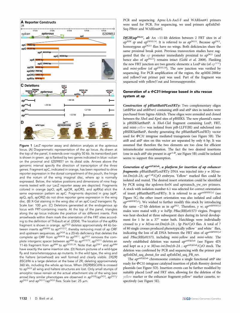
Figure 2 The mutations apblot and apXa affect theap wing enhancer region. (A) Temperature-sensitivewing phenotypes obtained for the homozygousapblot allele. At 18�, less than 30% of the wingsare affected and most of them only show a disrup-tion of the posterior crossvein (arrow). At 29�, ~70%of the wings have a phenotype. In many of them,the posterior compartment is severely affected. (B)At the top of the panel, the coordinates of the ap-terous locus are indicated. The insertion site ofblood, a retrotransposable element, within theapRXa wing enhancer is depicted. (C) Sequencedata close to the insertion site of the blood elementin apblot. The insertion causes a four bp duplication(CTGA, underlined). Exact coordinates of the 4 bpduplication: 2R:5735176.0.5735179 (Flybase Re-lease FB2014_06). (D) Preparations of wild type
and apXa mutant wings. All apXa/+ flies show a dominant phenotype: the distal part of the wing blade is lost and the characteristic mittenphenotype is formed. In hemizygous condition, the wing tissue of apXa/apDG3 flies forms a short tube-like structure. Margin bristles are absentexcept for sometimes a few at the tip. All scale bars are 50 mm. (E) Molecular characteristics of the apXa mutation. A reciprocal translocationinvolving the right arms of the second and third chromosome causes a breakpoint just upstream of the apRXa wing enhancer (indicated in orange)and juxtaposes the daughters against dpp (dad) locus (indicated in blue) next to the ap gene. The dark blue rectangles represent the well-studiedcis-regulatory elements Dadint52 and Dad4 which are active in the wing disc (Weiss et al. 2010). (F) Chromatograph of the apXa sequence acrossthe rearrangement break point. The coordinates of the breakpoints are: 2R:5375319 and 3R:17065902 (Flybase Release FB2014_06).
Volume 5 June 2015 | apterous Wing Enhancer | 1135

the D2V compartment boundary (Figure 3B). This Wg stripe is es-sential for the proper formation of the wing margin (Couso et al. 1994).
The temperature sensitivity of apblot was faithfully recapitulated bythe expression patterns in 3rd instar wing discs. Although ap mRNAlevels were reduced at 18� as well as at 29�, an obvious deviation of theap mRNA pattern was only observed at 29� in the posterior compart-ment of the pouch. This change correlated with a size reduction of theposterior compartment and the appearance of additional tissue foldingin this region (arrow in Figure 3A). Consistent with the sharp bound-ary of the apmRNA expression pattern at 18�, the Wg stripe along theD-V compartment boundary remained unchanged (Figure 3B). Incontrast, at 29�, the fuzzy appearance of the ap mRNA pattern inthe posterior compartment correlated with the disruption of the Wgstripe. In summary, these results are consistent with the adult wingphenotypes and provide an explanation for the abnormalities in theposterior wing margin as well as for the reduced size of the posteriorcompartment in apblot flies raised at elevated temperature.
In apXa heterozygotes, a strong ectopic misexpression of the aptranscript was detected in the ventral compartment of the wing disc,
with the highest signal along the medial part of the disc (Figure 3A). Asa consequence, the Wg stripe was disrupted in the medial region of thewing pouch (Figure 3B). Remarkably, the disruption of the Wg stripecorrelated well with the expression domain of the Dad4-GFP reporterconstruct (Figure 3C). Wherever GFP was detected, the expression ofWg was either very low or absent. Wing discs of hemizygous apXa/apDG3 flies showed strong ap expression in the entire pouch region.The characteristic Wg stripe in the wing pouch was lost, leaving behindonly a small dot of Wg expression in the middle of the pouch. More-over, the dimension of the wing pouch was reduced to about half thesize of a wild-type pouch.
In Drosophila, the somatic pairing of the two homologous chromo-somes can lead to a special situation of gene regulation called trans-vection (Lewis 1954; Sipos et al. 1998; Morris et al. 1999; Coulthardet al. 2005). In this case the regulatory elements of a gene can regulatethe expression of its homolog in trans. Transvection has been describedfor many gene loci (for reviews see Wu and Morris 1999; Duncan2002) including the ap locus (Gohl et al. 2008). Therefore, we decidedto test the transvection ability of apXa by crossing it with ap::GFP. Inthis combination only the gene in trans is labeled with GFP, allowingfor the independent detection of the gene product from this chromo-some. Trans-heterozygous ap::GFP/apXa flies displayed no ectopicexpression of Ap-GFP in the ventral wing pouch (Figure 3D). Thisresult demonstrates that the misexpression of ap is limited to thechromosome affected by the rearrangement.
As a selector gene, ap is known to regulate multiple downstreamgenes (Bronstein et al. 2010). We wished to know whether the ectopic
n Table 1 Temperature sensitivity of apblot
TemperatureTotal Wings
ScoredNormalWings
Wings withPhenotypes
18� 294 72% 28%25� 284 48% 52%29� 242 30% 70%
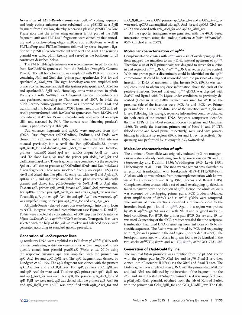
Figure 3 Wing disc phenotypes in apblot andapXa. All discs are shown anterior to the leftand dorsal side up. (A) in situ hybridizationagainst ap mRNA in late 3rd instar larval wingdiscs. In wild type, the dorsal compartment ofthe wing pouch is filled and outlined by the aptranscript. apblot discs show reduced ap mRNAlevels. At 18�, the ap expression pattern remainsvery similar as that in wild type. At 29�, expres-sion of ap in the posterior compartment is dis-turbed and the tissue is deformed (see arrow). Inheterozygous apXa discs, ectopic ap expressionis seen in the ventral part of the wing disc, withthe strongest signal in median regions. Theblack arrows point to the edges of the discwhere ap transcript is absent. In hemizygousapXa/apDG3 larvae, a similar pattern is observed.Note the change in shape of the wing disc. (B)a-Wg antibody staining of 3rd instar wing discs.In wild type, a characteristic thin stripe of Wgtraverses the wing pouch along the D-V com-partment boundary. In apblot, Wg expression isnormal at 18�C. At 29�C, the Wg stripe is muchweaker and less well defined in posteriorregions of the wing pouch. In apXa/+ discs, theWg stripe is interrupted in the median pouchregion. In hemizygous apXa discs, the Wg stripeis lost and only a dot of Wg expression in the
middle of the pouch is visible. In addition, the size of the pouch is reduced. (C) GFP expression driven by the Dad4 enhancer is detected in thecentral part of an apXa/+ wing disc. Note that absence of Wg stripe correlates well with higher GFP levels. Therefore, stripe formation is moreaffected in the anterior than in the posterior compartment. (D) a-GFP and a-Wg antibody staining of an ap::GFP/apXa wing disc. GFP expression isrestricted to the dorsal compartment of the wing pouch. In particular, Ap-GFP fusion protein does not spread ventrally where the Wg stripe isinterrupted. This indicates that dad enhancers on the apXasta chromosome are unable to activate ap::GFP located on the homologous chromo-some. (E) Expression of Beadex- and fringe-Gal4 enhancer trap lines in wild type and apXa/+ discs. Note that ectopic expression (white arrows) ofthese two validated Ap targets in the ventral compartment is only detected where the Wg stripe is interrupted. All scale bars are 100 mm.
1136 | D. Bieli et al.
Ap expression observed in heterozygous apXa flies was sufficient toinduce its targets also in the ventral compartment of the pouch. To-ward that end, we analyzed Gal4-enhancer trap lines of two validatedAp target genes, Beadex (Bx) and fringe (fng) (Irvine and Wieschaus1994; Milán et al. 2004). Their activity was monitored with the help ofa UAS-CD8-GFP transgene. Under wild-type conditions, Beadex .GFP expression was detected exclusively in the dorsal wing pouch,whereas fng . GFP was observed predominantly in the whole dorsalcompartment with weak ventral GFP outside the wing pouch (Figure3E). When analyzed in a heterozygous apXa background, the expres-sion of both reporters extended to the medial-ventral region of thewing discs (Figure 3E). In summary, the data presented in Figure 3strongly suggest that in apXa, ap is ectopically expressed due to thejuxtaposition of dad wing enhancer elements and the ap promoter. Asa consequence, Ap target genes are up-regulated in the ventral com-partment of the wing pouch. These molecular events correlate wellwith the disruption of the Wg stripe in median parts of the wing discand, finally, the altered adult wing morpholgy.
Requirements for ectopic wing margin inductionA puzzling observation is that Ap expression does not induce Wg inthe ventral part of the pouch, ultimately leading to extra marginformation in adult wings. One explanation for this finding is thatcompartment and compartment boundaries must be defined by a clearon-off state of selector gene activity. However, the dad gene and itsenhancers are regulated by a Dpp concentration gradient (high Dpp inmedial parts, low Dpp in lateral parts of the wing disc; for a review seeAffolter and Basler 2007). As a consequence, in apXa, Ap is expressedin the ventral part of the wing pouch in response to the dad enhancersin a gradient-like manner. Thus, no clear selector gene on-off statebetween neighboring cells would be generated. In this case, the initia-tion of the signaling cascade that usually induces the wg gene at thecompartment boundary fails to be activated.To explore this possibilityin more detail, we used the Gal4/UAS-system (BRAND and PERRIMON
1993). Preliminary test crosses indicated that upon Gal4 activation,a UAS-ap transgene leads to lethality or pleiotropic phenotypes withall Gal4 drivers tested except dpp-Gal4. For this reason, we tested aninsertion into ap, EY03046, which contains a UAS-driven promoterlocated several 100 bp upstream of the ap TSS (Figure 4F). In contrastto Gal4 . UAS-ap combinations, Gal4 . EY03046 flies were viableand obvious phenotypes were restricted to the dorsal thoracic appen-dages. One possible explanation for the difference is that the activationof EY03046 by Gal4 is reduced or eliminated by the ap PRE (Schwartzet al. 2006; Tolhuis et al. 2006; Oktaba et al. 2008; D. Bieli et al.,unpublished data) in most tissues outside of the wing disc. To activateEY03046 expression in the wing pouch, we used the following Gal4drivers: actin-Gal4, dad4-Gal4, salE-Gal4, dpp-Gal4, and ptc-Gal4.Their expression domains are depicted in Figure 4, A2E. For ourpurposes, they can be grouped into three classes: (1) actin . GFP isfound in all cells of the pouch; (2) Dad4 . GFP and salE . GFPexpression domains are rather broad with a rather ill-defined edge andcentered on the A-P axis; and (3) dpp . GFP and ptc . GFP forma narrow stripe along the A-P axis.
To analyze the effects of ectopic Ap expression, we examined Wgstripe formation along the D-V compartment boundary (see Figure 4,A´2E´) and adult wing morphology (Figure 4, A´´2E´´). UbiquitousAp expression in the pouch using actin::Gal4 prevents Wg activation.As a consequence, margin formation in the tiny adult wings wasabolished. As expected from the data in Figure 3, Dad4 and spalt-mediated Ap expressions led to Xasta phenocopies. Wg stripe forma-tion was abolished in the center of the pouch. Occasionally, small
ectopic Wg stripes extended into the ventral compartment inDad4 . EY03046 wing discs. Nevertheless, both Gal4 drivers elicitedsimilar moderate Xasta-like phenotypes in adult wings. Finally, al-though the expression patterns mediated by the dpp-Gal4 and ptc-Gal4drivers were remarkably similar, their phenotypic consequences weredramatically different. dpp. EY03046 caused the appearance of a faintectopic Wg patch on the A-P axis in the ventral compartment. A tuft ofectopic bristles was observed on the ventral side at the intersection ofthe A-P axis and the wing margin in less than 10% of the adult wings(black arrowhead in Figure 4D´´). ptc . EY03046, on the other hand,interrupted the Wg stripe in the center of the pouch and frequentlyinduced a well-defined Wg stripe which traversed the whole ventralcompartment. A Wg stripe of variable length also is formed in theanterior compartment. Adult wings of this genotype often formedthree-dimensional, balloon-like structures with an oval-shaped poste-rior margin extending from the proximal edge of the wing appendageto its distal end and back to proximal. In addition, anterior and pos-terior margins were not continuous at the tip of the wing.
These observations corroborate our expectations. First, the presenceof Ap in the ventral compartment at sufficiently high levels impedesthe activation of the signaling cascade that induces Wg expressionalong the D2V compartment boundary. Second, an ectopic compart-ment boundary can only be formed between cells with sharp on-offlevels of Ap. This prerequisite is only satisfied by the ptc-Gal4 driver. Inthe wing disc, Ptc is expressed in a straight line immediately abuttingthe posterior compartment where it serves as a receptor for the Hedgehogligand (Capdevila et al. 1994; Alexandre et al. 1996). Its anterior limitof expression is more graded and less well defined and ectopic anteriormargin in the adult wing can only rarely be observed. The questionremains why dpp . EY03046 is only marginally active in this exper-iment. It is possible that ectopic Ap levels remain below a certainthreshold because the levels of Gal4 do not suffice. Alternatively, theonset of Gal4 activity in this driver line might be delayed. However,very similar observations were made with a UAS-ap transgene in placeof EY03046 and also with a different dpp-Gal4 driver line. In addition,Klein et al. have reported a similar phenotype for ectopic Ser expres-sion by dpp-Gal4 (Klein et al. 1998).
The in situ rescue systemTo extend our analysis of the cis-regulatory elements directing ap ex-pression, we decided to characterize and manipulate possible regulatorysequences directly at the endogenous locus. For this purpose, we engi-neered an in situ rescue system. The establishment of this system wasa multistep procedure and is described in detail in the sectionMaterialsand Methods. A diagrammatic summary is presented in Figure 5. Inbrief, we deleted the 27-kb intergenic spacer between the ap and l(2)09851 loci and replaced it with an attP site located 400 bp upstream ofthe ap TSS (Figure 5, A2D). This ap allele is referred to as apattPDEnh
(Figure 5D). The deleted region is identical to that of apDG1. Therefore,homo- or hemi-zygous apattPDEnh flies have no wings (data not shown).The attP site of apattPDEnh serves as docking site for FC31-mediatedintegration of any desired DNA located on a plasmid containing an attBsite and the yellow selection marker (Figure 5, D and E).
As proof of principle, two control plasmids were first introducedinto apattPDEnh: (1) the empty pEnh-Reentry vector gave rise to a flyline called apempty; (2) pEnh-Reentry-full-length contained the com-plete 27-kb intergenic spacer and the corresponding transgenic linewas called apfull-length (Figure 6A). The “wing-forming” activity of thesetwo controls as well as all subsequent transgenic lines analyzed in thisstudy was determined in hemizygous condition. Therefore, balancedapempty and apfull-length males were crossed with apDG3/SM6 virgins and
Volume 5 June 2015 | apterous Wing Enhancer | 1137

the wings of trans-heterozygous progeny were carefully inspected. Asexpected, apempty/apDG3 flies generated no detectable wing material. Incontrast, the reconstituted ap locus produced wild-type wings inapfull-length/apDG3 flies. Taken together, these observations demonstrate thefeasibility of our in situ rescue system and suggest that the backbone ofthe pEnh-Reentry plasmid does not cause any disturbances.
DadInt52 and Dad4 enhancers contribute significantlyto the Xasta phenotypeOur model for the Xasta wing phenotype posits that the wing specificdad enhancers Dad4 and DadInt52 are responsible for ectopic Apexpression in the ventral pouch compartment. We wished to test this
hypothesis with the in situ rescue system. Two fly lines were estab-lished: apapRXaDad52.4 and apDad52.4 (Figure 6B). The former combinedthe three identified wing specific enhancers apRXa, DadInt52 andDad4. The latter contained only the two dad regulatory elements.When the 2 transgenics were initially isolated, it was immediatelyapparent that both phenocopied the dominant Xasta allele. However,a semi quantitative analysis also showed that the severity of theirphenotypes was weaker than observed for apXa/+ wings (Table 2).Although roughly 50% of the apDad52.4/+ wings were as strongly af-fected as those of apXa/+ flies, hardly any such wings appeared inapapRXaDad52.4/+ flies. These observations indicate that apart fromDadInt52 and Dad4, other factors contribute to the production ofa full blown Xasta wing phenotype.
The wings of apapRXaDad52.4 and apDad52.4 were also analyzed in hemi-zygous condition. The phenotypes were comparable to the one seen inapXa/ apDG3 flies: only tube-like wing stumps were formed which lackedwing margin completely except for the occasional occurrence of a fewmargin hairs at the very tip. It is conceivable that the latter arise due to theWg spot seen in the center of the pouch of apXa/apDG3 wing discs (seeFigure 3B). We have never seen homozygous apXa flies but did inspectadult wings of apXa/apDad52.4 animals. They appeared as even smallerversions of those observed in hemizygous apXa flies (data not shown).
The apRXA enhancer is required but not sufficient forwing formationIn Figure 1 of this paper, we have presented evidence that the ~8 kbapC fragment harbors an 874-bp wing specific enhancer that is es-sential for wing formation. However, the experimental approaches weused are not adequate to test whether the enhancer is also sufficientfor the formation of a wild-type wing. Therefore, four overlappingfragments covering the whole apC were introduced into the ap locusand the corresponding transgenic lines were obtained: apapP, apapY,apapR, and apapRXa. Their wing enhancer activity was tested in a hemi-zygous genetic background (Figure 6C). apapP/apDG3 flies, which con-tained the apP fragment that did not yield any LacZ reporter activity(see Figure 1A), also did not develop any wing or haltere tissue andphenotypically resembled ap null alleles. When apY, a fragment whichis shifted by 2 kb toward the ap TSS, was tested in apapY/apDG3 flies,wing development was partially restored. However, most of the mar-gin, the alula and the hinge region were poorly formed. Similar phe-notypes as for apapY were observed in apapR/apDG3 and apapRXa/apDG3
flies. Note that these three apC derivatives were sufficient to driveap-specific LacZ expression in our reporter assay (see Figure 1A).“Homozygotes” obtained by pairwise combinations of apapY, apapR
or apapRXa were also studied. Such wings looked improved comparedto the phenotypes observed in hemizygotes, because the margins,particularly along the anterior but also along the posterior edges ofthe wing, were formed to a large degree (data not shown). Somewhatunexpectedly, heterozygous apapY, apapR and apapRXa flies showeda weak dominant wing phenotype, associated with a small notch inthe tip region in 10–20% of the cases. This phenotype was not ob-served in apapY/+ or apfull-length/apDG3 flies (data not shown).
These results demonstrate that the 874 bp apRXa wing enhancerelement is required but not sufficient in the endogenous context tocorrectly regulate ap expression. Our observations imply the existence offurther unidentified wing enhancer elements elsewhere in the ap region.
DISCUSSIONIn the past, cis-regulatory elements were mainly investigated usingreporter-based assay systems, in which putative regulatory DNA
Figure 4 Margin formation in adult wings depends on well-definedOn-Off Apterous expression levels during larval development. Alldiscs are shown anterior to the left and dorsal side up. (A2E) 3rd instarimaginal wing discs showing UAS-GFP patterns (in green) elicited bythe five Gal4 drivers indicated at the top of the panel. a-Wg antibodystaining (in red) outlines the pouch and the position of the D2V com-partment boundary. (A´2E´) a-Wg antibody staining. The effect ofectopic Ap production as a consequence of Gal4 . EY03046 on D-Vboundary formation is shown. (A´´2E´´) Adult wings as obtained afterectopic Ap expression in (A´´) actin . EY03046, (B´´) Dad4 . EY03046,(C´´) salE . EY03046, (D´´) dpp . EY03046, and (E´´) ptc . EY03046animals. In (D´´), the arrowhead points to a small lesion near the tip ofthe wing. Scale bars in (A2F) and (A´2F´) are 100 mm. Scale bars in(A´´2F´´) are 50 mm. (F) Insertion site of P{EPgy2}EY03046 relative tothe ap TSS is shown. The triangle depicts the structure of the trans-gene. The red box corresponds to the mini-white marker, the yellowbox to the yellow marker and the blue oval to an array of UAS sites.Arrows specify the transcriptional direction of mini-white, yellow, andthe UAS-driven promoter. P{EPgy2} transgenes are intended for regu-lated expression of genes proximate to the site of the insertion: genesin direct orientation with respect to the UAS-controlled promoter canbe conditionally expressed via transgene-derived Gal4 activity (Bellenet al. 2004). Note that at apterous, the UAS-driven promoter is ata considerable distance from and in opposite orientation to the apter-ous promoter (shown in green). We propose that in Gal4 . EY03046 flies,Gal4 activates ap transcription in much the same way as the eye-specificGMR-Gal4{w-} driver boostsmini white expression in GMR-Gal4{w-}/EY03046flies. These have red eyes while the eye pigmentation in EY03046/+ flies isfaint yellow (M. Müller, unpublished data). Drawing not to scale.
1138 | D. Bieli et al.

fragments were tested for their ability to drive reporter gene expres-sion when present on a transgene inserted randomly in the genome(Simon et al. 1985; Hiromi and Gehring 1987). Although this methodproved to be a highly useful and valuable approach, it has some short-comings. Enhancer fragments are tested in a genomic environmentthat may differ considerably from their native position. Additionally,the results of such studies yield little or no information about whetherthe investigated elements are sufficient, permissive or even dispensablefor the regulation of gene expression at their original location. Re-cently, some improvements were achieved by using bacterial artificialchromosomes to investigate cis-regulatory elements in a broader ge-nomic context (Dunipace et al. 2013).
To circumvent the problem of positional effects, we performed ourclassical reporter assay at a single FC31-system docking site located on3R. Our laboratory has successfully used this insertion site for theanalysis of wing specific enhancer elements (Weiss et al. 2010). Fur-thermore, we investigated the relevance of the reporter data with twopowerful genetic approaches. We used methods from the Drosophilagenetic tool kit and generated useful materials for the in situ dissectionof regulatory elements directly at the ap locus. First, a set of smalloverlapping deletions within the ap region was isolated with the helpof different transposable elements carrying FRT sites. Second, the insitu rescue system was established. The novel fly strain generatedgreatly facilitates the introduction of any DNA fragment by meansof integrase-mediated recombination into the apterous locus. It hasand will serve us as a tool to dissect important ap regulatory sequencesin great detail.
In this study, the combined application of reporter assay, deletionanalysis and in situ rescue system has allowed us to firmly establish the874 bp apRXa fragment as an essential wing-specific regulatory element
for apterous transcription. We show that apRXa is sufficient to drivereporter gene expression within the dorsal compartment of the wingpouch. Flies hemizygous for an 11-kb deletion encompassing the apRXaelement develop no wing structures. This observation proves that thislarger DNA interval including apRXa is essential for ap function. Finally,when tested in the context of the endogenous ap locus, we documentthat apRXa is required but not sufficient to form wild-type wings.
The importance of the apRXa enhancer element is furtherhighlighted through the molecular characterization of 2 classical apalleles, apblot and apXa. apblot contains an insertion of a retrotransposonfrom the blood family. This insertion is located within the apRXaenhancer. We have not attempted to prove the presence of the fulllength 7.4-kb blood element in apblot, but we have completely se-quenced both LTRs. So far, all blood elements detected in the Dro-sophila genome are full-length insertions. None of them was found tobe truncated (Kaminker et al. 2002). Hence, it appears likely that apblot
also contains an intact, full length blood element and that it is account-able for the mutagenic effect. For example, it is possible that the in-sertion destroys an important transcription factor binding site withinthe apRXa wing enhancer. Alternatively, the inserted DNA mightseparate important transcription factor binding sites.
The other ap allele we investigated is apXa. In this mutant, a re-ciprocal translocation event between the right arms of the second andthird chromosomes caused a breakpoint immediately upstream of theapRXa wing enhancer. This rearrangement juxtaposes the dad locusnext to apterous. Our experimental evidence strongly indicates that inthis mutant, ap transcription falls under the control of dad wing specificenhancers Dad4 and DadInt52. As a consequence, ap and its targetgenes are ectopically expressed in the medial section of the ventral partof the wing disc, conferring ventral cells with a dorsal cell fate identity.
Figure 5 Generation of the in situ rescue sys-tem at the endogenous ap locus. (A2B) Directgene conversion at apterous. P-element inser-tion apMM located ~400 bp upstream of the apTSS was previously isolated. By mobilization ofapMM and concomitant injection of plasmidpLAPGPRA, fly line apc1.4b could be isolated.It contains two inverted attP sites flankinga GFP reporter. (B2C) FC31-integrase medi-ated site-specific recombination. By injectionof plasmid pBSattBattPLoxFRTy, new attP,LoxP, and FRT sites were introduced into theap locus. Note that pBSattBattPLoxFRTy can in-sert in two different attP sites leading to oppo-sitely oriented insertions. apattBPFRTy2 is theappropriate one for our purpose. (C2D) Fli-pase-mediated deletion. Trans-heterozygousapattBPFRTy2/ape01573 animals were repeatedlytreated with Flipase during larval stages.Among the progeny of these flies, apattPDEnh
could be isolated. It lacks the 27kb intergenicspacer but retains a strategically positioned attPsite. (D2E) apattPDEnh serves as a platform toreinsert enhancer fragments. These are clonedinto pEnh-Reentry. This plasmid is injected intoyoung embryos and integrates into the ap locusby FC31-integrase mediated recombination.Transgenics of the type apEnh-Reentry can be iso-lated thanks to the yellow marker. If desired,yellow can be removed by Cre-treatment. Inaddition, the complete insert can be excisedby Flipase treatment.
Volume 5 June 2015 | apterous Wing Enhancer | 1139

This, in turn, likely interferes with signaling at the D-V compartmentboundary and causes the disruption of the Wg stripe in the center of thewing pouch. dad-controlled ap expression also provides an explanationwhy the anterior compartment is more strongly affected than the pos-terior one in adult wings. As evidenced by asymmetric Dad4-GFPexpression along the A-P compartment boundary, a wider domain withhigher levels of GFP is produced in the anterior compartment (seeFigure 3C). We propose that a similar asymmetrical distribution ofAp causes differential Wg stripe expression in the two compartments.
Our observations also suggest that dad-mediated transcriptionalactivation of ap is not the sole cause for the explanation of the Xastaphenotype. The dominant phenotypes of apapRXaDad52.4/+ andapDad52.4/+ flies are clearly less pronounced than that documentedfor apXa/+. Why should this be the case? It is known that the apterouslocus is a target of the repressive Polycomb Group (PcG) system.Scm2/2 clones reaching into the ventral compartment elicit ectopicAp expression (Oktaba et al. 2008). In addition, it is well documentedthat the silencing activity of isolated Polycomb Response Elements ispairing dependent (Kassis et al. 1991; Fauvarque and Dura 1993; Chanet al. 1994; Gindhart and Kaufman 1995; Muller et al. 1999; reviewedin Kassis 2002). It is therefore conceivable that in apXa/+ flies, thechromosomal rearrangement prevents efficient homologous chromo-some pairing and thus reduced PcG-mediated silencing. This hypoth-esis is supported by the fact that the mini-white markers of transgenesinserted in the ap locus are partially derepressed in a Xasta heterozy-
gous background (M. Müller, unpublished data). An alternative expla-nation could be that as yet-uncharacterized wing-specific enhancers arepresent in the genomic dad locus which lack in apapRXaDad52.4/+ andapDad52.4/+ flies. It thus might be that the stronger phenotype observedin apXa/+ flies is a consequence of stronger Ap misexpression due tothe combined effect of more than two enhancers.
Our findings imply the existence of other, as yet unidentified wing-specific regulatory elements within the realms of the apterous locus. Ahint about the possible location of such sequences has previously beenobtained through the genetic analysis of insertion PBac{WH}f00451. Thistransposon is located about 3 kb distal to apRXa. The PBac{WH} ele-ment contains an array of Su(Hw) binding sites at its 39 end (Thibaultet al. 2004). It is well established that on transgenes, a cluster of Su(Hw)binding sites acts as enhancer blocker. It interferes with enhancer-promoter interaction when placed in between two such regulatory
Figure 6 Testing dad and ap enhancers in the en-dogenous apterous locus. (A) Positive control: thewhole 27-kb ap wing enhancer region was re-inserted and apfull-length flies obtained. In apfull-
length/apDG3 animals, perfectly wild-type wings areformed. Negative control: the empty pEnh-Reentryplasmid gave rise to apempty flies. No wing tissue isformed in apempty/apDG3 adults. (B) Xasta pheno-copies are obtained with apapRXaDad52.4 andapDad52.4 alleles. apapRXaDad52.4 contains three en-hancer elements: apRXa, DadInt52, and Dad4.Heterozygous flies only produce rather weakphenocopies. The junction between wing vein L2and the margin (see arrow head) is present in al-most 100% of the wings. apDad52.4 contains onlythe 2 dad enhancers. Faithful phenocopies ofapXa/+ wings where the junction between vein L2and the margin is missing are often observed. Thewing phenotypes of hemizygous apapRXaDad52.4,apDad52.4 and apXa are similar: tube-like wingstumps of variable length are formed. Margin bris-tles are absent except for sometimes a few at thetip of the wing. (C) Testing the wing enhancer ac-tivity of four apC derivatives. At the top of thepanel, the ap locus is depicted. Below, the posi-tions of fragments apP, apY, apR and apRXa areshown relative to apC. The respective wing pheno-types in hemizygous condition are shown to theright of the corresponding fragments. Flies trans-genic for the gray apP fragment behave like a truenull allele: no wings are formed. Fragments drawnin orange have partial rescue activity: inflated wingsare formed, where most of the margin and the alulaare missing. The hinge is poorly formed. Note thatin B and C, for space reasons, parts of the reentryplasmid have been omitted. All scale bars are50 mm.
n Table 2 Penetrance of the dominant apXa wing phenotype
GenotypeNumber of
Wings ScoredaL2 JunctionPresent
L2 JunctionAbsent
apXa/+ 262 6.5% 93.5%apapRXaDad52.4/+ 160 98.1% 1.9%apDad52.4/+ 546 58.6% 41.4%a
Wings were scored for the presence or absence of the junction between wingvein L2 and the wing margin.
1140 | D. Bieli et al.
elements (Holdridge and Dorsett 1991; Hagstrom et al. 1996; Scottet al. 1999). Furthermore, the mutagenic effect of an array of Su(Hw) binding sites located on the gypsy mobile genetic element canoften be attributed to a similar mechanism (Geyer et al. 1986; Peiferand Bender 1988; Dorsett 1993; Hogga et al. 2001). Homozygous aswell as hemizygous apf00451 flies have been reported to cause a ratherstrong wing phenotype. Importantly, the phenotype is completely sup-pressed in a su(Hw)- background (Gohl et al. 2008). This observationsuggests the presence of other ap wing enhancer elements distally toPBac{WH}f00451. We are currently exploring this possibility with anal-ogous experimental approaches as outlined above, in particularthrough the use of the in situ rescue system established and describedin this study (D. Bieli et al., unpublished results).
ACKNOWLEDGMENTSWe thank Johannes Bischof, John B. Thomas, Jorgos Pyrowolakis,Marco Milán, Steven Henikoff, and Fisun Hamarotoglu and theBloomington and Kyoto stock centers for sending stocks and JackBateman for important DNA clones. Stefan Thor is acknowledged forhelpful discussions at initial stages of this work. Our direct gene con-version approach benefitted a lot from Laci Sipos’ suggestions. AlexWeiss and Emmanuel Caussinus helped greatly by sharing their in-jection skills during the screen for gene conversion events at apterous.Thanks are also due to Mario Metzler for verifying the distal foot ofgene convertant apc1.4b. Tetsuya Tabata kindly provided us with un-published information about DadP1883D32. This work would not havebeen possible without the recent efforts of the following labs: the Baslerand Karch labs adopted the FC31 system for Drosophila and sent usimportant stocks prior to publication; Hugo Bellen et al. developed theMiMIC tool and shared with us useful insertions in apterous. We wouldlike to thank the Biozentrum Imaging Core Facility for unceasing sup-port. Last but not least, a big thank you to Bernadette Bruno, GinaEvora, and Karin Mauro for constant and reliable supply with world’sbest fly food. This study was supported by grants from the KantonsBasel-Stadt and Basel-Land, and the Swiss National Science Foundation.
LITERATURE CITEDAffolter, M., and K. Basler, 2007 The decapentaplegic morphogen gradient:
from pattern formation to growth regulation. Nat. Rev. Genet. 8: 663–674.Ahmad, K., and S. Henikoff, 2001 Modulation of a transcription factor
counteracts heterochromatic gene silencing in Drosophila. Cell 104: 839–847.Alexandre, C., A. Jacinto, and P. W. Ingham, 1996 Transcriptional activa-
tion of hedgehog target genes in Drosophila is mediated directly by thecubitus interruptus protein, a member of the GLI family of zinc fingerDNA-binding proteins. Genes Dev. 10: 2003–2013.
Alexandre, C., A. Baena-Lopez, and J.-P. Vincent, 2014 Patterning andgrowth control by membrane-tethered Wingless. Nature 505: 180–185.
Ashburner, M., 1989 Drosophila: A Laboratory Handbook and Manual(two volumes). Cold Spring Harbor Laboratory Press, Cold Spring Har-bor, New York.
Bateman, J. R., A. M. Lee, and C. T. Wu, 2006 Site-specific transformationof Drosophila via phiC31 integrase-mediated cassette exchange. Genetics173: 769–777.
Bellen, H. J., R. W. Levis, G. Liao, Y. He, J. W. Carlson et al., 2004 TheBDGP gene disruption project: single transposon insertions associatedwith 40% of Drosophila genes. Genetics 167: 761–781.
Bingham, P. M., and C. H. Chapman, 1986 Evidence that white-blood isa novel type of temperature-sensitive mutation resulting from temperature-dependent effects of a transposon insertion on formation of white tran-scripts. EMBO J. 5: 3343–3351.
Bischof, J., R. K. Maeda, M. Hediger, F. Karch, and K. Basler, 2007 Anoptimized transgenesis system for Drosophila using germ-line-specificphiC31 integrases. Proc. Natl. Acad. Sci. USA 104: 3312–3317.
Blair, S. S., D. L. Brower, J. B. Thomas, and M. Zavortink, 1994 The role ofapterous in the control of dorsoventral compartmentalization and PSintegrin gene expression in the developing wing of Drosophila. Devel-opment 120: 1805–1815.
Bourgouin, C., S. E. Lundgren, and J. B. Thomas, 1992 apterous is a dro-sophila LIM domain gene required for the development of a subset ofembryonic muscles. Neuron 9: 549–561.
Bronstein, R., L. Levkovitz, N. Yosef, M. Yanku, E. Ruppin et al.,2010 Transcriptional regulation by CHIP/LDB complexes. PLoS Genet.6: e1001063.
Butterworth, F. M., and R. C. King, 1965 The developmental genetics ofapterous mutants of Drosophila melanogaster. Genetics 52: 1153–1174.
Capdevila, J., M. P. Estrada, E. Sánchez-Herrero, and I. Guerrero, 1994 TheDrosophila segment polarity gene patched interacts with decapentaplegicin wing development. EMBO J. 13: 71–82.
Caussinus, E., O. Kanca, and M. Affolter, 2011 Fluorescent fusion proteinknockout mediated by anti-GFP nanobody. Nat. Struct. Mol. Biol. 19:117–121.
Chan, C. S., L. Rastelli, and V. Pirrotta, 1994 A Polycomb response elementin the Ubx gene that determines an epigenetically inherited state of re-pression. EMBO J. 13: 2553–2564.
Cohen, B., M. E. McGuffin, C. Pfeifle, D. Segal, and S. M. Cohen,1992 apterous, a gene required for imaginal disc development in Dro-sophila encodes a member of the LIM family of developmental regulatoryproteins. Genes Dev. 6: 715–729.
Coulthard, A. B., N. Nolan, J. B. Bell, and A. J. Hilliker, 2005 Transvection atthe vestigial locus of Drosophila melanogaster. Genetics 170: 1711–1721.
Couso, J. P., S. A. Bishop, and A. Martinez Arias, 1994 The wingless sig-nalling pathway and the patterning of the wing margin in Drosophila.Development 120: 621–636.
Couso, J. P., E. Knust, and A. Martinez Arias, 1995 Serrate and winglesscooperate to induce vestigial gene expression and wing formation inDrosophila. Curr. Biol. 5: 1437–1448.
Dahmann, C., and K. Basler, 1999 Compartment boundaries: at the edge ofdevelopment. Trends Genet. 15: 320–326.
Diaz-Benjumea, F. J., and S. M. Cohen, 1993 Interaction between dorsaland ventral cells in the imaginal disc directs wing development in Dro-sophila. Cell 75: 741–752.
Diaz-Benjumea, F. J., and S. M. Cohen, 1995 Serrate signals through Notchto establish a Wingless-dependent organizer at the dorsal/ventral com-partment boundary of the Drosophila wing. Development 121: 4215–4225.
Dorsett, D., 1993 Distance-independent inactivation of an enhancer by thesuppressor of Hairy-wing DNA-binding protein of Drosophila. Genetics134: 1135–1144.
Duncan, I. W., 2002 Transvection effects in Drosophila. Annu. Rev. Genet.36: 521–556.
Dunipace, L., A. Saunders, H. Ashe, and A. Stathopoulos,2013 Autoregulatory feedback controls sequential action of cis-regulatorymodules at the brinker locus. Dev. Cell 26: 536–543.
Fauvarque, M. O., and J. M. Dura, 1993 polyhomeotic regulatory sequencesinduce developmental regulator-dependent variegation and targetedP-element insertions in Drosophila. Genes Dev. 7: 1508–1520.
Garcia-Bellido, A., P. Ripoll, and G. Morata, 1973 Developmental com-partmentalisation of the wing disk of Drosophila. Nat. New Biol. 245:251–253.
Geyer, P. K., and V. G. Corces, 1987 Separate regulatory elements are re-sponsible for the complex pattern of tissue-specific and developmentaltranscription of the yellow locus in Drosophila melanogaster. Genes Dev.1: 996–1004.
Geyer, P. K., C. Spana, and V. G. Corces, 1986 On the molecular mecha-nism of gypsy-induced mutations at the yellow locus of Drosophila mel-anogaster. EMBO J. 5: 2657–2662.
Gindhart, J. G., and T. C. Kaufman, 1995 Identification of Polycomb andtrithorax group responsive elements in the regulatory region of theDrosophila homeotic gene Sex combs reduced. Genetics 139: 797–814.
Volume 5 June 2015 | apterous Wing Enhancer | 1141
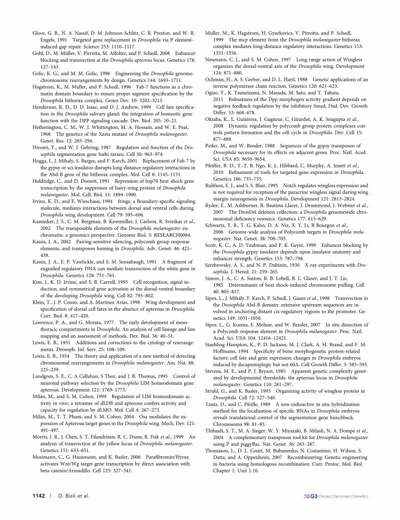
Gloor, G. B., N. A. Nassif, D. M. Johnson-Schlitz, C. R. Preston, and W. R.Engels, 1991 Targeted gene replacement in Drosophila via P element-induced gap repair. Science 253: 1110–1117.
Gohl, D., M. Müller, V. Pirrotta, M. Affolter, and P. Schedl, 2008 Enhancerblocking and transvection at the Drosophila apterous locus. Genetics 178:127–143.
Golic, K. G., and M. M. Golic, 1996 Engineering the Drosophila genome:chromosome rearrangements by design. Genetics 144: 1693–1711.
Hagstrom, K., M. Muller, and P. Schedl, 1996 Fab-7 functions as a chro-matin domain boundary to ensure proper segment specification by theDrosophila bithorax complex. Genes Dev. 10: 3202–3215.
Henderson, K. D., D. D. Isaac, and D. J. Andrew, 1999 Cell fate specifica-tion in the Drosophila salivary gland: the integration of homeotic genefunction with the DPP signaling cascade. Dev. Biol. 205: 10–21.
Hetherington, C. M., W. J. Whittington, M. A. Hossain, and W. E. Peat,1968 The genetics of the Xasta mutant of Drosophila melanogaster.Genet. Res. 12: 285–294.
Hiromi, Y., and W. J. Gehring, 1987 Regulation and function of the Dro-sophila segmentation gene fushi tarazu. Cell 50: 963–974.
Hogga, I., J. Mihaly, S. Barges, and F. Karch, 2001 Replacement of Fab-7 bythe gypsy or scs insulator disrupts long-distance regulatory interactions inthe Abd-B gene of the bithorax complex. Mol. Cell 8: 1145–1151.
Holdridge, C., and D. Dorsett, 1991 Repression of hsp70 heat shock genetranscription by the suppressor of hairy-wing protein of Drosophilamelanogaster. Mol. Cell. Biol. 11: 1894–1900.
Irvine, K. D., and E. Wieschaus, 1994 fringe, a Boundary-specific signalingmolecule, mediates interactions between dorsal and ventral cells duringDrosophila wing development. Cell 79: 595–606.
Kaminker, J. S., C. M. Bergman, B. Kronmiller, J. Carlson, R. Svirskas et al.,2002 The transposable elements of the Drosophila melanogaster eu-chromatin: a genomics perspective. Genome Biol. 3: RESEARCH0084.
Kassis, J. A., 2002 Pairing-sensitive silencing, polycomb group responseelements, and transposon homing in Drosophila. Adv. Genet. 46: 421–438.
Kassis, J. A., E. P. VanSickle, and S. M. Sensabaugh, 1991 A fragment ofengrailed regulatory DNA can mediate transvection of the white gene inDrosophila. Genetics 128: 751–761.
Kim, J., K. D. Irvine, and S. B. Carroll, 1995 Cell recognition, signal in-duction, and symmetrical gene activation at the dorsal-ventral boundaryof the developing Drosophila wing. Cell 82: 795–802.
Klein, T., J. P. Couso, and A. Martinez Arias, 1998 Wing development andspecification of dorsal cell fates in the absence of apterous in Drosophila.Curr. Biol. 8: 417–420.
Lawrence, P. A., and G. Morata, 1977 The early development of meso-thoracic compartments in Drosophila. An analysis of cell lineage and fatemapping and an assessment of methods. Dev. Biol. 56: 40–51.
Lewis, E. B., 1951 Additions and corrections to the cytology of rearrange-ments. Drosoph. Inf. Serv. 25: 108–109.
Lewis, E. B., 1954 The theory and application of a new method of detectingchromosomal rearrangements in Drosophila melanogaster. Am. Nat. 88:225–239.
Lundgren, S. E., C. A Callahan, S Thor, and J. B. Thomas, 1995 Control ofneuronal pathway selection by the Drosophila LIM homeodomain geneapterous. Development 121: 1769–1773.
Milán, M., and S. M. Cohen, 1999 Regulation of LIM homeodomain ac-tivity in vivo: a tetramer of dLDB and apterous confers activity andcapacity for regulation by dLMO. Mol. Cell 4: 267–273.
Milán, M., T. T. Pham, and S. M. Cohen, 2004 Osa modulates the ex-pression of Apterous target genes in the Drosophila wing. Mech. Dev. 121:491–497.
Morris, J. R., J. Chen, S. T. Filandrinos, R. C. Dunn, R. Fisk et al., 1999 Ananalysis of transvection at the yellow locus of Drosophila melanogaster.Genetics 151: 633–651.
Mosimann, C., G. Hausmann, and K. Basler, 2006 Parafibromin/Hyraxactivates Wnt/Wg target gene transcription by direct association withbeta-catenin/Armadillo. Cell 125: 327–341.
Muller, M., K. Hagstrom, H. Gyurkovics, V. Pirrotta, and P. Schedl,1999 The mcp element from the Drosophila melanogaster bithoraxcomplex mediates long-distance regulatory interactions. Genetics 153:1333–1356.
Neumann, C. J., and S. M. Cohen, 1997 Long-range action of Winglessorganizes the dorsal-ventral axis of the Drosophila wing. Development124: 871–880.
Ochman, H., A. S. Gerber, and D. L. Hartl, 1988 Genetic applications of aninverse polymerase chain reaction. Genetics 120: 621–623.
Ogiso, Y., K. Tsuneizumi, N. Masuda, M. Sato, and T. Tabata,2011 Robustness of the Dpp morphogen activity gradient depends onnegative feedback regulation by the inhibitory Smad, Dad. Dev. GrowthDiffer. 53: 668–678.
Oktaba, K., L. Gutiérrez, J. Gagneur, C. Girardot, A. K. Sengupta et al.,2008 Dynamic regulation by polycomb group protein complexes con-trols pattern formation and the cell cycle in Drosophila. Dev. Cell 15:877–889.
Peifer, M., and W. Bender, 1988 Sequences of the gypsy transposon ofDrosophila necessary for its effects on adjacent genes. Proc. Natl. Acad.Sci. USA 85: 9650–9654.
Pfeiffer, B. D., T.-T. B. Ngo, K. L. Hibbard, C. Murphy, A. Jenett et al.,2010 Refinement of tools for targeted gene expression in Drosophila.Genetics 186: 735–755.
Rulifson, E. J., and S. S. Blair, 1995 Notch regulates wingless expression andis not required for reception of the paracrine wingless signal during wingmargin neurogenesis in Drosophila. Development 121: 2813–2824.
Ryder, E., M. Ashburner, R. Bautista-Llacer, J. Drummond, J. Webster et al.,2007 The DrosDel deletion collection: a Drosophila genomewide chro-mosomal deficiency resource. Genetics 177: 615–629.
Schwartz, Y. B., T. G. Kahn, D. A. Nix, X. Y. Li, R Bourgon et al.,2006 Genome-wide analysis of Polycomb targets in Drosophila mela-nogaster. Nat. Genet. 38: 700–705.
Scott, K. C., A. D. Taubman, and P. K. Geyer, 1999 Enhancer blocking bythe Drosophila gypsy insulator depends upon insulator anatomy andenhancer strength. Genetics 153: 787–798.
Serebrovsky, A. S., and N. P. Dubinin, 1930 X-ray experiments with Dro-sophila. J. Hered. 21: 259–265.
Simon, J. A., C. A. Sutton, R. B. Lobell, R. L. Glaser, and J. T. Lis,1985 Determinants of heat shock-induced chromosome puffing. Cell40: 805–817.
Sipos, L., J. Mihály, F. Karch, P. Schedl, J. Gausz et al., 1998 Transvection inthe Drosophila Abd-B domain: extensive upstream sequences are in-volved in anchoring distant cis-regulatory regions to the promoter. Ge-netics 149: 1031–1050.
Sipos, L., G. Kozma, E. Molnár, and W. Bender, 2007 In situ dissection ofa Polycomb response element in Drosophila melanogaster. Proc. Natl.Acad. Sci. USA 104: 12416–12421.
Staehling-Hampton, K., P. D. Jackson, M. J. Clark, A. H. Brand, and F. M.Hoffmann, 1994 Specificity of bone morphogenetic protein-relatedfactors: cell fate and gene expression changes in Drosophila embryosinduced by decapentaplegic but not 60A. Cell Growth Differ. 5: 585–593.
Stevens, M. E., and P. J. Bryant, 1985 Apparent genetic complexity gener-ated by developmental thresholds: the apterous locus in Drosophilamelanogaster. Genetics 110: 281–297.
Struhl, G., and K. Basler, 1993 Organizing activity of wingless protein inDrosophila. Cell 72: 527–540.
Tautz, D., and C. Pfeifle, 1989 A non-radioactive in situ hybridizationmethod for the localization of specific RNAs in Drosophila embryosreveals translational control of the segmentation gene hunchback.Chromosoma 98: 81–85.
Thibault, S. T., M. A. Singer, W. Y. Miyazaki, B. Milash, N. A. Dompe et al.,2004 A complementary transposon tool kit for Drosophila melanogasterusing P and piggyBac. Nat. Genet. 36: 283–287.
Thomason, L., D. L. Court, M. Bubunenko, N. Costantino, H. Wilson, S.Datta, and A. Oppenheim, 2007 Recombineering: Genetic engineeringin bacteria using homologous recombination. Curr. Protoc. Mol. Biol.Chapter 1: Unit 1.16.
1142 | D. Bieli et al.
Tolhuis, B., E. de Wit, I. Muijrers, H. Teunissen, W. Talhout et al.,2006 Genome-wide profiling of PRC1 and PRC2 Polycomb chromatinbinding in Drosophila melanogaster. Nat. Genet. 38: 694–699.
Tsuneizumi, K., T. Nakayama, J. L. Christian, and T. Tabata,1997 Daughters against dpp modulates dpp organizing activity inDrosophila wing development. Nature 389: 627–631.
Venken, K. J. T., K. L. Schulze, N. A. Haelterman, H. Pan, Y. He et al.,2011 MiMIC: a highly versatile transposon insertion resource for en-gineering Drosophila melanogaster genes. Nat. Methods 8: 737–743.
Vuilleumier, R., A. Springhorn, L. Patterson, S. Koidl, M. Hammerschmidtet al., 2010 Control of Dpp morphogen signalling by a secreted feed-back regulator. Nat. Cell Biol. 12: 611–617.
Waddington, C. H., 1940 The genetic control of wing development inDrosophila. J. Genet. 41: 75–139.
Weiss, A., E. Charbonnier, E. Ellertsdóttir, A. Tsirigos, C. Wolf et al.,2010 A conserved activation element in BMP signaling during Dro-sophila development. Nat. Struct. Mol. Biol. 17: 69–76.
Whittle, J. R., 1979 Replacement of posterior by anterior structures in theDrosophila wing caused by the mutation apterous-blot. J. Embryol. Exp.Morphol. 53: 291–303.
Wieschaus, E., and W. Gehring, 1976 Clonal analysis of primordial disc cells inthe early embryo of Drosophila melanogaster. Dev. Biol. 50: 249–263.
Wilanowski, T. M., J. B. Gibson, and J. E. Symonds, 1995 Retrotransposoninsertion induces an isozyme of sn-glycerol-3-phosphate dehydrogenasein Drosophila melanogaster. Proc. Natl. Acad. Sci. USA 92: 12065–12069.
Williams, J. A., S. W. Paddock, and S. B. Carroll, 1993 Pattern formation ina secondary field: a hierarchy of regulatory genes subdivides the devel-oping Drosophila wing disc into discrete subregions. Development 117:571–584.
Williams, J. A., S. W. Paddock, K. Vorwerk, and S. B. Carroll, 1994 Organizationof wing formation and induction of a wing-patterning gene at thedorsal/ventral compartment boundary. Nature 368: 299–305.
Wu, C. T., and J. R. Morris, 1999 Transvection and other homology effects.Curr. Opin. Genet. Dev. 9: 237–246.
Zecca, M., and G. Struhl, 2002a Subdivision of the Drosophila wing imag-inal disc by EGFR-mediated signaling. Development 129: 1357–1368.
Zecca, M., and G. Struhl, 2002b Control of growth and patterning of theDrosophila wing imaginal disc by EGFR-mediated signaling. Develop-ment 129: 1369–1376.
Communicating editor: H. D. Lipshitz
Volume 5 June 2015 | apterous Wing Enhancer | 1143