the divided visual field paradigm: methodological ... · the divided visual field paradigm:...
TRANSCRIPT

The divided visual field paradigm: Methodological
considerations
Victoria J. Bourne
University of Dundee, UK
The divided visual field methodology has been used to examine a wide variety of
lateralised processes. When conducting such studies it is important to employ a
number of strict controls in order to maximise the effectiveness of the paradigm for
examining the processing of stimuli by each hemisphere. The use of these controls is
discussed in this paper. The following issues are discussed: selection of participants;
methods of fixation control; presenting stimuli unilaterally; methods of responding;
and measures that can be taken. The use of the divided visual field paradigm to
examine interhemispheric cooperation is also discussed. Employing the recom-
mended controls provides an effective and relatively easy method of examining the
role of each hemisphere in the processing of stimuli.
Since the work of Broca at the end of the nineteenth century (see Joynt, 1964,
for a review of Broca’s work) it has been widely accepted that the two
cerebral hemispheres are differentially specialised for processing distinct
forms of information. Much of the early work examining these hemispheric
specialisations used clinical participants, such as split-brain patients (e.g.,
Levy & Trevarthen, 1976) or patients suffering from unilateral brain lesions
(e.g., DeRenzi & Spinnler, 1966). Although this work provided great
advances in understanding how the brain is lateralised, it has somewhat
limited ability to provide valid extrapolations to non-clinical populations.
More recently, research methodologies have been developed that have
enabled the examination of lateralised processing in non-clinical partici-
pants, e.g., neuroimaging techniques. However, the use of neuroimaging
equipment is costly and not widely or easily available to all researchers. In
contrast, the divided visual field (DVF) methodology provides an easy way
of examining lateralisation that is accessible to all researchers.
Address correspondence to Victoria J. Bourne, Department of Psychology, University of
Dundee, Dundee, Scotland, UK DD1 4HN. Email: [email protected]
I would like to thank Claude Braun and an anonymous reviewer for helpful and insightful
comments on an earlier draft of this paper.
LATERALITY, 2006, 11 (4), 373�393
# 2006 Psychology Press Ltd
http://www.psypress.com/laterality DOI: 10.1080/13576500600633982
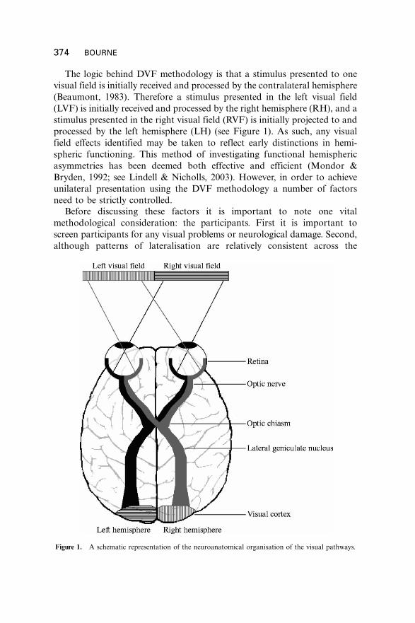
The logic behind DVF methodology is that a stimulus presented to one
visual field is initially received and processed by the contralateral hemisphere
(Beaumont, 1983). Therefore a stimulus presented in the left visual field
(LVF) is initially received and processed by the right hemisphere (RH), and a
stimulus presented in the right visual field (RVF) is initially projected to and
processed by the left hemisphere (LH) (see Figure 1). As such, any visual
field effects identified may be taken to reflect early distinctions in hemi-
spheric functioning. This method of investigating functional hemispheric
asymmetries has been deemed both effective and efficient (Mondor &
Bryden, 1992; see Lindell & Nicholls, 2003). However, in order to achieve
unilateral presentation using the DVF methodology a number of factors
need to be strictly controlled.
Before discussing these factors it is important to note one vital
methodological consideration: the participants. First it is important to
screen participants for any visual problems or neurological damage. Second,
although patterns of lateralisation are relatively consistent across the
Figure 1. A schematic representation of the neuroanatomical organisation of the visual pathways.
374 BOURNE
population, they are not universal. When considering lateralised processes it
is important to identify individuals with atypical patterns of asymmetry, as
these individuals will provide data that are anomalous with respect to the
majority of the population. One of the simplest ways of identifying such
individuals is to restrict the participants to right-handers. Previous research
has found patterns of lateralisation to be far more consistent in right-handers than in left-handers. For example, a functional magnetic resonance
imaging (fMRI) study (Pujol, Deus, Losilla, & Capdevila, 1999) found that
96% of right-handed people were lateralised to the LH for language
processing, with the remaining 4% bilaterally distributed. For left-handed
people this trend was reduced, with 76% being lateralised to the LH for
language function, 10% lateralised to the RH, and the remaining 14%
bilaterally organised. It is therefore recommended that participant groups be
restricted to those that are strongly right-handed in order to reduce possiblecontamination of the data by participants with atypical asymmetry.
Handedness can be assessed easily using a simple questionnaire that can
be completed in a few minutes. One frequently used handedness ques-
tionnaire is the Edinburgh Handedness Inventory (Oldfield, 1971). This
questionnaire comprises 10 items. In each item a task is described (e.g.,
throwing) and participants have to make a binary response to say which
hand they use to complete the task. Using this method of handedness
assessment, any participant completing eight or more tasks with their righthand would be deemed strongly right-handed and would be suitable for
inclusion in a study of laterality.
The Edinburgh Handedness Inventory has been found to have high test�retest reliability (Dorthe, Blumenthal, Jason, & Lantz, 1995) over periods of
up to 18 months (Ransil & Schachter, 1994). Although reliable, the
Edinburgh Handedness Inventory has been criticised for treating handed-
ness as dichotomous*treating people as either right-handed or left-handed
(Bishop, Ross, Daniels, & Bright, 1996). Furthermore, it has been found thatthe Edinburgh Handedness Inventory is only reliable in assessing cases of
extreme handedness (Verdino & Dingman, 1998) and becomes unreliable
when assessing cases of mid-range handedness (Schachter, 1994). Other
researchers have suggested that handedness should be considered on a
continuum (Annett, 1985) or as subgroups (Peters & Murphy, 1992).
Handedness questionnaires developed more recently have measured hand-
edness on a continuum. For example, Dorthe et al. (1995) used a 7-point
Likert scale with preference scores ranging from �3 (‘‘Always with lefthand’’) through 0 (‘‘Equal or no preference’’) to �/3 (‘‘Always with right
hand’’). Measuring handedness on a continuum is particularly relevant in
the light of recent evidence that the degree of handedness may be related to
the magnitude of lateralisation (Papousek & Schulter, 1999). In order to
select participants that are strongly right-handed a questionnaire, such as the
DIVIDED VISUAL FIELD METHODOLOGY 375

Edinburgh Handedness Inventory, would provide a quick and simple
method of screening participants. However, studies that may wish to
examine lateralisation in relation to the strength of handedness may wish
to use a questionnaire that measures handedness on a continuum.
Using the DVF methodology, each trial consists of a number of events,
each designed to maximise the ability to present stimuli unilaterally. In order
to control for the placement of the stimuli, it is also important to control the
participant’s head position. This is necessary to ensure that the participant’s
head is a correct and constant distance away from the monitor, therefore
maintaining the visual angle of stimulus presentation. This can be achieved
easily with the use of a chin rest.
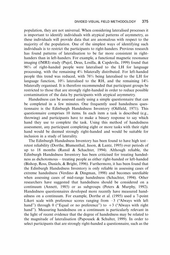
The DVF methodology recommended in this paper suggests four main
events in each trial (see Figure 2): the first is to ensure that the participant is
fixating centrally before the test stimulus is presented, the second is the
presentation of the test stimulus, the third is to backward mask the test
stimulus, and the fourth involves the participant’s response. Backward
masking is not widely used in DVF research. However, it enables greater
control over stimulus presentation and is therefore recommended. Its
advantages are discussed in greater detail later in this paper. Each of the
events that together form the DVF paradigm requires strict methodological
controls when attempting to achieve unilateral presentation and measure the
response given. These will now be considered in turn.
“Response”
X+
Fixation control
Stimulus presentation
Backward mask
Figure 2. Graphic representation of the events occurring in each trial.
376 BOURNE

FIXATION CONTROL
In the DVF methodology the test stimulus is presented at a specified
distance from a central fixation point. It is therefore important to control
where the participant is fixating when the stimulus is presented to ensure
that it is presented in the correct portion of the visual field. If, for example, a
participant is looking to the left of the display when a test stimulus is
presented, the subsequent processing of that stimulus may be affected, as it
may effectively have been presented bilaterally, rather than unilaterally as
intended, and any possible asymmetric effects would disappear.
A number of methods of fixation control have been used, each with
varying ease and efficacy. These methods can be divided into two types:
direct and indirect. The simplest and most frequently used is the indirect
method of instructing the participant to fixate centrally, often using a small
cross or some other marker to indicate the desired fixation point
(e.g., Kitterle, Christman, & Hellige, 1990; Rapaczynski & Ehrlichman,
1979; Van Kleek, 1989; Weismann & Banich, 1999, 2000). Although some
work has suggested that simple instruction may provide a reliable method of
fixation control (Jones & Santi, 1978; Posner, Nissen, & Ogden, 1978), it is
possible that participants may not necessarily maintain fixation, and
anticipatory saccades may be initiated prior to stimulus presentation.
Such eye movements may reduce the possibility of unilateral presentation.
Batt, Underwood, and Bryden (1995) found that participants fixated more
than 18 away from the fixation point on up to 17% of trials. Further,
directional biases have been found with studies showing both rightward
(Jones & Santi, 1978; Jordan, Patching, & Milner, 1998; Terrace, 1959)
and leftward (Batt et al., 1995) biases. Jordan et al. (1998, Experiment 1)
found that participants only fixated centrally on 23% of the trials, with
28% falling to the left of centre and 49% falling to the right of centre.
It therefore seems that the indirect method of verbal instruction to fixate
may not always provide adequate control over participants’ fixation
locations.
A further indirect method of fixation control is to present a letter or digit
at the central fixation point. This letter or digit has to be reported verbally
by the participant. The rationale behind this method is that the participant
has to be fixating the digit or letter in order to report it, and therefore will be
fixating centrally when the test stimulus is presented. If the fixation target is
incorrectly reported then central fixation cannot be assumed and the
subsequent response to the trial should not be included in the analyses.
This method of fixation control has been used in a number of studies (e.g.,
Belger & Banich, 1998; Bourne & Hole, 2006; Leehay, Carey, Diamond, &
Cahn, 1978; Luh & Levy, 1995). Although this method provides more
control than the simple instruction method, two possible limitations have
DIVIDED VISUAL FIELD METHODOLOGY 377

been identified (Jordan et al., 1998). First, it is assumed that the fixation
target can only be reported when fixation is central. However, if the target
can be reported when in peripheral vision, central fixation may not
be guaranteed. This may become particularly problematic following a
number of trials if there are possible practice effects. Second, it is possible
that the fixation task may interfere with the response to the experimental
test stimulus. For example, asking a participant to report a letter as a
fixation control may have a subsequent effect on responses to the test
stimulus if it is a word-based task, such as a word�nonword distinction. This
possibility has been examined by Carter and Kinsbourne (1979) who found
that changes to the fixation control target changed the patterns of
asymmetry identified.
Direct methods of fixation control can resolve some of the methodolo-
gical and theoretical difficulties encountered by the indirect methods. Direct
methods involve delaying the presentation of the test stimulus until the
participant is fixating centrally. The simplest direct method involves the
experimenter observing the participant’s eye movements and presenting
the test stimulus when the participant is judged to be fixating in the desired
location (e.g., Deruelle & de Schonen, 1998; Marzi & Berlucchi, 1977; Mohr,
Pulvermuller, Rayman, & Zaidel, 1994). However, this method is highly
dependent on the experimenter’s subjective decisions about the participant’s
fixation locations.
A more precise direct method involves the objective monitoring of eye
movements to ensure that stimulus presentation only occurs if the
participant is fixating centrally (e.g., Christman, 1990; Hardyck, Chiarello,
Dronkers, & Simpson, 1985). Such monitoring can be achieved using either
eye-tracking equipment or electro-oculography. This method overcomes
some of the limitations associated with the previous methods discussed
because (a) central fixation can be ensured, (b) the participant does not have
to complete the additional task of reporting a fixation control digit or letter,
and (c) it is not dependent on the experimenter’s subjective decisions. It
therefore seems that the most effective method of fixation control is to
objectively monitor eye movements and control test stimulus presentation.
However, this method involves costly equipment, is more time consuming in
terms of experimental preparation and the running of participants, and is
less flexible in terms of the participants that may be used (e.g., the use of eye-
tracking equipment may be unsuitable when using children or certain patient
groups as participants). Thus, it is preferable to monitor eye movements and
stimulus presentation using either eye-tracking equipment or electro-
oculography. However, the alternative method of monitored central fixation
may also be deemed adequate and effective.
378 BOURNE

PRESENTING STIMULI UNILATERALLY
When presenting a stimulus unilaterally there are two important factors that
need to be taken into consideration and experimentally controlled: the
placing of the stimulus in the visual field and the exposure duration of the
stimulus. These will now be discussed in turn.
When deciding where in the visual field to present stimuli, the
neuroanatomy of the visual system must be taken into consideration. The
nerve fibres of the primary visual system are organised in such a way that
stimuli received by the nasal hemiretinae are projected to the contralateral
hemisphere, whereas stimuli received by the temporal hemiretinae are
projected to the ipsilateral hemisphere (see Figure 1). However, the
ipsilateral and contralateral projections are not neatly divided and a certain
amount of overlap occurs between the visual fields as a result of crossover in
the commissural connections in the corpus callosum (Stone, Leicester, &
Sherman, 1973). Therefore, in order to maximise the chances of unilateral
presentation, it is important to present stimuli outside this region of overlap.
However, there has been some disagreement as to how wide this region may
be, and consequently where stimuli should be presented. Estimates of the
size of this bilateral strip range from 0.58 wide (Wyatt, 1978) to 38 wide
(Bunt, Minckler, & Johanson, 1977). Recently it has been suggested that, at
least behaviourally, the bilateral strip does not exist. Rather, that the fovea is
vertically split, with each hemi-fovea projecting to the contralateral hemi-
sphere (Lavidor & Ellis, 2003). However, Lindell and Nicholls (2003) suggest
that this finding should be treated with caution and offer a wide range of
evidence supporting bilateral projection of stimuli presented foveally (for
more detail see Lindell & Nicholls, 2003). In order to avoid potential
problems of bilateral presentation of stimuli, previous papers have recom-
mended that a stimulus be presented with the inside edge at least 28 from
central fixation (Young, 1982). However, given that some research estimates
that stimuli can be projected up to 38 from central fixation (Bunt et al.,
1977), this paper gives a slightly more conservative recommendation of
presenting a stimulus with its inside edge 2.5�38 from central fixation.
One potential issue when presenting stimuli unilaterally in peripheral
vision is the decrease in visual acuity with increasing eccentricity (e.g.,
Østerberg, 1935). Thus stimuli presented at different distances from fixation
are not perceptually equivalent. Along with controlling the distance of the
inside edge of the stimulus from central fixation, it is also important to
consider how wide the stimulus is. The outside edge of a stimulus is
perceived with lower visual acuity, which may have serious consequences for
the processing of that stimulus, particularly for stimuli such as words.
Whether stimuli are perceptually equivalent depends on both their
position in the visual field and the properties of the monitor used to display
DIVIDED VISUAL FIELD METHODOLOGY 379

them. The position of the stimulus in the visual field is important due to the
heterogeneity of retinal sampling discussed above. Changes in peripheral
acuity are not symmetrical, with nasal peripheral acuity poorer than
temporal peripheral acuity (Regan & Beverley, 1983; both are represented
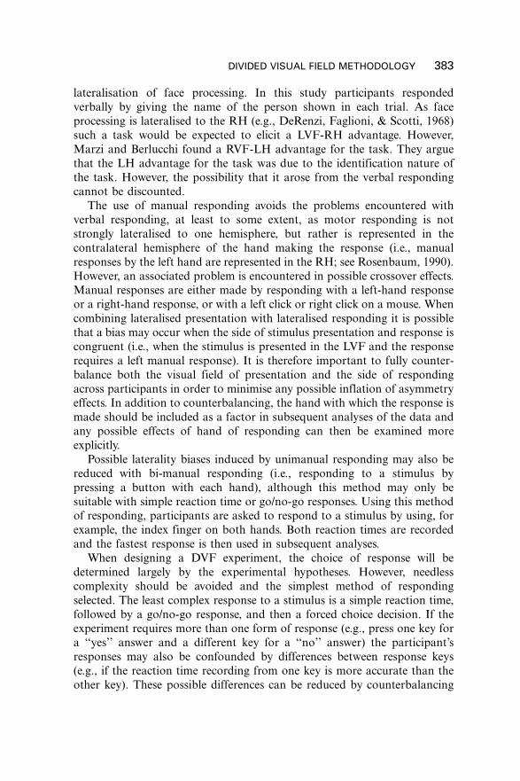
in Figure 3). The properties of the monitor will limit the resolution at which
stimuli can be presented; this is particularly important if stimulus presenta-
tion is at a resolution lower than visual acuity. Take, for example, a monitor
with a display resolution of 800�/600 pixels, subtending 308 horizontally in
the participant’s visual field. This monitor would therefore be able to display
stimuli at a resolution of 27 pixels per degree; therefore the maximum spatial
frequency that could be displayed by the monitor would be 13.5 cycles per
degree (cpd). Peripheral vision would only limit the sampling of stimuli when
presented at an eccentricity where acuity is below 13.5 cpd. If we assume a
foveal acuity of 60 cpd (Curcio, Sloan, Kalina, & Hendrickson, 1990) and
calculate peripheral acuity using values and equations taken from Rovamo
and Virsu (1979), it can be seen that acuity only falls below 13.5 cpd at
Figure 3. Spatial acuity as a function of eccentricity for both nasal and temporal retinae. Acuity was
calculated using values and equations taken from Rovamo and Virsu (1979) and assuming a central
acuity of 60 cpd (Curcio et al., 1990). The maximum spatial frequency that could be displayed by the
monitor in the example given in the text would be 13.5 cpd (as shown by the bold solid line) therefore
any stimuli presented within 10.58 from fixation (as shown by the bold dashed lines) would be sampled
by the visual system with equivalent acuity.
380 BOURNE

eccentricities of greater than 10.58 from the central fixation point (see Figure
3). Therefore any stimuli presented within 10.58 of fixation would be limited
by the resolution of the monitor and thus perceived with the equivalent
acuity of 13.5 cpd.
The decrease in visual acuity with increasing eccentricity from the central
fixation point should be taken into consideration when designing a DVFexperiment that involves presenting word stimuli. With the exception of a
few languages, such a Hebrew and Chinese, words are typically read from
left to right and a strong bias exists for looking at the left end of a word
(Pynte, Kennedy, & Murray, 1991). This may introduce a bias in DVF
experiments that involve words, as the beginning of words presented in the
LVF is perceived with poorer visual acuity than the beginning of words
presented in the RVF. It is therefore important to take into account possible
lateralised biases in the processing that may be introduced as a result ofreduced visual acuity in peripheral vision, and consider alternative methods
of presentation. For example, words may be presented vertically rather than
horizontally (e.g., Eviatar, 1999).
The duration for which stimuli are displayed is also important in
successfully achieving unilateral presentation. Whichever method of fixation
control is used, it is assumed that the participant is fixating centrally when
the test stimulus is presented. In order to maintain unilateral presentation
the stimulus presentation should be completed before an eye movementtowards the stimulus can be executed and the stimulus is fixated upon.
Saccades are rapid, ballistic eye movements that move the fovea (the region
of highest visual acuity) towards the target stimulus (Westheimer, 1973).
Mean saccadic latencies range from 150 ms to 200 ms (Carpenter, 1988) with
less than 2% of latencies falling below 150 ms. Therefore stimulus
presentation needs to be completed within 150 ms in order to minimise
the possibility of the test stimulus being foveated, which would cause the
stimulus to be presented bilaterally rather than unilaterally. Previous papershave suggested that stimulus exposures of up to 200 ms may be acceptable
(Young, 1982). However, it is recommended that stimulus presentation is
limited to a maximum exposure of 180 ms, with exposure ideally limited to
150 ms if the task is a simple one.
Although saccade latencies typically range from 150 ms to 200 ms,
anticipatory saccades may jeopardise the unilateral nature of the stimulus
presentation and should therefore be considered. Anticipatory saccades have
latencies of less than 100 ms (Bronstein & Kennard, 1987), and therefore, ifinitiated, may enable the test stimulus to be fixated and therefore presented
bilaterally. Anticipatory saccades may occur if the location of the target
stimulus is predictable and if there is a gap between the fixation control and
the presentation of the target stimulus (Carpenter, 1988). Therefore visual
field of presentation must be randomised in order to reduce the participant’s
DIVIDED VISUAL FIELD METHODOLOGY 381

ability to predict the visual field of presentation for the following trial and
the presentation of the target stimulus must immediately follow the fixation
control. Using these two controls the likelihood of anticipatory saccades will
be greatly reduced.
BACKWARD MASKING STIMULI
One further control can be included in an attempt to limit the presentation
of the stimulus to the desired duration. Following presentation the
participant may experience afterimage effects, either resulting from theirown subjective afterimage or, for some monitors, phosphor persistence (see
Felsten & Wasserman, 1980; Kahneman, 1968; Van Kleek, 1989). Introdu-
cing a backward visual mask immediately following stimulus presentation
ensures that such afterimage effects will not occur and that the exposure
duration of the stimulus is controlled (see Enns & DiLollo, 2000, for more
detail about backward visual masking). If a mask were not used then it is
possible that an afterimage may occur, effectively extending the presentation
duration beyond that recommended above in an uncontrollable manner.
METHODS OF RESPONDING AND MEASURES TAKEN
Two main methods of responding have been used in the DVF paradigm:
verbal (e.g., Klein, Moscovitch, & Vigna, 1976; Leehay et al., 1978; Marzi &
Berlucchi, 1977) and manual responding (e.g., Brown, Jeeves, Dietrich, &
Burnison, 1999; Mohr, Pulvermuller, Mittelstadt, & Rayman, 1996;
Schweinberger, Baird, Blumler, Kaufmann, & Mohr, 2003). From these
responses latency and/or accuracy can be measured. Each method of
responding has distinct advantages and disadvantages. From manual
responses it is possible to take precise measurements of both the speedand accuracy of responding. However, the types of responses that can be
given by the participant are typically limited to go/no-go responding or
forced choice binary decisions (e.g., yes/no, word/nonword, famous/not
famous). Verbal responding is far more flexible in terms of the types of
responses that a participant can make (e.g., reading out a word or giving the
name of a face presented). However, it may be difficult to record reaction
times with high accuracy from verbal responses.
With both methods of responding there is an assumption that theresponse will not influence the measurement of hemispheric differences;
however, this is not necessarily this case (Beaumont, 1983). For example, it is
possible that, as language is LH dominant, verbal responding may lead to an
overestimation of the performance of the LH. One example of this is work
by Marzi and Berlucchi (1977), who conducted a DVF study examining the
382 BOURNE
lateralisation of face processing. In this study participants responded
verbally by giving the name of the person shown in each trial. As face
processing is lateralised to the RH (e.g., DeRenzi, Faglioni, & Scotti, 1968)
such a task would be expected to elicit a LVF-RH advantage. However,
Marzi and Berlucchi found a RVF-LH advantage for the task. They argue
that the LH advantage for the task was due to the identification nature ofthe task. However, the possibility that it arose from the verbal responding
cannot be discounted.
The use of manual responding avoids the problems encountered with
verbal responding, at least to some extent, as motor responding is not
strongly lateralised to one hemisphere, but rather is represented in the
contralateral hemisphere of the hand making the response (i.e., manual
responses by the left hand are represented in the RH; see Rosenbaum, 1990).
However, an associated problem is encountered in possible crossover effects.Manual responses are either made by responding with a left-hand response
or a right-hand response, or with a left click or right click on a mouse. When
combining lateralised presentation with lateralised responding it is possible
that a bias may occur when the side of stimulus presentation and response is
congruent (i.e., when the stimulus is presented in the LVF and the response
requires a left manual response). It is therefore important to fully counter-
balance both the visual field of presentation and the side of responding
across participants in order to minimise any possible inflation of asymmetryeffects. In addition to counterbalancing, the hand with which the response is
made should be included as a factor in subsequent analyses of the data and
any possible effects of hand of responding can then be examined more
explicitly.
Possible laterality biases induced by unimanual responding may also be
reduced with bi-manual responding (i.e., responding to a stimulus by
pressing a button with each hand), although this method may only be
suitable with simple reaction time or go/no-go responses. Using this methodof responding, participants are asked to respond to a stimulus by using, for
example, the index finger on both hands. Both reaction times are recorded
and the fastest response is then used in subsequent analyses.
When designing a DVF experiment, the choice of response will be
determined largely by the experimental hypotheses. However, needless
complexity should be avoided and the simplest method of responding
selected. The least complex response to a stimulus is a simple reaction time,
followed by a go/no-go response, and then a forced choice decision. If theexperiment requires more than one form of response (e.g., press one key for
a ‘‘yes’’ answer and a different key for a ‘‘no’’ answer) the participant’s
responses may also be confounded by differences between response keys
(e.g., if the reaction time recording from one key is more accurate than the
other key). These possible differences can be reduced by counterbalancing
DIVIDED VISUAL FIELD METHODOLOGY 383
the key of response between participants (e.g., half of the participants press
the left key for ‘‘yes’’ and the right key for ‘‘no’’ and the other half press the
right key for ‘‘yes’’ and the right key for ‘‘no’’) and can also be accounted for
in the analysis of data collected.
DESIGN AND ANALYSIS OF A DVF EXPERIMENT
While researchers using the DVF are often primarily interested in laterality
effects, they are also often interested in additional experimentally manipu-
lated variables. Having taken great pains to ensure that the stimuli presented
in each visual field do not differ systematically, the same fastidious approach
should be taken with other independent variables. The ideal solution is to
use the same stimuli in each experimental condition, but to vary the task that
the participants have to complete. However, this is often not possible, and in
these instances psychophysical properties should be comparable across
conditions. For example, if hemispheric specialisations in processing familiar
and unfamiliar faces are to be examined, both sets of faces should be
equivalent in terms of luminance, contrast, spatial frequency, etc. With such
controls implemented, conclusions can be drawn regarding the cognitive
process in question (e.g., familiar face recognition) without the possible
confound of lower-level processes. A similar problem may occur if the
difficulty of the task varies across conditions; for example, if the successful
categorisation of a face as familiar elicits far more errors than categorising a
face as unfamiliar. When designing the experiment, try to ensure that the
difficulty of the task is comparable in all conditions. Alternatively, in some
experimental designs it is possible to implement an algorithm that modifies
the difficulty of the task so that the error rate is constant throughout the
experiment.It is also important to consider which event triggers the following trial.
One possibility is for each trial to commence at a set inter-stimulus interval;
however, this method may be problematic. For example, if a participant
makes a response that is longer than the inter-stimulus interval, the data for
that trial may not be recorded. Such a response might also impact on the
participant’s response in the subsequent trials. It is recommended that each
trial be initiated at a set interval following the participant’s response to avoid
missed trials or contaminated responses.Typically, two types of data are collected from DVF experiments: speed
and accuracy. Appropriate analyses of these data require careful considera-
tion. Following data collection it is important to categorise all responses.
Broadly, this involves categorising responses as either correct or incorrect.
There may be different types of errors: decision errors (an incorrect
response), not responding to a stimulus (an omission error), or responding
384 BOURNE

too quickly (an anticipation error). Usually omission and anticipation errors
are infrequent and can be discarded from subsequent analyses. However, if
there are a large number of omission and/or anticipation errors, these should
be subjected to statistical analyses.
The measurement of error is often quantified as the number or percentage
of errors made. However, in some paradigms, such as one using a forced
choice binary decision, a wider range of possible responses can be made, and
more sophisticated methods exist that can be used to analyse all types of
response. While it is often assumed that there are only correct and incorrect
responses, responses can actually be categorised into four types. Say, for
example, that participants are asked to classify a string of letters as a word or
a nonword. They may correctly categorise a word as a word (a hit), correctly
categorise a nonword as a nonword (a correct rejection), incorrectly
categorise a word as a nonword (a miss), or incorrectly categorise a nonword
as a word (a false alarm). In this situation it may be preferable to use the
signal detection theory measure of d?, which takes into account all four
possible responses, rather than a simple error rate (for more information on
the signal detection method of analysis see Swets, Dawes, & Monahan,
2000).
When analysing reaction time data, the responses should also be split into
correct and incorrect responses, or into the four response types if using
signal detection theory. Reaction times within each condition are then often
summarised by calculating the mean reaction time in that condition. One
common difficulty that can occur with reaction time analysis is that they
tend to be asymmetrically distributed. An extra complication can occur as
this asymmetry may be exaggerated in more difficult conditions. This can be
addressed by using either medians or trimmed means to summarise reaction
times within each condition, or by log transforming the mean reaction times
(for more detail see Ratcliff, 1993).
The data acquired from DVF experiments is most often analysed using
analysis of variance (ANOVA). In its simplest form the analysis will require a
two-way ANOVA, with visual field of presentation forming one independent
variable, and the experimental manipulation as another independent
variable. It may also be of interest to include other independent variables
in the analysis, such as hand/key of responding or other factors that maybe
of interest (e.g., sex of the participant if a possible sex difference in the task is
expected).
PRACTICAL AND ENVIRONMENTAL CONSIDERATIONS
Having taken precautions to ensure effective unilateral presentation of
stimuli using the DVF paradigm, it is important to ensure that other factors
DIVIDED VISUAL FIELD METHODOLOGY 385

(such as the monitor used to present stimuli or the experimental room) are
controlled with equal consideration. When running a DVF experiment, the
primary aim will be to compare responses to stimuli presented in each visual
field. It is important to ensure that stimuli presented in one visual field do
not differ systematically from those presented in the other visual field. It is
important that stimuli are presented equidistant from the central fixationpoint in both visual fields. Even when a computer program controls stimulus
placement, the accuracy with which the stimulus is positioned (i.e., the
distance between central fixation and stimulus presentation) should be
manually checked and, if necessary, adjusted. When using a cathode ray tube
monitor (for a more detailed discussion of lateralised biases that may be
induced when using a cathode ray tube monitor see Ratinckx, Brysbaert, &
Vermeulen, 2001), there can be variation in luminance across the screen.
This might pose a problem in the DVF paradigm if one side of the monitor isbrighter, as this may artificially bias laterality effects. Luminance irregularity
tends to be greatest while the monitor is warming up, so it is advisable to
leave the monitor on for as long as possible prior to testing a participant.
Once the monitor has warmed up, luminance can be tested using a
photometer to ensure equal luminance across the screen.
When designing stimuli it is important to consider how certain choices
might affect the DVF paradigm. It is preferable to use a black, or dark-
coloured, stimulus against a white luminant background. Leakage of lightmay occur if presenting a white stimulus against a black background,
effectively increasing the size of the stimulus, which may be problematic
when trying to control the eccentricity and size of stimulus presentation. It is
important to bear in mind display time limitations when using a cathode ray
tube monitor. It only takes microseconds for each line of pixels to refresh,
but if the stimulus is excessively large there is a risk of introducing a bias in
stimulus presentation, as the left side of the monitor displays slightly before
the right side, and the top displays slightly before the bottom. To avoid thispossible confound, use small stimuli.
It is preferable to run the experiment in a darkened room. Ensure that the
participant has a few moments with the lights dimmed, or off, before
beginning the experiment to ensure that their retinae have adapted to the
level of light (for a recent review of dark adaptation see Lamb & Pugh,
2004). It is also important to make certain that ambient light is equalised on
either side of the participant (e.g., light from a door, window, or light) to
avoid biasing any lateralised presentation of stimuli or responding.When participating in a DVF experiment, some individuals may need to
blink a great deal due to either dry air or constant gazing and concentration
(particularly if wearing contact lenses). This can be uncomfortable for the
participant and may lead to missed trials. Placing a wet sponge at the foot of
the chinrest can easily solve such a problem.
386 BOURNE

USING DVF METHODOLOGY TO EXAMINEINTERHEMISPHERIC COOPERATION
Whilst the DVF methodology is useful for examining lateralised processing
of stimuli, it can also be used effectively to examine the role of interhemi-
spheric cooperation. In all studies using non-clinical participants it is likely
that a certain amount of information is passed between the hemispheres in
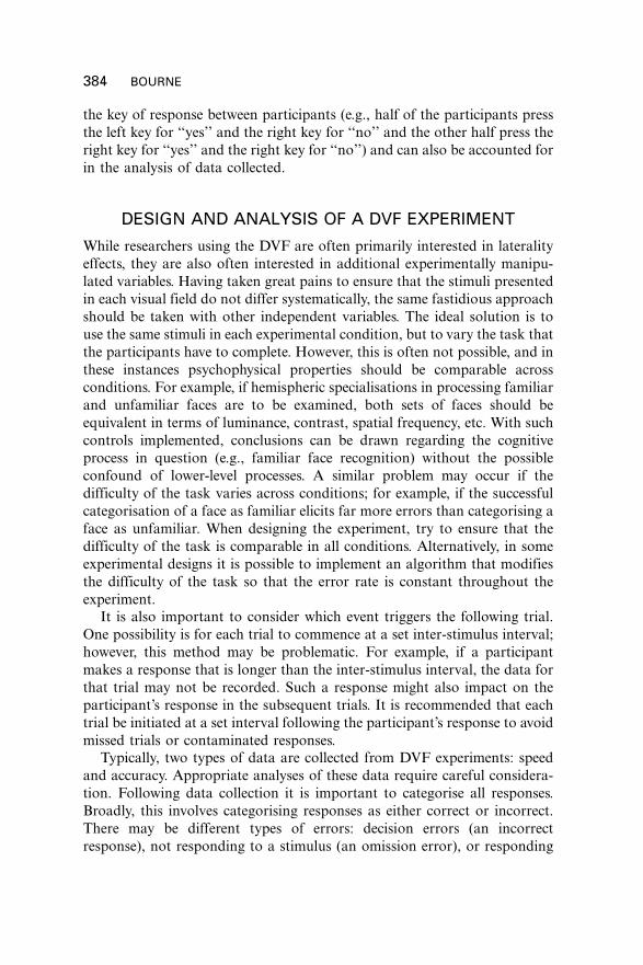
order to complete the task. Four methods of examining interhemispheric
cooperation using DVF methodology have been developed, each of which
will be discussed (see Figure 4).The first and oldest experimental approach that has been used to examine
interhemispheric cooperation, and one that is still used today, is the
Poffenberger paradigm (Poffenberger, 1912). Using this method, visual
targets are presented in each visual field and participants have to respond
using either the hand on the opposite side to that in which the visual
stimulus was presented (crossed, Figure 4a right), or the hand on the same
side (uncrossed, Figure 4a left). The rationale behind this method is that
responding in the uncrossed condition requires processing from only one
hemisphere, whereas responding in the crossed condition requires transfer of
information from one hemisphere to the other. Therefore, the crossed�uncrossed difference can be used as an estimate of the amount of time it
takes for information to be transferred from one hemisphere to another. A
recent study, using event-related fMRI, has provided evidence for transfer of
information across the corpus callosum for the crossed�uncrossed difference
(Weber et al., 2005).
The second method of examining interhemispheric cooperation is the
redundant target paradigm. This method has developed from a classic
finding that a participant will respond faster to two simultaneously
presented copies of the same stimulus (Figure 4b right) than to a single
copy (Figure 4b left; Todd, 1912). Although participants are presented with
the same information in both conditions, when presented with an additional
‘‘redundant’’ copy, reaction times are facilitated; hence this is known as the
redundant target effect, or redundancy gain. This paradigm can be used to
investigate interhemispheric cooperation by comparing reaction times to a
single copy presented in one visual field to reaction times to two copies,
where one copy is presented in each visual field. In the redundant target
condition, faster reaction times are attributed to interhemispheric coopera-
tion. This suggestion is supported by studies that have shown an increased
effect of redundancy gain in split-brain patients (Corballis, 1998; Reuter-
Lorenz, Nowaza, Gazzaniga, & Hughes, 1995).
The third method, developed by Dimond and Beaumont (1972), involves
a simple comparison between the processing of pairs of stimuli presented
unilaterally (Figure 4c left) and the processing of pairs of stimuli presented
DIVIDED VISUAL FIELD METHODOLOGY 387

No IHC needed IHC needed
Poff
enbe
rger
par
adig
mR
edun
dant
targ
ets
para
digm
Dim
ond
para
digm
Ban
ich
para
digm
A
B
C
D
Figure 4. Four experimental paradigms that can be used to examine interhemispheric cooperation
(IHC). For each paradigm the left panel shows the condition in which IHC is not necessary to respond
to the stimulus/stimuli and the right panel shows the condition in which IHC facilitates responding to
the stimulus/stimuli.
388 BOURNE

bilaterally (Figure 4c right), where one copy is presented in each visual field
(e.g., Mohr et al., 1996, Schweinberger et al., 2003). The rationale behind
experiments using this methodology is that any bilateral advantage identified
reflects the advantage provided by the activation of interhemispheric
cooperation. Evidence in support of this can be gained from split-brain
patients who do not show an advantage in bilateral trials (Mohr et al., 1994).The final method to be discussed has been developed by Banich and
colleagues (see Banich & Shenker, 1994) and has been used in a wide variety
of studies using alphabetic (e.g., Weissman & Banich, 2000), geometric (e.g.,
Weissman & Banich, 1999), and face (e.g., Compton, 2002) stimuli. Using
this paradigm, participants are presented with three stimuli: two in the top
half of the display, one in each visual field, and the third in the bottom half
of the display. The participant’s task is to decide whether the bottom
stimulus matches either of the stimuli presented above it. When two
matching stimuli are presented within the same visual field (Figure 4d
left), it is assumed that the hemisphere that they are presented to functions
as an isolated unit to complete the task. When the two matching stimuli are
presented across the two visual fields (Figure 4d right), it is assumed that
interhemispheric cooperation must occur in order to complete the task.
Comparing performance in each of these two conditions, any change in
performance is thought to reflect the time taken for interhemispheric
cooperation to occur.
When designing an interhemispheric cooperation experiment, it is
preferable to select the simplest paradigm that will enable the hypothesis
to be addressed. One important issue to consider and control for is the
placement of the stimuli and the effect this may have on the deployment of
visual attention. In the DVF paradigm, participants are required to fixate
centrally prior to stimulus presentation, it is therefore difficult to discern
whether an advantage for responses to centrally (bilaterally) presented
stimuli in comparison to unilaterally presented stimuli results from an
interhemispheric cooperation advantage or from not having to shift
attention to complete the task. A similar problem may also occur if stimuli
are presented at varying distances from the central fixation point, and
therefore all stimuli should be presented equidistant from the central fixation
point.
SUMMARY AND RECOMMENDATIONS
Strict methodological controls are necessary to maximise the validity of
conclusions drawn from DVF studies. It is important to include only
participants who are strongly right-handed. The use of a chin rest is
DIVIDED VISUAL FIELD METHODOLOGY 389

necessary to ensure that participant’s head movements are limited and to
maintain the distance of the participant’s eyes from the monitor.
Each trial within the DVF comprises a number of events (see Figure 2).
First it is necessary to control the participant’s fixation location prior to
presentation of the test stimulus. A direct method of fixation control is
recommended, using eye-tracking equipment or electro-oculography tomonitor the participant’s eye movements and withholding stimulus pre-
sentation until fixation is on the desired central location. Second, the test
stimulus is presented. It is important that both the location and exposure
duration of the test stimulus are strictly controlled in order to ensure
unilateral presentation. It is recommended that stimuli be presented for 150�180 ms (with shorter exposure time if the task is a simple one), at 2.5�38from central fixation and that presentation is followed immediately by a
backward visual mask to reduce any possible afterimage effects. Visual fieldof presentation must be randomised to reduce the likelihood of anticipatory
saccades occurring. The DVF methodology can be relatively difficult for a
participant to master and may be quite tiring, particularly with more
complex or difficult tasks. It is recommended that participants be given
enough practice trials to become comfortable with the speed of stimulus
presentation and the method of responding. Participants should also be
given regular breaks, and be able to indicate easily if they need a break, in
order to maintain high levels of concentration and avoid ‘‘noisy’’ respond-ing. When used with appropriate controls the DVF methodology provides
and effective, inexpensive, and relatively easy way to examine the lateralisa-
tion of processing.
Manuscript received 13 May 2004
Revised manuscript received 10 January 2006
First published online 01 May 2006
REFERENCES
Annett, M. (1985). Left, right, hand and brain: The right-shift theory. Hove, UK: Lawrence
Erlbaum Associates Ltd.
Banich, M. T., & Shenker, J. I. (1994). Investigations of interhemispheric processing: Methodo-
logical considerations. Neuropsychology, 8 , 263�277.
Batt, V., Underwood, G., & Bryden, M. P. (1995). Inspecting asymmetric presentation of words
differing in informational and morphemic structure. Brain and Cognition , 49 , 202�223.
Beaumont, J. G. (1983). Methods for studying cerebral hemispheric function. In A. W. Young
(Ed.), Functions of the right cerebral hemisphere (pp. 114�146). London: Academic Press.
Belger, A., & Banich, M. T. (1998). Costs and benefits of integrating information between the
cerebral hemispheres: A computational perspective. Neuropsychology, 12 , 380�398.
Bishop, D. V. M., Ross, V., Daniels, M. S., & Bright, P. (1996). The measurement of hand
preference: A validation study comparing three groups of right-handers. British Journal of
Psychology, 87 , 269�285.
390 BOURNE
Bourne, V. J. & Hole, G. J. (2006). Lateralised repetition priming for familiar faces: Evidence for
asymmetric interhemispheric cooperation. Quarterly Journal of Experimental Psychology, 59,
1117�1133 .
Bronstein, A. M., & Kennard, C. (1987). Predictive eye saccades are different from visually
triggered saccades. Vision Research , 27 , 517�520.
Brown, W. S., Jeeves, M. A., Dietrich, R., & Burnison, D. S. (1999). Bilateral field advantage and
evoked potential interhemispheric transmission in commissurotomy and callosal agenesis.
Neuropsychologia , 37 , 1165�1180.
Bunt, A. H., Minckler, D. S., & Johanson, G. W. (1977). Demonstration of bilateral projection of
the central retina of the monkey with horseradish peroxidase neuronography. Journal of
Comparative Neurology, 17 , 619�630.
Carpenter, R. H. S. (1988). Movements of the eyes. London: Pion.
Carter, G. L., & Kinsbourne, M. (1979). The ontogeny of right cerebral lateralization of spatial
mental set. Developmental Psychology, 15 , 241�245.
Christman, S. (1990). Effects of luminance and blur on hemispheric asymmetries in temporal
integration. Neuropsychologia , 28 , 361�374.
Compton, R. (2002). Inter-hemispheric interaction facilitates face processing. Neuropsychologia ,
40 , 2409�2419.
Corballis, M. C. (1998). Interhemispheric neural summation in the absence of the corpus callosum.
Brain , 121 , 1795�1807.
Curcio, C. A., Sloan, K. R., Kalina, R. E., & Hendrickson, A. E. (1990). Human photoreceptor
topography. Journal of Comparative Neurology, 292 , 497�523.
De Renzi, E., Faglioni, P., & Scotti, G. (1968). Tactile spatial impairment and unilateral cerebral
damage. Journal of Nervous and Mental Disorders , 146 , 468�475.
De Renzi, E., & Spinnler, H. (1966). Facial recognition in brain-damaged patients. An experimental
approach. Neurology, 16 , 145�152.
Deruelle, C., & de Schonen, S. (1998). Do the right and left hemispheres attend to the same
visuospatial information within a face in infancy? Developmental Neuropsychology, 14 , 535�554.
Dimond, S., & Beaumont, G. (1972). Processing in perceptual integration between the cerebral
hemispheres. British Journal of Psychology, 63 , 509�514.
Dorthe, N. J., Blumenthal, T. D., Jason, D. R., & Lantz, P. E. (1995). The use of next-of-kin in
assessing handedness. Perceptual and Motor Skills , 81 , 203�208.
Enns, J. T., & DiLollo, V. (2000). What’s new in visual masking? Trends in Cognitive Sciences , 4 ,
345�352.
Eviatar, Z. (1999). Cross-language tests of hemispheric strategies in reading nonwords.
Neuropsychology, 13 , 498�515.
Felsten, G., & Wasserman, G. S. (1980). Visual masking: Mechanisms and theories. Psychological
Bulletin , 88 , 329�353.
Hardyck, C., Chiarello, C., Dronkers, N. F., & Simpson, G. V. (1985). Orienting attention within
the visual fields: How efficient is interhemispheric transfer? Journal of Experimental
Psychology: Human Perception and Performance , 11 , 650�666.
Jones, B., & Santi, A. (1978). Lateral asymmetries in visual perception with or without eye
movements. Cortex , 14 , 164�168.
Jordan, T. R., Patching, G. R., & Milner, A. D. (1998). Central fixations are inadequately
controlled by instruction alone: Implications for studying cerebral asymmetry. Quarterly
Journal of Experimental Psychology: Human Experimental Psychology, 51 , 371�391.
Joynt, R. J. (1964). Paul Pierre Broca: His contribution to the knowledge of aphasia. Cortex , 1 ,
206�213.
Kahneman, D. (1968). Method, findings, and theory in studies of visual masking. Psychological
Bulletin , 70 , 404�425.
DIVIDED VISUAL FIELD METHODOLOGY 391

Kitterle, F. L., Christman, S., & Hellige, J. B. (1990). Hemispheric differences are found in the
identification, but not the detection, of low versus high spatial frequencies. Perception and
Psychophysics , 48 , 297�306.
Klein, D., Moscovitch, M., & Vigna, C. (1976). Attentional mechanisms and perceptual
asymmetries in tachistoscopic recognition of words and faces. Neuropsychologia , 14 , 55�66.
Lamb, T. D., & Pugh, E. N. (2004). Dark adaptation and the retinoid cycle of vision. Progress in
Retinal and Eye Research , 23 , 307�380.
Lavidor, M., & Ellis, A. W. (2003). Interhemispheric integration of letter stimuli presented foveally
or extra-foveally. Cortex , 39 , 69�83.
Leehay, S. C., Carey, S., Diamond, R., & Cahn, A. (1978). Upright and inverted faces: The right
hemisphere knows the difference. Cortex , 14 , 411�419.
Levy, J., & Trevarthen, C. (1976). Metacontrol of hemispheric function in human split-brain
patients. Journal of Experimental Psychology: Human Perception and Performance , 2 , 299�312.
Lindell, A. K., & Nicholls, M. E. R. (2003). Cortical representation of the fovea: Implications for
visual half-field research. Cortex , 39 , 111�117.
Luh, K. E., & Levy, J. (1995). Interhemispheric cooperation: Left is left and right is right, but
sometimes the twain shall meet. Journal of Experimental Psychology: Human Perception and
Performance , 21 , 1243�1258.
Marzi, C. A., & Berlucchi, G. (1977). Right visual field superiority for accuracy of recognition of
famous faces in normals. Neuropsychologia , 15 , 751�756.
Mohr, B., Pulvermuller, F., Mittelstadt, K., & Rayman, J. (1996). Multiple simultaneous
presentation facilitates lexical processing. Neuropsychologia , 34 , 1003�1013.
Mohr, B., Pulvermuller, F., Rayman, J., & Zaidel, E. (1994). Interhemispheric cooperation during
lexical processing is mediated by the corpus-callosum: Evidence from the split-brain.
Neuroscience Letters , 181 , 17�21.
Mondor, T. A., & Bryden, M. P. (1992). On the relation between visual spatial attention and visual-
field asymmetries. Quarterly Journal of Experimental Psychology, 44A , 529�555.
Oldfield, R. C. (1971). The assessment and analysis of handedness: The Edinburgh inventory.
Neuropsychologia , 9 , 97�113.
Østerberg, G. (1935). Topography of the layer of rods and cones in the human retina. Acta
Opthalmologica , 6 , 1�103.
Papousek, I., & Schulter, G. (1999). Quantitative assessment of five behavioural laterality
measures: Distributions of scores and intercorrelations among right-handers. Laterality, 4 ,
345�362.
Peters, M., & Murphy, K. (1992). Cluster analysis reveals at least three, and possibly five distinct
handedness groups. Neuropsychologia , 30 (4), 373�380.
Poffenberger, A. (1912). Reaction time to retinal stimulation with special reference to the time lost
in conduction through nervous centers. Archives of Psychology, 23 , 1�73.
Posner, M. I., Nissen, M. J., & Ogden, W. C. (1978). Attended and unattended processing modes:
The role of set for spatial location. In H. L. Pick & I. J. Saltzman (Eds.), Modes of perceiving
and processing information (pp. 137�157). Hillsdale, NJ: Lawrence Erlbaum Associates Inc.
Pujol, J., Deus, J., Losilla, J. M., & Capdevila, A. (1999). Cerebral lateralization of language in
normal left-handed people studied by functional MRI. Neurology, 52 , 1038�1043.
Pynte, J., Kennedy, A., & Murray, W. S. (1991). Within-word inspection strategies in continuous
reading: Time course of perceptual, lexical, and contextual processes. Journal of Experimental
Psychology: Human Perception and Performance , 17 , 458�470.
Ransil, B. J., & Schachter, S. C. (1994). Test�retest reliability of the Edinburgh Handedness
Inventory and Global Handedness preference measurements, and their correlation. Perceptual
and Motor Skills , 79 , 1355�1372.
Rapaczynski, W., & Ehrlichman, H. (1979). Opposite visual hemifield superiorities in face
recognition as a function of cognitive style. Neuropsychologia , 17 , 645�652.
392 BOURNE
Ratcliff, R. (1993). Methods for dealing with reaction time outliers. Psychological Bulletin , 114 ,
510�532.
Ratinckx, E., Brysbaert, M., & Vermeulen, E. (2001). CRT screens may give rise to biased estimates
of interhemispheric transmission time in the Poffenberger paradigm. Experimental Brain
Research , 136 , 413�416.
Regan, D., & Beverley, K. I. (1983). Visual-fields described by contrast sensitivity, by acuity, and by
relative sensitivity to different orientations. Investigative Ophthalmology & Visual Science , 24 ,
754�759.
Reuter-Lorenz, P. A., Nozawa, G., Gazzaniga, M. S., & Hughes, H. C. (1995). Fate of neglected
targets: A chronometric analysis of redundant target effects in the bisected brain. Journal of
Experimental Psychology-Human Perception and Performance , 21 , 211�230.
Rosenbaum, D. A. (1990). Human motor control . New York: Academic Press.
Rovamo, J., & Virsu, V. (1979). An estimation and application of the human cortical magnification
factor. Experimental Brain Research , 37 , 495�510.
Schachter, S. C. (1994). Evaluating the Bryden-McManus-Bulman-Fleming critique of the
Geschwind-Behan-Galaburda model of cerebral lateralization. Brain and Cognition , 26 ,
199�205.
Schweinberger, S. R., Baird, L. M., Blumler, M., Kaufmann, J. M., & Mohr, B. (2003).
Interhemispheric cooperation for face recognition but not for affective facial expressions.
Neuropsychologia , 41 , 407�414.
Stone, J., Leicester, J., & Sherman, S. M. (1973). The naso-temporal division of the monkey’s
retina. Journal of Comparative Neurology, 150 , 333�348.
Swets, J. A., Dawes, R. M., & Monahan, J. (2000). Better decisions through science. Scientific
American , 283 , 82�87.
Terrace, H. S. (1959). The effects of retinal locus and attention on the perception of words. Journal
of Experimental Psychology, 58 , 382�385.
Todd, J.W. (1912). Reaction to multiple stimuli . New York: The Science Press.
Van Kleek, M. H. (1989). Hemispheric differences in global versus local processing of hierarchical
visual stimuli by normal subjects: New data and a meta-analysis of previous studies.
Neuropsychologia , 27 , 1165�1178.
Verdino, M., & Dingman, S. (1998). Two measures of laterality in handedness: The Edinburgh
handedness inventory and the Purdue pegboard test of manual dexterity. Perceptual and Motor
Skills , 86 , 476�478.
Weber, B., Treyer, V., Oberholzer, N., Jaermann, T., Boesiger, P., Brugger, P., et al. (2005). Attention
and interhemispheric transfer: A behavioral and fMRI study. Journal of Cognitive Neu-
roscience , 17 , 113�123.
Weissmann, D. H., & Banich, M. T. (1999). Global� local inference modulated by communication
between the hemispheres. Journal of Experimental Psychology: General , 128 , 283�308.
Weissmann, D. H., & Banich, M. T. (2000). The cerebral hemispheres cooperate to perform
complex but not simple tasks. Neuropsychology, 14 , 41�59.
Westheimer, G. (1973). Saccadic eye movements. In V. Zickmund (Ed.), The oculomotor system and
brain functions (pp. 59�77). Sevenoaks, UK: Butterworth.
Wyatt, H. J. (1978). Nasotemporal overlap and visual field sparing. Investigative Ophthalmology
and Visual Science , 17 , 1128�1130.
Young, A. W. (1982). Methodological theoretical bases. In J. G. Beaumont (Ed.), Divided visual
field studies of cerebral organisation (pp. 11�27). London: Academic Press.
DIVIDED VISUAL FIELD METHODOLOGY 393