the digestive system chapter 23 – lecture notes to accompany anatomy and physiology: from science...
TRANSCRIPT
The Digestive SystemChapter 23 – Lecture Notes
to accompany
Anatomy and Physiology: From Science to Life
textbook by
Gail Jenkins, Christopher Kemnitz, Gerard Tortora

Chapter Overview23.1 Gastrointestinal (GI) Tract23.2 Accessory Organs of the Head23.3 Swallowing23.4 Stomach23.5 Accessory Organs of the Abdomen23.6 Small Intestine23.7 Large Intestine23.8 Phases of Digestion 23.9 Food Molecules23.10 Metabolism

Essential Terms
digestion process of mechanically or chemically breaking
down foodabsorption passage of small molecules into blood and lymphdigestive system organs which carry out process of digestion and
absorptionmetabolism all the chemical reactions of the body
IntroductionDigestive System
1. Composed of GI tract and accessory organs
2. Breaks down ingested food for use by the body
3. Digestion occurs by mechanical and chemical mechanisms
4. Excretes waste products or feces through process of defecation
Concept 23.1
Gastrointestinal (GI) Tract
GI Tract / Alimentary Canal
Continuous tube from mouth to anus Mouth Pharynx Esophagus Stomach Small intestine Large intestine
Accessory Digestive Organs Provide mechanical and chemical mechanisms
to aid digestion Teeth Tongue Salivary glands Liver Gallbladder Pancreas

Figure 23.1

Functions of Digestive System1. Ingestion
2. Secretion
3. Mixing and propulsion• Motility
4. Digestion• Mechanical and chemical
5. Absorption
6. Defecation

Layers of GI Tract
Same in all areas of GI tract
From deep to superficial: Mucosa Submucosa Muscularis Serosa

Figure 23.2

Layers of GI Tract
Mucosa Epithelium
Type varies Lamina propria – areolar connective tissue
MALT – mucus-associated lymphatic tissue Muscularis mucosae – smooth muscle
Submucosa Areolar connective tissue Blood and lymphatic vessels Neurons – submucosal plexus
Layers of GI Tract
Muscularis Skeletal and smooth muscle Neurons – myenteric plexus
Serosa Areolar and simple squamous epithelium Visceral peritoneum

Peritoneum
Mesothelium Parietal peritoneum Visceral peritoneum Peritoneal cavity Retroperitoneal

Figure 23.3a

Figure 23.3b

Figure 23.3c

Figure 23.3d

Folds of Peritoneum
Greater omentum Adipose tissue
Falciform ligament Liver to anterior abdominal wall
Lesser omentum Mesentery
Small intestine to posterior abdominal wall Mesocolon
Neural Innervation of GI Tract Regulated by autonomic nervous system
Enteric division Myenteric plexus / plexus of Auerbach Submucosal plexus / plexus of Meissner
Able to function independently from rest of nervous system
Linked to CNS by extrinsic sympathetic and parasympathetic nerves
Sympathetic nerves decrease GI secretions & motility Parasympathetic nerves increase GI secretion and motility

Concept 23.2 Accessory Organs of the Head
Mouth Parts of Digestive System Mouth formed by several parts:
Cheeks Lips / labia Labial frenulum Orbicularis Vestibule Oral cavity proper Fauces Hard and soft palate Uvula Palatoglossal and palatopharyngeal arch
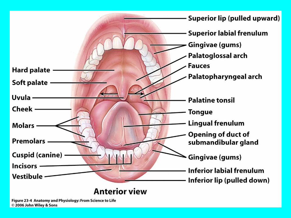
Figure 23.4
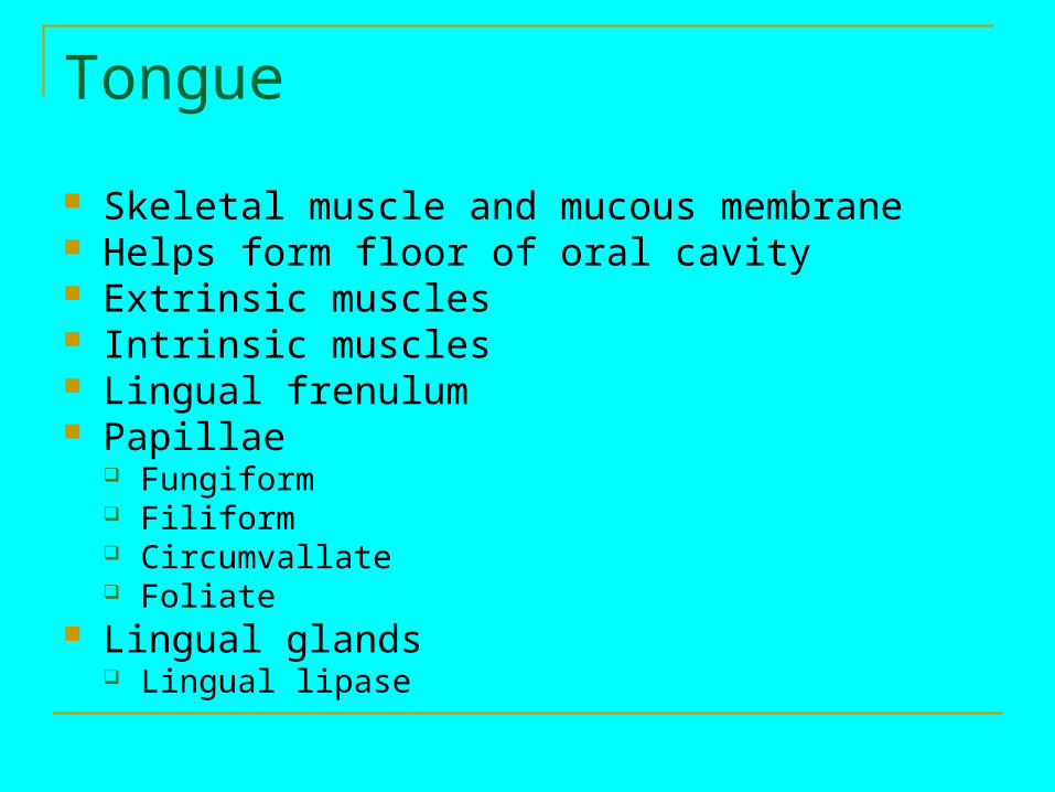
Tongue
Skeletal muscle and mucous membrane Helps form floor of oral cavity Extrinsic muscles Intrinsic muscles Lingual frenulum Papillae
Fungiform Filiform Circumvallate Foliate
Lingual glands Lingual lipase

Salivary Glands
Release saliva to oral cavity
3 pairs of salivary glands Parotid Submandibular Sublingual
Composition of Saliva
99.5 % water 0.5% other solutes
Ions Mucus Immunoglobulin A Enzymes
Salivation controlled by autonomic nervous system
Stimulated by various mechanisms
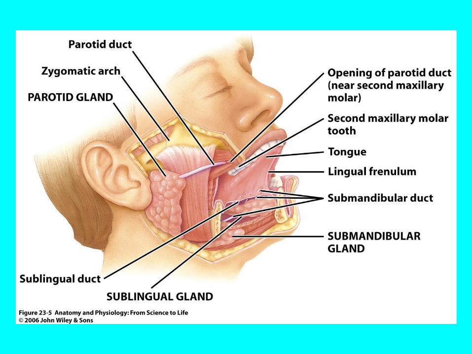
Figure 23.5
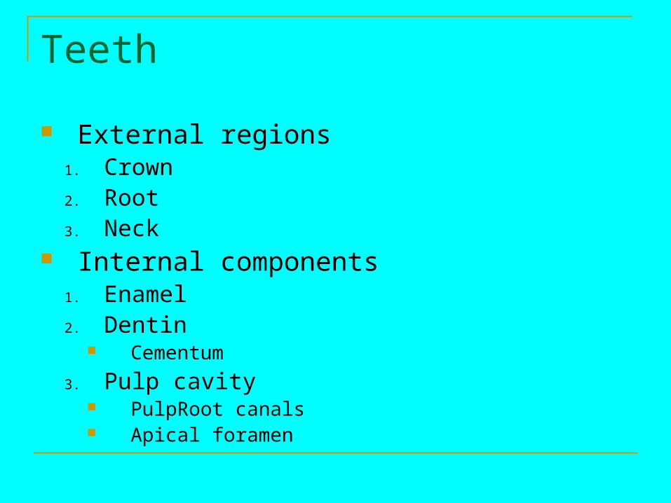
Teeth
External regions1. Crown2. Root3. Neck
Internal components1. Enamel2. Dentin
Cementum
3. Pulp cavity PulpRoot canals Apical foramen

Figure 23.6
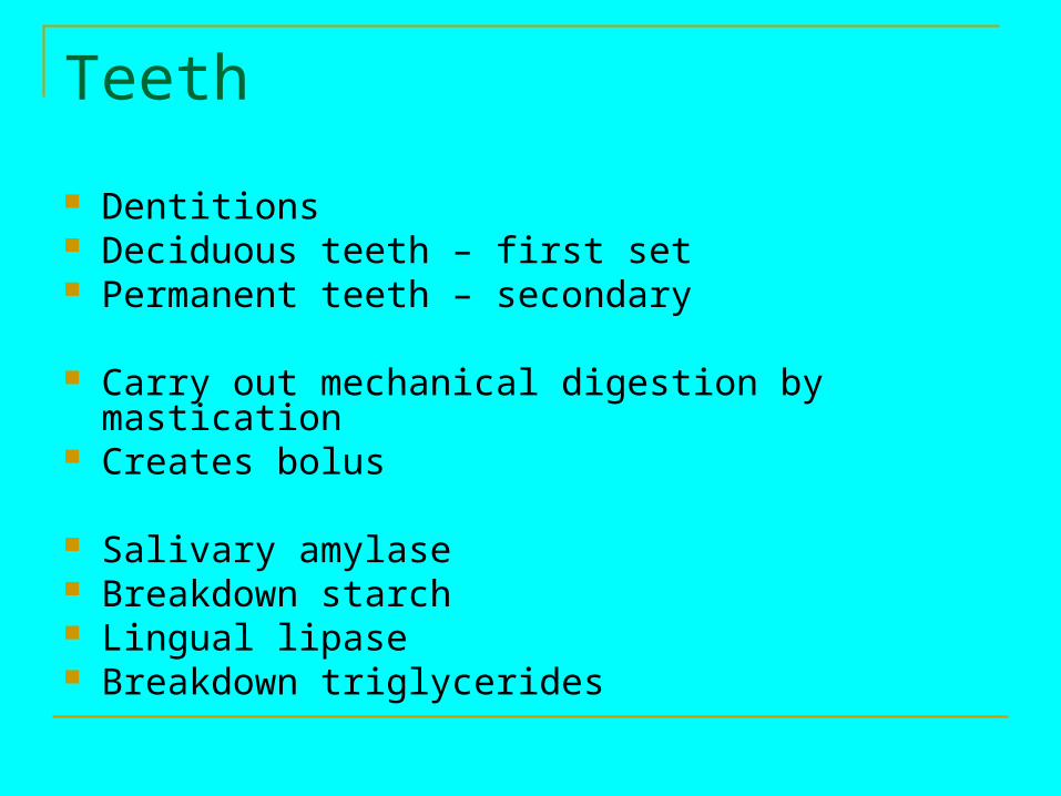
Teeth
Dentitions Deciduous teeth – first set Permanent teeth – secondary
Carry out mechanical digestion by mastication Creates bolus
Salivary amylase Breakdown starch Lingual lipase Breakdown triglycerides

Figure 23.7
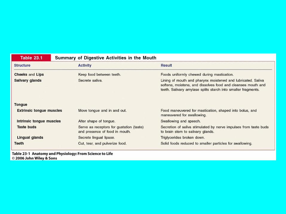
Table 23.1

Concept 23.3 Swallowing
Pharynx
Composed of skeletal muscle Lined by mucous membrane
Nasopharynx Oropharynx Laryngopharynx
Esophagus
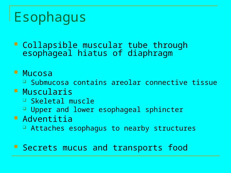
Collapsible muscular tube through esophageal hiatus of diaphragm
Mucosa Submucosa contains areolar connective tissue
Muscularis Skeletal muscle Upper and lower esophageal sphincter
Adventitia Attaches esophagus to nearby structures
Secrets mucus and transports food

Figure 23.8

Deglutition
Stages of swallowing Voluntary
Mouth to oropharynx Pharyngeal
Deglutition center in medulla oblongata and pons Closing of epiglottis Involuntary
Esophageal Involuntary Peristaltic contractions

Figure 23.9a,b
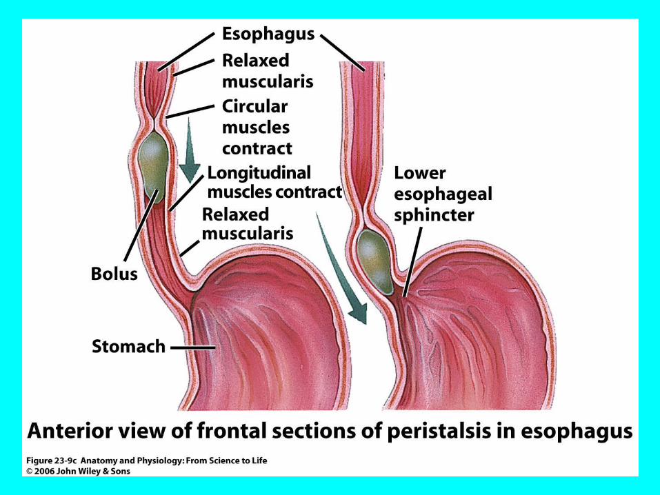
Figure 23.9c

Table 23.2
Concept 23.4 Stomach
Stomach
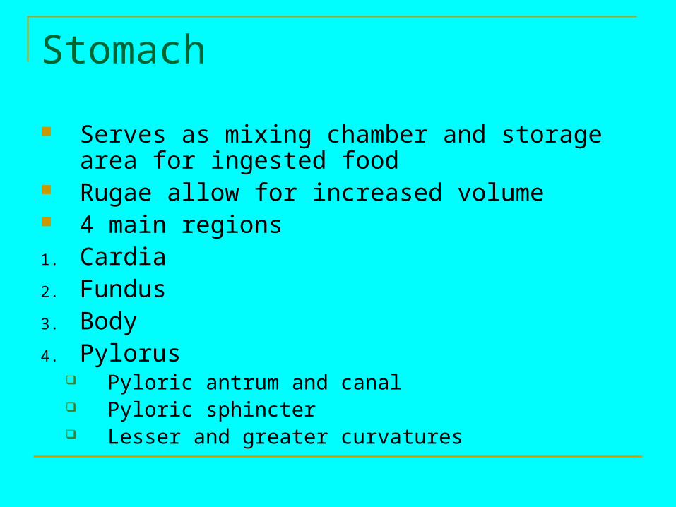
Serves as mixing chamber and storage area for ingested food
Rugae allow for increased volume 4 main regions1. Cardia2. Fundus3. Body4. Pylorus
Pyloric antrum and canal Pyloric sphincter Lesser and greater curvatures
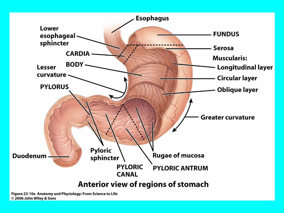
Figure 23.10a
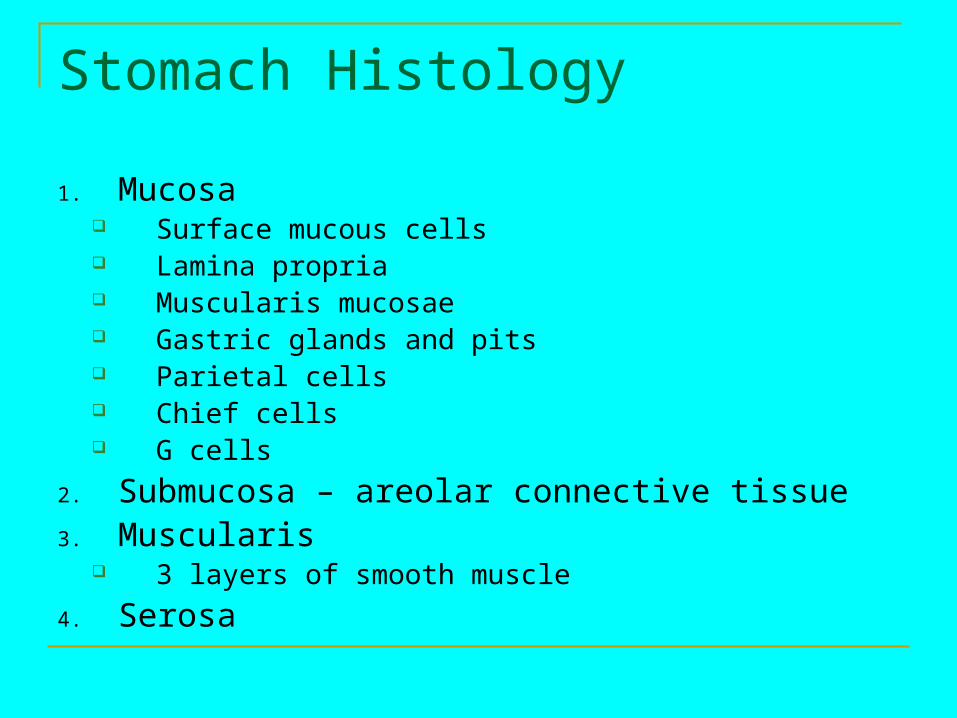
Stomach Histology
1. Mucosa Surface mucous cells Lamina propria Muscularis mucosae Gastric glands and pits Parietal cells Chief cells G cells
2. Submucosa – areolar connective tissue3. Muscularis
3 layers of smooth muscle
4. Serosa
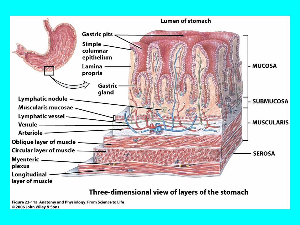
Figure 23.11a

Figure 23.11b

Figure 23.11c
Mechanical and Chemical Digestion Mixing waves caused by peristaltic movement Chyme released in process of gastric emptying Proton pumps bring H+ into the lumen Carbonic anhydrase forms carbonic acid to
provide H+ and bicarbonate ions (HCO3-)

Figure 23.12

Mechanical and Chemical Digestion
Chemical digestion stimulated by nervous system Parasympathetic neurons release acetylcholine
Works with gastrin HCl released in presence of histamine
Pepsin begins digestion of proteins Stomach protected by alkaline mucus secretion
Gastric lipase digests triglycerides Few molecules absorbed by stomach
Water, ions, short-chain fatty acids, alcohol
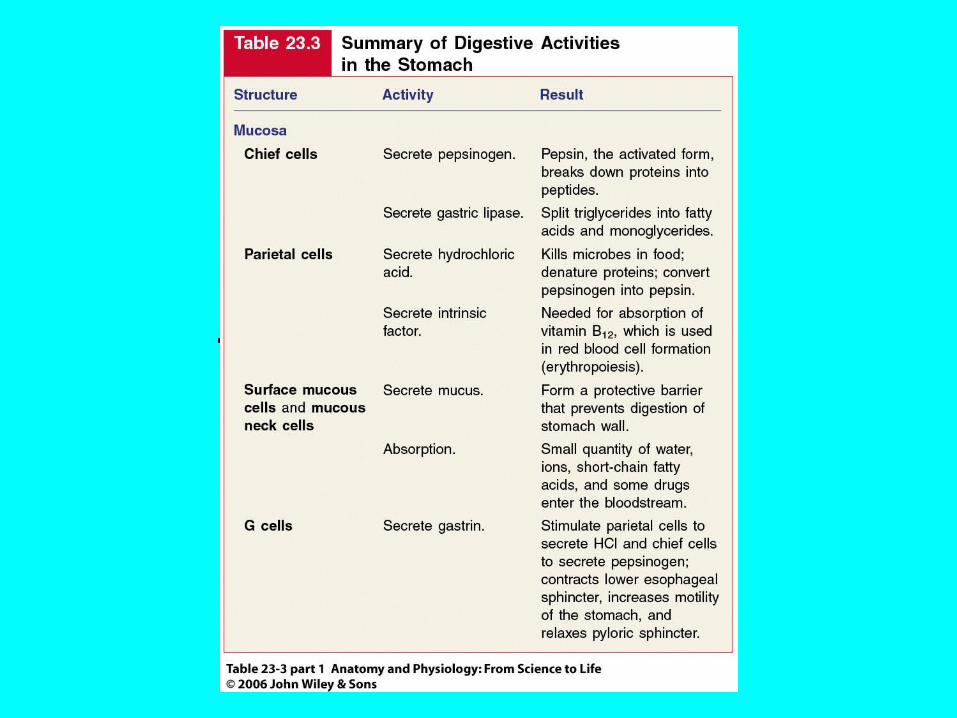
Table 23.3 pt 1
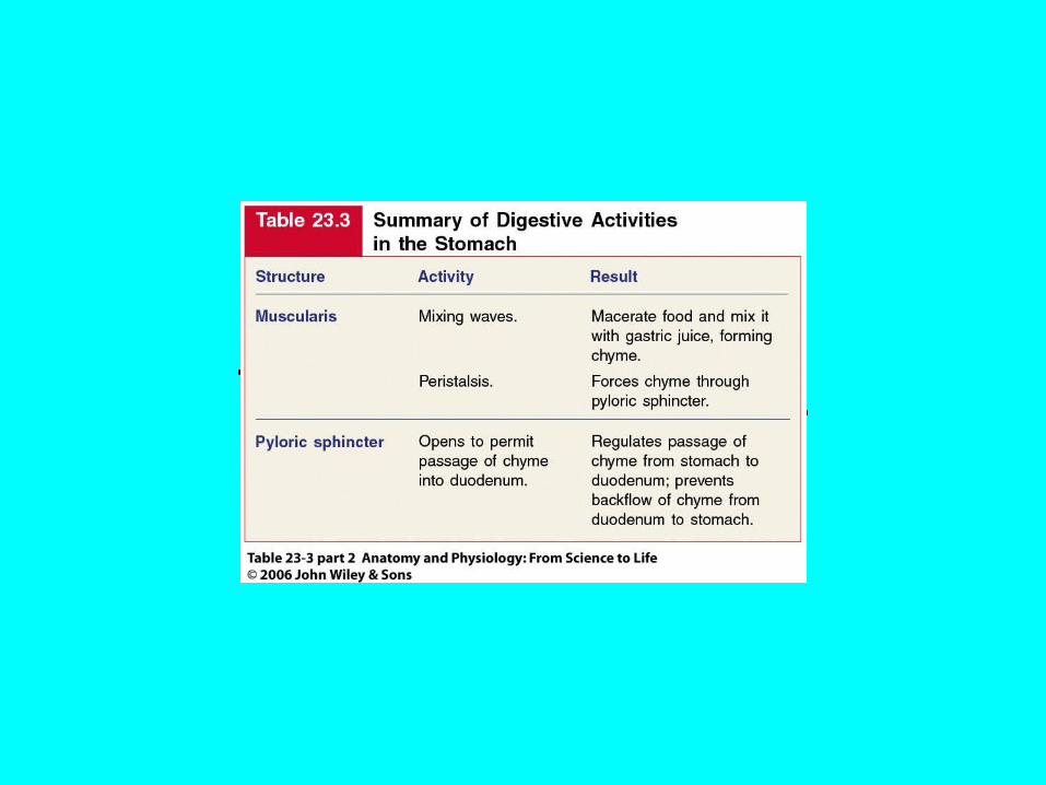
Table 23.3 pt 2
Concept 23.5 Accessory Organs of the Abdomen

Pancreas Produces secretions to aid digestion Head Body Tail Pancreatic duct /duct of Wirsung
Hepatopancreatic ampulla Sphincter of the heatopancreatic ampulla
(sphincter of (Oddi) Regulates passage of pancreatic juice and bile
Accessory duct (duct of Santorini)

Figure 23.13a

Figure 23.13b
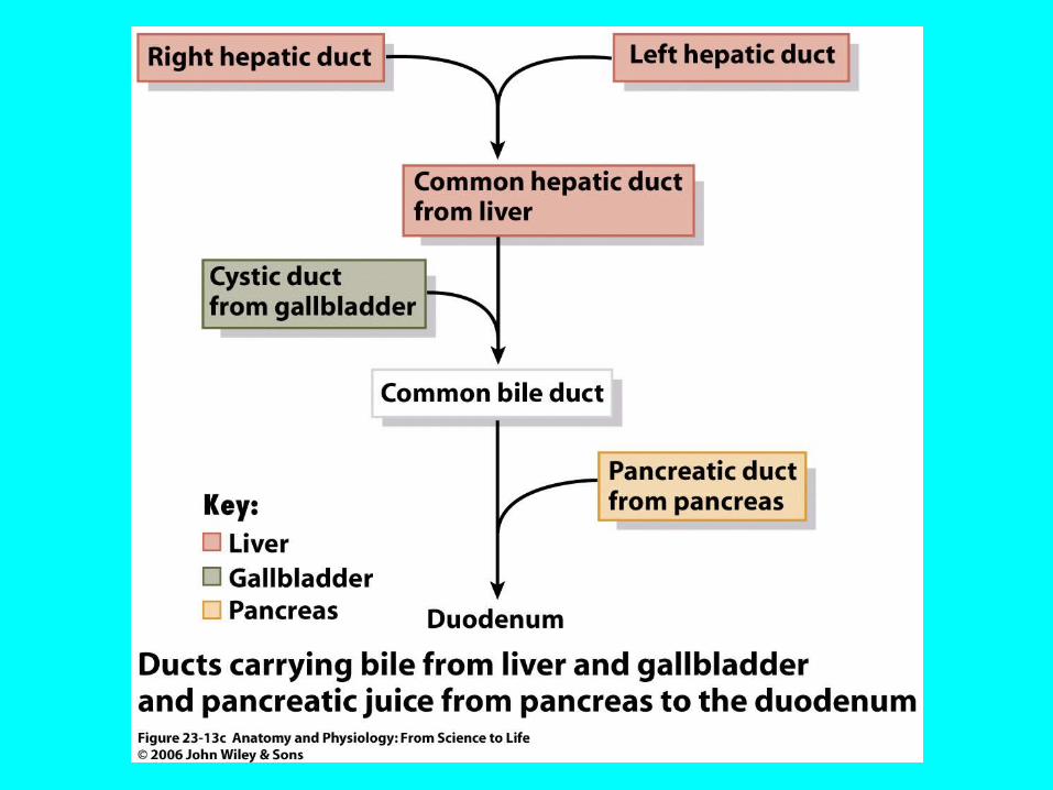
Figure 23.13c
Histology of Pancreas
Glandular epithelial cells 99% exocrine clusters Secrete pancreatic juice
Fluid and enzymes
Pancreatic islets (islets of Langerhans) 1% endocrine cells Hormones
Glucagon Insulin Somatostatin
Pancreatic polypeptide
Pancreatic Juice
1200-1500 mL/day pH 7.1-8.2 Water Salts Sodium bicarbonate Enzymes
Pancreatic amylase Trypsin
Entereokinase Chymotrypsin Carboxypeptidase Elastase Pancreatic lipase Ribonuclease and deoxyribonuclease

Liver and Gallbladder
Liver Largest gland at 1.4 kg (~3 lb)
Gallbladder Closely associated with liver
Anatomy of Liver
Right and left lobe separated by falciform ligament
Quadrate lobe Caudate lobe
Round ligament (ligamentum teres) Remnant of umbilical vein
coronary ligaments
Histology of Liver
Lobule Hepatocytes radiating from central vein Sinusoids
Reticuloendothelial (Kupffer) cells Stationary phagocytes

Figure 23.14a

Figure 23.14b
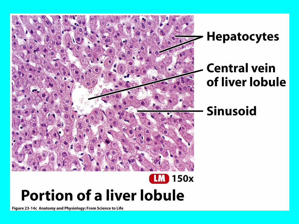
Figure 23.14c
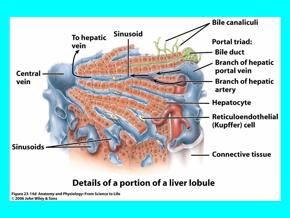
Figure 23.14d

Bile Duct System
Bile secreted by hepatocytes Bile canaliculi Bile ducts Right and left hepatic ducts Common hepatic duct Common bile duct
Gallbladder for temporary storage of bile Cystic duct
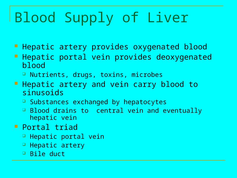
Blood Supply of Liver
Hepatic artery provides oxygenated blood Hepatic portal vein provides deoxygenated blood
Nutrients, drugs, toxins, microbes Hepatic artery and vein carry blood to sinusoids
Substances exchanged by hepatocytes Blood drains to central vein and eventually hepatic vein
Portal triad Hepatic portal vein Hepatic artery Bile duct
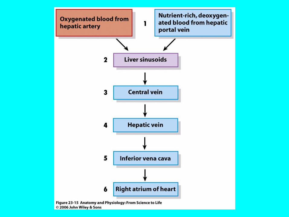
Figure 23.15

Bile
800-1000 mL/day pH 7.6 – 8.6 Water Bile acids Bile salts
Emulsification Cholesterol Lecithin Bile pigments
Bilirubin Stercobilin
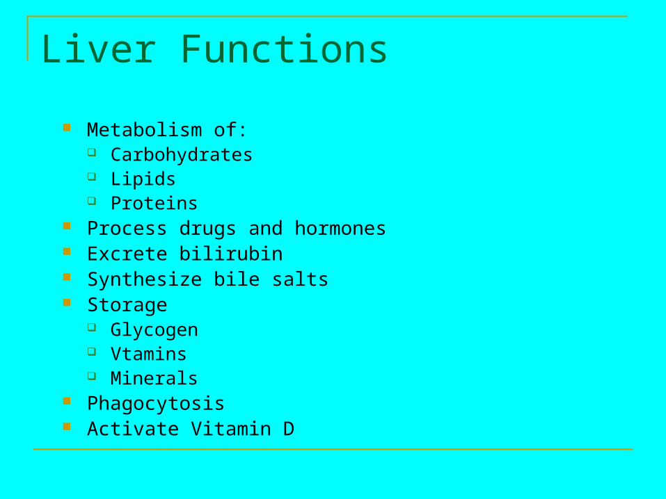
Liver Functions
Metabolism of: Carbohydrates Lipids Proteins
Process drugs and hormones Excrete bilirubin Synthesize bile salts Storage
Glycogen Vtamins Minerals
Phagocytosis Activate Vitamin D
Concept 23.6 Small Intestine
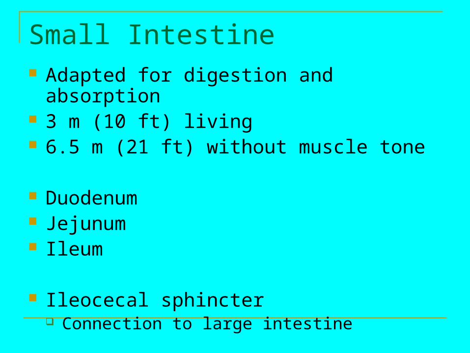
Small Intestine Adapted for digestion and absorption 3 m (10 ft) living 6.5 m (21 ft) without muscle tone
Duodenum Jejunum Ileum
Ileocecal sphincter Connection to large intestine

Figure 23.16a
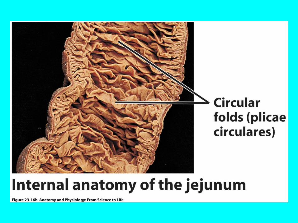
Figure 23.16b
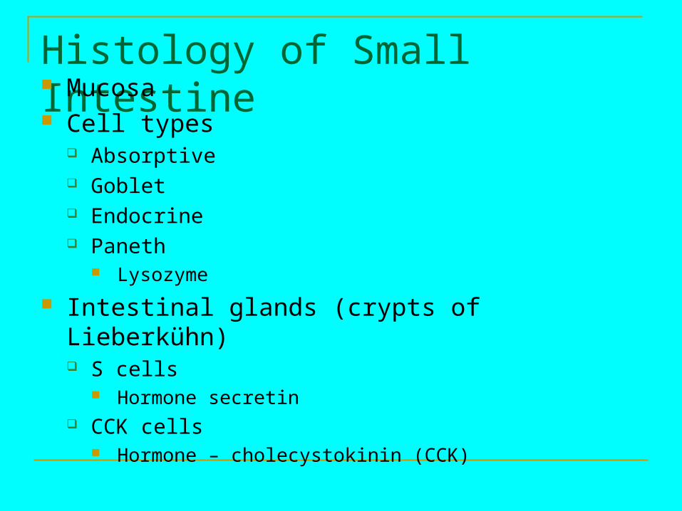
Histology of Small Intestine Mucosa Cell types
Absorptive Goblet Endocrine Paneth
Lysozyme
Intestinal glands (crypts of Lieberkühn) S cells
Hormone secretin CCK cells
Hormone – cholecystokinin (CCK)
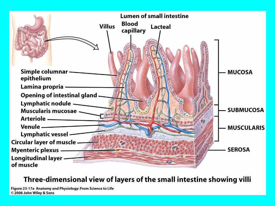
Figure 23.17a