the conundrum of estrogen receptor oscillatory activity in the
TRANSCRIPT

The Conundrum of Estrogen Receptor OscillatoryActivity in the Search for an Appropriate HormoneReplacement Therapy
Sara Della Torre, Andrea Biserni, Gianpaolo Rando, Giuseppina Monteleone,Paolo Ciana, Barry Komm, and Adriana Maggi
Center of Excellence on Neurodegenerative Diseases and Department of Pharmacological Sciences(S.D.T., G.R., G.M., P.C., A.M.), University of Milan, 20133 Milan, Italy; Transgenic Operative Productss.r.l. (A.B.), 26900 Lodi, Italy; and Pfizer Inc. (B.K.), Collegeville, Pennsylvania 19426
By the use of in vivo imaging, we investigated the dynamics of estrogen receptor (ER) activity inintact, ovariectomized, and hormone-replaced estrogen response element-luciferase reportermice. The study revealed the existence of a long-paced, noncircadian oscillation of ER transcrip-tional activity. Among the ER-expressing organs, this oscillation was asynchronous and its ampli-tude and period were tissue dependent. Ovariectomy affected the amplitude but did not suppressER oscillations, suggesting the presence of tissue endogenous oscillators. Long-term administra-tion of raloxifene, bazedoxifene, combined estrogens alone or with basedoxifene to ovariecto-mized estrogen response element-luciferase mice showed that each treatment induced a distinctspatiotemporal profile of ER activity, demonstrating that the phasing of ER activity among tissuesmay be regulated by the chemical nature and the concentration of circulating estrogen. This pointsto the possibility of a hierarchical organization of the tissue-specific pacemakers. Conceivably, therhythm of ER transcriptional activity translates locally into the activation of specific gene networksenabling ER to significantly change its physiological activity according to circulating estrogens. Inreproductive and nonreproductive organs this hierarchical regulation may provide ER with thesignaling plasticity necessary to drive the complex metabolic changes occurring at each femalereproductive status. We propose that the tissue-specific oscillatory activity here described is animportant component of ER signaling necessary for the full hormone action including thebeneficial effects reported for nonreproductive organs. Thus, this mechanism needs to betaken in due consideration to develop novel, more efficacious, and safer hormone replacementtherapies. (Endocrinology 152: 2256 –2265, 2011)
The normal aging process in women results in meno-pause, which is characterized by the cessation of ovar-
ian function and the decrease of sex hormone synthesis.Among the most common effects associated with meno-pause are vasomotor instability, loss of body mass, andaltered lipid profile; in addition, several epidemiologicalstudies indicated that after menopause there is a signifi-cant increase of cardiovascular (1, 2), immune (3), skeletal(4, 5), and central nervous system (6, 7) disorders, sug-gesting a protective action of estrogens also in tissues notdirectly associated with reproductive functions. The ex-
tension of the negative effects after the reduced synthesisof estrogens may be explained by the lack of activation ofthe two estrogen receptors (ER; ER� and ER�) becauseERs are expressed in most mammalian cells in which, bycontrolling specific transcription programs, they aredeeply involved in the whole-cell metabolism (8).
Attempts to reinstate estrogen beneficial effects withhormone replacement therapies (HRT) did not provide theexpected results so far. HRT has been carried out admin-istering 17�-estradiol or conjugated estrogens (CE) to hys-terctomized women; in nonhysterectomized women a
ISSN Print 0013-7227 ISSN Online 1945-7170Printed in U.S.A.Copyright © 2011 by The Endocrine Societydoi: 10.1210/en.2011-0173 Received February 15, 2011. Accepted March 25, 2011.First Published Online April 19, 2011
Abbreviations: BZA, Bazedoxifene; CCD, charge-coupled device; CE, conjugated estrogen;E2, 17�-estradiol; ER, estrogen receptor; ERE, estrogen response element; HRT, hormonereplacement therapy; HT, hormone therapy; Luc, luciferase; ovx, ovariectomized; RAL,raloxifen; SERM, selective estrogen receptor modulator; TSEC, tissue selective estrogencomplex.
G E N E R A L E N D O C R I N O L O G Y
2256 endo.endojournals.org Endocrinology, June 2011, 152(6):2256–2265

combined therapy has been applied with progestagens op-posing the hyperplastic effects of estrogen therapy in theuterus (9). To avoid estrogen unwanted effects on uterusand mammary gland but retain their beneficial effects inother organs efforts were made to identify ER ligands ableto mimic the hormone activity in nonreproductive organsand to antagonize its effects in the uterus and mammarygland. Several such molecules, named selective estrogenreceptor modulators (SERM) in virtue of their tissue-se-lective ER agonist or antagonist properties, were devel-oped and entered clinical practice (10). To date, however,none of the SERM developed appear to be provided withthe ideal balance of ER agonist and antagonist activity foran optimal postmenopausal therapy (11). For instance,the SERM used so far tend to be ER antagonists in thecentral nervous system, thus accentuating menopause va-somotor symptoms (11). To overcome these limitations, anovel HRT concept was proposed, which consists in thecombination of natural estrogens with a SERM: in thisway the whole spectrum of ER agonists effects can beobtained and the activity in the reproductive organs isblocked. This HRT has been named tissue selective estro-gen complex (TSEC) (12, 13).
In all cases, HRTs are carried out by the continuousadministration of the hormone aimed at maintaining aconstant level of circulating estrogens: this is in contrastwith the systemic, periodic fluctuations of ER activity dueto the changes of ovarian functions during the menstrualcycle. The impact of continuous stimulation of a receptorsystem developed to respond to oscillating levels of hor-mone is unknown; thus, a better understanding of the pe-riodic nature of ER signaling in the whole organism maybe an important factor for the generation of more effica-cious HRT. The goal of the present study was to investi-gate the effects of HRTs on the temporal organization ofER transcriptional activity in reproductive and nonrepro-ductive tissues. To this aim, we took advantage of theestrogen response element (ERE)-luciferase (Luc) trans-genic mouse characterized by the ubiquitous, ER-regu-lated expression of the luciferase gene (14). In this animalmodel, the possibility to measure luciferase activity in vivoby bioluminescence-based imaging provides the opportu-nity to investigate the ER systemic activity in time (14–17). Here we studied in a model of surgical menopause theextent to which current modalities of HRT, based on es-trogens, SERM, and TSEC administration, were able torestore the physiological oscillation occurring in healthy,cycling mice (12, 13).
We show that in intact mice ER activity oscillates at apace that is similar in each tissue but is not always syn-chronized on estrogen synthesis in the ovaries. In addition,our results demonstrate that, in each tissue, the continuous
administration of natural or synthetic estrogens inducesprofiles of ER oscillation with an amplitude and frequencythat are characteristic of each compound administered.Because of that, upon hormone therapy (HT) the phasing ofER activity among target organs may be significantly dis-rupted compared with what is observed in healthy, cyclingmice. We propose that the decentralized ER oscillatory be-haviorobserved in intactmicemight respondtoprecisephys-iological needs and that the study of HT effects on ER oscil-latory profile might provide a novel mean for theidentification of safer and more efficacious HRT.
Materials and Methods
Animal studiesIn the present study, we used heterozygous C57BL/6
repTOPERE-Luc (Transgenic Operative Products srl, Lodi, Italy)females 2–3 months of age (18). Mice were housed in individuallyventilated plastic cages with hardwood chip bedding and animalhouse, fed ad libitum with a standard diet (4RF21 standard diet;Mucedola, Settimo Milanese, Italy) and provided with filtered wa-ter. The animal room was maintained within a temperature rangeof22–25C,relativehumidityof50�10%,andunderanautomaticcycle of 12-h light, 12-h dark (lights on at 0700 h).
All animal experimentation was carried out in accordancewith the Guide for the Care and Use of Laboratory Animals asadopted and promulgated by the U.S. National institutes ofHealth and also in accordance with the European Guidelines forAnimal Care and Use of Experimental Animals, approved by theItalian Ministry of Research and University and controlled by thepanel of experts of the Department of Pharmacological Sciences(University of Milan, Mila, Italy). Mice were ovariectomized 3wk before the beginning of the study. Animals were assigned toa specific experimental group and treated per os (gavage) for 6 h(acute treatment) or daily for 21 d (chronic treatment) with ve-hicle, CE (3 mg/kg), bazedoxifen (BZA; 2 and 10 mg/kg), or BZAin association with 3 mg/kg CE (TSEC) and raloxifen (RAL; 2and 10 mg/kg). All compounds tested (Wyeth, Collegeville, PA)were dissolved in dimethylsulfoxide and subsequently diluted invehicle (2% Tween 80 and 0.5% carboxymethylcellulose watersolution). Controls were treated with vehicle. Compounds wereadministered always at 0930 h; the in vivo imaging session tookplace at 1500 h.
In vivo imagingMice were injected ip with 80 mg/kg D-luciferin (beetle lu-
ciferin potassium salt; Promega, Madison, WI) 15 min beforeeach in vivo imaging session. Previous experiments had demon-strated that this dose and time are sufficient to obtain an uniformbiodistribution of the substrate (19, 20). Mice were anesthetizedusing isofluorane (Isofluorane-Vet; Merial, Lyon, France) andXenogen XGI-8 gas anesthesia system (Caliper Life Sciences,Hopkinton, MA) and kept under anesthesia during the wholeimaging session [gas anesthesia system setting: vaporizer value2.5%; oxygen flow 1.5 l/min in the induction chamber and 0.25l/min to the mice during the in vivo imaging in the charge-coupleddevice (CCD) camera]. Photon emission in mice was measuredfor 5-min-long periods using a CCD camera (Xenogen IVIS Lu-
Endocrinology, June 2011, 152(6):2256–2265 endo.endojournals.org 2257
mina system; Caliper Life Sciences) consisting of a scientific-grade thermoelectrically cooled CCD camera mounted on alight-tight imaging chamber. Bioluminescence was measuredwith tiff images of 512 � 512 pixels at 16 bits. Each pixel con-tained the number of photon counts detected over the exposureperiod at the resolution of about 0.3 pixels/mm. Instrumentalefficiency was measured with appropriate luminescent sub-strates (Glowell; Lux Biotechnology, Edinburgh, UK).
Automatic analysis of mouse images:segmentation algorithm
Anatomical areas (head, limbs, tail, reproductive area, he-patic area, abdominal area) were segmented in Matlab environ-ment (TOP Transgenic Operative Products srl) using an algo-rithm previously described (21). The anatomical areas analyzedwere: skeletal areas (bone and limbs), reproductive areas (genitaland mammary glands), and hepatic and abdominal areas. In eachanatomical area, photon emission was defined as the number ofphotons (p) per second per centimeter squared corrected for in-strument efficiency. All the measurements were in the linearityrange of the detector (IVIS Lumina; Caliper Life Sciences) andwere previously validated by the comparison with the manualanalysis carried out using the CCD camera software (Living Im-age 3.0; Caliper Life Sciences).
Luciferase enzymatic assayTissues were homogenized in 200 �l of lysis buffer (100 mM
KPO4; 1 mM dithiothreitol; 4 mM EGTA; 4 mM EDTA, pH 7.8)with a 5-mm inox bead in a TissueLyser (QIAGEN GmbH,Hilden, Germany), subjected to one freezing-thawing cycle, andcentrifuged for 30 min at 4900 � g at 4 C (Allegra 25R; BeckmanCoulter, Brea, CA), and supernatants containing luciferase werecollected in ice. In samples containing luciferase, protein con-centration was measured by the Bradford assay, following themanufacturer’s instructions (Thermo Fisher Scientific Inc.,Waltham, MA). Luciferase enzymatic activity was then assayedwith a commercial luciferase assay buffer (Promega). Light in-tensity was measured with a luminometer (Glomax96 micro-plate luminometer; Promega) and expressed as relative light unitsover 10 sec per microgram of proteins.
Vaginal smearVaginal smears were carried out at 0, 7, 14, and 21 d at 0900 h
by vaginal flushing with sterile physiological solution, which wassubsequently air dried on glass microscope slides and stainedwith the May-Grunwald and Giemsa methods (MGG Quickstain kit,; Bio-Optica, Milan, Italy) following the protocol pro-vided by the manufacturer.
Measurement of the phasing of ER oscillatory indifferent body areas
The synchronic phasing of ER activity was analyzed manuallyby counting each body area by the total number of peaks of ERactivity/mouse during the 21-d-long experiment and then thenumber of coincident peaks for each couple of body areas. Thepercentages of coincident/noncoincident peaks obtained fromthe six to 10 individual animals observed were then averaged.
Real-time PCRTotal liver RNA was extracted with RNeasy minikit
(QIAGEN). cDNA was prepared as described (22). RT-PCR ex-periments were done by Sybr Green and TaqMan technology usingthe following primers: Fasn (forward, 5�-cctctgatcagtggcctcctc-3�;reverse, 5�-ggattcgggaatacaagtggc-3�); Acly (forward, 5�-gaagct-gaccttgctgaaccc-3�; reverse, 5�-ccgtaattcgccagttcattg-3�); Pmvk(forward, 5�-atggggctgtgatacagacag-3�; reverse, 5�-caaagttc-ccaaagttgtcca-3�); and 36b4 as reference gene (forward,5�-ggcgacctggaagtccaact-3�; reverse, 5�-ccatcagcaccacagccttc-3�).TaqMan gene expression assays for Foxo1 (Mm00490672_m1),Igf-1 (Mm00439561_m1), and as a reference gene assay 18SrRNA VIC-MGB-PDAR (Applied Biosystems by Life Technolo-gies, Carlsbad, CA). The reaction was carried out according to themanufacturer’s protocol using the 7900HT fast real-time PCR sys-tem (Applied Biosystems by Life Technologies). Data were ana-lyzed using the 2���Ct method (23).
Statistical analysisP values were calculated with one-way or two-way ANOVA
followed by Bonferroni post hoc test or t test with GraphPadPrism version 5.02 for Windows (GraphPad Software, SanDiego, CA).
Results
ER transcriptional activity is not centrallysynchronized
We first investigated the extent to which ER activityoscillated in intact and ovariectomized (ovx) mice. To thisaim, we measured luciferase-dependent photon emissionin nine intact and nine ovx mice for 21 d. In the cyclingmice (Fig. 1A), luciferase activity oscillated with time. Theamplitude of the oscillation was different in each bodyarea: for instance, it was more pronounced in hepatic thanthe genital area or limbs. In most body areas, the frequencyof oscillation appeared to be around 4 d (average in alltissues 4.4d); this was expected, considering that in micethe length of estrous cycle is 4–5 d; however, ER oscilla-tions did not occur synchronously in all the body areas(e.g. in the limbs the peak of photon emission was gener-ally retarded by 1 d with respect to the hepatic and genitalareas that tended to oscillate in phase). Quite unexpect-edly in these animals, ER activity fluctuated after ablationof the ovaries with an oscillation period of about 4 d (Fig.1B), but the amplitude of the oscillation was lower than inintact mice and the phasing among organs was altered: forinstance, photon emission in the hepatic and genital areaswas seldom in phase.
These results suggested that, in intact mice, ER activityoscillateswitha frequency that is compatiblewith theestrouscycle; however, 17�-estradiol (E2) cannot be the master reg-ulator of ER oscillatory activity because the oscillations wereasynchronous and persisted after ovariectomy.
2258 Della Torre et al. ER Oscillatory Activity Endocrinology, June 2011, 152(6):2256–2265

The measurement of mRNAs encoded byendogenous ER target genes confirms that ERactivity is asynchronous in liver and bone ofintact mice
To assess the validity of the observation based on lu-ciferase as an indicator of ER activity, we measured thecontent of several endogenous genes in bone and liver dur-ing the estrous cycle. The target genes were selected on thebasis of previous reports (24–26) and of studies carriedout in our laboratory (Stell A., personal communication).First, we demonstrated that the genes selected were goodindicators of ER activity by measuring their mRNA con-tent in tissue extracts of ovx mice treated with E2 for 6 hat the dose of 10 �g/kg sc. This treatment was sufficient toincrease photon emission from 12,800 to 150,000 p/sec �
cm2 in liver (Fig. 2A); the effect of acute E2 treatment onluciferase in bone was less visible (Fig. 2C). Real-time PCRanalysis carried out with liver extracts showed that afterthe E2 treatment, Foxo1 and Igf-1 mRNA were signifi-cantly increased compared to controls (�140 and �31%,respectively). This is in line with previous publications byour and other laboratories. Conversely mRNAs such asfatty acid synthase (Fasn; �38%), ATP citrate lyase (Acly;�27%), and phosphomevalonate kinase (Pmvk; �43%),known to be encoded by genes repressed by ER, weresignificantly decreased by the treatment (Fig. 2B). In bone,the mRNAs encoding for Foxo1, Nrip1, and Bmp-6 were
significantly increased by E2; in all experimental animals,Igf-1 mRNA content appeared to be higher than in con-trols, but the increase did not reach a statistically signifi-cant value (Fig. 2D). Next, we measured luciferase activityin 15 cycling ERE-Luc mice that were euthanized whenluciferase activity was low in liver and high in bone (group1) or, on the contrary, high in liver and low in bone (group2) (Fig. 3, A and C). Real-time quantitative PCR analysisdemonstrated a full correspondence between the contentof the endogenous genes and luciferase activity as shownin Fig. 3, B and D. Indeed, Fig. 3B shows that in liver thegenes induced by estrogens were low in group 1 (low liverluciferase) and high in group 2 (high liver luciferase).Conversely, the genes repressed by estrogens were lowerin group 2 than in group 1. Similarly in bone (Fig. 3D),in which the genes selected were all positively regulatedby E2, we observed that the mRNA content of Foxo1,Nrip1, and Bmp-6 was higher in group 1 than in group2. These results led us to conclude that luciferase is areliable indicator of ER transcriptional activity and canbe used as a surrogate target gene in our studies on theeffect of selected HT.
Tissue-specific action of selected HT in short-termtreatment (6 h)
The HT selected for the present study were: 1) naturalestrogens largely used in HT (Premarin or CE); 2) twoSERMs, BZA and RAL; and 3) TSEC (BZA together withCE) (12, 13). The selection of the dosage to be adminis-
FIG. 1. Profile of photon emission in time in cycling and ovx ERE-Lucmice. Photon emission was measured daily at 1500 h in 6-month-oldERE-Luc mice cycling or ovariectomized 3 wk before the initiation ofthe study. Each animal group was constituted of nine mice. The figurerepresents daily photon emission (plotted as photons per second persquare centimeter) in head, genital, and hepatic areas of a singlemouse representative of the pattern of ER activity in intact (A) and ovx(B) female mice.
FIG. 2. Luciferase activity in selected tissues of ERE-Luc mice andrelative endogeneous gene target expression after acute treatmentwith E2. Photon emission in the hepatic area (A) and in limbs (C) wasmeasured by a 5-min exposure time to a CCD camera (see Materialsand Methods) in mice at 6 h after treatment with 10 �g/kg of E2 orvehicle (ctrl). After the imaging session, mice were euthanized andtissues rapidly dissected. B, Foxo1, Igf-1, Fasn, Acly, and Pmvk mRNAcontent in liver of control and E2-treated mice. D, Foxo1, Igf-1, Nrip1,and Bmp-6 mRNA content in bone of control and E2-treated mice.Columns represent means � SEM of groups of six mice each. *, P �0.05; **, P � 0.01. P values were calculated with t test.
Endocrinology, June 2011, 152(6):2256–2265 endo.endojournals.org 2259

tered with regard to CE, BZA, and RAL was based onprevious reports (27). Before starting the long-term studywith the different types of HRT currently in use, we dem-onstrated their ability to regulate the luciferase reporterwhen administered orally at a dosage previously found tomimic HT in humans (10, 11, 15, 27). For the TSEC treat-ment, we tested two different concentrations of BZA (2and 10 mg/kg) to identify the concentration necessary andsufficient to block CE effects when used in the combinedtherapy.
The study was carried out in ovx ERE-Luc mice at 2–3months of age; surgery was performed 3 wk before HT.Two weeks after surgery, circulating levels of estradiolwere undetectable (not shown) and the uterus weight wassignificantly decreased (�73% vs. proestrus, the phase ofthe reproductive cycle during which estrogen circulatinglevels are highest) (Supplemental Fig. 1A, published onThe Endocrine Society’s Journals Online web site at http://endo.endojournals.org). Ovarian ablation reduced signif-icantly ER activity on the reporter gene (Supplemental Fig.2), yet the persistence of luciferase pointed to the existenceof factors other than ovarian estrogens able to elicit ERactivity.
Six hours after treatment, CE induced a strong tran-scriptional activity of ER in the hepatic and genital areas(Supplemental Fig. 1, B and C). With other treatments, atrend to increase was observed in several of the areas stud-ied, but none of the changes reached statistical signifi-
cance. These results were confirmed by a more quantita-tive study in which luciferase activity was directlymeasured in tissue extracts (Supplemental Fig. 3): in CE-treated mice, luciferase activity was increased in the uterus(�550%), breast (�230%), liver (�3451%), and bone(�145%). The high, but not the low, concentration ofboth SERMs caused an increased ER activity in the breast(BZA, �289%; RAL, �286% vs. vehicle); RAL at bothdosages was able to increase ER activity in the intestine(�159%). In this short-term study, BZA and RAL did notincrease ER activity in bone despite the well-known pro-tective effects of the two compounds against ovx-inducedosteoporosis.
This preliminary study established that the sensitivityof the bioluminescence imaging was sufficient to investi-gate the effects of the treatments.
Long-term �21-d effect of HT with CE, SERM,or TSEC
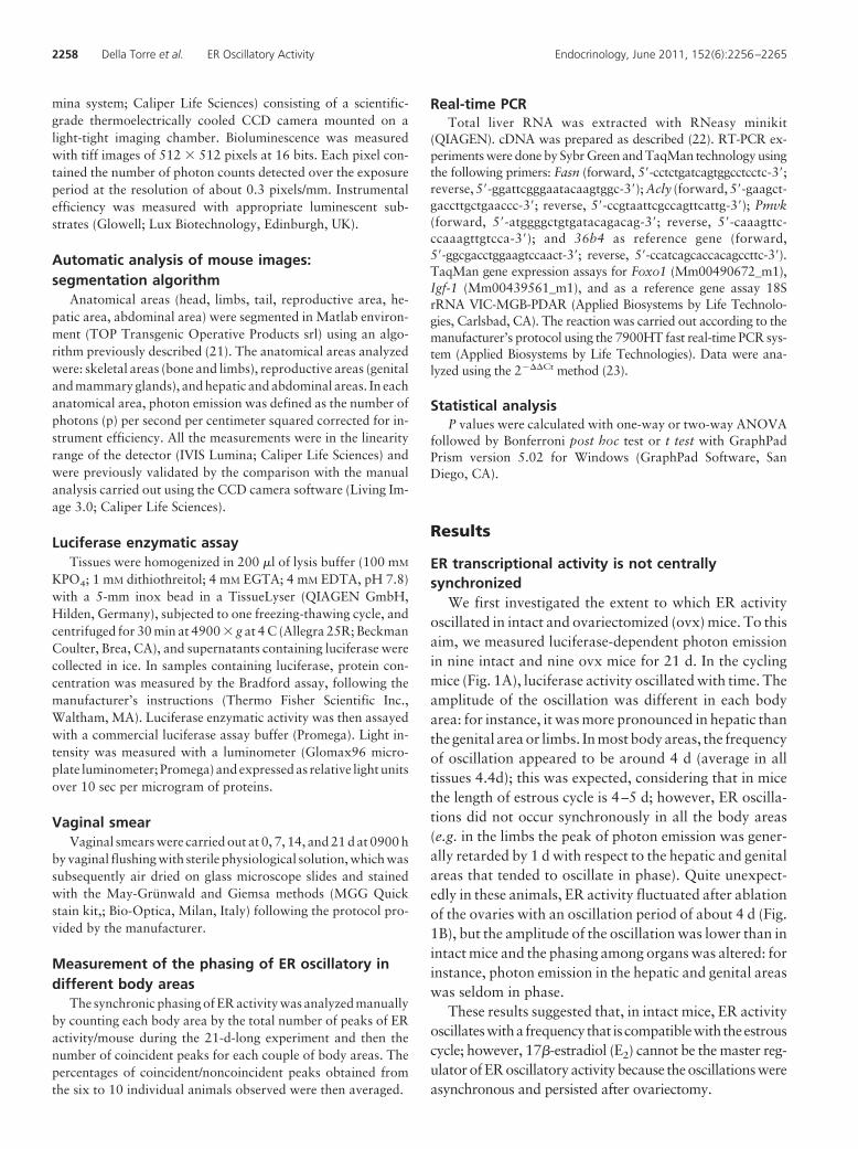
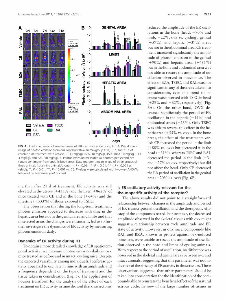
For the long-term study, ovx mice were treated daily bygavage with CE (3 mg/kg); BZA (10 mg/kg); TSEC (CE 3mg/kg � BZA 10 mg/kg), and RAL (10 mg/kg). Details ofthe treatments are described in the methodology section.Photon emission at 3, 7, 14, and 21 d of treatment (Fig. 4,A and B) indicated the efficacy of each HT in specific bodyareas. In line with the short-term treatment, CE signifi-cantly increased ER activity in the genital area (up to�318% vs. vehicle at d 7); this effect was blocked by BZA.In this body area, SERM affected ER activity only tran-siently (d 14). Measurement of the uterus weight in long-term-treated animals further supported the lack of estro-genic effects of BZA, RAL, and TSEC in this organ(Supplemental Fig. 4). In limbs, CE and BZA were able tosignificantly increase ER activity transiently (CE, �98%at d 7 and BZA, �105% at d 3); although a trend to anincrease was present all through out the study, TSEC hada strong effect augmenting ER activity in limbs from d 3until the end of the treatment (d 3: �111%; d 7: �120%;d 14: �118%; d 21: �101%), and RAL had a delayedeffect (d 14: �88%), which lasted to d 21 (�70%). In linewith previous reports of estrogens in liver (14), CE had amajor effect in the hepatic area that was blocked by BZA;BZA and RAL alone did not affect ER activity in this bodyregion. Interestingly, in the hepatic area, but not in thegenital area, the response to CE decreased with time, sug-gesting a tissue-specific mechanism diminishing ER activ-ity upon prolonged stimulation. We failed to see any sig-nificant change in the abdominal area with the exceptionof TSEC, which had a delayed effect (d 21: �160%). Thestudy of luciferase enzymatic activity in the tissues dis-sected from the animals at the end of the treatment (Sup-plemental Fig. 5) fully supported the in vivo data indicat-
FIG. 3. Comparative analysis of luciferase activity mice andendogeneous gene target expression in selected tissues of intact ERE-Luc. Photon emission in the hepatic area (A) and limbs (C) of mice ofgroup 1 (low liver/high bone photon emission) and group 2 (high liver/low bone photon emission). B, Foxo1, Igf-1, Fasn, Acly, and PmvkmRNA content measured by real-time quantitative PCR in tissueextracts of liver of group 1 and group 2 mice. D, Foxo1, Igf-1, Nrip1,and Bmp-6 mRNA content in bone of mice belonging to group 1 andgroup 2. Columns represent means � SEM of groups of six mice each.*, P � 0.05; **, P � 0.01; ***, P � 0.001. P values were calculatedwith t test.
2260 Della Torre et al. ER Oscillatory Activity Endocrinology, June 2011, 152(6):2256–2265
ing that after 21 d of treatment, ER activity was stillelevated in the uterus (�435%) and the liver (�868%) ofmice treated with CE and in the bone (�64%) and theintestine (�333%) of those exposed to TSEC.
The observation that during the long-term treatment,photon emission appeared to decrease with time in thehepatic area but not in the genital area and limbs and thatin selected areas the changes were transient, led us to fur-ther investigate the dynamics of ER activity by measuringphoton emission daily.
Dynamics of ER activity during HTTo obtain a more detailed knowledge of ER spatiotem-
poral activity, we measure photon emission daily in ovxmice treated as before and in intact, cycling mice. Despitethe expected variability among individuals, luciferase ac-tivity appeared to oscillate in time with an amplitude anda frequency dependent on the type of treatment and thetissue taken in consideration (Fig. 5). The application ofFourier transform for the analysis of the effect of eachtreatment on ER activity in time showed that ovariectomy
reduced the amplitude of the ER oscil-lations in the bone (head, �70% andlimb, �22%, ovx vs. cycling), genital(�59%), and hepatic (�39%) areasbut not in the abdominal area. CE treat-ment increased significantly the ampli-tude of photon emission in the genital(�90%) and hepatic areas (�481%)but in the bone and abdominal area wasnot able to restore the amplitude of os-cillation observed in intact mice. Theeffect of BZA, TSEC, and RAL was notsignificant in any of the areas taken intoconsideration, even if a trend to in-crease was observed with TSEC in head(�29% and �62%, respectively) (Fig.6A). On the other hand, OVX de-creased significantly the period of ERoscillation in the hepatic (�14%) andabdominal areas (�23%). Only TSECwas able to reverse this effect in the he-patic area (�55% vs. ovx). In the boneareas, the effect of the treatments var-ied: CE increased the period in the limb(�88% vs. ovx) but decreased it in thehead (�31%), whereas TSEC and RALdecreased the period in the limb (�31and �27% vs. ovx, respectively) but didnot affect the head. Only CE decreasedthe ER period of oscillation in the genitalarea (�20% vs. ovx) (Fig. 6B).
Is ER oscillatory activity relevant for thetissue-specific activity of the receptor?
The above results did not point to a straightforwardrelationship between changes in the amplitude and periodof ER transcriptional oscillation and the therapeutic effi-cacy of the compounds tested. For instance, the decreasedamplitude observed in the skeletal tissues with ovx mightsuggest a relationship between cycle amplitude and ERstate of activity. However, in ovx mice, compounds likeRAL and BZA, known to protect against ovx-inducedbone loss, were unable to rescue the amplitude of oscilla-tion observed in the head and limbs of cycling animals.With respect to the period of oscillation, no difference wasobserved in the skeletal and genital areas between ovx andintact animals, suggesting that this parameter was not in-dicative of the efficacy of ER activity in those tissues. Theseobservations suggested that other parameters should betaken into consideration for the identification of the com-pounds able to reinstate the beneficial effects of the naturalestrous cycle. In view of the large number of tissues in
FIG. 4. Photon emission of selected areas of ERE-Luc mice undergoing HT. A, Pseudocolorimage of photon emission from one representative animal/group at 0, 3, 7, and 21 d ofchronic oral treatment with vehicle, CE (3 mg/kg), BZA (10 mg/kg), TSEC (BZA 10 mg/kg � CE3 mg/kg), and RAL (10 mg/kg). B, Photon emission measured as photons per second persquare centimeter from specific body areas. Data represent mean � SEM of three groups ofthree animals (total nine animals/group). *, P � 0,05; **, P � 0,01; ***, P � 0,001 vs.vehicle; °°, P � 0,01; °°°, P � 0,001 vs. CE. P values were calculated with two-way ANOVAfollowed by Bonferroni post hoc test.
Endocrinology, June 2011, 152(6):2256–2265 endo.endojournals.org 2261

which ER is active, we speculated that the beneficial effectsof the hormone are due to a harmonic sequence of eventsin which the relative synchronization of ER activity in thedifferent tissue is critical. We tried to test this hypothesisby counting the number of synchronous cycles in two bodyareas at the time during the 21 d of the experiment (Fig. 7).We reasoned that during the estrous cycle, the body areasrepresenting functionally related tissues should cycle insynchrony. Indeed, in the skeletal areas (photon emissionfrom head and limb) of cycling mice, the percentage ofsynchronic cycling was generally quite high (76.5%); also,reproductive (breast and genital area) and genital and he-patic areas were cycling relatively in phase (56 and 55% ofthe cycles were in phase, respectively). The latter obser-vation had been previously reported (14). In the hepaticand abdominal areas, the percentage of synchrony wassignificantly lower (29.7%).
Ovariectomy had a disruptive effect by decreasing theextent of synchronous phasing in the hepatic and abdom-inal areas (from 29.7 to 16%) and in the genital and he-patic area (from 55 to 28%). No significant change wasobserved in the skeletal and reproductive areas, whereas a
trend to an increased phasing was measured in the headand genital areas.
Among HT, only TSEC and BZA were able to com-pletely rescue the effect of ovx in the phasing betweenhepatic and abdominal areas (from 16 to 35 and 34%,respectively).
All HT, including CE, tended to decrease the synchronyof cycling of reproductive areas (from 57% to CE, 31%;TSEC, 32%; BZA, 40%; RAL, 40%). A trend to increasein the synchronic oscillation among skeletal tissues wasobserved with TSEC (from 67 to 84%), BZA (to 81%),and RAL (to 76%). When we compared head and genitalareas, the only significant change was observed with theCE treatment that increased the phasing with respect tocycling mice (�138%).
Discussion
Pulsatility characterizes the secretion of several hormones(e.g. GH, GnRH, insulin), and the maintenance of a spe-
FIG. 5. Profile of photon emission in time in ERE-Luc mice undergoingHT. Photon emission was measured daily in head, limbs, and genital,hepatic, and abdominal areas at 1500 h (6 h after the treatment) usinga segmentation algorithm (22). The experiment was done inexperimental groups each composed of nine mice. Graphs reproducedata obtained from a single, representative mouse/group.
FIG. 6. Fourier transform (FT) analysis of the profile of ER activity intime to measure the amplitude and frequency of luciferase oscillationin different body areas of ERE-Luc intact (cyc) and ovx mice and in ovxmice with HT. FT was applied to the data described in Figs. 4 and 5. A,Average amplitude of cycles in each group of nine mice estimated bymeasuring the degree of displacement from the resting state(calculated as the square root of the 95th percentile of the powerspectra). cyc, Cycling mice (or intact mice). B, Period of oscillationestimated by the inverse of the frequencies under the amplitudepreviously calculated. Bars represent average � SEM of groups of ninemice each. *, P � 0.05; **, P � 0.01 vs. cycling animals; °, P � 0.05;°°, P � 0.01 vs. ovx mice. P values were calculated with one-wayANOVA followed by Bonferroni post hoc test.
2262 Della Torre et al. ER Oscillatory Activity Endocrinology, June 2011, 152(6):2256–2265

cific pattern of secretion is necessary for the hormone fullendocrine effects (25–28). This may be valid also for es-trogens; indeed, current view predicts that a finely tunedfeedback system in the hypothalamic-pituitary-gonadalaxis ensures the maintenance of the reproductive cycleregulated by the cyclic synthesis of ovarian sex hormonesand the activation of ER transcriptional activity in repro-ductive as well as nonreproductive organs. The observa-tions reported here argue against such a unicentric modelof ER regulation because it is shown that: 1) in intact micethe systemic production of estrogens by the ovaries fails tosynchronize ER activity among target organs; 2) afterovariectomy ER maintains a cyclic activity; and 3) eachorgan regulates autonomously the frequency and the am-plitude of ER transcriptional activity in response to theadministration of estrogens of different chemical nature orof a combination of estrogenic compounds.
At the present time, the mechanisms underlying thelong-paced, rhythmic oscillation of ER activity as well asits physiological function may be only object of specula-tion. Western blot analysis and RT-PCR carried out onliver and uterus extracts show that during the long-pacedER transcriptional oscillation, the content of the receptorprotein and mRNA is unchanged (not shown). Thus, wedo not believe that the oscillation described here is a con-sequence of ER down-regulation.
The observation that the phasing of the oscillation isindependent from a central control and is very susceptibleto the nature of circulating estrogens may suggest that ineach cell the ER activity is controlled autonomously viainterlocking transcription/translation feedback loops.This would be in line with what reported for the rhythmicfluctuationsof the circadiangenes,whichappear tobe able
to oscillate independently from the cen-tral oscillator located in the suprachias-matic nucleus (28–30). Further support-ing the hypothesis of cell autonomousregulatory mechanisms are recent studiescarriedout in isolatedcells inwhichitwasdemonstrated that, in the constant pres-ence of the ligand, ER activity fluctuatesrapidly (in the order of minutes). Themechanisms driving the receptor oscilla-tory activity include changes in coregula-tor recruitment (31), assembly of thecomponents of the preinitiation complex(32), and posttranslational regulation ofthe receptor leading to proteolysis of theER complex (33). These oscillations arebelieved to be necessary to poise the re-ceptors for a proper response to hor-monal stimulus.
The fluctuations of ER activity de-scribed here have a periodicity very similar to the estrouscycle (4.4 d as average in the different tissues of cyclingmice) yet are not synchronized on the fluctuations of thesex hormones in the blood stream. Because it is wellknown that several nonsteroidal stimuli may activate ERtranscriptionally (15, 34–38), it is tempting to speculatethat endocrine factors other than ovarian estrogen are re-sponsible for the waves of receptor activation observed inthe different ER-expressing cells. Supporting this hypoth-esis is a recent study on mice carrying a liver-selective ab-lation of ER� (39) in which it was demonstrated that he-patic ER play a major role in the synthesis and secretion ofIgf-1, a liver hormone essential for several physiologicalfunctions including the maturation of the uterus epithe-lium and the full execution of the reproductive cycle. Con-sidering the major involvement of ER in reproductivefunctions, a decentralized control of the activity of thesereceptors would serve the purpose to grant the reciprocalregulatory feedbacks to ensure that reproduction occurs inthe most favorable energetic/metabolic/health conditionsand to enable the significant metabolic adaptations asso-ciated with changes in the reproductive status (e.g. pu-berty, pregnancy, lactation). It is conceivable that in eachtarget tissue the significant changes in estrogen synthesisand metabolism reported in different reproductive condi-tions are instrumental to modulate large transcription pro-grams that trigger the metabolic response necessary for thesuccessful reproduction. Several studies have underlinedthe relevance of the chemical stimulus triggering ER ac-tivity for the selection of the genes to be transcribed (40–43). We also showed by chip-on-chip analysis (data notshown) that in liver, ER associated with very distinct
FIG. 7. Effect of HT on the phasing of luciferase oscillation among different body areas ofliving ERE-Luc mice. The number of synchronous cycles/total cycles in the 21-d observationperiod was scored analyzing the profile of ER activity of the body areas of each single animal.Data represent the average percentage of synchronic cycling in four clusters of anatomicalregions: skeletal area (head and limbs), reproductive area (breast and genital area), hepatic-abdominal areas, reproductive-hepatic areas, and head-genital areas. The experiment was donein experimental groups each composed of nine animals. cyc, Cycling mice (or intact mice).
Endocrinology, June 2011, 152(6):2256–2265 endo.endojournals.org 2263

classes of promoters during the different phases of theestrous cycle. On the other hand, the ability of factorsother than estrogens to regulate ER activity may be re-quired to prevent pregnancy in case of disease or insuf-ficient nutritional contribution. According to this viewand in agreement with the results of the present study,the nature of the estrogenic stimulus may act as a triggerfor the differential, rhythmic, and harmonic modula-tion of ER in the different organs necessary for the ac-tivation of the gene programs fulfilling the necessarymetabolic program.
It remains to be established which is the hormonal set-ting that needs to be reestablished in the case of HT in thepostmenopause. The study indicated that the use of SERMor a combination of natural hormones and SERM mayhave a significant effect on the relative phasing and inten-sity of ER activity in the target organs; this prospects thepossibility to reproduce pharmacologically the desiredcomplexity of ER action in the whole organism. What islacking at the present time is a clear view of the pattern ofER activity that would have the most favorable effects forwomen’s health during aging. In the absence of suchknowledge, we believe that the mere analysis of the effectsof HRT on a single parameter (e.g. the effect on the periodor amplitude of ER activity in different organs) is not suf-ficient to establish the superiority of a treatment on others.In a recent study, Rando et al. (44) applied an algorithmdeveloped for the comparative analysis of multivariate pa-rameters to the study of the activity of synthetic ER li-gands. Most interestingly, the study showed that the ap-plication of such an algorithm enables the identification ofstructurally related compounds by comparing their spa-tiotemporal effects on the ERE-Luc promoter. In addition,the study showed that the method enables one to measurethe ability of each family of compounds to reproduce thestate of activation of ER that characterize the intact, cy-cling mouse. We believe that these methodologies may, atthe present time, facilitate the identification of HT to beapplied.
Our current hypothesis that the hierarchical ER acti-vation in different tissues is a mechanism set in evolutionto enable ER to recognize the changes in the reproductivestatus and to alert and adapt the entire organism to thenovel energetic requirements may help in a better under-standing of the metabolic changes occurring after meno-pause and in devising novel, more efficacious therapeuticinterventions.
Acknowledgments
We thank Paolo Sparaciari for veterinary assistance andA. Buscemi, C. Roncoroni, E. Galioto, and V. Benedusi for their
assistance in the generation of the data and for their commentsduring the preparation of the manuscript.
Address all correspondence and requests for reprints to:Adriana Maggi, Center of Excellence on NeurodegenerativeDiseases and Department of Pharmacological Sciences,University of Milan, Via Balzaretti 9, 201313, Milan, Italy.E-mail: [email protected].
This work was supported by European Union Grant STREPEWA LSHM-CT-2005-518245; National Institutes of HealthGrant RO1AG027713; and Pfizer Pharmaceutical Co.
Disclosure Summary: S.D.T., A.B., G.R., G.M., and P.C. havenothing to declare. B.K. is employed by Pfizer and has stock/stock options in Pfizer. A.M. received grant support from Wyeth/Pfizer and received consulting fees from Wyeth/Pfizer.
References
1. Mendelsohn ME, Karas RH 1999 The protective effects of estrogenon the cardiovascular system. N Engl J Med 340:1801–1811
2. Bolego C, Vegeto E, Pinna C, Maggi A, Cignarella A 2006 Selectiveagonists of estrogen receptor isoforms: new perspectives for cardio-vascular disease. Arterioscler Thromb Vasc Biol 26:2192–2199
3. Straub RH 2007 The complex role of estrogens in inflammation.Endocr Rev 28:521–574
4. Imai Y, Kondoh S, Kouzmenko A, Kato S 2009 Regulation of bonemetabolism by nuclear receptors. Mol Cell Endocrinol 310:3–10
5. Cauley JA, Robbins J, Chen Z, Cummings SR, Jackson RD, LaCroixAZ, LeBoff M, Lewis CE, McGowan J, Neuner J, Pettinger M,Stefanick ML, Wactawski-Wende J, Watts NB 2003 Effects of es-trogen plus progestin on risk of fracture and bone mineral density:the Women’s Health Initiative randomized trial. JAMA 290:1729–1738
6. Sherwin BB 2003 Estrogen and cognitive functioning in women.Endocr Rev 24:133–151
7. Maggi A, Ciana P, Belcredito S, Vegeto E 2004 Estrogens in thenervous system: mechanisms and nonreproductive functions. AnnuRev Physiol 66:291–313
8. Dahlman-Wright K, Cavailles V, Fuqua SA, Jordan VC, Katzenel-lenbogen JA, Korach KS, Maggi A, Muramatsu M, Parker MG,Gustafsson JA 2006 International Union of Pharmacology. LXIV.Estrogen receptors. Pharmacol Rev 58:773–781
9. Anderson GL, Limacher M, Assaf AR, Bassford T, Beresford SA,Black H, Bonds D, Brunner R, Brzyski R, Caan B, Chlebowski R,Curb D, Gass M, Hays J, Heiss G, Hendrix S, Howard BV, Hsia J,Hubbell A, Jackson R, Johnson KC, Judd H, Kotchen JM, Kuller L,LaCroix AZ, Lane D, Langer RD, Lasser N, Lewis CE, Manson J,Margolis K, Ockene J, O’Sullivan MJ, Phillips L, Prentice RL, Riten-baugh C, Robbins J, Rossouw JE, Sarto G, Stefanick ML, Van HornL, Wactawski-Wende J, Wallace R, Wassertheil-Smoller S 2004 Ef-fects of conjugated equine estrogen in postmenopausal women withhysterectomy: the Women’s Health Initiative randomized controlledtrial. JAMA 291:1701–1712
10. Turgeon JL, McDonnell DP, Martin KA, Wise PM 2004 Hormonetherapy: physiological complexity belies therapeutic simplicity. Sci-ence 304:1269–1273
11. Johnson KA 2006 Editorial: the SERM of my dreams. J Clin Endo-crinol Metab 91:3754–3756
12. Kharode Y, Bodine PV, Miller CP, Lyttle CR, Komm BS 2008 Thepairing of a selective estrogen receptor modulator, bazedoxifene,with conjugated estrogens as a new paradigm for the treatment ofmenopausal symptoms and osteoporosis prevention. Endocrinology149:6084–6091
2264 Della Torre et al. ER Oscillatory Activity Endocrinology, June 2011, 152(6):2256–2265

13. Komm BS 2008 A new approach to menopausal therapy: the tissueselective estrogen complex. Reprod Sci 15:984–992
14. Ciana P, Raviscioni M, Mussi P, Vegeto E, Que I, Parker MG, LowikC, Maggi A 2003 In vivo imaging of transcriptionally active estrogenreceptors. Nat Med 9:82–86
15. Klotz DM, Hewitt SC, Ciana P, Raviscioni M, Lindzey JK, Foley J,Maggi A, DiAugustine RP, Korach KS 2002 Requirement of estro-gen receptor-� in insulin-like growth factor-1 (IGF-1)-induced uter-ine responses and in vivo evidence for IGF-1/estrogen receptor cross-talk. J Biol Chem 277:8531–8537
16. Di Lorenzo D, Villa R, Biasiotto G, Belloli S, Ruggeri G, AlbertiniA, Apostoli P, Raviscioni M, Ciana P, Maggi A 2002 Isomer-specificactivity of dichlorodyphenyltrichloroethane with estrogen receptorin adult and suckling estrogen reporter mice. Endocrinology 143:4544–4551
17. Mussi P, Liao L, Park SE, Ciana P, Maggi A, Katzenellenbogen BS,Xu J, O’Malley BW 2006 Haploinsufficiency of the corepressor ofestrogen receptor activity (REA) enhances estrogen receptor func-tion in the mammary gland. Proc Natl Acad Sci USA 103:16716–16721
18. Ciana P, Di Luccio G, Belcredito S, Pollio G, Vegeto E, Tatangelo L,Tiveron C, Maggi A 2001 Engineering of a mouse for the in vivoprofiling of estrogen receptor activity. Mol Endocrinol 15:1104–1113
19. Biserni A, Giannessi F, Sciarroni AF, Milazzo FM, Maggi A, Ciana P2008 In vivo imaging reveals selective peroxisome proliferator acti-vated receptor modulator activity of the synthetic ligand 3-[1-(4-chlorobenzyl)-3-t-butylthio-5-isopropylindol-2-yl]-2,2-dimethylpropanoic acid (MK-886). Mol Pharmacol 73:1434–1443
20. Rando G, Biserni A, Ciana P, Maggi A 2010 Profiling of drug actionusing reporter mice and molecular imaging. Methods Mol Biol 602:79–92
21. Rando G, Casiraghi E, Arca S, Campadelli P, Maggi A Automaticsegmentation of mouse images. In: Capasso V, et al., Stereology andimage analysis. Proc 10th European Congress of ISS, Bologna (Italy),2009, Esculapio, 60–64
22. Ciana P, Biserni A, Tatangelo L, Tiveron C, Sciarroni AF, OttobriniL, Maggi A 2007 A novel peroxisome proliferator-activated recep-tor responsive element-luciferase reporter mouse reveals genderspecificity of peroxisome proliferator-activated receptor activity inliver. Mol Endocrinol 21:388–400
23. Livak KJ, Schmittgen TD 2001 Analysis of relative gene expressiondata using real-time quantitative PCR and the 2[��� C(T)] method.Methods 25:402–408
24. Stossi F, Barnett DH, Frasor J, Komm B, Lyttle CR, Katzenellen-bogen BS 2004 Transcriptional profiling of estrogen-regulated geneexpression via estrogen receptor (ER) � or ER� in human osteosar-coma cells: distinct and common target genes for these receptors.Endocrinology 145:3473–3486
25. Krum SA, Miranda-Carboni GA, Lupien M, Eeckhoute J, Carroll JS,Brown M 2008 Unique ER� cistromes control cell type-specific generegulation. Mol Endocrinol 22:2393–2406
26. Bland R 2000 Steroid hormone receptor expression and action inbone. Clin Sci (Lond) 98:217–240
27. Peano BJ, Crabtree JS, Komm BS, Winneker RC, Harris HA 2009Effects of various selective estrogen receptor modulators with orwithout conjugated estrogens on mouse mammary gland. Endocri-nology 150:1897–1903
28. Vujovic N, Davidson AJ, Menaker M 2008 Sympathetic input mod-ulates, but does not determine, phase of peripheral circadian oscil-lators. Am J Physiol Regul Integr Comp Physiol 295:R355–R360
29. Mahoney CE, Brewer D, Costello MK, Brewer JM, Bittman EL2010 Lateralization of the central circadian pacemaker output: a testof neural control of peripheral oscillator phase. Am J Physiol RegulIntegr Comp Physiol 299:R751–R761
30. Escobar C, Cailotto C, Angeles-Castellanos M, Delgado RS, BuijsRM 2009 Peripheral oscillators: the driving force for food-antici-patory activity. Eur J Neurosci 30:1665–1675
31. Metivier R, Penot G, Hubner MR, Reid G, Brand H, Kos M, GannonF 2003 Estrogen receptor-� directs ordered, cyclical, and combina-torial recruitment of cofactors on a natural target promoter. Cell115:751–763
32. Stenoien DL, Patel K, Mancini MG, Dutertre M, Smith CL,O’Malley BW, Mancini MA 2001 FRAP reveals that mobility ofoestrogen receptor-� is ligand- and proteasome-dependent. Nat CellBiol 3:15–23
33. Lonard DM, Nawaz Z, Smith CL, O’Malley BW 2000 The 26Sproteasome is required for estrogen receptor-� and coactivator turn-over and for efficient estrogen receptor-� transactivation. Mol Cell5:939–948
34. Ignar-Trowbridge DM, Nelson KG, Bidwell MC, Curtis SW, Wash-burn TF, McLachlan JA, Korach KS 1992 Coupling of dual signal-ing pathways: epidermal growth factor action involves the estrogenreceptor. Proc Natl Acad Sci USA 89:4658–4662
35. Kato S, Endoh H, Masuhiro Y, Kitamoto T, Uchiyama S, Sasaki H,Masushige S, Gotoh Y, Nishida E, Kawashima H, Metzger D,Chambon P 1995 Activation of the estrogen receptor through phos-phorylation by mitogen-activated protein kinase. Science 270:1491–1494
36. Coleman KM, Smith CL 2001 Intracellular signaling pathways:nongenomic actions of estrogens and ligand-independent activationof estrogen receptors. Front Biosci 6:D1379–D1391
37. Walters MR, Dutertre M, Smith CL 2002 SKF-82958 is a subtype-selective estrogen receptor-� (ER�) agonist that induces functionalinteractions between ER� and AP-1. J Biol Chem 277:1669–1679
38. Veeneman GH 2005 Non-steroidal subtype selective estrogens.Curr Med Chem 12:1077–1136
39. Della Torre S, Rando G, Meda C, Stell A, Chambon P, Krust A,Ibarra C, Magni P, Ciana P, Maggi A 2011 Amino acid-dependentactivation of liver estrogen receptor � integrates metabolic and re-productive functions via IGF-1. Cell Metab 13:205–214
40. Sismondi P, Biglia N, Ponzone R, Fuso L, Scafoglio C, Cicatiello L,Ravo M, Weisz A, Cimino D, Altobelli G, Friard O, De Bortoli M2007 Influence of estrogens and antiestrogens on the expression ofselected hormone-responsive genes. Maturitas 57:50–55
41. Davis AM, Mao J, Naz B, Kohl JA, Rosenfeld CS 2008 Comparativeeffects of estradiol, methyl-piperidino-pyrazole, raloxifene, and ICI182 780 on gene expression in the murine uterus. J Mol Endocrinol41:205–217
42. Miki Y, Suzuki T, Nagasaki S, Hata S, Akahira J, Sasano H 2009Comparative effects of raloxifene, tamoxifen and estradiol on hu-man osteoblasts in vitro: estrogen receptor dependent or indepen-dent pathways of raloxifene. J Steroid Biochem Mol Biol 113:281–289
43. Chang KC, Wang Y, Bodine PV, Nagpal S, Komm BS 2010 Geneexpression profiling studies of three SERMs and their conjugatedestrogen combinations in human breast cancer cells: insights into theunique antagonistic effects of bazedoxifene on conjugated estro-gens. J Steroid Biochem Mol Biol 118:117–124
44. Rando G, Horner D, Biserni A, Ramachandran B, Caruso D, CianaP, Komm B, Maggi A 2010 An innovative method to classify SERMsbased on the dynamics of estrogen receptor transcriptional activityin living animals. Mol Endocrinol 24:735–744
Endocrinology, June 2011, 152(6):2256–2265 endo.endojournals.org 2265