the chloride conductance of tight junctions of rat ileum can be increased by camp but not by...
TRANSCRIPT

The Chloride Conductance of Tight Junctions of Rat Ileum Can Be Increased by cAMPBut Not by Carbachol
P.B. Bijlsma, R. Bakker, J.A. GrootGraduate School for the Neurosciences, Institute of Neurobiology, Faculty of Biology, University of Amsterdam, Kruislaan 320, 1098 SMAmsterdam, The Netherlands
Received: 7 September 1996/Revised: 5 November 1996
Abstract. It is well known, that in mammalian smallintestine, cAMP increases Cl− permeability of the apicalmembrane of enterocytes as part of its secretory action.Paradoxically, this is usually accompanied by an increaseof the transepithelial resistance. In the present study wereport that in the presence of bumetanide (to block ba-solateral Cl− uptake) cAMP always decreased the trans-epithelial resistance. We examined whether this de-crease in resistance was due to a cAMP-dependent in-crease of the paracellular electrolyte permeability inaddition to the increase of the Cl− permeability of theapical cell membrane. We used diffusion potentials in-duced by serosal replacement of NaCl, and transepithe-lial current passage to evoke transport number effects.The results revealed that cAMP (but not carbachol) couldincrease the Cl− permeability of the tight junctions in ratileum. Moreover, we observed a variation in transepi-thelial resistance of individual tissue preparations, in-versely related to the cation selectivity of the tissue, sug-gesting that Na+ permeability of the tight junctions canvary between preparations.
Key words: Paracellular pathway — Sodium conduc-tance — Transport number — Ion selectivity — Trans-epithelial resistance — Tetrodotoxin — Bumetanide
Introduction
Since the pioneering studies of Fro¨mter [14] it is wellknown that in leaky epithelia the paracellular pathwaycan comprise more than 90% of the transepithelial con-
ductance. The paracellular pathway consists of a seriesarray of the tight junctions and the lateral intercellularspace. The tight junctions are considered to be the mostimportant barrier in this array. It is evident, however,that the interspace becomes more important when itsdiameter decreases [14].
The epithelia of small intestine and gallbladder be-have as cation selective barriers [6, 13]. This is becausethe paracellular pathway dominates the transepithelialpermeability and the tight junctions have a larger con-ductance for cations. It has been reported that the cationselectivity of these epithelia can be decreased by cAMP-generating drugs [4, 11, 27]. This conclusion is primar-ily based on measurements of diffusion potentialsinduced by replacing part of the NaCl in the bathingsolution at one side of the epithelium by an inert non-electrolyte. These so called dilution potentials are indi-cators for the ion selectivity of the epithelium andchanges in these transepithelial potential differences re-flect changes in ion selectivity of the paracellular path-way if the ion permeability of the cell membranes re-mains unaltered. The conclusion that the ion selectivityof tight junctions ofNecturusgallbladder [11] can bemodulated by cAMP must be questioned since (i) it isknown now that in this tissue cAMP also induces anincrease of the Cl− conductance in the apical cell mem-branes [26], thereby reducing the cation selectivity of thetissue, (ii) cAMP induced a decrease of the transepithe-lial conductance in the gallbladder due to a collapse ofthe lateral intercellular space [19]. This makes this partof the paracellular pathway more important and becausethe transport numbers (t) in the lateral intercellularspaces are based on free solution mobility, withtCl >tNa, the cation selectivity of the paracellular pathwayshould decrease. In contrast, in fish intestine, wherecAMP has no effect on the ion permeability of the apicalCorrespondence to:J.A. Groot
J. Membrane Biol. 157, 127–137 (1997) The Journal of
MembraneBiology© Springer-Verlag New York Inc. 1997

membrane of the epithelial cells [4, 20, 28], it has beendemonstrated that cAMP can increase the conductivity ofthe tight junctions for specifically some anions, includingCl− [3, 5]. Although the details of the mechanism remainto be elucidated, it is evident that the anion conductanceof the tight junctions is regulated by cAMP because itcan be increased by the permeable form 8-Br-cAMP andby the adenylate cyclase activator forskolin and can beregulated by cAMP-related transmitters like VIP and epi-nephrine and the cAMP sparing inhibitors of phospho-diesterase [3, 4]. Cytoskeletal-active [21] agents in-duced anonselectivepermeability increase [5].
The main goal of the present study was to answer thequestion whether, like in fish intestine, the Cl− conduc-tance of the tight junctions of rat ileum can be increasedby cAMP. The absence of the cAMP-sensitive Cl− chan-nels in the apical membrane of fish enterocytes makes itpossible to use cAMP mediated changes in dilution po-tentials as indicator for modulation of the ion selectivityof the tight junctions. In rat ileum, however, the mucosaldilution potential (serosa-negative potential change) isdiminished and the serosol dilution potential (serosa-positive potential change) is increased by cAMP becauseof the activation of the transcellular Cl− secretion whichin itself causes a serosa positive potential change.Would it have been possible to block the apical cAMP-activated Cl− channels in the rat ileum then one wouldhave a tissue like fish intestine. However, we foundnone of the tested Cl− channel blockers (DPC, 9AC,SITS, NPPB) effective in preventing the serosa positivepotential change upon cAMP application in rat ileum(R.B. Bajnath, P.B. Bijlsma, J.A. Groot,unpublished ob-servations). As an alternative we compared the effect ofcAMP on the serosal dilution potential in the absenceand presence of the NaK2Cl cotransport-inhibitor, bu-metanide. Bumetanide causes a strong reduction of theforskolin-evoked potential change in rat ileum [16]. TheCl− permeability at the basolateral side of the epithelialcells is not affected by forskolin [11] and, in the presenceof a K+-channel blocker in the serosal bath, application offorskolin will not change the cellular component of theion selectivity at the basolateral side.
Another test we used to determine the influence offorskolin, is the effect of current passage through theepithelium. This technique has previously been used tostudy the ion-selectivity of tight junctions in leaky epi-thelia, including fish intestine [5, 7, 14]. The back-ground of this method is described in detail in themethod section. Furthermore, we compared the effectsof cAMP and forskolin to the effects of carbachol, an-other secretagogue, acting via a different pathway.
Materials and Methods
ELECTROPHYSIOLOGICAL MEASUREMENTS
Female Wistar rats (200–300 g) were anesthetized by intraperitonealinjection of sodium-pentobarbital (60 mg/kg). A midline abdominal
ventral incision was made, segments of distal ileum were ligated, in-cised next to the ligatures and rinsed with Ringer’s solution to removeintestinal contents. After ligating the blood supply to the segment itwas removed, stripped of muscle layers and flat sheets of tissue weremounted in tissue holders described elsewhere in detail [3]. The timebetween cutoff of blood supply and mounting of tissue holders in theUssing chambers was less than 2 min. Two sheets of tissue wereprepared simultaneously and were used as control and experimentaltissue. The exposed area was 0.2 cm2 and free of Peyer’s patches.Both sides of the epithelium were perfused with Ringer’s solution,gassed with humidified 5% CO2 + 95% O2. Solutions were maintainedat 37°C with water jackets and recirculated (total volume 3 ml on eitherside) with a roller pump. No hydrostatic pressure differences occurredduring changes of the perfusion solutions. Transepithelial potentialdifferences was measured using Ag-AgCl electrodes connected to theperfusion medium by Ringer-agar bridges. The tips were placed at lessthan 1 mm from the epithelium in the middle of the exposed area.The resistance was calculated from voltage deflections evoked by 1-secbipolar current injections of +10 and −10mA through platinumelectrodes placed 2 cm from the epithelium and is given as ohm.cm2.When serosa-to-mucosa or mucosa-to-serosa direct currents werepassed, the output of a constant current source was connected to thecurrent passing electrodes so that the effect of direct current on thetransepithelial resistance could be followed continuously. The directcurrent was passed as long as necessary to reach a plateau value ofpotential and resistance. Corrections were made for the resistance ofthe perfusion solutions and, when necessary, for potential differencesacross the agar bridges which were calculated by the Henderson equa-tion. The input resistance of the amplifiers was higher than 109 ohm.The amplifiers were connected to recorders so that potential and resis-tance could be followed continuously.
FLUX MEASUREMENTS
Intestinal sheets, were mounted on tissue holders to separate two com-partments filled with 3-ml Ringer’s and stirred with magnetic buttons.A stream of humidified 95% O2 and 5% CO2 was passed over thesolutions. Six intestinal sheets were used in one experiment.36Cl wasadded to either the mucosal or serosal compartment. At 30 min, bywhich time steady state fluxes had been achieved, three 0.1-ml sampleswere taken and this was repeated at 40 min. Dibutyryl cyclic AMP (1mM) was added thereafter to four serosal compartments and in two ofthese also 0.1-mM bumetanide. Further samples were taken at 50 and60 min. The changes in the fluxes were calculated from the 10-minperiod before and after drug addition. Serosa-to-mucosa fluxes of22Nain the presence and absence of 8-Br-cAMP were monitored in separateexperiments with bumetanide present throughout.
CALCULATIONS
ThegNa,tj andgCl,tj, the conductance of the tight junctions for Na+ andCl− respectively was calculated with the assumption that the paracel-lular conductance comprises 95% of the tissue conductance [24] andthat the lateral intercellular space contributes 15% to the resistance,based on the mean decrease of the resistance due to mucosa to serosacurrent (this paper). This allows to calculateGtj (conductance of tightjunctions) which is considered to be the sum ofgNa,tj and gCl,tj. Theparticipation of K+ has been left out because of its much lower con-centration.
The change of the transepithelial potential (dEtj) induced by mu-cosal dilution of NaCl was corrected for the tip potential, calculatedfrom Henderson’s equation, and used to calculate the transport numberof Cl and Na, using the Hodgkin and Horowitz equation:dEtj 4
128 P.B. Bijlsma et al.: Modulation of Paracellular Conductance

tNa z dENa + tCl z dECl with tNa + tCl 4 1. In this calculation, the con-tribution of changes in the apical membrane potential was ignoredbecause the changes are very small [1, 24] and, because of the voltagedivider ratio in leaky epithelia, their contribution to the transepithelialpotential is even smaller.
From measurements of mucosal dilution potentials [7.6 ± 0.3 mV;n 4 55, see Results) the average Na transport number of the tightjunctions was estimated totNa 4 0.7, while in free solutiontNa 4 0.4,as can be calculated from the ionic mobility in free solution.
EFFECT OFPASSAGE OFTRANSEPITHELIAL CURRENT
To study the ion selectivity of the tight junctions we made use of theso-called transport number effect [7, 14] which would decrease theamount of NaCl in the lateral intercellular space when a serosa-positivedirect current is passed through the intestinal epithelium (see Fig. 1).This is because most current through the tight junctions is carried fromthe interspace to the lumen by Na+ (cation selective tight junctions)while at the serosal side most current is carried from the interspace byCl− (in free solution the mobility of Cl− is larger than the mobility ofNa+). The depletion of NaCl in the lateral intercellular space will leadto increased electrical resistance of this compartment and thus to anincrease of the transepithelial resistance. When a larger part of thecurrent at the tight junctional border can be carried by Cl−, the differ-ence in transport number at the two ends of the paracellular pathwaywill decrease and the effect of current passage on transepithelial resis-tance will also decrease.
Current injection from mucosa to serosa will increase the amountof NaCl in the interspace and may induce a decrease of the resistanceof the epithelium. Demonstration of the latter change depends on theinitial contribution of the lateral intercellular spaces to the total resis-tance of the paracellular pathway. The change will be hardly detectablewhen the initial contribution of the resistance of the interspaces to theparacellular resistance is small.
CHEMICALS
The Ringer’s composition was (in mM): NaCl 117.5, KCl 5.7, NaHCO325, NaH2PO4 1.2, CaCl2 2.5, Mg SO4 1.2, mannitol (mucosal perfusateonly) 27.8, glucose (serosal perfusate only) 27.8. After gas equilibra-
tion the pH was 7.3 and the osmolarity 320 mOsm. The final concen-trations of the secretagogues and inhibitors were: 8-Br-cAMP anddBcAMP 10−3 M, forskolin 10−5 M, carbachol 10−5 M, bumetanide10−4 M, BaCl2 10−3 M, TTX 10−6 M, quinidine 10−3 M.
Forskolin was dissolved in ethanol, bumetanide and quinidine inmethanol, tetrodotoxin (TTX) in Ringer’s solution and the other com-pounds used, in water. The final concentrations of ethanol and metha-nol were 0.1%. This concentration was without detectable effect. Di-lution potentials were induced by replacing 59 mM NaCl by 118 mM
mannitol.All chemicals were obtained from Sigma Chemical (St Louis,
MO) except for bumetanide which was a gift of Leo PharmaceuticalProducts (Ballerup, Denmark) and radiochemicals which were fromAmersham (Amersham International, England).
Statistical significance was tested by paired or unpaired Student’st-test and regression analysis was performed using GraphPad software.
Results
FORSKOLIN- AND CARBACHOL EFFECTS AREPARTIALLY
MEDIATED BY NEURONAL ACTIVATION
Figure 2 shows the change of the transepithelial potentialinduced by forskolin in the presence and absence ofTTX. From the inhibitory effect of TTX on the secretoryresponse, we conclude that the effect of forskolin is par-tially due to activation of the underlying neuronal tissue.The carbachol-response was also partially sensitive toTTX (Fig. 3) To prevent, as far as possible, the release ofunknown transmitters by forskolin or carbachol we ap-plied TTX in most of the further experiments.
CHANGES IN TRANSEPITHELIAL RESISTANCE INDUCED BY
FORSKOLIN AND CARBACHOL
Table 1 shows that the increase in transepithelial poten-tial difference induced by either forskolin (row 1) or
Fig. 1. Schematic representation of the effect of direct current passingfrom serosa to mucosa. The difference in transport number at the mu-cosal barrier and the serosal barrier causes a depletion of NaCl in thelateral intercellular space and, because of osmotic forces, of water. Thisinduces an increase of the electrical resistance of the interspace andtherefore of the epithelium.
Fig. 2. Transepithelial potential change induced by forskolin (10−5 M)added att 4 0 to the serosal side in the absence (triangles) and pres-ence (squares) of TTX 10−6 M in paired experiments. The potentialchange reached a maximum at about 10 min and declined to a levelsimilar to TTX (n 4 7, mean ±SE). Data points are significantlydifferent till t 4 30 min (P < 0.05, one-tailed Student’st-test).
129P.B. Bijlsma et al.: Modulation of Paracellular Conductance

carbachol (row 3) is accompanied by an increase intransepithelial resistance. To exclude a cAMP-unrelatedeffect of forskolin, 8-Br-cAMP (1 mM) or dBcAMP (1mM) was used instead of forskolin. The results werequalitatively not different from the results obtained withforskolin (not shown). Therefore, results with cAMP-analogues and forskolin are discussed together. Both,the opening of apical Cl− and basolateral K+ channelsand a possible increase of Cl− permeability of the tightjunctions woulddecreasethe transepithelial resistance.Hence, the observedincreaseis, most probably, due to acollapse of the lateral intercellular spaces [8, 10, 18, 27].The collapse may result from the net loss of salt andwater from the lateral intercellular spaces via the cells tothe luminal side. Inhibition of uptake of Cl− through thebasolateral membrane would impede the collapse of theinterspace and possibly the resistance-increase induced
by secretagogues. Therefore, bumetanide was applied,which is known to block the NaK2Cl cotransporter in thebasolateral membrane and thereby transcellular Cl−
transport [29]. As shown in row 2, the presence of TTXand bumetanide reduced the effect of forskolin on thetransepithelial potential change andreversedthe effect offorskolin on the resistance. The application of bu-metanide and TTX had no significant effect on the trans-epithelial resistance when applied without forskolin (Theresistance remained at 99 ± 1% of its original value).Apparently, the prevention of transcellular Cl− transportand thereby of a collapse of the lateral spaces unmasks acAMP induceddecreaseof the transepithelial resistance.
The data in row 4 indicate that the blockers alsoprevented the carbachol-induced increase of the resis-tance. However, in this case the presence of bumetanidedid not reveal a decrease of the resistance as observedwith forskolin.
DISCRIMINATION BETWEEN CELLULAR AND
PARACELLULAR EFFECTS
To study whether cAMP can decrease the paracellularresistance, in addition to the decrease of the transcellularresistance [1], we performed experiments in which theparacellular pathway plays a dominant role.
Diffusion Potentials Induced by Serosal Dilutionof NaCl
It has been shown earlier in our laboratory that iso-osmotic replacement of NaCl in the serosal compartmentof isolated rat ileum caused a smaller (serosa positive)potential change when dBcAMP or forskolin was present[16]. When only Na+ in the serosal compartment waspartially replaced by N-methylglucamine (NMG) thepresence or absence of forskolin had no significant effecton the (serosa positive) biionic potential. In contrast the(serosa negative) biionic potential was larger in the pres-ence of one of these secretagogues when only serosal Cl−
was partially replaced by gluconate. These observations
Table 1. Effect of forskolin and carbachol on transepithelial resistance and potential
Ro V z cm2 Rt V z cm2 D R V z cm2 Dct (max) mV n
1) Fskln 21.2 ± 0.9 24.2 ± 1.0 +3.0 ± 0.9 6.2 ± 0.3 442) Fskln + bum + TTX 23.8 ± 1.5 19.8 ± 1.2 −4.0 ± 0.3 1.5 ± 0.2 193) Carbachol 18.2 ± 1.5 24.0 ± 1.9 +5.8 ± 0.8 7.4 ± 0.6 124) Carbachol + bum + TTX 21.6 ± 1.5 21.1 ± 1.5 −0.5 ± 0.5 1.0 ± 0.1 17
The changes in resistance and potential are all significantly different from 0 (Studentst test,P < 0.001)except for the change of resistance in row 4. Abbreviations: fskln4 forskolin, bum4 bumetanide, TTX4 tetrodotoxin, Ro4 initial resistance, Rt4 resistance at 10 min after forskolin or 7 min aftercarbachol.Dct (max) were taken at the peak of the potential change. Bumetanide and TTX were added10 min before the secretagogue.
Fig. 3. Transepithelial potential change induced by carbachol (10−5 M)added att 4 0 to the serosal side in the absence (triangles) and pres-ence (squares) of TTX 10−6 M in paired experiments. The potentialchange reached a maximum at about 2 min and declined to a levelsimilar to TTX (n 4 7, mean ±SE). Data points are significantlydifferent till t 4 7 min (P < 0.05, one-tailed Student’st-test).
130 P.B. Bijlsma et al.: Modulation of Paracellular Conductance
can be explained by one or more of the following pos-sibilities: (i) an increased permeability for Cl− of the tightjunctions, (ii) increased influence of the lateral intercel-lular space because of its collapse caused by secretioninduced by cAMP or (iii) reduced secretory current dueto the lower Cl− concentration in the serosal bath andtherefore lower substrate concentration for the NaK2Clcotransporter.
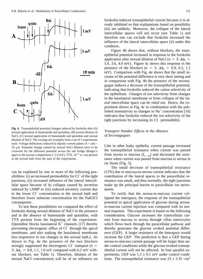
To test these possibilities we compared the effect offorskolin during serosal dilution of NaCl in the presenceand in the absence of bumetanide and quinidine, withTTX present from the beginning of the experiment.(Quinidine blocks basolateral K+ channels [17] therebypreventing electrogenic efflux of Cl− through the apicalmembrane, and also making the basolateral membraneless responsive to ion changes in the serosal bath.). Asshown in Fig. 4a the presence of the two blockersstrongly suppressed the electrogenic Cl− transport (n 43: Dct 4 0.8, 1.1, 1.0 mV compared with 6.2 mV with-out blockers, see Table 1). Therefore, dilution of theserosal NaCl concentration will be of no influence on
forskolin-induced transepithelial current because it is al-ready inhibited so that explanations based on possibility(iii) are unlikely. Moreover, the collapse of the lateralintercellular spaces will not occur (see Table 1) andtherefore one can exclude that forskolin increased theinfluence of the lateral intercellular space (ii) under thiscondition.
Figure 4b shows that, without blockers, the trans-epithelial potential increased in response to the forskolinapplication after serosal dilution of NaCl (n 4 3: Dct 45.0, 3.6, 4.0 mV). Figure 4c shows this response in thepresence of the blockers (n 4 3: Dct 4 0.8, 0.2, 1.2mV). Comparison with Fig. 4a shows that the small in-crease of the potential difference is very short lasting andin comparison with Fig. 4b the presence of the secreta-gogue induces a decrease of the transepithelial potential,indicating that forskolin reduced the cation selectivity ofthe epithelium. Changes of ion selectivity from changesin the basolateral membrane or from collapse of the lat-eral intercellular space can be ruled out. Hence, the ex-periment shown in Fig. 4c in combination with the pub-lished insensitivity to changes in Na+ concentration [16]indicates that forskolin reduced the ion selectivity of thetight junctions by increasing its Cl− permeability.
Transport Number Effects in the Absenceof Secretagogues
Like in other leaky epithelia, current passage increasedthe transepithelial resistance when current was passedfrom serosa to mucosa (Is→m) and decreased the resis-tance when current was passed from mucosa to serosa inrat ileum (Fig. 5).
The small decrease of transepithelial resistance(15%) due to mucosa-to-serosa current indicates that thecontribution of the lateral spaces to the paracellular re-sistance was small, and thus normally, the tight junctionsmake up the principal barrier to paracellular ion move-ments.
To verify that the serosa-to-mucosa current col-lapsed the interspace, the response of the transepithelialpotential to apical application of glucose during serosa-to-mucosa current injection was compared with its nor-mal response. This experiment is based on the followingconsideration. Glucose increases the transcellular cur-rent from mucosa to serosa through villus enterocyteswhich flows back through the paracellular pathway andthereby generates the glucose evoked potential differ-ence (GEP). A larger resistance of the interspace wouldincrease the GEP. The prediction is that a GEP duringserosa-to-mucosa current passage will be larger than un-der control conditions while the glucose-evoked transep-ithelial current will not be affected. In four parallel ex-periments, GEP was 1.3 ± 0.1 mV under control condi-tions. The transepithelial resistance was 23 ± 3V z cm2
Fig. 4. Transepithelial potential changes induced by forskolin after (A)serosal application of bumetanide and quinidine, (B) serosal dilution ofNaCl, (C) serosal application of bumetanide and quinidine and serosaldilution of NaCl. The tracings are examples from a set of 3 experimentseach. Voltage deflections induced by bipolar current pulses of + and −10 mA. Potential change caused by serosal NaCl dilution have to becorrected for the diffusion potential across the salt bridge (Ringer’sagar) in the serosal compartment (−1.9 mV). TTX, 10−6 M, was presentin the serosal bath from the start of the experiments.
131P.B. Bijlsma et al.: Modulation of Paracellular Conductance

and the glucose evoked current was calculated fromOhm’s law as 56 ± 7mA/cm2. GEP duringIs→m of 150mA was 2.3 ± 0.2 mV, transepithelial resistance 38 ± 2V z cm2 and the glucose induced current 61 ± 7mA/cm2.Thus, the glucose evoked potential was significantlylarger duringIs→m (P < 0.001) while the current was notdifferent, indicating thatIs→m increased the resistance ofthe paracellular shunt pathway.
Effect of Forskolin and Carbachol on TransportNumber Effects
If cAMP can increase the Cl− conductance in tight junc-tions, the difference betweentNa in the tight junctions(0.7, see Materials and Methods) and that in free solution(0.4, see Materials and Methods) would become smallerand thus the changes in the resistance of the paracellularshunt pathway induced by current passages would bedecreased in the presence of forskolin.
This was tested by passing serosa-to-mucosa currentin the absence and presence of forskolin. Experimentswere done in the presence of TTX, bumetanide and Ba2+
(see footnote1). Figure 6 shows the time course of the
change in resistance due to direct current passage fromserosa to mucosa. The serosa-to-mucosa current in theabsence of forskolin led to a resistance increase as shownin Fig. 5. Apparently, the presence of the blockers hadno effect on this phenomenon. The presence of forskolinprevented the increase of the resistance, and applicationof forskolin at t 4 12.5 min after starting the currentpassage, immediately decreased the resistance. Applica-tion of TTX and bumetanide during current passage waswithout effect on the resistance (not shown). The sup-pression by forskolin adds to the evidence for a reductionof the cation selectivity of the tight junctions by cAMP.
The reduction in cation selectivity of the tight junc-tions combined with the cAMP dependent decrease ofthe transepithelial resistance in the presence of bu-metanide (Table 1), indicate that in rat ileum, a rise inintracellular cAMP can increase the Cl− conductance intight junctions.
Figure 7 shows a comparison between the effects offorskolin and carbachol on the change of the resistancedue to serosa-to-mucosa current passage. Carbacholcould not prevent the current-induced increase in trans-1 Direct currents passed through the epithelium, will predominantly
take the paracellular route as this shunt comprises about 95% of theconductance of rat ileum (24). However, in the presence of forskolinthe cellular pathway increases its conductance. Current from serosa tomucosa may induce an influx of K+ through the basolateral membranesand of Cl− through the apical membranes, thus leading to an increase ofcellular KCl and cell volume and presumably, a decrease of the diam-eter of the lateral intercellular space, possibly leading to an increase intransepithelial resistance. This hypothetical effect would be minimized
by blocking the basolateral K+ conductance with Ba2+. In comparingexperiments with and without Ba2+, however, we have not found dif-ferences. The maximal relative resistance under control conditions after100 mA serosa-to-mucosa current injection was 135 ± 5% and withBa2+ 127 ± 5% (n4 8).
Fig. 5. The histogram shows the mean transepithelial resistance as apercentage of the resistance before current injection of 100mA. Pas-sage of the current from mucosa to serosa induced a decrease of theresistance while direct current from serosa to mucosa increased theresistance. (Initial resistance 21 ± 1 ohmz cm2; mean andSE of 17experiments.)
Fig. 6. Time course of the change of the transepithelial resistanceinduced by direct current of 200mA from serosa to mucosa in thepresence of TTX, bumetanide and BaCl2. The lower curve is in thepresence of forskolin, added at 10 min before start of current injection.At the arrow forskolin was added to the other sheets. (mean ±SE of 5experiments).
132 P.B. Bijlsma et al.: Modulation of Paracellular Conductance

epithelial resistance. Thus, it appears that the increase ofthe Cl− conductance in tight junctions is a cAMP-relatedphenomenon.
Effect of cAMP on Ion Fluxes
If cAMP can increase the Cl− permeability of the tightjunctions, one would expect that even in the presence ofbumetanide the fluxes from serosa to mucosa and frommucosa to serosa should increase. Table 2 shows that inthe presence of bumetanide cAMP can still increase theserosa-to-mucosa but also the mucosa-to-serosa flux.Without bumetanide, the addition of cAMP increased theserosa-to-mucosa flux and decreased the mucosa-to-serosa flux.
To test for the possibility that the paracellular so-dium permeability was changed, the serosa-to-mucosaNa+ flux was measured in another set of experiments.The control flux in the presence of bumetanide (19.1 ±1.3 mmol/cm2 z hr, n 4 12) was not different from theflux in the presence of 8-Br-cAMP plus bumetanide(21.1 ± 1.0,mmol/cm2 z hr, n 4 12).
THE EFFECT OF CAMP ON TRANSEPITHELIAL RESISTANCE
VARIES BETWEEN PREPARATIONS
Table 1, row 1 shows that the average effect of forskolinon the transepithelial resistance is an increase. However,when considering the individual experiments, it appearedthat, depending on the initial resistance, the addition offorskolin or 8-Br-AMP either increased or decreased theconductance. (The application of forskolin or 8-Br-cAMP in the presence of bumetanide always increasedthe conductance.) Figure 8 shows the relation betweenthe forskolin and cAMP induced change in transepithe-lial conductance and the initial resistance. The slope of
the regression line of data in the absence of bumetanidediffers significantly from zero, whereas in the presenceof bumetanide the deviation of the slope from zero is notsignificant. This suggests that the cAMP-induced col-lapse of the lateral spaces correlates with the initial re-sistance. We will argue in the discussion that the varia-tion in behavior of the preparations and initial resistancemay be due to differences in the Na+ permeability of thetight junctions. This is corroborated by the observed ionselectivity of the tight junctions as determined from themucosal dilution potentials. Figure 9 shows the relationbetween the dilution potential (mean 7.6 ± 0.3 mV,n 455) and the initial resistance (mean 22.5 ± 0.8 ohmz cm2,
Fig. 7. Comparison of the increase of the relative resistance (% frominitial resistance) induced by serosa-to-mucosa direct current with car-bachol (middle) or forskolin (right). Carbachol could not prevent thetransport number effect. (100mA in control and carbachol experiments,200 mA in the presence of forskolin mean ±SE of 8 experiments).
Table 2. Cl− fluxes and the effects of dBcAMP and bumetanide
J m → smmol/cm2 hr (n)
J s→ mmmol/cm2 z hr
Control 18.0 ± 0.9 (19) 13.9 ± 0.9 (21)DJ(dB-cAMP) −1.8 ± 0.5 (12) 3.1 ± 1.0 (12)DJ(bum + dB-cAMP) 1.5 ± 0.4 (7) 1.5 ± 0.5 (9)
Control fluxes were calculated from samples taken att 4 30 andt 4
40 min after start of experiment. dBcAMP or dBcAMP + bumetanidewere added thereafter. Changes induced by these additions were cal-culated from samples taken att 4 50 andt 4 60 min and comparedto their respective control values.
Fig. 8. Change of the transepithelial conductance (mS/cm2) induced by8-Br-cAMP or forskolin in the absence or presence of bumetanide as afunction of the initial resistance of the intestinal sheet. The slope of theregression lines of data without bumetanide (forskolin, open squares:1.37 ± 0.14, 8-Br-cAMP, open circles: 0.78 ± 0.15) differ significantlyfrom zero (P < 0.001) and significantly from the slope of the regressionline of data with bumetanide (0.14 ± 0.09) which is not different fromzero. Note that in the presence of bumetanide (filled circles) the con-ductance always increases while without bumetanide (open symbols)the chance of an increase is much larger with larger initial resistances.
133P.B. Bijlsma et al.: Modulation of Paracellular Conductance

n 4 55). The results show that low resistances are as-sociated with large dilution potentials i.e., larger cationselectivity and vice versa. From the initial resistance andthe dilution potentials the conductance of the tight junc-tion for Na+ (gNa,tj) and Cl− (gCl,tj) can be estimated.Figure 10 showsgNa,tj andgCl,tj as a function of the initialresistance. Apparently, the variation in transepithelialresistance results primarily from endogenous differencesin Na+ permeability of the tight junctions.
Similarly, analysis of the individual data of transportnumber effects upon serosa-to-mucosa current injection(Fig. 11) also reveals a correlation with the initial resis-tance; the lower resistances (larger cation selectivity)showing the larger transport number effects.
Discussion
THE MODULATION OF THE Cl− CONDUCTANCE OF THE
TIGHT JUNCTION
The principal findings in this study are that cAMP in ratileum reduced the transepithelial potential evoked by se-rosal dilution of NaCl and prevented the increase of theresistance induced by passage of direct current from se-rosa to mucosa. Both findings can be interpreted as evi-dence that cAMP decreased the cation selectivity of thetight junctions. This interpretation is based on the as-sumption that (i) the properties of the leak pathway pre-dominate in the serosal dilution potential and that (ii) theincrease of the transepithelial resistance by serosa-to-mucosa current is due to the collapse of the lateral in-
tercellular space by the transport number effect. Underthe conditions that were used to measure the serosal di-lution potential, i.e., presence of K+ channel blocker plusbumetanide to prevent uptake of Cl− and thereby thetranscellular Cl− transport, the first assumption seemsvalid. The second assumption was confirmed by thelarger GEP during serosa to mucosa current passage.The observed increase of the GEP indicate that a path-
Fig. 9. Relation between the change of diffusion potential induced byreplacing 59 mm NaCl in the mucosal compartment with 118 mM
mannitol (Dilution potential) and the initial resistance of the intestinalsheets. The slope of the line differs signficantly from zero (P < 0.005).
Fig. 10. Conductance of tight junctions (mS/cm2) for Na+ and for Cl−
vs. initial resistance of the epithelium. Slopes of regression lines aresignificantly different (P < 0.001). Cation selectivity decreased withresistance. For estimation of ion conductances see methods.
Fig. 11. Individual data showing the increase of the relative resistancecaused by serosa-to-mucosa current passage. The negative slope of theregression line is significantly different from zero (P < 0.001). Thissuggests that transport number effects are smaller with larger initialresistance of the epithelium. (Data from experiments with 100mAserosa-to-mucosa current injection.)
134 P.B. Bijlsma et al.: Modulation of Paracellular Conductance

way parallel to the villus epithelial cells had increased itsresistance. In a leaky epithelium like rat ileum this in-dicates a resistance increase of the paracellular pathwayby a collapse of the interspaces. This is because the dif-fering effect of serosa-to-mucosa current and the mu-cosa-to-serosa current makes it inconceivable that theresistance increase is in tight junctions. (It should bepointed out here, that in rat ileum, under the conditionsof these experiments, glucose does not increase the trans-epithelial conductance (P.B. Bijlsma,unpublished obser-vations). This increase has been found in other intestinalpreparations with differing experimental conditions [25].Even if glucose can modulate the tight junctions in a waythat could not be detected in our experiments it wouldnot affect the conclusion that during current passagefrom serosa to mucosa the paracellular resistance ishigher.) Therefore, it is concluded that both types ofexperimental conditions, i.e., serosal dilution potentialsand transport number effects, which amplify the visibil-ity of changes in ion selective properties of the tightjunctions, indicate that cAMP can reduce the cation se-lectivity of the tight junctions.
The cation selectivity of the tight junctions may bereduced by a decrease of the Na+ conductance and/or byan increase of the Cl− conductance. From the results ofthe serosa-to-mucosa Na+ flux measurements we con-sider the first possibility less likely because the diffu-sional flux in the presence of bumetanide appears to beunaffected by cAMP. This observation corroborates ear-lier results in rat small intestine [17] where it has beenshown that the diffusional flux of Na+ was not affectedby secretagogues. An increase of the conductance in thetight junction for Cl−, in combination with the decreaseof the cellular resistance because of activation of theapical Cl− channels [1, 30] would lead one to expect tosee a decrease of the transepithelial resistance. How-ever, secretion usually goes together with an increasedtransepithelial resistance. Apparently, this is not becauseof a decrease of the Na+ conductance in the tight junc-tions but is caused by the decrease of the diameter of thelateral intercellular spaces [8, 10, 18, 27]. We proposethat the interspace collapses because of the depletion ofNaCl caused by a faster uptake through the basolateralmembrane than diffusion of NaCl into the interspace, asdiscussed in the next paragraph. By decreasing the up-take of NaCl through the basolateral membrane with bu-metanide the resistance-increase could indeed be re-versed to a decrease.
From the combination of the results it is concludedthat the most plausible explanation for the forskolin-induced decrease in transepithelial resistance, as ob-served in the presence of bumetanide, is an increasedconductance for Cl− in the tight junctions as well as inthe apical membranes.
This correlates with the observation in filter-grown
monolayers of T84 cells, a mammalian intestinal cell line,where it has been found that VIP, which activates ade-nylate cyclase, increased not only the secretory serosa-to-mucosa flux but also the Cl− flux from mucosa toserosa, suggesting an increase of the diffusional, para-cellular pathway in addition to the transcellular Cl− se-cretion [9]. In HT-29cl.19A cells (another model intes-tinal cell line), the conductance of the transcellular path-way can reach its maximum while the transepithelialconductance continues to increase. This suggests themodulation of the paracellular conductance with a largertime constant than the change in conductance of the api-cal membrane [1]. Thus, the cAMP mediated increase ofthe Cl− conductance in tight junctions seems to occur notonly in fish intestine but also in rat ileum and human celllines.
The results demonstrate an interesting differencebetween the action of forskolin and carbachol. Carba-chol is thought to activate multiple signaling effectorsincluding Ca2+ and Protein kinase C [12] leading toan increased conductance of apical Cl− channels inHT29cl.19A cells [2] and an increase of the NaCl secre-tion in rat ileum like with cAMP [17]. However, carba-chol did not change the ion selectivity of the tight junc-tions and did not decrease the transepithelial resistance inthe presence of bumetanide. Thus, the modulation of ionselectivity of the tight junctions appears to be specific forcAMP. This is like in fish intestine, where it has beenfound that carbachol or the increase of cellular Ca2+ byionomycin or activation of PKC by phorbol esters couldnot mimic the effect of cAMP on the tight junctions [3].
VARIATION IN BEHAVIOR OF INDIVIDUAL
TISSUEPREPARATIONS
To explain the correlation between the cAMP-inducedchange in conductance and the initial resistance of thetissue in the absence of bumetanide (Fig. 8), we will firstillustrate how the collapse of the lateral spaces may re-sult from transcellular Cl− secretion. Figure 12 shows asimplified scheme of the ion movements during Cl− se-cretion in an open-circuited preparation. The combinedresult of the NaK2Cl cotransporter, the NaKpump andthe K+ permeability in the basolateral membrane is thenet transport of Cl− ions from the lateral spaces acrossthe basolateral membrane and through the apical mem-brane into the lumen. This is equivalent to a transcellularmucosa-to-serosa direct current. In an open-circuitedepithelium, this current must be compensated by an equalserosa-to-mucosa current, i.e., from the lateral spacesthrough the tight junctions to the lumen. The ion selec-tivity of the tight junctions determines the proportion ofthis current carried by Na+ ions moving from the spacesto the lumen, or by Cl− ions in the opposite direction.In other words, the ion selectivity of the tight junctions
135P.B. Bijlsma et al.: Modulation of Paracellular Conductance

determines the (partial) recycling of Cl− ions, and thusthe required inflow of NaCl from the serosal solution.When the supply of NaCl by diffusion or fluid movementis insufficient, the spaces will collapse. Therefore, thestrength of the secretory transcellular current and theion-selective properties of the tight junctions determineto a large extent whether transcellular Cl− secretion willbe accompanied by a partial collapse of the lateral spacesand thus by a decrease of the transepithelial conductance.In view of this mechanism, the observation shown in Fig.8, namely that application of cAMP to specimen with thelower initial resistances decreased the transepithelialconductance, suggests that the Na+ conductance andhence the cation selectivity of the tight junctions is stilltoo high to allow sufficient Cl− recycling—despite theincrease of the Cl− conductance in tight junctions—andto prevent a collapse of the lateral spaces. Further evi-dence for the role of the Na+ conductance of the tightjunctions in the collapse of the interspace can be drawnfrom Holman et al. [18]. These authors have shown thattheophylline induces a collapse of the intercellular spacein rabbit ileum. However, no collapse was observedwhen the Na+ permeability in the tight junctions wasreduced by 2,4,6-triaminopyrimidine [23].
The variation in dilution potentials (the larger dilu-tion potentials observed in the tissues with the lowerinitial resistance) and the transport number effect (thelarger effect in tissues with the lower initial resistance)likewise indicates that the transport number for Na+ inthe tight junctions is larger in tissues with the lowerinitial resistances.
Thus, the three findings in tissues with lower initialtransepithelial resistances: (i) that they have larger dilu-tion potentials (ii) that they show an increase of the re-sistance when forskolin was applied and (iii) that they
show a larger increase of the resistance upon serosa-to-mucosa current injection, appear to be based on thelarger conductance of the tight junctions for Na+.
The regulation of the ion selectivity of the tight junc-tions may be of physiological relevance to modulate saltand water transport through the paracellular pathway[15]. It may be of relevance to prevent a collapse of thelateral intercellular spaces when under open-circuit con-ditions like during sugar or amino acid uptake or duringsecretory activity in vivo a transepithelial current is flow-ing from serosa to mucosa, predominantly through thecrypts [22] because of their lower paracellular resistance.
In conclusion, the results show evidence that the ionselectivity of tight junctions in rat ileum is under controlof a cAMP-related mechanism that can increase the Cl−
conductance in that structure. In contrast, carbacholcould not increase the Cl− conductance in the tight junc-tions. (In a parallel study [6a] we report that carbachol,but not forskolin, can induce an increased uptake of in-tact protein from the mucosal side and an increasedtransepithelial transport of nonelectrolytes and of intactprotein by, primarily, the paracellular route.) In addi-tion, the variation in Na+ conductance of the tight junc-tions may be evidence for the presence of an independentmechanism for the regulation of the Na+ conductance.
We thank K. Dekker and Dr. W.P. Oosterhuis for their participation inthe flux experiments.
References
1. Bajnath, R.B., Augeron, C., Laboisse, C.L., Bijman, J., de Jonge,H.R., Groot, J.A. 1991. Electrophysiological studies of forskolin-induced changes in ion transport in the human colon carcinoma cellline HT-29 cl.19A—lack of evidence for a cAMP-activated baso-lateral K+ conductance.J. Membrane Biol.122:239–250
2. Bajnath, R.B., Dekker, K., Vaandrager, A.B., De Jonge, H.R.,Groot, J.A. 1992. Biphasic increase of apical Cl− conductance bymuscarinic stimulation of HT− 29cl.19A human colon carcinomacell line—evidence for activation of different Cl− conductances bycarbachol and forskolin.J. Membrane Biol.127:81–94
3. Bakker, R., Dekker, K., De Jonge, H.R., Groot, J.A. 1993. VIP,serotonin, and epinephrine modulate the ion selectivity of tightjunctions of goldfish intestine.Am. J. Physiol.264:R362–R368
4. Bakker, R., Groot, J.A. 1984. cAMP-mediated effects of ouabainand theophylline on paracellular ion selectivity.Am. J. Physiol.246:G213–G217
5. Bakker, R., Groot, J.A. 1989. Further evidence for the regulation ofthe tight junction ion selectivity by cAMP in goldfish intestinalmucosa.J. Membrane Biol.111:25–35
6. Barry, P.H., Diamond, J.M., Wright, E.M. 1971. The mechanismof cation permeation in rabbit gallbladder.J. Membrane Biol.4:358–394
6a. Bijlsma, P.B., Kiliaan, A.J., Scholten, G., Heyman, M., Groot,J.A., Taminiau, J.M. 1996. Carbachol, but not forskolin, increasesmucosal-to-serosal transport of intact protein in rat ileum in vitro.Am. J. Physiol.271:G147–G155
7. Bindslev, N., Tormey, J.M.D., Wright, E.M. 1974. The effects ofelectrical and osmotic gradients on lateral intercellular spaces and
Fig. 12. Schematic representation of the ion movements during Cl−
secretion under open-circuit condition. For clarity NaCl fluxes, dis-cussed in the text are shown in bold.
136 P.B. Bijlsma et al.: Modulation of Paracellular Conductance

membrane conductance in a low resistance epithelium.J. Mem-brane Biol.19:357–380
8. Corbett, C.L., Isaacs, P.E.T., Hawker, P.C., Turnberg, L.A. 1977.Theophylline-induced changes in ion transport and conductance inhuman small intestinal mucosa.Nature267:714–717
9. Dharmsathaphorn, K., Mandel, K.G., Masui, H., McRoberts, J.A.1985. Vasoactive intestinal polypeptide-induced chloride secretionby a colonic epithelial cell line: Direct participation of a basolat-erally localized Na,K,Cl cotransport system.J. Clin. Invest.75:462–471
10. DiBona, D.R., Chen, I.C., Sharp, G.W.G. 1974. A study of inter-cellular spaces in the rabbit jejunum during active volume expan-sion and after treatment with cholera toxin.J. Clin. Invest.53:1300–1307
11. Duffey, M.E., Hainau, S., Ho, S., Bentzel, C. 1981. Regulation ofepithelial tight junction permeability by cAMP.Nature294:451–453
12. Felder, C.C. 1995. Muscarinic acetylcholine receptors: signaltransduction through multiple effectors.FASEB J.9:619–625
13. Frizzell, R.A., Schultz, S.G. 1972. Ionic conductances of extracel-lular shunt pathway in rabbit ileum.J. Gen. Physiol.59:318–346
14. Fromter, E. 1972. The route of passive ion movement through theepithelium of Necturus gallbladder.J. Membrane Biol.8:259–301
15. Groot, J.A., Bakker, R. 1988. NaCl transport in the VertebrateIntestine.In: Advances in Comparative and Environmental physi-ology. R. Greger, editor, pp. 103–152. Springer, Berlin
16. Groot, J.A., Bakker, R., Dekker, K., Oosterhuis, W.P. 1986. Modu-lation of transepithelial ion permeability by neurohumoral agents:comparison of fish and rat intestine.In: Ion Gradient CoupledTransport. F. Alvarado and C.H. van Os, editors. pp. 411–414.Elsevier, Amsterdam
17. Hardcastle, J., Hardcastle, P.T., Noble, J.M. 1984. The involve-ment of calcium in the intestinal response to secretagogues in therat. J. Physiol.355: 465–478
18. Holman, G.D., Naftalin, R.J., Simmons, N.L., Walker, M. 1979.Electrophysiological and electron-microscopical correlations withfluid and electrolyte secretion in rabbit ileum.J. Physiol.290:367–386
19. Kottra, G., Haase, W., Fro¨mter, E. 1993. Tight-junction tightnessof Necturus gall bladder epithelium is not regulated by cAMP orintracellular Ca2+. I. Microscopic and general electrophysiologicalobservations.Pfluegers Arch.-Eur. J. Physiol.425:528–534
20. Krasny, E.J., Frizzell, R.A. 1984. Intestinal ion transport in marineteleosts.In: Chloride Transport Coupling in Biological Membranesand Epithelia. G.A. Gerencser, editor. pp. 205–218. Elsevier Sci-ence Publishers, Amsterdam
21. Madara, J.L., Barenberg, D., Carlson, S. 1986. Effects of cytocha-lasin D on occluding junctions of intestinal absorptive cells: furtherevidence that the cytoskeleton may influence paracellular perme-ability and junctional charge selectivity.J. Cell. Biol.97:125–136
22. Marcial, M.A., Carlson, S.L., Madara, J.L. 1984. Partitioning ofparacellular conductance along the crypt-villus axis: A hypothesisbased on structural analysis with detailed consideration of tightjunction structure function relationships.J. Membrane Biol.80:59–70
23. Moreno, J.H. 1975. Blockage of gallbladder tight junction cation-selective channels by 2,4,6-tiaminopyrimidinium (TAP).J. Gen.Physiol.66:97–115
24. Okada, Y., Irimajiri, A., Inouye, A. 1977. Electrical properties andactive solute transport in rat small intestine. II. Conductive prop-erties of transepithelial routes.J. Membrane Biol.31: 221–232
25. Pappenheimer, J.R. 1987. Physiological regulation of transepithe-lial impedance in the intestinal mucosa of rats and hamsters.J.Membrane Biol.100:137–148
26. Petersen, K.U., Reuss, L. 1983. Cyclic AMP-induced chloride per-meability in the apical membrane of Necturus gallbladder epithe-lium. J. Gen. Physiol.81:705–729
27. Powell, D.W. 1974. Intestinal conductance and permselectivitychanges with theophylline and choleragen.Am. J. Physiol.227:1436–1443
28. Rao, M.C., Nash, M.T., Field, M. 1984. Differing effects of cGMPand cAMP on ion transport across flounder intestine.Am. J. Phys-iol. 246: C167–C171
29. Scheffler, A., Heintze, K., Mussler, K. 1984. Bumetanide as aninhibitor of the PGE1- and theophylline-stimulated electrogenicchloride secretion in rabbit colonin vitro. In: Intestinal Absorptionand Secretion. E. Skadhauge and K. Heintze, editors. pp. 335–342.MTP Press, Lancaster
30. Stewart, C.P., Turnberg, L.A. 1989. A microelectrode study ofresponses to secretagogues by epithelial cells on villus and crypt ofrat small intestine.Am. J. Physiol.257:G334–G343
137P.B. Bijlsma et al.: Modulation of Paracellular Conductance