the chemokine ccl2 mediates the seizure-enhancing … · 2016-03-25 · detection of seizures was...
TRANSCRIPT
Neurobiology of Disease
The Chemokine CCL2 Mediates the Seizure-enhancingEffects of Systemic Inflammation
Chiara Cerri,1,2* Sacha Genovesi,3* Manuela Allegra,1,2 Francesco Pistillo,1 Ursula Puntener,4 Angelo Guglielmotti,5
V. Hugh Perry,4 Yuri Bozzi,1,3† and X Matteo Caleo1†
1Neuroscience Institute, National Research Council, 56124 Pisa, Italy, 2Accademia Nazionale dei Lincei, 00165 Rome, Italy, 3Laboratory of MolecularNeuropathology, Centre for Integrative Biology, University of Trento, 38123 Trento, Italy, 4Centre for Biological Sciences, University of Southampton,Southampton SO16 6YD, United Kingdom, and 5Angelini SpA, S. Palomba-Pomezia, 00040 Rome, Italy
Epilepsy is a chronic disorder characterized by spontaneous recurrent seizures. Brain inflammation is increasingly recognized as acritical factor for seizure precipitation, but the molecular mediators of such proconvulsant effects are only partly understood. Thechemokine CCL2 is one of the most elevated inflammatory mediators in patients with pharmacoresistent epilepsy, but its contribution toseizure generation remains unexplored. Here, we show, for the first time, a crucial role for CCL2 and its receptor CCR2 in seizure control.We imposed a systemic inflammatory challenge via lipopolysaccharide (LPS) administration in mice with mesial temporal lobe epilepsy.We found that LPS dramatically increased seizure frequency and upregulated the expression of many inflammatory proteins, includingCCL2. To test the proconvulsant role of CCL2, we administered systemically either a CCL2 transcription inhibitor (bindarit) or a selectiveantagonist of the CCR2 receptor (RS102895). We found that interference with CCL2 signaling potently suppressed LPS-induced seizures.Intracerebral administration of anti-CCL2 antibodies also abrogated LPS-mediated seizure enhancement in chronically epilepticanimals. Our results reveal that CCL2 is a key mediator in the molecular pathways that link peripheral inflammation with neuronalhyperexcitability.
Key words: systemic inflammation; temporal lobe epilepsy; CCL2; EEG; seizures
IntroductionInflammatory processes within the brain parenchyma are well-known determinants of seizure propensity (Fabene et al., 2008;Friedman and Dingledine, 2011; Pernot et al., 2011; de Vries et
al., 2012; Devinsky et al., 2013; Vezzani et al., 2013). In particular,seizures upregulate inflammatory mediators in animal models,and increased levels of many cytokines/chemokines such asinterleukin-1� (IL-1�) and C-C motif ligand 2 (CCL2; alsoknown as monocyte chemoattractant protein-1, MCP-1) can bedetected in brain tissue of patients with intractable epilepsy(Aronica and Gorter, 2007; Choi et al., 2009). Importantly, braininflammatory pathways play a key role in recurrence and precip-
Author contributions: C.C., S.G., A.G., V.H.P., Y.B., and M.C. designed research; C.C., S.G., M.A., F.P., and U.P.performed research; C.C., S.G., M.A., F.P., and U.P. analyzed data; C.C., S.G., V.H.P., Y.B., and M.C. wrote the paper.
This work was supported by Italian Ministry of Health Grant RF-TAA-2008-1141282 to Y.B. and M.C. andFondazione Pisa Grant 158/2011 to M.C. C.C. and M.A. are supported by fellowships from Accademia dei Lincei(Rome, Italy). F.P. was supported by a postdoctoral fellowship from Fondazione Veronesi (Milan, Italy). Wethank Valentina Adami (High-Throughput Screening Facility, Centre for Integrative Biology, University ofTrento, Trento, Italy) and Tarcisio Fedrizzi (Bioinformatics Facility, Centre for Integrative Biology, Universityof Trento, Trento, Italy) for help with microarray experiments; and Francesca Biondi (National ResearchCouncil, Pisa, Italy) for excellent animal care.
A.G. is an Angelini employee. The remaining authors declare no competing financial interests.*C.C. and S.G. contributed equally as first authors.†Y.B. and M.C. contributed equally as senior authors.
Correspondence should be addressed to either of the following: Dr. Matteo Caleo, Neuroscience Institute, Na-tional Research Council, via G. Moruzzi 1, 56124 Pisa, Italy, E-mail: [email protected]; or Dr. Yuri Bozzi, Centre forIntegrative Biology, University of Trento, via Sommarive 9, 38123 Trento, Italy, E-mail: [email protected].
S. Genovesi’s present address: Armenise/Harvard Laboratory of Cancer Biology and Genetics, Centre for Integra-tive Biology, University of Trento, Italy.
DOI:10.1523/JNEUROSCI.0451-15.2016Copyright © 2016 the authors 0270-6474/16/363777-12$15.00/0
Significance Statement
Substantial evidence points to a role for inflammation in epilepsy, but currently there is little insight as to how inflammatorypathways impact on seizure generation. Here, we examine the molecular mediators linking peripheral inflammation with seizuresusceptibility in mice with mesial temporal lobe epilepsy. We show that a systemic inflammatory challenge via lipopolysaccharideadministration potently enhances seizure frequency and upregulates the expression of the chemokine CCL2. Remarkably, selec-tive pharmacological interference with CCL2 or its receptor CCR2 suppresses lipopolysaccharide-induced seizure enhancement.Thus, CCL2/CCR2 signaling plays a key role in linking systemic inflammation with seizure susceptibility.
The Journal of Neuroscience, March 30, 2016 • 36(13):3777–3788 • 3777
itation of seizures (Vezzani et al., 2000; Maroso et al., 2010). Forexample, IL-1� causes potent proconvulsant effects by mediatingenhanced calcium influx through NMDA receptors (Vezzani etal., 2013).
Peripheral inflammatory stimuli can also impact on seizurepropensity. Clinical and experimental data provide solid evi-dence for a role of systemic infection in triggering or sustainingseizures (Cross, 2012; Marchi et al., 2014). Specifically, systemicinflammation reduces the threshold for pharmacologically in-duced acute seizures in animals (Sayyah et al., 2003; Riazi et al.,2008), and this has been linked to upregulation of proinflamma-tory cytokines (Riazi et al., 2008).
A systemic inflammatory challenge during a critical period inearly development leaves a lasting impact on brain excitabilityand seizure susceptibility later in life (Galic et al., 2008). Periph-eral inflammatory stimuli trigger a local brain inflammatory“mirror” reaction (i.e., cytokine and chemokine production)similar to the response elicited in the periphery (Perry and Hol-mes, 2014). The diseased brain displays an amplified, exaggeratedresponse to a systemic inflammatory challenge, as a result of glialactivation and “priming” (Perry and Holmes, 2014).
We have exploited a systemic inflammatory challenge in ani-mals with chronic mesial temporal lobe epilepsy (MTLE) toidentify novel molecular pathways involved in seizure regulation.Microarray and ELISA analyses indicated a potential role for thechemokine CCL2 in mediating seizure upregulation followingsystemic LPS. Accordingly, functional blocking experimentshighlighted a crucial role for CCL2 in inflammation-inducedseizures.
Materials and MethodsAnimals. Experiments were conducted in accord with the EuropeanCommunity Directive 2010/63/EU and were approved by the ItalianMinistry of Health. Animals were housed in a 12 h light/dark cycle withfood and water available ad libitum. Adult (8 –12 weeks old) C57BL/6Nmale mice were used in all experiments. All efforts were made to mini-mize animal suffering.
Kainic acid (KA) injection and placement of electrodes. Mice were uni-laterally injected with 50 nl of a 20 mM solution of KA in PBS into the leftdorsal hippocampus under Hypnorm/Hypnovel anesthesia (Antonucciet al., 2008, 2009). Stereotaxic injections into the dorsal blade of thedentate gyrus were made at the following coordinates with respect tobregma: anteroposterior �2.0, mediolateral 1.5, 1.7 mm below dura.Mice were then implanted with a bipolar electrode inserted into theinjected hippocampus. The bipolar electrode was formed of two enamel-insulated nichrome wires (120 �m). A ground electrode was placed overthe cerebellum. Electrodes were connected to a multipin socket and se-cured to the skull by acrylic dental cement. In a subset of animals, a guidecannula was glued to the bipolar electrode and positioned on top of durafor anti-CCL2 or control IgG injection.
EEG recordings. EEG recordings were performed in freely moving miceduring the chronic phase of epilepsy. In all animals, recordings beganbetween 15 and 21 d after KA. All mice were recorded for 1 h daily duringbaseline periods. Mice were then intraperitoneally injected with LPS(L5886, Sigma; from Salmonella enterica, serotype abortus equi; 100�g/kg in PBS, n � 7) or saline (n � 7). Additional EEG recording sessions(2 h long) were made 2 and 24 h after treatment. For bindarit experi-ments, after a baseline recording period (4 –5 d), bindarit (100 mg/kg in0.5% methylcellulose [MC]; n � 10 mice) or MC (0.5% in aqueoussolution; n � 11 mice) was daily intraperitoneally injected for 4 d. Thirtyminutes after the third and the fourth injection, EEG activity was re-corded for 30 min. On the fourth day, mice received an intraperitonealinjection of LPS, and EEG recordings were performed between 2 and 4 hafter the systemic challenge. The choice of bindarit dose (100 mg/kg) wasbased on our previous studies, showing that a similar dose was able toreduce CCL2 expression in the brain and suppress neuropathological
signs of experimental autoimmune encephalomyelitis in vivo (Ge et al.,2012).
To interfere with signaling via the CCL2 receptor, we injected system-ically a selective CCR2 antagonist (RS102895; 5 mg/kg, Tocris Biosci-ence; n � 7) or vehicle (4% DMSO in saline; n � 7) as control. Injectionsof RS102895/vehicle were given twice (i.e., immediately after and 1.5 hfollowing LPS delivery in chronically epileptic animals). RS102895 hasbeen previously used as a potent and specific antagonist of CCR2 (Hunget al., 2013; Ren et al., 2015).
In anti-CCL2 experiments, after a baseline recording period (2–3 d),mice were injected with goat anti-CCL2 (1 �l of a 100 �g/ml solution;AF-479-NA, R&D Systems; n � 7) or control goat IgG (AB-108-C, R&DSystems; n � 6) via the implanted microcannula. This blocking antibodyhas been previously shown to efficiently neutralize the biological activityof CCL2 in vitro and in vivo (e.g., Stamatovic et al., 2006; Fujimoto et al.,2009). One hour after injection, mice received an intraperitoneal LPSinjection and EEG recordings were performed between 2 and 4 h after thesystemic challenge.
All recordings were performed between 10:00 A.M. and 6:00 P.M., andcare was taken to record from each animal at the same time of the day.Signals were amplified (10,000-fold), bandpass filtered (0.3–100 Hz),digitized (National Instruments card), and conveyed to a computer forstorage and analysis (Antonucci et al., 2008, 2009; Mainardi et al., 2012).Detection of seizures was performed with custom software written inLabView (National Instruments). The program first identified spikes inthe EEG using a voltage threshold. This voltage threshold was set to 4.5times the SD of the EEG signal (determined in a period devoid of spikeactivity). Spontaneous recurrent seizures (SRSs) were defined as spikeclusters lasting for �4 s, whereas clusters lasting �4 s and isolated spi-kes were considered as interictal events. For each recording session, wedetermined the frequency and duration of SRS and interictal clusters, thenumber of single spikes, and the total time spent in seizures or in inter-ictal activity (calculated by adding together the duration of either ictal orinterictal episodes) (Antonucci et al., 2008, 2009; Mainardi et al., 2012;Vannini et al., 2015). For histological controls, naive and chronicallyepileptic animals were deeply anesthetized and perfused with 4% PFA.Serial coronal sections (40 �m) throughout the dorsal hippocampuswere processed for Nissl staining.
Tissue dissections for molecular analyses. To analyze mRNA expressionchanges by microarray and qRT-PCR, KA-injected hippocampi weredissected from epileptic mice during the chronic phase, 4 and 24 h afterLPS or saline intraperitoneal administration. For CCL2 mRNA in situhybridization and CCL2/CCR2 immunohistochemistry, brains were dis-sected from KA-treated epileptic mice during the chronic phase, 4 h afterLPS administration. For ELISA experiments, hippocampi were dissectedfrom chronically epileptic and naive animals, 4 h after LPS or salineadministration; at the same time, serum was collected by cardiac punc-ture from each animal. For ELISA experiments, two additional groups ofnonepileptic, naive animals received LPS (100 �g/kg in PBS) or salineand tissues were collected 4 h after treatment. In bindarit experiments,epileptic mice received daily intraperitoneal injection of bindarit or MCfor 4 d. On the fourth day, 1 h after bindarit or MC administration, micewere challenged with LPS. Hippocampal tissues and blood were collected4 h after LPS delivery. RS102895 was administered immediately after and1.5 h following LPS delivery in chronically epileptic animals (see above)and hippocampi were collected 4 h after LPS.
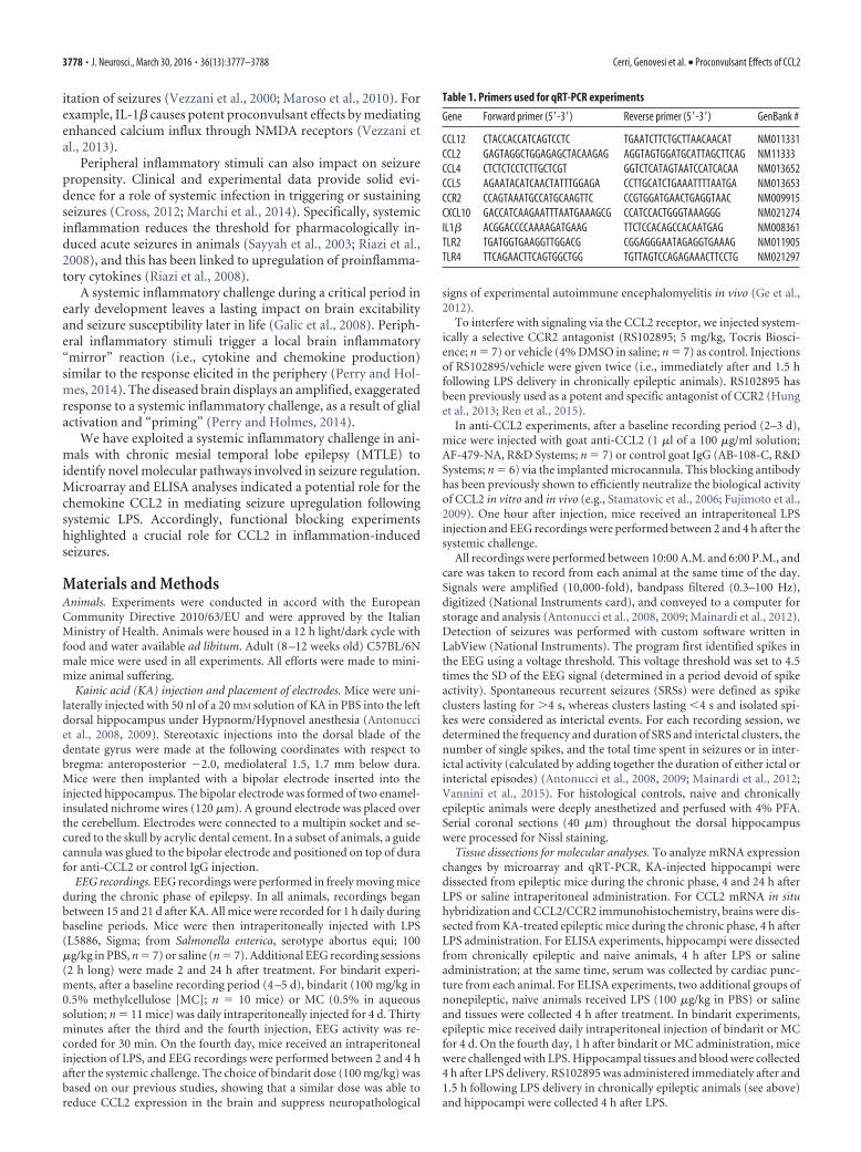
Table 1. Primers used for qRT-PCR experiments
Gene Forward primer (5�-3�) Reverse primer (5�-3�) GenBank #
CCL12 CTACCACCATCAGTCCTC TGAATCTTCTGCTTAACAACAT NM011331CCL2 GAGTAGGCTGGAGAGCTACAAGAG AGGTAGTGGATGCATTAGCTTCAG NM11333CCL4 CTCTCTCCTCTTGCTCGT GGTCTCATAGTAATCCATCACAA NM013652CCL5 AGAATACATCAACTATTTGGAGA CCTTGCATCTGAAATTTTAATGA NM013653CCR2 CCAGTAAATGCCATGCAAGTTC CCGTGGATGAACTGAGGTAAC NM009915CXCL10 GACCATCAAGAATTTAATGAAAGCG CCATCCACTGGGTAAAGGG NM021274IL1� ACGGACCCCAAAAGATGAAG TTCTCCACAGCCACAATGAG NM008361TLR2 TGATGGTGAAGGTTGGACG CGGAGGGAATAGAGGTGAAAG NM011905TLR4 TTCAGAACTTCAGTGGCTGG TGTTAGTCCAGAGAAACTTCCTG NM021297
3778 • J. Neurosci., March 30, 2016 • 36(13):3777–3788 Cerri, Genovesi et al. • Proconvulsant Effects of CCL2

Microarray analysis. RNAs from dissected hippocampi (n � 4 perexperimental group) were purified using standard column purificationaccording to the manufacturer’s protocol (RNAeasy Mini Kit, QIAGEN).RNA quality was analyzed by microfluidic gel electrophoresis on RNA6000 NanoChips using the Agilent 2100 Bioanalyzer. Only RNA with ahigh (�9) RNA integrity number was selected and used for subsequentretrotranscription, labeling, and array hybridization according to Agilentprotocols. Mouse gene expression arrays (Agilent 4 � 44K slides) werehybridized and scanned with the Agilent microarray station. Intensityvalues were processed with GeneSpring GX software (Agilent) using de-
fault parameters to remove low-quality probes (Sgado et al., 2013). Sig-nals were then normalized by means of the quantile normalizationmethod, and data were statistically filtered by two-way ANOVA impos-ing a fold change �2 for each gene in at least one of the four experimentaldata groups. Differentially expressed genes were classified by DAVIDsoftware, applying Benjamini-corrected p value cutoff of 0.05. Gene clas-sification was performed according the Kyoto Encyclopedia of Genesand Genomes (KEGG; http://www.genome.jp/kegg/), Panther Molec-ular Function (http://www.pantherdb.org/panther/ontologies.jsp), andGOterm Molecular Function (http://geneontology.org) databases.
Figure 1. Histopathological changes and EEG seizures in the KA model of MTLE. A, Nissl staining in coronal sections from a naive mouse (top) and from a mouse killed 21 d after intrahippocampalKA (bottom). In the epileptic hippocampus, note the typical granule cell dispersion and extensive neuronal loss in CA1 and CA3. Scale bar, 500 �m. B, Representative EEG recordings displayingactivity in the hippocampus during the chronic phase of epilepsy. Three types of epileptiform events are distinguished: seizures (spike clusters lasting for�4 s; a, red), interictal clusters (spike clusterslasting �4 s; b, green), and isolated spikes (c, blue).
Figure 2. Impact of LPS challenge on the epileptic brain. A, Representative intrahippocampal EEG recordings from chronically epileptic mice, before and 2– 4 h after saline/LPS administration.LPS clearly increases SRS frequency. B, Number of EEG seizures per 10 min of recording in epileptic mice, before and after saline (black circles) or LPS (red circles). Seizure frequency is significantlyenhanced by LPS (two-way repeated-measures ANOVA followed by Holm–Sidak test, p � 0.05). C, Total time spent in ictal activity per 10 min of recording. Epileptic animals treated with LPSsignificantly spent more time in ictal activity than saline-injected mice (two-way repeated-measures ANOVA followed by Holm–Sidak test, p � 0.05). D, Mean duration of EEG seizures in saline- andLPS-treated mice (one way ANOVA; p � 0.7). E, Total time spent in interictal activity per 10 min of recording. Epileptic animals treated with LPS (red circles) significantly spent more time in interictalactivity than saline-injected mice (black circle) 2– 4 h following the systemic challenge (two-way repeated-measures ANOVA followed by Holm–Sidak test, p � 0.05). F, Number of isolated spikesper 10 min of recordings. No difference was found between LPS and saline-injected epileptic mice (two-way repeated-measures ANOVA followed by Holm–Sidak test, p � 0.5). Data are � SEM.*p � 0.05.
Cerri, Genovesi et al. • Proconvulsant Effects of CCL2 J. Neurosci., March 30, 2016 • 36(13):3777–3788 • 3779
qRT-PCR. Total RNAs were extracted by Trizol reagent (Invitrogen)from explanted hippocampi (n � 4 mice per experimental group).DNase-treated RNAs were purified by RNA extraction RNAeasy Kit(QIAGEN). cDNA was synthesized from pooled RNAs by SuperScriptVILO cDNA Synthesis Kit (Invitrogen) according to the manufacturer’instructions. qRT-PCR was performed in a C1000 Thermal Cycler(Bio-Rad) with real-time detection of fluorescence, using the KAPASYBR FAST Master Mix reagent (KAPA Biosystems). Mouse mitochon-drial ribosomal protein L41 (mRPL41) was used as a standard for quan-
tification. Primer sequences (Sigma Genosys) are reported in Table 1.Ratios of comparative concentrations of each mRNA with respect to L41mRNA were then calculated and plotted as the average of three indepen-dent reactions (technical replicates) obtained from each RNA. Expres-sion analyses were performed using the CFX3 Manager (Bio-Rad)software (Sgado et al., 2013).
ELISA. Hippocampi (n � 4 – 6 mice per experimental group) werehomogenized in a Tris buffer containing a protease inhibitor mix-ture (150 mM NaCl, 25 mM Tris, 1% Triton X-100 pH 7.4, complete
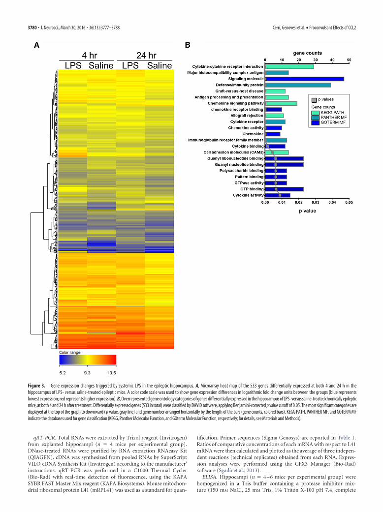
Figure 3. Gene expression changes triggered by systemic LPS in the epileptic hippocampus. A, Microarray heat map of the 533 genes differentially expressed at both 4 and 24 h in thehippocampus of LPS- versus saline-treated epileptic mice. A color code scale was used to show gene expression differences in logarithmic fold change units between the groups (blue representslowest expression; red represents higher expression). B, Overrepresented gene ontology categories of genes differentially expressed in the hippocampus of LPS- versus saline-treated chronically epilepticmice, at both 4 and 24 h after treatment. Differentially expressed genes (533 in total) were classified by DAVID software, applying Benjamini-corrected p value cutoff of 0.05. The most significant categories aredisplayed at the top of the graph to downward ( p value, gray line) and gene number arranged horizontally by the length of the bars (gene counts, colored bars). KEGG PATH, PANTHER MF, and GOTERM MFindicate the databases used for gene classification (KEGG, Panther Molecular Function, and GOterm Molecular Function, respectively; for details, see Materials and Methods).
3780 • J. Neurosci., March 30, 2016 • 36(13):3777–3788 Cerri, Genovesi et al. • Proconvulsant Effects of CCL2
protease inhibitor mixture; Roche Diagnostics). Samples were centri-fuged for 30 min at 15,000 � g, and supernatants assayed for total proteincontent using a Pierce BCA protein assay kit (Thermo Fisher Scientific).Cytokine levels in tissue and serum samples (n � 4 – 6 mice per experi-mental group, each sample in triplicate) were assessed using MSD mul-tiplex kit for mouse proinflammatory cytokines (K15012B; Meso ScaleDiscovery). CCL2 protein quantitation in blood and hippocampal ex-tracts was performed using CCL2 ready-set-go ELISA kit (eBioscience),according to the manufacturer’s instructions.
CCL2 mRNA in situ hybridization. Brains from 3 chronically epilepticmice were rapidly removed and frozen on dry ice, 4 h after LPS challenge.Coronal cryostat sections (20 �m thick) were fixed in 4% PFA. Nonra-dioactive in situ hybridization was performed as previously described(Tripathi et al., 2009) using a mix containing a CCL2 (GenBank ID: NM011333.1) digoxigenin-labeled riboprobe. Signal was detected by alkalinephosphatase-conjugated anti-digoxigenin antibody followed by alkalinephosphatase staining. The specificity of the results was confirmed bythe use of sense riboprobes (data not shown). Images were acquired usinga Zeiss Axio Observer z1 microscope.
Immunostaining. Immunohistochemical characterization of CCL2and CCR2 protein expression was performed on brains from 3 chroni-cally epileptic mice treated with LPS. Brains were fixed by transcardialperfusion with 4% PFA followed by 1 h postfixation at 4°C, and coronalsections (40 �m thick) were cut on a vibratome. Serial sections at thelevel of the dorsal hippocampus were incubated overnight with appro-priate antibodies (all from Abcam: CCL2, 1:200 dilution; CCR2, 1:200dilution; CD68, 1:400 dilution). Signals were revealed with biotin-
conjugated secondary antibody and streptavidin conjugated to appropri-ate fluorophores (AlexaFluor-488/-594, Invitrogen). Images wereacquired using a Zeiss Axio Observer z1 microscope.
Immunoblotting. To assess NF-kB phosphorylation, hippocampal pro-teins from LPS (n � 4) and LPSRS (n � 6) treated mice were extractedwith lysis buffer (1% Triton X-100, 10% glycerol, 20 mM Tris HCl, pH 7.5,150 mM NaCl, 10 mM EDTA, 1 �g/ml leupeptin, 1 mg/ml aprotinin, 1 mM
PMSF, PhosSTOP Roche phosphatase inhibitors mixture). Protein concen-tration was determined through BCA protein assay kit (Euroclone). Equalamounts of each sample (30 �g) were separated on 4%–12% precast gels(Invitrogen), transferred to nitrocellulose membranes, blocked, and thenincubated with primary antibody overnight at 4°C (p65, 1:1000, Cell Signal-ing Technology; phospho p65 S536, 1:1000, Cell Signaling Technology; andactin 1:8000, Sigma-Aldrich). Secondary antibodies (anti-rabbit Ly-CorIRDye800RD; anti-mouse Ly-Cor IRDye680RD) were used at 1:10,000 di-lution for 1 h at room temperature.
The membranes were dried overnight in the dark at room temperatureand the signal was measured using an Odyssey CLx-Infrared ImagingSystem. The signal intensity of immunoblotting bands was quantifiedusing iStudio software. Data are mean � SEM.
Statistical analyses. For EEG experiments, statistical analysis was per-formed by two-way repeated-measures ANOVA followed by Holm–Sidak test for post hoc comparisons. Two-way ANOVA followed byTukey test was used for comparing gene expression at 4/24 h followingLPS/saline treatment. One-way ANOVA (followed by Dunn’s or Holm–Sidak test) was used for comparing mean seizure duration and cytokine/chemokine concentrations in blood and hippocampi. Differences
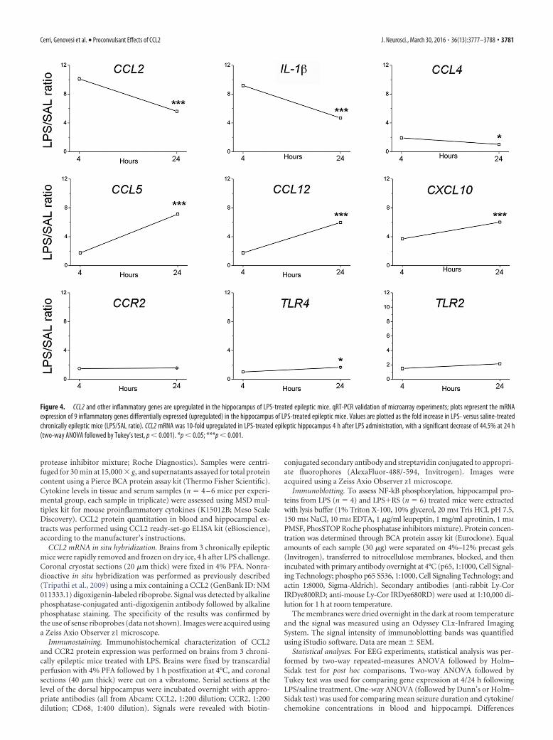
Figure 4. CCL2 and other inflammatory genes are upregulated in the hippocampus of LPS-treated epileptic mice. qRT-PCR validation of microarray experiments; plots represent the mRNAexpression of 9 inflammatory genes differentially expressed (upregulated) in the hippocampus of LPS-treated epileptic mice. Values are plotted as the fold increase in LPS- versus saline-treatedchronically epileptic mice (LPS/SAL ratio). CCL2 mRNA was 10-fold upregulated in LPS-treated epileptic hippocampus 4 h after LPS administration, with a significant decrease of 44.5% at 24 h(two-way ANOVA followed by Tukey’s test, p � 0.001). *p � 0.05; ***p � 0.001.
Cerri, Genovesi et al. • Proconvulsant Effects of CCL2 J. Neurosci., March 30, 2016 • 36(13):3777–3788 • 3781
between two groups were assessed with Student’s t test because our datawere normally distributed. For microarray data analysis, DAVID (http://david.abcc.ncifcrf.gov) was used to assess for functional categories over-represented in the differentially expressed 533 common genes between 4and 24 h dataset. Pairwise comparisons of qRT-PCR and immunoblot-ting data were assessed by a two-tailed Student’s t test. Level of signifi-cance was set at p � 0.05.
ResultsSystemic inflammation enhances spontaneous seizuresMTLE was induced by unilateral injection of KA into the adultmouse hippocampus (Riban et al., 2002; Antonucci et al., 2008).All experiments were conducted during the chronic phase of ep-ilepsy (i.e., starting from 2 weeks after KA), when the injected sidepresents typical hippocampal sclerosis (Fig. 1A), and EEG re-cordings show frequent SRSs, interictal clusters, and isolatedspikes (Fig. 1B).
We first examined the impact of a systemic inflammatory chal-lenge on seizure activity. Baseline SRS were recorded for 4–5 d be-fore intraperitoneal treatment with LPS (100 �g/kg) or saline (SAL)as control. EEG recordings were performed 2–4 h after systemicchallenge (i.e., when the behavioral response to LPS is highest, Teel-ing et al., 2007) and at 24 h. We found that injection of saline had noeffect on SRS at either time point, whereas injection of LPS clearlyincreased seizure incidence within 4 h (two-way repeated-measuresANOVA followed by Holm–Sidak test, p � 0.05; Fig. 2A,B). Totaltime spent in ictal and interictal activity was also significantly in-
creased by LPS (p � 0.05; Fig. 2C,E). Interestingly, the inflammatorychallenge did not impact on mean seizure duration and frequency ofisolated spikes (one way ANOVA, p � 0.6; Fig. 2D,F). LPS effectswere transient, as mice recorded 24 h after treatment showed a re-turn to the baseline, pre-LPS level of seizures and interictal dis-charges (post-ANOVA Holm–Sidak test, p � 0.05; Fig. 2B,C,E).
CCL2 upregulation in the epileptic hippocampusfollowing LPSWe then performed a microarray analysis to investigate the geneexpression pathways activated by LPS in KA-injected hip-pocampi. To this purpose, we compared the transcriptomicprofile of chronically epileptic hippocampi in LPS- versus saline-treated mice. The expression profile of epileptic hippocampimarkedly differed between LPS- and saline-treated animals, atboth 4 and 24 h after treatment (Fig. 3A). A total of 533 geneswere differentially expressed in epileptic hippocampi at both 4and 24 h after LPS, compared with saline-treated animals; path-way analysis revealed that most of these differentially expressedgenes belong to chemokine/cytokine pathways (Fig. 3B). We nextused qRT-PCR to validate nine of these differentially expressedgenes. Compared with saline-treated epileptic mice, CCL2 andIL-1� mRNA were 10-fold and 8-fold, respectively, upregulatedin LPS-treated epileptic mice 4 h after LPS administration, signif-icantly decreasing at 24 h (two-way ANOVA followed by Tukey’stest, p � 0.0001; Fig. 4). CCL4 mRNA was only 1.9-fold upregu-
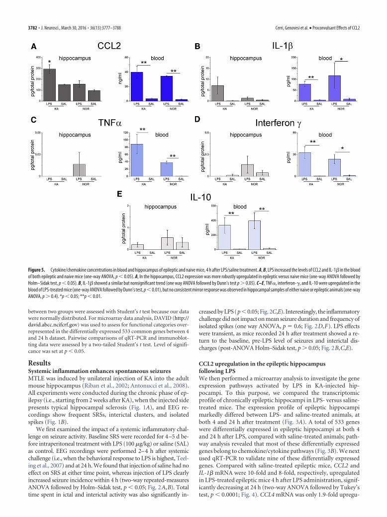
Figure 5. Cytokine/chemokine concentrations in blood and hippocampus of epileptic and naive mice, 4 h after LPS/saline treatment. A, B, LPS increased the levels of CCL2 and IL-1� in the bloodof both epileptic and naive mice (one-way ANOVA, p � 0.05). A, In the hippocampus, CCL2 expression was more robustly upregulated in epileptic versus naive mice (one-way ANOVA followed byHolm–Sidak test, p � 0.05). B, IL-1� showed a similar but nonsignificant trend (one way ANOVA followed by Dunn’s test p � 0.05). C–E, TNF�, interferon-�, and IL-10 were upregulated in theblood of LPS-treated mice (one-way ANOVA followed by Dunn’s test, p�0.01), but no consistent mirror response was observed in hippocampal samples of either naive or epileptic animals (one-wayANOVA, p � 0.4). *p � 0.05; **p � 0.01.
3782 • J. Neurosci., March 30, 2016 • 36(13):3777–3788 Cerri, Genovesi et al. • Proconvulsant Effects of CCL2

lated in LPS-treated mice at 4 h and returned to basal levels 24 hafter LPS administration (two-way ANOVA followed by Tukey’stest, p � 0.02). The chemokines CCL5, CCL12, and CXCL10showed a stronger upregulation at 24 h with respect to 4 h (two-way ANOVA followed by Tukey’s test, p � 0.0001). The CCL2receptor CCR2 as well as the Toll-like receptors TLR4 and TLR2showed modest changes in LPS-treated mice (Fig. 4). Together,the strong increase of CCL2 and IL-1� expression at the time ofLPS-induced seizure aggravation (4 h) points to a possible in-volvement of these molecules in mediating LPS effects.
We then analyzed CCL2 and IL-1� protein levels 4 h followingLPS challenge (Fig. 5A,B) using ELISA. LPS increased CCL2 andIL-1� levels in the blood of both epileptic and naive mice (Fig.
5A,B). Analysis of hippocampal samples revealed a strong “prim-ing” effect, with CCL2 expression more robustly upregulated inthe hippocampus of epileptic versus naive mice (one-wayANOVA followed by Holm–Sidak test, p � 0.05; Fig. 5A). Similarresults were obtained for IL-1� in the blood, whereas IL-1� levelsdid not differ significantly in the hippocampus of epileptic ornaive mice treated with LPS (Fig. 5B). Multiplex ELISA revealed arobust upregulation of TNF�, interferon-�, and IL-10 in theblood of LPS-treated mice, but increases in the brain were ex-tremely modest and independent of the epileptic phenotype (Fig.5C–E). These data indicate selectivity in the “mirror” inflamma-tory cascades triggered by a systemic challenge within the epilep-tic brain, and a potential role of CCL2 in translating the CNSeffects of systemic inflammation.
We next examined the cell populations expressing CCL2 andits receptor CCR2 in the epileptic hippocampus. CCL2 mRNA insitu hybridization and immunostaining indicated expression ofCCL2 in pyramidal neurons and dispersed granule cells of thedentate gyrus following LPS challenge (Fig. 6A–C). Several small,likely glial cells of stratum oriens, stratum radiatum, stratumlacunosum moleculare, and hilus were also labeled (Fig. 6A,B).Immunohistochemical localization of CCR2 was found predom-inantly in activated microglia/macrophages within the sclerotictissue (Fig. 6D,E), consistent with a role for CCR2 in microgliaand monocyte recruitment (Shi and Pamer, 2011; Cunningham,2013).
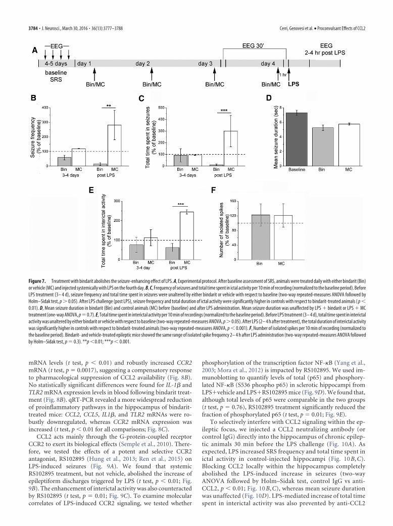
Interference with CCL2/CCR2 signaling abrogates LPS-induced seizure enhancementWe next asked whether interfering with CCL2 would prevent theenhancement of chronic seizures following systemic LPS treat-ment. We first used the anti-inflammatory drug bindarit thatpreferentially inhibits transcription of the monocyte chemoat-tractant subfamily of CC chemokines, including CCL2 (Mirolo etal., 2008; Ge et al., 2012; Mora et al., 2012). We recorded baselineEEG seizure activity; then epileptic animals received daily intra-peritoneal injections of bindarit (100 mg/kg) (Ge et al., 2012) orvehicle solution (MC) for 4 d. EEG recording sessions were per-formed 30 min after the third and the fourth daily injection. Atthe end of the recording session on the fourth day, mice werechallenged with LPS and recorded 2– 4 h later (Fig. 7A). Record-ings performed before LPS challenge showed that seizure fre-quency and total time spent in seizures were unaltered by eitherbindarit or vehicle with respect to baseline (two-way repeated-measures ANOVA followed by Holm–Sidak test, p � 0.05; Fig.7B,C). Importantly, LPS triggered the expected seizure upregu-lation in controls but failed to do so in animals treated withbindarit (two-way repeated-measures ANOVA, bindarit vs con-trol, 2– 4 h after LPS, p � 0.01; Fig. 7B,C). Mean seizure durationfollowing LPS administration was unaffected by either bindaritor vehicle treatment (one-way ANOVA, p � 0.3; Fig. 7D). TheLPS-induced increase in interictal discharges was also abolishedby bindarit (Fig. 7E), whereas the number of isolated interictalspikes did not vary between bindarit- and vehicle-treated epilep-tic mice (Fig. 7F). Thus, treatment with bindarit abolishes theseizure-enhancing effect of systemic inflammation.
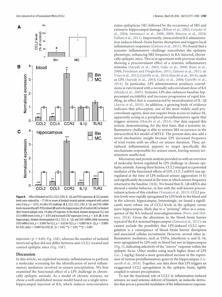
To probe the effects of bindarit at the molecular level, wecollected epileptic hippocampi and blood samples from bindarit-and vehicle-treated mice, 4 h after LPS delivery. ELISA showedthat CCL2 protein was reduced by 77.4% in blood of bindarit-treated animals, compared with control mice (t test, p � 0.01; Fig.8A). qRT-PCR confirmed this reduction at the mRNA level (p �0.001; Fig. 8B). In blood samples, bindarit also dampened CCL5
Figure 6. Cellular localization of CCL2 and CCR2 in the sclerotic hippocampus after LPS. A–C,CCL2 mRNA (A, B) and protein (C) staining on representative sections from the dorsal hippocam-pus of an epileptic mouse, 4 h after LPS. CCL2 staining is present in pyramidal neurons, dispersedgranule cells of the dentate gyrus (A, B, arrowhead) and several small, likely glial cells of stratumoriens, stratum radiatum (A, B, arrows), stratum lacunosum moleculare, and hilus. Details oflabeled cells are shown in B. Left, CA1 stratum radiatum. Right, Dentate granule cells. Scalebars: A, 200 �m; B, 40 �m; C, 600 �m. D, Immunostainings of CCR2 on representative sectionsfrom the epileptic (left) and contralateral (right) hippocampus, 4 h after LPS. CCR2 stainingis mainly restricted to the CA1 subfield and dentate gyrus of the epileptic hippocampus and isabsent from the contralateral side. Scale bar, 600 �m. E, Immunostainings of CCR2 (green),CD68 (red), and their colocalization (yellow) on a representative section from the CA1 subfield ofan epileptic mouse, 4 h after LPS. Expression of CCR2 in activated microglia/macrophages, asindicated by CCR2/CD68 colocalization (yellow), is evident in high-magnification figures shownin insets. Scale bar: 150 �m; insets, 30 �m.
Cerri, Genovesi et al. • Proconvulsant Effects of CCL2 J. Neurosci., March 30, 2016 • 36(13):3777–3788 • 3783
mRNA levels (t test, p � 0.01) and robustly increased CCR2mRNA (t test, p � 0.0017), suggesting a compensatory responseto pharmacological suppression of CCL2 availability (Fig. 8B).No statistically significant differences were found for IL-1� andTLR2 mRNA expression levels in blood following bindarit treat-ment (Fig. 8B). qRT-PCR revealed a more widespread reductionof proinflammatory pathways in the hippocampus of bindarit-treated mice: CCL2, CCL5, IL1�, and TLR2 mRNAs were ro-bustly downregulated, whereas CCR2 mRNA expression wasincreased (t test, p � 0.01 for all comparisons; Fig. 8C).
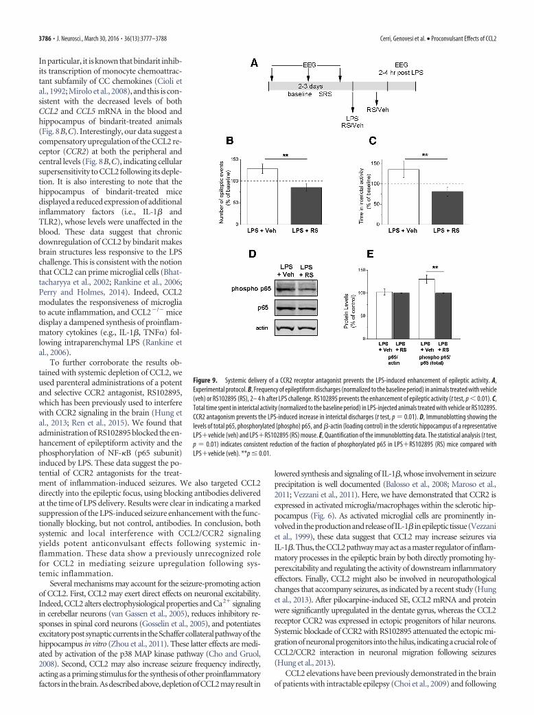
CCL2 acts mainly through the G-protein-coupled receptorCCR2 to exert its biological effects (Semple et al., 2010). There-fore, we tested the effects of a potent and selective CCR2antagonist, RS102895 (Hung et al., 2013; Ren et al., 2015) onLPS-induced seizures (Fig. 9A). We found that systemicRS102895 treatment, but not vehicle, abolished the increase ofepileptiform discharges triggered by LPS (t test, p � 0.01; Fig.9B). The enhancement of interictal activity was also counteractedby RS102895 (t test, p � 0.01; Fig. 9C). To examine molecularcorrelates of LPS-induced CCR2 signaling, we tested whether
phosphorylation of the transcription factor NF-�B (Yang et al.,2003; Mora et al., 2012) is impacted by RS102895. We used im-munoblotting to quantify levels of total (p65) and phosphory-lated NF-�B (S536 phospho p65) in sclerotic hippocampi fromLPSvehicle and LPSRS102895 mice (Fig. 9D). We found that,although total levels of p65 were comparable in the two groups(t test, p � 0.76), RS102895 treatment significantly reduced thefraction of phosphorylated p65 (t test, p � 0.01; Fig. 9E).
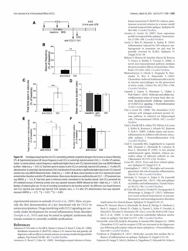
To selectively interfere with CCL2 signaling within the ep-ileptic focus, we injected a CCL2 neutralizing antibody (orcontrol IgG) directly into the hippocampus of chronic epilep-tic animals 30 min before the LPS challenge (Fig. 10A). Asexpected, LPS increased SRS frequency and total time spent inictal activity in control-injected hippocampi (Fig. 10 B, C).Blocking CCL2 locally within the hippocampus completelyabolished the LPS-induced increase in seizures (two-wayANOVA followed by Holm–Sidak test, control IgG vs anti-CCL2, p � 0.01; Fig. 10 B, C), whereas mean seizure durationwas unaffected (Fig. 10D). LPS-mediated increase of total timespent in interictal activity was also prevented by anti-CCL2
Figure 7. Treatment with bindarit abolishes the seizure-enhancing effect of LPS. A, Experimental protocol. After baseline assessment of SRS, animals were treated daily with either bindarit (Bin)or vehicle (MC) and injected systemically with LPS on the fourth day. B, C, Frequency of seizures and total time spent in ictal activity per 10 min of recording (normalized to the baseline period). BeforeLPS treatment (3– 4 d), seizure frequency and total time spent in seizures were unaltered by either bindarit or vehicle with respect to baseline (two-way repeated-measures ANOVA followed byHolm–Sidak test, p � 0.05). After LPS challenge (post LPS), seizure frequency and total duration of ictal activity were significantly higher in controls with respect to bindarit-treated animals ( p �0.01). D, Mean seizure duration in bindarit (Bin) and control animals (MC) before (baseline) and after LPS administration. Mean seizure duration was unaffected by LPS bindarit or LPS MCtreatment (one-way ANOVA, p�0.7). E, Total time spent in interictal activity per 10 min of recordings (normalized to the baseline period). Before LPS treatment (3– 4 d), total time spent in interictalactivity was unaltered by either bindarit or vehicle with respect to baseline (two-way repeated-measures ANOVA, p � 0.05). After LPS (2– 4 h after treatment), the total duration of interictal activitywas significantly higher in controls with respect to bindarit-treated animals (two-way repeated-measures ANOVA, p � 0.001). F, Number of isolated spikes per 10 min of recording (normalized tothe baseline period). Bindarit- and vehicle-treated epileptic mice showed the same range of isolated spike frequency 2– 4 h after LPS administration (two-way repeated-measures ANOVA followedby Holm–Sidak test, p � 0.3). **p �0.01; ***p � 0.001.
3784 • J. Neurosci., March 30, 2016 • 36(13):3777–3788 Cerri, Genovesi et al. • Proconvulsant Effects of CCL2
injection ( p � 0.05; Fig. 10E), whereas the number of isolatedinterictal spikes did not differ between anti-CCL2-treated andcontrol epileptic mice (Fig. 10F ).
DiscussionIn this article, we exploited systemic inflammation to performa molecular screening for the identification of novel inflam-matory mediators involved in seizure precipitation. We firstexamined the functional effect of a LPS challenge in chroni-cally epileptic animals. As a model of chronic seizures, wechose a well-established mouse model based on a single intra-hippocampal injection of KA, which induces nonconvulsive
status epilepticus (SE) followed by the occurrence of SRS andextensive hippocampal damage (Riban et al., 2002; Gouder etal., 2004; Antonucci et al., 2008, 2009; Maroso et al., 2010;Pallud et al., 2011). Importantly, intracerebral KA administra-tion induces blood– brain barrier disruption and triggers localinflammatory responses (Zattoni et al., 2011). We found that asystemic inflammatory challenge exacerbates the epilepticphenotype, enhancing SRS frequency in KA-injected, chroni-cally epileptic mice. This is in agreement with previous studiesshowing a proconvulsant effect of a systemic inflammatorystimulus (Sayyah et al., 2003; Galic et al., 2008; Riazi et al.,2008; Friedman and Dingledine, 2011; Zattoni et al., 2011; deVries et al., 2012; Gyorffy et al., 2014; Marchi et al., 2014), suchas LPS (Sayyah et al., 2003; Galic et al., 2008; Gyorffy et al.,2014). In particular, LPS administration produces convul-sions in rats treated with a normally subconvulsant dose of KA(Heida et al., 2005). Systemic LPS also enhances baseline hip-pocampal excitability and increases progression of rapid kin-dling, an effect that is counteracted by neutralization of IL-1�(Auvin et al., 2010). In addition, a growing body of evidenceindicates that pilocarpine, one of the most widely used pro-convulsant agents, does not require brain access to induce SE,apparently acting as a peripheral proinflammatory agent thattriggers seizures (Marchi et al., 2014). Our data expand thisnotion, demonstrating, for the first time, that a systemic in-flammatory challenge is able to worsen SRS occurrence in theintracerebral KA model of MTLE. The present data also add anovel mechanistic insight because LPS increased frequencyof ictal events with no effect on seizure duration. Thus, pe-ripheral inflammation appears to target specifically themechanisms responsible for seizure onset, leaving seizure ter-mination unaffected.
Microarray and protein analysis provided us with an overviewof molecular factors regulated by LPS challenge in chronic epi-leptic animals. Among these factors, CCL2 emerged as a potentialmediator of the functional effects of LPS. CCL-2 mRNA was up-regulated at the time of LPS-induced seizure aggravation (4 h)and significantly decreased at the time at which seizure frequencyreturned to the baseline (24 h). We found that IL-1� mRNA alsoshowed a similar behavior, in line with the well-known procon-vulsant actions of this cytokine (Vezzani et al., 2013). CCL2 pro-tein levels were rapidly upregulated by LPS both in the blood andin the sclerotic hippocampus. Interestingly, we found a signifi-cantly more robust rise of CCL2 levels in the epileptic versusnaive hippocampus, likely due to a “priming” effect as a conse-quence of the KA-induced neurodegeneration (Perry and Hol-mes, 2014). Given the alterations in the blood– brain barriertypical of the KA-lesioned hippocampus (Zattoni et al., 2011), wecannot exclude the possibility that LPS-induced CCL2 upre-gulation is a consequence of blood– brain barrier disruptionand associated cellular recruitment. However, several other in-flammatory mediators, such as TNF�, interferon-�, and IL-10,were upregulated by LPS only in blood but not in hippocampus(Fig. 5), indicating selectivity of the “mirror” response within theepileptic focus. Other studies using much higher doses of LPS(i.e., 2 mg/kg) found a more generalized increase in the expres-sion of various proinflammatory genes in the hippocampus (Ca-zareth et al., 2014). Together, our data show that LPS triggers anexaggerated CCL2 upregulation in the epileptic brain, tightlycoupled to seizure precipitation.
To test the functional role of CCL2 in inflammation-inducedseizures, we used systemic delivery of bindarit, an indazolic deriva-tive that acts as a powerful modulator of the inflammatory response.
Figure 8. Effect of bindarit on CCL2, CCL5, CCR2, IL-1�, and TLR2 expression. A, CCL2 proteinlevels were reduced by �77.4% in serum of bindarit-treated animals compared with controlmice (t test, p � 0.01), 4 h after LPS challenge. B, C, CCL2, CCL5, CCR2, IL-1�, and TLR2 mRNAlevels measured by qRT-PCR in blood (B) and in the hippocampus (C) of control (MC) or bindarit(Bin) treated epileptic mice, 4 h after LPS injection. In the blood, bindarit dampened CCL2 andCCL5 mRNA levels (t test, p � 0.01) and increased CCR2 expression (t test, p � 0.01; B). In thehippocampus, bindarit downregulated CCL2, CCL5, IL- 1�, and TLR2 mRNAs while increasingCCR2 mRNA (t test, p � 0.0001 for CCL2, p � 0.02 for CCL5, p � 0.0001 for IL-1�, p � 0.0005for TLR2, and p � 0.0001 for CCR2; C). *p � 0.05; **p � 0.01; ***p � 0.001.
Cerri, Genovesi et al. • Proconvulsant Effects of CCL2 J. Neurosci., March 30, 2016 • 36(13):3777–3788 • 3785
In particular, it is known that bindarit inhib-its transcription of monocyte chemoattrac-tant subfamily of CC chemokines (Cioli etal., 1992; Mirolo et al., 2008), and this is con-sistent with the decreased levels of bothCCL2 and CCL5 mRNA in the blood andhippocampus of bindarit-treated animals(Fig. 8B,C). Interestingly, our data suggest acompensatory upregulation of the CCL2 re-ceptor (CCR2) at both the peripheral andcentral levels (Fig. 8B,C), indicating cellularsupersensitivity to CCL2 following its deple-tion. It is also interesting to note that thehippocampus of bindarit-treated micedisplayed a reduced expression of additionalinflammatory factors (i.e., IL-1� andTLR2), whose levels were unaffected in theblood. These data suggest that chronicdownregulation of CCL2 by bindarit makesbrain structures less responsive to the LPSchallenge. This is consistent with the notionthat CCL2 can prime microglial cells (Bhat-tacharyya et al., 2002; Rankine et al., 2006;Perry and Holmes, 2014). Indeed, CCL2modulates the responsiveness of microgliato acute inflammation, and CCL2�/� micedisplay a dampened synthesis of proinflam-matory cytokines (e.g., IL-1�, TNF�) fol-lowing intraparenchymal LPS (Rankine etal., 2006).
To further corroborate the results ob-tained with systemic depletion of CCL2, weused parenteral administrations of a potentand selective CCR2 antagonist, RS102895,which has been previously used to interferewith CCR2 signaling in the brain (Hung etal., 2013; Ren et al., 2015). We found thatadministration of RS102895 blocked the en-hancement of epileptiform activity and thephosphorylation of NF-�B (p65 subunit)induced by LPS. These data suggest the po-tential of CCR2 antagonists for the treat-ment of inflammation-induced seizures. We also targeted CCL2directly into the epileptic focus, using blocking antibodies deliveredat the time of LPS delivery. Results were clear in indicating a markedsuppression of the LPS-induced seizure enhancement with the func-tionally blocking, but not control, antibodies. In conclusion, bothsystemic and local interference with CCL2/CCR2 signalingyields potent anticonvulsant effects following systemic in-flammation. These data show a previously unrecognized rolefor CCL2 in mediating seizure upregulation following sys-temic inflammation.
Several mechanisms may account for the seizure-promoting actionof CCL2. First, CCL2 may exert direct effects on neuronal excitability.Indeed, CCL2 alters electrophysiological properties and Ca2 signalingin cerebellar neurons (van Gassen et al., 2005), reduces inhibitory re-sponses in spinal cord neurons (Gosselin et al., 2005), and potentiatesexcitatorypostsynapticcurrents intheSchaffercollateralpathwayofthehippocampus in vitro (Zhou et al., 2011). These latter effects are medi-ated by activation of the p38 MAP kinase pathway (Cho and Gruol,2008). Second, CCL2 may also increase seizure frequency indirectly,acting as a priming stimulus for the synthesis of other proinflammatoryfactors inthebrain.Asdescribedabove,depletionofCCL2mayresult in
lowered synthesis and signaling of IL-1�, whose involvement in seizureprecipitation is well documented (Balosso et al., 2008; Maroso et al.,2011; Vezzani et al., 2011). Here, we have demonstrated that CCR2 isexpressed in activated microglia/macrophages within the sclerotic hip-pocampus (Fig. 6). As activated microglial cells are prominently in-volvedintheproductionandreleaseofIL-1� inepileptictissue(Vezzaniet al., 1999), these data suggest that CCL2 may increase seizures viaIL-1�.Thus, theCCL2pathwaymayactasamasterregulatorof inflam-matory processes in the epileptic brain by both directly promoting hy-perexcitability and regulating the activity of downstream inflammatoryeffectors. Finally, CCL2 might also be involved in neuropathologicalchanges that accompany seizures, as indicated by a recent study (Hunget al., 2013). After pilocarpine-induced SE, CCL2 mRNA and proteinwere significantly upregulated in the dentate gyrus, whereas the CCL2receptor CCR2 was expressed in ectopic progenitors of hilar neurons.Systemic blockade of CCR2 with RS102895 attenuated the ectopic mi-grationofneuronalprogenitors intothehilus, indicatingacrucialroleofCCL2/CCR2 interaction in neuronal migration following seizures(Hung et al., 2013).
CCL2 elevations have been previously demonstrated in the brainof patients with intractable epilepsy (Choi et al., 2009) and following
Figure 9. Systemic delivery of a CCR2 receptor antagonist prevents the LPS-induced enhancement of epileptic activity. A,Experimental protocol. B, Frequency of epileptiform discharges (normalized to the baseline period) in animals treated with vehicle(veh) or RS102895 (RS), 2– 4 h after LPS challenge. RS102895 prevents the enhancement of epileptic activity (t test, p � 0.01). C,Total time spent in interictal activity (normalized to the baseline period) in LPS-injected animals treated with vehicle or RS102895.CCR2 antagonism prevents the LPS-induced increase in interictal discharges (t test, p � 0.01). D, Immunoblotting showing thelevels of total p65, phosphorylated (phospho) p65, and �-actin (loading control) in the sclerotic hippocampus of a representativeLPSvehicle (veh) and LPSRS102895 (RS) mouse. E, Quantification of the immunoblotting data. The statistical analysis (t test,p � 0.01) indicates consistent reduction of the fraction of phosphorylated p65 in LPSRS102895 (RS) mice compared withLPSvehicle (veh). **p � 0.01.
3786 • J. Neurosci., March 30, 2016 • 36(13):3777–3788 Cerri, Genovesi et al. • Proconvulsant Effects of CCL2
experimental seizures in animals (Foresti et al., 2009). Here, we pro-vide the first demonstration of a key functional role for CCL2 inseizure precipitation. Drugs interfering with CCL2 signaling are cur-rently under development for several inflammatory brain disorders(Semple et al., 2010) and may be tested in epileptic syndromes thatremain resistant to currently available medications.
ReferencesAntonucci F, Di Garbo A, Novelli E, Manno I, Sartucci F, Bozzi Y, Caleo M (2008)
Botulinum neurotoxin E (BoNT/E) reduces CA1 neuron loss and granule celldispersion,withnoeffectsonchronicseizures, inamousemodeloftemporal lobeepilepsy. Exp Neurol 210:388–401. CrossRef Medline
Antonucci F, Bozzi Y, Caleo M (2009) Intrahippocampal infusion of botu-
linum neurotoxin E (BoNT/E) reduces spon-taneous recurrent seizures in a mouse modelof mesial temporal lobe epilepsy. Epilepsia 50:963–966. CrossRef Medline
Aronica E, Gorter JA (2007) Gene expressionprofile in temporal lobe epilepsy. Neuroscien-tist 13:100 –108. CrossRef Medline
Auvin S, Shin D, Mazarati A, Sankar R (2010)Inflammation induced by LPS enhances epi-leptogenesis in immature rat and may bepartially reversed by IL1RA. Epilepsia 51[Suppl 3]:34 –38.
Balosso S, Maroso M, Sanchez-Alavez M, RavizzaT, Frasca A, Bartfai T, Vezzani A (2008) Anovel non-transcriptional pathway mediatesthe proconvulsive effects of interleukin-1beta.Brain 131:3256 –3265. CrossRef Medline
Bhattacharyya S, Ghosh S, Dasgupta B, Maz-umder D, Roy S, Majumdar S (2002)Chemokine-induced leishmanicidal activityin murine macrophages via the generationof nitric oxide. J Infect Dis 185:1704 –1708.CrossRef Medline
Cazareth J, Guyon A, Heurteaux C, Chabry J,Petit-PaitelA (2014) Molecularandcellularneu-roinflammatory status of mouse brain after sys-temic lipopolysaccharide challenge: importanceof CCR2/CCL2 signaling. J Neuroinflammation11:132. CrossRef Medline
Cho J, Gruol DL (2008) The chemokine CCL2activates p38 mitogen-activated protein ki-nase pathway in cultured rat hippocampalcells. J Neuroimmunol 199:94 –103. CrossRefMedline
Choi J, Nordli DR Jr, Alden TD, DiPatri A Jr, LauxL, Kelley K, Rosenow J, Schuele SU, RajaramV, Koh S (2009) Cellular injury and neuro-inflammation in children with chronic intrac-table epilepsy. J Neuroinflammation 6:38.CrossRef Medline
Cioli V, Ciarniello MG, Guglielmotti A, LupariniMR, Durando L, Martinelli B, Catanese B,Fava L, Silvestrini B (1992) A new proteinantidenaturant agent, bindarit, reduces sec-ondary phase of adjuvant arthritis in rats.J Rheumatol 19:1735–1742. Medline
Cross JH (2012) Fever and fever-related epilep-sies. Epilepsia 53 [Suppl 4]:3– 8.
Cunningham C (2013) Microglia and neurode-generation: the role of systemic inflammation.Glia 61:71–90. CrossRef Medline
Devinsky O, Vezzani A, Najjar S, De LanerolleNC, Rogawski MA (2013) Glia and epilepsy:excitability and inflammation. Trends Neuro-sci 36:174 –184. CrossRef Medline
de Vries HE, Kooij G, Frenkel D, Georgopoulos S,Monsonego A, Janigro D (2012) Inflamma-tory events at blood-brain barrier in neuroin-flammatory and neurodegenerative disorders:
implications for clinical disease. Epilepsia 53 [Suppl 6]:45–52.Fabene PF, Navarro Mora G, Martinello M, Rossi B, Merigo F, Ottoboni L,
Bach S, Angiari S, Benati D, Chakir A, Zanetti L, Schio F, Osculati A,Marzola P, Nicolato E, Homeister JW, Xia L, Lowe JB, McEver RP, Oscu-lati F, et al. (2008) A role for leukocyte-endothelial adhesion mecha-nisms in epilepsy. Nat Med 14:1377–1383. CrossRef Medline
Foresti ML, Arisi GM, Katki K, Montanez A, Sanchez RM, Shapiro LA (2009)Chemokine CCL2 and its receptor CCR2 are increased in the hippocam-pus following pilocarpine-induced status epilepticus. J Neuroinflamma-tion 6:40. CrossRef Medline
Friedman A, Dingledine R (2011) Molecular cascades that mediate the in-fluence of inflammation on epilepsy. Epilepsia 52 [Suppl 3]:33–39.
Fujimoto H, Sangai T, Ishii G, Ikehara A, Nagashima T, Miyazaki M, Ochiai A
Figure 10. Intrahippocampal injection of a CCL2 neutralizing antibody completely abrogates the increase in seizures followingLPS. A, Experimental protocol. B, Seizure frequency in anti-CCL2 or control IgG-injected animals (Ctrl), 2– 4 h after LPS adminis-tration. In control animals, seizure frequency is significantly higher than in anti-CCL2 injected animals (two-way ANOVA followedby Holm–Sidak test, p � 0.01). C, Total time spent in seizures in anti-CCL2 or control IgG-injected (Ctrl) animals, 2– 4 h after LPSadministration. In control IgG-injected animals (Ctrl), total duration of ictal activity is significantly higher than in anti-CCL2 injectedanimals (two-way ANOVA followed by Holm–Sidak test, p � 0.001). D, Mean seizure duration in anti-CCL2-injected and controlanimals before (baseline) and after LPS administration. Mean seizure duration was unaffected by anti-CCL2 LPS treatment (oneway ANOVA, p � 0.3). E, Total time spent in interictal activity (normalized to the baseline period). Anti-CCL2 prevented theLPS-mediated increase of interictal activity (two-way repeated-measures ANOVA followed by Holm–Sidak test, p � 0.01). F,Number of isolated spikes per 10 min of recording (normalized to the baseline period). No difference was found betweenanti-CCL2 injected and control IgG-injected (Ctrl) epileptic mice, 2– 4 h after LPS administration (two-way repeated-measures ANOVA, p � 0.7). **p � 0.01; ***p � 0.001.
Cerri, Genovesi et al. • Proconvulsant Effects of CCL2 J. Neurosci., March 30, 2016 • 36(13):3777–3788 • 3787
(2009) Stromal MCP-1 in mammary tumors induces tumor-associatedmacrophage infiltration and contributes to tumor progression. Int J Can-cer 125:1276 –1284. CrossRef Medline
Galic MA, Riazi K, Heida JG, Mouihate A, Fournier NM, Spencer SJ, Kalynchuk LE,Teskey GC, Pittman QJ (2008) Postnatal inflammation increases seizure sus-ceptibility in adult rats. J Neurosci 28:6904–6913. CrossRef Medline
Ge S, Shrestha B, Paul D, Keating C, Cone R, Guglielmotti A, Pachter JS(2012) The CCL2 synthesis inhibitor bindarit targets cells of the neuro-vascular unit, and suppresses experimental autoimmune encephalomy-elitis. J Neuroinflammation 9:171. CrossRef Medline
Gosselin RD, Varela C, Banisadr G, Mechighel P, Rostene W, Kitabgi P,Melik-Parsadaniantz S (2005) Constitutive expression of CCR2 chemo-kine receptor and inhibition by MCP-1/CCL2 of GABA-induced currentsin spinal cord neurones. J Neurochem 95:1023–1034. CrossRef Medline
Gouder N, Scheurer L, Fritschy JM, Boison D (2004) Overexpression ofadenosine kinase in epileptic hippocampus contributes to epileptogen-esis. J Neurosci 24:692–701. CrossRef Medline
Gyorffy B, Kovacs Z, Gulyassy P, Simor A, Volgyi K, Orban G, Baracskay P,Szabo Z, Janaky T, Dobolyi A, Juhasz G, Czurko A, Kekesi KA (2014)Brain protein expression changes in WAG/Rij rats, a genetic rat model ofabsence epilepsy after peripheral lipopolysaccharide treatment. Brain Be-hav Immun 35:86 –95. CrossRef Medline
Heida JG, Teskey GC, Pittman QJ (2005) Febrile convulsions induced by thecombination of lipopolysaccharide and low-dose kainic acid enhance sei-zure susceptibility, not epileptogenesis, in rats. Epilepsia 46:1898 –1905.CrossRef Medline
Hung YW, Lai MT, Tseng YJ, Chou CC, Lin YY (2013) Monocyte chemoat-tractant protein-1 affects migration of hippocampal neural progenitorsfollowing status epilepticus in rats. J Neuroinflammation 10:11. CrossRefMedline
Mainardi M, Pietrasanta M, Vannini E, Rossetto O, Caleo M (2012) Tetanusneurotoxin-induced epilepsy in mouse visual cortex. Epilepsia 53:e132–e136. CrossRef Medline
Marchi N, Granata T, Janigro D (2014) Inflammatory pathways of seizuredisorders. Trends Neurosci 37:55– 65. CrossRef Medline
Maroso M, Balosso S, Ravizza T, Liu J, Aronica E, Iyer AM, Rossetti C, Mol-teni M, Casalgrandi M, Manfredi AA, Bianchi ME, Vezzani A (2010)Toll-like receptor 4 and high-mobility group box-1 are involved in icto-genesis and can be targeted to reduce seizures. Nat Med 16:413– 419.CrossRef Medline
Maroso M, Balosso S, Ravizza T, Iori V, Wright CI, French J, Vezzani A(2011) Interleukin-1beta biosynthesis inhibition reduces acute seizuresand drug resistant chronic epileptic activity in mice. Neurotherapeutics8:304 –315. CrossRef Medline
Mirolo M, Fabbri M, Sironi M, Vecchi A, Guglielmotti A, Mangano G, BiondiG, Locati M, Mantovani A (2008) Impact of the anti-inflammatoryagent bindarit on the chemokinome: selective inhibition of the monocytechemotactic proteins. Eur Cytokine Netw 19:119 –122. CrossRef Medline
Mora E, Guglielmotti A, Biondi G, Sassone-Corsi P (2012) Bindarit: an anti-inflammatory small molecule that modulates the NFkappaB pathway.Cell Cycle 11:159 –169. CrossRef Medline
Pallud J, Haussler U, Langlois M, Hamelin S, Devaux B, Deransart C, Depau-lis A (2011) Dentate gyrus and hilus transection blocks seizure propaga-tion and granule cell dispersion in a mouse model for mesial temporallobe epilepsy. Hippocampus 21:334 –343. CrossRef Medline
Pernot F, Heinrich C, Barbier L, Peinnequin A, Carpentier P, Dhote F, BailleV, Beaup C, Depaulis A, Dorandeu F (2011) Inflammatory changes dur-ing epileptogenesis and spontaneous seizures in a mouse model of mesio-temporal lobe epilepsy. Epilepsia 52:2315–2325. CrossRef Medline
Perry VH, Holmes C (2014) Microglial priming in neurodegenerative dis-ease. Nat Rev Neurol 10:217–224. CrossRef Medline
Rankine EL, Hughes PM, Botham MS, Perry VH, Felton LM (2006) Braincytokine synthesis induced by an intraparenchymal injection of LPS isreduced in MCP-1-deficient mice prior to leucocyte recruitment. EurJ Neurosci 24:77– 86. CrossRef Medline
Ren F, Jiao H, Cai H (2015) Analgesic effect of intrathecal administration ofchemokine receptor CCR2 antagonist is related to change in spinal NR2B,nNOS, and SIGIRR expression in rat with bone cancer pain. Cell Biochem
Biophys. Advance online publication. Retrieved Feb. 5, 2015. doi:10.1007/s12013-014-0510-7. CrossRef Medline
Riazi K, Galic MA, Kuzmiski JB, Ho W, Sharkey KA, Pittman QJ (2008)Microglial activation and TNFalpha production mediate altered CNS ex-citability following peripheral inflammation. Proc Natl Acad Sci U S A105:17151–17156. CrossRef Medline
Riban V, Bouilleret V, Pham-Le BT, Fritschy JM, Marescaux C, Depaulis A(2002) Evolution of hippocampal epileptic activity during the develop-ment of hippocampal sclerosis in a mouse model of temporal lobe epi-lepsy. Neuroscience 112:101–111. CrossRef Medline
Sayyah M, Javad-Pour M, Ghazi-Khansari M (2003) The bacterial endo-toxin lipopolysaccharide enhances seizure susceptibility in mice: involve-ment of proinflammatory factors: nitric oxide and prostaglandins.Neuroscience 122:1073–1080. CrossRef Medline
Semple BD, Kossmann T, Morganti-Kossmann MC (2010) Role of chemokines inCNS health and pathology: a focus on the CCL2/CCR2 and CXCL8/CXCR2networks. J Cereb Blood Flow Metab 30:459–473. CrossRef Medline
Sgado P, Provenzano G, Dassi E, Adami V, Zunino G, Genovesi S, Casarosa S,Bozzi Y (2013) Transcriptome profiling in engrailed-2 mutant mice re-veals common molecular pathways associated with autism spectrum dis-orders. Mol Autism 4:51. CrossRef Medline
Shi C, Pamer EG (2011) Monocyte recruitment during infection and in-flammation. Nat Rev Immunol 11:762–774. CrossRef Medline
Stamatovic SM, Keep RF, Mostarica-Stojkovic M, Andjelkovic AV (2006)CCL2 regulates angiogenesis via activation of Ets-1 transcription factor.J Immunol 177:2651–2661. CrossRef Medline
Teeling JL, Felton LM, Deacon RM, Cunningham C, Rawlins JN, Perry VH (2007)Sub-pyrogenic systemic inflammation impacts on brain and behavior, indepen-dent of cytokines. Brain Behav Immun 21:836–850. CrossRef Medline
Tripathi PP, Sgado P, Scali M, Viaggi C, Casarosa S, Simon HH, Vaglini F,Corsini GU, Bozzi Y (2009) Increased susceptibility to kainic acid-induced seizures in Engrailed-2 knockout mice. Neuroscience 159:842–849. CrossRef Medline
van Gassen KL, Netzeband JG, de Graan PN, Gruol DL (2005) The chemo-kine CCL2 modulates Ca 2 dynamics and electrophysiological propertiesof cultured cerebellar Purkinje neurons. Eur J Neurosci 21:2949 –2957.CrossRef Medline
Vannini E, Restani L, Pietrasanta M, Panarese A, Mazzoni A, Rossetto O,Middei S, Micera S, Caleo M (2015) Altered sensory processing and den-dritic remodeling in hyperexcitable visual cortical networks. Brain StructFunct. Advance online publication. Retrieved Jul. 12, 2015. doi: 10.1007/s00429-015-1080-1. CrossRef Medline
Vezzani A, Conti M, De Luigi A, Ravizza T, Moneta D, Marchesi F, De SimoniMG (1999) Interleukin-1beta immunoreactivity and microglia are en-hanced in the rat hippocampus by focal kainate application: functionalevidence for enhancement of electrographic seizures. J Neurosci 19:5054 –5065. Medline
Vezzani A, Moneta D, Conti M, Richichi C, Ravizza T, De Luigi A, De SimoniMG, Sperk G, Andell-Jonsson S, Lundkvist J, Iverfeldt K, Bartfai T (2000)Powerful anticonvulsant action of IL-1 receptor antagonist on intracere-bral injection and astrocytic overexpression in mice. Proc Natl Acad SciU S A 97:11534 –11539. CrossRef Medline
Vezzani A, Maroso M, Balosso S, Sanchez MA, Bartfai T (2011) IL-1receptor/Toll-like receptor signaling in infection, inflammation, stressand neurodegeneration couples hyperexcitability and seizures. Brain Be-hav Immun 25:1281–1289. CrossRef Medline
Vezzani A, Aronica E, Mazarati A, Pittman QJ (2013) Epilepsy and braininflammation. Exp Neurol 244:11–21. CrossRef Medline
Yang F, Tang E, Guan K, Wang CY (2003) IKK beta plays an essential role inthe phosphorylation of RelA/p65 on serine 536 induced by lipopolysac-charide. J Immunol 170:5630 –5635. CrossRef Medline
Zattoni M, Mura ML, Deprez F, Schwendener RA, Engelhardt B, Frei K,Fritschy JM (2011) Brain infiltration of leukocytes contributes to thepathophysiology of temporal lobe epilepsy. J Neurosci 31:4037– 4050.CrossRef Medline
Zhou Y, Tang H, Liu J, Dong J, Xiong H (2011) Chemokine CCL2 modula-tion of neuronal excitability and synaptic transmission in rat hippocam-pal slices. J Neurochem 116:406 – 414. CrossRef Medline
3788 • J. Neurosci., March 30, 2016 • 36(13):3777–3788 Cerri, Genovesi et al. • Proconvulsant Effects of CCL2