the brain is conscious -...
TRANSCRIPT

C H A P T E R
8
The brain is conscious
O U T L I N E
1.0 Introduction 2121.1 Three global brain states 213
2.0 Waking: purposeful thoughtsand actions 2142.1 The stadium analogy: chattering,
cheering, and chanting 2162.2 How does the chattering brain
do its work? 2212.3 What we expect from
conscious people 2242.4 Waking has conscious and
unconscious threads 224
3.0 Attention 2293.1 Attention selects conscious events 2293.2 The Posner flanker task 2303.3 A model of attention 2313.4 Voluntary attention 2383.5 Synchrony enables attention 238
4.0 Executive control 2394.1 Losing voluntary control 241
5.0 Dreaming 2415.1 Dreams are conscious 242
5.2 Nonrational thinking 2435.3 Working memory in dreams 2435.4 Brainstem oscillations trigger
dreaming 2445.5 Lucid dreaming 245
6.0 Deep sleep: ups and downs 2456.1 The need for sleep 2456.2 Memory replay and consolidation 246
7.0 Exceptional states of mind 2467.1 Epilepsy, drugs, and psychoses 2487.2 Out-of-body experiences 2487.3 Neurofeedback 2497.4 Sensorimotor rhythm feedback 2497.5 Rhythmic entrainment 2497.6 Hypnosis and conversion 2497.7 Meditation and yoga 250
8.0 Summary 250
9.0 Study questions and drawingexercises 2519.1 Study questions 2519.2 Drawing exercise 252
211Fundamentals of Cognitive Neuroscience: A Beginner’s Guide # 2013 Elsevier Inc. All rights reserved.

Event-related EEG
Mic
rovo
lts
Milliseconds4000
+
–
(a)
(b)
Cortex and white matter
Brainstem and subcortex
The waking brain. Waking activity recorded frommany scalp electrodes. Patches of cortical neu-
rons generate electrical activity that is recorded as EEG and averaged over brief stretches of time.
These local averaged evoked potentials look much like tiny waves in the sea. The small graph shows
AEPs over .7 second from more than 100 scalp electrodes. Notice that waves in the center look dif-
ferent from the periphery.Whydo you think thatmight be?Howdo you think theywould look if the
subject fell asleep?
Source: Electrical Geodesics, Inc., with permission.
1.0 INTRODUCTION
Consciousness is the water in which we swim. Like fish in the ocean, we can’t jump out tosee how it looks from the outside. As soon as we lose consciousness, we can no longer seeanything. Ancient people knew about waking, sleep, and dreaming because they experiencedthose states in themselves, and they could see other peoplewhen theywere sleeping andwak-ing. Sometimes clan members would wake up from a dream and tell others about their innerjourneys. All human cultures know about the three basic states.
Scientists also combine objective evidence with subjective reports. Scientific studies usually haveways to double-check the reports people give about their experiences. For example, we knowthat REM dreams show an unmistakable pattern of activity: The EEG shows low voltage, fast,and irregular waves, and the eyes move back and forth in fairly slow and large movements.
212 8. THE BRAIN IS CONSCIOUS
When we awaken people during REM dreams, they tend to tell us about vivid, dramatic butfrequently disrupted experiences. Whenever we find both the objective signs of REM dream-ing and subjective reports from people awoken during those periods, we can feel confidentthat we have converging evidence that people are telling us about “real” dreams.
1.1 Three global brain states
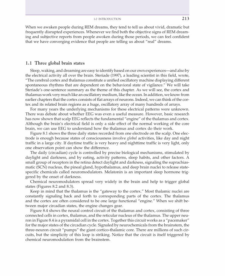
Sleep,waking, anddreaming are easy to identify basedonour ownexperiences—andalso bythe electrical activity all over the brain. Steriade (1997), a leading scientist in this field, wrote,“The cerebral cortex and thalamus constitute a unified oscillatorymachine displaying differentspontaneous rhythms that are dependent on the behavioral state of vigilance.” We will takeSteriade’s one-sentence summary as the theme of this chapter. As we will see, the cortex andthalamusworkverymuch like anoscillatorymedium, like the ocean. In addition,weknow fromearlier chapters that the cortex consists of flat arrays of neurons. Indeed,we can think of the cor-tex and its related brain regions as a huge, oscillatory array of many hundreds of arrays.
For many years the underlying mechanisms for these electrical patterns were unknown.There was debate about whether EEG was even a useful measure. However, basic researchhas now shown that scalp EEG reflects the fundamental “engine” of the thalamus and cortex.Although the brain’s electrical field is only a side effect of the normal working of the corebrain, we can use EEG to understand how the thalamus and cortex do their work.
Figure 8.1 shows the three daily states recorded from one electrode on the scalp. One elec-trode is enough because states of consciousness involve global activities, like day and nighttraffic in a large city. If daytime traffic is very heavy and nighttime traffic is very light, onlyone observation point can show the difference.
The daily (circadian) cycle is controlled by precise biological mechanisms, stimulated bydaylight and darkness, and by eating, activity patterns, sleep habits, and other factors. Asmall group of receptors in the retina detect daylight and darkness, signaling the suprachias-matic (SCN) nucleus, the pineal gland, hypothalamus, and deep brain nuclei to release state-specific chemicals called neuromodulators. Melatonin is an important sleep hormone trig-gered by the onset of darkness.
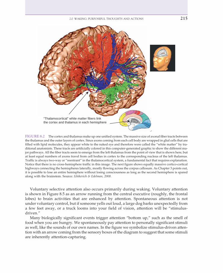
Chemical neuromodulators spread very widely in the brain and help to trigger globalstates (Figures 8.2 and 8.3).
Keep in mind that the thalamus is the “gateway to the cortex.” Most thalamic nuclei areconstantly signaling back and forth to corresponding parts of the cortex. The thalamusand the cortex are often considered to be one large functional “engine.” When we shift be-tween major circadian states, the engine changes gear.
Figure 8.4 shows the neural control circuit of the thalamus and cortex, consisting of threeconnected cells in cortex, thalamus, and the reticular nucleus of the thalamus. The upper neu-ron in Figure 8.4 is a pyramidal cell in the cortex. Together this circuit works as a “pacemaker”for themajor states of the circadian cycle. Signaled by neurochemicals from the brainstem, thethree-neuron circuit “pumps” the giant cortico-thalamic core. There are millions of such cir-cuits, but the simplicity of this loop is striking. Notice that the circuit is itself triggered bychemical neuromodulation from the brainstem.
2131.0 INTRODUCTION

2.0 WAKING: PURPOSEFUL THOUGHTS AND ACTIONS
Waking is our time to open up to the world around us, perceive and explore, think aboutourselves and one another, learn and prepare for the future, copewith challenges, express ouremotions, and advance our personal and social goals. From a biological point of view, all ofour purposeful survival and reproductive activities take place during the conscious state.
We have used a “functional diagram” of human cognition since Chapter 2.Whatwe haven’tsaid is that the waking state is the necessary condition for all those mental functions. That fact hasmany surprising consequences for our understanding of the brain. For example, the global fea-tures of the conscious state (Figures 8.1–8.4)maynot beobvious, butweknowtheyare necessaryfor us to have conscious sensations, working memory, and voluntary control of our muscles.
It will help to take another look at our basic functional diagram (Figure 8.5) and think aboutthe boxes that work only in the waking state. They are generally the colorful ones. The grayboxes (long-term memories) continue to store information 24 hours per day. As we will see,conscious experiences we learn during waking periods are often consolidated in slow wavesleep.
Sensory consciousness obviously also depends on the waking state, as do the “innersenses” of verbal rehearsal (inner speech), the visuospatial sketchpad (imagery), and the like.Normal, voluntary control is mainly limited to the waking state.
FIGURE 8.1 EEG of waking, dreaming,and deep sleep. To identify brain states, weonly need one electrode on the scalp (and areference electrode that is often attached tothe ear). Global states can be identified justby looking at EEG. Waking consciousnessand REM dreaming look remarkably similar,consistent with the fact that we experience arich flow of conscious events during bothwaking and dreams. Deep sleep looks verydifferent: it is high in voltage, slow, and regu-lar. Source: Squire, et al. 2008.
214 8. THE BRAIN IS CONSCIOUS
Voluntary selective attention also occurs primarily during waking. Voluntary attentionis shown in Figure 8.5 as an arrow running from the central executive (roughly, the frontallobes) to brain activities that are enhanced by attention. Spontaneous attention is notunder voluntary control, but if someone yells out loud, a large dog barks unexpectedly froma few feet away, or a truck looms into your field of vision, attention will be “stimulus-driven.”
Many biologically significant events trigger attention “bottom up,” such as the smell offood when you are hungry. We spontaneously pay attention to personally significant stimulias well, like the sounds of our own names. In the figure we symbolize stimulus-driven atten-tionwith an arrow coming from the sensory boxes of the diagram to suggest that some stimuliare inherently attention-capturing.
“Thalamocortical” white matter fibers linkthe cortex and thalamus in each hemisphere
FIGURE 8.2 The cortex and thalamusmake up one unified system. Themassive size of axonal fiber tracts betweenthe thalamus and the outer layers of cortex. Since axons coming from each cell body are wrapped in glial cells that arefilled with lipid molecules, they appear white to the naked eye and therefore were called the “white matter” by tra-ditional anatomists. These tracts are artificially colored in this computer-generated graphic to show the different ma-jor pathways. All the fiber tracts seem to emerge from the left thalamus from the point of view that is shown here, butat least equal numbers of axons travel from cell bodies in cortex to the corresponding nucleus of the left thalamus.Traffic is always two-way or “reentrant” in the thalamocortical system, a fundamental fact that requires explanation.Notice that there is no cross-hemisphere traffic in this image. The next figure shows equally massive cortico-corticalhighways connecting the hemispheres laterally, mostly flowing across the corpus callosum. As Chapter 3 points out,it is possible to lose an entire hemisphere without losing consciousness as long as the second hemisphere is sparedalong with the brainstem. Source: Izhikevich & Edelman, 2008.
2152.0 WAKING: PURPOSEFUL THOUGHTS AND ACTIONS

Later in this chapter wewill see that dreaming has some features of waking consciousness,such as vivid conscious imagery and working memory. Still, the fact remains that our goal-directed actions happen during the waking state, including thinking and problem solving,food gathering, social behavior and mate seeking. It makes sense therefore that task-relatedsignaling in the brain is mostly found in waking consciousness.
2.1 The stadium analogy: chattering, cheering, and chanting
Waking EEG has puzzled scientists since 1929, since it looks very irregular, even random,as if it’s a kind of “white noise,” such as the random noise we hear from waterfalls and oceanwaves. If we record the sound of a waterfall and then average separate stretches of sound, theaveragewill look like a flat line with zero voltage. That is because random activity is so unpre-dictable that it adds up to zero. The chance of a voltage at anymoment being above and belowzero is about equal. As we’ve seen with the averaged evoked potential in Chapter 5, we can
Cortex:pyramidal neuron
Thalamus:reticular neuron Thalamus:
Feedback to cortex
Brainstem:Cholinergic On/Offswitch
FIGURE 8.3 The state control circuit: three neurons in a thalamocortical loop. The basic rhythmic “pump” of thebrain. Waking, sleep, and dreaming are driven by thalamocortical oscillations. Thalamic nuclei interact closely withcorresponding regions of cortex. This core brain is shared by other mammals and birds. Source:Adapted from Steriade,
2006.
216 8. THE BRAIN IS CONSCIOUS

use that fact to obtain beautiful AEP curves that are time-locked to a stimulus. The randomEEG just drops out of the averaging process.
2.1.1 Chattering in the waking brain
If we think of the brain as a huge football stadiumwith thousands of people just chatteringwith one another, the averaged sound is so irregular that it resembles white noise. However,every conversation in the stadium is very meaningful to the people doing the talking. We seelocal synchrony between two individuals in a conversation and global randomness because noneof the conversations are linked to one another. It’s convenient to call this the “chattering” stateof the football stadium.
Figure 8.6 shows that during the waking state, neurons in different parts of the cortex andhippocampus are “phase-locked” to one another. (Phase-locked simplymeans “synchronizedwith a small lag time.”)
The evidence comes from Cantero and colleagues (2005), who recorded from hundreds ofneurons directly in the brain of an epileptic patient. Notice that the “Waking” column showsvery high correlated activity between different parts of the cortex (top) and also between thehippocampus and neocortex. Both of those regions are very active during waking, but wenow know that they are also highly correlated with each other at the single neuron level. Thatis analogous to spectators in the stadium talking “in sync” with one another. But like the
FIGURE 8.4 Chemical switching controls the state circuit. The daily states of consciousness are turned on and offby surprisingly small numbers of neurons, located at the bottomof the brain in nuclei like the substantia nigra (SN), as inthe figure (dark substance). Their widespread axons spray special neurochemicals called “neuromodulators” tomodifylocal neurotransmitters. Thebundleof axons thatproject from the small SN in thebottomof thebrain are called the nigro-striatal neurons because the start at the substantia nigra and terminate in the striatum ("striped region") of the basalganglia. Source: U.S. National Institute of General Medicine.
2172.0 WAKING: PURPOSEFUL THOUGHTS AND ACTIONS

chattering people in the stadium, their conversations are independent of one another. If werecord the overall sound in the stadium, it seems random, but if we record the talk of twopeople in a conversation, we can see the synchronized activity.
Although the stadium analogy is not supposed to be taken literally, two people talking toeach other do “dance” in synchrony with each other, so a slowed-down video of a conversa-tion will show them “micro-dancing” with each other. We can think of Figure 8.6 as showingrelatively localized synchrony between regions of the brain that are working together. Duringdeep sleep and dreaming, that kind of synchrony breaks down, as you can see in the secondand third column of the figure.
The conscious state therefore seems to support local synchrony (or phase-locking), whiledeep sleep and dreaming do not. As we will see next, there is very direct evidence thatsynchrony serves as an important coordinating rhythm for neurons that may be widelydispersed but that are supporting the same cognitive task, whether it is sensory perception,motor control, memory storage, or the other active tasks in our functional diagram(see Figure 8.5).
SensoryInput
Vision
Hearing Consciouscontent Working
storage
Centralexecutive
A functional framework.
Top-downvoluntaryattention
Actionplanning
Verbalrehearsal
Visuospatialsketchpad
Learningand retrievalStored memories, knowledge, and skills:
Perceptualmemory
Autobiographicalmemory
Linguistic andsemantic
Visualknowledge
Declarativeknowledge
Habits andmotor skills
Responseoutput
Working memory
Touch
SensorybuffersBottom-up
attentionalcapture
FIGURE 8.5 Weare not conscious of all cognitive functions at the same time, butwe can get rapid conscious accessto all the colored boxes in the diagram. The gray boxes at the bottom are never directly conscious. However, we canretrieve conscious information from long-termmemories. Notice that the diagram looks very different in dreams andsleep. Source: Baars, with permission.
218 8. THE BRAIN IS CONSCIOUS

2.1.2 Cheering in the waking brain
Football spectators are not always chattering to one another locally. They do at least twomassively coordinated actions. Onewewill call cheering, such aswhen one team scores a thril-ling goal. Ten thousand people suddenly applaud (or boo) out loud. Since this is an event-related cheer, it is analogous to the event-related potential. In the brain we commonly averageevoked potentials over a number of triggering events, like the thrilling football play that trig-gers a cheer. That is the averaged evoked potential. Sending a big flash or a loud noise throughthe brain is very much like a simultaneous cheer going through a football stadium. Suddenlyall the noisy background chat turns into a giant “hooray!”
2.1.3 Chanting in the unconscious brain
What about unconscious periods, like deep sleep (slow wave sleep)? You can see inFigure 8.1 that it consists of very large, very slow (by brain standards), and very coordinatedbrain activity. We know that billions of neurons are highly coordinated during SWS because
Waking SWS REM
800
600
400
200
00.1 0.3 0.5 0.7 0.9
% showing phase-locking
% showing phase-locking
% showing phase-locking
% showing phase-locking % showing phase-locking
Pai
red
iEE
G e
lect
rode
sP
aire
d iE
EG
ele
ctro
des
N = 2575Mean = 0.43SEM = 0.03
0
300 1000
800
600
400
200
0
200
100
0.1 0.3 0.5 0.7 0.9 0.1 0.3 0.5 0.7 0.9
N = 889Mean = 0.48SEM = 0.02
N = 889Mean = 0.02SEM = 0.004
N = 2575Mean = 0.03SEM = 0.03
N = 2575Mean = 0.04SEM = 0.07
cortico-hippocampal gamma phase-locking
3000
2000
1000
00.1 0.3 0.5 0.7 0.9
3000
2000
1000
00.1 0.3 0.5 0.7 0.9
% showing phase-locking
1000
800
600
400
200
00.1 0.3 0.5 0.7 0.9
N = 889Mean = 0.03SEM = 0.005
long-range gamma phase-locking in neocortex
FIGURE 8.6 Synchronized “conversations” between neurons duringwaking. If we think of each neuron as a spec-tator in a large football arena, during the conscious state thousands of spectators are chatteringwith one another at thesame time. Each dialogue synchronizes just two people, but hundreds of different conversations are happening atthe same time. There is local synchrony but not global synchrony. Source: Cantero & Atienza, 2005.
2192.0 WAKING: PURPOSEFUL THOUGHTS AND ACTIONS
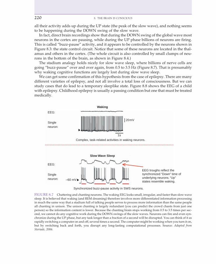
all their activity adds up during the UP state (the peak of the slow wave), and nothing seemsto be happening during the DOWN swing of the slow wave.
In fact, direct brain recordings show that during the DOWN swing of the global wavemostneurons in the cortex are pausing, while during the UP phase billions of neurons are firing.This is called “buzz-pause” activity, and it appears to be controlled by the neurons shown inFigure 8.3: the state control circuit. Notice that some of these neurons are located in the thal-amus and others in the cortex. (The whole circuit is also controlled by small clumps of neu-rons in the bottom of the brain, as shown in Figure 8.4.)
The stadium analogy holds nicely for slow wave sleep, where billions of nerve cells aregoing “buzz-pause” over and over again, from 0.5 to 3.5 Hz (Figure 8.7). That is presumablywhy waking cognitive functions are largely lost during slow wave sleep.
We can get some confirmation of this hypothesis from the case of epilepsy. There are manydifferent varieties of epilepsy, and not all involve a total loss of consciousness. But we canstudy cases that do lead to a temporary sleeplike state. Figure 8.8 shows the EEG of a childwith epilepsy. Childhood epilepsy is usually a passing condition but one that must be treatedmedically.
EEG:
Singleneuron:
EEG:
Singleneuron:
Slow Wave Sleep
Waking
Complex, task-related activities in waking neurons.
EEG troughs reflect thesynchronized “Down” time ofunderlying neurons. “Up” states resemble waking.–60 mV
20mV
1s
Synchronized buzz-pause activity in SWS neurons.
FIGURE 8.7 Chattering and chanting neurons. The waking EEG looks small, irregular, and faster than slowwavesleep. It is believed that waking (and REM dreaming) therefore involves more differentiated information processingin much the same way that a stadium full of talking people serves to process more information than the same peopleall chanting in unison. The unison chanting is largely redundant (you can predict the crowd chants from just oneperson) so the information content is lower. Because the chanting brain stops working from 0.5 to 3.5 times per sec-ond, we cannot do any cognitive work during the DOWN swings of the slow waves. Neurons can fire and even syn-chronize during the UP phase, but any task longer than a fraction of a second will be disrupted. You can think of it asrapidly switching a computer on and off, several times a second. The computermight beworkingwhen you turn it on,but by switching back and forth, you disrupt any long-lasting computational processes. Source: Adapted fromSteriade, 2006.
220 8. THE BRAIN IS CONSCIOUS

Other unconscious states also show slow EEG waves, except for deep coma, which mayshow a very low voltage level so that peakwaves simply drop out. However, scalp EEG some-times fails to pick up EEG activity deep in the brain, so a low overall level of the EEG does notnecessarily mean there is no consciousness at all. It is important to remember that scalp EEGonly picks up 0.1 percent of the voltage level at the cortex. It is therefore possible to miss brainactivity if we only look at scalp EEG.
In some cases, patients have been wrongly diagnosed as being in an irreversible comawhen closer observations showed signs of consciousness (Laureys et al., 2002). Medical sci-entists have therefore proposed a new diagnosis called theminimally conscious state (MCS) forconditions that may look like coma on the outside but that may actually involve patients whohave conscious periods.
2.2 How does the chattering brain do its work?
If the stadium analogy is right, most cognitive tasks happen during chattering states. Thatincludes both waking and dreaming (see Figure 8.1). Waking and dreaming EEGs look irreg-ular and low in voltage because there is no global synchrony. However, we expect there to belocal synchrony (technically, local phase-locking).
Fp1-F7
F7-T3
T3-T5
T5-O1
Fp2-F8
F8-T4
F4-T6
T6-O2
Eyes flutter, unresponsive
1 s 200 µV
FIGURE 8.8 Slow hypersynchrony in epileptic loss of consciousness. The surface EEG of a 7-year-old girl duringan epileptic seizurewith loss of consciousness. Epileptic loss of consciousness shows slow, hypersynchronized globalEEG similar to deep sleep but characteristically more jagged. Other unconscious states, like coma and general anes-thesia, also show slow, high-voltage, and synchronized waves spreading over the cortex, suggesting that synchronyby itself is not the marker of consciousness. Tonic-clonic seizures are cortical. Source: Blemenfled, 2005.
2212.0 WAKING: PURPOSEFUL THOUGHTS AND ACTIONS
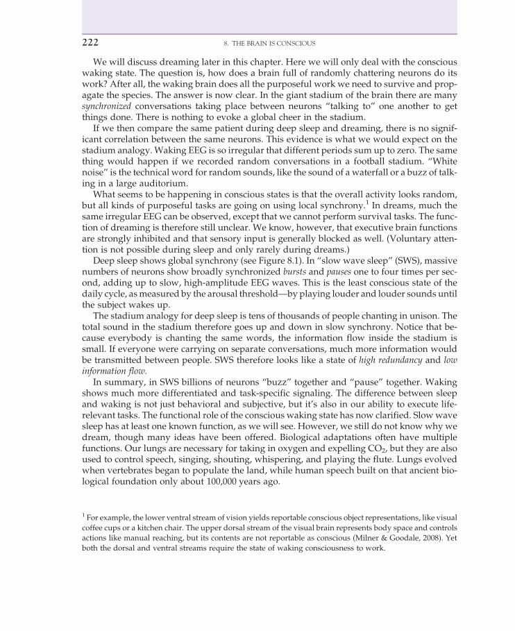
We will discuss dreaming later in this chapter. Here we will only deal with the consciouswaking state. The question is, how does a brain full of randomly chattering neurons do itswork? After all, the waking brain does all the purposeful work we need to survive and prop-agate the species. The answer is now clear. In the giant stadium of the brain there are manysynchronized conversations taking place between neurons “talking to” one another to getthings done. There is nothing to evoke a global cheer in the stadium.
If we then compare the same patient during deep sleep and dreaming, there is no signif-icant correlation between the same neurons. This evidence is what we would expect on thestadium analogy. Waking EEG is so irregular that different periods sum up to zero. The samething would happen if we recorded random conversations in a football stadium. “Whitenoise” is the technical word for random sounds, like the sound of a waterfall or a buzz of talk-ing in a large auditorium.
What seems to be happening in conscious states is that the overall activity looks random,but all kinds of purposeful tasks are going on using local synchrony.1 In dreams, much thesame irregular EEG can be observed, except that we cannot perform survival tasks. The func-tion of dreaming is therefore still unclear. We know, however, that executive brain functionsare strongly inhibited and that sensory input is generally blocked as well. (Voluntary atten-tion is not possible during sleep and only rarely during dreams.)
Deep sleep shows global synchrony (see Figure 8.1). In “slow wave sleep” (SWS), massivenumbers of neurons show broadly synchronized bursts and pauses one to four times per sec-ond, adding up to slow, high-amplitude EEG waves. This is the least conscious state of thedaily cycle, asmeasured by the arousal threshold—by playing louder and louder sounds untilthe subject wakes up.
The stadium analogy for deep sleep is tens of thousands of people chanting in unison. Thetotal sound in the stadium therefore goes up and down in slow synchrony. Notice that be-cause everybody is chanting the same words, the information flow inside the stadium issmall. If everyone were carrying on separate conversations, much more information wouldbe transmitted between people. SWS therefore looks like a state of high redundancy and lowinformation flow.
In summary, in SWS billions of neurons “buzz” together and “pause” together. Wakingshows much more differentiated and task-specific signaling. The difference between sleepand waking is not just behavioral and subjective, but it’s also in our ability to execute life-relevant tasks. The functional role of the conscious waking state has now clarified. Slowwavesleep has at least one known function, as we will see. However, we still do not know why wedream, though many ideas have been offered. Biological adaptations often have multiplefunctions. Our lungs are necessary for taking in oxygen and expelling CO2, but they are alsoused to control speech, singing, shouting, whispering, and playing the flute. Lungs evolvedwhen vertebrates began to populate the land, while human speech built on that ancient bio-logical foundation only about 100,000 years ago.
1 For example, the lower ventral stream of vision yields reportable conscious object representations, like visual
coffee cups or a kitchen chair. The upper dorsal stream of the visual brain represents body space and controls
actions like manual reaching, but its contents are not reportable as conscious (Milner & Goodale, 2008). Yet
both the dorsal and ventral streams require the state of waking consciousness to work.
222 8. THE BRAIN IS CONSCIOUS

In the same way, the brain’s circadian states are likely to have more than one function. Theonsets of both waking and sleep trigger gene expression in hundreds of different neural genes.In waking we have easy access to a vast repertoire of skills and capacities. One of these is vol-untary attention; we can decide to pay attention to this book or to yesterday’s notes. Conscious-ness and attention are intimately related but are not identical.Whenwe selectively attend to onething over another,we become conscious of the attended object. Those consciousmomentsmaythen trigger further attentional selection, and so on.We cycle between experiencing things (con-sciousness) and deciding what we want to experience next (selective attention).
Figure 8.2 is a reminder of waking state functions, including selective attention. With 100billion neurons firing about ten times per second, and excitatory neurons triggering off otherexcitatory neurons, you can imagine what an ocean of mutually pushing waves and troughswe have oscillating in our brains every second. Fortunately, most excitatory neurons are con-trolled by inhibitory neurons so the brain can regulate its state of excitation. Otherwise wemight get epileptic seizures, with giant waves of excitation constantly triggering new wavesso the cortex goes into overdrive. Like all the organs of your body, the activity of the brain isunder careful homeostatic control.
During waking consciousness, the thalamocortical system reveals a constant flow of signaltraffic, with thousands of messages going back and forth. Figure 8.3 shows a large-scalesimulation of the known neurons and synaptic signal flow in the thalamocortical system(Izhikevich et al., 2004, 2008).
Waking consciousness resembles the back-and-forth flow of traffic in a busy city. At night,street traffic might dwindle to a few cars, but during the rush hour, vehicles are constantlygoing from any location to any other. It is that ability to go anywhere, guided by local goals,that makes cars useful. Waking tasks serve a huge variety of functions with great flexibility.
TABLE 8.1 Major Features of the Conscious State
1. A very wide range of conscious contents2. Widespread brain effects beyond the specific contents of consciousness3. Informative conscious contents4. Rapid adaptation (learning and coping)5. The “fleeting present” of a few seconds6. Internal consistency7. Limited capacity and seriality8. Sensory binding9. Self-attribution10. Accurate reportability11. Subjectivity12. Focus-fringe structure13. Consciousness facilitates learning14. Stability of conscious contents15. Object and event-centered16. Consciousness is useful for voluntary decision making17. Involvement of the thalamocortical core
Source: Modified from Seth & Baars, 2008.
2232.0 WAKING: PURPOSEFUL THOUGHTS AND ACTIONS

Humans have access to a remarkable range of conscious events—the sight of a single staron a dark night, the difference between the sounds “ba” and “pa,” and the goal of studying foran exam. All sensory systems do a great deal of unconscious processing, leading to conscious,reportable sights, sounds, and feelings.
Recalling an autobiographical memory also results in conscious experiences, like yourmemory of seeing this book for the first time. Inner speech and imagery have conscious as-pects. Asking someone to rehearse a list of words usually results in conscious inner speech.People vary in the vividness of their visual images, but dreams are generally reported to havevivid imagery (Stickgold et al., 2001). Finally, action planning can have conscious compo-nents, especially when we have to make choices about our actions (Lau et al., 2004a, b). Nor-mally, wemay not have to think much about the action of walking, but if we injure a leg, evenstanding up and walking can become a consciously planned process (Sacks, 1984).
2.3 What we expect from conscious people
Table 8.1 presents 17 properties of consciousness that are generally recognized in scientificand medical literature. (Of course, the humanities, arts, philosophy, and religion have a longhistory of exploring consciousness, too.) People with head injuries are often given a mentalstatus examination, which tests for abilities we expect normal, conscious people to have(McDougall, 1990). The test includes the following:
1. Orientation to time, place, and person: “What day of the week, date, and season is it?”“Where are you now?” “Who are you?” “Who am I?” (asked by the examiner)
2. Simple working memory: “Remember these three objects. . ..”3. Attention and calculation: “Count down by sevens starting from 100.” (100, 93, 86, 79,
72 . . .)4. Intermediate recall: “What were the three objects I asked you to remember a few
minutes ago?”5. Language: Asking the patient to identify a wristwatch and a pencil, to repeat a sentence,
to follow three simple commands, to read and understand a simple sentence, and towrite their own sentence.
6. A basic visuomotor skill: Asking the patient to copy a drawing of a line pentagon.
The mental status exam gives a broad overview of normal mind and brain functions. Somedisorders involve disorientation, others affect short-term memory, and still others impairvisual and motor coordination.
We also expect healthy, conscious adults to do realistic thinking. While dreams are unreal-istic, waking consciousness has been believed to be necessary for logical, mature, and reality-oriented thought. Nevertheless, we routinely experience waking fantasies, unfocused states,daydreams, emotional thinking, and mind wandering.
2.4 Waking has conscious and unconscious threads
It is important to keep inmind that waking cognition is woven of both conscious and uncon-scious threads, constantly weaving back and forth. For example, the process of reading thesewords is only partly conscious. You are a highly practiced reader, and you have learned over
224 8. THE BRAIN IS CONSCIOUS

many hours of practice to automatically convert these tiny marks on paper into your own innerspeech and then into unconscious processes like word recognition, grammar, and meaning.
We do not become conscious of every mental step in reading a sentence. In fact, there is agreat deal of scientific evidence today that all cognitive tasks have many conscious and un-conscious components. Figure 8.9 shows an example of unconscious detection of the pictureof a snake. Human beings are attuned to detecting some things unconsciously, it seems, in-cluding snakes and even facial expressions, which are processed through the visual system,and trigger the fear-sensitive amygdala, a major subcortical center for processing emotions.
One can imagine how that ability may have evolved, since humans (and our ancestors)lived for millions of years in environments where snakes were an everyday, deadly threat.Human beings learn a vast amount of information from other people, such as the dangerof crossing busy streets without looking. We are not innately attuned to cars rushing througha street, although we may be afraid of “looming” objects like a fast-approaching elephant.
Until recently, however, the evidence was still hotly debated on whether humans respondto genuinely unconscious stimuli. Studies like this one seem to prove the point convincingly(see Figure 8.9) (Ohman et al., 2007).
Even conscious sensory percepts have stages of unconscious processing (Figure 8.10). Wehave both psychological and brain evidence to that effect. Social psychologists like Banaji andGreenwald (1995) have found evidence that social perception and inference seem to have un-conscious properties as well.
FIGURE 8.9 Comparing conscious versus unconscious events during the waking state. The subject can reportthe snake on the left side but not on the right side, even though the experimental stimulus is identical. This canbe done using binocular rivalry, for example. In this experiment the emotional brain (the amygdala) canrecognize the unconscious snake. Humans evolved in environments where deadly snakes were not unusual.Source: Ohman et al., 2007.
2252.0 WAKING: PURPOSEFUL THOUGHTS AND ACTIONS

Almost all cognitive tasks we know take place during the waking state and have both con-scious (reportable) and unconscious (nonreportable) components. Most cognitive tasks weknow are therefore consciously mediated. Working memory, for example, has both consciousand unconscious components. If you rehearse seven numbers, you will notice that someare conscious at any moment, but others are not. The instructions to rehearse and rememberare obviously conscious and so is the set of items as you see or hear them. But we are notaware of the nonrehearsed items at any moment, of the important role of the basal gangliain controlling inner speech, or of the automatic (habitual) components of any task. Thereare no completely conscious cognitive tasks, as far as we know, and there may be nocompletely unconscious ones (Baars & Franklin, 2003). (Franklin has suggested the term “con-sciously mediated” for cognitive tasks that have a consciousness but are otherwiseunconscious.)
The basal ganglia and cerebellum are very large brain structures that are believed to functionwithout supporting moment-to-moment conscious contents, even in the waking state. The cer-ebellum can actually be lesioned on both sides and people will continue to behave much asbefore but without the ability to control fine motor movements. In humans those structureshave many other cognitive roles but without direct conscious contents.
What can we do completely unconsciously? We still do not know the answer, because it isdifficult to do careful studies on sleepwalking, sleep movement disorders, epileptic “auto-matic behaviors,” and other “zombie states” (Crick & Koch, 2003). There are many reportsabout automatic behaviors from individuals with sleep disorders and epilepsy. To verifythose reports we need brain recordings that are difficult to obtain in freely moving people.It is also possible that epileptic behavioral automatisms, for example, reflect momentary con-scious “flashes” that are known to exist (Kranczioch et al., 2006). It is therefore hard to testwhether there are complex but entirely unconscious behaviors, in part because we simplydo not know the distinctive brain correlates of consciousness as yet (but see Gaillard et al.,
Controlled processing Automatic processing
PMFCPPC
PPCDLPFC
A/G
FIGURE 8.10 Voluntary and automaticskills. The brain images show cortical activityduring two identical behavioral tasks. Thedifference is that the left-side task is noveland therefore requires more conscious andvoluntary control (labeled “controlled pro-cessing”). The red-orange regions indicatefMRI peak activity mainly in the prefrontaland parietal lobes. The brain images on theright only show activity in the auditory cor-tex, probably because the specific auditorystimulus is never entirely predictable. Justlike riding a bicycle, the most predictableparts of a practiced task tend to fade fromconsciousness. Instead of controlling everymovement, we control higher levels, likewhich direction to steer. PMFC ¼ premotorfrontal cortex; PPC ¼ posterior parietal cor-tex; DLPFC ¼ dorso-lateral prefrontal cortex;A/G ¼ anterior gyrus. Source: Scheider, 2009.
226 8. THE BRAIN IS CONSCIOUS

2009; Revonsuo, 2006). We do not know yet what difference enables consciousness of the ven-tral but not the dorsal stream of the visual cortex. There are, however, ongoing efforts toanswer those questions (Laureys & Tononi, 2008).
Most cognitive tasks we know are therefore consciously mediated. Problem incubation is afamous example of unconscious mental processing.
2.4.1 Task-related signaling between linked neurons
Because new findings about brain rhythms for cognitive functions are constantly appear-ing, it helps to use just three ranges: slow, midrange, and fast. Slow oscillations include thedelta waves of deep sleep (less than 4 Hz). Delta waves are important for consolidating tem-porary memories into long-term memories. (In addition, a slower rhythm has been discov-ered that continues throughout the 24-hour cycle (Steriade, 1997).) There is no certaintyyet about their role in waking tasks.
Delta waves occur during sleep and drowsy states, and similar slow waves occur duringlight anesthesia and in epileptic loss of consciousness. These are unconscious states. Fasterwaves occur during waking and dreaming.
MID-RANGE AND FAST WAVES
Cognitive tasks during waking are known to involve both midrange and faster waves.Until the evidence settles down, therefore, it is useful to talk about two frequency rangesfor waking cognition.
ALPHA AND THETA RHYTHMS
Midrange oscillations include theta and alpha waves. Alpha rhythms of 8 to 12 Hz werefirst observed over the occipital cortex when human subjects were relaxed or closed theireyes. However, alpha and theta (4–7 Hz) are now known to be involved in many differentwaking tasks in many parts of the brain. In many cases these near–10 Hz waves seem to co-ordinate faster oscillations. In a very broad sense, near–10 Hz waves may function as a wide-spread “system clock” for many parts of the brain. For example, theta waves are known tofacilitate encoding of temporary episodic memories into long-term episodic memory. Inthe motor cortex alpha-like rhythms have been reported to be involved in the inhibition ofplanned actions. In the frontal lobe, alpha-like waves are involved in momentary memorystorage, and some researchers find that both synchrony and desynchrony of alpha wavesmay play a role in cognitive processes. Even the boundary between theta and alpha is notnecessarily clear, and some researchers believe that these waves are not necessarily stablein their conventional range.
Scientific periods of rapid discovery often seem confusing until they settle into some stablepattern of evidence. Because empirical science is unpredictable, we do not know at this timewhether the brain wave spectrumwill be divided up neatly into frequency ranges or whetherdifferent brain locations will turn out to have quite different oscillations.
There is reasonable agreement, however, that alpha/theta oscillations near 10 Hz interactwith faster oscillations. One proposal is that brain waves resemble the radio spectrum, with“carrier frequencies” being modulated (by amplitude, as in AM radio), or by frequency(FM). In the case of radio waves, broadcasting stations generate electromagnetic radiation atspecific tuning frequencies (as you can see on your AM or FM dial). Radio receivers can be
2272.0 WAKING: PURPOSEFUL THOUGHTS AND ACTIONS

tuned to the major frequencies. Since speech and music involve faster oscillations, these are“carried” by the standard tuning frequencies.
In the case of the brain, it is believed that theta waves sometimeswork as carrier waves andthat individual neurons can tune their own firing patterns relative to some widespread thetawave (Canolty et al., 2006). Since these are open issues on the scientific frontiers, we simple donot know precisely how they will settle over the longer term.
There is no agreement currently on the range of faster oscillations, often called beta andgamma. Functional rhythms have been reported up to 200 Hz and even (briefly) 600 Hz. Be-cause new findings are constantly appearing, it makesmore sense to describe three frequencyranges (see Figure 8.1). Midrange oscillations include classical alpha and theta, near 10 Hz.The pace of new findings is now so rapid that we can expect to see much greater clarificationon these issues.
A range of frequencies have now been observed for sensory processing, attentional en-hancement of sensory input, and both working and long-term memory. Synchrony is bothnatural and useful for signaling in an oscillatory system like the brain. Sometimes perfect syn-chrony is not attainable, so there is a brief time lag between the peak of the wave in one place(like the hippocampus) and another place (like the frontal lobe). In those cases, the better termis phase locking or phase coherence, a little bit like a syncopated “off-beat” rhythm in music. It issynchrony with a time lag.
Individual neurons have a temporal integration time of about 10 ms, the period when den-dritic inputs can add up to increase the probability of a single axonal output spike (seeChapter 3). A group of interconnected neurons can strengthen one another’s firing rates be-tween 30 and 100 Hz by supplying synaptic inputswithin the 10 mswindow. If two excitatoryneurons are signaling each other at a rate of 50 Hz, for example, it is possible to sustain anexcitatory feedback loop, because converging signals can arrive within the critical 10 ms pe-riod. However, neuronal firing rates below 30 Hz may not be integrated by target neuronsbecause different spikes may arrive too late to have additive effects. It is therefore believedthat a group of neurons firing in the beta-gamma range will exert a stronger drive to down-stream neurons than lower frequencies. Obviously, real brain networks are more complexand have inhibitory as well as excitatory elements. Nevertheless, these basic points applyto neurons in general and have gained a good deal of direct empirical support.
Radio transmission has some similarities to oscillatory synchrony in the brain. The existenceof AM and FM radio suggests at least two ways in which brain rhythms may process informa-tion in the brain. But there aremanymore coding schemes. Brain rhythms could serve as clocks,and they can use single pulses or a series of pulses like Morse code. Different neurons may usesignals in different ways, perhaps in combination with different molecules and synapses.
Television is an example of a spatiotemporal code, in which the broadcast signal scansacross every line of the screen from top to bottom.Computer screens use similar spatiotemporalcoding. Brain rhythms are also likely to coordinate visuotopic maps, somatotopical maps, andmotor maps. As we have mentioned, the brain is rich in topographical maps, which representsensory input arrays or neuromuscular maps at various levels of abstraction (see Chapter 5).
Evolution has exploited the rhythmic properties of neurons over hundreds of millions ofyears. For that reason, we should not expect to find only a single neural code. What we doknow is that brain rhythms are very widespread and that they are associated with knownfunctions.
228 8. THE BRAIN IS CONSCIOUS

Finally, waves can also interfere with one another. When you place a radio receiver next toa computer, you will hear a burst of noise whenever you press the keyboard. That is becauseeach key press triggers an electromagnetic signal that radiates into the surrounding space.Wave interference is a fundamental phenomenon in the physics of radiation. Interferencemay have important uses in the brain, but it might also degrade neural information processing.We are only beginning to understand the role of brain rhythms, but it is likely that wave inter-ference will turn out to have effects as well.
3.0 ATTENTION
Common sense makes a distinction between attention and consciousness. The word atten-tion seems to imply an ability to bring something to mind. If you can’t remember a word inthis book, for example, you might try to “pay more attention.” What trying to pay more at-tention comes down to is allowing the forgottenword to be in consciousness for a longer time.So we rehearse the forgotten word (consciously), or we make a note about it (making it con-scious again), or we write a definition about it (same thing). The traditional “law of effect”about learning states that the more we make something conscious, the more we will learnit. When we call someone’s attention to a speeding car, we expect him or her to become con-scious of it.
In everyday language, “consciousness” refers to an experience—of reading this sentence,for example, or conscious sensory perception, conscious thoughts, feelings, and images.Those are the experiences we can talk about to one another. Selective attention implies a se-lection among possible conscious events. When we make an attentional selection, we expectto become conscious of what we’ve chosen to experience.
With careful studies we can separate the phenomena of attention and consciousness. Tofocus on conscious events “as such,” we typically study them experimentally in contrast withclosely matched unconscious events, as we have seen in previous chapters (see Chapters 1, 3,6, and 7). By contrast, experiments on attention typically ask subjects to select one of twoalternative stimuli. “Attention” is therefore concerned with the process of selection andconsciousness, with reportable experiences themselves. Some key questions for cognitiveneuroscience are: What is distinctive about conscious events in the brain? What does it reallymean for someone to be conscious? How does the brain basis of attentional selection relate toour private, conscious experiences of the world?
3.1 Attention selects conscious events
Attention has two aspects: the source of attentional control, which decides what to pay at-tention to, and the target of attention, which is selected for additional processing. Considerthe case of a college student sitting in a lecture room, with many sensory inputs at the sametime andmany simultaneous tasks to perform. The studentmust stay alert, orient to the visualand auditory input, keep track of the lecture, take notes, andmore. In fact, students are alwaysmultitasking, and aswe know, that is inherently difficult. That is why it is important to reviewlectures. Live lectures can easily overwhelm our attentional capacities.
2293.0 ATTENTION

3.2 The Posner flanker task
Michael Posner and colleagues devised a simplemethod called the “flanker task.” They asksubjects to pay attention to a stimulus at a known location on the right or left side of the fix-ation point (marked with a dot or a plus sign) (Figure 8.11). Because humans have a very lim-ited foveal “keyhole” through which they fixate a small part of the visual field at any singlemoment, it is possible to control the exact information that is available to a subject. (We seeonly about 3–6 degrees of visual arc when the eyes are fixed on a point. Try it!
The arrow points away from the target location
The arrow points to the target location
The cue appears
(b)
(a)
FIGURE 8.11 The flanker task for studying attention. The Posner flanker task has long been used to assess visualattention and its brain bases. The subject looks only at a fixation point in the center of the screen (a) Directional cuessuch as an arrow (b) draw attention to the left or right flank (side) of the fixation point, but no eye movements areallowed. It is the subject’s attention that is cued to one or the other side of the screen, not the eyes. Source: Reynoldset al., 2003.
230 8. THE BRAIN IS CONSCIOUS

The flanker task is simple, effective, and adaptable. For example, the target stimuli can beemotional faces, allowing us to explore how the brain pays attention to emotional events (Fanet al., 2002; Posner & Petersen, 1990).
Subjects keep their gaze on the fixation point. When flanking stimuli appear on the right orleft side, they can be detected even when the eyes are kept fixed on the crosshairs. InFigure 8.11, the target is flashed in the expected location for a fraction of a second. Subjectsrespond as quickly as possible. When their cued expectations are correct, their reaction timesand accuracy are optimal.
The flanker task allows for testing of both voluntary and nonvoluntary attention, by givingsubjects either correct or incorrect information about the flank on which the stimulus will ap-pear. The task is simple enough to administer in an fMRI scanner in a half hour or so, and theresulting brain scans provide separate information about expectation-driven trials and unex-pected trials. By subtracting the “unexpected attention” brain activity from the “expected at-tention” scans, Posner and colleagues were able to obtain a relatively pure measure of thebrain regions involved in voluntary visual attention.
3.3 A model of attention
Itti and Koch (2001) developed a model of attention that combines a number of importantfeatures. It shows a simplified layered concept of the visual system, with multiple topo-graphic visual maps. The visual maps show line orientation, stimulus intensity (contrast),color, and salience. Salience is defined in terms of feature contrast in any visual map. Inlight-sensitive regions, it is the contrast between light and dark patches on the map. Inmotion-sensitive areas, like area MT, it may be a stable object against the background of awaterfall. A combined salience map may combine all the contrasting features into a singlesaliency map, one that reflects the unusual, unexpected, or noteworthy features of a visualscene at any level of analysis. A “winner-take-all” computation selects the most salient loca-tion on a combined map and inhibits competing locations. Obviously, salience can also bemisleading; for example, when you are watching a visually excitingmusic video that containsa variety of attention-driving features, youmay want to think about something else. You mayhave to override what is most salient at any moment.
Visuotopic neurons respond to optical stimuli at different levels of analysis (see Chapter 6).Figure 8.12 gives us a convenient overview. Each layer of the pictured model by Itti and Kochresponds to a particular feature of the stimulus: color, line orientation, contrast, and objectidentity. This is a simplification of the visual brain, which is far more complex and flexibleand that must deal with complications such as the constant motion of the eyes and the head,the very narrow limits on foveal vision, and much more. But Figure 8.12 helps to clarify ourquestion.
Each visuotopic map is a two-dimensional mosaic of neurons with rather narrow receptivefields (see Chapter 6). We can therefore ask a more focused question: Does attention to someevent or location enhance signal processing in the correct receptive field? If the watcher inFigure 8.12 is hot and thirsty while wandering in the Sahara Desert, will his or her attentionalsystem enhance visual processing for ice-cold mugs of beer located on the left side of his vi-sual field? This question is much more specific and testable.
2313.0 ATTENTION

The concept of a saliencymap reflects significance,motivational relevance, andvividness ofthe input. Many topographical maps in the visual brain are sensitive to motivation and rele-vance. The man at the bottom of the figure is imagined standing in a hot desert with a coldmugofbeer on the far left sideofhis visual field—just outsideofhisdirect visual field. Selectiveattentionallowssignificant stimuli like thecoldbeer toemerge into consciousness.These canbeexpressed as “top-downattentional biases” that alter the saliencymapon topof the stack. Priorlearning also is input into the saliency map. All the topographical maps resonate together insynchrony and jointlymake decisions in caseswhere the corresponding points on themajorityof maps lead to the same overall result. The output may be an eye movement, allowing theviewer to see the cold beermug, or itmaybe a covert shift in attention to the left, again allowing
Current biology
Input image
Atte
nded
loca
tion
Winner-takes-allnetwork
Top-downattentional bias
and training,learning
Color
IntensityOrientation
Saliency Map
FIGURE 8.12 How attention might acti-vate sensorymaps.Multiple visuotopicmapssupport our conscious experiences of beermugs and deserts. Notice that the person atthe bottom seems to be looking for a colddrink on a hot day in the Sahara. How doour brains select the cold beer? What do webecome conscious of? Source: Thier et al.,
2002; figure adapted from Itti & Koch, 2001.
232 8. THE BRAIN IS CONSCIOUS

the beer stein to come to visual consciousness. Again, it is possible that the man may want tooverride the perception of the cold beer and focus attention on crossing the desert instead.There are potentially competing decisions in this multilayered network.
An important aspect is the “winner-take-all” (WTA) network (Figure 8.13). WTA networksessentially allow themost active point on the joint topographical maps to “win,” input from sa-liency,which represents such things asmotivation, hunger, and thirst; learning about relevance;and so on. WTA also suggests an explanation for conscious experiences of ambiguous figures.Conscious experiences are marked by internal consistency, even when sensory inputs are not.Most of the words in the English lexicon are highly ambiguous, for example, but in context(as in this sentence), ambiguous words are consciously experienced in terms of just one in-terpretation. Thus, a WTA network may be involved in an attentional system, as shown inFigure 8.12, but they are also a very powerful feature of conscious perception. Indeed,we canconsider conscious perception to be the outcome of many attentional acts. In reality, theremay be no difference in the brain between those two mechanisms.
The term attention is used most intuitively when there is a clear voluntary or executive as-pect. We ask people to pay attention to things, which implies they can choose to do so or not,depending on some decision-making processes. Voluntary attention is the kind that is studiedmost often, and as youmight guess from the other chapters, it is likely to use prefrontal cortexin humans (see Chapter 12).
Corbetta and colleagues (2002) recently wrote that voluntary attention “is involved in pre-paring and applying goal-directed (top-down) selection for stimuli and responses.” Auto-matic attention, on the other hand, “is not involved in top-down selection. Instead, thissystem is specialized for the detection of behaviorally relevant stimuli, particularly when theyare salient or unexpected.” When we hear a sudden loud noise, our attention is “captured,”even without executive guidance. As you might expect, visual attention can be captured byhuman faces, emotional expressions, and bodies, when compared with neutral stimuli. In-tense or sudden stimuli, or unexpected events generate larger brain responses than controlstimuli. Thus we can talk about “bottom-up” capture of selective attention, driven by stimuli.
In the real world, voluntary and automatic types of attention are generally mixed. We cantrain ourselves to pay attention to the sound of the telephone ringing. When it rings and wesuddenly pay attention to it, is that voluntary or automatic? Well, it began being voluntaryand becamemore automatic. The dimension of voluntary versus automatic attention is there-fore a continuum. Perhaps the strongest case of voluntary attention is the one where we mustexert intense mental effort over a period of time. A clear example of the opposite pole of the
FIGURE 8.13 Awinner-take-all (WTA) network, in which the mostactivated point in the horizontal plane inhibits all the surroundingpoints. The vertical axis is labeled activity and may represent thesummed activity of multiple visuotopic maps. WTA networks are verycommon in decision-making neural nets. In the brain, both selective at-tention and conscious perception may make their final “decisions”using a WTA mechanism. In the case of ambiguous stimuli, the brainmakes one of two competing interpretations conscious. Source: Standageet al., 2005.
2333.0 ATTENTION

continuum might be a case of a loud sound or a biologically important event like a cryingbaby, which is hard not to pay attention to.
Therefore, attention is defined here as the ability to select information for cognitive pur-poses. Selection may be shaped by emotion, motivation, and salience, and it is at least partlyunder executive control. Thus selective attention works closely with all the other componentsof our framework diagram (see the chapter opening figure). Without flexible, voluntaryaccess control, human beings could not deal with unexpected emergencies or opportunities.We would be unable to resist automatic tendencies when they became outdated or changeattentional habits to take advantage of new opportunities.
As Figure 8.14 shows, attention is a selective capacity, either under voluntary control ordriven by a stimulus. The result of selective attention is to enhance the quality of the selectedinformation or at least try to do so. What is the evidence for attentional enhancement?
Figure 8.15 shows a current set of hypotheses about specific brain regions involved in vol-untary attention to a visual location or stimulus. Notice that voluntary control of attention isattributed to the prefrontal cortex. Top-down activity descends to visual maps related to eyemovements (prefrontal eye field, parietal eye field, and superior colliculus) and visuotopicalfeature maps (V1-IT). The pulvinar nucleus of the thalamus also contains a visuotopical mapand is hypothesized to bring together saliency cues, basically representing contrasting fea-tures and their locations in all the sensory feature maps. Notice that this brain model lacksa WTA mechanism, as postulated by the abstract model shown in Figure 8.13.
Top-down attention is driven by expectations, and in the delay interval (see Figure 8.16a)subjects know where to look, but the stimulus has not yet appeared. Yet visuotopical syn-chrony still occurs in motion-sensitive areas and the posterior intra-parietal sulcus (pIPS)on the right hemisphere. During this period of attentional expectancy there is significant cou-pling between MT and IPS. After the delay, the stimulus is presented to one side of the visualfield, so its first effect will occur on the opposite side of the stimulus. If subjects know when toexpect a visual stimulus, anticipatory synchrony occurs (Figure 8.16).
Figure 8.17 shows that visualmaps often synchronize, but that they synchronize differentlyin different tasks. Sometimes the frontal eye fields (FEF) are not in sync with other visualmaps. You can imagine a rock band with players who are in sync some of the time, butnot all the time. The vocalist might rest for a while and let other instruments take over, ordifferent instruments might play in syncopation.
How is it that you can learn thematerial in this book?What we do in daily life is simply payattention to new and interesting things—exploring them with our senses, rehearsing themmentally, and repeatedly directing our attention to them—and, magically, learning seemsto occur. The next day, we suddenly realize that yesterday’s new information seems morefamiliar. We must have learned it. Orienting tasks are important to enable learning. Mere ex-posure to information is often enough to enable recognition memory. In cognitive science jar-gon, most of our everyday learning is incidental.
What we generally do is just pay attention to newmaterial, even if it seems hard to under-stand. The biggest challenge is paying attention to new and difficult information and be pa-tient enough to allow our brains to wonder, ask questions, and, ultimately, comprehend anynew material. Once we perceive and comprehend something new, learning seems to occur.Brain evidence indicates that spontaneous attention to some sensory content also activatesthe hippocampal complex (Stark & Okado, 2003). Though there is reliable evidence for
234 8. THE BRAIN IS CONSCIOUS

Possible sources of attentional control signals
Frontal eye fieldSuperior parietal lobule
Intraparietal sulcus
MT
V1-V4
Targets ofattentionalcontrol
Ventral temporal cortex
FIGURE 8.14 Voluntary attention. Fromfrontoparietal to sensory cortex. Voluntaryattention in perception is directed to sensorycortex by frontal and parietal regions. Parietalregions are believed to be involved in spa-tially directed attention. Visual regions (inred) are enhanced by attentional mechanisms,such as gamma synchrony among multiplevisuotopic maps for the selected spatial loca-tion and visual features. Source: Yantis, 2008.
Location Maps:prefrontal eye fieldparietal eye field
Salient objectsand locations
Pulvinarsaliency map
(Thalamus)
Superiorcolliculus
Visual input
V1occipitallobe Visual
featuremaps
V2
V4
IT
visual objects represented ininferior temporal lobe
Voluntary control ofattention
Prefrontal
lobe
FIGURE 8.15 A brain model for visual attention. Shipp (2005) explores a number of brain models of visual at-tention. Notice that in addition to cortical maps, two subcortical maplike regions are shown. They are the pulvinarnucleus of the thalamus and the superior colliculus. Many of these same regions are involved in the control of overteyemovements, raising the possibility that in evolution visuospatial attentionmay have emerged on the prior basis ofselective eye movements. Source: Adapted from Shipp, 2005.
2353.0 ATTENTION

subliminal perception and learning, we will limit this discussion to episodic and declarativememory of conscious events.
Cognitive neuroscientists believe that declarative and episodic learning occurs somethinglike this (Seitz & Watanabe, 2005): We pay attention to new information so that it tends tobecome conscious. As soon as we experience the new information with enough clarity, ourbrains are able to store it. Repeated attention to new or difficultmaterial often is needed beforewe get a sense of clarity. By using sensitive measures of episodic and declarative (conscious)memory, like recognition measures, we can show that humans learn very quickly after clearconscious experiences of new information. There is reliable evidence for subliminal learning,but here we will focus on learning of conscious events.
That does not mean we can recall all memories on cue. It does mean that we can recognizeconsciously experienced events far above chance.
For example, you may recognize a scene in a movie you may have seen ten years ago. Itwasn’t necessary to memorize the movie scene. All you had to do was to watch it once—consciously—and then see it again ten years later. Consciousness of an event seems to beenough to establish it in memory. Much the same is true of recognizing yearbook photosof high school classmates, news photos, headlines from years ago, and the like.
Unfortunately, academic exams rarely use recognition tests. Rather, exams test associativerecall by asking questions like “What is the capital of France?” College exams would be easierif they gave us a part of the answer, like the partial recognition item “Par__ is the capital ofFrance.”
Associative recall tests give much lower memories than recognition tests. That is why ac-ademic exams are difficult. It is not our stored memories that are at fault but our ability toretrieve them on demand. By analogy, you can file a book randomly in a giant library. Thebook (the memory) is somewhere in there, but you cannot retrieve it on demand unless
FIGURE 8.16 Attention works by synchro-nizing multiple visual maps. Here the MEGsignal shows high synchrony in the right hemi-sphere. The cortex has been mathematically“puffed up” tomake its hidden valleys (sulci) vis-ible. Source: Siegel et al., 2008.
236 8. THE BRAIN IS CONSCIOUS

you have a very good retrieval system, like a search engine or a card catalog. Otherwise, thebook might as well be lost forever.
Paying attention does not always make things conscious. For example, we can pay atten-tion to an ambiguous figure without seeing interpretations (see Chapter 1). Learning a com-plicated subject like brain science is often like learning an ambiguous stimulus. At first, newmaterial may seem vague, hard to understand, or confusing. Then we may spend some timethinking about it, paying attention to it, trying to draw it by hand, or answering questionsabout it. Over time, a clearer sense of meaning tends to come. At the point when we becomeclearly conscious of the information, learning tends to follow quickly. Most of our effort instudying new material is therefore devoted to the task of comprehension. Once we under-stand things clearly, memory tends to come along.
Pow
er (log)
–0.08
+0.08
(a)
Stimulus-related Y-band activity(50-110 Hz, 150-500 ms post-stimulus)
Freq
uenc
y (H
z)
F-score
–15
+15
(b) (c)
Awareness-related effect
n.s.
120
100
80
60
400 100 400300
Time (ms)
200 500
Freq
uenc
y (H
z)
Attention-related effect
n.s.
120
100
80
60
400 100 400300
Time (ms)
200 500
FIGURE 8.17 Separate brain correlates of attention and awareness. Wyart and Tallon-Baudry (2008) were able todissociate the effect of visual awareness and selective attention. (a) MEG activity in the back of the brain (occipital) isshown in a simplified cartoon of the brain, as seen from above. The color bar shown on the right side indicates the in-tensity of the MEG signal. (b) Awareness has a different effect from selective attention. This figure shows MEG fre-quency on the vertical axis, with time on the horizontal axis. The zero point of time corresponds to the onset of thestimulus. We can see a burst of intense MEG activity around 60 Hz, starting 230 ms after the stimulus, and going onfor several hundredmilliseconds. In panel (c) we can see a separate attention effect at a higher frequency and startinga little later. Awareness and attention therefore seem like twodifferent processes. Source:Wyart &Tallon-Baudry, 2008.
2373.0 ATTENTION

In sum, paying attention may be a means toward conscious clarity, which in turn enableslearning. The hippocampal complex is believed to turn conscious experiences into memories.At first, hippocampal memory is believed to be unstable. Consolidation serves to make long-lasting changes to the neocortex. Theta oscillations play a role in local hippocampal functionsand in long-range coordination between the hippocampus and neocortex.
3.4 Voluntary attention
Voluntary selective attention is controlled by the frontal lobes (executive functions) andparietal regions (for spatial localization, which is often needed for paying attention). The net-work of cortical and other areas has been called the cognitive control network. In the case ofvision, many of these areas overlap with the control of eye movements.
If wewant to pay attention to a spokenword in a stream of words, wemaywant to increasethe sensitivity of our auditoryandspeechperception cortex towords that sound like“cognitivecontrol network.” If we are reading, we want to do the same thing to visual word recognitionareas. In general, therefore, we can think of the control of attention and the targets of attention.
Oneway to increase the signal strength in cortical area is to synchronize it. (See Chapter 2 onneural synchrony.) The idea is similar tovolumecontrol on a loudspeaker. Because the brain canbe viewed as a very large collection of topographical arrays that oscillate togetherwith other to-pographical arrays, selective attention may simply “dance together” with attentional targetarrays.
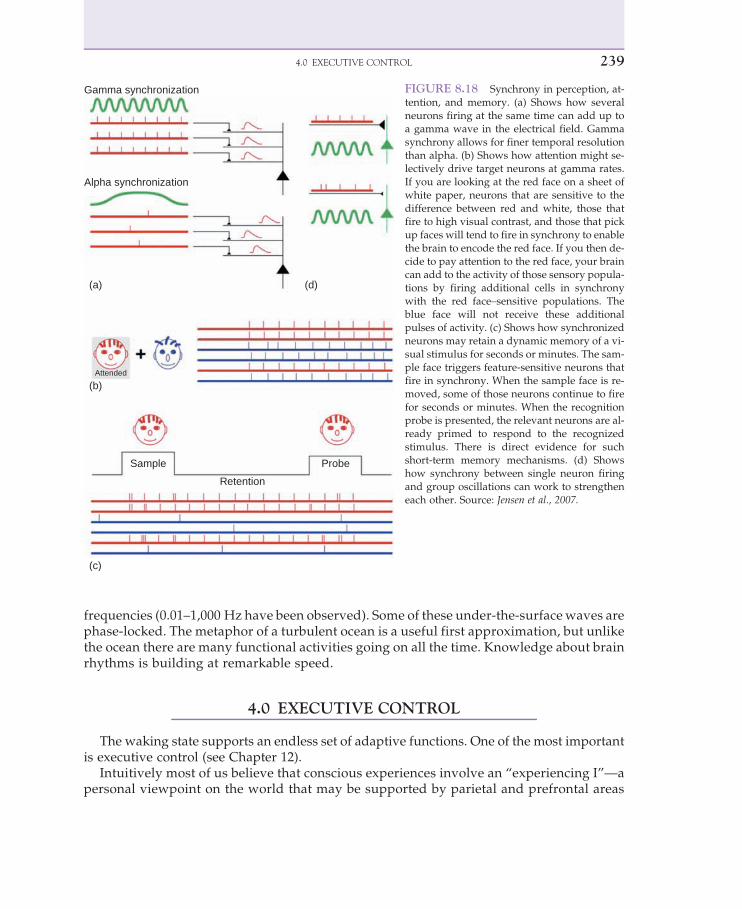
For example, the attentional network discovered by Posner and colleagues may dance tothe same beat as face-selective visual maps of the temporal lobe (Figure 8.18a-d). Figure 8.18bshows that the attentional influences could work by adding to the overall synchronystrengthens the red face signal and decreases the blue face activity by breaking up its synchro-nous activity. Finally, in Figure 8.18c, a synchronous population can respond to a stimulusand may keep running for some seconds or minutes after the stimulus has ended. Synchro-nouswave activity can therefore store a temporarymemory, but it may fade fairly quickly andmay also be vulnerable to interference from other stimuli.
These are only some of the coding possibilities of synchronous neurons and populations.Obviously, these hypotheses require evidence, as we will see.
3.5 Synchrony enables attention
Gamma synchrony may amplify neuronal population amplitude because synchronizedneurons can add to one another’s activity. Synchrony tends to increase the size of the signalin much the same way a microphone that picks up sounds from a loudspeaker will tend toamplify the signal over and over again. In biological systems, such self-amplifying feedbackcan never be allowed to run out of control for the same reason that sound systems can’t allowinfinite self-amplification. Audio systems have control circuits to prevent amplifier feedbackfrom overloading the speakers, not to mention the ears of the listeners. Epileptic seizures mayactually represent the wrong kind of self-amplification of slow rhythms, interfering with nor-mal brain functions and even leading to a loss of consciousness.
The raw EEG shows only the surface waves of a deep and turbulent ocean. Underneaththe visible EEG there are multiple oscillatory streams interacting over a wide range of
238 8. THE BRAIN IS CONSCIOUS
frequencies (0.01–1,000 Hz have been observed). Some of these under-the-surface waves arephase-locked. The metaphor of a turbulent ocean is a useful first approximation, but unlikethe ocean there are many functional activities going on all the time. Knowledge about brainrhythms is building at remarkable speed.
4.0 EXECUTIVE CONTROL
The waking state supports an endless set of adaptive functions. One of the most importantis executive control (see Chapter 12).
Intuitively most of us believe that conscious experiences involve an “experiencing I”—apersonal viewpoint on the world that may be supported by parietal and prefrontal areas
Gamma synchronization
Alpha synchronization
Attended
Sample Probe
Retention
(a)
(b)
(c)
(d)
FIGURE 8.18 Synchrony in perception, at-tention, and memory. (a) Shows how severalneurons firing at the same time can add up toa gamma wave in the electrical field. Gammasynchrony allows for finer temporal resolutionthan alpha. (b) Shows how attention might se-lectively drive target neurons at gamma rates.If you are looking at the red face on a sheet ofwhite paper, neurons that are sensitive to thedifference between red and white, those thatfire to high visual contrast, and those that pickup faces will tend to fire in synchrony to enablethe brain to encode the red face. If you then de-cide to pay attention to the red face, your braincan add to the activity of those sensory popula-tions by firing additional cells in synchronywith the red face–sensitive populations. Theblue face will not receive these additionalpulses of activity. (c) Shows how synchronizedneurons may retain a dynamic memory of a vi-sual stimulus for seconds or minutes. The sam-ple face triggers feature-sensitive neurons thatfire in synchrony. When the sample face is re-moved, some of those neurons continue to firefor seconds or minutes. When the recognitionprobe is presented, the relevant neurons are al-ready primed to respond to the recognizedstimulus. There is direct evidence for suchshort-term memory mechanisms. (d) Showshow synchrony between single neuron firingand group oscillations can work to strengtheneach other. Source: Jensen et al., 2007.
2394.0 EXECUTIVE CONTROL

of the cortex (Baars et al., 2003) (see Chapter 12). In daily life we use phrases like“I’m awake,” “I see the coffee cup,” “I lost consciousness,” “I couldn’t control myself,”and so on.
For many years the “observing I” was criticized as “the homunculus fallacy” (from theLatin word for “little man”). The problem, according to the critics, is that to make sense ofthe observing self, a little observer would have to sit in the brain, looking at the sensory in-flow. But to make sense of the little observer, it would also need another little observer insideof it, and so on ad infinitum. Such an infinite regress does not explain anything. It just movesthe burden of explanation to another level.
However, not all versions of an observing self lead to an infinite regress (Baars, 1988). Forexample, executive programs are routinely needed in robots, without leading to aninfinite set of control routines. Similarly, it is possible to have an executive capability inthe frontal and parietal lobes to interpret self-relevant information, such as “Is this new in-formation good for me? If not, will it hurt? How much? Should I run away? Would I feelembarrassed if I did?” The emotions are generally believed to process self-relevant informa-tion, but even body movement requires the visual system to interpret a flow of visual vistasin these terms. Watching a visual flow while sitting still (in a car or in a theater) is not thesame as actively walking or driving. Moving one’s eyes spontaneously is different frompushing the eyeball from the outside, as Helmholtz pointed out in a famous demonstration.The weight of scientific opinion may now be swinging back to the idea of an executive “I.”
There is a great deal of evidence for an executive network in the human brain (seeChapter 12). The brain areas in the cognitive control network (CCN) are shown inFigure 8.19, with connections—fiber tracts—that form the neural basis for the CCN. For exam-ple, certain kinds of brain damage seem to damage executive functions without necessarilyimpairing conscious perception. The classic case of Phineas Gage involved a radical changein personality and impaired self-control. The sense of agency in voluntary control is also
FIGURE 8.19 The Cognitive Control Net-work proposed by Cole and Schneider. The col-ored tracts are white matter fibers denselyconnected in the frontal lobes (on the left),and making parietal connections (on the right).The tractography images are in very high reso-lution. Source: W. Schneider, personal communi-
cation, 2009.
240 8. THE BRAIN IS CONSCIOUS

dependent on the frontal regions, and the right parietal region is a crucial area for “perspectiveof the self” in the neurological condition of parietal neglect (Baars, 2002; Baars et al., 2003).
4.1 Losing voluntary control
The outer muscles of the body and head are inhibited during sleep. Sleep-related muscleinhibition is easy to noticewhen your head starts to drop downwhen you feel drowsy. In yourbrain, a small circuit in the brainstem simply switches to its sleeping mode. It is now inhibit-ing body muscles. When you are awake, the same circuit maintains your upright posture andnormal muscle tone. Muscle inhibition in sleep probably evolved to avoid sleepwalking oracting out one’s dreams.
5.0 DREAMING
Hobson and colleagues (2000) define dreams as:
Mental activity occurring in sleep characterized by vivid sensorimotor imagery that is experienced as wak-ing reality, despite such distinctive cognitive features as impossibility or improbability of time, place, person,and actions; emotions, especially fear, elation, and anger predominate over sadness, shame, and guilt andsometimes reach sufficient strength to cause awakening; memory for even very vivid dreams is evanescentand tends to fade quickly upon awakening unless special steps are taken to retain it.
In a sense, dreaming is our most creative state. In normal waking we could never spin outthe flow of visual scenes, dramatic characters, or strange plots we constantly make up in ourdreams. It is almost as if we are creating a running movie but we can’t recall the storyline fromone scene to the next. Spontaneouswaking thoughts tend to be about our current concerns, anddream contents may refer to those concerns in a more visually symbolic way.
Both sensory input andmuscular output are blocked during REMdreaming, so the brain istalking only to itself (Figure 8.20). The existence of muscular inhibition may give rise to some“paralysis dreams,” in which we feel paralyzed or want to move but feel we cannot. This can
TABLE 8.2 What Makes Dreams Different
1. Hallucinations—especially visual and motoric, but occasionally in any and all sensory modalities2. Bizarreness—incongruity (imagery is strange, unusual, or impossible; discontinuity (imagery and plot can
change, appear or disappear rapidly); uncertainty (persons, places, and events often bizarrely uncertain bywaking standards)
3. Delusion—we are consistently duped into believing that we are awake (unless we cultivate lucidity)4. Self-reflection absent or greatly reduced—relative to waking5. Lack of orientational stability—people, times, and places are fused, plastic, incongruous, and discontinuous6. Narrative story lines—explain and integrate all the dream elements in a confabulatory manner7. Emotions increased—intensified and predominated by fear-anxiety8. Instinctual programs—(especially fight/flight) often incorporated9. Volitional control—greatly attenuated10. Memory deficits—across dream-wake, wake-dream, and dream-dream transitions
Source: Hobson in Squire et al., 2008.
2415.0 DREAMING

lead to unpleasant “locked-in” feelings during dreaming if we become aware of being unableto move. But muscular inhibition is an entirely normal part of dreaming. It keeps us from act-ing out our dreams.
Memory is also impaired during rapid eyemovement (REM) dreaming.When dreamers arewoken up from REM, they may report vivid visual memories, but they tend to fade in secondsunlesswemake a special effort to rehearse them. Even peoplewith good dream recall are likelyto remember only a tiny fraction of their nightly 90 to 120 minutes of REM (Figure 8.21).
5.1 Dreams are conscious
Dreams are reported as conscious experiences when people are woken up during REM.The EEG of REM dreaming is strikingly similar to wakefulness. It therefore seems that weactually have two daily conscious states: waking and REMdreaming. The activity of wakingand dreaming reflects a similar operating style by the thalamocortical system. Duringdreams, the EEG looks very much like waking consciousness (see Figure 8.1). The functionof dreams is still debated, but similar physiological states are found in many mammals.
FIGURE 8.20 The grayed-out boxes show impaired functions, including sensory and motor flow, executivefunctions, and some kinds of memory. Dreams are conscious states, but voluntary control is lost. Visual imageryis unusually vivid. Source: Baars, with permission.
242 8. THE BRAIN IS CONSCIOUS

Dreaming is therefore likely to have an evolutionary function. However, it is important tokeep in mind that there are important differences during REM as well, such as sensory andmotor blocking, a high level of emotional activity, and a low level of executive (frontal lobe)control.
5.2 Nonrational thinking
The surreal and nonrational nature of dreams may reflect this lower level of executive con-trol. Dreaming is a “hypofrontal” state, like drowsiness, alcohol inebriation, delirium, andother states of lowered self-reflection and self-control (Dietrich, 2003). In addition, the limbicregions of the brain, which are involved with emotions, show higher fMRI activity than in thewaking state (Figure 8.22).
5.3 Working memory in dreams
In dreams we can unexpectedly jump from one imagined scene to another, and dreamcharacters can change their identities, as if the brain is constantly trying to reinterpret the flowof imagined experiences from moment to moment. Waking events tend to be much morecoherent.
One possibility is that dreams overload our immediate memory with their creative flow ofemotions, images, and imaginary encounters. If we tried to spin dreamlike fantasies duringthe waking state, we would certainly lose track of our narrative, just because it’s hard to re-member all the details. Story writers work for hours and days to imagine what all of us ex-perience from minute to minute during sleep. The jumpy transitions in dreams may reflectmoments when we simply lose track of the plot.
FIGURE 8.21 The architecture of sleep. The thalamocortical core changes dramatically in sleep. In deep sleep, thelow, irregular oscillations ofwaking EEG change to high, slow, and regular activity, called deltawaves. The four levelsof arousability (Stages 1–4) show increasing delta waves, until Stage 4 of Non-REM (NREM-4) is almost all delta.Every 90 minutes or so, sleepers show rapid eye movements (REM) as the eyes swing back and forth. At the sametime the EEG shows striking waking-like activity in the EEG: fast, irregular, and low in amplitude. People who areawoken from REM tend to report vivid, narrative dreams, whereas SWS sleepers may report waking-like innerspeech. REM periods become longer in the second half of the night. Source: Stickgold & Walker, 2007.
2435.0 DREAMING

5.4 Brainstem oscillations trigger dreaming
REMdreaming is triggered by “PGOwaves”—pons-geniculate-occipital activation—alongwith neuromodulation. Sharp bursts of neuronal spikes come from the pons (P), activate thelateral geniculate nucleus of the thalamus (G), and then trigger visual experiences by way ofthe occipital cortex (O). Hobson and McCarley (1977), who advanced the early PGO hypoth-esis, called this the“activation-synthesishypothesis,”with theemphasison theactivationburstfrom the pons and its conscious interpretation or “synthesis” by the cortex (see Figure 8.22).Pace-Schott andHobson (2002)write thatREMsleep is also triggeredbychanges inmajor brainmodulators, especially norepinephrine, acetylcholine, serotonin (5-HT), and glutamine.
However, the PGO theory does not explain the content of dreams. It only suggests that thecortex tries to make a coherent story out of meaningless brainstem signals. What determinesdream contents remains a puzzle. Valli & Revonsuo (2006) proposed a “threat simulation”theory of dreams, an evolutionary hypothesis based on the fact that the REM state is widelyobserved in mammals and birds and therefore may have an important evolutionary function.
Motor perception
Lack of distinction between first-and third-person perspectives
Visual perception
Mind representation
Emotions
Bizarreness, discontinuity, andimprobable combinations
Delusional beliefof being awake
Lack of control on theflow of dream events
Amnesia at awakening
Fragmented episodicmemory recall
Uncritical acceptance ofcontent
(a)
(b)
(c)
FIGURE 8.22 Functional brain imaging of REM dreams. Red areas represent increased blood oxygen–related ac-tivity—for example, in emotion areas. Blue areas correspond to below-baseline activity during dreaming, as in thefrontal lobes. Higher activity in occipital regions may reflect vivid visual dream imagery. Low frontal lobe metabo-lismmay also explain the delusional beliefs, bizarreness, and suddendiscontinuities of dreams, such as rapid changesin scenes, dream characters, and narrative coherence. Low frontal lobe functioning is consistent with amnesia fordreams upon awakening and low levels of voluntary control. Source: Dang-Vu et al., 2005.
244 8. THE BRAIN IS CONSCIOUS
Valli & Revonsuo (2006) suggests that dreams allow us to mentally rehearse threatening sit-uations. Traumatized individuals may re-experience events related to the trauma (in somecases even during the waking state). Consistent with their theory, Valli & Revonsuo (2006)found that traumatized children from a war zone had significantly more threatening dreamsthan controls. Other major life events, such as loss of a loved one, or even normal stressfulevents, might also be reflected in dream contents.
5.5 Lucid dreaming
Some people can learn to experience their dreams knowing they are dreaming, and evenwith some voluntary control over dream events. LaBerge and colleagues (1986) made a casefor such “lucid dreaming” in the sleep laboratory by showing that some dreamers could learnto voluntarily move their eyes back and forth three times in a row, on cue, when given anauditory signal. Some lucid dreamers could also count to ten in the dream and then showthe experimenters that they had done so by repeating the triple eye movement to signalthe end of the ten-second period. Lucid dreamers in this experiment had to recall and carryout their task instructions learned during the waking period before going to sleep. Thus luciddreaming required executive functions and semantic memory retrieval, as well as linguisticand auditory processing. Lucid dreamers may therefore be able to control some of the cog-nitive functions that usually are impaired in the REM state.
6.0 DEEP SLEEP: UPS AND DOWNS
Deep sleep is the least conscious state of the daily cycle. It is also called “slow wave sleep”(SWS) because of the predominance of slow EEG waves over the cortex during this time. (seeFigure 8.2). In slow EEG, billions of neurons in the cortex and thalamus turn on and off in uni-son at the rate of the delta wave (about 2–4 Hz). The flow of signals is forced to screech to a haltevery half second, then start again, stop again, and so on. The fast and flexible flow of signalingis therefore constantly disrupted. Unconsciousness is the state where we cannot perform ournormal, waking activities because the engine of consciousness cannot maintain continuity.
Why do we sleep and dream for a third of the day? We only know a few of the answers.Deep sleep is the best time for consolidating what we learn during conscious periods intolong-termmemories. Sleep and dreaming probably have other functions. We know that hun-dreds of neuronal genes turn on and off whenever we fall asleep or we wake up. But there isno scientific agreement on the functions of our “offline” states.
Waking consciousness is the best time for learning, but learned material is “consolidated”during the next deep sleep period. If you study for an exam, it’s good to “sleep on it” and thenrefresh your memory in the morning. Much of yesterday’s learning will then be stabilized.
6.1 The need for sleep
Sleep-deprived rats die after only threeweeks.Weknow therefore that sleep is needed for sur-vival in rats and very likely in other mammals. Sleep duration is under precise biological regu-lation. We tend to make up for lost sleep by sleeping longer. Sleep-deprived individuals show
2456.0 DEEP SLEEP: UPS AND DOWNS

“microsleeps”—moments of “dropping off”—that can seriously interfere with actions like driv-ing. In a secondor two, yourneckmuscles can lose tone, andyourheadwill tend todrop forward.
As a major biological adaptation, sleep presumably does things that help us to survive andreproduce as a species. Many of its functions are programmed genetically, causing DNA-controlled proteins to build new synapses to consolidate learning. And yet, sleeping animalsare also more vulnerable to predators. Animals seek shelter before going to sleep, in trees, inearth hollows, or in dense bushes that are hard for predators to penetrate. Some animals relyon large colonies that sleep together so they can warn one another about predators. Never-theless, sleep is still a period of special danger because animals cannot run, flee, fight, or en-gage in mate selection and reproduction.
Our nightly eight hours of unconsciousness must therefore have some compensatory ad-vantages. We know that sleep seems to enable memory consolidation, converting new andunstable memories into lasting ones. The function of dreaming is still debated. Because thesleep-waking cycle is a very widely conservative feature among animals, it is likely to havemultiple functions.
Surprisingly, a single night of sleep deprivation can also improve depression. Staying up forone night therefore has been suggested as a safe, inexpensive, and effective treatment. Butchronic (long-lasting) sleepdeprivation is stressful anddegrades normal conscious functioning.
6.2 Memory replay and consolidation
SWS is more common in the first half of the night, whereas REM predominates in the sec-ond half. Researchers therefore have studied memory consolidation after “SWS-rich” and“REM-rich” periods of sleep. In general, SWS-rich periods appear to strengthen explicit mem-ories, and REM-rich periods strengthen procedural tasks and perhaps implicit memories.Increased SWS has been observed after intensive episodic learning, and increased REM isseen after procedural training.
Episodic memories are thought to be transferred from the hippocampus to the neocortex formemories to become enduring. The hippocampus plays a major role in place learning and nav-igation, and it has beenpossible to pinpoint hippocampal “place cells” that firewhen the animalpasses a particular point in the maze. As the rat runs the learned maze, place cells fire in se-quence.Afterward the samepattern of cell firing is observed during slowwave sleep, activatingother parts of the brain, like the thalamus, neocortex, and basal ganglia (Figures 8.23 and 8.24).
In rats, we can actually record neurons when the animals are exploring locations in se-quence and observe that the brain replays a learned spatial sequence during SWS. The thetarhythm appears to be a major rhythm for the hippocampal-neocortical dialogue that leads tomemory consolidation (Buzsaki, 2006; Jensen & Colgin, 2007).
7.0 EXCEPTIONAL STATES OF MIND
To achieve altered states, humans have used a huge variety of methods: taking psychoac-tive plants, fasting, drinking alcohol, special exercises, visualization, disorientation, hypoxia,self-mutilation, sexual practices, dramatic rites, lucid dreaming, dancing, whirling, chantingand music, suggestion, trauma, sleep deprivation, and social isolation. Traditions like
246 8. THE BRAIN IS CONSCIOUS

Buddhism and Vedanta Hinduism believe that humans should change their states of con-sciousness and self. That belief can be found in many times and places.
Simply asking people whether some practice makes them happier does not make for con-vincing evidence. Brain evidence may be more convincing. We cannot alter our EEG or fMRIscans to prove a favorite hypothesis. Decreased metabolism in the frontal lobes may be a
Amygdala
Hippocampus
Thalamus: Fast spindles
Slow+spindleSlow
Spindle
1s
20m
V
Cortex: Slow Up State + spindle
Hippocampus: Sharp memory spike
0.1 s
FIGURE 8.23 Slow wave sleep enables memory consolidation during “up states.”. Three brain regions work to-gether: early episodic learning during the waking state encodes unstable hippocampal and neocortical memorytraces. During slow wave sleep, thalamic spindles ignite, which triggers hippocampal sharp spikes, which are be-lieved to activate synchronized memory traces in the hippocampus. Thus thalamic spindle activity helps to triggerthe next up state, as shown by cortical iEEG. These regions act in concert to reactivate recent memory traces. Duringthe following down state of slow wave sleep, the activated neurons are believed to express proteins that lead to syn-aptic plasticity for more enduring memory coding. Source: Adapted from Steriade, 2000.
Sharp Wave-Ripple Complex
Ripple
Sharp Wave
CA3
CA1Subiculum
DentateGyrus
FIGURE 8.24 Hippocampal sharp andripple waves. A cartoon of the hippocampusshows its basic arrays of neurons,which gener-ate sharp waves and ripple complexes in acti-vating retrieval and neocortical consolidationduring slow wave sleep. Source: Beenhakker &Huguenard, 2009.
2477.0 EXCEPTIONAL STATES OF MIND
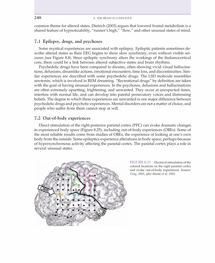
common theme for altered states. Dietrich (2003) argues that lowered frontal metabolism is ashared feature of hypnotizability, “runner’s high,” “flow,” and other unusual states of mind.
7.1 Epilepsy, drugs, and psychoses
Some mystical experiences are associated with epilepsy. Epileptic patients sometimes de-scribe altered states as their EEG begins to show slow synchrony, even without visible sei-zures (see Figure 8.8). Since epileptic synchrony alters the workings of the thalamocorticalcore, there could be a link between altered subjective states and brain rhythms.
Psychedelic drugs have been compared to dreams, often showing vivid visual hallucina-tions, delusions, dreamlike actions, emotional encounters, time loss, and discontinuities. Sim-ilar experiences are described with some psychedelic drugs. The LSD molecule resemblesserotonin, which is involved in REM dreaming. “Recreational drugs” by definition are takenwith the goal of having unusual experiences. In the psychoses, delusions and hallucinationsare often extremely upsetting, frightening, and unwanted. They occur at unexpected times,interfere with normal life, and can develop into painful persecutory voices and distressingbeliefs. The degree to which these experiences are unwanted is one major difference betweenpsychedelic drugs and psychotic experiences.Mental disorders are not amatter of choice, andpeople who suffer from them cannot stop at will.
7.2 Out-of-body experiences
Direct stimulation of the right posterior parietal cortex (PPC) can evoke dramatic changesin experienced body space (Figure 8.25), including out-of-body experiences (OBEs). Some ofthe most reliable results come from studies of OBEs, the experience of looking at one’s ownbody from the outside. Some epileptics experience alterations in body space, perhaps becauseof hypersynchronous activity affecting the parietal cortex. The parietal cortex plays a role inseveral unusual states.
FIGURE 8.25 Electrical stimulation of thecolored locations on the right parietal cortexand evoke out-of-body experiences. Source:Tong, 2003, after Blanke et al, 2002.
248 8. THE BRAIN IS CONSCIOUS

7.3 Neurofeedback
Neurofeedback training is defined as learning to control brain events by giving sensory(conscious) feedback, contingent on the event. Simply by holding a thermometer one canlearn to increase warmth, for example, which seems to help people relax and lower bloodpressure. Neurofeedback studies of animals have shown positive results over several de-cades. In a brain with billions of neurons, simple sensory feedback can allow training of vol-untary control over otherwise involuntary neuronal firing.
EEG neurofeedback show significant results, but long-term studies are often lacking. Be-cause neurofeedback may work in cases where other medical treatments have failed, long-term trials would seem to be vitally important.
7.4 Sensorimotor rhythm feedback
The sensorimotor response (SMR) is an alpha-like EEG pattern found over themotor cortexwhen people voluntarily suppress some planned action. SMR feedback training has beenshown to be an effective treatment in cases of human epilepsy, ADHD, and impulse controldisorders (Sterman, 2006). On average, 80 percent of patients trained to enhance SMR hadsignificant improvements.
Drug treatments for epilepsy do not always work, and having another treatment is helpfulfor many patients. Scientifically, because epilepsy shows slow, sychronous, and high wavesin the EEG, the ability to modify it by training reveals an interesting fact about the voluntarycontrol of brain rhythms.
7.5 Rhythmic entrainment
If brain rhythms constitute a basic neural code, it would be interesting to know if we candrive rhythms externally. If we listen to a 10-Hz auditory stream, will alpha waves increase?Brain wave entrainment has been described with TMS (transcranial magnetic stimulation)and tACS (transcranial alternating current). However, although there are many popularclaims about the effects of auditory entrainment, they have not been demonstrated to workby demanding medical and scientific standards so far.
7.6 Hypnosis and conversion
About one-fourth of the normal population is highly hypnotizable, as assessed by standardhypnotic procedures. Hypnotic suggestions can change brain events, as in reducing the av-eraged evoked potential (AEP) to a flashing light simply by “hallucinating” a cardboard boxcovering the stimulus (Spiegel, 2003). We do not know how hypnotic induction works. About5 percent of the population are said to be “hypnotic prodigies,” who can hallucinate percep-tual events, such as the buzzing of a fly; they can also enter into dissociated identity states(Hilgard, 1977). fMRI studies show that hypnotically suggested pain activates some ofthe same brain regions as real pain. Hypnotic procedures can alleviate chronic pain(Spiegel, 2003).
2497.0 EXCEPTIONAL STATES OF MIND

Hypnosis may involve a dissociation between voluntary control (dorsolateral prefrontalcortex, DL-PFC) and the ability to monitor errors (the anterior cingulate cortex, ACC). Egnerand colleagues (2005) reported an fMRI-EEG studyof hypnosis in the Stroop task showing that“hypnosis decouples cognitive control from conflict monitoring processes of the frontal lobe.”
Mild conversion symptoms are quite common in medicine. Medical students who arestudying serious diseases may become worried when they seem to notice “real” symptomsin themselves. “Medical student syndrome” is common and generally fades over time. Con-version disorders might be a result of the general human tendency toward autosuggestion.The placebo effect is a positive version of autosuggestion.
7.7 Meditation and yoga
The term meditation covers many mental techniques. One is silent repetition of a wordcalled a “mantra.” Asian and other traditions describe mantra meditation as a method forchanging mind states, as taught in Vedanta, Buddhism, and Chinese medicine. It has alsobeen widely practiced in Europe and the Middle East.
Meditation methods have been reported to increase coherence (synchrony), especially intheta, alpha, and beta-gamma EEG. Frontal-lobe coherence is also reported, as well as im-proved attentional functioning (Lazar et al., 2000; Lutz et al., 2004; Tang et al., 2007).
One of the surprises with mantra repetition is a significant drop in metabolic activity,reflected in “breath suspensions”—spontaneous stopping of the breath without compensa-tory breathing afterward. This is different from holding our breath voluntarily. In swimming,for example, we may take a deep breath before diving and then feel the need to take someextra breaths after coming up for air. The lack of compensatory breathing suggests that energydemand has indeed dropped more than an ordinary resting state, as has been demonstratedby measuring O2/CO2 exchange (Benson et al., 1975).
Herbert Benson and colleagues (1975) proposed that these results represent a “relaxationresponse,” like other physiological reflexes. Spontaneous breath suspensions are reported tobe associated with reports of periods of “pure consciousness,” defined as relaxed alertnesswithout any specific mental contents. Converging evidence has come from measures of me-tabolism, sympathetic nervous system tone and, recently, large-scale changes in gene expres-sion. Because the exact functions of “relaxation-related” genes are not yet clear, thispromising direction requires additional studies (Dusek et al., 2006; Jacobs et al., 1996).
A different procedure is called “mindfulness meditation.” Cahn and Polich (2006, 2009)describe it as “allowing any thoughts, feelings, or sensations to arise while maintaining a spe-cific attentional stance: awareness of the phenomenal field as an attentive and nonattachedobserver without judgment or analysis.” Mindfulness meditation has been shown to improvedepression and even suicidal thinking.
8.0 SUMMARY
Consciousness has intrigued human beings since the beginning of history. With improvedscientific tools we have seen considerable progress in recent years. Attention and conscious-ness can be considered complementary processes (see Figure 8.5). Conscious contents often
250 8. THE BRAIN IS CONSCIOUS

are thought to involve the widespread distribution of focal contents, like the sight of a rabbit.As soon as we see a rabbit (consciously), we can also judge whether it’s real or if it’s some-body’s pet.We can laugh or scream for fear of being bitten and try to remember if rabbits carryrabies. The point is that a great variety of brain events can be evoked by a conscious object,including a great variety of unconscious processes.
Conversely, we can select a source of information to focus on. Selective attention allows us tochoosewhatwewillbeconsciousof. Inorder to learnabout thebrain,wedirectattention to itoverand over again. Learning a difficult subject involves allocating attentional resources over time.
Consciousness typically involves a small amount of focal contents at any given moment,like the sight of coffee cup at a distance, which then may recruit and mobilize many uncon-scious brain functions. In attention, major functions like motivation, planning, emotionalneeds, and the like all interact with consciousness of a specific object in the world. Episodicand declarative types of learning seem to occur automatically as a function of conscious con-tents, which in turn may be guided by voluntary attention.
The three major brain states of the circadian cycle show different operating styles. A majorfunction of slowwave sleep is to consolidate the memory of events that were initially learnedin the waking state. Waking consciousness is by far the most active state in terms of survivaland reproduction, the two essential activities of any species. Food seeking, infant protection,fight or flight, tracking, hunting and gathering, social interactions, mate seeking—all thesegoal-directed activities require the conscious state. It follows that the brain (particularlythe cortex and its great input hub, the thalamus) must be able to organize itself around manydifferent goal-directed actions during waking.
REM dreaming looks similar to waking as measured by EEG. We can remember the last10 or 20 seconds of a dream as a conscious drama. The contents of dreams are hallucinatory,visually vivid, and often emotionally dramatic. Dream contents come from the brain itself,triggered by brainstem (PGO).
Deep sleep (slow wave sleep) appears to be unconscious, but people can remember somecontent when they are woken up from SWS. Slow delta waves occur at 2 to 4 Hz, and it hasbeen suggested that we are only truly unconscious during the valleys of the slow waves, the"down" cycle of the slow wave. At those times billions of neurons pause at the same time,making it impossible for neurons to signal one another. However, SWS is not inactive. Rather,it is the best time to convert learning into permanent synaptic changes.
9.0 STUDY QUESTIONS AND DRAWING EXERCISES
9.1 Study questions
1. What brain region triggers the circadian rhythm of waking, sleep and dreaming?2. What is the function of slow wave sleep? Of waking? Of dreaming? (If no function is
established, say so.)3. When you learn to ride a bicycle really well, what happens to your cortical
activity for sensorimotor control?4. How might two brain areas that represent the visual field be coordinated?5. What are the physiological signs of dreaming?
2519.0 STUDY QUESTIONS AND DRAWING EXERCISES

6. What are the main effects of selective attention? What part of the brain appears tocontrol voluntary, selective attention?
7. What’s meant by the “architecture of sleep”? Describe its features and timing.8. What are some hypotheses about deep or slow-wave sleep? What kind of function might
it have?9. What are the features of the conscious waking state? What kind of cortical waveforms
seem to occur?10. Describe the up state and the down state of slow-wave sleep. What are the implications
for brain processing?11. Describe neurofeedback training. What is it useful for?12. Is there convincing evidence that hypnosis reflects a brain state?13. What are the pros and cons of surface (scalp) EEG compared to intracranial EEG (iEEG)?14. Meditationmethods have been described for thousands of years.What are some common
features? What evidence do we have pro or con?
9.2 Drawing exercise
In Figure 8.26, indicate how dreams differ from waking. (Hint: What capacities are avail-able during dreaming but not waking? During waking but not dreaming?)
FIGURE 8.26 Source: Baars, with permission.
252 8. THE BRAIN IS CONSCIOUS