the blood chapter 10. composition and function of blood - components blood: transports everything...
TRANSCRIPT

The Blood
Chapter 10

Composition and Function of Blood - Components
Blood: Transports everything that is carried from one
place to another in the body – nutrients, wastes and body heat.
It is the only true FLUID tissue in the body because it has both solid and liquid components
It is a complex connective tissue in which living blood cells are suspended in a non-living fluid matrix called PLASMA
Plasma: The non-living fluid make-up of the blood – the
liquid component

Composition and Function of Blood - Components
Erythrocytes: Thickened portion of blood when put through a
centrifuge will separate from the plasma and appear as a red mass at the bottom of the tube
Buffy Coat: Thin, whitish layer between the formed elements
of the blood plasma (separates is from the erythrocytes) it contains:
Leukocytes: White blood cells – protect the body Platelets: Cell fragments that help in the blood-clotting
process

Composition and Function of Blood - Components
Hematocrit: Percentage of total blood volume
45 % of which is erythrocytes White blood cells and platelets are less than 1% Plasma makes up the remaining 55%
Composition and Function of Blood -Physical Characteristics and Volume
Blood: Sticky, opaque fluid Color depends on the amount of oxygen that it is carrying
Scarlet = oxygen rich Dull red = oxygen poor
Heavier than water and 5 x thicker (viscous) because of its formed elements
It is slightly alkaline with a pH of 7.35-7.45 The temperature of blood is slightly higher than body
temperature at approx. 100.4˚F It accounts for 8% of body weight and its volume is
approximately 5-6 liters

Composition and Function of Blood -Plasma
Approximately 90% water and is the liquid part of blood.
Contains dissolved substances such as: Nutrients Metal Ions Respiratory gases Hormones Plasma proteins Various wastes and products of cell metabolism

Composition and Function of Blood -Plasma
Plasma Proteins: Most abundant solute in plasma Made by the liver Serve a variety of functions:
Albumin: contributes to osmotic pressure of blood, helping to keep water in the bloodstream, clot blood vessels when blood is lost through an injured blood vessel, antibodies protect the body from pathogens.

Composition and Function of Blood -Plasma
Composition of plasma changes as cells remove or add substances to the blood
Homeostatic mechanisms help to keep it relatively consistent Example: when blood proteins drop to a low level
the liver will become stimulated to create more proteins and when it becomes to acid or to basic the respiratory system and kidneys take action to bring it back to its pH balance
Helps to distribute body heat evenly through the body

Composition and Function of Blood -Formed Elements
Erythrocytes make up most of the formed elements in blood Consist of 4-6 million red blood cells Transport oxygen and help to transport carbon dioxide
Leukocytes: 4,000-11,0000 white blood cells Responsible for body defenses and immunity Consist of Basophils, Neutrophils, Eosinohil, Lymphocytes
and Monocytes Platelets:
250, 000-500,000 Responsible for blood clotting

Composition and Function of Blood -Formed Elements
Erythrocytes: Red Blood Cells (RBC’s) Carry oxygen in blood to all cells of the body Anucleate: have no nucleus They contain very few organelles Mature RBC’s in the blood are sacs of hemoglobin
molecules Hemoglobin: Iron containing protein transports bulk of
oxygen carried in blood They lack mitochondria and make ATP by anaerobic
mechanisms and do NOT use any of the oxygen they are transporting

Composition and Function of Blood -Formed Elements
Erythrocytes: Small cells shaped like flat disks with an indented
center Their small size and shape provide a large surface
area relative to volume and make them ideal for gas exchange
RBC’s outnumber white blood cells by 1000 to 1 and contribute greatly to blood viscosity
Normally we have approximately 5 million RBC’s per cubic millimeter blood – when this number increases the blood viscosity increases and when the number decreases the blood viscosity decreases making the blood thin and flow rapidly

Composition and Function of Blood -Formed Elements
Erythrocytes: The amount of hemoglobin in the blood stream will
determine how well the erythrocytes can perform oxygen transport
The more hemoglobin molecules the RBC’s have the more oxygen they can carry
A single RBC contains 250 million hemoglobin molecules each of which can bind 4 molecules of oxygen enabling the RBC to carry approximately 1 billion molecules of oxygen!
Men have a slightly higher hemoglobin content than women

Composition and Function of Blood -Formed Elements Erythrocytes:
Anemia: a decrease in the oxygen carrying ability of the blood. Can result from lower than normal RBC’s or abnormal or deficient hemoglobin content in the RBC’s
Sickle Cell Anemia: genetic disorder. Abnormal hemoglobin forms and becomes spiky and sharp when the RBC’s unload oxygen or when oxygen content is lower than normal. This deformity causes them to rupture easily and block the small blood vessels interfering with oxygen delivery and causing pain. It is most common in black people who live or have ancestors who lived in the malaria belt of Africa

Composition and Function of Blood -Formed Elements
Erythrocytes: Polycythemia: An excessive or abnormal
increase in the number of erythrocytes Results from bone marrow cancer or can be a
normal physiological response to living at a high altitude
Major problem resulting from polycythemia is that the viscosity of blood is increased causing it to move slowly through our systems.

Composition and Function of Blood -Formed Elements
Leukocytes: White Blood Cells (WBC’s) Less numerous than RBC’s Crucial to defense of the body systems against
disease The only complete cells in blood – containing a
nucleus, nuclei and the usual organelles Perform a protective “army” that defends
against damage from bacteria, viruses, parasites and tumor cells

Composition and Function of Blood -Formed Elements
Leukocytes: Unlike RBC’s which are confined to the blood only
WBC’s can move in and out of the blood vessels through a process called diapedesis.
The circulatory system is just their way of getting to the areas where they are needed for inflammatory or immune responses
They move to damaged or infected areas by responding to certain chemicals that are released when a cell is damaged
Positive chemotaxis: the capability to respond to the damaged cells chemical release.
Composition and Function of Blood -Formed Elements
Leukocytes: Amoeboid Motion: the formation of flowing
extensions to move them through the vessels to the damaged area by a diffusion gradient
Once the WBC’s have reached the damaged cell they will form around the cell and destroy it
When WBC’s are called to action the body will produce up to twice the normal WBC’s = Leukocytosis. If leukocytosis is read in a persons blood draw this indicates the body is preparing for some bacterial or viral infection.

Composition and Function of Blood -Formed Elements
Leukocytes Leukopenia: is the opposite of leukocytosis and
means that the WBC count is lower than normal. This may be caused by certain drugs like corticosteroids (anti-inflammatory) or cancer fighting agents.
Leukemia: meaning “white blood”. The bone marrow becomes cancerous and produces huge numbers of WBC’s and continually releases them into the body.
This would not normally be a problem but the newly created WBC’s are immature and cannot carry out their normal functions allowing the body to become “prey” for bacteria and viruses.

Composition and Function of Blood -Formed Elements
Leukocytes: White blood cells are categorized in two ways
depending on if they contain visible granules in their cytoplasm or not.
Granulocytes: granule containing WBC’s• They contain lobed nuclei which are rounded nuclear
areas connected by thin strands of nuclear material• The granules in the cytoplasm contain neutrophils,
eosinophils and basophils
Composition and Function of Blood -Formed Elements
Leukocytes: Granulocytes:
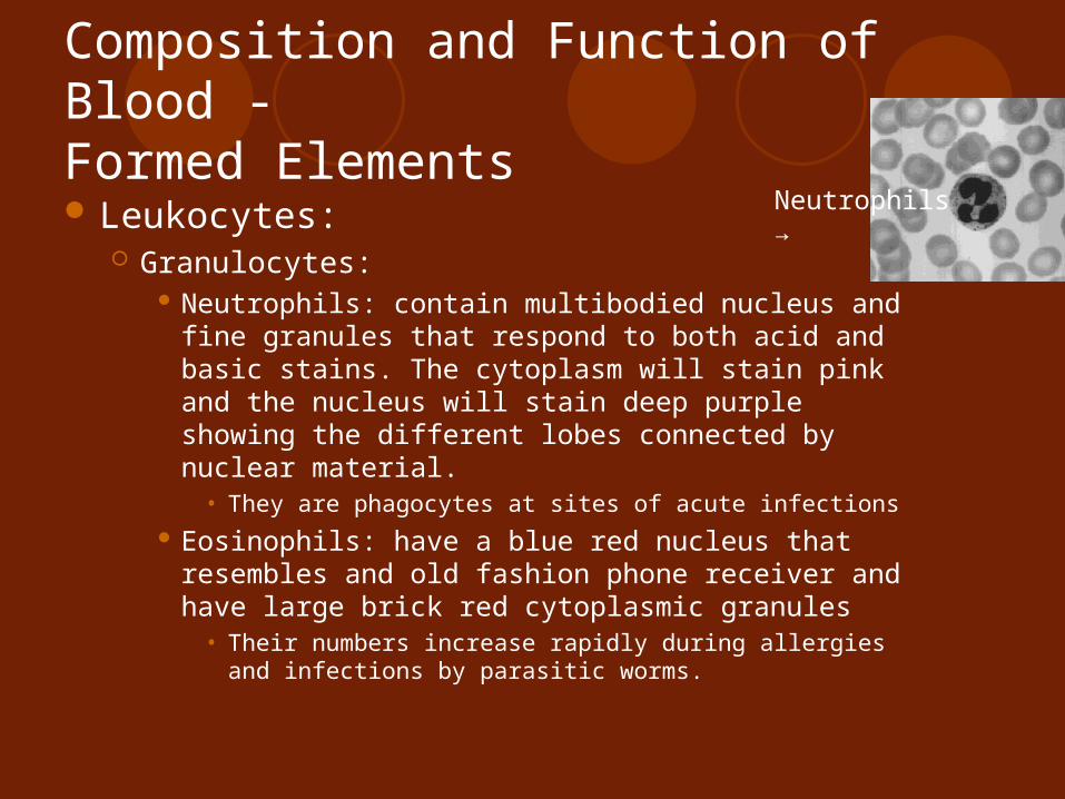
Neutrophils: contain multibodied nucleus and fine granules that respond to both acid and basic stains. The cytoplasm will stain pink and the nucleus will stain deep purple showing the different lobes connected by nuclear material.
• They are phagocytes at sites of acute infections Eosinophils: have a blue red nucleus that
resembles and old fashion phone receiver and have large brick red cytoplasmic granules
• Their numbers increase rapidly during allergies and infections by parasitic worms.
Neutrophils →

Composition and Function of Blood -Formed Elements
Leukocytes: Granulocytes:
Basophils: the rarest of WBC’s contain large histamine containing granules that stain dark blue
• Histamine is a inflammatory chemical that makes blood vessels leak and attracts other WBC’s to the infected area.

Composition and Function of Blood -Formed Elements
Leukocytes The second type of WBC is Agranulocytes: They lack visible cytoplasmic granules and their
nuclei are closer to normal looking They include lymphocytes and monocytes
Lymphocytes: have large dark purple nucleus that occupies most of the cells volume. They are slightly larger than RBC’s and are found in the lymphatic tissues where they play important roles in immune response.
Composition and Function of Blood -Formed Elements Leukocytes:
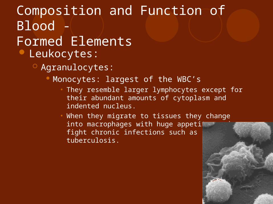
Agranulocytes: Monocytes: largest of the WBC’s
• They resemble larger lymphocytes except for their abundant amounts of cytoplasm and indented nucleus.
• When they migrate to tissues they change into macrophages with huge appetites and fight chronic infections such as tuberculosis.

Composition and Function of Blood -Formed Elements
Platelets: Are not cells but are fragments of bizarre multinucleate
cells called Megakaryocytes which rupture and release thousands of anucleate pieces that seal themselves off from surrounding fluids.
These platelets appear as dark staining irregularly shaped bodies scattered among other blood cells
They are important for the blood clotting processes that occurs in vessels when they are ruptured or broken.

Hematopoiesis (blood cell formation)
The formation of blood cells occurs in the red bone marrow or Myeloid tissue which is found mostly in the flat bones of the adult skeleton.
Each type of blood cell is produced in different numbers in response to changing body needs and different stimuli.
Once they are produced and mature they will be discharged into the blood vessels that surround the area

Hematopoiesis (blood cell formation)
Hemocytoblast: the stem cell from which all formed elements will come from
The development of the cells will differ and once they are designated to a specific body area/blood pathway they cannot change
There are two types of stem cells which are produced by the hemocytoblasts: Lymphoid stem cell Myeloid stem cell

Hematopoiesis (blood cell formation)
Lymphoid stem cell: produces lymphocytes Myeloid stem cell: can produce all other classes
of formed elements Since RBC’s are anucleate they cannot
synthesize proteins, grow or divide so as they age they become brittle and fragment/fall apart in approximately 100-120 days
Their remains will then be eliminated by phagocytes in the spleen, liver and other body tissues

Hematopoiesis (blood cell formation)
The lost cells are replaced by the division of the hemocytoblasts within the red bone marrow.
The developing RBC’s can divide many times and synthesize large amounts of hemoglobin
When enough hemoglobin has been built up the nucleus and other organelles are ejected and the cell collapses inward becoming a young RBC or reticulocyte because it still contains some endoplasmic reticulum.
Hematopoiesis (blood cell formation)
Reticulocytes enter the bloodstream and begin transporting oxygen and within 2 days of being released in the bloodstream they will get rid of the remaining ER and become fully functional erythrocytes
The development from a hemocytoblast to a mature RBC takes approximately 3-5 days.

Hematopoiesis (blood cell formation)
Erythropoietin: the hormone which controls the production of erythrocytes.
Normally we have small amounts of erythropoietin in our blood circulation at all times and RBC’s are constantly being produced.
The liver produces some of this hormone but most of it is made in the kidneys
When blood levels and oxygen decline for any reason the kidneys release their erythropoietin which targets the red bone marrow and stimulates it to produce more RBC’s
If there is excessive amounts of oxygen in the blood stream the amount of erythropoietin released will be decreased.

Hematopoiesis (blood cell formation)
The formation of leukocytes and platelets is also stimulated by hormones.
Colony Stimulating Factors (CSF’s) and Interleukins are what prompt the red bone marrow to turn out leukocytes and enhance the ability of mature leukocytes to protect the body.
Chemical signals in the body found in inflammatory conditions also stimulate leukocytes to be produced or be enhanced.

Hematopoiesis (blood cell formation)
Exposure to bacteria or toxins will also stimulate macrophages and lymphocytes to release CSF’s and interleukins which will pull together WBC’s to fight off the attack
Thrombopoietin: Hormone which accelerates the production of platelets
Hemostasis
The stopping of blood flow because of some type of damage to the endothelium of the blood vessel wall
A very fast response which is localized to a particular area
Involves three phases that occur in a rapid sequence: Vascular spasms Platelet plug formation Coagulation Blood clotting

Hemostasis
Steps of Hemostasis: Platelets are repelled by an intact endothelium.
When the endothelium is broken the underlying collagen fibers are exposed and the platelets become “sticky” and cling to the damaged area.
These platelets will then release chemicals to attract more platelets to the area.
As more and more platelets accumulate a small mass called a platelet plug or white thrombus is formed.

Hemostasis
Steps of Hemostasis: Once anchored the platelets release serotonin
which causes the blood vessel to go into spasms.
These spasms narrow the blood vessel at that point, decreasing the blood flow until clotting can occur.
At the same time the injured tissues release thromboplastin which will play an important role in the clotting process.
Hemostasis Steps of Hemostasis:
PF3: a phospholipid that coats the surfaces of the platelets will interact with thromboplastin and calcium ions to form an activator that triggers the “clotting cascade”
PF3 the prothrobin activator converts prothrobin present in plasma to thrombin – an enzyme
Once this occurs the thrombin will join soluble fibrinogen proteins and create long hairlike molecules of insoluble fibrin
Fibrin then forms a “mesh” trap for RBC’s to form the basis of a blood clot.
Within an hour of this the clot will begin to react and squeeze serum from the mass and pull the ruptured edges of the blood vessel together.

Hemostasis
Normal blood clotting occurs within 3-6 minutes and as quickly as it is started it can stop so that widespread clotting does not occur – which would make the blood hard.
Applying pressure to a wound will help to speed up the clotting process.

Disorders of Hemostasis
Undesirable clotting: For unknown reasons sometimes clots will form
in intact blood vessels – most commonly the legs.
Thrombus: a clot that develops and persists in an unbroken blood vessel
If it is large enough it may block the vessel and prevent blood flow to the cells beyond the blockage
Embolus: a thrombus that breaks away from the blood vessel wall and floats freely in the blood stream.

Disorders of Hemostasis
Undesirable clotting: An embolus is usually not a problem unless it
gets lodged in a blood vessel that is too narrow for it to pass through.
Possible causes of undesirable clotting may be: Anything that roughens the endothelium of a blood
vessel and encourages clinging of platelets such as severe burns, physical blows or an accumulation of fatty material.
Slow flowing blood or blood pooling is a risk factor especially in people who are unable to move
Disorders of Hemostasis
Bleeding Disorders: The most common cause of abnormal bleeding
is platelet deficiency or thrombocytopenia = decreased number of platelets
Thrombocytopenia: result of an insufficient number of circulating platelets
Petechia: small purple colored blotches on the skin that indicate small vessel breakages that can occur with normal movement.
Usually arises from conditions where the myeloid tissue is suppressed, bone marrow cancer, radiation or certain drugs.

Disorders of Hemostasis
Bleeding Disorders: When the liver cannot synthesize its usual
clotting factors severe bleeding may occur. Vitamin K is used by the liver to produce these
clotting factors and may be deficient, causing the severe bleeding. If so then vitamin K supplements will solve the problem.
If liver function is severely impaired (Hepatitis, Cirrhosis, etc.) then blood transfusions will help.

Disorders of Hemostasis
Bleeding Disorders: Hemophilia: Hereditary bleeding disorder that
results from a lack of any of the factors that are present during clotting.
With hemophilia a simple cut or scratch will bleed for a prolonged period of time and could potentially be life threatening.
If the bleeding is occurring internally in the joints it will be painful and they could become disabled.

Disorders of Hemostasis
Bleeding disorders: Hemophiliacs are given transfusions of fresh
plasma or injections of the clotting factor they are missing in order to stop the bleeding episodes.
Because of their need for transfusions and lack of clotting factors hemophiliacs can become victims of AIDS and Hepatitis more easily because they cannot defend as well against the viruses.

Blood Groups and Transfusions
The body can lose up to 15% of its blood without too many problems.
If blood loss is between 15-30% pallor (white coloring) or weakness can occur
Losses over 30% will cause severe shock and can be fatal.
Blood transfusions can be given to replace this lost blood. For transfusions blood is collected from a donor and mixed with an anticoagulant to prevent clotting. This blood can then be stored for approximately 35 days until its needed.

Blood Groups and Transfusions
Blood Groups: People have different blood groups and if a
transfusion is given with mismatched blood it can be fatal.
The plasma membrane of an RBC has genetically determined proteins (Antigens) that identify you as unique!
Because the body normally recognizes antigens as a foreign body it will defend against it – thus making a transfusion with the wrong blood type fatal.

Blood Groups and Transfusions
Blood Groups: Agglutination: the binding of the antibodies on
the surface of the antigens which will cause RBC’s to clump together.
After agglutination occurs the foreign RBC’s will rupture and release hemoglobin into the bloodstream. This will cause a decrease in the ability to carry oxygen to areas of the body.
In severe cases of mismatched transfusions the hemoglobin molecules may block the kidney tubules and cause kidney failure.

Blood Groups and Transfusions
Blood Groups: Treatment to prevent kidney damage is to
infuse alkaline fluids to dilute and dissolve the hemoglobin along with diuretics which will flush it out of the body through urine.
There are over 30 different RBC antigens in humans allowing each persons blood cell to be classified into different blood groups.
The antigens of the ABO and Rh blood groups cause the most vigorous transfusion reactions.

Blood Groups and Transfusions
Blood Groups: ABO blood groups: based on which of two antigens,
type A or type B, you inherit. Absence of both of these antigens results in a type O
blood. Presence of both will lead to type AB If you only have antigen A you will have type A blood
and if only antigen B is present you will have type B blood.
You will build up antibodies to antigen A or antigen B depending on which you are missing.

Blood Groups and Transfusions
Rh Blood groups: Named because one of the Rh antigens
(antigen D) was originally found in rhesus monkeys then later found in humans.
Most Americans are Rh positive which means we carry the Rh antigen in our RBC’s.
Anti-Rh antibodies are not automatically formed and present in the blood of Rh- individuals
If an Rh- receives Rh+ blood shortly after the transfusion their immune system becomes sensitized and will produce antibodies against the foreign blood type.

Blood Groups and Transfusions
Rh Blood Groups: Hemolysis: rupture of the RBC’s that creates antigens
will not occur with the first transfusion because it takes time for the body to react and begin its antibody production. Every time after the first the body will react to the transfusion and attack and rupture the donated RBC’s
An important reaction occurs when Rh- women are carrying Rh+ babies. The first child born will be healthy but because the mother is sensitized by the Rh+ antigen that have passed through the placenta to her bloodstream she will form Rh+ antibodies unless she gets treated with RhoGAM shortly after giving birth.
RhoGAM is an immune serum that prevents the sensitization and decreases her immune response to the Rh+

Blood Groups and Transfusions
Rh Blood Groups: Hemolytic disease of the newborn: if the mother
is not treated with the RhoGAM then with her next pregnancy her body will send out the Rh+ antibodies and destroy the babies RBC’s. The baby will be anemic and become hypoxic (lack of oxygen). Brain damage and death could occur if fetal transfusions are not done before birth to provide more RBC’s to transport oxygen.

Blood Groups and Transfusions
Blood typing: In order to make sure that an accurate and safe
transfusion is made both the recipient and donor’s blood type must be determined.
To determine blood type a blood draw is taken and mixed with two different types of immune serum – anti-A and anti-B
Agglutination will occur when the RBC’s of a group A person are mixed with the anti-A serum and likewise when group B is mixed with anti-B

Blood Typing
Go to the link below and read the statement on blood typing and do the activity linked with it.
http://nobelprize.org/medicine/educational/landsteiner/readmore.html
Developmental Aspects of Blood