the biology of colletotrichum acutatum
TRANSCRIPT
The biology of Colletotrichum acutatum
by
Phillip S. Wharton1 & Javier Diéguez-Uribeondo2
1 Department Plant Pathology, Michigan State University, USA. [email protected] Real Jardín Botánico, Plaza de Murillo 2, E-28014 Madrid, Spain. [email protected] (corresponding author)
Abstract
Colletotrichum acutatum is major pathogen of fruit crops, cau-sing economically important losses of temperate, subtropicaland tropical fruits worldwide. However, few studies have beencarried out on key aspects of its biology. This is mainly becausetraditionally isolates of C. acutatum were often wrongly identi-fied as C. gloeosporioides. Effective separation of the two spe-cies was not possible until the introduction of molecular tools fortaxonomy. The life cycle of C. acutatum comprises a sexual andan asexual stage and much remains to be resolved regarding thegenetics of sexuality and the effects of the sexual stage on po-pulation structure. Colletotrichum acutatum exhibits both infec-tion strategies described for Colletotrichum species, i.e. intrace-llular hemibiotrophy and subcuticular-intramural necrotrophy,and may also undergo a period of quiescence in order to over-come resistance mechanisms in immature fruit such as pre-for-med toxic compounds and phytoalexins, or due to the unsuita-bility of unripe fruit to fulfill the nutritional and energy require-ments of the pathogen. Colletotrichum acutatum may overwin-ter as mycelium and/or appressoria in or on different parts of thehost. Conidia are water-born and spread by rain episodes so in-fections are usually highest during the wettest periods of thegrowing season. Current management strategies for this funguscomprise the exploitation of cultivar resistance, cultural, chemi-cal, and biological control methods, and preventive strategiessuch as disease-forecasting models. This review focuses on thecurrent knowledge of biological aspects of C. acutatum and re-lated Colletotrichum species and includes a discussion of theprogress towards their control.
Key words: Anthracnose, Ascomycete taxonomy, fungal disea-ses, infection, appressorium, host pathogen interactions, post-harvest, fungicide, fruit, and disease control.
Resumen
Colletotrichum acutatum es uno de los principales hongos pató-genos en agricultura y responsable de importantes pérdidaseconómicas en frutales en áreas tanto de climas templadoscomo subtropicales y tropicales. Sin embargo, existen pocos es-tudios sobre aspectos clave de su biología. Esto es debido a que,tradicionalmente, muchos aislamientos de C. acutatum se hanidentificado como C. gloeosporioides. El uso de técnicas de bio-logía molecular ha posibilitado la distinción entre ambas. El ciclovital de C. acutatum comprende una fase sexual y otra asexual,y todavía quedan por conocer muchos aspectos genéticos de sufase sexual y de su relevancia en la estructura de la población.Colletotrichum acutatum posee los dos tipos de estrategias deinfección descritas en el género Colletotrichum, intracelular he-mibiotrófica y subcuticular-intramural necrotrófica, y puede in-cluso establecer un periodo de latencia con la finalidad de hacerfrente a los mecanismos de defensa del hospedante tales como:existencia de compuestos tóxicos preformados y fitoalexinas, laescasez de nutrientes del propio tejido del hospedante para ha-cer frente a los requerimientos energéticos del patógeno. Colle-totrichum acutatum generalmente inverna como micelio y/oapresorios en distintas partes del hospedante. Los conidios re-quieren la presencia de agua para ser producidos y su dispersiónse produce con la lluvia. Las actuales medidas de manejo de estehongo comprenden el aprovechamiento de la distinta resisten-cia de cultivares, formas y manejo de los cultivos, métodos quí-micos y de control biológico, así como estrategias preventivas ta-les como modelos de predicción de las enfermedades. Así pues,el objetivo de este trabajo es presentar los conocimientos actua-les sobre distintos aspectos de la biología de C. acutatum y otrasespecies relacionadas e incluye una discusión sobre los adelan-tos para el control de este hongo.
Palabras clave: antracnosis, taxonomía de ascomicetes, enfer-medades fúngicas, infección, apresorio, interacciones patóge-no-hospedante, postcosecha, fungicida, fruta, control de enfer-medades.
Anales del Jardín Botánico de Madrid 61(1): 3-22www.rjb.csic.es
4 Anales del Jardín Botánico de Madrid 61(1) 2004
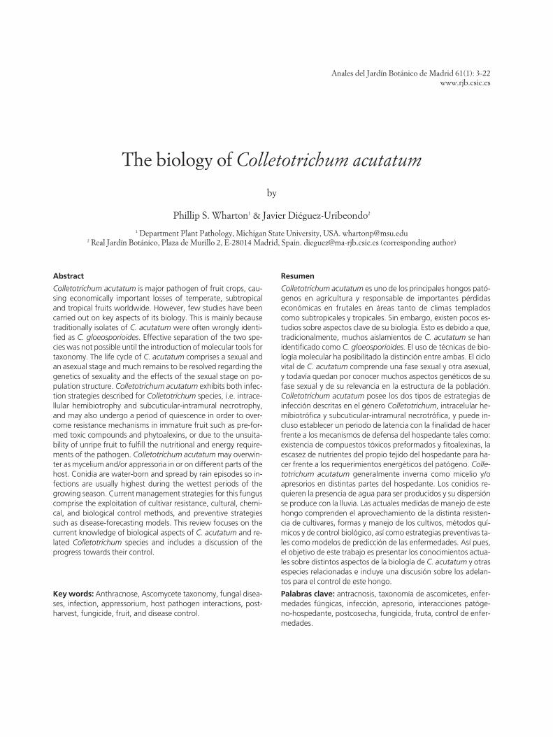
Fig. 1. Symptoms of infection of Colletotrichum acutatum in almond tissues. Colletotrichum acutatum causes pre- and post-harvest los-ses by affecting most parts of the tree. A, Blossom blight. Disease symptoms first become apparent on almond blossoms. B, The infec-tion may continue into the spurs and shoots resulting in shoot dieback. C, Hyphae of the fungus growing in a senescent leaf. (D-F), Fruitcan be infected at all stages of development. D, Infected almond fruit. E, Quiescent infection manifesting after incubation under highhumidity. F, Quiescent infection in almond kernels. Infected kernels may show internal bluish staining after the nuts are harvested.
Introduction
Colletotrichum Corda is a large genus of As-comycete fungi, containing species that are amongstthe most successful plant pathogenic fungi, causingsignificant economic damage to crops in tropical, sub-tropical, and temperate regions (Bailey & Jeger, 1992).Their economic impact has lead to extensive studieson diverse aspects of its biology such as, host specifici-ty (Correl & al., 2000; Freeman, 2000), cell biology ofinfection processes (Bailey & al., 1992; O’Connell &al., 2000), fungal-host interaction (Prusky & Plumbley,1992; Prusky, 1996; Prusky & al., 2000), genetic diver-sity (Freeman, 2000), and epidemiology (Förster &Adaskaveg, 1999; Timmer & Brown, 2000). Species ofthis genus have been used as models for studying in-fection strategies and host-parasite interactions (Per-fect & al., 1999), defining the genetic basis of fungalsymbiotic life styles (Rodriguez & al., 2000), and fordeveloping infection (Fitzell & al., 1984) and diseaseforecasting systems (Dannenberger & al., 1984; Tim-mer & Zitko, 1993, 1996; Monroe & al., 1997;Adaskaveg & al., 2001, 2002; Uddin & al., 2002).
One of the most pathogenic species of this genus isColletotrichum acutatum J.H. Simmonds, which causesanthracnose and blight in agriculturally importanthosts such as almond (Prunus dulcis (Mill.) D.A. Web.)(Ogawa & English, 1991; Adaskaveg & Hartin, 1997;Förster & Adaskaveg, 1999) (Fig. 1), avocado (Perseaspp.) (Freeman, 2000), peach (Prunus persica L.)(Adaskaveg & Hartin, 1997; Zaitlin & al., 2000), blue-berries (Vaccinium spp.) (Smith & al., 1996; Schilder &al., 2001; Yoshida & Tsukiboshi, 2002) (Fig. 2), citrus(Citrus spp.) (Zulfiqar & al., 1996; Timmer & Brown,2000), mango (Magnifera indica L.) (Fitzell, 1979;Arauz, 2000), olive (Olea europaea L.) (Martín & Gar-cía-Figueres, 1999), and strawberry (Fragaria × ananas-sa Duch.) (Smith & Black, 1990; Howard & al., 1992;Curry & al., 2002).
Colletotrichum acutatum can affect most parts ofthe plant, from the roots to the leaves, blossoms,twigs, and fruit, causing diseases such crown root rot,defoliation, blossom blight, and fruit rot (Figs. 1 and2). However, as for most Colletotrichum species, themost significant losses due to infection by C. acutatumare incurred when fruit is attacked (Bailey & Jeger,

1992). There are two distinct types of Colletotrichumdiseases affecting fruit, those causing disease on im-mature and developing fruit in the field (pre-harvest)and those damaging mature fruit at harvest and dur-ing storage (post-harvest). Fruit affected by post-har-vest Colletotrichum species often appear completelyhealthy at the time of harvest, with disease symptomsonly manifesting themselves during storage (Figs. 1E-F and 2B). This is due the ability of many Colleto-trichum species to cause latent or quiescent infectionsin which the fungus infects immature fruit in the fieldand then becomes dormant until the fruit ripens, atwhich time it resumes its growth causing disease onthe fruit (Prusky & Plumbey, 1992; Prusky, 1996).
In spite of its economic impact, few studies havebeen carried out on key aspects of the biology of C. acu-
P.S. Wharton & J. Diéguez-Uribeondo: The biology of Colletotrichum acutatum 5
tatum. An improved understanding of its developmen-tal biology, infection processes, host pathogen interac-tions and epidemiology may lead to the development ofmore efficient control and management strategies.Thus, the purpose of this review is to compile the cur-rent knowledge on the biology of C. acutatum and re-lated pathogenic species in relation to anthracnose ofeconomically important fruit crops such as almond,blueberry and citrus. A discussion of the recentprogress towards control of this fungus is also included.
The taxonomic status
Fungi classified in the ascomycete (telomorphic)genus Glomerella Spauld. & H. Schrenk and thecoelomycete (anamorphic) genus Colletotrichum have
Fig. 2. Symptoms of infection of Colletotrichum acutatum on blueberry tissues. A, Anthracnose fruit-rot. Fruit do not develop symptomsuntil they are mature (blue). Bright orange spore masses are produced within shriveled, sunken areas on the fruit surface. The sticky spo-re masses spread to other fruit by splashing water and contact. B, Under optimal conditions in storage, the fungus may sporulate cove-ring the entire fruit surface with bright orange spore masses. C (i-iv), Growth and colonization of blueberry twigs by C. acutatum. Thisfungus overwinters in flower buds. In the spring the fungus grows out of the buds (i) and into surrounding tissue. The fungus then growsdown the twig killing the tissue and eventually sporulates (ii-iv). D, Bright orange C. acutatum spore masses on a dead blueberry twig.
proved some of the most challenging to taxonomists.While the generic limits are today relatively well de-fined, the concept of a species in the genus Col-letotrichum is not well established nor universally ac-cepted (Sutton, 1992). The present taxonomic con-cepts of the group largely follow von Arx (1957, 1970)and Sutton (1980). Morphological characteristics andhost range have traditionally been used to define thespecies, although excessive reliance on the latter hasled to a proliferation of unnecessary names. This maybe partly due to the wide host range of a number ofColletotrichum species and the fact that several Col-letotrichum species may be associated with a singlehost (Freeman & al., 1998). The problem is exempli-fied by the confusion that exists with regard to the Col-letotrichum species that infect fruit. Colletotrichumacutatum and C. gloeosporioides (Penz.) Penz. & Sacc.are the two members of the genus that are most com-monly associated with fruit rots in the literature. Col-letotrichum gloeosporioides is considered a cumulativespecies and is found on a wide variety of fruits, includ-ing almond, apple, avocado, citrus, mango olive, andstrawberry (Fitzell, 1979; Sutton, 1992; Freeman &Shabi, 1996; Freeman & al., 1998; Martín & García-Figueres, 1999; Arauz, 2000; Timmer & Brown, 2000).Likewise, C. acutatum has also been reported to infecta large number of fruit crops (Freeman & al., 1998;Martín & García-Figueres, 1999; Adaskaveg &Förster, 2000; Yoshida & Tsukiboshi, 2002).
Colletotrichum acutatum and C. gloeosporioides aremorphologically very similar and because of their over-lapping host ranges and the extensive variability thattheir isolates show in culture, it has been very difficultto separate them by traditional taxonomical methods.Nonetheless, these two species have been successfullyseparated based on a number of characteristics includ-ing culture morphology, conidium shape and size, andhost-range (Smith & Black, 1990; Sutton, 1992; Förster& Adaskaveg, 1999). However, these techniques haveto be used with caution, as they are prone to error. Forexample, Förster & Adaskaveg (1999) and Adaskaveg& Förster (2000) included culture morphology, andconidium size and shape in a comparison of strains iso-lated from almond assigned to C. acutatum, and strainsisolated from citrus identified as C. gloeosporioides. Iso-lates of each species could be distinguished by conidi-um shape when cultures were grown on potato dex-trose agar (conidia with rounded ends were identifiedas C. gloeosporioides and conidia with pointed ends asC. acutatum). However, on pea straw agar the conidialsize of the two species overlapped, showing that thischaracter is not reliable for distinguishing between thetwo species. Another character often used for isolate
description is colony morphology. Colonies ofC. gloeosporioides are usually gray in appearance whileC. acutatum colonies had a pink or orange phenotype(Zulfiqar & al., 1999; Martín & García-Figueres, 1999).Förster & Adaskaveg (1999) found that almond iso-lates of C. acutatum had two different phenotypes, onegray and one pink, and therefore much precautionneeded to be taken when using this character forspecies segregation. Other characters, however, havebeen helpful for separation of isolates of C. acutatumfrom C. gloeosporioides, e.g. growth rates (slow, inC. acutatum and fast in C. gloeosporioides), optimumgrowth temperature (25 ºC in C. acutatum vs. 30 ºCin C. gloeosporioides), and sensitivity to benomyl(Adaskaveg & Förster, 2000).
Isozyme electrophoresis has also been used to dis-criminate between similar Colletotrichum species suchas C. fragariae Brooks and C. gloeosporioides (Bonde &al., 1991). Recent studies have also shown that C. acu-tatum and C. gloeosporioides isolates from olive also dif-fer in their enzymatic properties, i.e. their ability to hy-drolyze casein (Martín & García-Figueres, 1999).These properties could be used to distinguish betweenisolates of C. acutatum and C. gloeosporioides in otherpathosystems, and may represent a new and usefulproperty to be included for differentiating betweenthese two species.
Many problems still remain in providing a workabletaxonomy of the genus Colletotrichum. However, mol-ecular biology has provided new insights into system-atics, particularly in the delimitation of species anddefining inter- and intraspecific relationships. In re-cent years, the use of molecular biological techniqueshas led to the reclassification of a number of C. gloeos-porioides isolates as C. acutatum (Smith & al., 1996;Jayasinghe, & al., 1997; Martín & García-Figueres,1999; Peres, & al., 2002). Several laboratories have alsonow begun to decipher the relationships among Col-letotrichum isolates from fruit-rots (Freeman & Shabi,1996; Shi & al., 1996; Johnston & Jones, 1997; Kura-mae-Izioka & al., 1997; Lardner & al., 1999; Freeman& al., 2001). A detailed study on a diverse populationof C. acutatum from fruit rots, lupin, and pine in NewZealand, reported that this species can be consideredas a “group species”, C. acutatum sensu lato (broadsense) (Lardner & al., 1999). Within this collectivegroup, four distinct C. acutatum sensu stricto (narrowsense) groups, including the original one first de-scribed by Simmonds were distinguished, based on se-quence analysis of the D2 domain of the rDNA largesubunit (Johnston & Jones, 1997). Recently, Freeman& al. (2001) characterized isolates of C. acutatum sen-su Simmonds from several diverse hosts and different
6 Anales del Jardín Botánico de Madrid 61(1) 2004

geographical regions using various molecular meth-ods. They showed that there was considerable diversi-ty among C. acutatum isolates, and identified four sub-groups within C. acutatum. Group I included U.S. iso-lates from almond, apple, peach, and pecan, group IIisolates from anemone, olive and strawberry, group IIIisolates from almond in Israel and strawberry in Spain,and group IV contained a single isolate from anemonein the Netherlands.
Future molecular studies on C. acutatum and otherfruit-rotting Colletotrichum species should be orientedtowards defining these species more accurately in ge-netic terms. Current methods of obtaining genomicdata for use in systematics can be laborious, expensiveand potentially error prone (Camacho & al., 1997;Zhang & al., 1997). Therefore, the current species con-cept should also take into account morphological cri-teria, considering the overall biology and ecology ofthe organism in question. Without such an approachwe could encounter similar problems with molecular-based methodologies that were encountered whenspecies classification was solely based on morphologi-cal criteria.
Reproduction and Genetics
The life cycle of Colletotrichum species comprises asexual and an asexual stage. In general terms, the sex-ual stage accounts for the genetic variability and theasexual stage is responsible for the dispersal of the fun-gus. Sexual recombination in most Colletotrichumspecies is rare in nature and to date only 11 out of about20 Colletotrichum species have Glomerella teleo-morphs. Furthermore, sexual reproduction in Glo-merella is more complex than is usual for most as-comycete fungi. Fungal species that reproduce sexual-ly can usually be classified as either self-fertile (ho-mothallic), or self-sterile (heterothallic). However,Glomerella is unusual because within a single speciessome strains are both self-fertile and cross-fertile, whileothers are cross-fertile but self-sterile (Chilton &Wheeler, 1949; Wheeler, 1954). Based on extensivestudies on the genetics of mating in G. cingulata, it wasconcluded that heterothallism in this species is derivedfrom homothallism via mutations in genes controllingsteps in the morphogenetic pathway necessary for self-fertility (Wheeler, 1954).
The sexual stage of Colletotrichum acutatum hasnever been found in nature. However, studies haveshown that there is extensive genetic diversity andheterogeneity within this species (Johnston & Jones,1997; Lardner & al., 1999; Freeman & al., 2001). Onehypothesis for this diversity is the occurrence of sexu-al recombination between strains of the fungus. Fur-
P.S. Wharton & J. Diéguez-Uribeondo: The biology of Colletotrichum acutatum 7
ther evidence for the existence of sexual recombina-tion in C. acutatum has come from recent studies inwhich the teleomorph, Glomerella acutata, was gener-ated in artificial culture (Guerber & Correll 1997,2001). Moreover, it was found that Colletotrichumacutatum isolates from the same host were in general,self-sterile but crosses between C. acutatum isolatesfrom different hosts readily produced the teleo-morph, G. acutata.
Another mechanism by which genetic diversity maybe generated in Colletotrichum acutatum populations isthrough vegetative compatibility. The term vegetativecompatibility refers to the ability of individual fungalstrains to undergo mutual hyphal anastomosis, result-ing in viable fused cells containing nuclei of bothparental strains in a common cytoplasm (Katan, 2000).Hyphal anastomosis is a common phenomenon inmany fungi (e.g. Neurospora Shear & B.O Dodge andAspergillus Link), and the genetic status of the anasto-mosed cell reflects the genetic relatedness of the com-ponent nuclei. When the nuclei are genetically identical(e.g. due to fusion between two hyphae of the samemonoconidial culture), the anastomosed cell is ahomokaryon. On the other hand, when the anastomos-ing hyphae belong to genetically different strains, theresultant anastomosed cell is a heterokaryon. Since re-production in many Colletotrichum populations ismainly or exclusively vegetative, the only means of ex-changing genetic material between two strains wouldbe anastomosis and heterokaryosis. These processesoccur between some Colletotrichum isolates but notothers and, in some cases, seem to be restricted by theexistence of vegetative incompatibility (Brooker & al.,1991; Chacko & al., 1994). Isolates that cannot form aviable heterokaryon with each other are, in effect, ge-netically isolated. Isolates that can anastomose with oneanother and form viable heterokaryons are placed inthe same vegetative-compatibility group (VCG) to in-dicate this fact. They may potentially share a commongene pool, and are isolated from other strains or VCGswithin the species by the incompatibility mechanism(Katan, 2000). Vegetative compatibility groups havebeen used quite widely to study the population struc-tures of a number of Colletotrichum species includingC. gloeosporioides and C. acutatum (Chacko & al., 1994;Correll & al., 1994). These studies indicate that the ge-netics of sexual and vegetative compatibility inC. gloeosporioides and C. acutatum are quite similar(Correll & al., 2000). However, much remains to be re-solved regarding the genetics of sexual and vegetativecompatibility in C. acutatum and the effects of thesemechanisms on population structure.

Microscopical events in the Host-Pathogen Interaction
Pre-penetration events and conditions affecting earlydevelopment. The early stages of fungal developmentduring the infection process (Figs. 3 and 4) are essen-tially the same for all Colletotrichum species and can beseparated into stages including: 1) the deposition ofconidia on plant surfaces, 2) attachment of conidia tothose surfaces, 3) germination of conidia, 4) produc-tion of appressoria, 5) penetration of the plant epider-mis, 6) growth and colonization of plant tissues, and 7)production of acervuli and sporulation (Jeffries & al.,1990; Prusky & al., 2000).
In C. acutatum, some conidia (Fig. 3A) do not followthe usual stages of development. These conidia under-go microcyclic conidiation in which the conidium ger-minates and produces a secondary conidium directlyfrom the first without producing a germ-tube (Fig. 3B)or undergoing vegetative growth (Leandro & al., 2001;Diéguez-Uribeondo & al., 2003a) (Fig. 3C). In others,the conidia germinate and produce a germ-tube thatgrows along the plant surface until it comes into contactwith other C. acutatum hyphae or conidia. Upon con-tact, the germ-tubes undergo hyphal anastomosis (Fig.3D) (Diéguez-Uribeondo, 2003a; Wharton & Schilder,2003).
The occurrence and relevance of each stage in theinfection process may vary depending on the condi-
tions of growth, the host tissue, the particular species,and or the fungal isolate (Bailey & al., 1992; Zulfiqar &al., 1996; Diéguez-Uribeondo & al., 2003a). Assumingthat the fungal conidia encounter the right host, themost important microclimatic parameters influencingthe timing of fungal development are wetness and tem-perature (Duthie, 1997). The chronology of infectionby C. acutatum has been established on several hostsincluding citrus, almond, strawberry, and blueberry(Zulfiqar & al., 1996; Leandro & al., 2001; Curry & al.,2002; Diéguez-Uribeondo & al., 2003a, 2003b; Whar-ton & Schilder, 2003). These studies have shown thatgermination and germ tube differentiation (i.e. appres-sorium formation or microcyclic conidiation), occurwithin a few hours (ca. 3 to 48 h), and consequently in-fections by this fungus can occur rapidly under favor-able conditions.
In spite of the extensive studies on the influence ofenvironmental conditions in the development of Col-letotrichum diseases (Dannenberger & al., 1984; Tim-mer & Zitko, 1993, 1996; Monroe & al., 1997; Uddin& al., 2002), only a few studies have focused on the in-fluence of microclimatic parameters on early develop-ment and differentiation during pre-penetrationevents (Fitzell & al., 1984; Dodd & al., 1991). Studiesof this kind are important for determining the exactconditions and timings required for infection by Col-letotrichum species, and have been successfully used to
8 Anales del Jardín Botánico de Madrid 61(1) 2004
Fig. 3. Light micrographs of the early development stages of Colletotrichum acutatum on almond leaves. A, Conidia (C). Bar = 5 µm. B, Sep-tated conidia (C) with germ tubes (GT). Bar = 5 µm. C, Secondary conidiation. A conidium (C) is forming a new conidium (NC). Bar = 5 µm.D, On almond petal, conidia (C) produce long germ tubes (GT) that anastomosed (arrow) 24 h after inoculation. Bar = 10 µm. E, Early stagesof appressoria formation. Unmelanized appressorium (A) and early melanization (EM). Bar = 5 µm. F, A conidium (C) forming an appresso-rium (A). No infection peg has been formed yet. Bar = 5 µm. G, An appressorium (A) with an internal light spot (ILS), i.e. penetration peg. Bar= 5 µm. H, hypha penetrating (HP) petal tissue. The penetrating hypha has been developed from an appressorium (A). Bar = 5 µm.
P.S. Wharton & J. Diéguez-Uribeondo: The biology of Colletotrichum acutatum 9
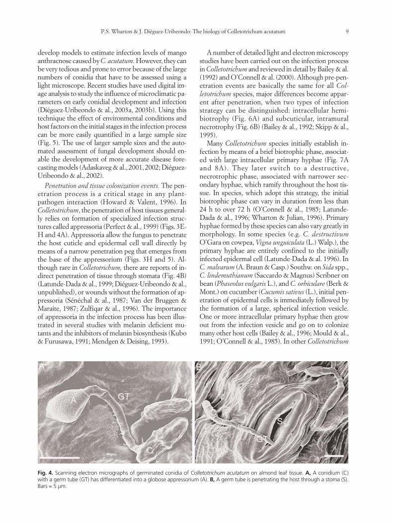
Fig. 4. Scanning electron micrographs of germinated conidia of Colletotrichum acutatum on almond leaf tissue. A, A conidium (C) with a germ tube (GT) has differentiated into a globose appressorium (A). B, A germ tube is penetrating the host through a stoma (S).Bars = 5 µm.
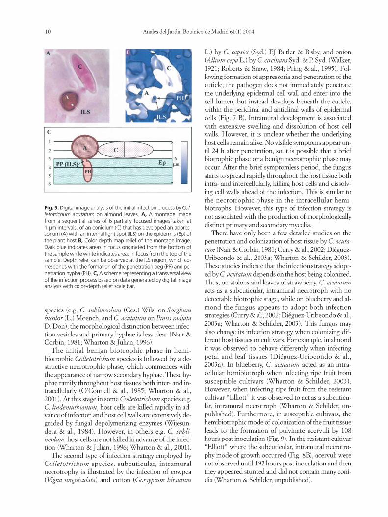
develop models to estimate infection levels of mangoanthracnose caused by C. acutatum. However, they canbe very tedious and prone to error because of the largenumbers of conidia that have to be assessed using alight microscope. Recent studies have used digital im-age analysis to study the influence of microclimatic pa-rameters on early conidial development and infection(Diéguez-Uribeondo & al., 2003a, 2003b). Using thistechnique the effect of environmental conditions andhost factors on the initial stages in the infection processcan be more easily quantified in a large sample size(Fig. 5). The use of larger sample sizes and the auto-mated assessment of fungal development should en-able the development of more accurate disease fore-casting models (Adaskaveg & al., 2001, 2002; Diéguez-Uribeondo & al., 2002).
Penetration and tissue colonization events. The pen-etration process is a critical stage in any plant-pathogen interaction (Howard & Valent, 1996). InColletotrichum, the penetration of host tissues general-ly relies on formation of specialized infection struc-tures called appressoria (Perfect & al., 1999) (Figs. 3E-H and 4A). Appressoria allow the fungus to penetratethe host cuticle and epidermal cell wall directly bymeans of a narrow penetration peg that emerges fromthe base of the appressorium (Figs. 3H and 5). Al-though rare in Colletotrichum, there are reports of in-direct penetration of tissue through stomata (Fig. 4B)(Latunde-Dada & al., 1999; Diéguez-Uribeondo & al.,unpublished), or wounds without the formation of ap-pressoria (Sénéchal & al., 1987; Van der Bruggen &Maraite, 1987; Zulfiqar & al., 1996). The importanceof appressoria in the infection process has been illus-trated in several studies with melanin deficient mu-tants and the inhibitors of melanin biosynthesis (Kubo& Furusawa, 1991; Mendgen & Deising, 1993).
A number of detailed light and electron microscopystudies have been carried out on the infection processin Colletotrichum and reviewed in detail by Bailey & al.(1992) and O’Connell & al. (2000). Although pre-pen-etration events are basically the same for all Col-letotrichum species, major differences become appar-ent after penetration, when two types of infectionstrategy can be distinguished: intracellular hemi-biotrophy (Fig. 6A) and subcuticular, intramuralnecrotrophy (Fig. 6B) (Bailey & al., 1992; Skipp & al.,1995).
Many Colletotrichum species initially establish in-fection by means of a brief biotrophic phase, associat-ed with large intracellular primary hyphae (Fig. 7Aand 8A). They later switch to a destructive,necrotrophic phase, associated with narrower sec-ondary hyphae, which ramify throughout the host tis-sue. In species, which adopt this strategy, the initialbiotrophic phase can vary in duration from less than24 h to over 72 h (O’Connell & al., 1985; Latunde-Dada & al., 1996; Wharton & Julian, 1996). Primaryhyphae formed by these species can also vary greatly inmorphology. In some species (e.g. C. destructivumO’Gara on cowpea, Vigna unguiculata (L.) Walp.), theprimary hyphae are entirely confined to the initially infected epidermal cell (Latunde-Dada & al. 1996). InC. malvarum (A. Braun & Casp.) Southw. on Sida spp.,C. lindemuthianum (Saccardo & Magnus) Scribner onbean (Phaseolus vulgaris L.), and C. orbiculare (Berk &Mont.) on cucumber (Cucumis sativus (L.), initial pen-etration of epidermal cells is immediately followed bythe formation of a large, spherical infection vesicle.One or more intracellular primary hyphae then growout from the infection vesicle and go on to colonizemany other host cells (Bailey & al., 1996; Mould & al.,1991; O’Connell & al., 1985). In other Colletotrichum
species (e.g. C. sublineolum (Ces.) Wils. on Sorghumbicolor (L.) Moench, and C. acutatum on Pinus radiataD. Don), the morphological distinction between infec-tion vesicles and primary hyphae is less clear (Nair &Corbin, 1981; Wharton & Julian, 1996).
The initial benign biotrophic phase in hemi-biotrophic Colletotrichum species is followed by a de-structive necrotrophic phase, which commences withthe appearance of narrow secondary hyphae. These hy-phae ramify throughout host tissues both inter- and in-tracellularly (O’Connell & al., 1985; Wharton & al.,2001). At this stage in some Colletotrichum species e.g.C. lindemuthianum, host cells are killed rapidly in ad-vance of infection and host cell walls are extensively de-graded by fungal depolymerizing enzymes (Wijesun-dera & al., 1984). However, in others e.g. C. subli-neolum, host cells are not killed in advance of the infec-tion (Wharton & Julian, 1996; Wharton & al., 2001).
The second type of infection strategy employed byColletotrichum species, subcuticular, intramuralnecrotrophy, is illustrated by the infection of cowpea(Vigna unguiculata) and cotton (Gossypium hirsutum
L.) by C. capsici (Syd.) EJ Butler & Bisby, and onion(Allium cepa L.) by C. circinans Syd. & P. Syd. (Walker,1921; Roberts & Snow, 1984; Pring & al., 1995). Fol-lowing formation of appressoria and penetration of thecuticle, the pathogen does not immediately penetratethe underlying epidermal cell wall and enter into thecell lumen, but instead develops beneath the cuticle,within the periclinal and anticlinal walls of epidermalcells (Fig. 7 B). Intramural development is associatedwith extensive swelling and dissolution of host cellwalls. However, it is unclear whether the underlyinghost cells remain alive. No visible symptoms appear un-til 24 h after penetration, so it is possible that a briefbiotrophic phase or a benign necrotrophic phase mayoccur. After the brief symptomless period, the fungusstarts to spread rapidly throughout the host tissue bothintra- and intercellularly, killing host cells and dissolv-ing cell walls ahead of the infection. This is similar tothe necrotrophic phase in the intracellular hemi-biotrophs. However, this type of infection strategy isnot associated with the production of morphologicallydistinct primary and secondary mycelia.
There have only been a few detailed studies on thepenetration and colonization of host tissue by C. acuta-tum (Nair & Corbin, 1981; Curry & al., 2002; Diéguez-Uribeondo & al., 2003a; Wharton & Schilder, 2003).These studies indicate that the infection strategy adopt-ed by C. acutatum depends on the host being colonized.Thus, on stolons and leaves of strawberry, C. acutatumacts as a subcuticular, intramural necrotroph with nodetectable biotrophic stage, while on blueberry and al-mond the fungus appears to adopt both infectionstrategies (Curry & al., 2002; Diéguez-Uribeondo & al.,2003a; Wharton & Schilder, 2003). This fungus mayalso change its infection strategy when colonizing dif-ferent host tissues or cultivars. For example, in almondit was observed to behave differently when infectingpetal and leaf tissues (Diéguez-Uribeondo & al.,2003a). In blueberry, C. acutatum acted as an intra-cellular hemibiotroph when infecting ripe fruit fromsusceptible cultivars (Wharton & Schilder, 2003).However, when infecting ripe fruit from the resistantcultivar “Elliott” it was observed to act as a subcuticu-lar, intramural necrotroph (Wharton & Schilder, un-published). Furthermore, in susceptible cultivars, thehemibiotrophic mode of colonization of the fruit tissueleads to the formation of pulvinate acervuli by 108hours post inoculation (Fig. 9). In the resistant cultivar“Elliott” where the subcuticular, intramural necrotro-phy mode of growth occurred (Fig. 8B), acervuli werenot observed until 192 hours post inoculation and thenthey appeared stunted and did not contain many coni-dia (Wharton & Schilder, unpublished).
10 Anales del Jardín Botánico de Madrid 61(1) 2004
Fig. 5. Digital image analysis of the initial infection process by Col-letotrichum acutatum on almond leaves. A, A montage imagefrom a sequential series of 6 partially focused images taken at1 µm intervals, of an conidium (C) that has developed an appres-sorium (A) with an internal light spot (ILS) on the epidermis (Ep) ofthe plant host B, Color depth map relief of the montage image.Dark blue indicates areas in focus originated from the bottom ofthe sample while white indicates areas in focus from the top of thesample. Depth relief can be observed at the ILS region, which co-rresponds with the formation of the penetration peg (PP) and pe-netration hypha (PH). C, A scheme representing a transversal viewof the infection process based on data generated by digital imageanalysis with color-depth relief scale bar.

Latency (quiescent infections). The term quiescentinfection describes a “quiescent or dormant parasiticrelationship, which after a time, changes to an activeone” (Verhoeff, 1974; Swinburne, 1983). The infec-tion process and the phenomenon of quiescence has
P.S. Wharton & J. Diéguez-Uribeondo: The biology of Colletotrichum acutatum 11
been extensively studied in other Colletotrichumspecies, e.g. C. gloeosporioides on avocado, and C.musae (Berkeley & Curtis) von Arx on bananas (Musaespp.) (Prusky & al., 1982; Jeffries & al., 1990; Prusky& al., 1991a; Prusky, 1996). This allows us to speculate
Fig. 7. Light micrographs of almond petal tissue inoculated with Colletotrichum acutatum. A, Intracellular colonization of petal tissue.The fungus penetrates the epidermal cells (EC) of the petal from an appressorium (A) and forms thick primary hyphae (PH) characteris-tic of biotrophic stage. B, Subcuticular and intramural colonization of almond petal tissue. The fungus does not immediately enter intothe cell lumen and instead grows beneath the cuticle within the epidermal cell walls (CW). Bars = 5 µm.
Fig. 6. Infection strategies adopted by Colletotrichum species as described by Bailey & al. (1992) and O’Connell & al. (2000). The initial sta-ges of early differentiation are the same for both strategies. A conidium (C) germinates and forms and appressorium (A). The appresso-rium produces a penetration pore and peg (PP) which penetrates the cuticle (Cu) of the host and as a result an internal light spot (ILS) canbe seen in the appressorium. A, In intracellular hemibiotrophs the penetration peg penetrates the epidermal cell and swells to produce aninfection vesicle and broad hyphae, named primary hyphae (PH), which may colonize adjacent epidermal (E) and mesophyll cells (M). Du-ring the early stages of this type of colonization, the interaction between the host and the pathogen is biotrophic (living cell representedin green). The subsequent necrotrophic (N) (represented in brown) interaction is characterized by the formation of thin secondary hyphae(SH). These secondary hyphae grow intracellularly and intercellularly while secreting cell wall degrading enzymes and killing the host cells.B, In subcuticular intramural necrotrophs, host colonization is initially by subcuticular (ScH), and intramural hyphae the biotro-phic phase is very short or does not occur. The fungus quickly spreads throughout the tissue and grows both inter- and intracellularly.

as to the events that may occur during the infectionand colonization of host tissue by C. acutatum. Fromthe limited studies on post-penetration events thatlead to successful colonization of host tissue by C. acu-tatum, it has been determined that this fungus can un-dergo a period of quiescence in almond (Adaskaveg &Förster, 2000), apple (Biggs, 1995), blueberry (Daykin& Milholland, 1984), and strawberry (Howard & al.,1992) similar to other fruit-rotting fungi.
In most Colletotrichum species exhibiting a quies-cent period, quiescence occurs after the formation ofappressoria and/or initial penetration of the host cuti-cle (Muirhead & Deverall, 1984; Rappel & al., 1989;Prusky & al., 1991a; Prusky & Plumbey, 1992). Therehave been conflicting reports as to whether dormancyactually occurs in ungerminated melanized appressoriaor after the formation of infection pegs. Chakravaty(1957) found subcuticular hyphae of C. musae in im-mature bananas soon after infection. Studying the samesystem, Muirhead & Deverall (1984) found bothmelanized and unmelanized appressoria on the unripefruit surface. The latter produced penetrating hyphaeon unripe fruits, which resulted in necrosis of epider-mal cells and the initiation of a host defense response.Melanized appressoria remained quiescent and unger-minated until the fruit ripened. Thus, they suggestedthat ungerminated melanized appressoria were respon-sible for the majority of quiescent infections observedin ripe bananas. Anatomical studies of C. gloeospo-rioides infection of avocado revealed that appressoriapersisted on unripe fruit (Binyamini & Schiffmann-Nadel, 1972). In a re-examination of the infectionprocess, Prusky & al. (1991a) and Rappel & al. (1989),found that the appressoria produced a short infectionpeg in the peel of unripe fruit. In blueberry, light mi-croscopy studies by Daykin & Milholland (1984), ofthe early stages of infection by C. acutatum on unripefruit showed that some appressoria germinated andproduced subcuticular hyphae. However, it has yet tobe determined whether or not these infections result in:i) cell necrosis and the initiation of a host defense re-sponse as in the C. musae-banana pathosystem, ii) as aquiescent ungerminated melanized appressoria, or iii)as a quiescent germinated appressoria.
Biochemical Basis of Host-Pathogen Interactions
The successful colonization of host tissues by apathogen depends on its ability to overcome the hostdefenses. The resistance of immature fruits to colo-nization by Colletotrichum species may be related toone of four mechanisms within the host: 1) Pre-formedtoxic compounds that inhibit pathogen growth are
present in unripe but not ripe fruit; 2) Unripe fruitdoes not provide a suitable substrate to fulfill the nu-tritional and energy requirements of the pathogen; 3)The enzyme ‘potential’ of the fungus is inadequate tocolonize unripe fruit; and 4) Phytoalexin productionin unripe fruit (Jeffries & al., 1990).
Preformed fungitoxic compounds. The role of pre-formed compounds in quiescence and resistance havedescribed in detailed in the C. gloeosporioides-avocadopathosystem (Prusky & al., 1982, 1985, 1988). Initialstudies identified a preformed antifungal diene, (cis,cis-1-acetoxy-2-hydroxy-4-oxo-heneicosa-12, 15-di-ene) in the peel of unripe avocado fruit and showedthat it was present at concentrations above that re-quired to inhibit growth of C. gloeosporioides in vitro,and subsequently decreased in concentration as thefruits ripened. Further studies revealed that fungallipoxygenase activity degraded the diene and was in-hibited by epicatechin present in the peel of unripefruit. The concentration of epicatechin also declinedduring ripening. Thus, it was concluded that a declinein epicatechin levels lead to increased lipoxygenase ac-tivity, which in turn reduced the quantity of antifungaldiene and consequently allowed colonization of thehost tissue by the pathogen.
Pathogen nutrition. Ripening fruit undergo a vastarray of biochemical changes, the most obvious ofwhich are the conversion of storage carbohydrates tosoluble sugars. In a number of fungal diseases, resis-tance has been related to sugar content (Horsfall &Dimond, 1957). In guava (Psidium guajava L.), Sing &Sharma (1981) observed that fruit from varieties thatwere resistant to Glomerella cingulata had high levelsof soluble solids and higher levels of ascorbic acidthan fruit from susceptible varieties. In blueberry,studies carried out on the antifungal properties of ex-tracts from ripe fruit from wild highbush blueberryplants (Cipollini & Stiles, 1992a, 1992b, 1993) indi-cated that the main antifungal compounds present inripe fruit were water-soluble, non-alkaloidal chemi-cals such as phenolics and acids. Furthermore, the au-thors noted an increase in the antifungal activity of ex-tracts in the presence of added organic acids (1% cit-ric acid) and suggested that this was due to an interac-tion between the phenolic constituents of the extractsand the acids. Thus, they proposed that resistance inripe blueberries might be due to a combination of fac-tors including acid levels in the fruit and an interac-tion between simple phenolic compounds and organ-ic acids, and not necessarily individual fungitoxiccompounds. However, recent studies on the infectionof resistant blueberry cultivars by C. acutatum haveshown that phenolic compounds accumulate in in-
12 Anales del Jardín Botánico de Madrid 61(1) 2004
fected cells and surround fungal hyphae in a processsimilar to that observed in other resistant host-Col-letotrichum interactions (Wharton & Julian, 1996;Wharton & Schilder, unpublished).
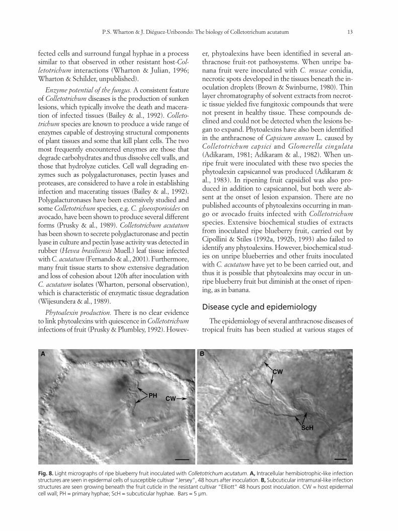
Enzyme potential of the fungus. A consistent featureof Colletotrichum diseases is the production of sunkenlesions, which typically involve the death and macera-tion of infected tissues (Bailey & al., 1992). Colleto-trichum species are known to produce a wide range ofenzymes capable of destroying structural componentsof plant tissues and some that kill plant cells. The twomost frequently encountered enzymes are those thatdegrade carbohydrates and thus dissolve cell walls, andthose that hydrolyze cuticles. Cell wall degrading en-zymes such as polygalacturonases, pectin lyases andproteases, are considered to have a role in establishinginfection and macerating tissues (Bailey & al., 1992).Polygalacturonases have been extensively studied andsome Colletotrichum species, e.g. C. gloeosporioides onavocado, have been shown to produce several differentforms (Prusky & al., 1989). Colletotrichum acutatumhas been shown to secrete polyglacturonase and pectinlyase in culture and pectin lyase activity was detected inrubber (Hevea brasiliensis Muell.) leaf tissue infectedwith C. acutatum (Fernando & al., 2001). Furthermore,many fruit tissue starts to show extensive degradationand loss of cohesion about 120h after inoculation withC. acutatum isolates (Wharton, personal observation),which is characteristic of enzymatic tissue degradation(Wijesundera & al., 1989).
Phytoalexin production. There is no clear evidenceto link phytoalexins with quiescence in Colletotrichuminfections of fruit (Prusky & Plumbley, 1992). Howev-
P.S. Wharton & J. Diéguez-Uribeondo: The biology of Colletotrichum acutatum 13
er, phytoalexins have been identified in several an-thracnose fruit-rot pathosystems. When unripe ba-nana fruit were inoculated with C. musae conidia,necrotic spots developed in the tissues beneath the in-oculation droplets (Brown & Swinburne, 1980). Thinlayer chromatography of solvent extracts from necrot-ic tissue yielded five fungitoxic compounds that werenot present in healthy tissue. These compounds de-clined and could not be detected when the lesions be-gan to expand. Phytoalexins have also been identifiedin the anthracnose of Capsicum annum L. caused byColletotrichum capsici and Glomerella cingulata(Adikaram, 1981; Adikaram & al., 1982). When un-ripe fruit were inoculated with these two species thephytoalexin capsicannol was produced (Adikaram &al., 1983). In ripening fruit capsidiol was also pro-duced in addition to capsicannol, but both were ab-sent at the onset of lesion expansion. There are nopublished accounts of phytoalexins occurring in man-go or avocado fruits infected with Colletotrichumspecies. Extensive biochemical studies of extractsfrom inoculated ripe blueberry fruit, carried out byCipollini & Stiles (1992a, 1992b, 1993) also failed toidentify any phytoalexins. However, biochemical stud-ies on unripe blueberries and other fruits inoculatedwith C. acutatum have yet to be been carried out, andthus it is possible that phytoalexins may occur in un-ripe blueberry fruit but diminish at the onset of ripen-ing, as in banana.
Disease cycle and epidemiology
The epidemiology of several anthracnose diseases oftropical fruits has been studied at various stages of
Fig. 8. Light micrographs of ripe blueberry fruit inoculated with Colletotrichum acutatum. A, Intracellular hemibiotrophic-like infectionstructures are seen in epidermal cells of susceptible cultivar “Jersey”, 48 hours after inoculation. B, Subcuticular intramural-like infectionstructures are seen growing beneath the fruit cuticle in the resistant cultivar “Elliott” 48 hours post inoculation. CW = host epidermalcell wall; PH = primary hyphae; ScH = subcuticular hyphae. Bars = 5 µm.
crop development. In most Colletotrichum diseasesconidia are water-borne with the occurrence of quies-cent infections being highest during the wettest peri-ods of the growing season (Denham & Waller, 1981;Fitzell & Peak, 1984; Darvas & Kotze, 1987). In avo-cado, citrus and mango, it was shown that infectedleaves in the tree canopy were the main source of in-oculum, with conidia being rain–splash dispersed tounripe fruit (Denham & Waller, 1981; Fitzell & Peak,1984; Fitzell, 1987). However, in mango and citrus in-fected flowers also contributed to inoculum levels(Fitzell & Peak, 1984; Zulfiqar & al., 1996). In almond,mummified fruit represent the main source of conidiafor infections (Adaskaveg & al., 2000).
Infection by Colletotrichum can take place at allstages of fruit development (Hartung, & al., 1981;Daykin & Milholland, 1984, Adaskaveg & al., 2000).In blueberry, the fungus is thought to overwinter asmycelium in and on blighted twigs, which act as themain source of inoculum in the spring (Milholland,1995). However, recent data suggest that the primarysource of overwintering inoculum may be from dor-mant flower buds (Fig. 2C) (DeMarsay & Oudemans,2002; Wharton & Schilder, unpublished). In studiescarried out on the cultivar ‘Bluecrop’ in New Jersey,flower buds accounted for 72% of overwintering in-fections (DeMarsay & Oudemans, 2002). In screen-ing experiments carried out on the susceptible culti-var ‘Jersey’ in Michigan, 57% of healthy lookingflower buds were found to be infected, and of thoseinfected, 82% of the infections were caused by C. acu-tatum (Wharton & Schilder, unpublished). It was ob-served that as the flower buds broke dormancy, thefungus grew out of the buds and colonized the sur-rounding stem tissue, causing black lesions aroundthe infected buds (Fig. 2C). These lesions grew up to2 cm in length, and resulted in the death of the flowerbud and any tissue above the lesion (Fig. 2C). Afterabout 7 days, sporulating masses were observed in thedead tissue (Fig. 2C-D).
In the field, the fungus sporulates on infected tis-sues during periods of extended wetness in the spring,and conidia of C. acutatum are dispersed by rain splash(Caruso & Ramsdell, 1995). As in citrus and strawber-ry, secondary conidiation, may play a role in early-sea-son dispersal of C. acutatum conidia on blueberries(Zulfiqar & al., 1996; Timmer & Brown, 2000; Lean-dro & al., 2001; P. Oudemans, personal communica-tion). In citrus, the conidia densities decline with timein the absence of bloom, and through normal leaf dropand mortality of buttons and twigs (Timmer & Brown,2000). In blueberry, peak spore dispersal coincideswith flowering and early fruit development stages
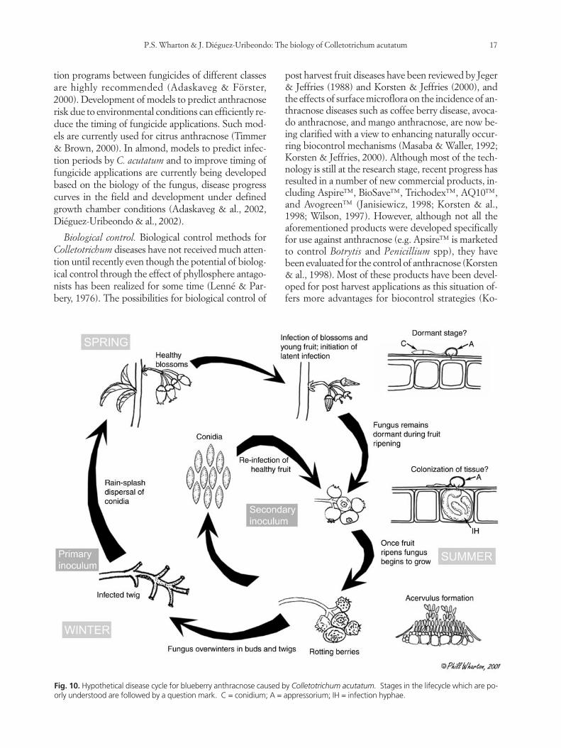
(Hartung & al., 1981; Wharton & al., 2002). A secondpeak occurs at fruit maturity, apparently coincidingwith sporulation of the fungus on ripe fruit (Wharton& al., 2002). As described above, on immature blue-berry fruit, the fungus initiates quiescent infections,and disease symptoms are not observed until the fruitbegins to ripen. A proposed disease cycle for thegrowth and sporulation of Colletotrichum on blueber-ry fruit is shown in Fig. 10.
Fungal control
Effective control of Colletotrichum diseases usuallyinvolves the use of one or a combination of the follow-ing: 1) resistant cultivars, 2) cultural control, 3) chemi-cal control, and 4) biological control using antagonis-tic organisms. The applicability of control strategiesdepends as much on the characteristics of the crop onwhich they are being used as on the disease at whichthey are targeted.
Resistant Cultivars. Resistance to disease is perhapsthe most significant aspect of disease control in agri-cultural crops, but has been exploited to a lesser extentin fruit crops mainly due to the longer time frame re-quired for breeding and selecting for resistance andthe shorter-term advantages of chemical control. Cul-tivar resistance in fruit crops is complicated by theability of most Colletotrichum fruit pathogens to formquiescent infections. In most host-pathogen interac-tions, resistance involves the triggering of host defenseresponses that prevent or retard pathogen growth andmay be conditioned by a single gene pair, a host resis-tance gene and a pathogen avirulence gene (Flor,1971). However, such gene-for-gene interactions arenot usually involved in the resistance of fruits topostharvest diseases caused by Colletotrichum (Prusky& al., 2000). Instead, resistance is usually the result ofseveral genes interacting in a way that is not well un-derstood. Resistance in fruit to postharvest pathogenshas been described as a “dynamic incompatibility”(Prusky & al., 2000). The response of the host’s resis-tance genes to products of a pathogen’s avirulencegenes prevents or retards pathogen growth under spe-cific host physiological conditions, as described above.However, the physiological status of the host changesas it matures, ripens, and senesces. Storage, mechani-cal injury, temperature extremes, and anoxia also alterhost physiology, and when physiological changes in thehost inhibit defense responses to pathogen activities,the interaction becomes compatible, leading to hostcolonization and disease. Thus, postharvest differ-ences in resistance among cultivars may be due asmuch to the conditions under which the fruit is stored
14 Anales del Jardín Botánico de Madrid 61(1) 2004

as to the occurrence of defense compounds (Prusky &al., 1991b). Therefore it is probably more useful to de-fine resistance and susceptibility of fruit cultivars topostharvest disease in terms of the incubation periodafter fruit ripening, with resistant cultivars having alonger storability and shelf life than susceptible ones.
Differences in susceptibility of almond cultivars ofCalifornia have been noted for blossoms, leaves andfruits both in the field and laboratory (Adaskaveg & al.,2002; Diéguez-Uribeondo & al., 2002). Cultivar ‘Non-pareil’ appeared to be most tolerant to C. acutatum in-oculations and ‘NePlus Ultra’ the most susceptible,while cultivars ‘Carmel’ and ‘Wood Colony’ had inter-mediate susceptibility. In blueberry, there are a fewcommercially available cultivars (e.g. ‘Elliott’, ‘Brigitta
P.S. Wharton & J. Diéguez-Uribeondo: The biology of Colletotrichum acutatum 15
Blue’) that are considered “resistant” to anthracnose(Ehlenfeldt & Stretch, 1997). The most widely grownresistant cultivar in Michigan is ‘Elliott’, which has be-come popular in the last 25 years due to its very late har-vest and long storage life (Jim Hancock, personal com-munication). It is interesting to note that most blueber-ry cultivars in which resistance has been found are lateseason cultivars, with fairly high acid contents. How-ever, as mentioned above few biochemical studies havebeen carried out into the biochemical basis of resis-tance in blueberry, and screening trials for resistance toanthracnose have not been able to correlate high acidcontent to resistance (Ballinger & al., 1978).
Host plant resistance would seem to be a logical andefficient way to control anthracnose disease. However,
Fig. 9. Confocal scanning laser microscopy micrographs of acervulus formation by Colletotrichum acutatum in ripe fruit of the suscepti-ble blueberry cultivar “Jersey”. A-C, Conidiophores (Co) develop from hyphae in hyphal aggregates (HA) in epidermal cells. A septum (S)usually formed between conidiophores and hyphae in the aggregates; 96 h after inoculation (images A-C are optical sections taken usingdifferential interference contrast optics at 0.16 µm z intervals). D-F, Acervuli are fully developed by 108 h after inoculation. Hyphae (H) andconidia (C) had ruptured the epidermal cell wall (arrow head) and cuticle (Cu) (images D-F are confocal micrographs taken at 1 µm z intervals). G-I, Acervuli are pulvinate, do not contain setae, and were observed in abundance by 120 h after inoculation (images G-I areprojections of 90 optical sections taken at 1, 0.61 and 0.81 µm z intervals respectively). Bars = 5 µm (A-C), 20 µm (D-F).
aside from the costs associated with replacing an es-tablished crop with a resistant or more tolerant culti-var, most growers tend to select cultivars based on cri-teria other than disease resistance.
Cultural control. This usually refers to the range ofmethods used to control diseases, mostly using tacticsaimed at disease avoidance through phytosanitation,manipulation of cropping patterns or by enhancing re-sistance and avoiding predisposition. However, in re-lation to fruit crops it also involves the use of propersanitation techniques during processing of the harvest-ed fruit, transportation, packaging and storage, toavoid exposure of fruit to the pathogen. It also involvesproper handling to avoid abiotic factors such as me-chanical injury, temperature extremes, and anoxia,which can predispose the fruit to infection by thepathogen. The ubiquitous nature of inoculum sourcesof Colletotrichum diseases and their often-rapid epi-demic development under suitable conditions reducesthe effectiveness of many pre-harvest general phy-tosanitary practices. However, general orchard hy-giene has a place in integrated disease control, as re-moval of obvious inoculum sources such as diseasedleaves and fruit can increase the efficiency of chemicalcontrol (Waller, 1972, 1988).
The prerequisite for wet conditions to coincide withsusceptible crop stages for development of Col-letotrichum epidemics also offers an opportunity fordisease control through the manipulation of croppingpatterns and or irrigation (Fitzell & Peak, 1984; Fitzell& al., 1984). In blueberry, this can be achieved to someextent by pruning techniques such as the fine pruningof old, fruited and dead twigs that remain in the bushfrom year to year. As described above, such twigs arethought to act as one of the main sources of Col-letotrichum inoculum for the infection of susceptibleyoung fruit in the spring. Overhead irrigation is oftenused in the spring to mediate frosts since blueberrysoils are often found in topographically low areas thatare prone to frosts. Although the use of overhead irri-gation may mediate frost damage it provides ideal con-ditions for the infection of young fruit.
Chemical control. Chemical control methods arewidely used on fruit crops, partly because the value ofthe produce gained usually offsets the relatively ex-pensive inputs, in terms of machinery, materials and la-bor, and transportation and storage, which are re-quired, and partly because the availability and efficien-cy of chemical control is relatively greater than that ofother control methods (Jeger & Plumbley, 1988). Gen-erally, Colletotrichum diseases can be controlled by awide range of chemical such as copper compounds,dithiocarbamates, benzimidazole and triazole com-
pounds, and other fungicides such as chlorothalonil,imazalil and prochloraz (Waller, 1992). Newer classesof fungicides such as the strobilurins (e.g. azoxys-trobin and pyraclostrobin) are also proving highly ef-fective against Colletotrichum species that infect fruits(Schilder & al., 2001). However, the problem of fungi-cide tolerance may arise quickly if a single compoundis relied upon too heavily.
For successful chemical control of many Col-letotrichum diseases timing and placement are of criticalsignificance. In general, fungicides must be applied toprotect the young expanding crop tissues, whetherleaves, blossoms or fruit, against infection during peri-ods of wetness (Fitzell & al., 1984). Both rapid expan-sion of the fruit surfaces and the natural erosion of thefungicide by rainfall make adequate fungicide protec-tion often difficult to achieve, and repeated applicationsare often necessary to maintain protection in diseasessuch as mango anthracnose (Fitzell & Peak, 1984).However, poorly timed fungicide applications may ac-tually lead to an increase in the severity of disease due tothe disturbance of natural biocontrol mechanisms andincreased crop susceptibility (Griffiths, 1981).
Currently, control of blueberry and almond an-thracnose is primarily accomplished by chemicalmeans. The following fungicides are reported to haveactivity against C. acutatum: fosetyl-AL (Alliette), cap-tan (Captan), benomyl (Benlate), chlorothalonil (Bra-vo), ziram (Ziram), fenbuconazole (Indar 75 WP), mi-crobutanil (Rally 40WP), thiophanate methyl (Topsin75WP), azoxystrobin (Abound) and pyraclostrobin(Cabrio) (Adasakaveg & Förster, 2000; Schilder,2002). However, the use and effectiveness of thesefungicides may be limited by various factors. For ex-ample, benomyl has recently been withdrawn by themanufacturer and only limited stocks remain. The useof fosetyl-Al tends to be costly, and chlorothalonil can-not be used after petal fall in blueberry because of phy-totoxicity to the fruit. In addition, Captan is currentlyconsidered a B-2 carcinogen and its use is restricted bysome fruit processors. Ziram has a minimum14-daypre-harvest interval, whereas the number or applica-tions of azoxystrobin and pyraclostrobin allowed perseason is limited as part of a fungicide resistance man-agement protocol. This means that growers have toutilize their fungicide options wisely to attain effectivecontrol. Although treatment with fungicides can sig-nificantly reduce the incidence and severity of disease,eradication cannot normally be achieved (Adasakaveg& Förster, 2000). Thus, if treatments are stopped andconditions favorable for disease re-occur, the diseasein the crop may subsequently increase. Applicationsprior conducive conditions are thus required and rota-
16 Anales del Jardín Botánico de Madrid 61(1) 2004
tion programs between fungicides of different classesare highly recommended (Adaskaveg & Förster,2000). Development of models to predict anthracnoserisk due to environmental conditions can efficiently re-duce the timing of fungicide applications. Such mod-els are currently used for citrus anthracnose (Timmer& Brown, 2000). In almond, models to predict infec-tion periods by C. acutatum and to improve timing offungicide applications are currently being developedbased on the biology of the fungus, disease progresscurves in the field and development under definedgrowth chamber conditions (Adaskaveg & al., 2002,Diéguez-Uribeondo & al., 2002).
Biological control. Biological control methods forColletotrichum diseases have not received much atten-tion until recently even though the potential of biolog-ical control through the effect of phyllosphere antago-nists has been realized for some time (Lenné & Par-bery, 1976). The possibilities for biological control of
P.S. Wharton & J. Diéguez-Uribeondo: The biology of Colletotrichum acutatum 17
post harvest fruit diseases have been reviewed by Jeger& Jeffries (1988) and Korsten & Jeffries (2000), andthe effects of surface microflora on the incidence of an-thracnose diseases such as coffee berry disease, avoca-do anthracnose, and mango anthracnose, are now be-ing clarified with a view to enhancing naturally occur-ring biocontrol mechanisms (Masaba & Waller, 1992;Korsten & Jeffries, 2000). Although most of the tech-nology is still at the research stage, recent progress hasresulted in a number of new commercial products, in-cluding Aspire™, BioSave™, Trichodex™, AQ10™,and Avogreen™ (Janisiewicz, 1998; Korsten & al.,1998; Wilson, 1997). However, although not all theaforementioned products were developed specificallyfor use against anthracnose (e.g. Apsire™ is marketedto control Botrytis and Penicillium spp), they havebeen evaluated for the control of anthracnose (Korsten& al., 1998). Most of these products have been devel-oped for post harvest applications as this situation of-fers more advantages for biocontrol strategies (Ko-
Fig. 10. Hypothetical disease cycle for blueberry anthracnose caused by Colletotrichum acutatum. Stages in the lifecycle which are po-orly understood are followed by a question mark. C = conidium; A = appressorium; IH = infection hyphae.

rsten & al., 1998). For example, environmental condi-tions during fruit transportation and storage are gener-ally more uniform than in the field and can often bemanipulated. The biomass of the harvested fruit is alsomuch less than that of the standing crop, easier to treatin a uniform manner, and more suited to directly targetthe pathogen with an appropriate biocontrol formula-tion.
Currently there are no commercial products regis-tered in the USA for use against C. acutatum on blue-berry or almond. However, bio-fungicides containingthe antagonistic bacteria Bacillus subtilis (e.g. Sere-nadeTM, RhapsodyTM) and Candida oleophila (e.g.AspireTM) are in the testing phase to determine theirefficacy against C. acutatum.
Conclusions
Studies on mango anthracnose and Colletotrichumdiseases of citrus have shown that development ofcost-effective and efficient control strategies for themanagement of these diseases was facilitated by an ac-curate identification of the pathogen and gaining anunderstanding of the host-pathogen interactions thatdetermine successful pathogenesis (Fitzell & Peak,1984; Fitzell, & al., 1984; Timmer & Brown, 2000). Aknowledge of factors such as the conditions requiredfor germination and appressorium formation on thehost surface, infection and colonization of host tissuesand spore production, enabled disease predictionmodels to be developed which significantly reducedthe number of fungicide sprays required to controlthese diseases (Fitzell & Peak, 1984; Fitzell & al., 1984;Timmer & Brown, 2000). However, the precise deter-mination of the etiology of Colletotrichum diseases re-quires more than just the isolation and identification ofa Colletotrichum species from plant tissue. Col-letotrichum species can be easily and readily isolatedfrom both diseased and apparently healthy tissue ofmany crops and several ecological studies have shownthat Colletotrichum species can co-inhabit lesionsformed by other pathogens and exist as epiphytes andasymptomatic endophytes in a large variety of plantspecies (Rodriguez & al., 2000; Waller & Bridge, 2000;Leandro & al., 2001). Thus, a prerequisite to under-standing the lifecycles of these fungi is a thoroughknowledge of the infection processes and colonizationof tissues.
There have been only a few studies on the infectionprocess of C. acutatum and no detailed studies on thepre- and post-infection events (including the quiescentphase) that lead to successful colonization of fruit(Daykin & Milholland, 1984; Curry & al., 2002;Diéguez-Uribeondo & al., 2003a; Wharton &
Schilder, 2003). As described above, the infection offruit by Colletotrichum species can be separated intostages including the deposition of conidia, germina-tion, appressoria formation, growth and colonizationof plant tissues, and production of acervuli and sporu-lation. The proposed disease cycle of blueberry an-thracnose in Fig. 10 indicates several stages (followedby a question mark) that are currently not well under-stood. The nature of the dormant stage, for instance, isstill unclear. The dormant stage is very important inthe survival of the fungus from the initial infection ofthe young, green fruit, until the fruit ripens and colo-nization takes place. Understanding in what form, andwhere exactly the fungus survives this period, whichcan last for more than a month, could help elucidatethis crucial component of the lifecycle. It could also in-dicate a very vulnerable stage in the fungal lifecyclethat could be used in developing specific controlstrategies. Next is an understanding of the timing andnature of the colonization process. Again, it is not clearwhen and to what extent the fungus colonizes fruit tis-sues before it reemerges and sporulates. This is also astage that we believe could yield some clues as to pos-sible resistance mechanisms.
Host resistance in fruit is often described in terms ofdynamic incompatibility and host barriers can occur atdifferent stages of the infection and colonizationprocess. Dynamic incompatibility could occur at thestage of inhibition of appressorium development, inhi-bition of fungal penetration, or inhibition of fungalcolonization. In blueberry, the question remains as towhat potential defense responses are triggered, or sup-pressed in fruits at different physiological stages? Andwhat the roles of pre-formed or inducible barriers areto pathogen attack? As described above, several stud-ies have been carried out into the antifungal propertiesof extracts from ripe blueberry fruit from wild high-bush blueberry plants as they relate to fruit decay andherbivore preference (Cipollini & Stiles, 1992a,1992b, 1993). These studies indicated that the mainantifungal compounds present in ripe blueberry fruitwere water-soluble, non-alkaloidal chemicals such asphenolics and acids. They proposed that resistance inripe blueberries may be due to a combination of fac-tors including acid levels in the fruit and an interactionbetween simple phenolic compounds and organicacids, and not necessarily individual fungitoxic com-pounds. To date, no studies have investigated the bio-chemical composition of unripe blueberry fruit. This isimportant as studies have shown that there are differ-ences in the chemical composition of blueberries bothat different stages of maturity and between cultivars(Connor & al., 2002; Hakkinen & Torronen, 2000;
18 Anales del Jardín Botánico de Madrid 61(1) 2004

Kalt & McDonald, 1996). Furthermore, an improvedunderstanding of the physiology and underlying bio-chemical processes involved in the induction andmaintenance of quiescence could lead to new diseasecontrol measures, such as have been developed in avo-cado (Prusky & al., 1991b).
Given the epidemiological versatility of Col-letotrichum diseases, the production of high-qualityfruit, should follow an integrated approach in order toachieve maximum yields. Where possible, resistantcultivars can be combined with judicious pruning andproper irrigation timing to reduce inoculum levels anddisease pressure. More detailed information on the bi-ology of the pathogen will be needed to help optimizeuse and timing of the available fungicides. As more re-duced-risk and biological control products becomeavailable, they may be substituted for older chemistriesto lessen negative impacts on users, consumers, andthe environment.
Acknowledgements
We would like to acknowledge Dr. James E. Adaskaveg (Uni-versity of California Riverside) for providing pictures for the figureson almond anthracnose, and Dr. Annemiek Schilder (MichiganState University) for her contributions to our understanding ofblueberry anthracnose. This work was supported in part, by ProjectGREEEN (Generating Research and Extension to meet Economicand Environment Needs) at Michigan State University and projectFlora Mycologica Iberica V (REN2002-04068-C02-01GLO), Mi-nisterio de Ciencia y Tecnología, Spain.
References
Adaskaveg, J.E. & Förster, H. 2000. Occurrence and managementof anthracnose epidemics caused by Colletotrichum species ontree fruit crops in California. In: Prusky, D., Freeman, S. &Dickman, M.B. (eds.), Colletotrichum: Host Specificity, Patho-logy, and Host-Pathogen Interaction, pp. 317-336. The Ameri-can Phytopathological Society. St. Paul MN.
Adaskaveg, J.E. & Hartin, R.J. 1997. Characterization of Colleto-trichum acutatum isolates causing anthracnose of almond andpeach in California. Phytopathology 87: 979-987.
Adaskaveg, J.E., Diéguez-Uribeondo, J., Förster, H., Thompson,D., Teviotdale, B.L., Connell, J., Edstrom, J., Duncan, R., Free-man, M. & Verdegaal, P. 2001. Epidemiology and managementof almond anthracnose and brown rot in California. In: Proc. ofthe 29th Annual Almond Conference, pages 45-53. The AlmondBoard of California. Modesto, CA.
Adaskaveg, J.E., Diéguez-Uribeondo, J., Förster, H., Erickson, E.,Teviotdale, B.L., Connell, J., Edstrom, J., Duncan, R., Hen-dricks, L., Freeman, M. & Verdegaal, P. 2002. Epidemiologyand management of almond anthracnose and brown rot in Ca-lifornia. In: Proc. of the 30th Annual Almond Conference, pages79-487. The Almond Board of California. Modesto, CA.
Adikaram, N.K.B. 1981. A study of the latent infection of fruit ofCapsicum spp. by Colletotrichum capsici and Glomerella cin-gulata. Ph D Thesis, Queen’s University of Belfast.
Adikaram, N.K.B., Brown, A.E. & Swinburne, T.R. 1982. Phytoa-lexin involvement in latent infection of Capsicum annum L.
P.S. Wharton & J. Diéguez-Uribeondo: The biology of Colletotrichum acutatum 19
fruit by Glomerella cingulata (Stonem.). Physiological Plant Pat-hology 21: 161-170.
Adikaram, N.K.B., Brown, A.E. & Swinburne, T.R. 1983. Obser-vations on infection of Capsicum annum fruit by Glomerellacingulata and Colletotrichum capsici. Transactions of the BritishMycological Society 80: 395-401.
Arauz, L.F. 2000. Mango anthracnose: Economic impact and cu-rrent options from integrated management. Plant Disease 84:600-611.
Bailey, J.A. & Jeger, M.J. (eds). 1992. Colletotrichum: Biology, Pat-hology and Control. CAB International. Wallingford UK.
Bailey, J.A., O’Connell, R.J., Pring, R.J. & Nash, C. 1992. Infectionstrategies of Colletotrichum species. In: Bailey, J.A. & Jeger,M.J. (eds.), Colletotrichum: Biology, Pathology and Control, pp.88-120. CAB International. Wallingford UK.
Bailey, J.A., Nash, C., Morgan, L.W. & O’Connell, R.J. 1996. Mo-lecular taxonomy of Colletotrichum species causing anthracno-se on the Malvaceae. Phytopathology 86: 1076-1083.
Ballinger, W.E., Maness, E.P. & McClure, W.F. 1978. Relations-hip of stage of ripeness and holding temperature to decay deve-lopment of blueberries. Journal of the American HorticulturalSociety 103: 130-134.
Biggs, A.R. 1995. Detection of latent infections in apple fruit withparaquat. Plant Disease 79: 1062-1067.
Binyamini, N. & Schiffmann-Nadel, M. 1972. Latent infection inavocado due to Colletotrichum gloeosporioides. Phytopatho-logy 62: 592-594.
Bonde, M.R., Peterson, G.L. & Maas, J.L. 1991. Isozyme compari-sons for identification of Colletotrichum species pathogenic tostrawberry. Phytopathology 81: 1523-1528.
Brooker, N.L., Leslie, J.F. & Dickman, M.B. 1991. Nitrate non-utilizing mutants of Colletotrichum and their use in studies ofvegetative compatibility and genetic relatedness. Phytopatho-logy 81: 672-677.
Brown, A.E. & Swinburne, T.R. 1980. The resistance of immaturebanana fruits to anthracnose (Colletotrichum musae [Berk. &Curt.] Arx.). Journal of Phytopathology - PhytopathologischeZeitschrift 99: 70-80.
Camacho, F.J., Gernandt, D.S., Liston, A., Stone, J.K. & Klein,A.S. 1997. Endophytic fungal DNA, the source of contamina-tion in spruce needle DNA. Molecular Ecology 6: 983-987.
Caruso, F.L. & Ramsdell, D.C. 1995. In: F.L. Caruso & D.C.Ramsdell (eds.). Compendium of Blueberry and Cranberry Dise-ases, 87 pp. APS Press. St. Paul MN.
Chacko, R.J., Weidemann, G.J., TeBeest, D.O. & Correll, J.C.1994. The use of vegetative compatibility and heterokaryosis todetermine potential asexual gene exchange in Colletotrichumgloeosporioides. Biological Control 4: 382-389.
Chakravaty, T. 1957. Anthracnose on banana (Gloesporium musa-rum Cke & Massae) with special reference to latent infection in sto-rage. Transactions of the British Mycological Society 40: 337-345.
Chilton, S.J.P. & Wheeler, H.E. 1949. Genetics of Glomerella.VII. Mutation and segregation in plus cultures. American Jour-nal of Botany 36: 717-721
Cipollini, M.L. & Stiles, E.W. 1992a. Antifungal activity ofripe ericaceous fruits-phenolic-acid interactions and palatabi-lity for dispersers. Biochemical Systematics and Ecology 20: 501-514.
Cipollini, M.L. & Stiles, E.W. 1992b. Relative risks of fungal rotfor temperate ericaceous fruits-effects of seasonal-variation onselection for chemical defense. Canadian Journal of Botany 70:1868-1877.
Cipollini, M.L. & Stiles, E.W. 1993. Fruit rot, antifungal defense,and palatability of fleshy fruits for frugivorous birds. Ecology74: 751-762.
Connor, A.M., Luby, J.J. & Tong, C.B.S. 2002. Variation and heri-tability estimates for antioxidant activity, total phenolic con-tent, and anthocyanin content in blueberry progenies. Journalof the American Society for Horticultural Science 127: 82-88.
Correll, J.C., Guerber, J.C. & Rhodes, D.D. 1994. Genetic andmolecular diversity of populations of Glomerella cingulata, Co-lletotrichum gloeosporioides and Colletotrichum acutatum onapple fruit. In: Proceedings of Fifth International MycologicalConference. Vancouver B. C. Canada.
Correll, J.C., Guerber, J.C., Walsiwa, L.A., Sherril, J.F. & More-lock, T.E. 2000. Inter and intra species variation in Colletotri-chum and mechanism which affect population structure. In:Prusky, D., Freeman, S. & Dickman, M.B. (eds.), Colletotri-chum: Host Specificity, Pathology, and Host-Pathogen Interac-tion, pp. 145-179. The American Phytopathological Society.St. Paul MN.
Curry, K.J., Abril, M., Avant, J.B. & Smith, B.J. 2002. Strawberryanthracnose: Histopathology of Colletotrichum acutatum andC. fragariae. Phytopathology 92: 1055-1063.
Danneberger, T.K., Vargas, J.M. & Jones, A.L. 1984. A model forweather-based forecasting of anthracnose on annual bluegrass.Phytopathology 74: 448-451.
Darvas, J.M. & Kotze, J.M. 1987. Avocado fruit diseases and theircontrol in South Africa. South African Avocado Grower’s Asso-ciation Yearbook 10: 117-119.
Daykin, M.E. & Milholland, R.D. 1984. Infection of blueberry fruitby Colletotrichum gloeosporioides. Plant Disease 68: 948-950.
DeMarsay, A. & Oudemans, P.V. 2002. Reservoirs of Colletotri-chum acutatum in dormant and growing highbush blueberry.Phytopathology 92: S143.
Denham, T.G. & Waller, J.M. 1981. Some epidemiological aspectsof post-bloom fruit drop disease (Colletotrichum gloeosporioi-des) in citrus. Annals of Applied Biology 98: 65-77.
Diéguez-Uribeondo, J., Förster, H. & Adaskaveg, J.E. 2002. Tem-perature and wetness periods requirements for almond anth-racnose development on leaves and blossoms. Phytopathology92: S19.
Diéguez-Uribeondo, J., Förster H. & Adaskaveg, J.E. 2003a. Sub-cuticular and intracellular hemibiotrophic development of Co-lletotrichum acutatum on almond. Phytopathology 93: S21.
Diéguez-Uribeondo, J., Förster, H. & Adaskaveg, J.E. 2003b. Di-gital image analysis of the internal light spots of appressoria ofColletotrichum acutatum. Phytopathology 92: 923-930.
Dodd, J.C., Estrada, A.B., Matcham, J., Jeffries, P. & Jeger, M.J.1991. The effect of climatic factors on Colletotrichum gloeos-porioides, causal agent of mango anthracnose, in the Philippi-nes. Plant Pathology 40: 568-575.
Duthie, J.A. 1997. Models of the response of foliar parasites to thecombined effects of temperature and duration of wetness. Phy-topathology 87: 1088-1095.
Ehlenfeldt, M.K. & Stretch, A.W. 1997. Identifying sources of re-sistance to mummy berry and anthracnose in highbush blue-berry. Acta Horticulturae 446: 281-286.
Fernando, T.H.P.S., Jayasinghe, C.K. & Wijesundera, R.L.C.2001. Cell wall degrading enzyme secretion by Colletotrichumacutatum, the causative fungus of secondary leaf fall of Heveabasiliensis (rubber). Mycological Research 105: 195-201.
Fitzell, R.D. 1979. Colletotrichum acutatum as a cause of anthrac-nose of mango in New-South-Wales. Plant Disease Reporter 63:1067-1070.
Fitzell, R.D. 1987. Epidemiology of anthracnose disease of avoca-dos. South African Avocado Grower’s Association Yearbook 10:113-116.
Fitzell, R.D. & Peak, C.M. 1984. The epidemiology of anthracno-se disease of mango - Inoculum sources, spore production anddispersal. Annals of Applied Biology 104: 53-59.
Fitzell, R.D., Peak, C.M. & Parnell, R.E. 1984. A model for esti-mating infection levels of anthracnose disease of mango. Annalsof Applied Biology 104: 451-458.
Flor, H.H. 1971. The current status of the gene-for-gene concept.Annual Review of Phytopathology 9: 275-296.
Förster, H. & Adaskaveg, J.E. 1999. Identification of subpopula-tions of Colletotrichum acutatum and epidemiology of almondanthracnose in California. Phytopathology 89: 1056-1065.
Freeman, S. 2000. Genetic diversity and host specificity of Colleto-trichum species on various fruits. In: Prusky, D., Freeman, S. &Dickman, M.B. (eds.), Colletotrichum: Host Specificity, Patho-logy, and Host-Pathogen Interaction, pages 131-144. The Ameri-can Phytopathological Society. St. Paul MN.
Freeman, S. & Shabi, E. 1996. Cross-infection of subtropical andtemperate fruits by Colletotrichum species from various hosts.Physiological and Molecular Plant Pathology 49: 395-404.
Freeman, S., Katan, T. & Shabi, E. 1998. Characterization of Colletotrichum species responsible for anthracnose diseases ofvarious fruits. Plant Disease 82: 596-605.
Freeman, S., Minz, D., Maymon, M. & Veibil, A. 2001. Genetic di-versity within Colletotrichum acutatum sensu Simmonds. Phy-topathology 91: 586-592.
Griffiths, E. 1981. Pathogenic plant diseases. Annual Review Phy-topathology 19: 69-82.
Guerber, J.C. & Correll, J.C. 1997. The first report of the teleo-morph of Colletotrichum acutatum. Plant Disease 81: 1334
Guerber, J.C. & Correll, J.C. 2001. Characterization of Glomerellaacutata, the teleomorph of Colletotrichum acutatum. Mycologia93: 216-229
Hakkinen, S.H. & Torronen, A.R. 2000. Content of flavonols andselected phenolic acids in strawberries and Vaccinium species:influence of cultivar, cultivation site and technique. Food Rese-arch International 33: 517-524.
Hartung, J.S., Burton, C.C. & Ramsdell, D.C. 1981. Epidemiologi-cal studies of blueberry anthracnose disease caused by Colleto-trichum gloeosporioides. Phytopathology 71: 449-453.
Horsfall, J.G. & Dimond, A.E. 1957. Interactions of tissue sugar,growth substances and disease susceptibility. Zietschrift furPflanzenkrankheiten und Pflanzenschutz 64: 415-419.
Howard, C.M., Maas, J.L., Chandler, C.K. & Albregts, E.E. 1992.Anthracnose of strawberry caused by Colletotrichum complexin Florida. Plant Disease 76: 796-981.
Howard, R.J. & Valent, B. 1996. Breaking and entering: Host pe-netration by the fungal rice blast pathogen Magnaporthe grisea.Annual Review of Microbiology 50: 491-512.
Janisiewicz, W.J. 1998. Biocontrol of postharvest diseases of tem-perate fruits. In: Boland, G.J. & Kuykendall, L.D. (eds.), Plant-Microbe Interactions and Biological Control, pages 171-198.Marcel Dekker NY.
Jayasinghe, C.K., Fernando, T.H.P.S. & Priyanka, U.M.S. 1997.Colletotrichum acutatum is the main cause of Colletotrichumleaf disease of rubber in Sri Lanka. Mycopathologia 137: 53-56.
Jeffries, P., Dodd, J.C., Jeger, M.J. & Plumbey, R.A. 1990. The bio-logy and control of Colletotrichum species on tropical fruitcrops. Plant Pathology 39: 343-366.
Jeger, M.J. & Jeffries, P. 1988. Alternatives to chemical usage fordisease management in the post-harvest environment. Aspectsof Applied Biology 17: 47-57.
Jeger, M.J. & Plumbley, R.A. 1988. Post-harvest losses caused byanthracnose (Colletotrichum gloeosporioides) of tropical fruitand vegetables. Biodeterioration 7: 642-646.
Johnston, P.R. & Jones, D. 1997. Relationships among Colletotri-chum isolates from fruit-rots assessed using rDNA sequences.Mycologia 89: 420-430.
20 Anales del Jardín Botánico de Madrid 61(1) 2004

Kalt, W. & McDonald, J. E. 1996. Chemical composition of low-bush blueberry cultivars. Journal of the American Society forHorticultural Science 121: 142-146.
Katan, T. 2000. Vegetative compatibility in Colletotrichum. In:Prusky, D., Freeman, S. & M.B. Dickman (eds.), Colletotrichum:Host Specificity, Pathology, and Host-Pathogen Interaction, pp.145-179. The American Phytopathological Society, St. Paul MN.
Korsten, L. & Jeffries, P. 2000. Potential of biological control of di-seases caused by Colletotrichum. In: Bailey, J.A. & Jeger, M.J.(eds.), Colletotrichum: Biology, Pathology and Control, pp. 266-291. CAB International. Wallingford UK.
Korsten, L., Towsen, E. & Claasens, V. 1998. Evaluation of avo-green as a postharvest treatment for controlling anthracnoseand stem-end rot on avocado fruit. South African Avocado Gro-wer’s Association Yearbook 21 (in press).
Kubo, Y. & Furusawa, I. 1991. Melanin biosynthesis: prerequisitefor successful invasion of the plant host by appressoria of Colle-totrichum and Pyricularia. In: Cole, G.T. & Hoch, H.C. (eds.),The Fungal Spore and Disease Initiation in Plants and Animals,pp. 205-218. Plenum Press NY.
Kuramazae-Izioka, E.E., Lopes, C.R., Souza, N.L. & Machado,M.A. 1997. Morphological and molecular characterization ofColletotrichum spp. from citrus orchards affected by postblo-om fruit drop in Brazil. European Journal of Plant Pathology103: 323-329.
Lardner, R., Johnston, P.R., Plummer, K.M. & Pearson, M.N.1999. Morphological and molecular analysis of Colletotrichumacutatum sensu lato. Mycological Research 103: 275-285.
Latunde-Dada, A.O., O’Connell, R.J., Nash, C. & Lucas, J.A.1999. Stomatal penetration of cowpea (Vigna unguiculata) lea-ves by a Colletotrichum species causing latent anthracnose.Plant Pathology 48: 777-784.
Latunde-Dada, A.O., O’Connell, R.J., Nash, C., Pring, R.J., Lucas,J.A. & Bailey, J.A. 1996. Infection process and identity of thehemibiotrophic anthracnose fungus (Colletotrichum destructi-vum) from cowpea (Vigna unguiculata). Mycological Research100: 1133-1141.
Leandro, L.F.S., Gleason, M.L., Nutter, F.W., Wegulo, S.N. & Di-xon, P.M. 2001. Germination and sporulation of Colletotri-chum acutatum on symptomless strawberry leaves. Phytopatho-logy 91: 659-664.
Lenné, J.M. & Parbery, D.G. 1976. Phyllosphere antagonists andappressoria formation in Colletotrichum gloeosporioides.Transactions of the British Mycological Society 66: 334-336.
Masaba, D. & Waller, J.M. 1992. Coffee Berry Disease: The cu-rrent status. In: Bailey, J.A. & Jeger, M.J. (eds.), Colletotrichum:Biology, Pathology and Control, pages 237-249. CAB Internatio-nal. Wallingford UK.
Martín, M.P. & García-Figueres, F. 1999. Colletotrichum acuta-tum and C. gloeosporioides caused anthracnose on olives. Eu-ropean Journal of Plant Pathology 105: 733-745.
Mendgen, K. & Deising, H. 1993. Infection structures of fungalplant pathogens - a cytological and physiological evaluation.New Phytologist 124: 193-213.
Milholland, R.D. 1995. Anthracnose Fruit Rot (Ripe Rot). In: Ca-ruso, F.L. & Ramsdell, D.C. (eds.). Compendium of Blueberryand Cranberry Diseases, 17 pp. APS Press. St Paul, MN.
Monroe, J.S., Santini, J.B. & Latin, R. 1997. A model defining therelationship between temperature and leaf wetness duration,and infection of watermelon by Colletotrichum orbiculare.Plant Disease 81: 739-742.
Mould, M.J.R., Boland, G.J. & Robb, J. 1991. Ultrastructure of theColletotrichum trifolii-Medicago sativa pathosystem. II. Post-penetration events. Physiological and Molecular Plant Pathology38: 195-210.
Muirhead, I.F. & Deverall, B.J. 1984. Role of appressoria in latent
P.S. Wharton & J. Diéguez-Uribeondo: The biology of Colletotrichum acutatum 21
infections of banana fruits by Colletotrichum musae. Physiolo-gical Plant Pathology 19: 77-84.
Nair, J. & Corbin, J.B. 1981. Histopathology of Pinus radiata se-edlings infected with Colletotrichum acutatum f. sp. pinea. Phy-topathology 71: 777-783.
O’Connell, R.J., Bailey, J.A. & Richmond, D.V. 1985. Cytologyand physiology of infection of Phaseolus vulgaris by Colletotri-chum lindemuthianum. Physiological and Molecular Plant Pat-hology 27: 75-98.
O’Connell, R.J., Perfect, S.E., Hughes, H.B., Carazaniga, R. & Bai-ley, J.A. 2000. Dissecting the cell biology of Colletotrichum in-fection processes. In: Prusky, D., Freeman, S. & Dickman, M.B.(eds.), Colletotrichum: Host Specificity, Pathology, and Host-Pathogen Interaction, pages 57-76. The American Phytopatho-logical Society. St. Paul MN.
Ogawa, J.M. & English, H. 1991. Diseases of Temperate Zone TreeFruit and Nut Crops. In: University of California, Division ofAgriculture and Natural Resources, pp. 345- 461. Oakland CA.
Peres, N.A.R., Kuramae, E.E., Dias, M.S.C. & de Souza, N.L.2002. Identification and characterization of Colletotrichumspp. affecting fruit after harvest in Brazil. Journal of Phytopat-hology - Phytopathologische Zeitschrift 150: 128-134.
Perfect, S.E., Hughes, H.B., O’Connell, R.J. & Green, J. 1999. Colletotrichum: A model genus for studies on pathology andfungal-plant interactions. Fungal Genetics Biology 27: 186-198.
Pring, R.J., Nash, C., Zakaria, M. & Bailey, J.A. 1995. Infectionprocess and host range of Colletotrichum capsici. Physiologicaland Molecular Plant Pathology 46: 137-152.
Prusky, D. 1996. Pathogen quiescence in postharvest diseases. An-nual Review of Phytopathology 34: 413-434.
Prusky, D. & Plumbley, R. A. 1992. Quiescent infections of Colle-totrichum in tropical and subtropical fruit. In: Bailey, J.A. & Je-ger, M.J. (eds.), Colletotrichum: Biology, Pathology and Control,pages 337-357. CAB International. Wallingford UK.
Prusky, D., Gold, S. & Keen, N.T. 1989. Purification and charac-terization of an endopolygalacturonase produced by Colletotri-chum gloeosporioides. Physiological and Molecular Plant Patho-logy 35: 121-133.
Prusky, D., Kobiler, I. & Jacoby, B. 1988. Involvement of epicate-chin in cultivar susceptibility of avocado fruits to Colletotri-chum gloeosporioides after harvest. Journal of Phytopathology -Phytopathologische Zeitschrift 123: 140-146.
Prusky, D., Plumbley, R. A. & Kobiler, I. 1991b. Effect of CO2 tre-atment on the induction of antifungal diene in unripe avocadofruits. Physiological and Molecular Plant Pathology 39: 325-334.
Prusky, D., Keen, N.T., Sims, J.J., & Midland, S.L. 1982. Possibleinvolvement of an anti-fungal diene in the latency of Colletotri-chum gloeosporioides on unripe avocado fruits. Phytopathology72: 1578-1582.
Prusky, D., Kobiler, I., Ardi, R., Beno-Moalem, D., Yakoby, N. &Keen, N.T. 2000. Resistance mechanisms of subtropical fruitsto Colletotrichum gloeosporioides. In: Prusky, D., Freeman, S.& Dickman, M.B. (eds.), Colletotrichum: Host Specificity, Pat-hology, and Host-Pathogen Interaction, pp. 232-244. The Ame-rican Phytopathological Society. St. Paul MN.
Prusky, D., Kobiler, I., Fishman, Y., Sims, J.J., Midland, S.L. &Keen, N.T. 1991a. Identification of an antifungal compound inunripe avocado fruits and its possible involvement in the quies-cent infections of Colletotrichum gloeosporioides. Journal ofPhytopathology - Phytopathologische Zeitschrift 132: 319-327.
Prusky, D., Kobiler, I., Jacoby, B., Sims, J.J. & Midland, S.L. 1985.Effect of inhibitors of lipoxygenase activity and its possible re-lation to latency of Colletotrichum gloeosporioides on avocadofruits. Physiological and Molecular Plant Pathology 27: 269-279.
Rappel, L., Irwin, J.A.G. & Muirhead, I.F. 1989. The secret life ofavocado anthracnose. Queensland Agricultural Journal 115: 31-32.
Roberts, R.G. & Snow, J.P. 1984. Histopathology of cotton bollrot caused by Colletotrichum capsici. Phytopathology 74: 390-397.
Rodriguez, R.J., US Geological Survey, WA & Redman, R.S. 2000.Colletotrichum as model system for defining the genetic basis offungal symbiotic life styles. In: Prusky, D., Freeman, S. & Dick-man, M.B. (eds.), Colletotrichum: Host Specificity, Pathology,and Host-Pathogen Interaction, pp. 232-244. The AmericanPhytopathological Society. St. Paul MN.
Schilder, A.M.C. 2002. Small fruit fungicides. In: Fruit SprayingCalendar of Michigan State University Extension Bulletin E-154,pp. 95-105. Michigan State University. East Lansing, MI.
Schilder, A.M.C., Gillett, J.M. & Sysak, R.W. 2001. Evaluation offungicides for control of anthracnose fruit rot of blueberries.Fungicide and Infanticide Tests 2001: SMF5, [http://www.sci-soc.org/online/FNtests/2001/top.htm].
Sénéchal, Y., Sanier, C., Gohet, E. & D’Auzac, J. 1987. Différentsmodes de pénétration du Colletotrichum gloeosporioides dansles feuilles d’Hevea brasiliensis. Comptes Rendus De L’ Acade-mie Des Sciences Serie Iii-Sciences De La Vie-Life Sciences 305:537-542.
Shi, Y.J.C. Correll, J.C. Guerber J.C. & Rom, C.R. 1996. Fre-quency of Colletotrichum species causing bitter rot of apple inthe southeastern United States. Plant Disease 80: 692-696.
Singh, J.P. & Sharma, S.K. 1981. Screening and chemical basis ofresistance in guava varieties to anthracnose (Glomerella cingu-lata). Haryana Journal of Horticultural Sciences 10:155-157.
Skipp, R.A., Beever, R.E., Sharrock, K.R., Rikkerink, E.H.A. &Templeton, M.D. 1995. Histopathological, genetic, Biochemi-cal and molecular basis in Colletotrichum. In: Singh, U.S., Koh-moto, K. & Singh, R.P. (eds.), Pathogens and Host Parasite Spe-cificity in Plant Disease, pp. 119-143. Elsevier Science Ltd. Ox-ford UK.
Smith, B.J. & Black, L.L. 1990. Morphological, cultural, and pat-hogenic variation among Colletotrichum species isolated fromstrawberry. Plant Disease 74: 69-76.
Smith, B.J., Magee, J.B. & Gupton, C.L. 1996. Susceptibility ofrabbiteye blueberry cultivars to postharvest diseases. Plant Di-sease 80: 215-218.
Sutton, B.C. 1980. The Coelomycetes: Fungi Imperfecti with Pycni-dia, Acervuli and Stromata. Commonwealth Mycological Insti-tute. London.
Sutton, B.L. 1992. The genus Glomerella and its Colletotrichumanamorph. In: Bailey, J.A. & Jeger, M.J. (eds.), Colletotrichum:Biology, Pathology and Control, pp. 1-28. CAB Internationa.Wallingford UK.
Swinburne, T.R. 1983. Quiescent infections in post-harvest disea-ses. In: Dennis, C. (ed.), Post-harvest Pathology of Fruits and Ve-getables, pp. 1-21. Academic Press. London UK.
Timmer, L.W. & Brown, G.E. 2000. Biology and control of anth-racnose diseases of citrus. In: Prusky, D., Freeman, S. & Dick-man, M.B. (eds.), Colletotrichum: Host Specificity, Pathology,and Host-Pathogen Interaction, pp. 300-316. The AmericanPhytopathological Society. St. Paul MN.
Timmer, L.W. & Zitko, S.E. 1993. Relationships of environmentalfactors and inoculum levels to the incidence of postbloom fruitdrop of citrus. Plant Disease 77: 501-503.
Timmer, L.W. & Zitko, S.E. 1996. Evaluation of a model for pre-diction of postbloom fruit drop of citrus. Plant Disease 80: 380-383.
Uddin, W., Serlemitsos, K. & Viji, G. 2002. A temperature and leafwetness duration-based model for prediction of gray leaf spot ofperennial ryegrass turf. Phytopathology 93: 336-343.
Van der Bruggen, P. & Maraite, H. 1987. Histopathology of cassa-va anthracnose disease caused by Colletotrichum gloeosporioi-des f. sp. manihotis. Parasitica 43: 3-21.
Verhoeff, K. 1974. Latent infection by fungi. Annual Review ofPhytopathology 12: 99-107.
von Arx, J.A. 1957. Die Arten der Gattung Colletotrichum Corda.Journal of Phytopathology - Phytopathologische Zeitschrift 29: 413.
von Arx, J.A. 1970. A revision of the fungi classified as Gloeospo-rium. Bibliotheca Mycologica 24:1.
Walker, J.C. 1921. Onion smudge. Journal of Agricultural Research20: 685-721.
Waller, J.M. 1972. Water-borne spore dispersal in coffee berry dise-ase and its relation to control. Annals of Applied Biology 71: 1-18.
Waller, J.M. 1988. New developments in integrated disease mana-gement of tropical plantation crops. In: Proceedings of theBrighton Crop Protection Conference - Pests and Diseases, pp.297-305.
Waller, J.M. 1992. Colletotrichum diseases of perennial and othercash crops. In: Prusky, D., Freeman, S. & Dickman, M.B. (eds.),Colletotrichum: Host Specificity, Pathology, and Host-PathogenInteraction, pp. 167-185. The American Phytopathological So-ciety. St. Paul MN.
Waller, J.M. & Bridge, P.D. 2000. Recent advances in understan-ding Colletotrichum diseases of some tropical perennial crops.In: Bailey, J.A., & Jeger, M.J. (eds.), Colletotrichum: Biology,Pathology and Control, pp. 337-345. CAB International. Wa-llingford UK.
Wharton, P.S. & Julian, A.M. 1996. A cytological study of compa-tible and incompatible interactions between Sorghum bicolorand Colletotrichum sublineolum. New Phytologist 134: 25-34.
Wharton, P.S. & Schilder, A.M.C. 2003. Infection and coloniza-tion of blueberry fruit by Colletotrichum acutatum. Phytopat-hology 93: S90.
Wharton, P.S., Dickman J. & Schilder, A.M.C. 2002. Timing ofspore release by Colletotrichum acutatum in Michigan blue-berry fields. Phytopathology 92: S86.
Wharton, P.S., Julian A.M. & O’Connell, R.J. 2001. Ultrastructu-re of the infection of Sorghum bicolor by Colletotrichum subli-neolum. Phytopathology 91: 149-158.
Wheeler, H.E.1954. Genetics and evolution of heterothallism inGlomerella. Phytopathology 44: 342-353.
Wijesundera, R.L.C., Bailey, J.A., & Byrde, R.J.W. 1984. Produc-tion of pectin lyase by Colletotrichum lindemuthianum in cul-ture and in infected bean (Phaseolus vulgaris) tissue. Journal ofGeneral Microbiology 130: 285-290.
Wijesundera, R.L.C., Bailey, J.A., Byrde, R.J.W. & Fielding, A.H.1989. Cell wall degrading enzymes of Colletotrichum lindemut-hianum - Their role in the development of bean anthracnose.Physiological and Molecular Plant Pathology 34: 403-413.
Wilson, C.L. 1997. Biocontrol of aerial plant diseases in agricultu-re and horticulture: Current approaches and future prospects.Journal of Industrial Microbiology and Biotechnology 19: 188-191.
Yoshida, S. & Tsukiboshi, T. 2002. Shoot blight and leaf spot ofblueberry anthracnose by Colletotrichum acutatum. Journal ofGeneral Plant Pathology 68: 246-248
Zaitlin, B., Zehr, E.I. & Dean, R.A. 2000. Latent infection on pe-ach caused by Colletotrichum gloeosporioides and by Colleto-trichum acutatum. Canadian Journal of Botany 22: 224-228.
Zhang, W., Wendel, J.F. & Clark, L.G. 1997. Bamboozled again!Inadvertent isolation of fungal rDNA sequences from bamboos(Poaceae: Bambusoideae). Molecular Phylogenetics and Evolu-tion 8: 205-217.
Zulfiqar, M., Brlansky, R.H. & Timmer, L.W. 1996. Infection offlower and vegetative tissues of citrus by Colletotrichum acuta-tum and C. gloeosporioides. Mycologia 88: 121-128.
Received: 27-I-2004Accepted: 15-III-2004
22 Anales del Jardín Botánico de Madrid 61(1) 2004