the biological basis of the diffusion signal outline
TRANSCRIPT

The Biological Basis of the Diffusion SignalThe Biological Basis of the Diffusion Signal
Christian Beaulieu, PhDAssociate Professor and AHFMR Senior Scholar
Dept of Biomedical Engineering, University of Alberta
June 15, 2008Human Brain Mapping
Melbourne
OutlineOutlineOutline1.1. Diffusion AnisotropyDiffusion Anisotropy
2.2. Magnetic SusceptibilityMagnetic Susceptibility
3.3. NeurofibrilsNeurofibrils / Axonal Transport/ Axonal Transport
4.4. Axonal MembranesAxonal Membranes
5.5. MyelinMyelin
6.6. Interpretation of AnisotropyInterpretation of Anisotropy
1. DIFFUSION
ANISOTROPY
1. DIFFUSION 1. DIFFUSION
ANISOTROPYANISOTROPY
Tissue Water
cellular structureshinder water diffusion
Start
Finish
Pure Water
unhindered random walk
Diffusion Samples the MicrostructureDiffusion Samples the Microstructure
Isotropic Diffusion Anisotropic Diffusion
similar moleculardisplacements in all directions
greater molecular displacementalong cylinders than across
Perpendicular to TractPerpendicular to Tract Parallel to TractParallel to Tract
Slower water diffusionSlower water diffusion Faster water diffusionFaster water diffusion
White Matter
&Diffusion
White White Matter Matter
&&DiffusionDiffusion

Diffusion Ellipsoids for Different Fractional Anisotropy
Diffusion Ellipsoids for Different Diffusion Ellipsoids for Different Fractional AnisotropyFractional Anisotropy
“CSF” “CentrumSemiovale”
“CorpusCallosum”
All have same Mean Diffusivity = 0.7 x 10⁻³ mm2/s
Anisotropy in Neural FibresAnisotropy in Neural Anisotropy in Neural FibresFibresQuite high degree of anisotropic water diffusion Quite high degree of anisotropic water diffusion
relative to other tissuesrelative to other tissues
Diffusing molecules sample local environment Diffusing molecules sample local environment and then infer microand then infer micro--structural characteristicsstructural characteristics
Obvious that anisotropy is related to ordered Obvious that anisotropy is related to ordered arrangement of the myelinated arrangement of the myelinated fibresfibres
Water diffusion distances are ~ 8 Water diffusion distances are ~ 8 μμm in the m in the diffusion time (~40 ms) used in MRIdiffusion time (~40 ms) used in MRI
Anisotropy in Neural FibresAnisotropy in Neural Anisotropy in Neural FibresFibres
myelinaxonal membrane neurofilament
microtubule
axon
D(//)
D(⊥)
What are the relative microWhat are the relative micro--structural structural contributions to anisotropic water diffusion ?contributions to anisotropic water diffusion ?
ADC(//)
ADC(⊥)
λλ11
λλ2,32,3
Useful Model SystemsUseful Model SystemsUseful Model Systems
Anisotropy – Not Unique to Neural FibresAnisotropy Anisotropy –– Not Unique to Neural Not Unique to Neural FibresFibres
Celery vascular bundlesCelery vascular bundles
Moseley, Topics MRI, 1991Moseley, Topics MRI, 1991
Diffusion Images With Diffusion Images With Different Gradient DirectionsDifferent Gradient Directions 2. MAGNETIC
SUSCEPTIBILITY
2. MAGNETIC2. MAGNETIC
SUSCEPTIBILITYSUSCEPTIBILITY
Susceptibility? No.Susceptibility? No.Susceptibility? No.Anisotropy, measured by NMR, could artificially result Anisotropy, measured by NMR, could artificially result
from local susceptibilityfrom local susceptibility--differencedifference--induced gradientsinduced gradients
Minimize these gradients by using bipolar gradient Minimize these gradients by using bipolar gradient pulse sequence or aligning pulse sequence or aligning fibresfibres parallel to Bparallel to Boo
Susceptibility Susceptibility notnot an issue :an issue :Porcine spinal cord inPorcine spinal cord in--vitro at 4.7Tvitro at 4.7TFour garfish nerves inFour garfish nerves in--vitro at 2.35Tvitro at 2.35THuman brain white matter at 1.5THuman brain white matter at 1.5T
Trudeau et al JMR 1995Trudeau et al JMR 1995
Beaulieu et al MRM 1996Beaulieu et al MRM 1996
Clark et al JMR 1999Clark et al JMR 1999
3. NEUROFIBRILS &
FAST AXONAL
TRANSPORT
3. NEUROFIBRILS &3. NEUROFIBRILS &
FAST AXONAL FAST AXONAL
TRANSPORTTRANSPORT
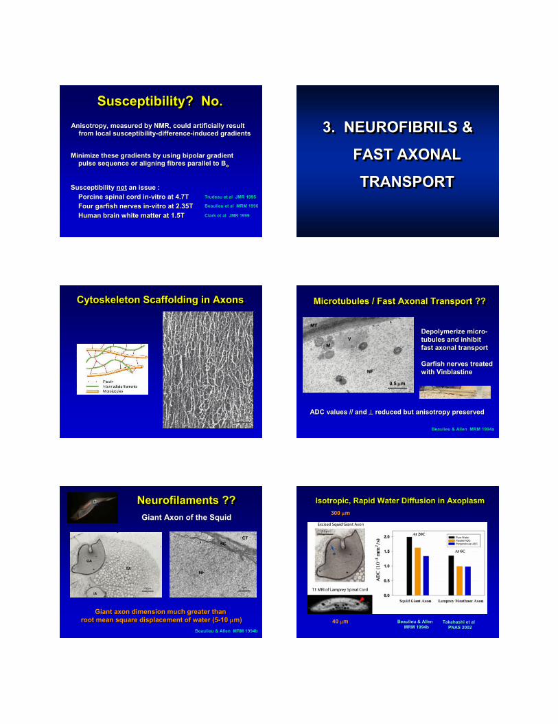
Cytoskeleton Scaffolding in AxonsCytoskeleton Scaffolding in AxonsCytoskeleton Scaffolding in Axons Microtubules / Fast Axonal Transport ??Microtubules / Fast Axonal Transport ??Microtubules / Fast Axonal Transport ??
DepolymerizeDepolymerize micromicro--tubules and inhibit tubules and inhibit fast axonal transportfast axonal transport
Garfish nerves treatedGarfish nerves treatedwith with VinblastineVinblastine
Beaulieu & Allen MRM 1994aBeaulieu & Allen MRM 1994a
ADC values // and ADC values // and ⊥⊥ reduced but anisotropy preservedreduced but anisotropy preserved
0.5 μm
Neurofilaments ??Neurofilaments ??Neurofilaments ??Giant Axon of the SquidGiant Axon of the Squid
Beaulieu & Allen MRM 1994bBeaulieu & Allen MRM 1994b
Giant axon dimension much greater than Giant axon dimension much greater than root mean square displacement of water (5root mean square displacement of water (5--10 10 μμm)m)
Isotropic, Rapid Water Diffusion in AxoplasmIsotropic, Rapid Water Diffusion in Isotropic, Rapid Water Diffusion in AxoplasmAxoplasm
Beaulieu & Allen Beaulieu & Allen MRM 1994bMRM 1994b
Takahashi et al Takahashi et al PNAS 2002PNAS 2002
300 μm
40 μm

SummaryNeurofibrils / Axonal Transport
SummarySummaryNeurofibrilsNeurofibrils / Axonal Transport/ Axonal Transport
Squid diffusion results matched Monte CarloSquid diffusion results matched Monte Carlosimulations of simulations of neurofilamentaryneurofilamentary latticelattice
Microtubules and neurofilaments Microtubules and neurofilaments do notdo not play aplay asignificant role in anisotropic water diffusionsignificant role in anisotropic water diffusion
Water diffusion in pure axoplasm is rapid and is Water diffusion in pure axoplasm is rapid and is ~ 70 ~ 70 -- 80% of that in pure water80% of that in pure water
4. AXONAL
MEMBRANES
4.4. AXONAL AXONAL
MEMBRANESMEMBRANES
Water Transport is ComplexWater Transport is ComplexWater Transport is Complex
www.bioscience.orgwww.bioscience.org, Watson, Watson
AquaporinsAquaporins
Garfish NervesGarfish NervesGarfish Nerves
Beaulieu & Allen, MRM, 1994aBeaulieu & Allen, MRM, 1994a
Non-myelinated olfactory Myelinated optic (CNS)
Myelinated trigeminal (PNS)
FA 0.59 FA 0.52
FA 0.59
Anisotropy in Non-myelinated Fibres : Other Studies
Anisotropy in NonAnisotropy in Non--myelinated myelinated FibresFibres : : Other StudiesOther Studies
Rat pups white matterRat pups white matter WimbergerWimberger et al,JCAT 1995et al,JCAT 1995Prayer et al, Prayer et al, NeuroradNeurorad 19971997
JimpyJimpy mouse optic nervemouse optic nerve Ono et alOno et alBrain Res 1995Brain Res 1995
Rat Rat vagusvagus nervenerve SeoSeo et alet alMRM 1999MRM 1999
Myelin deficient rat spinal cordMyelin deficient rat spinal cord GulaniGulani et al, MRM 2001et al, MRM 2001BitonBiton et al, MRI 2006et al, MRI 2006
Brain in developing miceBrain in developing mice Mori et al Mori et al MRM 2001MRM 2001
Anisotropy in Non-myelinated Fibres : Other Studies
Anisotropy in NonAnisotropy in Non--myelinated myelinated FibresFibres : : Other StudiesOther Studies
MyelinMyelin--deficient deficient shiverershiverer micemiceSong, Song, NeuroimageNeuroimage 2002 2002 Nair, Nair, NeuroimageNeuroimage 20052005TyszkaTyszka, , NeuroimageNeuroimage 20062006
Walking leg nerve of lobsterWalking leg nerve of lobster BeaulieuBeaulieuNMR NMR BiomedBiomed 20022002
Lamprey spinal cordLamprey spinal cord Takahashi et alTakahashi et alPNAS 2002PNAS 2002
Brain in baby rabbitsBrain in baby rabbits DrobyshevskyDrobyshevsky et alet alJ J NeurosciNeurosci 20052005
Neonatal human brain Neonatal human brain inin--vivovivo HuppiHuppi et al, et al, PedPed Res 1998Res 1998Neil et al, Radiology 1998Neil et al, Radiology 1998
Lamprey Spinal CordLamprey Spinal CordLamprey Spinal Cord
Takahashi et al PNAS 2002Takahashi et al PNAS 2002
Ani
sotr
o py
Ani
sotr
o py
Axon diameterAxon diameter
Packing DensityPacking Density
increasesincreases
decreasesdecreases
** all non** all non--myelinatedmyelinated ****
Anisotropy in Pre-myelination PeriodAnisotropy in PreAnisotropy in Pre--myelinationmyelination PeriodPeriod
DTI of white matter development in DTI of white matter development in perinatalperinatal rabbitsrabbits
DrobyshevskyDrobyshevsky et al et al J J NeurosciNeurosci 20052005
MyelinationMyelinationbegan at P5began at P5
MyelinationMyelinationbegan at P11began at P11
SummaryAxonal Membranes
SummarySummaryAxonal MembranesAxonal Membranes
Anisotropy in neural Anisotropy in neural fibresfibres NOT myelin specificNOT myelin specific
Axonal membranes sufficient for significant Axonal membranes sufficient for significant anisotropy and appear to play anisotropy and appear to play primaryprimary rolerole
Nonetheless, the loss and addition of myelin in a Nonetheless, the loss and addition of myelin in a given tract could certainly affect ADC(//), given tract could certainly affect ADC(//), ADC(ADC(⊥⊥), and the degree of anisotropy), and the degree of anisotropy
5. MYELIN5. MYELIN5. MYELIN
MyelinMyelin--Deficient Spinal Cord Rat ModelDeficient Spinal Cord Rat Model
X-linked recessive Wistar ratnear-total lack of myelination in the CNS
Normal
Toluidine Blue Stain
Myelin Deficient
FA 0.73
Anisotropy decrease of ~ 10% in absence of myelin
GulaniGulani et al, MRM, 2001et al, MRM, 2001
FA 0.66
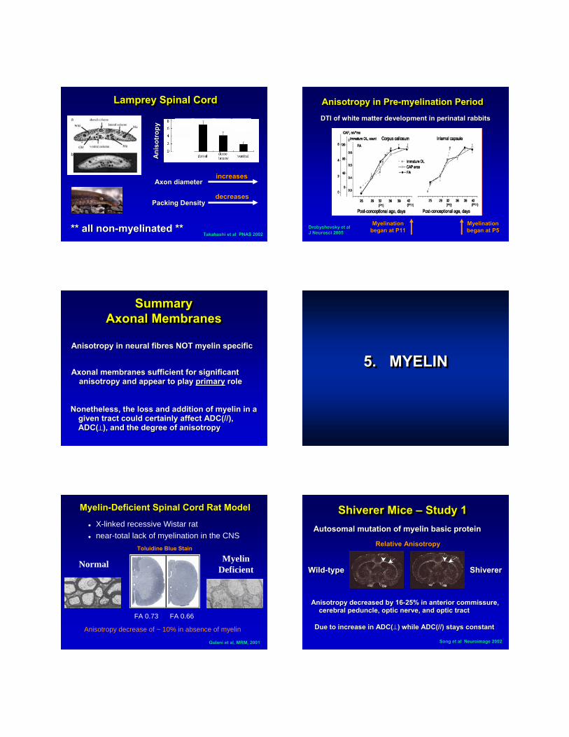
Shiverer Mice – Study 1ShivererShiverer Mice Mice –– Study 1Study 1AutosomalAutosomal mutation of myelin basic proteinmutation of myelin basic protein
Song et al Song et al NeuroimageNeuroimage 20022002
WildWild--typetype ShivererShiverer
Relative AnisotropyRelative Anisotropy
Anisotropy decreased by 16Anisotropy decreased by 16--25% in anterior 25% in anterior commissurecommissure, , cerebral peduncle, optic nerve, and optic tract cerebral peduncle, optic nerve, and optic tract
Due to increase in ADC(Due to increase in ADC(⊥) while ADC(//) stays constant ) while ADC(//) stays constant

Shiverer Mice – Study 2aShivererShiverer Mice Mice –– Study 2aStudy 2a
FA reduced by ~10% in corpus FA reduced by ~10% in corpus callosumcallosum
Nair et al Nair et al NeuroimageNeuroimage 20052005
Shiverer Mice – Study 2bShivererShiverer Mice Mice –– Study 2bStudy 2bDonorDonor--derived derived myelinationmyelination by transplanted by transplanted
precursor cells in corpus precursor cells in corpus callosumcallosum
Nair et al Nair et al NeuroimageNeuroimage 20052005
Myelin Basic Protein
T2T2--weighting did not show thisweighting did not show this
FA Map
Shiverer Mice – Study 3ShivererShiverer Mice Mice –– Study 3Study 3High resolution DTI microscopy of fixed brainsHigh resolution DTI microscopy of fixed brains
TyszkaTyszka et al et al NeuroimageNeuroimage 20062006
ShivererShivererFAFA
ControlControlFAFA
FAFADifferenceDifference
FA reduced byFA reduced by1111--19% in 19% in shiverershiverer micemice
Jimpy MiceJimpyJimpy MiceMice
HarsanHarsan et al J et al J NeuroNeuro Res 2007Res 2007
Myelin basicMyelin basicproteinprotein
ADCADC((⊥))
FAFA
P25P25
P75P75afterafter
recoveryrecovery
Transgenic Mice - DysmyelinationTransgenic Mice Transgenic Mice -- DysmyelinationDysmyelination
HarsanHarsan et al J et al J NeuroNeuro Res 2006Res 2006
Treated : P15Treated : P15 P60P60P30P30
//⊥ FA
Morphometry - Diffusion CorrelationsMorphometryMorphometry -- Diffusion CorrelationsDiffusion Correlations
Axon Axon morphometrymorphometry correlated with ADC(//), ADC(correlated with ADC(//), ADC(⊥), ), and anisotropy in normal rat cervical spinal cordand anisotropy in normal rat cervical spinal cord
Schwartz et al Schwartz et al NeuroreportNeuroreport 20052005
PositivePositive correlations with ADC(correlations with ADC(⊥))-- extraextra--cellular volume fractioncellular volume fraction-- axon spacing (most significant)axon spacing (most significant)
NegativeNegative correlations with ADC(correlations with ADC(⊥))-- axon countsaxon counts-- myelin volume fraction myelin volume fraction

What About Human Brain?What About Human Brain?What About Human Brain?PostPost--mortem DTI and histology in MS patientsmortem DTI and histology in MS patients
SchmiererSchmierer et al et al NeuroimageNeuroimage 20072007
More myelin
SummaryMyelin
SummarySummaryMyelinMyelin
Myelin has more marked effects on the mean Myelin has more marked effects on the mean diffusivity and diffusivity and eigenvalueseigenvalues than the anisotropythan the anisotropy
MyelinationMyelination can modulate degree of anisotropycan modulate degree of anisotropy
A reduction in ADC(A reduction in ADC(⊥) coincides with ) coincides with myelinationmyelinationfor a given neural for a given neural fibrefibre
Aside: small FA changes may reflect more severe pathology Aside: small FA changes may reflect more severe pathology than expectedthan expected
6. INTERPRETATION
OF ANISOTROPY
6.6. INTERPRETATIONINTERPRETATION
OF ANISOTROPYOF ANISOTROPY
How to Interpret Reductions in Anisotropy?
How to Interpret How to Interpret Reductions in Anisotropy?Reductions in Anisotropy?
Clues from Wallerian DegenerationClues from Clues from WallerianWallerian DegenerationDegenerationRat Spinal Cord Injury Rat Spinal Cord Injury
Ford et al MRM 1994Ford et al MRM 1994
PierpaoliPierpaoli et al et al NeuroimageNeuroimage 20012001Beaulieu et al MRM 1996Beaulieu et al MRM 1996
T2 T2 ADC(ADC(⊥⊥) ) ADC(//) ADC(//)
Similar findings in frog sciatic nerve and Similar findings in frog sciatic nerve and secondary secondary fibresfibres in human chronic strokein human chronic stroke
Same FA ReductionsSame FA ReductionsSame FA ReductionsNormal FANormal FA 20% FA20% FA
≠≠

FA Reductions Could Be Interpreted Differently
FA Reductions Could Be FA Reductions Could Be Interpreted DifferentlyInterpreted Differently
FAFAADC (//)ADC (//)
ADC (ADC (⊥⊥))
and/orand/or
Need to look at the eigenvaluesNeed to look at the Need to look at the eigenvalueseigenvalues
Mouse Optic Nerve IschemiaMouse Optic Nerve IschemiaMouse Optic Nerve Ischemia
Song et al Song et al NeuroimageNeuroimage 20032003
ADC (//)ADC (//) ADC (ADC (⊥⊥))
FAFA
Days post-injury
Cuprizone Treated MiceCuprizoneCuprizone Treated MiceTreated Mice
Sun et al MRM 2006Sun et al MRM 2006
λλ////drops firstdrops first
AxonalAxonaldamagedamage
λλ⊥⊥increases laterincreases later
DemyelinationDemyelination
Mouse Brain TraumaMouse Brain TraumaMouse Brain Trauma
MacDonald et al J MacDonald et al J NeurosciNeurosci 20072007
//
⊥
MacDonald et al J MacDonald et al J NeurosciNeurosci 20072007
Mouse Brain TraumaMouse Brain TraumaMouse Brain Trauma
SubacuteSubacute InjuryInjuryAcute InjuryAcute Injury
Reduced parallelReduced parallel Increased perpendicularIncreased perpendicular
Importance of AxonsImportance of AxonsImportance of Axons
Mouse Brain TraumaMouse Brain Trauma
MacDonald et al Exp MacDonald et al Exp NeurolNeurol 20072007Wu et al Wu et al NeuroimageNeuroimage 20072007
Mouse EAE ModelMouse EAE ModelOptic NerveOptic Nerve
Ani
sotr
opy
AD
C(//
)
Axolemmal Area # of Injured Axons

Human Callosotomy - FAHuman Human CallosotomyCallosotomy -- FAFA
ConchaConcha et al et al NeuroimageNeuroimage 20062006
Are these FA reductions the same?
Post-surgical transectionof corpus callosum
Human Callosotomy - EigenvaluesHuman Human CallosotomyCallosotomy -- EigenvaluesEigenvalues
ConchaConcha et al et al NeuroimageNeuroimage 20062006
FA – Eigenvalue HypothesisFA FA –– EigenvalueEigenvalue HypothesisHypothesis
FAFA
ADC (//)ADC (//)
ADC (ADC (⊥⊥))
Axonal DamageAxonal Damage
DemyelinationDemyelination
CONCLUSIONCONCLUSIONCONCLUSION
DTI – Limited ResolutionStill Probing the Microstructure
DTI – Limited ResolutionStill Probing the Microstructure
μm’s mm’s cm’s
ConclusionConclusionConclusionIntact membranes are confirmed to be the Intact membranes are confirmed to be the
primary determinant of anisotropic water primary determinant of anisotropic water diffusion in neural diffusion in neural fibresfibres
Changes in perpendicular diffusivity may be Changes in perpendicular diffusivity may be indicative of relative degree of myelin in a indicative of relative degree of myelin in a specific white matter tractspecific white matter tract
Primary role by axonal membranes Primary role by axonal membranes
Secondary role by Secondary role by myelinationmyelination (modulate)(modulate)