the binding site for ribosomal protein complex l8 within 23 s
TRANSCRIPT
THE JOURNAL OF BIOLOGICAL CHEMISTRY 0 1984 by The American Society of Biological Chemists, Inc.
Vol. 259, No. 10, Issue of May 25, pp, 6559-6563.1984 Printed in U.S.A.
The Binding Site for Ribosomal Protein Complex L8 within 23 S Ribosomal RNA of Escherichia coZi*
(Received for publication, November 2, 1983)
Alan A. D. Beauclerk$, Eric Cundliffe$$, and Jan Dijkn From the $Department of Biochemistty, University of Leicester, Leicester LEI 7RH, England and the 7lMax-Planck-Institut fur Molekulare Genetik, Abteilung Wittmann, Berlin-Dahlem, West Germany
The ribosomal protein complex L8 of Escherichia coli consists of two dimers of protein L7/L12 and one monomer of protein L10. This pentameric complex and ribosomal protein L11 bind in mutually cooperative fashion to 23 S rRNA and protect specific fragments of the latter from digestion with ribonuclease TI. Oli- gonucleotides protected either by the L8 complex alone or by the complex plus protein L11 were isolated from such digests and shown to rebind specifically to these proteins. They were also subjected to nucleotide se- quence analysis. The longest oligonucleotide, protected by the L8 complex alone, consisted of residues 1028- 1124 of 23 S rRNA and included all the other RNA fragments produced in this study. Previously, protein L11 had been shown to protect residues 1052-11 12 of 23 S rRNA. It is concluded that the binding sites for the L8 protein complex and for protein L11 are im- mediately adjacent within 23 S rRNA of E. coli.
Among the ribosomal proteins from Escherichia coli, protein L12 (and its NH2-terminally acetylated derivative, L7) is unique in being present as multiple copies (i.e. four) per 70 S particle (Thammana et al., 1973; Hardy, 1975; Subramanian, 1975). Protein L7/L12 is readily removed from ribosomes and dimerizes in solution (Moller et al., 1972) to produce an elongated structure (Wong and Paradies, 1974; Osterberg et al., 1976) to which another ribosomal protein, L10, can also bind (Pettersson et al., 1976). The resultant complex is pen- tameric and contains two dimers of protein L7/L12 plus one monomer of protein L10 (Pettersson and Liljas, 1979). A similar complex, formerly thought to represent a. discrete species, at which time it was designated “ribosomal protein L8,” can be extracted directly from ribosomes (Pettersson et al., 1976) and readily reassociates with ribosomal core parti- cles lacking the complex. During rebinding, the pentameric complex remains intact (Tokimatsu et al., 1981), although, off the ribosome, one L7/L12 dimer can selectively be displaced (Dijk et al., 1979; Zantema et al., 1982). These studies are readily reconciled with the observations that protein L7/L12 does not rebind to core particles lacking protein L10 (Schrier et al., 1973; Tokimatsu et at., 1981) or to those in which the latter is occluded by specific antibody (Stoffler et at., 1974).
A number of ribosomal proteins possess specific binding sites on rRNA, although in only a few cases have these been fully characterized (see Zimmermann (1980) for review).
* The costs of publication of this article were defrayed in part by the payment of page charges. This article must therefore be hereby marked “aduertisement” in accordance with 18 U.S.C. Section 1734 solely to indicate this fact.
Supported by a Research Project Grant from the Science and Engineering Research Council.
-
Thus, the protein complex L8 binds specifically and with saturation kinetics to a single site on 23 S rRNA (Dijk et al., 1977). In contrast, purified protein L10 bound to 23 S RNA in one study (Pettersson, 1979) but not in others (Dijk et al., 1979; Spierer et al., 1979), whereas protein L7/L12 interacted only weakly and nonspecifically with RNA. However, despite the conflicting observations with protein L10, there is general agreement that the L8 complex attaches to 23 S RNA via the L10 moiety (Dijk et al., 1979; Pettersson, 1979). It was also found that binding of the L8 complex was strongly promoted by ribosomal protein L11 (and vice versa) and that the at- tachment sites for both lay within an oligonucleotide, about 1200 residues long, derived from the 5’ terminus of 23 S RNA (Dijk et a!., 1979). Although an RNA fragment of that length could readily accommodate the proteins at fairly distant sites, mutual cooperativity in their binding would be plausibly ex- plained if the sites were close together. Such an hypothesis would also be consistent with the results from cross-linking studies (Clegg and Hayes, 1974; Expert-Bezancon et al., 1976) which suggest that proteins L7/L12, L10, and L11 are near neighbors in the 50 S ribosomal subunit.
The binding site for protein L11 within 23 S rRNA was recently determined (Schmidt et al., 1981) and shown to embrace nucleotides 1052-1112. Here, we have used similar methods, involving the use of proteins to protect RNA from nuclease digestion, to reveal the binding site of the pentameric L8 complex.
EXPERIMENTAL PROCEDURES
Materiak-Cellulose nitrate filters (0.45 pm, 13-mm diameter) were from Sartorius. Ribonuclease TI was from Sankyo Co., Ltd. Ribonucleases U, and Phy M, Bacillus cereus RNase, and polynucleo- tide kinase were from Bethesda Research Laboratories. Calf intestinal alkaline phosphatase was from Boehringer Mannheim and T4 RNA ligase was from P-L Biochemicals. [y3*P]ATP, [5’-32P]~ytidine his- phosphate, and [32P]phosphate were from Amersham International.
Preparation of Ribosomal Proteins-Protein L11 and L8 protein complex were prepared as previously described (Dijk and Littlechild, 1979) and were stored a t -70 “C. Protein L11 was stored in HMK buffer plus 0.1 mM benzamidine and 0.1 mM phenylmethylsulfonyl fluoride. This buffer contained 10 mM Hepes’/KOH, pH 7.45, at 20 “ c , 20 mM MgClz, 100 mM KCl, 3 mM 2-mercaptoethanol. Protein complex L8 was stored in buffer containing 20 mM sodium phosphate, pH 7.0,0.35 M NaCl and was dialyzed into HMK buffer immediately before use. Both buffers contained benzamidine and phenylmethyl- sulfonyl fluoride as described above.
Preparation of Uniformly Labeled [32Pl 23 S rRNA-This was purified from E. coli MRE 600 grown in low phosphate medium (Brownlee, 1972) supplemented with (32P)phosphate (10 mCi, carrier- free/25-ml culture). Cells were isolated by centrifugation, washed twice with 10 mM Tris-HC1, pH 8.1, at 20 “C, and resuspended in
The abbreviations used are: Hepes, 4-(2-hydroxyethyl)-l-pipera- zineethanesulfonic acid; SDS, sodium dodecyl sulfate; EF, elongation factor.
6559
by guest on April 12, 2018
http://ww
w.jbc.org/
Dow
nloaded from

6560 RNA Binding Site for Ribosomal Protein Complex L8 0.35 ml of this buffer. Then, Na2EDTA (1 mM final concentration) and lysozyme (50 pg) were added, followed, after 30 min a t 37 'C, by SDS (1% w/v final concentration). After two cycles of freezing and thawing in dry ice/methanol, the resultant lysate was cleared by centrifugation at 50,000 X g for 1 h at 15 "C. The supernatant was then applied to a linear gradient of 15-30% (w/v) sucrose in 10 HIM Tris-HCI, pH 7.5, a t 20 "C, 100 mM LiCl, 10 mM Na2EDTA. 0.2% (w/v) SDS. Centrifugation was at 25,000 rpm for 20 h at 15 "C in a Beckman SW 27 rotor. Fractions containing 23 S rRNA were pooled, and the RNA was precipitated with ethanol and redissolved in water. The 23 S rRNA was then extracted with phenol and reprecipitated three times from 0.6 M potassium acetate, dried in uacuo, and dis- solved in water.
Preparation of Unlabeled rRNA-Total ribosomal RNA was ex- tracted from 70 S ribosomes using acetic acid plus urea (Hochkeppel et al., 1976) and fractionated using LiCl plus SDS in a sucrose density gradient, from which 23 S rRNA was purified as described above.
Production of rRNA Fragments Protected by Specific Proteins- Ribosomal proteins (as below) were incubated with 23 S rRNA for 20 min at 44 "C and then for 5 min at 20 "C in 100 pl of buffer containing 20 mM Hepes/KOH, pH 7.5, a t 20 "C, 4 mM MgC12,380 mM NH4Cl, 20 mM KCI, 3 mM 2-mercaptoethanol, 0.1 mM phenylmethylsulfonyl fluoride, 0.1 mM benzamidine. Complexes formed between 23 S rRNA (50 pmol) and the L8 protein complex (150 pmol) were digested with Tl ribonuclease (0.5 units) for 5 min at 37 "C. Alternatively, those formed between 23 S rRNA (50 pmol) and a mixture of proteins L8 (113 pmol) and L11 (101 pmol) were incubated at 37 "C with T1 ribonuclease (2 units) for up to 15 min. In controls, protein complex L8 or a mixture of the L8 complex plus protein L11 was added after digestion of 23 S rRNA with Tl ribonuclease. Reaction mixtures were then treated in one of two ways prior to analysis by gel electrophoresis. Some reaction mixtures were filtered through cellulose nitrate discs (0.45 pm, 13-mm diameter), washed three times with 2 ml of incu- bation buffer, and then eluted with 0.5 ml of 1 M LiCl, 0.1% (w/v) SDS as described previously (Schmidt et al., 1981). The eluates were immediately extracted with phenol, and RNA fragments were precip- itated from the aqueous phase with 2.5 volumes of ethanol a t -20 "C followed by reprecipitation from 0.6 M sodium acetate. Other reaction mixtures, which had not been filtered, were directly extracted with phenol, and RNA fragments were precipitated as above.
Isolation of Uniformly Labeled f3'P/RNA Fragments-Following precipitation with ethanol, RNA fragments were taken up in 7 M urea containing 40% (w/v) sucrose, 0.1% (w/v) xylene cyanol FF, 0.1% (w/v) bromphenol blue and resolved by electrophoresis at 15 mA (constant current) for 2 h a t 20 'C in gels (10 cm X 1.5 mm; 6-mm wells) containing 12% (w/v) acrylamide and 7 M urea. Running buffer contained 90 mM Tris base, 90 mM boric acid, 2.8 mM Na2EDTA. RNA fragments in gels were visualized by autoradiography at -70 "C using Fuji RX film and were eluted from gel sections by maceration in 500 mM ammonium acetate, 10 mM magnesium acetate, 1 mM Na2EDTA, 0.1% (w/v) SDS followed by incubation a t 20 "C for 16 h. Pieces of gel were removed by filtration, and the RNA fragments were precipitated three times from 0.6 M sodium acetate in the presence of 10 pg of tRNA as carrier before being finally dissolved in water.
Preparation of T e r m i d y Labeled PPJRNA Fragments-Frag- ments, produced from unlabeled 23 S rRNA as described above, were reprecipitated twice from 0.6 M sodium acetate and dissolved in water. They were then labeled either at the 5' terminus using [Y-~~P]ATP and T4 polynucleotide kinase (Donis-Keller et al., 1977) or, after treatment with calf intestinal alkaline phosphatase (Chaconas and van de Sande, 1980), at the 3' terminus with [5'-32P]cytidine bis- phosphate and T4 RNA ligase (England et al., 1980, DAllessio, 1982). End-labeled RNA fragments were then resolved by electrophoresis a t 400 V (constant voltage) for 20 h at 20 "C in gels (35 cm X 1.5 mm; 6-mm wells) containing 12% (w/v) acrylamide and 7 M urea, visual- ized by autoradiography, and recovered as described above.
Rebinding of RNA Fragments to Ribosomal Proteins-Fragments of RNA (0.5 pmol) recovered from gels were incubated at 20 "C for 20 min with the L8 complex, with protein L11, or with a mixture of the two at a range of inputs up to 20-fold molar excess. In controls, protein L1 or L23 was added. The buffer (30 pl) contained 20 mM Hepes/KOH, pH 7.5, at 20 "C, 4 mM MgC12, 380 mM NH4CI, 20 mM KCl, 3 mM 2-mercaptoethanol, 0.1 mM phenylmethylsulfonyl fluoride, 0.1 mM benzamidine. Reaction mixtures were filtered through cellu- lose nitrate discs (0.45 pm, 13-mm diameter) which were washed with
2 ml of incubation buffer and subjected to liquid scintillation count- ing.
Nucleotide Sequence Analysis-Terminally labeled [ T I R N A frag- ments were partially degraded with RNase TI or Ut (Donis-Keller et al., 1977). with Phy M RNase (Donis-Keller, 1980), or with B. cereus ribonuclease (Lockhard et al., 1978), and the products were analyzed on sequence gels (D'Allessio, 1982).
RESULTS
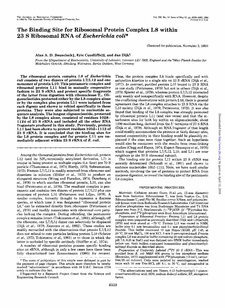
Preparation of Protected Fragments of 23 S RNA-Ribo- nuclease TI was used to digest 23 S rRNA under two sets of conditions which reflected the differing protective capabilities of the L8 protein complex plus and minus protein L11. When 23 S RNA was protected by a 3-fold molar excess of the L8 complex, digestion was carried out under relatively mild con- ditions. However, more severe conditions were used when the RNA was protected by a mixture of the L8 complex plus protein L11 (each present at 2-fold molar excess over 23 S RNA). The products of digestion were then analyzed directly on polyacrylamide gels (Fig. 1, tracks a, d, and g). Although there was some evidence for bands, possibly corresponding to specifically protected RNA fragments (for example, Fig. 1, track g), these were not resolved well enough to allow their isolation from such gels. Accordingly, an alternative method, which was described previously (Schmidt et al., 1981), was used for the recovery of RNA fragments protected by (and still bound to) specific proteins. This depended upon the ability of the latter to adhere to cellulose nitrate filters from which the bound oligonucleotides could subsequently be dis- placed by salt plus detergent and separated out on polyacryl- amide gels (see "Experimental Procedures"). At least two such oligonucleotides (designated 1 and 2) were recovered when 23 S RNA was digested gently with RNase TI in the presence of protein complex L8 (Fig. 1, track b). Also, since binding of the L8 complex to 23s RNA is strongly promoted by protein
0 . - - "
0
xc e 0
BPB 0 0
FIG. 1. Autoradiogram of a urea-polyacrylamide gel con- taining ribonuclease digests of E. coli 23 S rRNA. =P-labeled 23 S rRNA was digested with RNase T1 in the presence and absence of protein complex L8 or the L8 complex plus protein L11. Digestion was for 5 min (tracks a-f) or for 10 min (tracksg-i) as described under "Experimental Procedures." Digests were loaded onto urea-polyac- rylamide gels either directly or after filtration through cellulose nitrate followed by elution with LiCl plus SDS. The following were analyzed: unfractionated digests of 23 S RNA protected by protein complex L8 (track a) or the L8 complex plus protein L11 (tracks d and g); filtered digests of 23 S RNA protected by protein complex L8 (track b) or the L8 complex plus protein L11 (tracks e and h); filtered digests of unprotected 23 S RNA to which protein complex L8 (track c) or the L8 complex plus protein L11 (tracks f and i) was added after digestion but prior to filtration. RNA fragments are numbered as in text. XC and BPB indicate the positions of xylene cyanol FF and bromphenol blue dye markers, respectively. 0 is the origin.
by guest on April 12, 2018
http://ww
w.jbc.org/
Dow
nloaded from
RNA Binding Site for Ribosomal Protein Complex L8 6561
L11 (and vice versa), we sought protected oligonucleotides in digests of doubly protected RNA following more vigorous treatment with RNase TI (Fig. 1, tracks e and h). One such oligonucleotide appeared identical to RNA fragment 2 (above); the others were designated 3-5 in order of decreasing size. Given that similar oligonucleotides were not recovered from digests of unprotected RNA to which the L8 complex or the complex plus protein L11 was subsequently added (Fig. 1, tracks c, f, and i), we inferred that RNA fragments 1-5 had been specifically protected by the added proteins. Typical yields for their production are given in Table I, from which we concluded that no putative contaminant in our protein preparations could have been responsible for their protection.
Rebinding of Protected RNA Fragments to Proteins-In order to confirm that RNA fragments 1-5 were specifically protected oligonucleotides, they were eluted from preparative polyacrylamide gels (similar to that represented in Fig. 1) and assayed for their ability to rebind to the L8 complex and to protein L11 (Table 11). As controls, to score for nonspecific attachment, ribosomal proteins L1 and L23 were also included in these assays. Both these proteins bind to specific sites within 23 S RNA (see Zimmermann (1980) for details). As can be seen, none of the RNA fragments bound to protein L1 or L23, but all of them bound to protein L l l . In contrast, the lengths of the RNA fragments seemed crucial in determining how well they bound to the L8 complex. Thus, fragments 1 and 2 bound well to the complex, fragments 3 and 4 much less so, and fragment 5 not at all.
In each case where rebinding was observed, the shape of the binding curve was compatible with saturation kinetics and suggested a single binding site (data not shown). More- over, the L8 complex and protein L11 showed good evidence of cooperativity in promoting the binding of those oligonucle- otides (most notably RNA fragments 3 and 4) to which both could bind. This mimics their behavior with intact 23 S RNA and supports the authenticity of the RNA fragments gener-
TABLE I Yields of protected RNA fragments
Protecting Droteinfs) . , RNA fragment (duration ;;digestion)
produced L8” L8 + L1lb LB+ L11b L8+ Lllb (5 min) (5 min) (10min) (15min)
% input RNA (pmol/pmoU 1 4 2 6 11 3 7 4 11 13 8 5 9 19 21
a 1 unit of RNase T,/100 pmol of 23 S RNA, molar ratio of L8
1 unit of RNase TJ25 pmol of 23 S RNA molar ratio of proteins complex to RNA of 3:l.
L8 plus L11 to RNA of 2:1 (each).
TABLE I1 Rebinding of protectec’ RNA fragments to ribosomal proteins
Protein (protein/RNA
RNA fragment
molar ratio) 1 2 3 4 5 % rebinding“
L8 (2.5:l) 60 62 16 25 3 (201) 76 74 31 46 3
L11 (2.5:l) 48 36 44 43 39 (201) 71 67 68 65 74
L8 + L11 (2.5:l) 73 75 70 65 64 L1 (201) 2 3 3 3 2 L23 (201) 3 3 3 2 3
’ Per cent of input fragment bound to protein.
ated in these studies. As a further argument to that effect, we wish to emphasize that all the protection and rebinding ex- periments described here were carried out in buffer closely similar to that normally used during the first stage of the two- step reconstitution procedure (Nierhaus and Dohme, 1979), according to which active 50 S ribosomal subunits can be assembled in vitro. Under such conditions, nonspecific inter- actions between ribosomal proteins and rRNA are evidently minimized,
The rebinding data presented in Table I1 strongly suggested that we had isolated a “nest” of homologous oligonucleotides. Each one bound to protein L11 to about the same extent and evidently contained part, or all, of the recognition site for that protein. In contrast, the L8 protein complex appeared to interact primarily with nucleotide sequences absent from RNA fragment 5 but present in the larger oligonucleotides. In particular, although RNA fragment 4 was evidently only a few residues longer than fragment 5, this seemed crucial for binding of the L8 complex. We therefore concluded that protein complex L8 (presumably acting via the L10 moiety) binds to 23s RNA immediately adjacent to the site at which protein L11 binds.
Nucleotide Sequence Analysis of Protected RNA Frag- ments-In order to establish the nucleotide sequences of our RNA fragments, they were first prepared in nonradioactive form. This involved digestion of protected RNA followed by filtration on cellulose nitrate and elution as in Fig. 1. Then, oligonucleotides in the eluates were radioactively end-labeled and resolved by electrophoresis on polyacrylamide gels. After elution from such gels, RNA fragments were subjected to partial enzymic digestion and analysis on sequencing gels. In comparison with its behavior on the shorter gel depicted in Fig. 1, RNA fragment 2 was here resolved into doublet bands (designated 2a and 2b), the relative proportions of which varied with the conditions of digestion. We therefore deter- mined the nucleotide sequences of all these RNA fragments, including 2a and 2b made under the different conditions. Each was completely sequenced from either end, and the terminal residues were confirmed by end analysis. The results are summarized in Table 111. The protected oligonucleotides were indeed homologous and included the region of 23 S RNA (residues 1052-1112) previously shown to bind protein L11. In that study (see Tables I and I1 and Fig. 4a in Schmidt et al., 1981), there was some evidence for “fraying” at the 3‘ end of the RNA fragment protected by protein L11, so that residues derived from positions 1108-1112 were recovered with less than unit stoichiometry. Hence, fragment 5 repre- sents the binding site for protein L11, whereas the longer RNA fragments contain sequences recognized by the L8 pro- tein complex. Interestingly, the L8 complex appears to exert an “umbrella” effect upon 23 S RNA in the region represented
TABLE 111 Summary of protected oligonucleotide sequences
fragment t o protect RNA within 23 S rRNA
2a L8 complex” 1033-1124
2b L8 complex + L11 1033-1122 3 L8 complex + L11 1040-1120 4 L8 complex + L11 1044-1112 5 L8 complex + L11 1052-1110
L11 aloneb 1052-1112
RNA Protein(s) used Location of fragment
1 L8 complex” 1028-1124
a Mild digestion conditions. See Schmidt et al., 1981.
by guest on April 12, 2018
http://ww
w.jbc.org/
Dow
nloaded from
6562 RNA Binding Site for Ribosomal Protein Complex L8 G I A I
A I A -1028 I
A. / A G. G - C - G * U - A - G ~ C
1 1 2 4 1 A . A I 1 G * U I 1 U * G I I
I 1 U O G I I G * C - C I ‘C
A .A I I
1110- G G \ I
G . C I I
I t
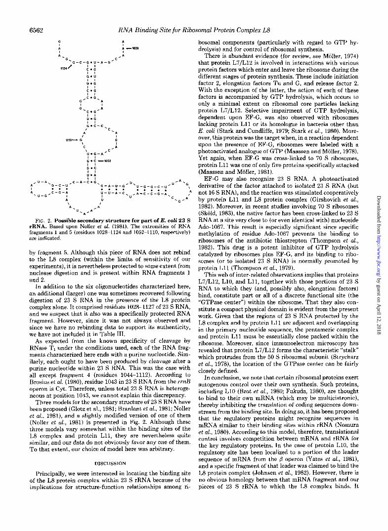
FIG. 2. Possible secondary structure for part of E. coli 23 S rRNA. Based upon Noller et al. (1981). The extremities of RNA fragments 1 and 5 (residues 1028-1124 and 1052-1110, respectively) are indicated.
by fragment 5. Although this piece of RNA does not rebind to the L8 complex (within the limits of sensitivity of our experiments), it is nevertheless protected to some extent from nuclease digestion and is present within RNA fragments 1 and 2.
In addition to the six oligonucleotides characterized here, an additional (larger) one was sometimes recovered following digestion of 23 S RNA in the presence of the L8 protein complex alone. I t comprised residues 1028-1127 of 23 S RNA, and we suspect that it also was a specifically protected RNA fragment. However, since it was not always observed and since we have no rebinding data to support its authenticity, we have not included it in Table 111.
As expected from the known specificity of cleavage by RNase TI under the conditions used, each of the RNA frag- ments characterized here ends with a purine nucleotide. Sim- ilarly, each ought to have been produced by cleavage after a purine nucleotide within 23 S RNA. This was the case with all except fragment 4 (residues 1044-1112). According to Brosius et al. (1980), residue 1043 in 23 S RNA from the rrnB operon is Cyt. Therefore, unless total 23 S RNA is heteroge- neous at position 1043, we cannot explain this discrepancy.
Three models for the secondary structure of 23 S RNA have been proposed (Glotz et al., 1981; Branlant et al., 1981; Noller et al., 1981), and a slightly modified version of one of them (Noller et al., 1981) is presented in Fig. 2. Although these three models vary somewhat within the binding sites of the L8 complex and protein L11, they are nevertheless quite similar, and our data do not obviously favor any one of them. To that extent, our choice of model here was arbitrary.
DISCUSSION
Principally, we were interested in locating the binding site of the L8 protein complex within 23 S rRNA because of the implications for structure-function relationships among ri-
bosomal components (particularly with regard to GTP hy- drolysis) and for control of ribosomal synthesis.
There is abundant evidence (for review, see Moller, 1974) that protein L7/L12 is involved in interactions with various protein factors which enter and leave the ribosome during the different stages of protein synthesis. These include initiation factor 2, elongation factors Tu and G, and release factor 2. With the exception of the latter, the action of each of these factors is accompanied by GTP hydrolysis, which occurs to only a minimal extent on ribosomal core particles lacking protein L7/L12. Selective impairment of GTP hydrolysis, dependent upon EF-G, was also observed with ribosomes lacking protein L11 or its homologue in bacteria other than E. coli (Stark and Cundliffe, 1979; Stark et al., 1980). More- over, this protein was the target when, in a reaction dependent upon the presence of EF-G, ribosomes were labeled with a photoactivated analogue of GTP (Maassen and Moller, 1978). Yet again, when EF-G was cross-linked to 70 S ribosomes, protein L11 was one of only five proteins specifically attacked (Maassen and Moller, 1981).
EF-G may also recognize 23 S RNA. A photoactivated derivative of the factor attached to isolated 23 S RNA (but not 16 S RNA), and the reaction was stimulated cooperatively by protein L11 and L8 protein complex (Girshovich et at., 1982). Moreover, in recent studies involving 70 S ribosomes (Skold, 1983), the native factor has been cross-linked to 23 S RNA a t a site very close to (or even identical with) nucleoside Ado-1067. This result is especially significant since specific methylation of residue Ado-1067 prevents the binding to ribosomes of the antibiotic thiostrepton (Thompson et al., 1982). This drug is a potent inhibitor of GTP hydrolysis catalyzed by ribosomes plus EF-G, and its binding to ribo- somes (or to isolated 23 S RNA) is normally promoted by protein L11 (Thompson et al., 1979).
This web of inter-related observations implies that proteins L7/L12, L10, and L11, together with those portions of 23 S RNA to which they (and, possibly also, elongation factors) bind, constitute part or all of a discrete functional site (the “GTPase center”) within the ribosome. That they also con- stitute a compact physical domain is evident from the present work. Given that the regions of 23 S RNA protected by the L8 complex and by protein L11 are adjacent and overlapping in the primary nucleotide sequence, the pentameric complex and protein L11 must be essentially close packed within the ribosome. Moreover, since immunoelectron microscopy has revealed that protein L7/L12 forms the characteristic “stalk” which protrudes from the 50 S ribosomal subunit (Strycharz et al., 1978), the location of the GTPase center can be fairly closely defined.
In conclusion, we note that certain ribosomal proteins exert autogenous control over their own synthesis. Such proteins, including L10 (Brot et al., 1980; Fukuda, 1980), are thought to bind to their own mRNA (which may be multicistronic), thereby inhibiting the translation of coding sequences down- stream from the binding site. In doing so, it has been proposed that the regulatory proteins might recognize sequences in mRNA similar to their binding sites within rRNA (Nomura et al., 1980). According to this model, therefore, translational control involves competition between mRNA and rRNA for the key regulatory proteins. In the case of protein L10, the regulatory site has been localized to a portion of the leader sequence of mRNA from the /3 operon (Yates et al., 1981), and a specific fragment of that leader was claimed to bind the L8 protein complex (Johnsen et al., 1982). However, there is no obvious homology between that mRNA fragment and our pieces of 23 S rRNA to which the L8 complex binds. I t
by guest on April 12, 2018
http://ww
w.jbc.org/
Dow
nloaded from

RNA Binding Site for Ribosomal Protein Complex L8 6563
therefore remains to be established whether any other portion of the mRNA from the 0 operon (Post et al., 1979) also binds protein L10 (or the L8 complfx) or whether this protein must recognize dissimilar sequences in mRNA and rRNA.
REFERENCES Branlant, C., Krol, A., Machatt, M. A., Pouyet, J., Ebel, J.-P.,
Edwards, K., and Kossel, H. (1981) Nucleic Acids Res. 9, 4303- 4324
Brosius, J., Dull, T. J., and Noller, H. F. (1980) Proc. Natl. Acad. Sci.
Brot, N., Caldwell, P., and Weissbach, H. (1980) Proc. Natl. Acad.
Brownlee, G. G. (1972) Determination of Sequences in RNA, North-
Chaconas, G., and van de Sande, J. H. (1980) Methods Enzymol. 65,
Clegg, C., and Hayes, D. (1974) Eur. J. Biochem. 42, 21-28 DAllessio, J. (1982) Gel Electrophoresis of Nucleic Acids-A Practical
Approach (Rickwood, D., and Hames, B. D., eds) pp. 173-197, IRL Press, Oxford
U. S. A. 77, 201-204
Sci. U. S. A. 77, 2592-2595
Holland/American Elsevier, Amsterdam
75-85
Dijk, J., and Littlechild, J. (1979) Methods Enzymol. 5 9 , 481-502 Dijk, J., Littlechild, J., and Garrett, R. A. (1977) FEBS Lett. 77 ,
Dijk, J., Garrett, R. A., and Muller, R. (1979) Nucleic Acids Res. 6,
Donis-Keller, H. (1980) Nucleic Acids Res. 8, 3133-3142 Donis-Keller, H., Maxam, A. H., and Gilbert, W. (1977) Nucleic Acids
England, T. E., Bruce, A. G., and Uhlenbeck, 0. C. (1980) Methods
Expert-Bezangon, A., Barritault, D., Milet, M., and Hayes, D. H.
Fukuda, R. (1980) Mol. Gen. Genet. 178 , 483-486 Girshovich, A. S., Bochkareva, E. S., and Gudkov, A. T. (1982) FEBS
Glotz, C., Zwieb, C., Brimacornbe, R., Edwards, K., and Kossel, H.
Hardy, S. J . S. (1975) Mol. Gen. Genet. 140, 253-274 Hochkeppel, H.-K., Spicer, E., and Craven, G. R. (1976) J. Mol. Biol.
Johnsen, M., Christensen, T., Dennis, P. P., and Fiil, N. P. (1982)
Lockhard, R. E., Alzner-Deweerd, B., Heckman, J . E., MacGee, M., Tabor, W., and RajBhandary, U. L. (1978) Nucleic Acids Res. 5,
Maassen, J. A., and Moller, W. (1978) J . Biol. Chem. 253,2777-2783 Maasen, J . A., and Moller, W. (1981) Eur. J. Biochem. 115,279-285 Moller, W. (1974) in Ribosomes (Nomura, M., Tissieres, A,, and
295-300
2717-2729
Res. 4,2527-2538
Enzymol. 65, 65-74
(1976) J. Mol. Biol. 108 , 781-787
Lett. 150 , 99-102
(1981) Nucleic Acids Res. 9, 3287-3306
101 , 155-170
EMEO J. 1,999-1004
37-56
Lengyel, P., eds) pp. 711-731, Cold Spring Harbor Laboratory, Cold Spring Harbor, NY
Moller, W., Groene, A., Terhorst, C., and Amons, R. (1972) Eur. J. Biochem. 25,5-12
Nierhbus, R.'H., and Dohme, F. (1979) Methods Enzymol. 59,443- 449
Noller, H. F., Kop, J., Wheaton, V., Brosius, J., Gutell, R. R., Kopylov, A. M., Dohme, F., Herr, W., Stahl, D. A., Gupta, R., and Woese, C. R. (1981) Nucleic Acids Res. 9, 6167-6189
Nomura, M., Yates, J. L., Dean, D., and Post, L. E. (1980) Proc. Natl. Acad. Sci. U. S. A. 77, 7084-7088
Osterberg, R., Sjoberg, B., Liljas, A., and Pettersson, I. (1976) FEBS
Pettersson, I. (1979) Nucleic Acids Res. 6 , 2637-2646 Pettersson, I., and Liljas, A. (1979) FEBS Lett. 98,139-144 Pettersson, I., Hardy, S. J. S., and Liljas, A. (1976) FEBS Lett. 6 4 ,
Post, L. E., Strycharz, G. D., Nomura, M., Lewis, H., and Dennis, P.
Schmidt, F. J., Thompson, J., Lee, K., Dijk, J., and Cundliffe, E.
Schrier, P. I., Maassen, J. A., and Moller, W. (1973) Biochem. Biophys.
Skold, S-E. (1983) Nucleic Acids Res. 11,4923-4932 Spierer, P., Wang, C.-C., Marsh, T. L., and Zimmermann, R. A.
Stark, M., and Cundliffe, E. (1979) J. Mol. Biol. 134 , 767-779 Stark, M. J. R., Cundliffe, E., Dijk, J., and Stoffler, G. (1980) Mol.
Stoffler, G., Hasenbank, R., Bodley, J. W., and Highland, J. H. (1974)
Strycharz, W. A,, Nomura, M., and Lake, J. A. (1978) J. Mol. Biol.
Subramanian, A. R. (1975) J. Mol. Biol. 9 5 , 1-8 Thammana, P., Kurland, C. G., Deusser, D., Weber, J., Maschler, R.,
Lett. 66,48-51
135-138
P. (1979) Proc. Natl. Acad. Sci. U. S. A. 76, 1697-1701
(1981) J. Biol. Chem. 256, 12301-12305
Res. Commun. 53.90-98
(1979) Nucleic Acids Res. 6, 1669-1682
Gen. Genet. 180 , 11-15
J. Mol. Biol. 86, 171-174
126 , 123-140
Stoffler, G., and Wittmann, H. G. (1973) Nature New Biol. 242 , , 47-41)
Thompson, J., Cundliffe, E., and Stark, M. (1979) Eur. J. Biochem.
Thommon. J.. Schmidt. F.. and Cundliffe. E. (19821 J. Biol. Chem. 98,261-265
, , I i I
257 , mi5-7917 Tokimatsu, H., Strycharz, W. A., and Dahlberg, A. E. (1981) J. Mol.
Wong, K.-P., and Paradies, H. H. (1974) Biochem. Biophys. Res.
Yates, J. L., Dean, D., Strycharz, W. A., and Nomura, M. (1981)
Zantema, A., Maassen, J. A,, Kriek, J., and Moller, W. (1982) Bio-
Zimmermann, R. A. (1980) in Ribosomes-Structure, Function and Genetics (Chambliss, G., Craven, G. R., Davies, J., Davis, K., Kahan, L., and Nomura, M., eds) pp. 135-169, University Park Press, Baltimore
Biol. 152,397-412
Commun. 61, 178-184
Nature (Lond.) 2 9 4 , 190-192
chemistry 21,3077-3082
by guest on April 12, 2018
http://ww
w.jbc.org/
Dow
nloaded from

A A Beauclerk, E Cundliffe and J DijkEscherichia coli.
The binding site for ribosomal protein complex L8 within 23 s ribosomal RNA of
1984, 259:6559-6563.J. Biol. Chem.
http://www.jbc.org/content/259/10/6559Access the most updated version of this article at
Alerts:
When a correction for this article is posted•
When this article is cited•
to choose from all of JBC's e-mail alertsClick here
http://www.jbc.org/content/259/10/6559.full.html#ref-list-1
This article cites 0 references, 0 of which can be accessed free at
by guest on April 12, 2018
http://ww
w.jbc.org/
Dow
nloaded from