the bcl-2 protein family: opposing activities that mediate ... · the bcl-2 protein family:...
TRANSCRIPT
The BCL-2 (B-cell lymphoma-2) gene was discovered at the t(14;18) chromosome translocation breakpoint in B-cell follicular lymphomas, where its transcription becomes excessively driven by the immunoglobulin heavy chain gene promoter and enhancer on chromosome 14 (Refs 1–3). One key early discovery that introduced a new paradigm for carcinogenesis was that overexpression of BCL-2 does not promote cell proliferation as most pre-viously discovered oncogenes do; rather, overexpression of BCL-2 inhibits cell death4. Apoptosis has now been widely accepted as a prominent tumour-suppression mechanism. Mutations in certain oncogenes that result in the activation of cell proliferation, such as deregulated MYC expression, require a second mutation to inhibit the apoptosis machinery so that tumour promotion can proceed efficiently5,6. Thus, the combined overexpression of BCL-2 and MYC synergize potently in the develop-ment of lymphomas and certain other types of cancer7. It has also become clear that, beyond roles in cancer, BCL-2 and other members of the family are essential for an array of apoptosis programmes, including developmentally programmed cell death, tissue turnover and host defence against pathogens.
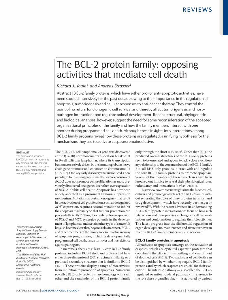
In mammals, there are at least 12 core BCL-2 family proteins, including BCL-2 itself and proteins that have either three-dimensional (3D) structural similarity or a predicted secondary structure that is similar to BCL-2 (fIG. 1). These proteins display a range of bioactivities, from inhibition to promotion of apoptosis. Numerous so-called BH3-only proteins share homology with each other and the remainder of the BCL-2 protein family
only through the short BH3 motif8. Other than BID, the predicted overall structures of the BH3-only proteins seem to be unrelated and appear to lack a close evolution-ary relationship to the core members of the BCL-2 family9. But, all BH3-only proteins interact with and regulate the core BCL-2 family proteins to promote apoptosis. Several of the members of these two classes have been knocked out in mice to reveal their physiological roles, redundancy and interactions in vivo (TABLe 1).
This review covers recent insights into the biochemical, cellular and physiological roles of the BCL-2 family with-out reiterating the roles of these proteins in cancer and drug development, which have recently been expertly reviewed5,10. With the recent advances in understanding BCL-2 family protein interactions, we focus on how such interactions lead these proteins to change subcellular local-ization and conformation to regulate their bioactivities. The latest progress into the differential regulation of organ development, maintenance and tissue turnover in mice by BCL-2 family members are also reviewed.
BCL-2 family proteins in apoptosisAll pathways to apoptosis converge on the activation of caspases, which are cysteinyl aspartate proteases that coordinate the efficient dismantling and engulfment of doomed cells (fIG. 2). Two pathways of cell death can be distinguished by whether they require BCL-2 family proteins and by which caspases are crucial for their exe-cution. The intrinsic pathway — also called the BCL-2-regulated or mitochondrial pathway (in reference to the role these organelles play) — is activated by various
*Biochemistry Section, Surgical Neurology Branch, National Institute of Neurological Disorders and Stroke, The National Institutes of Health, Bethesda, Maryland 20892, USA. ‡The Walter and Eliza Hall Institute of Medical Research, Parkville 3050, Melbourne, Australia. e-mails: [email protected]; [email protected] doi:10.1038/nrm2308
BH3 motifThe amino-acid sequence LXXXGD, in which X represents any amino acid. This motif is conserved between most core BCL-2 family members and among BH3-only proteins.
The BCL-2 protein family: opposing activities that mediate cell deathRichard J. Youle* and Andreas Strasser‡
Abstract | BCL-2 family proteins, which have either pro- or anti-apoptotic activities, have been studied intensively for the past decade owing to their importance in the regulation of apoptosis, tumorigenesis and cellular responses to anti-cancer therapy. They control the point of no return for clonogenic cell survival and thereby affect tumorigenesis and host–pathogen interactions and regulate animal development. Recent structural, phylogenetic and biological analyses, however, suggest the need for some reconsideration of the accepted organizational principles of the family and how the family members interact with one another during programmed cell death. Although these insights into interactions among BCL-2 family proteins reveal how these proteins are regulated, a unifying hypothesis for the mechanisms they use to activate caspases remains elusive.
R E V I E W S
NATure revIeWS | molecular cell biology vOLuMe 9 | jANuAry 2008 | 47
© 2008 Nature Publishing Group

3BIM/BOD
3BAD
3
BCL-G
BID (2BID)
PUMA/BBC3
BCL-XL (1R2D)
BCL-W (1MK3)
MCL1
BAX (1F16)
BAK (2IMS)
BIK/BLK/NBK
HRK/DP5
WWEUBA HECTMULE
A1
Boo/Diva/BCL-B/BCL2L10
BCL-RAMBO
BOK/MTD
NOXA
BMF
BCL-2 (1G5M)
BCL-
2 ho
mol
ogue
sBH
3-only
Anti-apoptotic
Pro-apoptotic
Nature Reviews | Molecular Cell Biology
BH1BH2BH3BH4TM regionα-helixUBAWWEHECT
TNF receptor familyCell-surface receptors in the tumour necrosis factor (TNf) family.
developmental cues or cytotoxic insults, such as viral infection, DNA damage and growth-factor deprivation, and is strictly controlled by the BCL-2 family of proteins. This pathway predominantly leads to the activation of caspase-9 (Ref. 11) but, at least in certain cell types, the intrinsic pathway can proceed in the absence of caspase-9 or its activator, apoptotic protease-activating factor-1 (APAF1)12.
The extrinsic or death-receptor pathway is triggered by ligation of so-called death receptors (members of the tumour necrosis factor (TNf) receptor family, such as Fas or TNF receptor-1 (TNFr1)) that contain an intra-cellular death domain, which can recruit and activate caspase-8 through the adaptor protein Fas-associated death domain (FADD; also known as MOrT1) at the cell surface. This recruitment causes subsequent activation of
Figure 1 | Sequence alignment of core bcl-2 family proteins and bH3-only proteins. Green bars depict α-helical segments from the determined structures (when labelled by Protein Data Bank (PDB) identifier in parentheses) or from secondary structure prediction (as predicted using PSIPRED). Red lines label regions of predicted transmembrane (TM) domains (as predicted using TMHMM). Sequence homologies of the BH1 (brown lines), BH2 (grey lines), BH3 (blue lines) and BH4 (orange lines) regions are shown. The BH1, BH2 and BH3 domains fold to line a hydrophobic pocket that can bind BH3-only peptides. The BH3 domain, particularly among the BH3-only proteins, mediates interaction between the BH3-only proteins and core BCL-2 family proteins and thereby promotes apoptosis. The upper five proteins (BCL-2, BCL-XL, BCL-W, A1 and MCL1) are generally anti-apoptotic. The three proteins in the shaded area are less well studied and cannot be categorized at this time. The lower 12 proteins are considered to be pro-apoptotic. MULE contains a ubiquitin-associated domain (UBA), the Trp-Trp-Glu interaction module (WWE) and a HECT ubiquitin ligase domain. BID has a unique role as both a BCL-2 homologue and a BH3-only protein and links the intrinsic and extrinsic apoptosis pathways (fIG. 2). BIM (also known as BOD), BAD and BMF are unstructured proteins.
R E V I E W S
48 | jANuAry 2008 | vOLuMe 9 www.nature.com/reviews/molcellbio
© 2008 Nature Publishing Group

Death domainA protein-interaction module that consists of six α-helices and that is involved in apoptosis and other signalling pathways.
downstream (effector) caspases, such as caspase-3, -6 or -7, without any involvement of the BCL-2 family. In some cells, most notably hepatocytes, the extrinsic pathway can intersect the intrinsic pathway through caspase-8 cleavage-mediated activation of the pro-apoptotic BH3-only protein BID13,14. The C-terminal truncated form of BID (tBID) translocates to mitochondria and promotes further caspase activation (caspase-9 and the effector caspases caspase-3, -6 and -7) through the intrinsic path-way. In these situations, loss of BID or overexpression of BCL-XL inhibits cell death13.
BCL-2 family proteins have opposing apoptotic activities. BCL-2 family members have classically been grouped into three classes. One class inhibits apoptosis (BCL-2, BCL-XL, BCL-W, MCL1, BCL-B (also known as BCL-2L10) and A1 (also known as BCL-2A1), whereas a second class promotes apoptosis (BAX, BAK and BOK (also known as MTD)). A third divergent class
of BH3-only proteins (BAD, BIK (also known as BLK or NBK), BID, HrK (also known as death protein-5 (DP5)), BIM (also known as BOD), BMF, NOXA and PuMA (also known as BBC3)) have a conserved BH3 domain that can bind and regulate the anti-apoptotic BCL-2 proteins to promote apoptosis (fIG. 1). It appears that the pro-apoptotic family members BAX and BAK are crucial for inducing permeabilization of the outer mitochondrial membrane (OMM) and the subsequent release of apoptogenic molecules (such as cytochrome c and DIABLO (also known as SMAC)), which leads to caspase activation. The anti-apoptotic family members, such as BCL-2 and BCL-XL, inhibit BAX and BAK. recent evidence indicates that BH3-only proteins de-repress BAX and BAK by direct binding and inhibition of BCL-2 and other anti-apoptotic family members15. By contrast, an opposing model postulates direct activ-ation of BAX and BAK by some BH3-only proteins (specifically BIM, tBID and PuMA)16 (fIG. 2).
Table 1 | Phenotypes of mice that are deficient in BCL-2 family members
bcl-2 family member
Defects caused by its deletion* refs
Pro-survival family members
BCL-2 Abnormal death of renal epithelial progenitors, melanocyte progenitors and mature B and T lymphocytes. Causes fatal polycystic kidney disease (100% mortality by 6 weeks), premature greying and lymphopoenia (but all of these effects can be rescued by concomitant loss of the BH3-only protein BIM).
130
BCL-XL Abnormal death of fetal erythroid progenitors and neuronal cells. Causes death around embryonic day 14 (100% mortality).
129
BCL-W Abnormal death of developing sperm cells. Causes male sterility. 132
A1A Abnormally accelerated death of granulocytes and mast cells in culture. 133
MCL1 Failure in implantation. Conditional knockout causes premature death of immature and mature B and T lymphoid cells, as well as haemopoietic stem cells.
128
Pro-apoptotic BAX/BAK family members
BAX Mild lymphoid hyperplasia, male sterility due to sperm-cell differentiation defect. 135
BAK No obvious defects detected so far. 136
Pro-apoptotic BH3-only proteins
BIM Lymphoid and myeloid cell hyperplasia, fatal SLE-like autoimmune disease (on mixed genetic C57BL/6x129SV background), many cell types are abnormally resistant to cytokine deprivation, deregulated calcium flux and the chemotherapeutic drug taxol; mild but significant resistance of many cell types to DNA damage and glucocorticoids.
143
BID BID-deficient mice are resistant to Fas-activation-induced hepatocyte killing and fatal hepatitis; however, some cell types (such as lymphoid cells) are normally sensitive to Fas-induced apoptosis.
13, 14
PUMA Many cell types are profoundly resistant to DNA damage; many are also resistant to cytokine deprivation, glucocorticoids and phorbol ester.
150,151
BAD Mild resistance of some cell types to deprivation of epidermal growth factor or insulin growth factor.
154
HRK Abnormal, although relatively mild, resistance of certain neuronal populations to deprivation of nerve growth factor.
155,156
BIK No obvious defects detected so far. 158
NOXA Relatively mild resistance of fibroblasts to γ-irradiation or etoposide, but profound resistance of these same cells and keratinocytes in the skin to ultraviolet irradiation.
150
*These are phenotypes found in mice. The roles of these proteins may differ in humans. BAD, BCL-2 antagonist of cell death; BAK, BCL-2-antagonist/killer-1; BAX, BCL-2-associated X protein; BCL-2, B-cell lymphoma-2; A1A, BCL-2-related protein A1A; BCL-W, BCL-2-like-2; BCL-XL, a BCL-2-like protein; BID, BH3-interacting domain death agonist; BIK, BCL-2-interacting killer; BIM, BCL-2-like-11; HRK, harakiri (also known as death protein-5); MCL1, myeloid cell leukaemia sequence-1; PUMA, BCL-2 binding component-3; SLE, systemic lupus erythematosus.
R E V I E W S
NATure revIeWS | molecular cell biology vOLuMe 9 | jANuAry 2008 | 49
© 2008 Nature Publishing Group

Nature Reviews | Molecular Cell Biology
Growth-factor deprivation, stress, UV, viruses
InactiveBH3-only
BCL-2
BAX/BAK
Cytochrome c release, mitochondrial fragmentation
APAF1 assemblyinto apoptosome
FAS
Caspase-8
TNFR1
APAF1Cytochrome c
BID cleavage
Intrinsic pathway Extrinsic pathway
ActiveBH3-only
Caspase-3
Apoptosis
Mitochondrial outer membrane permeabilizationThe process by which the outer membrane of mitochondria leaks certain soluble intermembrane space proteins, such as cytochrome c, into the cytoplasm.
ApoptosomeThe caspase-9 activation complex that is composed of APAf1 heptamers and that is assembled on binding of APAf1 monomers to cytochrome c.
BAX and BAK promote caspase activation by their effects on mitochondria. either directly or indirectly, these two pro-apoptotic BCL-2 family members induce the release of proteins from the space between the inner and outer mitochondrial membranes17. This process of mitochondrial outer membrane permeabilization (MOMP) results in the release of cytochrome c and other soluble proteins into the cytosol. Although it is commonly thought that BAX and BAK form pores in membranes, the biochemical nature of such pores and how anti-apoptotic BCL-2 family proteins might regulate them remains a key and controversial issue in the field of cell death18. At the same time as cytochrome c release (or immediately before), BAX and BAK induce mitochondria to fragment
into more numerous and smaller units, which suggests connections between mitochondrial division processes and the functions of the BCL-2 family19.
Once the OMM has been permeabilized, soluble proteins diffuse from the intermembrane space into the cytosol, where they promote caspase activation. The best studied of these proteins is cytochrome c, which binds to APAF1 and leads to the assembly of a heptameric protein ring called an apoptosome, which can bind pro-caspase-9 and induce its activation through a conformational change20,21. Cytochrome c–APAF1-dependent activa-tion of caspase-9 is absolutely required for neuronal and fibroblast cell-death processes22. However, in addition to this process, lymphocytes can probably use alternative APAF1-, caspase-9- and cytochrome c-independent, but pro-apoptotic BCL-2-family-member-dependent, pathways for caspase activation and cell killing12,22. Intriguingly, caspase activation in lymphocytes can be amplified by APAF1 even when APAF1 has not been incorporated into the apoptosome22.
One APAF1-independent pathway of caspase activation is the relief of caspase inhibition by inhibitor of apoptosis proteins (IAPs), such as XIAP, which bind and neutralize certain caspases (such as caspase-9 and caspase-3). This inhibitory action of IAPs can be antago-nized by the binding of DIABLO, which is released from mitochondria after the activation of BAX and/or BAK. However, DIABLO-deficient mice23, as well as XIAP-deficient mice24, do not display significant apop-totic phenotypes, which suggests that novel processes of caspase activation remain to be discovered. Several APAF1 related proteins, called NOD-like receptors, regulate alternative pathways of caspase activation that occur in non-apoptotic host defence processes that are associated with innate immunity and serve as examples of pathways that can also have roles during apoptosis25. One of these NOD-like receptors, NALP1, can be regulated by BCL-2 and BCL-XL26 in manner that is reminiscent of caspase activation in the worm (BOX 1).
BCL-2 and BCL-XL appear to control cell survival beyond the APAF1–caspase-9 axis. If caspase activation is inhibited by loss of APAF1 or caspase-9, or even by the combined loss of caspase-9 and caspase-2, the rate of acqui-sition of apoptotic morphology of myeloid progenitors and mast cells induced by growth-factor withdrawal or DNA damage can be significantly delayed. However, although the onset of apoptotic morphology can be delayed, the cells still lose clonogenic potential and thus effectively die, unlike cells that overexpress BCL-2 or BCL-XL27,28. Thus, the step of apoptosis regulation that is controlled by the BCL-2 family appears to be the most gen-eral final commitment step for the decision between cell life and death. The disruption of mitochondria by BAX and BAK may be one cause of eventual clonogenic cell death in the absence of apoptosome activation. Normally, caspase activation rapidly and efficiently mediates cell demoli-tion and removal. When caspases are blocked, certain features of apoptosis can be lost (or delayed), which causes the cells to die more slowly by BCL-2-family-mediated mitochondrial disruption or by novel caspase-activation pathways that have yet to be characterized.
Figure 2 | Scheme depicting intrinsic and extrinsic pathways of apoptosis. Apoptosis can be induced by cell surface receptors, such as Fas and tumour necrosis factor receptor-1 (TNFR1) (extrinsic pathway, right), or by various genotoxic agents, metabolic insults or transcriptional cues (intrinsic pathway, left). The intrinsic pathway starts with BH3-only protein induction or post-translational activation, which results in the inactivation of some BCL-2 family members. This relieves inhibition of BAX and BAK activation, which in turn promotes apoptosis. Some BH3-only proteins, such as BIM and PUMA, may also be able to activate BAX and/or BAK (as shown by the dotted line). Once activated, BAX and BAK promote cytochrome c release and mitochondrial fission, which leads to the activation of APAF1 into an apoptosome and activates caspase-9 to activate caspase-3. Caspases in turn cleave a series of substrates, activate DNases and orchestrate the demolition of the cell. The extrinsic pathway can bypass the mitochondrial step and activate caspase-8 directly, which leads to caspase-3 activation and cell demolition. The BCL-2 family regulates the intrinsic pathway and can modulate the extrinsic pathway when cleavage of BID communicates between the two pathways.
R E V I E W S
50 | jANuAry 2008 | vOLuMe 9 www.nature.com/reviews/molcellbio
© 2008 Nature Publishing Group

Nature Reviews | Molecular Cell Biology
EGL-1(BH3-only)
CED-9(core BCL-2 family)
CED-4(APAF1-like)
CED-3(caspase)
Inhibitor of apoptosis protein (IAP). One of a family of proteins that inhibits apoptosis by binding or degrading caspases.
NOD-like receptorA cytosolic receptor that is homologous to NOD1 and is involved in innate immunity pathways.
E3 ligaseOne of a family of proteins that facilitate the transfer of ubiquitin from a donor protein to a specific substrate protein that may signal the target for proteosomal degradation.
Structure and evolution The core multi-BH domain BCL-2 family members and, surprisingly, the BH3-only protein BID (fIG. 1) have con-served regions of sequence homology and similar pre-dicted secondary structure. Structures of seven of these proteins (BCL-XL29, BCL-2 (Ref. 30), BCL-W31,32, MCL1 (Ref. 33), BAX34, BAK35 and BID36,37) show remarkable similarity, which is intriguing considering that some are pro-apoptotic and others are anti-apoptotic. Ks-BCL-2, a viral homologue of BCL-2 (Ref. 38), as well as two viral proteins without apparent sequence similarity to BCL-2 family proteins, M11L39,40 and N1L41, display a helical fold that is similar to that of BCL-XL, and these inhibit apoptosis, which indicates that several viruses use BCL-2 family members to counteract host defence.
The 3D structures of the seven core BCL-2 family proteins mentioned above have yet to reveal any distin-guishing difference between anti-apoptotic members (such as BCL-XL and MCL1) and pro-apoptotic mem-bers (such as BAX and BID). All seven proteins are helical bundles with a hydrophobic helix-turn-helix hairpin that is flanked on both sides by pairs of amphipathic helices. excluding the viral anti-apoptotic BCL-2-like proteins, BCL-2 homologues appear to have C-terminal membrane- anchoring domains. In addition, pro-apoptotic BID appears to be myristoylated to mediate membrane anchorage42.
In three proteins, BAX, BCL-W and MCL1, the C-terminal anchor has been included in the structural analysis and fits into a hydrophobic pocket formed by the BH1, -2 and -3 regions. The same pocket that sequesters the C-terminal membrane anchor can also bind to peptides of the BH3-domain sequences of BAK, BAD and BIM43–45, which suggests that it also functions in dimerization with BH3-only proteins and/or multi-BH-domain-containing BCL-2 family members (fIG. 3). An extended BIM BH3 peptide that is 23 amino acids in length binds along the hydrophobic groove of BCL-XL, although it is inverted in the C- to N-terminal helical direction relative to the ori-entation of the BAX and BCL-W C-terminal membrane anchors45 (fIG. 3).
Classically, BH3-only proteins have been defined as having homology to the core BCL-2 family members in only the BH3 domain. recent sequence analyses indicate that, except for BID, the BH3-only proteins have pre-dicted secondary structures or determined 3D structures that are unrelated to the core BCL-2 family members, and, except for BID, they probably acquired BH3 motifs by convergent evolution9. One particular example of a BH3-motif-containing protein that is not otherwise related to the core BCL-2 family is MuLe, an e3 ligase that reportedly targets MCL1 for ubiquitylation and pro-teasomal degradation. MuLe has a BH3 domain and can loosely be considered to belong to the class of BH3-only proteins, which interact with and regulate other mem-bers of the BCL-2 family46,47 (fIG. 4). even the autophagy regulatory protein beclin-1 reportedly binds to BCL-2 through a BH3 domain, although further biochemical and genetic experiments are needed to establish a func-tional connection48. BAD, BMF and BIM are intrinsically unstructured49 and, along with PuMA, these proteins are not likely to be core BCL-2 family homologues on the basis of secondary structure predictions (fIG. 1).
So, BH3-only proteins include various proteins that share a single motif that allows them to bind and regu-late the core BCL-2 family members. BID, by contrast, is the one BH3-only protein with a determined structure that places it squarely in the core BCL-2 family mem-bers, which perhaps explains why BID shares certain properties with multidomain BCL-2 family members, such as the ability to oligomerize50 and to permeabilize membranes51. Thorough phylogenetic analyses of the BCL-2 family have generated important insights into the origins of the core BCL-2 family members (BOX 2) and the BH3-only proteins, and suggest that many of these proteins might have biological activities beyond regulation of cell death9,52.
BCL-2 family protein activation BH3-only proteins are pro-apoptotic and function as initial sensors of apoptotic signals that emanate from various cellular processes. BH3-only protein expression can be induced by transcription factors. For example, NOXA and PuMA are induced by the tumour sup-pressor p53 in response to DNA damage53–55, and BIM is induced by the class O forkhead box transcription factor-3A (FOXO3A) in response to growth-factor deprivation56 and by the transcription factors CeBPα
Box 1 | The mechanism of CED-9, the C. elegans orthologue of BCL-2
Genetic analyses of the apoptosis pathway in Caenorhabditis elegans and recent biochemical insights are consistent with the model shown in the figure above. EGL‑1, a BH3‑only protein, is transcriptionally induced by developmental cues for programmed cell death. EGL‑1 binds to the BCL‑2 homologue CED‑9, thereby freeing CED‑4, an AAA+ ATPase that is related to apoptotic protease‑activating factor‑1 (APAF1), which is normally sequestered by CED‑9. The released CED‑4 assembles into a tetrameric apoptosome and activates the protease activity of the caspase CED‑3. This model differs from the cytochrome c release model of mammalian cells (fIG. 2). It is not anticipated that homologous proteins regulate the same process by different mechanisms, so some underlying common process among BCL‑2 family members of C. elegans and mammals may await discovery.
Two studies indicate that CED‑9 may do more than prevent CED‑4 activation. In worms with mild ced‑3 loss‑of‑function mutations in which some excess cells survive, weak loss‑of‑function ced‑9 alleles actually increase cell survival, which suggests that CED‑9 also has pro‑apoptotic activity163. This might indicate that, depending on its conformation, CED‑9 can have BCL‑2‑like (that is, anti‑apoptotic) or BAX‑like (that is, pro‑apoptotic) activity94. In addition, loss of CED‑9 activity inhibits cell death due to overexpression of drp‑1 (Ref. 122), which further suggests that the sole core BCL‑2 family protein in C. elegans can function in both pro‑ and anti‑apoptotic modes. Certain mammalian BCL‑2 family members have also been reported to be convertible between anti‑ and pro‑apoptotic forms164,165. CED‑9 resides on mitochondria, as many mammalian BCL‑2 family proteins do, but how this localization relates to its biochemical action remains unclear. Similar to programmed cell death in mammals and flies, mitochondria become fragmented during apoptosis in the worm122 upstream of caspase activation, showing that there is one common denominator involving mitochondria in all three systems.
Future studies should explore how BCL‑2 family members function in sponges, echinoderms and insects. One recent study in D. melanogaster came to the surprising conclusion that although the two BCL‑2 family members are required for certain stress‑induced apoptosis pathways, they are not required for developmentally programmed cell death166.
R E V I E W S
NATure revIeWS | molecular cell biology vOLuMe 9 | jANuAry 2008 | 51
© 2008 Nature Publishing Group

Nature Reviews | Molecular Cell Biology
a
b
c
d
e
f
ER stressThe accumulation of unfolded or incompletely glycosylated proteins in the endoplasmic reticulum (eR) results in stress that may lead to apoptosis.
Dynein motor complexA molecular machine that transports cargo along microtubules.
JAK–STAT pathwayThe Janus kinase (JAK)–signal transducer and activator of transcription (sTAT) pathway is a signalling pathway that is activated by growth factors and cytokines.
(CCAAT-enhancer binding protein-α) or CHOP (CeBP homologous protein) in response to endoplasmic reticu-lum (eR) stress57. BH3-only proteins can also be activated post-translationally; for example, BAD is activated by loss of phosphorylation in response to growth-factor deprivation58; BID is activated by caspase-8-mediated proteolysis59,60; BIM is activated by release from the dynein motor complex61 or by loss of extracellular signal-regulated kinase (erK)-mediated phosphorylation (which targets it for ubiquitylation and proteasomal degradation in healthy cells)62,63; BMF is activated by release from actin–myosin motor complexes64; and BIK is activated by an unknown mechanism in response to inhibition of protein synthesis65.
regulation of the expression levels of anti-apoptotic BCL-2 family proteins is another way in which cells can regulate apoptosis. For example, BCL-XL can be transcriptionally induced by growth factors through the janus kinase–signal transducer and activator of trans-cription (JAK–sTAT) pathway to promote cell survival66.
MCL1 is rapidly degraded by the ubiquitin–proteasome pathway in response to cytokine deprivation or other death stimuli (such as ultraviolet (uv) radiation) and can be upregulated post-transcriptionally to prevent apoptosis by inhibiting the rate of degradation46,67. regulation of the expression levels of the pro-apoptotic proteins BAX and BAK is less apparent and the pro-teins appear to be constitutively expressed at more or less constant levels. BAX and BAK are primarily post-translationally regulated by other members of the BCL-2 family.
When BH3-only proteins are induced or activated, they interact with core BCL-2 family proteins to pro-mote apoptosis. The binding of BH3-only proteins or BH3 peptides to specific anti- and pro-apoptotic BCL-2 family members has been determined by using yeast two-hybrid analysis, plasmon resonance binding assays and by cell-free mitochondria and liposome permea-bilization studies68–72,73. Together, these assays indicate that some BH3-only proteins, such as BIM and PuMA, bind all anti-apoptotic BCL-2 family members, whereas others, such as BAD and NOXA, bind only certain anti-apoptotic BCL-2 family members (fIG. 4). In addition to interaction with anti-apoptotic BCL-2 family members, several reports show synergy of BID or BIM with BAX in cell-free membrane permeabilization assays, which suggests that some BH3-only proteins may directly bind and activate BAX and BAK70–72. However, it is dif-ficult to detect binding of full-length BID, tBID or BIM to BAX or BAK15, although a modified BH3 peptide can bind BAX and/or BAK74. Other models in which BH3-only proteins directly activate BAX and BAK are called into question owing to results from Bim/Bid double knockout mice and their cell lines, which show that these putative direct activators of BAX and BAK are not required for many apoptotic pathways15. Thus, known BH3-only proteins appear to induce apoptosis primarily by inhibiting anti-apoptotic BCL-2 family members, thereby liberating BAX and BAK to cause MOMP and activation of the caspase cascade (fIG. 2). The precise biochemical mechanisms that lead to the activation of BAX and BAK remain a mystery and constitute the ‘holy grail’ of apoptosis research.
Dynamics of subcellular localizationThe anti-apoptotic BCL-2 protein is embedded in the er, the nuclear envelope and the OMM by a hydrophobic C-terminal membrane-spanning domain, with most of its amino acids in the cytosol75,76. Although BCL-2 in any of these subcellular locations can block apoptosis, the functions of BCL-2 at the er and the nuclear envelope are less clear than those on mitochondria and have recently been reviewed77,78.
In contrast to BCL-2, BAX is mostly cytosolic and sequesters its hydrophobic C-terminal membrane anchor in its BH3-binding pocket (fIG. 3), with a minor fraction lightly bound to the OMM79. BAX appears to exist as a monomer in the cytosol of cells rather than being bound to any anti-apoptotic BCL-2 family members80. During apoptosis induction, BAX translocates specifically to mito-chondria (see Supplementary information S1 (movie)),
Figure 3 | Space-filling models of the structures of baX, bcl-W and bcl-Xl bound to a bim bH3-region peptide. Comparing the structures of full-length BCL-2 family members with those bound to BH3 peptides from other BCL-2 family members suggests how subcellular localization might be linked to protein–protein interactions among family members. (The structures in the lower panels are rotated 90 degrees from those in the top panels.) Full-length BAX (a,b) and BCL-W (c,d) fold with the C-terminal helix (grey) sequestered in a hydrophobic pocket. This C-terminal domain is experimentally deleted in BCL-XL (e,f), which binds to an extended BH3 domain peptide of BIM (yellow) in the homologous pocket that is occupied by the membrane anchor in BAX, BCL-W and MCL1. The BIM peptide orientates in the pocket in the opposite direction to the endogenous regions of the C terminus (that is, in the C- to N-terminal direction). Because the C-terminal helix is involved in membrane binding and is thought to penetrate deeply into membranes (fIG. 5), this helix would become displaced from the hydrophobic pocket on mitochondrial translocation (see Supplementary information S1 (movie)). Emptying this pocket of the C-terminal helix would enable it to bind BH3 domains (yellow) from other BCL-2 family members, allowing hetero- or homodimer formation. Alternatively, binding of BH3-only proteins to BAX or BCL-W in the cytosol could displace the C-terminal helix from the pocket and trigger mitochondrial translocation. Non-structured amino acids in BAX (1–12) and BCL-W (1–8 and 170–178) have been excluded from the models.
R E V I E W S
52 | jANuAry 2008 | vOLuMe 9 www.nature.com/reviews/molcellbio
© 2008 Nature Publishing Group

Nature Reviews | Molecular Cell Biology
BCL-2
BCL-XL
MCL1
A1
BCL-W
PUMA
tBID
BIM
NOXA
BAD
3
3
where it inserts into the OMM as an integral membrane protein81 using its C-terminal membrane anchor82, perhaps with organelle targeting specified by defined regions in the N terminus83. This translocation step of BAX, although reversible in certain situations, usually correlates closely with the irreversible commitment of cells to die and to the cytochrome c release step discussed below.
BOK also translocates from the cytosol to mito-chondria during apoptosis84, whereas BAK already resides on the OMM (see subcellular localization of BCL-2 family members in Supplementary information S2 (table)) in healthy cells, where it has been reported to be bound to MCL1 (Ref. 67) and to BCL-XL73. Notably, BAK–MCL1 and BAK–BCL-XL interaction experiments rely on detergent extraction of membrane proteins that, in some cases, can cause artefactual interactions among BCL-2 family proteins85. Although the OMM channel protein vDAC2 (voltage-dependent
anion channel-2) also reportedly binds to BAK86 and is important for BAK import into the OMM87, Vdac1/Vdac2/Vdac3 triple knockout mice display normal apoptosis88, indicating little or no role for vDACs in BCL-2 family protein regulation. The mitochondrial localization of BAK may be a consequence of MCL1 binding into the BH3 pocket of BAK and displacing the C-terminal membrane anchor, allowing it to interact with membranes. Although the BH3 domain is the obvious candidate domain of MCL1 for this inter-action, it is not exposed in the soluble MCL1 struc-ture33. The C-terminal membrane anchor of BCL-XL has been proposed to mediate binding to BAX by fitting into its BH3-binding pocket in trans89, which suggests another way by which BCL-XL and MCL1 can interact with BAK. On apoptosis induction, MCL1 is degraded (at least in certain cell types in response to certain cytotoxic stimuli)67,90 and/or the MCL1–BAK and BCL-XL–BAK interactions are disrupted by BH3-only proteins, such as NOXA, BIM73 or BIK65, which frees BAK to promote apoptosis.
Because the translocation of BAX to the mitochon-dria correlates with pro-apoptotic activity, it is curi-ous that BCL-2 is constitutively membrane bound but BCL-XL79,91, BCL-W92 and MCL1 (Ref. 90) exist partly in the cytosol and translocate from the cytosol to mito-chondria during apoptosis. Their binding to BH3-only proteins appears to be the trigger, perhaps by displacing the C-terminal membrane anchors of BCL-XL89 and BCL-W92 by occupancy of the hydrophobic pockets. The intracellular translocation probably correlates with conformational changes and deep insertion of BCL-XL and BCL-W into the OMM. Whether this translocation of anti-apoptotic BCL-2 family members represents a mechanism to inhibit apoptosis, inactiva-tion by BH3-only proteins or even conversion into pro-apoptotic effectors remains unclear92,93. It has, for example, been postulated that binding of a BH3-only protein changes the conformation of the pro-survival BCL-2-like protein so that it can then initiate formation of BAX and/or BAK oligomers in the mitochondrial and other intracellular membranes to cause initiator caspase activation94.
Figure 4 | bH3-only protein binding specificity for bcl-2 homologues. BIM and PUMA bind to all five anti-apoptotic BCL-2 family members tested. By contrast, NOXA only binds to MCL1 and A1, and BAD binds selectively to BCL-W, BCL-2 and BCL-XL. tBID binds avidly to BCL-XL, BCL-W, MCL1 and A1, but only weakly to BCL-2. BH3-only proteins do not appear to bind strongly to BAX or BAK. These binding specificities recapitulate the ability of these proteins to activate apoptosis. For example, BIM, BID or PUMA alone can induce apoptosis, whereas a combination of NOXA and BAD is required. This probably enables the fine specificity of apoptosis regulation in different tissues and during changes in cellular developmental stages. The BH3 domain is shown in red and the five anti-apoptotic BCL-2 family proteins are shown in the middle of the figure.
Box 2 | Phylogenomics of BCL-2 family proteins
BCL‑2 gene orthologues have been identified in all metozoan animals examined so far9. The earliest metazoan that has been analysed, the sponge52, contains two BCL‑2 related genes that most closely resemble mammalian BOK8, which has so far received little attention in mouse and human systems. Interestingly, Monosiga brevicollis, a single cell choanoflagellate that is closely antecedent to sponges, has no identifiable BCL‑2 family member in its recently sequenced genome (C. Wang, personal communication), which suggests that BCL‑2 family genes evolved with multicellular life forms. However, some recent viral gene products have been found with the signature helical fold of BCL‑2, and these function in apoptosis regulation39–41, which suggests that additional structural orthologues of BCL‑2 might exist in eukaryotes without discernible primary sequence homology. The number of apparent BCL‑2 family member genes in different orders varies widely. The sea urchin (Strongylocentrotus purpuratus) has ten homologous genes, significantly more than insects (Drosophila melanogaster; two core BCL‑2 family members) and round worms (Caenorhabditis elegans; one core BCL‑2 family member) (BOX 1). C. elegans also has one prominent BH3‑only gene, egl‑1, which appears to be crucial for most (and perhaps all) developmentally programmed somatic cell death160, and another BH3‑only gene, ced‑13, which might have a role in stress‑induced cell killing161. Zebrafish contain genes for nine versions of core BCL‑2 family members and homologues of eight of the best‑studied BH3‑only genes162. Humans and mice have a similar set of 12–13 structural homologues of BCL‑2 family proteins, which indicates that family member organization is stable among mammals.
R E V I E W S
NATure revIeWS | molecular cell biology vOLuMe 9 | jANuAry 2008 | 53
© 2008 Nature Publishing Group
Nature Reviews | Molecular Cell Biology
N terminus
C terminus
BAX
Soluble, cytosolic conformationin healthy cells
Model of membrane-insertedapoptotic state of BAX
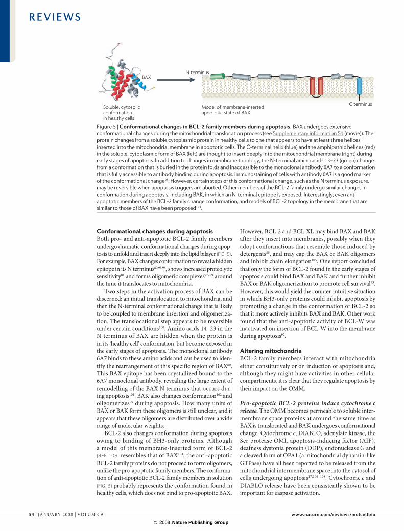
Conformational changes during apoptosis Both pro- and anti-apoptotic BCL-2 family members undergo dramatic conformational changes during apop-tosis to unfold and insert deeply into the lipid bilayer (fIG. 5). For example, BAX changes conformation to reveal a hidden epitope in its N terminus80,95,96, shows increased proteolytic sensitivity81 and forms oligomeric complexes97–99 around the time it translocates to mitochondria.
Two steps in the activation process of BAX can be discerned: an initial translocation to mitochondria, and then the N-terminal conformational change that is likely to be coupled to membrane insertion and oligomeriza-tion. The translocational step appears to be reversible under certain conditions100. Amino acids 14–23 in the N terminus of BAX are hidden when the protein is in its ‘healthy cell’ conformation, but become exposed in the early stages of apoptosis. The monoclonal antibody 6A7 binds to these amino acids and can be used to iden-tify the rearrangement of this specific region of BAX80. This BAX epitope has been crystallized bound to the 6A7 monoclonal antibody, revealing the large extent of remodelling of the BAX N terminus that occurs dur-ing apoptosis101. BAK also changes conformation102 and oligomerizes99 during apoptosis. How many units of BAX or BAK form these oligomers is still unclear, and it appears that these oligomers are distributed over a wide range of molecular weights.
BCL-2 also changes conformation during apoptosis owing to binding of BH3-only proteins. Although a model of this membrane-inserted form of BCL-2 (Ref. 103) resembles that of BAX104, the anti-apoptotic BCL-2 family proteins do not proceed to form oligomers, unlike the pro-apoptotic family members. The conforma-tion of anti-apoptotic BCL-2 family members in solution (fIG. 3) probably represents the conformation found in healthy cells, which does not bind to pro-apoptotic BAX.
However, BCL-2 and BCL-XL may bind BAX and BAK after they insert into membranes, possibly when they adopt conformations that resemble those induced by detergents85, and may cap the BAX or BAK oligomers and inhibit chain elongation105. One report concluded that only the form of BCL-2 found in the early stages of apoptosis could bind BAX and BAK and further inhibit BAX or BAK oligomerization to promote cell survival93. However, this would yield the counter-intuitive situation in which BH3-only proteins could inhibit apoptosis by promoting a change in the conformation of BCL-2 so that it more actively inhibits BAX and BAK. Other work found that the anti-apoptotic activity of BCL-W was inactivated on insertion of BCL-W into the membrane during apoptosis92.
Altering mitochondriaBCL-2 family members interact with mitochondria either constitutively or on induction of apoptosis and, although they might have activities in other cellular compartments, it is clear that they regulate apoptosis by their impact on the OMM.
Pro-apoptotic BCL-2 proteins induce cytochrome c release. The OMM becomes permeable to soluble inter-membrane space proteins at around the same time as BAX is translocated and BAK undergoes conformational change. Cytochrome c, DIABLO, adenylate kinase, the Ser protease OMI, apoptosis-inducing factor (AIF), deafness dystonia protein (DDP), endonuclease G and a cleaved form of OPA1 (a mitochondrial dynamin-like GTPase) have all been reported to be released from the mitochondrial intermembrane space into the cytosol of cells undergoing apoptosis17,106–108. Cytochrome c and DIABLO release have been consistently shown to be important for caspase activation.
Figure 5 | conformational changes in bcl-2 family members during apoptosis. BAX undergoes extensive conformational changes during the mitochondrial translocation process (see Supplementary information S1 (movie)). The protein changes from a soluble cytoplasmic protein in healthy cells to one that appears to have at least three helices inserted into the mitochondrial membrane in apoptotic cells. The C-terminal helix (blue) and the amphipathic helices (red) in the soluble, cytoplasmic form of BAX (left) are thought to insert deeply into the mitochondrial membrane (right) during early stages of apoptosis. In addition to changes in membrane topology, the N-terminal amino acids 13–27 (green) change from a conformation that is buried in the protein folds and inaccessible to the monoclonal antibody 6A7 to a conformation that is fully accessible to antibody binding during apoptosis. Immunostaining of cells with antibody 6A7 is a good marker of the conformational change95. However, certain steps of this conformational change, such as the N terminus exposure, may be reversible when apoptosis triggers are aborted. Other members of the BCL-2 family undergo similar changes in conformation during apoptosis, including BAK, in which an N-terminal epitope is exposed. Interestingly, even anti-apoptotic members of the BCL-2 family change conformation, and models of BCL-2 topology in the membrane that are similar to those of BAX have been proposed103.
R E V I E W S
54 | jANuAry 2008 | vOLuMe 9 www.nature.com/reviews/molcellbio
© 2008 Nature Publishing Group

ErythropoiesisThe production of red blood cells.
The initial finding that the structure of BCL-XL resem-bles that of the translocation domain of diphtheria toxin led to the proposal that BCL-XL might form pores in membranes29. In trying to understand how pore-formation activity is related to biological activity, a confusing factor is that both pro- and anti-apoptotic BCL-2 family members appear to be able to form membrane channels in vitro29,109,110. Incubation of BAX with isolated mitochon-dria induces cytochrome c release111, and incubating BAX with liposomes allows the release of large (up to 106 Da) dextran molecules112, which is consistent with the lip-idic pores that were identified in lipid bilayer studies113. BCL-XL inhibits BAX-induced cytochrome c release from isolated mitochondria111 and dextran release from liposomes70. Furthermore, the BH3-only protein BID can synergize with BAX to cause cytochrome c release in cell-free assays, either by activating BAX or by preventing anti-apoptotic BCL-2 family members from inhibiting BAX and BAK. Thus, one model for BAX and BAK activation is that they form large pores in the OMM that allow the release of proteins into the cytosol, inducing caspase activ-ation. However, the biochemical nature of this putative BAX–BAK pore, such as the number of molecules of BAX that comprise the pore, remains unknown.
Curiously, certain cell types (such as some neuro-nal populations and cardiomyocytes) can survive the cytochrome c release step, at least for a limited amount of time114,115. In such cells, caspases might be stringently regulated by caspase-inhibiting IAPs. In these cases, apoptosis requires the release of DIABLO from mito-chondria to relieve the IAP inhibition and thereby allow caspase activation. This might be a specialized adapta-tion of normally long-lived post-mitotic cells (which are essential for animal survival) to supply these cells with additional protection from cell-death activation and to prevent their accidental death.
Roles in mitochondrial fragmentation and morphology. Confocal and electron microscopy analyses of BAX translocation to mitochondria reveal that the earliest detectable form of ‘activated’ BAX does not localize to the entire OMM, but is found concentrated at small focal regions on the mitochondrial surface116. BAK also moves into these ‘BAX foci’ during apoptosis induction. This focal cluster form of BAX observed by microscopy has an altered N-terminal conformation and probably reflects the in situ state of BAX and BAK as oligomers. These sites of BAX coalescence often develop into mitochondrial division sites117, linking BAX to the pro-motion of the mitochondrial fragmentation processes that occur almost simultaneously with the release of cytochrome c118 (fIG. 2).
Inhibition of mitochondrial fission in vitro by downregulation of the mitochondrial dynamin family member DrP1 delays cytochrome c release and can decrease caspase activation, which suggests that the organelle division machinery somehow participates in the regulation of apoptosis119. Deletion or mutation of Drp1 in D. melanogaster120,121 and drp-1 in C. elegans122 also inhibits apoptosis in vivo (BOX 1). Conversely, inhi-bition of mitochondrial fusion by loss of a different
mitochondrial dynamin family member, OPA1, induces spontaneous apoptosis123.
unexpectedly, healthy cells that lack both BAX and BAK have altered mitochondrial morphology and slower mitochondrial fusion rates, which indicates that BAX and BAK affect mitochondrial morphogenesis machin-eries even in the absence of apoptotic stimuli124. recent work showing that OPA1 controls mitochondrial cristae formation and that tight cristae junctions can inhibit cytochrome c release during apoptosis suggests how mediators of mitochondrial fission and fusion might have a role in cytochrome c release and apoptosis125. In contrast to cytochrome c and OPA1, the release of other mitochondrial intermembrane space proteins (such as DIABLO) is not inhibited by DRP1 knockdown126, which underscores the suggestion that the role of the mitochondrial fission machinery in apoptosis might be indirectly linked to the cytochrome c release step. ectopic expression of human BCL-XL and C. elegans ced-9 in mammalian cells has been found to affect mitochondrial morphogenesis, which shows that it is possible to sepa-rate the process of organelle fusion regulation by BCL-2 family members from the regulation of apoptosis127.
Physiological roles of BCL-2 proteinsBCL-2 family members have essential roles in the mouse from early embryogenesis through to adult tissue homeo-stasis. The nervous system, haematopoietic tissues and spermatogenesis are particularly dependent on BCL-2 family protein regulation (TABLe 1).
Anti-apoptotic BCL-2 proteins. MCL1 and BCL-XL are both essential for normal embryogenesis. Mcl1–/– embryos die before implantation at the blastocyst stage128 and Bcl-x–/– mice (in which the entire Bcl-x locus (incor-porating Bcl-xl and Bcl-xs) was knocked out) survive only until fetal day 13.5, displaying severe defects in erythropoi-esis and neuronal development129. By contrast, although BCL-2-deficient mice survive to birth, they have defects in the immune system, hair follicles and renal epithe-lial cells, and all succumb to polycystic kidney disease by ~4–8 weeks of age (the age of death is influenced by genetic background)130,131. Bcl-w-knockout male mice are sterile owing to defective spermatogenesis, but otherwise both females and males are developmentally normal132. Analysis of the essential functions of A1 by gene targeting is complicated by the fact that, in contrast to humans, mice have four a1 genes. Mice that lack A1A are essen-tially normal, but their granulocytes and mast cells undergo apoptosis abnormally rapidly in culture133,134.
Pro-apoptotic BCL-2 proteins. Bax-knockout mice are viable and females are fertile, but both males and females have mild overgrowth of neurons and mild lymphoid hyperplasia, and males have a severe defect in sperm-cell differentiation, which results in sterility135. The Bak–/– phenotype in mice is even less pronounced than that of the Bax–/– mice: their fertility and most of their tis-sues are normal136, although they do exhibit mild platelet hypertrophy owing to a requirement for BAK to mediate the turnover of these anuclear cellular fragments137.
R E V I E W S
NATure revIeWS | molecular cell biology vOLuMe 9 | jANuAry 2008 | 55
© 2008 Nature Publishing Group

SLE-like autoimmune diseaseA rodent pathology that resembles human systemic lupus erythematosus, which is commonly known as lupus.
remarkably, however, Bax/Bak double knockout mice display various severe defects, indicating extensive redundancy in their activities136. A large fraction of Bax/Bak double knockout mice die during embryogenesis (particularly on an inbred C57BL/6 background; D.C.S. Huang, unpublished observations) or perinatally (on a mixed genetic background). The neonates display vari-ous developmental deficits — such as webs between their digits, imperforate vaginas and abnormally increased numbers of lymphoid and myeloid cells — that are caused by the persistent survival of cells that normally undergo developmentally programmed death136,138. In vitro experi-ments with cells from Bax/Bak double knockout mice have shown that BAX and BAK are required for most forms of stress-induced apoptosis139 and that these cells are even resistant to enforced expression of BH3-only proteins140,141. These results demonstrate that BAX and BAK are essential for apoptosis induction downstream of the BH3-only proteins.
Importantly, the heart, liver, lungs and many other organs develop normally in Bax/Bak double knockout mice136. This might indicate that apoptosis, or at least BAX/BAK-dependent apoptosis, is not crucial for normal morphogenesis and normal cell turnover in these organs. However, it remains possible that the closely related (but only relatively poorly studied) protein BOK has a crucial role in these tissues. It will therefore be informative to generate Bok–/–, Bax/Bok and Bak/Bok double knockout mice and, perhaps most importantly, Bax/Bak/Bok triple knockout mice.
BH3-only proteins. Gene-targeting experiments have also helped to define the essential functions of BH3-only proteins (reviewed in Ref. 142). Loss of BIM causes abnormal accumulation of lymphoid and myeloid cells and, on a mixed (C57BL/6x129Sv) genetic background, fatal sLe-like autoimmune disease143. BIM is crucial for the deletion of autoreactive T and B cells144,145 during their development and for the termination of immune responses146. In vitro experiments demonstrated that BIM is essential for apoptosis that is induced by growth-factor deprivation of a surprisingly broad range of cell types, including lymphocytes143, osteoclasts62, mast cells147, epithelial cells, endothelial cells63 and neurons148,149. BIM-deficient lymphocytes are also less vulnerable to deregulated calcium flux and have only minor resistance to γ-irradiation or treatment with glucocorticoids143.
Loss of BID has little effect on developmental apop-tosis and, although it renders mice resistant to Fas-induced hepatocyte apoptosis and fatal hepatitis13,14, lymphoid cells from Bid–/– mice are normally sensitive to Fas ligand14. PuMA, by contrast, is crucial for DNA-damage-induced apoptosis, which is mediated by p53 (Refs 150–152). Curiously, although γ-radiation and uv radiation both trigger apoptosis in a p53-dependent manner, PuMA is essential for γ-radiation-induced apoptosis and NOXA is essential for uv-radiation-induced apoptosis within the same cell type (trans-formed fibroblasts)153. This suggests that, depending on the type of DNA damage and the nature of the molecular mechanism of damage detection, p53 might be activated
in subtly different ways, thereby determining which of its two pro-apoptotic BH3-only target genes is activated preferentially. Alternatively, different forms of DNA damage might activate distinct pathways that act in par-allel with p53 signalling to determine whether PUMA, NOXA or both are induced. PuMA is also crucial for cell death that is induced by certain p53-independent apoptotic stimuli, including cytokine deprivation or treatment with glucocorticoids or phorbol ester150,151.
Mice that lack the BH3-only proteins that can only bind some pro-survival proteins (BAD, BIK, HrK, BMF or NOXA) have mild phenotypic abnormalities. This is consistent with the hypothesis that these BH3-only proteins are relatively weak killers compared to BIM, PuMA or BID, which bind to anti-apoptotic BCL-2 family members more promiscuously. Mice that lack BAD, BIK, HrK, BMF or NOXA are essentially normal in appearance and are normally fertile. In BAD-deficient mice, some cell types have subtle resistance to epidermal growth factor or insulin growth factor deprivation154; however, although these Bad–/– mice were reportedly abnormally prone to lymphoma development, this could not be reproduced in a subsequent study (P.N. Kelly and A.S., unpublished observations). Neuronal popu-lations from Hrk–/– mice exhibited some resistance to nerve-growth-factor deprivation155,156, but this was less pronounced than the protection afforded by loss of BAX157, which indicates that other BH3-only proteins are probably also involved.
Because many cells express more than one BH3-only protein and several apoptotic stimuli can activate more than one BH3-only protein, functional overlap appears to be likely, and this has indeed been confirmed in early studies on double knockout mice that lack two BH3-only proteins. Although Bim–/– and Bik–/– male mice both have normal spermatogenesis, severe defects that cause male sterility became apparent in Bim/Bik double knockout mice158. As in Bax–/– males135, the failure to produce mature sperm cells in Bim/Bik double knockout mice was a result of the abnormal accumulation and persistence of imma-ture progenitors, which prevent differentiating cells from getting access to specialized niches on stromal cells.
Analysis of mice that lack both BIM and PuMA has shown that these two BH3-only proteins are the most crucial for apoptosis initiation in response to many death stimuli in a broad range of cell types, particularly those of haemopoietic origin159. For example, although loss of either BIM or PuMA alone renders lymphoid and mye-loid cells resistant to cytokine deprivation or treatment with glucocorticoids, only the combined loss of both pro-teins provided as much protection as the overexpression of BCL-2 or the combined loss of BAX and BAK159.
BH3-only versus core BCL-2 proteins. The breeding of mice that lack both a BH3-only protein and a BCL-2 pro-survival family member has helped to clarify func-tional relationships between these proteins. remarkably, loss of a single allele of Bim prevents the fatal polycystic kidney disease and lymphopoenia caused by loss of BCL-2 (Bim+/– Bcl-2–/– mice), and loss of both Bim alleles even prevents the abnormal death of melanocyte
R E V I E W S
56 | jANuAry 2008 | vOLuMe 9 www.nature.com/reviews/molcellbio
© 2008 Nature Publishing Group

progenitors and premature greying131. These results indi-cate that when BCL-2 is absent in renal epithelial stem cells, lymphoid cells and melanocyte progenitors, the physiological levels of BIM are not sufficiently opposed and cause abnormal apoptosis, presumably by neutralizing the activity of other pro-survival BCL-2 family members, such as BCL-XL or MCL1.
Concluding remarksOur understanding of the regulation of BCL-2 family members and their roles in tissue dynamics of mam-mals has greatly expanded in recent years. BH3-only proteins sense signals to induce apoptosis and relay this information to core BCL-2 family members to initiate cell death. BAX and BAK are induced to change confor-mation and permeabilize the OMM. How BAX and BAK function in this process remains unclear despite inten-sive study. Difficulties in defining the structure of these
proteins after conformational change, oligomerization and membrane insertion, as well as in determining their intermolecular binding partners in membranes, has impeded progress. The molecular trigger that induces BAX translocation and BAK activation has so far also eluded discovery.
The difference between anti- and pro-apoptotic BCL-2 family proteins needs to be defined both on a structural and functional basis. One model to explain the difference is that anti-apoptotic members can act as dominant-negative inhibitors of the pro-apoptotic members. The functional effect of BH3-only proteins binding to the anti-apoptotic BCL-2 family members also deserves more study. recent advances in understanding the intermolecular interac-tions among the family members, corroborated at the level of animal studies, along with cell biology advances offer abundant clues for deciphering the remaining mysteries of cellular commitment to apoptosis.
1. Tsujimoto, Y., Cossman, J., Jaffe, E. & Croce, C. M. Involvement of the bcl-2 gene in human follicular lymphoma. Science 228, 1440–1443 (1985).
2. Bakhshi, A. et al. Cloning the chromosomal breakpoint of t(14;18) human lymphomas: clustering around JH on chromosome 14 and near a transcriptional unit on 18. Cell 41, 899–906 (1985).
3. Cleary, M. L., Smith, S. D. & Sklar, J. Cloning and structural analysis of cDNAs for bcl-2 and a hybrid bcl-2/immunoglobulin transcript resulting from the t(14;18) translocation. Cell 47, 19–28 (1986).References 1–3 describe the discovery of the human BCL-2 gene.
4. Vaux, D. L., Cory, S. & Adams, J. M. Bcl-2 gene promotes haemopoietic cell survival and cooperates with c-myc to immortalize pre-B cells. Nature 335, 440–442 (1988).Demonstrates that BCL-2 inhibits apoptotic cell death, thereby identifying the first cell death regulator, and shows that defects in apoptosis can promote tumorigenesis.
5. Adams, J. M. & Cory, S. The Bcl-2 apoptotic switch in cancer development and therapy. Oncogene 26, 1324–1337 (2007).
6. Evan, G. I. et al. Oncogene-dependent tumor suppression: using the dark side of the force for cancer therapy. Cold Spring Harb. Symp. Quant. Biol. 70, 263–273 (2005).
7. Strasser, A., Harris, A. W., Bath, M. L. & Cory, S. Novel primitive lymphoid tumours induced in transgenic mice by cooperation between myc and bcl-2. Nature 348, 331–333 (1990).
8. Zha, H., Aime-Sempe, C., Sato, T. & Reed, J. C. Proapoptotic protein Bax heterodimerizes with Bcl-2 and homodimerizes with Bax via a novel domain (BH3) distinct from BH1 and BH2. J. Biol. Chem. 271, 7440–7444 (1996).
9. Aouacheria, A., Brunet, F. & Gouy, M. Phylogenomics of life-or-death switches in multicellular animals: Bcl-2, BH3-only, and BNip families of apoptotic regulators. Mol. Biol. Evol. 22, 2395–2416 (2005).
10. Fesik, S. W. Promoting apoptosis as a strategy for cancer drug discovery. Nature Rev. Cancer 5, 876–885 (2005).
11. Hakem, R. et al. Differential requirement for caspase 9 in apoptotic pathways in vivo. Cell 94, 339–352 (1998).Demonstrated that caspase-9 is crucial for apoptosis that is induced by intrinsic apoptotic stimuli (such as growth-factor deprivation or DNA damage) but is dispensable for death-receptor-induced apoptosis.
12. Marsden, V. S. et al. Apoptosis initiated by Bcl-2-regulated caspase activation independently of the cytochrome c/Apaf-1/caspase-9 apoptosome. Nature 419, 634–637 (2002).
13. Yin, X. M. et al. Bid-deficient mice are resistant to Fas-induced hepatocellular apotosis. Nature 400, 886–891 (1999).
14. Kaufmann, T. et al. The BH3-only protein Bid is dispensable for DNA damage- and replicative stress-
induced apoptosis or cell-cycle arrest. Cell 129, 423–433 (2007).
15. Willis, S. N. et al. Apoptosis initiated when BH3 ligands engage multiple Bcl-2 homologs, not Bax or Bak. Science 315, 856–859 (2007).
16. Youle, R. J. Cell biology. Cellular demolition and the rules of engagement. Science 315, 776–777 (2007).
17. Newmeyer, D. D. & Ferguson-Miller, S. Mitochondria: releasing power for life and unleashing the machineries of death. Cell 112, 481–490 (2003).
18. Chipuk, J. E., Bouchier-Hayes, L. & Green, D. R. Mitochondrial outer membrane permeabilization during apoptosis: the innocent bystander scenario. Cell Death Differ. 13, 1396–1402 (2006).
19. Martinou, J. C. & Youle, R. J. Which came first, the cytochrome c release or the mitochondrial fission? Cell Death Differ. 13, 1291–1295 (2006).
20. Wang, X. The expanding role of mitochondria in apoptosis. Genes Dev. 15, 2922–2933 (2001).
21. Shi, Y. Mechanical aspects of apoptosome assembly. Curr. Opin. Cell Biol. 18, 677–684 (2006).
22. Hao, Z. et al. Specific ablation of the apoptotic functions of cytochrome c reveals a differential requirement for cytochrome c and Apaf-1 in apoptosis. Cell 121, 579–591 (2005).
23. Okada, H. et al. Generation and characterization of Smac/DIABLO-deficient mice. Mol. Cell. Biol. 22, 3509–3517 (2002).
24. Harlin, H., Reffey, S. B., Duckett, C. S., Lindsten, T. & Thompson, C. B. Characterization of XIAP-deficient mice. Mol. Cell. Biol. 21, 3604–3608 (2001).
25. Franchi, L., McDonald, C., Kanneganti, T. D., Amer, A. & Nunez, G. Nucleotide-binding oligomerization domain-like receptors: intracellular pattern recognition molecules for pathogen detection and host defense. J. Immunol. 177, 3507–3513 (2006).
26. Bruey, J. M. et al. Bcl-2 and Bcl-XL regulate proinflammatory caspase-1 activation by interaction with NALP1. Cell 129, 45–56 (2007).
27. Ekert, P. G. et al. Apaf-1 and caspase-9 accelerate apoptosis, but do not determine whether factor-deprived or drug-treated cells die. J. Cell Biol. 165, 835–842 (2004).
28. Marsden, V. S., Kaufmann, T., O’Reilly L, A., Adams, J. M. & Strasser, A. Apaf-1 and caspase-9 are required for cytokine withdrawal-induced apoptosis of mast cells but dispensable for their functional and clonogenic death. Blood 107, 1872–1877 (2006).
29. Muchmore, S. W. et al. X-ray and NMR structure of human Bcl-xL, an inhibitor of programmed cell death. Nature 381, 335 (1996).Revealed the first 3D structure of a BCL-2 family member.
30. Petros, A. M. et al. Solution structure of the antiapoptotic protein bcl-2. Proc. Natl Acad. Sci. USA 98, 3012–3017 (2001).
31. Denisov, A. Y. et al. Solution structure of human BCL-w: modulation of ligand binding by the C-terminal helix. J. Biol. Chem. 278, 21124–21128 (2003).
32. Hinds, M. G. et al. The structure of Bcl-w reveals a role for the C-terminal residues in modulating biological activity. EMBO J. 22, 1497–1507 (2003).
33. Day, C. L. et al. Solution structure of prosurvival Mcl-1 and characterization of its binding by proapoptotic BH3-only ligands. J. Biol. Chem. 280, 4738–4744 (2005).
34. Suzuki, M., Youle, R. J. & Tjandra, N. Structure of Bax: co-regulation of dimer formation and intracellular localization. Cell 103, 645–654 (2000).This paper presents the 3D structure of BAX, revealing that it is remarkably similar to that of BCL-XL, although BAX promotes apoptosis whereas BCL-XL promotes cell survival.
35. Moldoveanu, T. et al. The X-ray structure of a BAK homodimer reveals an inhibitory zinc binding site. Mol. Cell 24, 677–688 (2006).
36. McDonnell, J. M., Fushman, D., Milliman, C. L., Korsmeyer, S. J. & Cowburn, D. Solution structure of the proapoptotic molecule BID: a structural basis for apoptotic agonists and antagonists. Cell 96, 625–634 (1999).
37. Chou, J. J., Li, H., Salvesen, G. S., Yuan, J. & Wagner, G. Solution structure of BID, an intracellular amplifier of apoptotic signaling. Cell 96, 615–624 (1999).
38. Huang, Q., Petros, A. M., Virgin, H. W., Fesik, S. W. & Olejniczak, E. T. Solution structure of a Bcl-2 homolog from Kaposi sarcoma virus. Proc. Natl Acad. Sci. USA 99, 3428–3433 (2002).
39. Kvansakul, M. et al. A structural viral mimic of prosurvival bcl-2: a pivotal role for sequestering proapoptotic Bax and Bak. Mol. Cell 25, 933–942 (2007).
40. Douglas, A. E., Corbett, K. D., Berger, J. M., McFadden, G. & Handel, T. M. Structure of M11L: a myxoma virus structural homolog of the apoptosis inhibitor, Bcl-2. Protein Sci. 16, 695–703 (2007).
41. Aoyagi, M. et al. Vaccinia virus N1L protein resembles a B cell lymphoma-2 (Bcl-2) family protein. Protein Sci. 16, 118–124 (2007).
42. Zha, J., Weiler, S., Oh, K. J., Wei, M. C. & Korsmeyer, S. J. Posttranslational N-myristoylation of BID as a molecular switch for targeting mitochondria and apoptosis. Science 290, 1761–1765 (2000).
43. Sattler, M. et al. Structure of Bcl-xL–Bak peptide complex: recognition between regulators of apoptosis. Science 275, 983–986 (1997).
44. Petros, A. M. et al. Rationale for Bcl-xL/Bad peptide complex formation from structure, mutagenesis, and biophysical studies. Protein Sci. 9, 2528–2534 (2000).
45. Liu, X., Dai, S., Zhu, Y., Marrack, P. & Kappler, J. W. The structure of a Bcl-xL/Bim fragment complex: implications for Bim function. Immunity 19, 341–352 (2003).
46. Zhong, Q., Gao, W., Du, F. & Wang, X. Mule/ARF-BP1, a BH3-only E3 ubiquitin ligase, catalyzes the polyubiquitination of Mcl-1 and regulates apoptosis. Cell 121, 1085–1095 (2005).
R E V I E W S
NATure revIeWS | molecular cell biology vOLuMe 9 | jANuAry 2008 | 57
© 2008 Nature Publishing Group

47. Warr, M. R. et al. BH3-ligand regulates access of MCL-1 to its E3 ligase. FEBS Lett. 579, 5603–5608 (2005).
48. Oberstein, A., Jeffrey, P. & Shi, Y. Crystal structure of the BCL-XL–beclin 1 peptide complex: beclin 1 is a novel BH3-only protein. J. Biol. Chem. 282, 13123–13132 (2007).
49. Hinds, M. G. et al. Bim, Bad and Bmf: intrinsically unstructured BH3-only proteins that undergo a localized conformational change upon binding to prosurvival Bcl-2 targets. Cell Death Differ. 14, 128–136 (2007).
50. Grinberg, M. et al. tBID homooligomerizes in the mitochondrial membrane to induce apoptosis. J. Biol. Chem. 277, 12237–12245 (2002).
51. Schendel, S. L. et al. Ion channel activity of the BH3 only Bcl-2 family member, BID. J. Biol. Chem. 274, 21932–21936 (1999).
52. Wiens, M., Krasko, A., Muller, C. I. & Muller, W. E. Molecular evolution of apoptotic pathways: cloning of key domains from sponges (Bcl-2 homology domains and death domains) and their phylogenetic relationships. J. Mol. Evol. 50, 520–531 (2000).
53. Oda, E. et al. Noxa, a BH3-only member of the Bcl-2 family and candidate mediator of p53-induced apoptosis. Science 288, 1053–1058 (2000).
54. Nakano, K. & Vousden, K. H. PUMA, a novel proapoptotic gene, is induced by p53. Mol. Cell 7, 683–694 (2001).
55. Yu, J., Zhang, L., Hwang, P. M., Kinzler, K. W. & Vogelstein, B. PUMA induces the rapid apoptosis of colorectal cancer cells. Mol. Cell 7, 673–682 (2001).
56. Dijkers, P. F., Medema, R. H., Lammers, J. W., Koenderman, L. & Coffer, P. J. Expression of the pro-apoptotic Bcl-2 family member Bim is regulated by the forkhead transcription factor FKHR-L1. Curr. Biol. 10, 1201–1204 (2000).
57. Puthalakath, H. et al. ER stress triggers apoptosis by activating BH3-only protein Bim via de-phosphorylation and transcription induction. Cell 129, 1337–1349 (2007).
58. Zha, J., Harada, H., Yang, E., Jockel, J. & Korsmeyer, S. J. Serine phosphorylation of death agonist BAD in response to survival factor results in binding to 14-3-3 not BCL-X(L). Cell 87, 619–628 (1996).
59. Li, H., Zhu, H., Xu, C. J. & Yuan, J. Cleavage of BID by caspase 8 mediates the mitochondrial damage in the Fas pathway of apoptosis. Cell 94, 491–501 (1998).
60. Luo, X., Budihardjo, I., Zou, H., Slaughter, C. & Wang, X. Bid, a Bcl2 interacting protein mediates cytochrome c release from mitochondria in response to activation of cell surface death receptors. Cell 94, 481–490 (1998).
61. Puthalakath, H., Huang, D. C., O’Reilly, L. A., King, S. M. & Strasser, A. The proapoptotic activity of the Bcl-2 family member Bim is regulated by interaction with the dynein motor complex. Mol. Cell 3, 287–296 (1999).
62. Akiyama, T. et al. Regulation of osteoclast apoptosis by ubiquitylation of proapoptotic BH3-only Bcl-2 family member Bim. EMBO J. 22, 6653–6664 (2003).
63. Ley, R., Ewings, K. E., Hadfield, K. & Cook, S. J. Regulatory phosphorylation of Bim: sorting out the ERK from the JNK. Cell Death Differ. 12, 1008–1014 (2005).
64. Puthalakath, H. et al. Bmf: a proapoptotic BH3-only protein regulated by interaction with the myosin V actin motor complex, activated by anoikis. Science 293, 1829–1832 (2001).
65. Shimazu, T. et al. NBK/BIK antagonizes MCL-1 and BCL-XL and activates BAK-mediated apoptosis in response to protein synthesis inhibition. Genes Dev. 21, 929–941 (2007).
66. Grad, J. M., Zeng, X. R. & Boise, L. H. Regulation of Bcl-xL: a little bit of this and a little bit of STAT. Curr. Opin. Oncol. 12, 543–549 (2000).
67. Cuconati, A., Mukherjee, C., Perez, D. & White, E. DNA damage response and MCL-1 destruction initiate apoptosis in adenovirus-infected cells. Genes Dev. 17, 2922–2932 (2003).
68. Letai, A. et al. Distinct BH3 domains either sensitize or activate mitochondrial apoptosis, serving as prototype cancer therapeutics. Cancer Cell 2, 183–192 (2002).
69. Chen, L. et al. Differential targeting of prosurvival Bcl-2 proteins by their BH3-only ligands allows complementary apoptotic function. Mol. Cell 17, 393–403 (2005).
70. Kuwana, T. et al. BH3 domains of BH3-only proteins differentially regulate Bax-mediated mitochondrial membrane permeabilization both directly and indirectly. Mol. Cell 17, 525–535 (2005).
71. Kim, H. et al. Hierarchical regulation of mitochondrion-dependent apoptosis by BCL-2 subfamilies. Nature Cell Biol. 8, 1348–1358 (2006).
72. Certo, M. et al. Mitochondria primed by death signals determine cellular addiction to antiapoptotic BCL-2 family members. Cancer Cell 9, 351–365 (2006).
73. Willis, S. N. et al. Proapoptotic Bak is sequestered by Mcl-1 and Bcl-xL, but not Bcl-2, until displaced by BH3-only proteins. Genes Dev. 19, 1294–1305 (2005).
74. Walensky, L. D. et al. A stapled BID BH3 helix directly binds and activates BAX. Mol. Cell 24, 199–210 (2006).
75. Nguyen, M., Millar, D. G., Yong, V. W., Korsmeyer, S. J. & Shore, G. C. Targeting of Bcl-2 to the mitochondrial outer membrane by a COOH-terminal signal anchor sequence. J. Biol. Chem. 268, 25265–25268 (1993).
76. Lithgow, T., van Driel, R., Bertram, J. F. & Strasser, A. The protein product of the oncogene bcl-2 is a component of the nuclear envelope, the endoplasmic reticulum, and the outer mitochondrial membrane. Cell Growth Differ. 5, 411–417 (1994).
77. Heath-Engel, H. M. & Shore, G. C. Regulated targeting of Bax and Bak to intracellular membranes during apoptosis. Cell Death Differ. 13, 1277–1280 (2006).
78. Pinton, P. & Rizzuto, R. Bcl-2 and Ca2+ homeostasis in the endoplasmic reticulum. Cell Death Differ. 13, 1409–1418 (2006).
79. Hsu, Y.-T., Wolter, K. & Youle, R. J. Cytosol to membrane redistribution of members of the Bcl-2 family during apoptosis. Proc. Natl Acad. Sci. USA 94, 3668–3672 (1997).
80. Hsu, Y. T. & Youle, R. J. Bax in murine thymus is a soluble monomeric protein that displays differential detergent-induced conformations. J. Biol. Chem. 273, 10777–10783 (1998).
81. Goping, I. S. et al. Regulated targeting of BAX to mitochondria. J. Cell Biol. 143, 207–215 (1998).
82. Wolter, K. G. et al. Movement of Bax from the cytosol to mitochondria. J. Cell Biol. 139, 1281–1292 (1997).
83. Cartron, P. F. et al. Involvement of the N-terminus of Bax in its intracellular localization and function. FEBS Lett. 512, 95–100 (2002).
84. Gao, S., Fu, W., Durrenberger, M., De Geyter, C. & Zhang, H. Membrane translocation and oligomerization of hBok are triggered in response to apoptotic stimuli and Bnip3. Cell. Mol. Life Sci. 62, 1015–1024 (2005).
85. Hsu, Y.-T. & Youle, R. J. Nonionic detergent induced dimerization of members of the Bcl-2 family. J. Biol. Chem. 272, 13829–13834 (1997).
86. Cheng, E. H., Sheiko, T. V., Fisher, J. K., Craigen, W. J. & Korsmeyer, S. J. VDAC2 inhibits BAK activation and mitochondrial apoptosis. Science 301, 513–517 (2003).
87. Setoguchi, K., Otera, H. & Mihara, K. Cytosolic factor- and TOM-independent import of C-tail-anchored mitochondrial outer membrane proteins. EMBO J. 25, 5635–5647 (2006).
88. Baines, C. P., Kaiser, R. A., Sheiko, T., Craigen, W. J. & Molkentin, J. D. Voltage-dependent anion channels are dispensable for mitochondrial-dependent cell death. Nature Cell Biol. 9, 550–555 (2007).
89. Jeong, S. Y. et al. Bcl-x(L) sequesters its C-terminal membrane anchor in soluble, cytosolic homodimers. EMBO J. 23, 2146–2155 (2004).
90. Nijhawan, D. et al. Elimination of Mcl-1 is required for the initiation of apoptosis following ultraviolet irradiation. Genes Dev. 17, 1475–1486 (2003).
91. Hausmann, G. et al. Pro-apoptotic apoptosis protease-activating factor 1 (Apaf-1) has a cytoplasmic localization distinct from Bcl-2 or Bcl-x(L). J. Cell Biol. 149, 623–634 (2000).
92. Wilson-Annan, J. et al. Proapoptotic BH3-only proteins trigger membrane integration of prosurvival Bcl-w and neutralize its activity. J. Cell Biol. 162, 877–887 (2003).
93. Kim, P. K., Annis, M. G., Dlugosz, P. J., Leber, B. & Andrews, D. W. During apoptosis Bcl-2 changes membrane topology at both the endoplasmic reticulum and mitochondria. Mol. Cell 14, 523–529 (2004).
94. Strasser, A., O’Connor, L. & Dixit, V. M. Apoptosis signaling. Annu. Rev. Biochem. 69, 217–245 (2000).
95. Nechushtan, A., Smith, C. L., Hsu, Y.-T. & Youle, R. J. Conformation of the Bax C-terminus regulates subcellular location and cell death. EMBO J. 18, 2330–2341 (1999).
96. Desagher, S. et al. Bid-induced conformational change of Bax is responsible for mitochondrial cytochrome c release during apoptosis. J. Cell Biol. 144, 891–901 (1999).
97. Tan, Y. J., Beerheide, W. & Ting, A. E. Biophysical characterization of the oligomeric state of Bax and its complex formation with Bcl-XL. Biochem. Biophys. Res. Commun. 255, 334–339 (1999).
98. Antonsson, B., Montessuit, S., Lauper, S., Eskes, R. & Martinou, J. C. Bax oligomerization is required for channel-forming activity in liposomes and to trigger cytochrome c release from mitochondria. Biochem. J. 345, 271–278 (2000).
99. Mikhailov, V. et al. Association of Bax and Bak homo-oligomers in mitochondria. Bax requirement for Bak reorganization and cytochrome c release. J. Biol. Chem. 278, 5367–5376 (2003).
100. Valentijn, A. J., Metcalfe, A. D., Kott, J., Streuli, C. H. & Gilmore, A. P. Spatial and temporal changes in Bax subcellular localization during anoikis. J. Cell Biol. 162, 599–612 (2003).
101. Peyerl, F. W. et al. Elucidation of some Bax conformational changes through crystallization of an antibody-peptide complex. Cell Death Differ. 14, 447–452 (2006).
102. Griffiths, G. J. et al. Cell damage-induced conformational changes of the pro-apoptotic protein Bak in vivo precede the onset of apoptosis. J. Cell Biol. 144, 903–914 (1999).
103. Dlugosz, P. J. et al. Bcl-2 changes conformation to inhibit Bax oligomerization. EMBO J. 25, 2287–2296 (2006).
104. Annis, M. G. et al. Bax forms multispanning monomers that oligomerize to permeabilize membranes during apoptosis. EMBO J. 24, 2096–2103 (2005).
105. Ruffolo, S. C. & Shore, G. C. BCL-2 selectively interacts with the BID-induced open conformer of BAK, inhibiting BAK auto-oligomerization. J. Biol. Chem. 278, 25039–25045 (2003).
106. Ekert, P. G. & Vaux, D. L. The mitochondrial death squad: hardened killers or innocent bystanders? Curr. Opin. Cell Biol. 17, 626–630 (2005).
107. Green, D. R. & Kroemer, G. The pathophysiology of mitochondrial cell death. Science 305, 626–629 (2004).
108. Arnoult, D., Grodet, A., Lee, Y. J., Estaquier, J. & Blackstone, C. Release of OPA1 during apoptosis participates in the rapid and complete release of cytochrome c and subsequent mitochondrial fragmentation. J. Biol. Chem. 280, 35742–35750 (2005).
109. Antonsson, B. et al. Inhibition of Bax channel- forming activity by Bcl-2. Science 277, 370–372 (1997).
110. Minn, A. J. et al. Bcl-xL forms an ion channel in synthetic lipid membranes. Nature 385, 353–357 (1997).
111. Jurgensmeier, J. M. et al. Bax directly induces release of cytochrome c from isolated mitochondria. Proc. Natl Acad. Sci. USA 95, 4997–5002 (1998).
112. Kuwana, T. et al. Bid, Bax, and lipids cooperate to form supramolecular openings in the outer mitochondral membreane. Cell 111, 331–342 (2002).
113. Basanez, G. et al. Full length Bax disrupts planar phospholipid membranes. Proc. Natl Acad. Sci. USA 96, 5492–5497 (1999).
114. Martinou, I. et al. The release of cytochrome c from mitochondria during apoptosis of NGF-deprived sympathetic neurons is a reversible event. J. Cell Biol. 144, 883–889 (1999).
115. Potts, M. B., Vaughn, A. E., McDonough, H., Patterson, C. & Deshmukh, M. Reduced Apaf-1 levels in cardiomyocytes engage strict regulation of apoptosis by endogenous XIAP. J. Cell Biol. 171, 925–930 (2005).
116. Nechushtan, A., Smith, C. L., I., L., Yoon, S. H. & Youle, R. J. Bax and Bak coalesce into novel mitochondria-associated clusters during apoptosis. J. Cell Biol. 153, 1265–1276 (2001).
117. Karbowski, M. et al. Spatial and temporal association of Bax with mitochondrial fission sites, Drp1, and Mfn2 during apoptosis. J. Cell Biol. 159, 931–938 (2002).
118. Youle, R. J. & Karbowski, M. Mitochondrial fission in apoptosis. Nature Rev. Mol. Cell Biol. 6, 657–663 (2005).
119. Frank, S. et al. The role of dynamin-related protein 1, a mediator of mitochondrial fission, in apoptosis. Dev. Cell 1, 515–525 (2001).
R E V I E W S
58 | jANuAry 2008 | vOLuMe 9 www.nature.com/reviews/molcellbio
© 2008 Nature Publishing Group

120. Goyal, G., Fell, B., Sarin, A., Youle, R. J. & Sriram, V. Role of mitochondrial remodeling in programmed cell death in Drosophila melanogaster. Dev. Cell 12, 807–816 (2007).
121. Abdelwahid, E. et al. Mitochondrial disruption in Drosophila apoptosis. Dev. Cell 12, 793–806 (2007).
122. Jagasia, R., Grote, P., Westermann, B. & Conradt, B. DRP-1-mediated mitochondrial fragmentation during EGL-1-induced cell death in C. elegans. Nature 433, 754–760 (2005).
123. Olichon, A. et al. Loss of OPA1 perturbates the mitochondrial inner membrane structure and integrity, leading to cytochrome c release and apoptosis. J. Biol. Chem. 278, 7743–7746 (2003).
124. Karbowski, M., Norris, K. L., Cleland, M. M., Jeong, S. Y. & Youle, R. J. Role of Bax and Bak in mitochondrial morphogenesis. Nature 443, 658–662 (2006).
125. Cipolat, S. et al. Mitochondrial rhomboid PARL regulates cytochrome c release during apoptosis via OPA1-dependent cristae remodeling. Cell 126, 163–175 (2006).
126. Parone, P. A. et al. Inhibiting the mitochondrial fission machinery does not prevent Bax/Bak-dependent apoptosis. Mol. Cell. Biol. 26, 7397–7408 (2006).
127. Delivani, P., Adrain, C., Taylor, R. C., Duriez, P. J. & Martin, S. J. Role for CED-9 and Egl-1 as regulators of mitochondrial fission and fusion dynamics. Mol. Cell 21, 761–773 (2006).
128. Rinkenberger, J. L., Horning, S., Klocke, B., Roth, K. & Korsmeyer, S. J. Mcl-1 deficiency results in peri-implantation embryonic lethality. Genes Dev. 14, 23–27 (2000).Shows that the anti-apoptotic BCL-2 family member MCL1 is required for early steps in mouse embryonic development.
129. Motoyama, N. et al. Massive cell death of immature hematopoietic cells and neurons in Bcl-x-deficient mice. Science 267, 1506–1510 (1995).Shows that BCL-XL is essential for the survival of immature erythroid progenitors and neuronal cells during mouse embryonic development.
130. Veis, D. J., Sorenson, C. M., Shutter, J. R. & Korsmeyer, S. J. Bcl-2-deficient mice demonstrate fulminant lymphoid apoptosis, polycystic kidneys, and hypopigmented hair. Cell 75, 229–240 (1993).Shows that BCL-2 is essential for survival of renal epithelial progenitors, mature lymphocytes and melanocyte progenitors in the mouse.
131. Bouillet, P., Cory, S., Zhang, L. C., Strasser, A. & Adams, J. M. Degenerative disorders caused by Bcl-2 deficiency prevented by loss of its BH3-only antagonist Bim. Dev. Cell 1, 645–653 (2001).
132. Print, C. G. et al. Apoptosis regulator Bcl-w is essential for spermatogenesis but appears otherwise redundant. Proc. Natl Acad. Sci. USA 95, 12424–12431 (1998).
133. Hamasaki, A. et al. Accelerated neutrophil apoptosis in mice lacking A1-a, a subtype of the Bcl-2-related A1 gene. J. Exp. Med. 188, 1985–1992 (1998).
134. Xiang, Z. et al. Essential role of the prosurvival Bcl-2 homologue A1 in mast cell survival after allergic activation. J. Exp. Med. 194, 1561–1569 (2001).
135. Knudson, C. M., Tung, K. S., Tourtellotte, W. G., Brown, G. A. & Korsmeyer, S. J. Bax-deficient mice with lymphoid hyperplasia and male germ cell death. Science 270, 96–99 (1995).
136. Lindsten, T. et al. The combined functions of proapoptotic Bcl-2 family members Bak and Bax are essential for normal development of multiple tissues. Mol. Cell 6, 1389–1399 (2000).
137. Mason, K. D. et al. Programmed anuclear cell death delimits platelet life span. Cell 128, 1173–1186 (2007).
138. Rathmell, J. C., Lindsten, T., Zong, W. X., Cinalli, R. M. & Thompson, C. B. Deficiency in Bak and Bax perturbs thymic selection and lymphoid homeostasis. Nature Immunol. 3, 932–939 (2002).Along with reference 136, demonstrates that BAX and BAK have largely overlapping functions in developmentally programmed cell death and stress-induced apoptosis.
139. Wei, M. C. et al. Proapoptotic BAX and BAK: a requisite gateway to mitochondrial dysfunction and death. Science 292, 727–730 (2001).
140. Zong, W. X., Lindsten, T., Ross, A. J., MacGregor, G. R. & Thompson, C. B. BH3-only proteins that bind pro-survival Bcl-2 family members fail to induce apoptosis in the absence of Bax and Bak. Genes Dev. 15, 1481–1486 (2001).
141. Cheng, E. H. et al. BCL-2, BCL-X(L) sequester BH3 domain-only molecules preventing BAX- and BAK-mediated mitochondrial apoptosis. Mol. Cell 8, 705–711 (2001).References 140 and 141 demonstrate that BAX and/or BAK are required for apoptosis induced by BH3-only proteins.
142. Strasser, A. The role of BH3-only proteins in the immune system. Nature Rev. Immunol. 5, 189–200 (2005).
143. Bouillet, P. et al. Proapoptotic Bcl-2 relative Bim required for certain apoptotic responses, leukocyte homeostasis, and to preclude autoimmunity. Science 286, 1735–1738 (1999).Provides the first evidence that a BH3-only protein, BIM, is essential for developmentally programmed cell death in mammals.
144. Bouillet, P. et al. BH3-only Bcl-2 family member Bim is required for apoptosis of autoreactive thymocytes. Nature 415, 922–926 (2002).
145. Enders, A. et al. Loss of the pro-apoptotic BH3-only Bcl-2 family member Bim inhibits BCR stimulation-induced apoptosis and deletion of autoreactive B cells. J. Exp. Med. 198, 1119–1126 (2003).
146. Pellegrini, M., Belz, G., Bouillet, P. & Strasser, A. Shutdown of an acute T cell immune response to viral infection is mediated by the proapoptotic Bcl-2 homology 3-only protein Bim. Proc. Natl Acad. Sci. USA 100, 14175–14180 (2003).
147. Alfredsson, J., Puthalakath, H., Martin, H., Strasser, A. & Nilsson, G. Proapoptotic Bcl-2 family member Bim is involved in the control of mast cell survival and is induced together with Bcl-XL upon IgE-receptor activation. Cell Death Differ. 12, 136–144 (2005).
148. Putcha, G. V. et al. JNK-mediated BIM phosphorylation potentiates BAX-dependent apoptosis. Neuron 38, 899–914 (2003).
149. Whitfield, J., Neame, S. J., Paquet, L., Bernard, O. & Ham, J. Dominant-negative c-Jun promotes neuronal survival by reducing BIM expression and inhibiting mitochondrial cytochrome c release. Neuron 29, 629–643 (2001).
150. Villunger, A. et al. p53- and drug-induced apoptotic responses mediated by BH3-only proteins Puma and Noxa. Science 302, 1036–1038 (2003).
151. Jeffers, J. R. et al. Puma is an essential mediator of p53-dependent and -independent apoptotic pathways. Cancer Cell 4, 321–328 (2003).Together with reference 150, shows that the BH3-only protein PUMA is essential for p53-mediated apoptosis triggered by DNA damage and also for apoptosis that is induced by certain p53-independent stimuli, such as cytokine deprivation or treatment with glucocorticoids.
152. Erlacher, M. et al. BH3-only proteins Puma and Bim are rate-limiting for γ-radiation- and glucocorticoid-induced apoptosis of lymphoid cells in vivo. Blood 106, 4131–4138 (2005).
153. Naik, E., Michalak, E. M., Villunger, A., Adams, J. M. & Strasser, A. Ultraviolet radiation triggers apoptosis
of fibroblasts and skin keratinocytes mainly via the BH3-only protein Noxa. J. Cell Biol. 176, 415–424 (2007).
154. Ranger, A. M. et al. Bad-deficient mice develop diffuse large B cell lymphoma. Proc. Natl Acad. Sci. USA 100, 9324–9329 (2003).
155. Imaizumi, K. et al. Critical role for DP5/Harakiri, a Bcl-2 homology domain 3-only Bcl-2 family member, in axotomy-induced neuronal cell death. J. Neurosci. 24, 3721–3725 (2004).
156. Coultas, L. et al. Pro-apoptotic BH3-only Bcl-2 family member Hrk/DP5 contributes to the apoptosis of select neuronal populations but is dispensible for hemopoetic cell apoptosis. J. Cell Sci. 120, 2044–2052 (2007).
157. Deckwerth, T. L. et al. BAX is required for neuronal death after trophic factor deprivation and during development. Neuron 17, 401–411 (1996).
158. Coultas, L. et al. Concomitant loss of proapoptotic BH3-only Bcl-2 antagonists Bik and Bim arrests spermatogenesis. EMBO J. 24, 3963–3973 (2005).
159. Erlacher, M. et al. Puma cooperates with Bim, the rate-limiting BH3-only protein in cell death during lymphocyte development, in apoptosis induction. J. Exp. Med. 203, 2939–2951 (2006).
160. Conradt, B. & Horvitz, H. R. The C. elegans protein EGL-1 is required for programmed cell death and interacts with the Bcl-2-like protein CED-9. Cell 93, 519–529 (1998).Shows that the BH3-only protein EGL-1 is essential for developmentally programmed cell death in C. elegans.
161. Schumacher, B. et al. C. elegans ced-13 can promote apoptosis and is induced in response to DNA damage. Cell Death Differ. 12, 153–161 (2005).
162. Kratz, E. et al. Functional characterization of the Bcl-2 gene family in the zebrafish. Cell Death Differ. 13, 1631–1640 (2006).
163. Hengartner, M. O. & Horvitz, H. R. Activation of C. elegans cell death protein CED-9 by an amino-acid substitution in a domain conserved in Bcl-2. Nature 369, 318–320 (1994).Shows that CED-9, which is essential for cell survival during development in C. elegans, is a homologue of mammalian BCL-2, indicating that the control of apoptosis is evolutionarily conserved.
164. Cheng, E. H. et al. Conversion of Bcl-2 to a Bax-like death effector by caspases. Science 278, 1966–1968 (1997).
165. Lin, B. et al. Conversion of Bcl-2 from protector to killer by interaction with nuclear orphan receptor Nur77/TR3. Cell 116, 527–540 (2004).
166. Sevrioukov, E. A. et al. Drosophila Bcl-2 proteins participate in stress-induced apoptosis, but are not required for normal development. Genesis 45, 184–193 (2007).
DATABASESEntrez Gene: http://www.ncbi.nlm.nih.gov/entrez/query.fcgi?db=geneBCL-2UniProtKB: http://beta.uniprot.org/uniprotAPAF1 | BAD | BAK | BAX | A1 | BCL-B | BCL-W | BCL-XL | BID | BIK | BIM | BMF | BOK | caspase-8 | caspase-9 | DIABLO | HRK | MCL1 | PUMA | XIAP
FURTHER INFORMATIONRichard J. Youle’s homepage: http://neuroscience.nih.gov/Lab.asp?Org_ID=81 Andreas Strasser’s homepage: http://www.wehi.edu.au/facweb/indexresearch.php?id=24
SUPPLEMENTARY INFORMATIONSee online article: S1 (movie) | S2 (table)
all linkS are active in tHe online pDf
R E V I E W S
NATure revIeWS | molecular cell biology vOLuMe 9 | jANuAry 2008 | 59
© 2008 Nature Publishing Group