the association of early neonatal feeding on …
TRANSCRIPT

THE ASSOCIATION OF EARLY NEONATAL FEEDING ON
CLINICAL OUTCOMES AND CYTOTOXIC T LYMPHOCYTE (CTL)
RESPONSES IN HIV EXPOSED LOW BIRTH WEIGHT INFANTS
INVESTIGATOR:
RESHMI DASSAYE
Paediatric and Child Health
Nelson R. Mandela School of Medicine
University of KwaZulu-Natal
RESEARCH SUPERVISOR:
PROFESSOR ANNA COUTSOUDIS
Paediatric and Child Health
Nelson R. Mandela School of Medicine
University of KwaZulu-Natal
As the candidate’s supervisor I agree/do not agree to the submission of
this dissertation.
--------------------------------- Date: -------------------
SUBMITTED IN PARTIAL FULFILLMENT OF THE
REQUIREMENTS FOR THE DEGREE OF MASTERS IN MEDICAL
SCIENCE IN THE DEPARTMENT OF PAEDIATRICS AND CHILD
HEALTH, UNIVERSITY OF KWAZULU-NATAL

ii
AUTHOR’S DECLARARTION
I Reshmi Dassaye declare that
(i) The research reported in this dissertation, except where otherwise indicated, is my
original work.
(ii)This dissertation has not been submitted for any degree or examination at any
other university.
(iii) This dissertation does not contain any other persons’ data, pictures, graphs or
other information, unless specifically acknowledged as being sourced from other
persons.
(iv) This dissertation does not contain other person’s writing, unless specifically
acknowledged as being sourced from other researchers. Where other written sources
have been quoted, then:
a) their words have been re-written but the general information attributed to
them has been referenced;
b) where their exact words have been used, their writing has been placed inside
quotation marks, and referenced.
(v) Where I have reproduced a publication of which I am an author, co-author or
editor, I have indicated in detail which part of the publication was actually written by
myself alone and have fully referenced such publications.
(iv) This dissertation does not contain text, graphics or tables copied and pasted from
the internet, unless specifically acknowledged, and sourced being detailed in the
dissertation and in the reference sections.
Signed:---------------------------------------- Date:-----------------------------------

iii
ACKNOWLEGDEMENTS
“What we have done for ourselves alone dies with us; what we have done for others and
the world remains and is immortal” - Albert Pike
It is a pleasure to thank those that have made this dissertation possible:
Professor Anna Coutsoudis (I am heartily thankful for her words of encouragement,
supervision and support);
Professor Thumbi Ndung’u (for supervising the laboratory work and reviewing this thesis);
Professor Miriam Adhikari (Head of Department of Paediatrics and Child Health, UKZN);
Dr Nadia Nair (Clinician for the study);
Nozipho Makhanya and Nosipho Dludla (Study counselors);
I offer my regards and blessings to all of the mothers and their children involved in this
study;
And lastly, I owe my deepest gratitude to my loving husband and family who have
encouraged and believed in me during the time of my studies.

iv
CONTENTS
Title Page
Author’s declaration ii
Acknowledgements iii
List of figures viii
List of Tables ix
List of Abbreviations xii
Abstract xv
Chapter 1
1. Epidemiology of HIV/AIDS 1
2. Classification and origin of HIV 2
3. The structure of HIV 3
4. Gene organization 4
5. The HIV life cycle 5
5.1 Cell types infected by HIV 5
5.2 Binding and virus entry 6
5.3 Reverse transcription 6
5.4 Integration 6
5.5 Transcription and translation 6
5.6 Assembly, budding and maturation of new virions 7
6. The human immune system 9
6.1 Adaptive immunity 9
6.2 Cytotoxic T lymphocytes 10

v
6.3 Cross-presentation 11
7. Immunology of paediatric HIV disease 11
7.1. Primary Infection 12
7.2. Chronic HIV infection 13
7.3. Cross- presentation during HIV infection 14
7.4. CTL responses in HIV infected infants and children 14
7.5. CTL responses in HIV exposed uninfected infants and children 16
8. Breastfeeding and HIV infection 19
9. Benefits of breastfeeding 21
10. Heat treatment of expressed breast milk 23
11. Study rationale 26
12. Aim of study 27
13. Objectives 27
Chapter Two
2.1 Study design 28
2.1.1 Study site and study population 30
2.1.2 Inclusion and exclusion criteria 31
2.1.3 HIV diagnosis and clinical and growth monitoring 32
2.1.4 Patient management 33
2.1.5 Infant feeding practice 34
2.1.6 The Flash heat treatment method 34
2.1.7 Data collection methods and tools 35
2.1.8 Ethical approval 35

vi
2.2 Laboratory methodology 36
2.2.1 Blood sample collection 36
2.2.2 CD4+ T cell count monitoring 38
2.2.3 HIV RNA-PCR quantification (viral load) 38
2.2.4 Peripheral Blood Mononuclear cells (PBMCs) 38
2.2.5 Guava ViaCount – cell counting 39
2.2.6 Haemocytometer – cell counting 40
2.2.7 Freezing of PBMCs 40
2.2.8 ELISPOT assay 41
2.2.9 Intracellular Cytokine Staining (ICS) 43
Chapter Three
3.1 Descriptive analysis 46
3.1.1 Recruitment and follow-up for clinical outcome 46
investigations
3.2 Maternal and infant demographics 50
3.3 Maternal health and early infant infections 53
3.4 Maternal health and infant growth at birth 54
3.5 Maternal education and postnatal feeding 55
3.6 Infant infections and feeding modality 57
3.7 Maternal CD4 count at 6 weeks post delivery and 60
infant growth over time
3.8 The association between feeding mode and infant growth 68
3.9 The feasibility of each feeding mode 69

vii
3.10 Recruitment and follow-up for CTL investigations 72
3.11 Immune responses in HIV exposed low birth weight infants 76
Chapter 4
Discussion 79
Limitations 83
Conclusion 84
Chapter 5
Appendices 85
References 103

viii
LIST OF FIGURES
Title Page
Figure 1. AIDS deaths, non-AIDS deaths and annual new infection, 2
South Africa, 1985-2009, Source: ASSA model, 2003
Figure 2. Genomic organization of Human Immunodeficiency Virus 5
Figure 3. Viral life cycle of HIV 8
Figure 4. Schematic representation of the design of the main study 29
Figure 5. Schematic representation of the design of the sub-study 30
Figure 6. Schematic representation of the tests performed on the patient’s 37
blood samples upon each visit
Figure 7. Pooled peptides in a matrix format on an ELISPOT plate that 43
spanned the HIV genome
Figure 8. Monthly admissions at the King Edward VIII hospital nursery 47
(February 2008-September 2008)
Figure 9. The percentage of exposed admissions at the King Edward 47
VIII hospital nursery (February 2008-September 2008)
Figure 10. Monthly enrolment of mother-child pairs at the King Edward 47
VIII hospital nursery (February 2008-September 2008)
Figure 11. Participant flow from enrolment to 9 month follow-up 49
(clinical assessment)
Figure 12. The participants mean weight over time 63
Figure 13. The participants mean length over time 65
Figure 14 . The participants mean OFC over time 67
Figure 15. Participant flow from enrollment to 9 month follow-up 73
(CTL and clinical assessment)

ix
LIST OF TABLES
Title Page
Table 1. HIV-1 specific CTL responses in uninfected infants and 18
children with HIV-1 exposure
Table 2. Antibody Titrations 45
Table 3. Enrolment Statisitics at the King Edward VIII hospital 46
(February 2008-September 2009)
Table 4a. Descriptive characteristics of mother-child pairs 51
Table 4b. Continuation of the descriptive characteristics of mother-child 52
pairs
Table 5. Relationship between Maternal CD4 count 6 weeks post delivery 54
and early infant infection
Table 6. Maternal CD4 count and infant birth weight 54
Table 7. Maternal CD4 count and infant birth length 55
Table 8. Maternal CD4 count and infant birth OFC 55
Table 9. Relationship between maternal education and postnatal feeding 56
choice
Table 10. Relationship between maternal employment and postnatal 57
feeding choice
Table 11. Relationship between early feeding mode and infection at the 58
6 week follow-up visit
Table 12. Relationship between early feeding mode and infection at the 58
3 month follow-up visit
Table 13. Relationship between early feeding mode and infection at the 58
6 month follow-up visit

x
Table 14. Relationship between early feeding mode and infection at the 59
9 month follow-up visit
Table 15. Relationship between 6 week feeding choice and infection at 59
the 3 month follow-up visit
Table 16. Relationship between 6 week feeding choice and infection at 59
the 6 month follow-up visit
Table 17. Relationship between 6 week feeding choice and infection at 59
the 9 month follow-up visit
Table 18. Relationship between 3 month feeding choice and infection at 60
the 6 month follow-up visit
Table 19. Relationship between 3 month feeding choice and infection at 60
the 9 month follow-up visit
Table 20. The relationship between maternal CD4 count 6 weeks post 62
delivery and infant weight at each follow-up
Table 21. The association between maternal CD4 count and infant weight 63
Table 22. Relationship between maternal CD4 count 6 weeks post 64
delivery and infant length at each follow-up
Table 23. The association between maternal CD4 count and infant length 65
Table 24. The relationship between maternal CD4 count 6 weeks post 66
delivery and OFC at each follow up
Table 25. The association between maternal CD4 count and OFC 67
Table 26. The association of feed with weight 68
Table 27. The association of feed with length 69
Table 28. The association of feed with OFC 69
Table 29. Reason for choosing each feeding modality 71

xi
Table 30a. Descriptive characteristics of 55 mother-child pairs 74
(sub-study)
Table 30b. Continuation of the descriptive characteristics of 55
mother-child pairs (sub-study) 75
Table 31. Summary of the HIV-1 infected infants WHO staging, CDC 78
staging, CD4%, CD4+ count and viral load

xii
LIST OF ABBREVIATIONS:
AFASS Acceptable, feasible, affordable, sustainable and safe
AIDS Acquired immune deficiency syndrome
APC Antigen Presenting Cells
APGAR Appearance/Pulse/Grimace/Activity/Respiration/breathing of
the infant at birth
ART Antiretroviral therapy
ARV Antiretroviral
AZT Zidovudine
BCIP Bromo-Chloro-Indoylphosphate
CD4 Cluster of differentiation 4
CMV Cytomegalovirus
CPAP Continuous positive airway pressure
CTL Cytotoxic T Lymphocyte
DBS Dried blood spot
DNA Deoxyribonucleic acid
DOH Department of Health
DPBS Commercial PBS
EBM Expressed breastmilk
EDTA Ethylenediamine-tetraacetic acid
ELISpot Enzyme-linked immunosorbent spot
FACS Fluorescence activated cell sorter
FAS Apoptosis Stimulating Fragment (programmed cell death)
FBC Full bood count

xiii
FCS Fecal Calf Serum
FDC Follicular dendritic cell
FH Flash Heat
HIV Human immunodeficiency virus
HTEBM Heat treated expressed breastmilk
HTST High temperature short time
ICS Intracellular Cytokine Staining
IFNγ Interferon gamma
IVF Intravenous fluid
LCPUFA’s Long chain polyunsaturated fatty acids
MHC Major Histocompatibility complex
MO Month
NBT Nitroblue Tetrozolium Chloride
NEC Necrotizing enterocolitis
NVP Nevirapine
PBMCs Peripheral Blood Mononuclear Cells
PBS Phosphate Buffered Saline
PCR Polymerase Chain Reaction
PHA Phytohaemaglutinin A
PMTCT Prevention of mother-to-child transmission
RANTES Regulated on Activation, Normal T Expressed and
Secreted
RNA Ribonucleic acid
RT Room temperature
SdNVP Single dose Nevirapine

xiv
SIV Simian immunodeficiency virus
SLPI Secretory Leukocyte Protease Inhibitor
TB Tuberculosis
TLR Toll like Receptor
TNF Tumor necrosis factor
UNAIDS Joint United Nations Programme on HIV/AIDS
UNICEF United Nations Children’s Fund
URTI Upper respiratory tract infection
WHO World Health Organisation

xv
ABSTRACT
BACKGROUND
Sub-saharan Africa remains to date at the forefront of the HIV/AIDS epidemic. Despite
breastfeeding being a significant mode of postnatal HIV transmission it remains the main
nutritional source and pillar of child survival for the majority of infants born in Africa. It is
therefore, not surprising that considerable research has centred on making breastfeeding
safer in terms of HIV transmission. The flash heat treatment method (HTEBM) provides a
unique opportunity to safely breastfeed infants but prevent mother-to-child transmission of
HIV. Cytotoxic T lymphocyte (CTL) responses have been well documented in HIV-
infected adults and children. However, there is a lack of literature on CTL responses in
HIV exposed low birth weight infants. This pilot study attempted to examine the
association of early neonatal feeding on the clinical outcomes and CTL responses in HIV
exposed low birth weight infants.
METHODS Seventy-seven patients that fulfilled inclusion and exclusion criteria were enrolled. The
clinical outcomes of these patients were evaluated over a 9 month period. Fifty-five of
these patients were also investigated for cytotoxic T lymphocyte (CTL) responses by
means of the IFNγ ELISpot (megamatrix and confirmation) assays at the 6 weeks, 3, 6,and
9 months follow-up.
RESULTS Two HIV-1 infected infants generated a CTL response at a single time point using the
ELISPOT matrix screening assay. These responses could not be confirmed and were
undetectable at any of the consecutive visits. At the time of detection of responses the
infants were fed unheated breastmilk. HIV-1 exposed uninfected infants were unable to
elicit a HIV-1-specific CTL response irrespective of feed. With regards to clinical

xvi
outcomes, infants born o HIV infected mothers with a CD4 count < 500cells/µl were 2x
more likely to acquire other infections at birth compared to those infants born to HIV
infected mothers with a CD4 count >500cells/µl. Also, infants born to HIV infected
mothers with advanced disease (CD4 count 0-200 cells/µl) had a lower birth weight
compared to infants born to HIV-1 infected mothers with a CD4 count > 350 cells/µl. We
also investigated the feasibility of the flash heat treatment method at birth. While in-
hospital, 38 HIV-1 infected women fed their infants HTEBM after receiving counseling
and support from the nursing staff at the King Edward VIII hospital. The numbers
decreased rapidly post hospital discharge, mainly due to mixed feeding.
DISCUSSION
In conclusion we have shown that it is feasible for HIV infected mothers to heat treat their
expressed breastmilk during hospital admission. Furthermore, we were able to demonstrate
in this small cohort of patients that the clinical outcomes and growth parameters of infants
fed HTEBM were similar to that of infants fed either formula or unheated breastmilk. We
were unable to demonstrate HIV-specific responses in the infected infants or the uninfected
infants who had been exposed to heat inactivated virus in HTEBM. Our findings indicate
that this pilot study was limited in its ability to detect CTL responses in HIV exposed low
birth weight infants and further studies are warranted.

1
CHAPTER 1
1. EPIDEMIOLOGY OF HIV/AIDS
According to the Joint United Nations Programme on HIV/AIDS (UNAIDS), at the end of
2008; sub-Saharan Africa remained at the epicentre of the HIV/AIDS epidemic. This
region accounted for approximately 22.4 million HIV infected people and 1.9 million new
HIV infections. Sub-Saharan Africa also had 72% of AIDS deaths worldwide. Young
women of child bearing age are particularly prone to infection and comprise of 60% of all
infections in this region. In Kenya women are three times more likely to become HIV
infected than men. This vulnerability has resulted in sub-Saharan Africa being accountable
for 91% of all new HIV infections in children. Of note only 38% of these children
requiring antiretroviral therapy had access to it (UNAIDS, 2009).
The world’s largest population of HIV infected individuals reside in South Africa – 5.8
million in 2008 (figure 1). A key element driving this epidemic is the age-differential
partnering pattern, in which younger women partner with older men (Abdool Karim et al.,
2009). In 2008, the prevalence of HIV at the public antenatal clinic was estimated to be
29% .This projection has been stable over the last three years, indicating that the HIV
epidemic curve is reaching a plateau. The burden of disease is distributed unevenly across
the country. Currently, KwaZulu-Natal has the highest HIV prevalence of 38.7% among
women attending public antenatal clinic. This translates into more than a one third of the
population living with HIV in this province (DOH, 2009). Karim et al, in 2009 reported
that several factors have contributed to the spread of HIV/AIDS in South Africa including
overcrowded squatter settlements, migrant labour and poor health services.

2
Figure 1. AIDS deaths, non-AIDS deaths and annual new infections, South Africa,
1985-2009, Source: ASSA model, 2003
Adapted from: Department of Health (2010). Republic of South Africa country progress
report on the declaration of commitment on HIV/AIDS. Reporting period: January 2008 -
December 2009
[www.unaidsrstesa.org/…/southafrica-2010-country-progress-report-en.pdf (Accessed on
30th June 2010)]
2. CLASSIFICATION AND ORIGIN OF HIV
HIV belongs to the genus Lentivirus of the family Retroviridae. Simian immunodeficiency
virus (SIV) also belongs to the family Retroviridae. Accumulating evidence indicates that
the HIV epidemic arose when simian immunodeficiency viruses were transmitted from
non-human primates to humans in Africa perhaps through butchering and then diversified.
HIV is divided into two types: HIV-1 and HIV-2. HIV-2 is phylogenetically very closely
related to the SIV that infects the sooty mangabeys of West Africa and HIV-1 is
phylogentically very closely related to the SIV that infects the chimpanzee. HIV-1 is the
predominant type, and is responsible for the global pandemic and HIV-2 is less pathogenic

3
than HIV-1 and is confined to West Africa with limited spread to other countries. HIV-1 is
a highly variable virus and is distinguished into three groups: group M (major) viruses
include most of the HIV-1 isolates and group O (outliers) viruses are largely restricted to
the central African region and group N (non-M; non- O) viruses are rare and have only
been identified in a few individuals in Cameroon. Group M isolates have further been
subdivided into subtypes or clades, referred to alphabetically (A, B, C, D, E, F, G, H, J and
K). The subtypes are unevenly distributed globally with subtypes A, C, D and G being
most common in Africa and subtype B occurring in Europe and America. In regions where
multiple subtypes are circulating, recombinant viruses have been identified. A recombinant
virus is a virus with mosaic genomes made up of different subtypes e.g. CRF02_ AG ,
which is a mixture of subtypes A and G (Peeters et al., 2003; Tebit et al., 2007).
3. THE STRUCTURE OF HIV
The mature virion is spherical with a diameter of approximately 110nm. The virion has an
outer lipid bilayer that is host in origin and embedded within this lipid bilayer is the surface
glycoprotein (gp120) and transmembrane protein (gp41). The surface glycoprotein (gp120)
is attached to the transmembrane protein (gp41). The core of the virus is conical in shape
and made up of p24 capsid proteins. Between the core and the outer lipid bilayer is the
matrix protein (p7). Within the viral core are two identical copies of the single-stranded
RNA genome. Packaged within the virion are several proteins required for infecting the
host and the initiating viral replication including reverse transcriptase, protease and
integrase (Richman, 2003).

4
4. GENE ORGANIZATION
The human immunodeficiency virus (HIV) consists of three large open reading frames
namely, gag, pol and env which are unique to all retroviruses. The gag gene encodes the
Gag precursor protein (p53). Gag comprises of several functional domains, which when
cleaved by the viral protease produces the following structural proteins: matrix, capsid and
nucleocapsid. The gag and pol genes overlap by 241 nucleotides. The pol gene utilizes a
different reading frame to the gag gene and is not transcribed alone. In a few cases,
ribosomal frame shifting allows translation to continue through to the pol gene, to produce
Pol proteins. The Gag-Pol precursor protein is produced during this extended translation.
The Pol protein is then cleaved by viral protease to synthesize the following viral enzymes:
protease, reverse transcriptase, ribonuclease H and integrase. The env gene is transcribed to
produce the Env precursor polyprotein (gp160). The host protease cleaves gp120 to
produce the envelope surface and transmembrane glycoproteins (gp120 and gp41,
respectively). Tat and rev are two essential regulatory genes that control gene expression.
These genes partially overlap each other in different reading frames and are multiply
spliced. The virus also comprises of several accessory genes namely, vpu, vif and nef
which function in pathogenesis and replication (Richman, 2003).

5
Figure 2: Genomic organization of Human Immunodeficiency Virus
Adapted from: www.yale.edu/bio243/HIV/genome.html
(Accessed 23rd February 2010)
5. THE HIV LIFE CYCLE
5.1 Cell types infected by HIV
HIV entry into cells is facilitated by the interactions between viral envelope glycoprotein
and host cellular receptors and co-receptors. The receptors determine which cells the virus
will infect and are important in HIV transmission and disease progression. The virus
utilizes two receptors to enter cells, including CD4 and a second co-receptor belonging to
the chemokine receptor family, CCR5 and CXCR4. Cells that express these receptors on
their surfaces include a subset of T lymphocytes, monocytes, dendritic cells and microglial
cells in the brain. Viruses that use the CCR5 co-receptor are known as CCR5 tropic (R5)
and are non-syncitium inducing (NSI) and macrophage tropic in tissue culture. Viruses that
use the CXCR4 co-receptor (CXCR4 tropic) are known as X4 and are syncitium inducing
(SI) and T-cell line tropic in tissue culture. Some viruses can utilize both co-receptors and
are referred to as dual tropic while other viruses can utilize co-receptors other than CCR5
and CXCR4 (Wilson et al., 2008).

6
5.2 Binding and virus entry
The interaction between viral gp120 and the CD4 molecule is essential for HIV to bind to
host cells. Binding triggers a conformational change in gp120, facilitating interaction with
the viral co-receptor CCR5 or CXCR4 (Turner and Summers, 1999). Co-receptor binding
triggers the insertion of gp41 into the cell membrane and fusion of the virus with the cell
(Turner and Summers, 1999). The viral core containing the viral RNA is released into the
host cell cytoplasm.
5.3 Reverse Transcription
HIV reverse transcriptase occurs as a dimer (p51, p66) with both reverse transcriptase and
RNAase H nuclease activity. This enzyme is error prone in nature, due to the absence of a
proof reading function and on average one error is introduced per genome per replication
cycle.
5.4 Integration
The pre-integration complex contains viral DNA, reverse transcriptase, integrase, Vpr and
matrix proteins and is transported to the nucleus. The viral enzyme integrase catalyses
integration of viral DNA into the host chromosomal DNA. The life cycle of the virus
depends on this integration step and in its absence the transmission and the spread of the
virus is curtailed. The integrated virus is referred to as a provirus.
5.5 Transcription and translation
Proviral DNA is transcribed by host enzymes utilizing host cellular machinery. Structural
proteins are formed by singly spliced or non-spliced viral RNA. Regulatory and accessory

7
proteins are formed by multiple splicing of viral RNA. Full length unspliced RNA are
shuttled to the membrane surface for inclusion into new virus particles.
5.6 Assembly, budding and maturation of new virions
Gag, Gag-Pol and Env precursor polyproteins are synthesized and accumulate in the
plasma membrane, where they begin to assemble. The viral protease enzyme cleaves the
Gag-pol protein to form an infectious virus. The infectious virus buds through the
membrane, taking with it the cellular lipid bilayer, to form mature virus particles. Cellular
enzymes cleave gp160 which is embedded in the membrane of an infected cell to
synthesise functional gp120 and gp41 (Wilson et al., 2008).

8
Figure 3. Viral life cycle of HIV
Adapted from Pomerantz RJ and Horn DL (2003). Twenty years of therapy for HIV
infection. Nature Medicine 9:867-73. Nature Medicine adapted the figure from Turner BG
and Summers MF (1999). Structural Biology of HIV. The Journal of Molecular Biology,
2851:1-32. (Accessed 15th
May 2010)

9
6. THE HUMAN IMMUNE SYSTEM
The primary role of the immune system is to protect the host against exposure and
infection by pathogenic organisms. This protection is facilitated in a two step process of
innate and adaptive immunity. The innate immune response is the first line of defence
against many common microorganisms and infections and comprises of macrophages and
neutrophils. However, the innate system cannot always prevent or clear the infection and it
does not provide immunological memory. The adaptive immune response is the second
line of defence and involves T- and B-cells, which have evolved to provide a more
versatile means of defence, and provides protection against re-infection with the same
pathogen. There is close a relationship between the innate and adaptive immune system
which is ensured via the interaction of such components as toll-like receptors (TLR) and
dendritic cells (Janeway et al., 2005).
6.1. Adaptive immunity
The B and T lymphocytes originate in the bone marrow, but only the B lymphocytes
mature in this central lymphoid organ. The T lymphocytes migrate to the thymus to
undergo their maturation. The B- and T-lymphocytes are therefore named after the organs
they are derived from. Mature lymphocytes possess antigen-specific receptors on their
surface and continually circulate in the blood stream and peripheral/secondary lymphoid
organs. When a mature lymphocyte recognizes its specific antigen on the surface of an
activated dendritic cell, an adaptive immune response is triggered.
Lymphocytes are capable of detecting extracellular and intracellular pathogens via two
distinct recognition systems. B lymphocytes provide protection against extracellular
pathogens by bearing antigen-specific immunoglobulin receptor molecules on their surface

10
and once activated, secrete immunoglobulin as soluble antibody. T lymphocytes are
specialized to recognise foreign antigen as peptide fragments of intracellular pathogens
transported to the cell surface by the glycoprotein of the major histocompatibility complex
(MHC). The MHC is a group of genes on human chromosome 6 and it translates into a set
of membrane glycoprotein called the MHC molecules. There are two main types of T
lymphocytes viz. CD4+ T helper cells and CD8+ cytotoxic T cells. CD4 cells recognise
peptide fragments presented by the MHC class II molecule and the CD8 cell recognise
peptide fragments presented by the MHC class I molecule (Zinkernagel and Doherty,
1974; Townsend and McMichael, 1985; Morrison et al., 1986). The CD8 T cell functions
in killing infected target cells and the CD4 T cell amplifies the immune response (Bennett
et al., 1997; Pardoll and Topalian, 1998; Okada et al., 1989). Therefore, the T lymphocytes
are vitally important for both the humoral and cell mediated responses of adaptive
immunity (Janeway et al., 2005).
6.2. Cytotoxic T lymphocytes
Effector cytotoxic CD8 T lymphocytes are essential in host defence against pathogens that
reside in the cytosol, especially viruses. The CD8 T lymphocyte can recognise cells
infected with foreign pathogens by recognizing foreign peptides that are presented by the
MHC class I molecule on the cell surface (Doherty et al., 1992; Jamieson et al., 1987).
CD8 T lymphocytes are able to kill infected cells effectively by releasing two types of
preformed cytotoxic protein: the granzymes and perforin (Okada et al., 1989; Akashi et al.,
1994). The granzymes are able to induce apoptosis (programmed cell death) in any type of
target cell and perforin makes holes in the target cell membrane allowing granzymes to
enter. This ability allows CD8 T cells to destroy any cell infected with a cytosolic
pathogen. Fas (Apoptosis Stimulating Fragment) ligand is a member of the tumour necrosis

11
factor (TNF) receptor family and occurs on the membrane surface of CD8 and most CD4 T
cells. Binding of the Fas ligand to Fas expressed by the target cell induces apoptosis.
Interferon gamma (IFNγ) is a cytokine secreted by CD8 T cells and functions in inhibiting
viral replication and activating macrophages (Janeway et al., 2005).
6.3. Cross-Presentation
A cytotoxic T lymphocyte response can be induced by either direct presentation or cross-
presentation. Direct presentation is a process by which the cytotoxic T lymphocyte (CTL)
recognizes the target cell e.g. infected cell or tumour cell itself. In contrast, cross-
presentation/cross-priming is a process in which antigen presenting cells (APCs) e.g.
dendritic cells take up antigen and present it on their cell surface, the CTL then recognizes
the antigen bound to the APC and this leads to activation of the CTL. Several viral
infections release copious amounts of viral particles from the infected cells and this may
result in a relatively high number of cell deaths. A large amount of antigens are taken up
by antigen presenting dendritic cells which then elicit a CTL response via cross
presentation (Wordarz and Jansen, 2003).
7. IMMUNOLOGY OF PAEDIATRIC HIV DISEASE
As previously described, HIV-1 infects the host immune cells and impairs the immune
responses. The immune system mounts both a cell mediated and humoral response against
HIV; however, the response is unable to contain the infectious virions (Zeichner and Read,
2006).

12
7.1. Primary Infection
The majority of HIV-1 infections occur across the mucosal surface during sexual and
perinatal transmission. Within 48 hours of exposure the mucosal dendritic cells transport
the virus to the regional lymph nodes where CD4 T cells are the primary target of
infection. Between four to eleven days post infection infected CD4 T cells can be found
throughout the body. In adults, during the first few weeks of infection, the level of HIV in
the blood increases exponentially and then declines dramatically, reaching a stable set
point in approximately 6 months. The level of HIV-1 specific cytotoxic T lymphocytes is
associated with the decline in HIV viral load. Cell mediated HIV-1 specific CTLs develop
before antibodies and play an important role in suppression of initial viremia. Innate
immune response is also responsible for clearance of plasma HIV-1 levels. Cytotoxic T
lymphocytes suppress HIV-1 replication by secreting soluble factors including β-
chemokines Regulated on Activation, Normal T Expressed and Secreted (RANTES),
macrophage inflammatory protein-1 and α-defensins 1, 2 and 3. These soluble factors bind
to the co-receptors CCR5/CXCR4 and block entry into the target cell.
In children rapid progression of HIV disease is seen because their immune system only
reaches maturity between two and six years of age hence impacting on viral load reduction.
Perinatally infected infants reach peak viremia at 1-2 months of life but unlike adults have
a minimal decrease in plasma viral load over the next few months. Infants with rapid HIV
disease progression have no decrease in viral load over the first year of life. Whilst in
children with slow progression of disease show only a 0.5-1 log10 decline in the plasma
viral load. HIV infected infants mount a poor CTL response against the infection in the
first year of life. This has been hypothesized as an explanation for the high levels of
plasma viral load that persist in infants during initial infection. Another possible reason

13
may be that the vertically infected infant has acquired a strain of virus that has mutated to
escape the maternal immune response. The cytotoxic T cell response only matures to levels
similar to that found in adults during the second year of life (Zeichner and Read, 2006).
7.2. Chronic HIV infection
The progression of HIV disease destroys the architecture of the lymphoid tissue. The
germinal centres of the lymphoid tissue regress and there is an inability to mount a new
immune response. Lymph nodes harbour actively replicating HIV in follicular dendritic
cells (FDCs) and the FDC network is destroyed during chronic infection, resulting in the
spill over of HIV into the circulatory system. The virus is continually replicating causing
immune activation. Persistent immune system activation results in an increase in
programmed cell death, T-cell turnover and inability of the thymus to produce new T-cells.
This ultimately results in the exhaustion of the immune system (Appay and Sauce, 2008).
During advanced disease both the HIV infected adult and child can maintain detectable
HIV-1-specific cytotoxic T lymphocyte and antibody responses. But there is a decrease or
even absence of HIV-1-specific lymphoproliferative responses. HIV-1 preferentially
infects CD4+ T helper cells and may directly result in its destruction. CD4+ T helper cells
provide initial help in activating CD8+ T cells therefore the destruction of CD4+ T helper
cells has been attributed to a loss in CD8+ T cell function (Jansen, 2006). There are a
minority of adults and children who have no evidence of HIV disease for 10 years or
longer and posses low plasma viral loads. These individuals have been termed long term
non-progressors (LTNP) and both genetic and viral factors have been implicated
(Rowland-Jones et al., 2001).
14
7.3. Cross Presentation during HIV infection
During HIV infection there is an impairment of virus specific CD4+ T helper cells. The
main function of the CD+ 4 T helper cell is to activate antigen presenting cells which is
necessary for cross-presentation and CTL induction. Therefore, CD4+ T helper cell
impairment results in reduced cross-presentation relative to direct presentation due to the
failure of dendritic cells to become activated. The virus thus shifts the dynamics away from
efficient immunity towards tolerance (Wordarz and Jansen, 2003).
7.4. CTL responses in HIV infected infants and children
There have been reports that even the foetus is capable of generating an HIV-specific
CD8+ T cell response after in-utero exposure to HIV (Rowland-Jones et al., 1993;
Luzuriaga et al., 1995; Wasik et al., 1999). Several studies have shown that HIV
seropositive infants have variable and inconsistent CD8+ T cell responses (magnitude and
breadth) against HIV antigen (Buseyne et al., 1993; Luzuriaga et al., 1995; Scott et al.,
2001, Sandberg et al., 2003, Lohman et al., 2005, Thobakgale et al, 2007). These studies
suggested that the infrequent detection of HV-1 specific CTL responses in early infancy
may be due to a delayed capability of vertically infected infants to generate specific
cytokines. The difference in the immune response may also be attributed to the age-related
differences in the dynamics of antigen specific CD8+ T cell activation and expansion
(Scott et al., 2001). Infants infected in-utero had weaker responses to HIV-1 peptides than
infants infected peripartum. However, during the first year of life both groups had a similar
increase in the magnitude of HIV-1 specific IFN-γ responses (Lohman et al., 2005). The
timing of infection and the progression of disease in children has also been shown to play a
critical role in immune activation (Huang et al., 2008). Slow progressors have detectable
ex-vivo and in-vitro cytolytic activity against HIV. Infants that are rapid progressors have

15
transient and weak anti-HIV cytolytic activity and these responses are absent in infants that
progress to AIDS within the first year of life (Buseyne et al., 1998; Lohman-Payne et al.,
2009).
During acute infection the infant elicits an Env-specific CD8+ T cell response and during
chronic infection the principle target regions are Gag, Pol and Nef (Pikora et al., 1997;
Thobakgale et al., 2007). Env and Nef are characterized by a high degree of sequence
variability which are primary targets for a rapidly evolving virus, whereas, the Gag protein
is relatively conserved. Infants that generated a Gag specific T cell response had a decrease
in the HIV RNA level compared to those infants that did not mount such a response. The
frequency of Gag specific T cell responses correlated inversely with viral load (Huang et
al., 2008). Feeney et al, (2003) showed that older children between 6-17 years of age
mount a broader and more vigorous HIV-specific CTL response, which is similar to that
seen in adult infection.
Mansoor and colleagues (2009) compared the CD4+ and CD8+ T cell response in
untreated HIV infected infants, HIV exposed but uninfected infants and HIV unexposed
infants during the first year of life. The HIV infected infants had a skewing of
predominantly CD8+ T cell population compared to the other groups. At 3 months of age
the frequency of naïve CD8+ T cells were lowest in the HIV infected group. This may be
associated with thymic dysfunction and rapid progression of disease in perinatally infected
infants. The effector memory cells that re-express CD45RA were also higher in the HIV
infected infants than the HIV exposed uninfected infants.

16
7.5. CTL responses in HIV exposed uninfected infants and children
The majority of infants born to HIV-1 infected mothers remain uninfected despite recurrent
exposure in-utero, peripartum and postpartum via breastfeeding. One of the first studies to
describe the presence of HIV-specific CTL responses in exposed uninfected infants was
Cheynier et al., (1992). Three children who reverted to HIV Seronegative after clearance
of maternal antibodies had detectable HIV-specific cytotoxic activity. Two of these
children mounted an intense CTL response against Env, Gag and Nef viral peptides. This
response is indicative of continuous viral replication and persisted until 35 months in at
least one child. At 5 months of age an HIV-gag-specific CTL activity was detectable in an
infant born to an HIV infected mother. The level of CD45RO marker expression was also
elevated at 5 months, however, by 13 months the CTL response had disappeared and the
CD45RO marker expression was normal for age (Rowland-Jones et al., 1993). The infant
tested HIV negative up to 18 months of age using various assays (EIA, p24 antigen, HIV
DNA PCR and HIV RNA PCR). Several other studies have been able to detect HIV-
specific CTL activity in a minority of HIV exposed uninfected infants (De Maria et al.,
1994; McFarland et al., 1994; Wasik et al., 1999). The various studies describing HIV-1
specific CTL responses in exposed uninfected infants and children is summarised in Table
1.
There are various theories that have been proposed to explain this phenomenon. The first
theory is that in-utero exposure to human immunodeficiency virus may induce a cell
mediated immune response, and this may be true even in the presence of low viral stimulus
and at low level of immune activation. The foetus could have been exposed to either high
quantities of non-infectious HIV-1 particles in-utero or infectious maternal lymphocytes or
antigen presenting cells that may micro transfuse the placenta, activating the foetal

17
immune system. The second theory is that the foetus was transiently infected with HIV and
was able to effectively clear the virus prior to delivery. The last theory is that prolonged
low level exposure to antigen during gestation may trigger differentiation of memory cells
with a transition through the full effector phase (Legrand et al., 2006). Most recently, John-
Stewart (2009), showed that infant exposure to human immunodeficiency virus type 1
(HIV-1) via breastfeeding may elicit HIV-1-specific immunity against infection. Forty-
seven percent of the infants (HIV exposed but uninfected) enrolled in the study had at least
one positive ELISPOT assay during follow-up. The data are summarised in table 1.

18
Table. 1 HIV-1 specific CTL responses in uninfected infants and children with HIV-1
exposure
Adapted from: Farqhar C. and John-Stewart G. (2003). The role of infant immune
responses and genetic factors in preventing HIV-1 acquisition and disease progression.
Clini Exp Immunology, 134:367-377
AUTHOR, YEAR AGE RANGE PREVALENCE OF
RESPONSE
Cheynier, 1992 *2D – 35 months 3/3 (100%)
Buseyne, 1992 11D – 36months 0/4 (0%)
Rowland-Jones, 1993 Cord blood – 13
months
1/1 (100%)
Aldhous, 1994 6D – 18 months 2/11 (18%)
De Maria, 1994 12D – 50 months 7/23 (30%)
McFarland, 1994 6D – 23 months 2/8 (25%)
Luzuriaga, 1995 1D – 18 months 0/10 (0%)
Wasik, 1999 Cord blood – 14
months
2/9 (22%)
Legrand, 2006 1D - 9 months 9/9 (100%)
Thobakgale, unpublished
data
1D – 144 months 0/4 (0%)
John-Stewart, 2009 1D – 12 months 141/217 (47%)
*D = day/s
19
8. BREASTFEEDING AND HIV INFECTION
Mother-to-child transmission accounts for the majority of (> 95%) HIV infection in
children and occur either in uteri, perinatally or through breastfeeding. Vertical
transmission of HIV through breastfeeding was first described in 1985 in women newly
infected via blood transfusion or heterosexual exposure after delivery. In the developed
world, the use of antiretroviral therapy, prevention of mother-to-child transmission
(PMTCT) prophylaxis, caesarean section and the avoidance of breastfeeding has virtually
eliminated vertical HIV transmission (2% to 4%) (Townsend et al., 2008).
In the developing world, perinatal transmission rate in the absence of such interventions
varies between 20% and 30%. Breastfeeding especially when practiced as mixed feeding
contributes an additional 10 to 15%. Hence, HIV positive mothers in the developing
countries are faced with the difficult dilemma of whether to breastfeed or formula feed
their infants. Breastfeeding will expose their infants to HIV infection, while formula
feeding will increase their risk of developing gastrointestinal and respiratory related
morbidity and mortality during the first year of life. Because of the high prevalence of HIV
among antenatal clinic attendees in South Africa, and inadequate access to effective
prevention of mother-to-child transmission (PMTCT) programmes, large numbers of HIV
infected children will still be born.
The mechanism of postnatal HIV transmission by breastfeeding is unclear and several
hypotheses have been proposed. A previous study has shown that the tonsillar region is
laden with target cells and that this may serve as a primary portal of HIV entry (Campo et
al., 2006). During mucosal damage, inflammation may result in an increase in susceptible
target cells that may enhance HIV replication (Devito et al., 2000a). Several other
20
mechanisms of entry have been proposed with an intact mucosal surface including,
transcytosis through epithelial cells, M cells in the Peyer’s patches or enterocytes-
expressing galactosyl ceramide or Fc receptors and dendritic cells (Devito et al., 2000a;
Devito et al., 2000b).
During infection, breast milk contains both cell free and cell associated human
immunodeficiency virus. Mature milk has detectable levels of cell free HIV and if the
strain of virus is infectious then infants are more susceptible to acquiring HIV infection
(Lewis et al., 1998). Semrau and colleagues (2008) showed that consistent shedding of
human immunodeficiency virus in breast milk and high breast milk viral loads are strong
predictors of HIV mother to child transmission. Breast milk has also been shown to be a
reservoir of latently infected resting T cells that have a greater capacity to enter viral
replication than latently infected T cells in peripheral blood (Becquart et al., 2006). There
are maternal, infant and viral factors which contribute to an increase in postnatal
acquisition of HIV-1 through breastfeeding. These factors include the duration of
breastfeeding, non-exclusive breastfeeding in the first 6 months of life, maternal CD4+
count of less than 400 cells/µl, maternal nipple lesions, maternal acquisition of HIV
infection during breastfeeding, infant oral thrush, prematurity, viral load in breast milk and
plasma and clade C virus (Embree et al., 2000; John et al., 2001; Willumsen et al., 2003;
Coovadia, 2009; Lunney et al.,2010).
In a study conducted in Durban, HIV infected mothers self selected to either breastfeed or
formula feed there infants (Coutsoudis et al., 2001). Those infants that were exclusively
breastfed for at least 3 months had no excess risk of acquiring HIV infection compared to
those infants that were formula fed i.e. 19.4%. However, the authors showed that those

21
infants that were fed a mixture of breast milk and other liquids and solids (i.e. mixed fed)
were at increased risk (26.1%) of mother to child transmission of HIV (Coutsoudis et al.,
2001; Coutsoudis et al., 1999; Lunney et al, 2010). At 15 months the rate of transmission
was lower among those infants that were exclusively breastfed for at least 3 months
compared to those infants that were mixed fed, i.e. 24.75% vs. 35.9%. The authors
hypothesized that contaminated fluids and food given to infants during non-exclusive
breastfeeding damaged the infants gut and mediated the entry of HIV into the tissues.
In a similar study, results revealed that the risk of acquiring HIV infection in infants that
were mixed breastfed before three months of age was fourfold higher than those infants
that were exclusively breastfed (Iliff et al., 2005). In the presence of interventions
including the provision of short term antiretroviral prophylaxis and free infant formula in
Cote d’Ivoire, breastfed infants had no increased risk of mother to child transmission of
HIV when compared to the formula fed infants. However, the formula fed infants had
increased risk of morbidity and mortality from diarrhoeal and respiratory illnesses
(Becquet et al., 2007).
9. BENEFITS OF BREASTFEEDING
The immune system of the neonate is immature at birth and this is the primary cause of the
increase in neonatal morbidity and mortality. Breast milk contains a large number of
specific immunological and non-immunological factors that enhance the infant’s immune
response against infectious organisms, by providing both passive and active immunity
(Lawrence and Lawrence, 2005).

22
Breast milk contains large quantities of long chain polyunsaturated fatty acids
(LCPUFA’s) which is an essential component in neural and vascular membranes
(Koletzko et al., 2001). Children that are breastfed tend to have a lower fasting plasma
glucose level and a reduced diastolic blood pressure when compared to children that are
formula fed (Baur, 1998; Wilson et al., 1998; Singhal et al., 2001). A cohort of children
that are born prematurely and breastfed at birth were shown to have a higher intelligence
quotient at 7.5 – 8 years of age than the formula fed premature children (Lucas et al.,
1992).
In 1990 Lucas et al, conducted a prospective multicentre study to investigate the
relationship between early feeding choices and the development of necrotizing
enterocolitis (NEC). Results revealed that those infants that were exclusively formula fed
had a 6 fold increased risk of developing NEC compared to those infants that were
breastfed. Furthermore, the results also showed that those infants that were exclusively
formula fed had a 3.5 fold increased risk of developing NEC compared to those infants that
were fed a mixture of breast milk and formula.
In the first year of life infants are susceptible to the development of recurrent and non-
recurrent otitis media. Breastfeeding was found to play a protective role against this
common infant infection (Duncan et al., 1993). This study showed that increasing the
duration of exclusive breastfeeding in the first year of life decreases the total number of
acute otitis media episodes. This holds true for recurrent and non-recurrent otitis media.
Breast milk is a rich source of cholesterol and animal studies have suggested that postnatal
ingestion of large quantities of cholesterol protects against high cholesterol challenge later

23
in life (Mott et al., 1990) . A study by Wong (1993), demonstrated that adolescents that are
breastfed have a reduced low density lipoprotein to high density lipoprotein ratio.
High leptin concentrations relative to fat mass have been associated with obesity
(Considine and Caro, 1996; Considine et al., 1996). Adolescents that were breastfed had
lower leptin concentrations relative to fat mass compared to those adolescents that were
formula fed (Wilson et al., 1998; Singhal et al., 2002).
Exclusive breastfeeding has been associated with reduced morbidity and mortality due to
respiratory and diarrhoeal illnesses (Cesar et al., 1999; Oddy et al., 1999; Bahl et al.,
2005). Several mechanisms have been proposed that suggest that exclusive breastfeeding
offers protection against these illnesses. These include reduced exposure to environmental
pathogens and dietary antigens, an increase in the number of beneficial intestinal micro
flora, the antimicrobial, anti-inflammatory and immunomodulating factors present in breast
milk, and maintaining the integrity of mammary gland epithelial tight junctions ( Lawrence
and Lawrence, 2005; Kraehenbuhl and Neutra, 2000). In view of the many benefits of
breastfeeding it is not surprising that breastfeeding promotion was suggested to be able to
reduce 13% of the 11 million needless deaths among children < 5 years of age (Jones,
2003).
10. HEAT TREATMENT OF EXPRESSED BREAST MILK
During the time of the study the 2006 WHO infant feeding guidelines were implemented.
The WHO recommended that an HIV-infected woman exclusively breastfeed for the first
six months unless replacement feeding is acceptable, feasible, affordable, sustainable and
safe (AFASS criteria), followed by weaning only if a nutritionally adequate and safe diet
can be maintained (WHO, 2006). When replacement feeding fulfils the AFASS criteria

24
then the avoidance of all breastfeeding by the HIV-infected woman is recommended. In
2010 these guidelines were revised. Mothers known to be HIV-infected and whose infants
are HIV uninfected or of unknown HIV status should exclusively breastfeed for the first 6
months of life, introducing appropriate complementary foods thereafter, and continue
breastfeeding for the first 12 months of life. The guidelines further emphasized that
breastfeeding should only stop once a nutritionally adequate and safe diet without
breastmilk can be provided (WHO, 2010). And finally, when an HIV-infected mother
decides to stop breastfeeding they should do so gradually within one month. This
recommendation is based on the finding that risk of HIV transmission is lower in infants
exclusively breastfed compared to those that are mixed breastfed (Coutsoudis et al., 2001).
The WHO also recommend modifying breastfeeding to reduce HIV transmission while still
retaining the immunological benefits of breast milk to protect the infant from common
childhood illnesses. The alternative recommendation is the heat treatment of manually
expressed breast milk.
A few heat treatment methods have been proposed which include:
a) Direct boiling: which causes significant nutritional damage,
b) Pasteurization, which can be done in 3 ways
1. Holder Pasteurization (62.5ºC for 30 mins): commonly used in breast milk
banks and has been shown to inactivate HIV while retaining most of the breast
milk protective components. However, it requires the use of commercially
manufactured pasteurisers, temperature gauges and timing devices that are not
available in resource limited settings.

25
2. Pretoria Pasteurization (56 - 62.5ºC for 15 minutes): A simple method but
requires timing. The method is effective in inactivating HIV, destroying common
pathogens (E.coli and S.aureus) and retaining breast milk immune components
(Jeffery and Mercer, 2000; Jeffery et al., 2003; Jeffery et al., 2001).
3. Flash Pasteurisation (72ºC for 15 seconds): high temperature, short time
pasteurization method (Terpstra et al., 2007).
The Flash heat treatment method is a simple, cost effective method that can be used in the
domestic third world setting which imitates flash pasteurisation of breast milk. The method
is capable of inactivating cell free and cell associated human immunodeficiency virus in
naturally infected breast milk samples. Flash heating of expressed breast milk reaches a
temperature of 72ºC and higher for a few minutes and the authors hypothesize that cell
associated HIV provirus is inactivated and undergoes cell death under these conditions.
The surface protein on the cell free virus is disrupted during heating and the virus is
rendered non-infectious. The heat sensitive reverse transcriptase enzyme is exposed to the
high temperature and is destroyed (Israel-Ballard et al., 2007). There is an increase in the
level of vitamin A, B12, folate, riboflavin and thiamine post heat treatment. This effect
may be due to the release of vitamins from binding proteins during heating (Israel-Ballard
et al., 2005b; Israel-Ballard et al., 2008).The heat treatment reduces some of the
lactoferrin and the vitamin C and E content of breast milk. During the flash heat treatment
method the antigen binding capacity of most of the immunoglobulin’s present in breast
milk were retained (Chantry et al., 2009).
Israel-Ballard and colleagues also concurrently evaluated the Pretoria pasteurization
method and the Flash heat treatment method and showed that the Flash heat method is the

26
preferred method (Israel-Ballard et al., 2005b). The antimicrobial properties of breast milk
are also protected during flash heating. Heat treated samples that were spiked with S.
aureus and E. coli had the least growth of bacteria compared with unheated spiked
samples. Flash heating was also effective in eliminating bacteria and preventing substantial
bacterial growth for up to 8 hours when heat treated breast milk samples were stored at
room temperature (Israel-Ballard et al., 2006a). The flash heat treatment method is capable
of inactivating HIV while retaining the nutritional content of breast milk while being a
simple technology to implement in a resource limited settings burdened by the HIV
epidemic. Recent qualitative studies in Zimbabwe have shown that the flash heat treatment
method may be acceptable and feasible to implement in this setting but warrants further
investigations (Israel-Ballard et al., 2006b; Mbuya et al., 2010).
11. STUDY RATIONALE
Infants born to HIV-1 infected women are potentially exposed to virus in-utero, at
delivery and again via breastfeeding. This may be analogous to recipients of a prime
boost vaccine. In this study a portion of the infants were given heat treated breast milk
which contains heat inactivated virus or dead virus. We hypothesised that the mucosal
lining of the gut would induce CTLs in response to the heat inactivated virus from the
heat treated breast milk. Furthermore, although considerable work has been done on the
CTL responses in full term HIV infected infants very little is known about the CTL
responses in infants born prematurely. It is important to be able to elucidate the CTL
responses shown in preterm infants who receive formula feed from birth and those that
receive heat treated expressed breast milk from birth.

27
12. AIM OF STUDY
A pilot study to describe clinical outcomes and CTL responses in HIV exposed low birth
weight infants.
13. OBJECTIVES
1. To describe cumulative 9 month clinical outcomes (growth and morbidity) in HIV
exposed low birth weight infants fed on heat treated expressed breastmilk
(HTEBM) vs. a group never exposed to breastmilk (formula fed infants).
2. To describe immune responses to HIV at 6 weeks of age, in HIV exposed low birth
weight infants fed on heat treated expressed breastmilk (HTEBM) vs. a group never
exposed to breastmilk (formula fed infants).
3. To describe immune responses to HIV at 3, 6, and 9 months in infants exposed to
breastmilk and infected at 6 weeks vs. those who are not infected at 6 weeks.
4. To assess multifunctionality of HIV-1 specific T cells by multicolour flow
cytometry.

28
CHAPTER TWO
METHODOLOGY
2.1 Study design
This was a pilot study that was conducted in a prospective longitudinal design in order to
describe the clinical outcomes and cytotoxic T lymphocyte (CTL) responses in preterm
infants with different feeding methods. CTL response testing was only commenced after
the first 22 infants were enrolled. The sample size for the pilot sub-study (CTL response
testing) was set at 25 infants. Although we only planned to study the CTL responses in 25
infants because we had to make allowances for an expected large drop out rate due to
demise of the infant or other clinical complications, an extra 30 infants were recruited to
ensure success in obtaining complete longitudinal data on the 25 infants in the study.
A total of 77 infants were enrolled into this cohort. Of these 38 infants were enrolled into
the heat treated expressed breastmilk (HTEBM) arm, 10 infants were enrolled into the
expressed breastmilk arm (EBM) and 29 infants were enrolled into the formula fed arm
(Figure 4). Of the 77 infants, 55 were enrolled into the pilot sub-study. A schematic
representation of the design of the sub-study is illustrated in figure 5. Due to the
overwhelming success with the implementation of the Flash heat treatment method at the
King Edward VIIIth
Hospital Nursery, the majority of HIV positive mothers decided to
heat treat their expressed breastmilk instead of feeding their infants unheated (raw)
breastmilk. As a result, a decision was made to combine the expressed breastmilk feeding
group (EBM) and the heat treated expressed breastmilk feeding group (HTEBM), into one
group.

29
All infants enrolled into the study received a single dose of NVP at birth and either 7 or 28
days of AZT depending on whether the mother had received antiretroviral prophylaxis
during pregnancy. Each infant was followed longitudinally and provided four testing times
each allowing a longitudinal investigation over a 9 month period.
Figure 4. Schematic representation of the design of the main study
Baseline
n=77
(Assessed at 6wk, 3mo,
6mo and 9mo for
clinical info)
HTEBM
Feeding
Group
n=38
Formula
Feeding
Group
n=29 EBM
Feeding
Group
n=10
SdNVP at
birth
+ 7/28d
AZT
HIV RNA PCR at
birth

30
Figure 5. Schematic representation of the design of the sub-study
2.1.1 Study site and study population
Study site
Infants were be enrolled from the neonatal nursery at King Edward VIIIth Hospital,
Durban, South Africa from February 2008 – September 2009.
Study Population
All pregnant women who tested positive and delivered a preterm, low birth weight infant
between 1200-1800g were eligible for enrolment. All HIV positive mothers received
Baseline
n=55
(Assessed at 6wk, 3mo, 6mo
and 9mo for clinical
outcomes and CTL
responses)
HTEBM
Feeding
Group
n=30
Formula
Feeding
Group
n=17
EBM
Feeding
Group
n=8
HTEBM
Feeding
Group
n=38
SdNVP at
birth
+ 7/28d
AZT
HIV RNA PCR at
birth

31
counselling on infant feeding choices according to WHO/UNICEF/UNAIDS guidelines
and chose to either formula feed or breastfeed. Mothers who opted to breastfeed were
informed about the possibility of heat treating their expressed breastmilk. The method was
demonstrated to them and support was given to enable them to heat treat their milk in the
nursery. Enrolment took place once the feeding and counselling had been given. Mothers
were asked for written consent to have their infants participate in the study (Appendix 1).
2.1.2 Inclusion and exclusion criteria
The inclusion criteria were as follows:
• infants who were born to HIV infected mothers ≥ 18 years of age,
• infants with a birth weight of 1200-1700g,
• infants with a gestational age of < 34 weeks,
• infants whose mothers were not on HAART or eligible for HAART and
• Infants whose mothers had given written informed consent.
The exclusion criteria comprised the following:
• infants born to HIV infected mothers <18 years of age,
• infants with a birth weight of <1200g and >1800g,
• infants with a gestational age of >34 weeks,
• women who had advanced disease (CD4 < 200 or stage 3 and 4 disease) were not
eligible for participation and were referred to the national ARV treatment
programme,
• and finally, women who did not intend to stay within a 30 km radius of the clinic
and were not able to attend the follow up visits were not eligible to participate in
the study,

32
• any baby born with any congenital abnormality or with any complications during
birth requiring specialised management was not eligible to participate and
• Finally infants who switched their feeding method while in hospital (i.e. infants
who receive formula milk and switched to breastmilk during there hospital stay and
vice versa).
2.1.3 HIV diagnosis and clinical & growth monitoring
Follow up: Infants were followed up at 6 wk, 3mo, 6mo, and 9mo from birth. The 6 week
follow-up was conducted in the nursery if the patient was not yet discharged. Once
discharged, infant follow-up visits took place in the neonatal clinic for low birth weight
infants. Blood was taken at each visit. All infants had a 6 week HIV DNA PCR test as
per government guidelines. Additionally, at birth a blood spot on filter paper was taken
and stored for later testing by RNA-PCR if the 6wk sample was positive. At each follow
up visit, a blood spot was taken and stored for all infants for later checking of HIV status.
Final status was determined at 9 months of age by the Rapid test in infants who ceased
breastfeeding at least 6 weeks before and were still HIV negative. Infants who were still
being breastfed at 9 months of age had a DNA PCR test done to determine their status at
9 months, which was the end point of the study.
All babies were seen at 6wk, 3mo, 6mo, and 9mo for clinical, growth, developmental and
nutritional assessment and to identify clinical signs of disease progression and
opportunistic infections. At each follow up visit, a detailed feeding history was collected
in order to categorise infants into exclusive breastfeeding with non- heat treatment,
exclusive breastfeeding with heat treatment or formula feeding and the duration of each
feeding mode. The HIV infected infants had WHO disease staging at each visit. All

33
infants testing positive were assessed for eligibility for antiretroviral therapy (ART) as
per national guidelines and if requiring treatment were referred to an onsite ARV rollout
clinic.
2.1.4 Patient management
During the time of the study all infant feeding counselling was conducted in accordance
with the current WHO/South African guidelines for HIV infected mothers concerning
infant feeding (WHO, 2006). The standard of care is that women are counselled around
infant feeding options at the antenatal clinic visit and encouraged to make a choice to
either exclusively breastfeed or formula feed their infants. This choice is then recorded on
their maternal chart and made available to the nurses when the mother delivers. However,
some mothers may deliver prematurely before they have made an infant feeding choice as
a result the study had 2 groups of mothers; those who had been counselled and those who
did not receive counselling. For mothers who had already been counselled we spent a short
time with them ensuring that they understood the choice they had made and provided
reinforcement of the counselling they received. Mothers who did not receive antenatal
infant feeding counselling were provided with counselling. Basically the guidelines
recommend that women should exclusively breastfeed for 6 months unless formula feeding
is acceptable, feasible, affordable, sustainable and safe (AFASS criteria).
The counsellor discussed with the mother whether she has AFASS criteria in place for safe
formula feeding and based on this assisted the mother to make her choice. The study had
one dedicated counsellor who received training on the WHO breastfeeding counselling
course. Women who experienced infant feeding problems were encouraged to return to the
clinic for evaluation and care.

34
Mothers received additional counselling support at delivery (breastfeeding was encouraged
within the first hour of birth), and postnatal counselling. The counselling on infant feeding
was continued at the scheduled follow up visits. Counsellor training was based on infant
feeding training materials from the World Health Organization materials and Department
of Health (DOH) (WHO, 2006). The mother’s baseline and 6 monthly CD4 counts were
done as per national guidelines. All mothers requiring treatment as per national guidelines
were referred to the onsite ARV rollout clinic.
2.1.5 Infant feeding practice
A structured questionnaire of the infant dietary consumption was given at each clinic visit
to determine infant feeding patterns including exclusive breastfeeding. Mothers were
asked about infant feeding practices including current breastfeeding status, total number of
breast feeds over the last 24 hours, lactational amenorrhea, and mother’s working status,
and proximity to infant while at work. In addition the reasons for introduction of liquids or
foods were solicited.
2.1.6 The Flash heat treatment method
The Flash heat treatment method demonstrated to the mothers was adapted from Israel-
Ballard et al, (2005a). In brief, 50-100ml of breastmilk was manually expressed into a
glass jar; the uncovered jar of milk was placed into a 1L aluminium pan. The water level
was 2 finger widths above the level of milk, and the water and milk were heated on an
electric hot plate until the water reached a rolling boil. The breastmilk was removed from
heat source and allowed to cool to a lukewarm temperature tolerable to infant. The jar may
be held under cool running water for up to a minute, or by letting the milk to stand.
35
The breastmilk was fed to the child by nasogastric tube, syringe, cup or spoon based on the
child’s maturational level.
2.1.7 Data collection methods and tools:
Quantitative and qualitative data was collected from the nursery and POPD clinic at the
King Edward VIII hospital, Durban, South Africa. Data was collected by the counsellor
and PI by means of study specific data forms (see Appendix 1-5) and included:
• The enrolment questionnaires which comprised of baseline data on mother and
infant at enrolment and psychosocial data (Appendix 3),
• The in-hospital sheet which contained both morbidity and feeding data (Appendix
4),
• The infant follow-up form which consisted of morbidity and feeding data
(Appendix 5).
2.1.8 Ethical approval
Ethical approval for the study was obtained in 2008 from the Biomedical Research
Committee of the University of KwaZulu-Natal. Ethics Ref: BE 137/07.

36
2.2 LABORATORY METHODOLOGY
2.2.1 Blood sample collection
Approximately 3.5mls of whole blood sample were collected in a 4ml EDTA anticoagulant
tube at each clinical visit for the following tests: isolation of peripheral blood mononuclear
cells (PBMCs) for use on ELISpot assay (megamatrix and confirmation) and Intracellular
Cytokine Staining (ICS), CD4 cell counts and isolation of plasma for RNA-PCR (in HIV
positive infants). When insufficient amounts of PBMCs were recovered, the ELISpot
(confirmation) and ICS tests were performed at the next visit. Figure 5 is an illustration of
the various tests that were performed per individual on each visit. Blood samples were
collected at the following time points 6 weeks, 3 months, 6 months and 9 months. A dried
blood spot (DBS) was collected at birth for future HIV RNA PCR monitoring once the
infant tests positive at 6 weeks. A dried blood spot (DBS) was collected at each time point
for HIV DNA PCR to determine the timing of seroconversion and the end of the study.

37
Figure 6. Schematic representation of the tests performed on the patient’s blood
samples upon each visit
a) 3.5ml of
whole blood
in EDTA
b)
200µl aliquot
of whole blood
for CD4+ T
cell count
Centrifuge
remaining blood
at 1700rpm at RT
for 10mins
d) Recovered
Plasma (stored at
-85ºC
e)
Lymphocyte
separation
c)
Intra-Cellular
Cytokine Staining
g)
ELISPOT assay f)

38
2.2.2 CD4+ T cell count monitoring
CD4 counts were determined from fresh whole blood by Tru-Count technology using a
four-color FacsCalibur flow cytometer (Becton Dickinson) as previously described
(Thobakgale et al., 2007).
2.2.3 HIV RNA-PCR quantification (Viral Load)
COBAS® TaqMan® HIV-1 test (Roche Diagnostics)
The HIV RNA levels were determined from plasma using the COBAS® TaqMan® HIV-1
Test. This assay combines the automated sample processing and automated sample
detection and amplification which has been previously described (Schumacher et al.,
2007).
2.2.4 Peripheral Blood Mononuclear cells (PBMCs)
Separation of PBMCs
Preparation
Everything in the hood was sprayed with 70% ethanol before placing any other material in
the hood. For infant samples 15ml Falcon tubes were used and for adults 30ml Falcon
tubes were used. Tubes were pre-labelled with patient ID and time point. The exterior of
the EDTA tube was not sterile and to prevent contamination especially around the rim, it
was wiped down with 70% ethanol before transferring to the hood. The maximum amount
of blood that can be processed in a 15ml tube is 13.5ml.The Histopaque-1077, PBS
(Appendix 8), PBS with Antibiotics (Appendix 9) and R10 (Appendix 10) media were
removed from the fridge and left at room temperature to warm for approximately one hour.

39
The separation was based on a ratio of 1:1:1 of the Histopaque-1077: PBS + Antibiotics:
Blood Sample.
Methods
An equal volume of Histopaque-1077 was added to a sterile 15ml Falcon Tube. The blood
sample was diluted with an equal volume of PBS + Antibiotics (Appendix 9). This was
mixed well and carefully layered onto the Histopaque-1077. The tubes were then
centrifuged at room temperature for 30 minutes at 1600rpm with slow start and the brakes
off. The mononuclear cell layer (between the PBS and Ficoll) was transferred into a new
sterile 15ml Falcon tube. PBS was added to the mononuclear layer to a volume of 13.5ml.
If red blood cells were present, several drops of sterile water were added to lyses them, the
mix was allowed to stand for a few minutes and PBS was added immediately thereafter.
The tubes were centrifuged at 1600rpm for 10 minutes at room temperature ensuring that
when decanting the supernatant the cell pellet was not disturbed. PBS was added for the
second wash of the PBMCs. The supernatant was decanted and the tubes were taped gently
to break up the cell pellet and then resuspended with 10ml of R10 medium (Appendix 10).
The PBMCs were counted prior to subsequent investigations.
2.2.5 Guava ViaCount – cell counting
Method
A bead sample was prepared once the Guava Check beads and diluents were at room
temperature. A 1:20 dilution of Guava Check beads: diluents (25µl beads + 475µl diluents)
was freshly prepared and mixed well. The Guava check program was selected and the
beads were run three times. Each reading of the beads must flag in green. If this was not
the case then the clean cycle was selected, the machine was shutdown and the Guava

40
Check was re-run. If the Guava Check failed the Guava beads were re-prepared and read
three times. A 1:10 dilution of the PBMCs was prepared (20µl of cells + 180µl of counting
solution). The diluted sample was vortexed on high speed and incubated at room
temperature for 8 minutes.
2.2.6 Haemocytometer – cell counting
Method
The isolated PBMCs were resuspended in 10ml of R10 medium (Appendix 10). The cells
were thoroughly mixed with a pipette aid and a 10 ml pipette. The tube was then vortexed
and 10µl of the cell suspension was aliquoted into a sterile 1.5ml eppendorf. 10µl of
commercial PBS (DPBS) and 80µl of Tryphan blue was then added to the eppendorf and
the solution was vortexed well. 10µl of the diluted suspension was transferred into the well
of the haemocytometer and was then placed under the microscope and the cells were
counted.
2.2.7 Freezing of PBMCs
Method
The 15ml falcon tubes containing the cells were centrifuged at 4ºC for 10 minutes at
1700rpm. After the spin the tube was aspirated down to a volume of 200µl and placed on
ice. The pellet of cells was then resuspended in this volume. The cells were frozen at a
concentration of 10 million cells per vial in a 1ml volume using 10% DMSO added to
100% filtered fecal calf serum. To reduce shock to the cells, half the quantity of freezing
solution was added as FCS on its own and the cells were thoroughly suspended in this
formula. The FCS with DMSO was thereafter added in a drop wise manner whilst gently
shaking the cyropreserve vial.

41
The cryropreserve vials were immediately transferred to a Mr Frosty stratacooler box
(which contained 100% isopropanol, since DMSO is toxic to cells) and this was placed in
the -80ºC freezer overnight. The following day the cyropreserve vials were transferred to
the liquid nitrogen freezer.
2.2.8 ELISPOT assay
Synthetic HIV-1 peptides
A panel of 410 overlapping peptides (18mers with 10-12 amino acid overlap) spanning the
entire HIV-1 clade C consensus sequence was synthesized and used in a matrix screening
assay as previously described (Kiepiela et al, 2004). Previously defined confirmation
peptides were similarly synthesized.
Method
A sample of 5µl of anti-human IFN-γ (Mabtech) was diluted with 10ml commercial PBS.
The solution was vortexed well and 100µl was coated into each well of a sterile 96-well
polyvinylidene diflouride plate (Multiscreen, Millipore, Bedford, USA) with a
multichannel pipettor. The plate was then incubated overnight at 4ºC.
The plate is removed from the 4ºC fridge and washed six times with blocking buffer
(Appendix 12). Approximately 100µl of blocking buffer is added to each well to remove
the unbound antibody. 50µl of R10 medium (Appendix 10) were added to each well using
a multichannel pipette. For every patient’s sample tested, two wells contained 10µl of
Phytohaemaglutinin A (PHA) at a concentration of (33.3µg/ml) as positive controls and
four wells without any peptide as negative controls. 10µl of each individual peptide is
added (33.3µg/ml) giving a final concentration of 2µg/ml was added to each well.
42
100µl of freshly isolated PBMCs suspended in R10 medium at a final concentration of 0.5
X 106
was added to each well. The plate was incubated overnight at 37ºC in the 5% CO2
incubator.
The following day cells were discarded in a disinfectant container which contained 2%
Virkon solution (Appendix 11). The plate was washed six times with PBS using the
ELISPOT washer, 200µl/well. 5µl of biotinylated IFN-γ antibody was diluted with 10ml of
PBS and vortexed well. 100µl of the diluted biotinylated antibody was added to each well,
covered with aluminium to protect he plate from sunlight and incubated for 90 minutes at
room temperature. The plate was washed six times with PBS using the ELISPOT washer,
200µl/well. 5µl Streptavidin-alkaline phosphate conjugate was diluted with 10ml PBS and
vortexed well. 100µl diluted Streptavidin-alkaline phosphate was added to each well,
covered with aluminium foil and incubated for 45 minutes at room temperature. The plate
is then washed with PBS six times using the ELISPOT washer. 100µl of Bromo-Chloro-
Indoylphosphate (BCIP) (Appendix 13) and 100µl of Nitroblue Tetrozolium Chloride
(NBT) (Appendix 14) was diluted in 10ml 1M TRIS Buffer (pH 0.3) (Appendix 15) and
vortexed well. 100ul of the colour solution is added to each well and incubated at room
temperature until blue spots develop. This usually took 15-20 minutes and the reaction was
stopped before the background came up on the negative wells. The development process
was stopped by washing the plate six times under running water. The plate was left to air-
dry on absorbent tissue paper. The ELISPOT plate was read using the automated reader
(Autoimmune Diagnostics – Germany).

43
Figure 7. Pooled peptides in a matrix format on an ELISPOT plate that spanned the
HIV genome
2.2.9 Intracellular Cytokine Staining (ICS)
Method
The isolated PBMCs were counted and resuspended in a final concentration of 1 X 10⁶
cells/ml. A positive control (1µl of PMA and 0.5µl of Ionomycin was added) and a
negative control (10µl of R10 was added) were prepared for each patient sample being
tested. A master mix solution was prepared (per tube) with the following:
• 1µl of anti-CD28 antibody (Beckon Dickenson Immunocytometry Systems, San
Jose, CA)
• 1µl of anti-CD49 antibody (Beckon Dickenson Immunocytometry Systems, San
Jose, CA)
-VE controls
Gag Nef Vpu Rev
Pol
Env Vif
Selected optimal epitopes
Mixed Gag / Nef / Tat / Rev / Vpu
Mixed Pol / Vpr
Mixed Env / Vif
+VE
Vpr
Tat

44
The master mix was vortexed and 2µl was aliquoted into each tube. 10µl of pooled peptide
at a concentration of 200 µg/ml was added in the respective experimental tube. 60µl of
individual peptide at a concentration of 33µg/ml was added to the respective experimental
tube. 1ml of PBMCs resuspended in R10 was added to each of the experimental tubes. The
tubes were incubated for 15 minutes at 37ºC in a 5% CO2 incubator. The lids of the FACS
tubes were loosened to allow the aeration of cells. After the 15 minute incubation period
the FACS tubes were taken out of the incubator and 6µl of Brefeldin A (Sigma Chemical
Co., St Loius, USA) and 3µl of Golgi Stop was added to each tube. The tubes were then
incubated at 37ºC in a 5% CO2 incubator for 6 hours. The tubes were then refrigerated at
4ºC overnight and processed the following day. 2ml of PBS was added to each tube to
resuspend the cells and the tubes were centrifuged at 1500rpm for 10 minutes at room
temperature. The PBS is then removed by inverting each tube over the waste container
(making sure the pellet is not disrupted). A master mix is prepared which contains the
following: 3ml of commercial PBS and 3µl of vivid dye. 200µl of the master mix is added
to each tube and incubated 20 minutes on ice wrapped in foil. The cells were then washed
with 2ml of PBS and centrifuged at 1500rpm for 10 minutes at room temperature. The
supernatant is removed by inverting the tubes over the waste bucket and then added tittered
amounts of each surface stain. The tubes are wrapped in foil and incubated at 4ºC for 20
minutes. 2ml 2% FCS PBS was added to each tube and centrifuged at room temperature
for 10 minutes at 1500rpm. Cells were fixed with 100µl of Caltag Medium A (Caltag
Laboratories, Burlingame, CA) for 15 minutes at room temperature. 2ml of 2% FCS PBS
was added to each tube and centrifuged at room temperature for 7 minutes at1700rpm.
Cells were permeabilized by adding 100µl of Caltag Medium B (Caltag Laboratories,
Burlingame, CA). Tittered amounts of intracellular cytokine was added to each tube and
incubated for 30 minutes at room temperature wrapped n foil.2ml of 2% FCS PBS was

45
added to each tube and centrifuged at room temperature for 7 minutes at1700rpm. The
cells were then resuspended in 300 µl of PBS. The samples were acquired and analysed on
the FACS Calibur Flow Cytometer.
Table 2. Antibody Titrations
Antibody Fluorophore Amount
CD3 PE-CY5.5 2 µl
CD4 APC 4 µl
CD8 APC-CY7 5 µl
CD107a PE-CY5 15 µl
IFN-γ PE-CY7 5 µl
TNF-α Alexa 700 5 µl
IL-2 FITC 10 µl
MIP-1β PE 4 µl

46
CHAPTER THREE
RESULTS
3.1 Descriptive analysis
3.1.1 Recruitment and follow-up for clinical outcome investigations
From February 2008 through September 2009, 77 women who fulfilled inclusion and
exclusion criteria received counselling on infant feeding choices according to the
WHO/UNICEF/UNAIDS guidelines (WHO. 2006) at the King Edward VIII hospital
nursery and were enrolled into the study. Table 3 and figures 8 to 10 refer to background
information on the hospital population from which the cohort originated.
Table 3. Enrolment Statistics at the King Edward VIII hospital nursery (February
2008 – September 2009)
Month
Number
Admitted
Number of
HIV Exposed
Admissions
Percentage of
HIV Exposed
Admissions
Number
Enrolled into
study
Feb-08 252 81 32% 10
Mar-08 274 100 37% 8
Apr-08 300 117 39% 8
May-08 256 109 43% 9
Jun-08 253 86 34% 2
Jul-08 292 100 34% 2
Aug-08 304 106 35% 3
Sep-08 284 93 33% 5
Oct-08 246 92 37% 1
Nov-08 238 81 34% 12
Dec-08 229 70 31% 5
Jan-09 290 100 35% 4
Feb-09 260 99 38% 1
Mar-09 284 101 36% 6
Apr-09 248 98 40% 3
May-09 304 116 38% 5
Jun-09 247 114 46% 1
Jul-09 260 111 42% 1
Aug-09 249 91 36% 1
Sep-09 276 101 36% 2

47
Monthly Admis s ions from F ebruary 2008 to
S eptember 2009
0
50
100
150
200
250
300
350
F eb-
08
Mar-
08
Apr-
08
M
ay-
08
J un-
08
J ul-
08
A
ug-
08
S
ep-
08
Oc t-
08
Nov-
08
D
ec-
08
J an-
09
F eb-
09
Mar-
09
Apr-
09
M
ay-
09
J un-
09
J ul-
09
A
ug-
09
S
ep-
09
Month
Nu
mb
er
of
Ad
mis
sio
ns
Figure 8. Monthly admissions at the King Edward VIII hospital nursery (February
2008-September 2009)
P erc entag e E xpos ed Admis s ions
0.00
5.00
10.00
15.00
20.00
25.00
30.00
35.00
40.00
45.00
50.00
F
eb-
08
M
ar-
08
Apr-
08
M
ay-
08
J un-
08
J ul-
08
A
ug-
08
S
ep-
08
Oct-
08
Nov-
08
D
ec-
08
J an-
09
F
eb-
09
M
ar-
09
Apr-
09
M
ay-
09
Jun-
09
J ul-
09
A
ug-
09
S
ep-
09
Month
Pe
rce
nta
ge
of
Ex
po
se
d
Ad
mis
sio
ns
Figure 9. The percentage of exposed admissions at the King Edward VIII hospital
nursery (February 2008-September 2009)
Monthly S tudy E nrolments from F ebruary
2008 to S eptember 2009
0
2
4
6
8
10
12
14
F
eb-
08
M
ar-
08
Apr-
08
M
ay-
08
J un-
08
J ul-
08
A
ug-
08
S
ep-
08
Oct-
08
Nov-
08
D
ec -
08
J an-
09
F
eb-
09
M
ar-
09
Apr-
09
M
ay-
09
J un-
09
J ul-
09
A
ug-
09
S
ep-
09
Month
Nu
mb
er
of
En
roll
me
nts
Figure 10. Monthly study enrolments of mother-child pairs at the King Edward VIII
hospital nursery (February 2008-September 2009)

48
Delivery information was available for all 77 mother-child pairs. The HIV-1 status of the
infants were determined by HIV RNA PCR at birth (day 1) or HIV DNA PCR at 6 weeks
of age. By the time of discharge from hospital 19 infants were withdrawn from the study:
Three infants were transferred to Inkosi Albert Luthuli Chief (IALCH) hospital for
specialised care, 5 infants failed to meet inclusion criteria (mixed fed in-hospital), 9 infants
were lost to follow-up and 2 infants had demised. We therefore, had 58 eligible infants
with known HIV-1 status and clinical information.
Of the 58 infants, 52 infants were uninfected and 6 infants were HIV-1 infected at the 6
week follow-up visit. After the 6 week follow-up visit, 1 HIV-1 infected infant had
demised, 2 uninfected infants had demised and 2 uninfected infants were withdrawn from
the study due to loss to follow-up. An additional uninfected infant tested HIV-1 positive at
the 3 month follow-up visit. At this time point, 6 infants were HIV-1 infected and 47
infants were uninfected. After the 3 month follow-up visit, 1 uninfected infant demised and
2 uninfected infants were lost to follow-up. During the 6 month follow-up visit, 6 infants
became infected and 44 infants were uninfected. One infected infant demised and 13
uninfected infants were withdrawn from the study due to loss to follow-up after the 6
month visit. At the end of the study (9 months), 5 infants were HIV-1 infected and 31
infants were uninfected (Figure11).
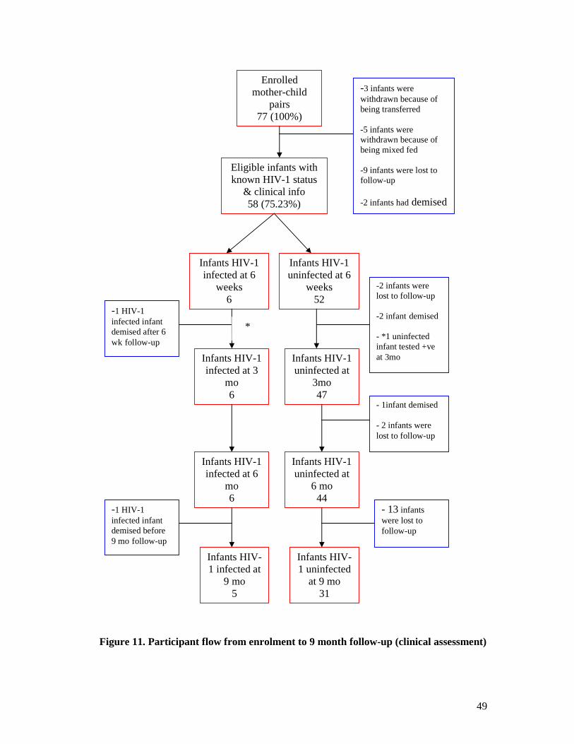
49
Figure 11. Participant flow from enrolment to 9 month follow-up (clinical assessment)
Enrolled
mother-child
pairs
77 (100%)
-3 infants were
withdrawn because of
being transferred
-5 infants were
withdrawn because of
being mixed fed
-9 infants were lost to
follow-up
-2 infants had demised
Eligible infants with
known HIV-1 status
& clinical info
58 (75.23%)
Infants HIV-1
uninfected at 6
weeks
52
Infants HIV-1
infected at 6
weeks
6
Infants HIV-1
infected at 3
mo
6
-1 HIV-1
infected infant
demised after 6
wk follow-up
Infants HIV-1
uninfected at
3mo
47
-2 infants were
lost to follow-up
-2 infant demised
- *1 uninfected
infant tested +ve
at 3mo
- 1infant demised
- 2 infants were
lost to follow-up
Infants HIV-1
infected at 6
mo
6
Infants HIV-1
uninfected at
6 mo
44
Infants HIV-
1 infected at
9 mo
5
Infants HIV-
1 uninfected
at 9 mo
31
- 13 infants
were lost to
follow-up
-1 HIV-1
infected infant
demised before
9 mo follow-up
*

50
3.2 Maternal and infant demographics
Tables 4a and 4b tabulate the baseline characteristics of the 77 mother-child pairs. The
mean age of HIV infected mothers was 27 years with a standard deviation of 5.6. The
mean parity was 2 with a standard deviation of 1.1 and the mean maternal CD4 count was
420 cells/µl with a standard deviation of 228 cells/µl. The mean and standard deviation of
each of the following: birth weight, birth length, birth occipito-frontal circumference
(OFC), gestational age, APGAR 1 min and APGAR 5 min is described in table 4a.
At the time of the study, government guidelines changed with regards to Prevention of
mother-to-child transmission (PMTCT). Previously, HIV exposed infants only received
single dose nevirapine (NVP) at birth. From September 2008 exposed infants received a
single dose of NVP at birth and 7 or 28 days of zidovudine (AZT) depending on the
maternal prophylactic history. In this cohort, 12(15.8%) infants did not receive NVP at
birth and 64(84.2%) infants received NVP at birth. Ten (13.2%) infants did not receive any
course of AZT, 18(23.7%) infants received a 7 day course of AZT and 48(63.2%) infants
received a 28 day course of AZT.
According to the PMTCT guidelines, HIV-1 infected women with a WHO clinical stage 4,
or women with a WHO clinical stage 3 with a CD4 <350cells/µl count or a woman in
WHO clinical stage 1 and 2 with a CD4 count <200cells/µl need to commence HAART
during pregnancy. HIV-1 infected women who do not need HAART should receive the
most effective prophylactic antiretroviral regimen. Twenty-three (29.9%) HIV-1 infected
women enrolled in the study did not receive any PMTCT prophylaxis; 23(29.9%) infected
women received NVP during onset of labor; 12(15.6%) infected women received AZT
only; and 19(24.7%) infected women received both NVP and AZT.

51
Table 4a. Descriptive characteristics of mother-child pairs
Mean ± SD 95% CI
Maternal age 27.2 ± 5.6 25.9 , 28.5
Maternal parity 2.1 ± 1.1 1.9 , 2.4
Maternal CD4 420.1 ± 228.4 362.1 , 478.1
Birth weight (kg) 1.44 ± 0.12 1.4 , 1.5
Birth length (cm) 40.3 ± 3.5 39.4, 41.1
OFC (cm) 29.6 ± 1.8 29.2, 30.05
Gestation 31 ± 2.00 31.0, 31.9
APGAR 5 min 9 ± 1.34 8.2, 8.9
APGAR 1 min 7 ± 1.82 6.7 , 7.6

52
Table 4b. Continuation of the descriptive characteristics of mother-child pairs Infant Characteristics N (%)
Gender Male 46(60.5%)
Female 30(39.5%)
Infant Received NVP No 12(15.0%)
Yes 64(84.2%)
Infant Received AZT No 10(13.2%)
Yes, for one week course 18(23.7%)
Yes, for 28 day course 48(63.16%)
Maternal Characteristics
Mode of Delivery NVD 36(46.8%)
Caesarean 40(51.0%)
Unknown 1(1.3%)
Reason for preterm delivery Spontaneous 41(53.0%)
Fetal distress 11(14.5%)
Maternal illness 9(11.9%)
Premature rupture of
membranes
2(2.7%)
Abruption 2(2.67%)
Fetal distress + abruption 1(1.3%)
Maternal illness + Abruption 1(1.3%)
Spontaneous preterm labor +
maternal illness(pre-eclampsia)
2(2.6%)
Fetal distress + maternal illness 2(2.6%)
Unknown 5(6.6%)
Maternal prophylaxis None 23(29.9%)
Nevirapine 23(29.9%)
AZT 12(15.6%)
Nevirapine + AZT 19(24.7%)
Maternal history of TB None 72(93.51%)
On treatment 2(2.6%)
Completed treatment 3(3.9%)
Maternal history of Syphilis Not confirmed 56(72.7%)
Confirmed 21(27.3%)
Maternal history of Pneumonia None 75(72.8%)
Yes 2(27.3%)
Other chronic conditions None 74(96.1%)
clinical 3(3.9%)
Employment of mother No 47(61.0%)
Yes 30(39.0)
Electricity/Fuel No 1(1.3%)
Yes 76(98.7%)
Piped running water No 3(3.9%)
Yes 74(96.1%)
Education Did not complete primary
school (grade 7)
3(3.9%)
Did not complete secondary
school (grade 12)
33(42.7%)
Completed secondary
education
36(46.8%)
Some tertiary education 1(1.3%)
Completed tertiary education 4(5.2%)

53
3.3 Maternal health and early infant infections
We examined the association between maternal CD4 counts and early infant infection
during the first two weeks of life (Table 5). Episodes of infection were categorized as mild,
moderate or severe. Mild or superficial infection included the skin, eye, umbilical, upper
respiratory tract infection (URTI) and gastroenteritis (not requiring rehydration). Moderate
infections were classified as infections that required supportive therapies without
continuous positive airway pressure (CPAP) or ventilation and included respiratory tract
infection/pneumonia, diarrheal disease (with intravenous fluid - IVF) and suspected sepsis
with full blood count (FBC) changes. Severe infections included infections that required
intensive therapies including CPAP and ventilation and include severe pneumonia,
necrotizing enterocolitis (NEC), meningitis, proven tuberculosis (TB), cytomegalovirus
(CMV) and zoster.
Due to the small sample size the infections were classified as absent or present. Early
infant infection was classified as any infection from birth to hospital discharge. The mean
number of day’s in-hospital was 14 days. Due to loss to follow-up CD4 counts were
available in only 61 HIV-1 infected women. HIV infected mothers were classified
according to their CD4 count, 6 weeks post delivery. The following observation was made:
for maternal CD4 count <500cells/µl vs. maternal CD4 count greater than >500cells/µl the
odds ratio for early infant infection was 1.69. This means that the risk of an infant
acquiring an infection at birth was approximately 2x higher if the maternal CD4 count was
less than 500cells/µl as compared to those infants born to an HIV infected women with a
maternal CD4 count > 500cells/µl.

54
Table 5: Relationship between maternal CD4 count 6 weeks post delivery and early
infant infection
Maternal CD4
count
(cells/µl)
Odds Ratio (95% CI)
<200 vs. >200 1.47 0.45,4.72
<350 vs. > 350 1.35 0.45,4.01
<500 vs. >500 1.69 0.55,5.17
3.4 Maternal health and infant growth at birth
The Kruskal-Wallis test, which is a nonparametric version of ANOVA, was used to test
whether there was a significant difference in mean birth weight, mean birth length, mean
birth OFC and maternal CD4 categories (tables 6-8). There was no significant difference
between mean birth weight and maternal CD4 count (p-value = 0.1394). However, the
following trend was observed: infants born to HIV-1 infected women with advanced
disease (CD4 count 0-200 cells/µl) were lighter in weight compared to infants born to
HIV-1 infected women with a CD4 count > 350 cells/µl. The mean length did not differ
significantly between categories of maternal CD4 count (p-value = 0.6533). The mean
OFC also did not differ significantly between categories of maternal CD4 count (p-
value=0.3770).
Table 6. Maternal CD4 count and infant birth weight (n=61)
Maternal CD4 N Mean Birth Weight (kg)
0-200 12 1.37
201-350 16 1.41
351-500 7 1.51
>500 26 1.50

55
Table 7. Maternal CD4 count and infant birth length (n=57)
Maternal CD4 N Mean Birth Length (cm)
0-200 12 29.33
201-350 14 29.45
351-500 7 30.86
>500 24 29.69
Table 8. Maternal CD4 count and infant birth OFC (n=55)
Maternal CD4 N Mean Birth OFC (cm)
0-200 12 41.00
201-350 13 39.46
351-500 7 41.86
>500 23 39.96
3.5 Maternal education and postnatal feeding choice
Of the 73 HIV-1 infected women that had data available on their feeding choice, 48
(65.8%) women decided to feed their infants breastmilk at birth. Of the 48 HIV-1 infected
women that decided to breastfeed their infants, 38(79.2%) women chose to heat-treat their
expressed breastmilk(HTEBM) and 10(20.8%) women chose to feed their infants
expressed breastmilk(EBM). The remaining 25(34.2%) HIV-1 infected women fed their
infants exclusive formula. Of the 38 HIV-1 infected women that decided to feed their
infants HTEBM, 2(5.3%) women did not complete primary school, 14(36.8%) women did
not complete secondary school and 22(57.9%) women completed secondary school. Ten
HIV-1 infected women fed their infants EBM birth, 6(60.0%) women did not complete
secondary school, 3(30.0%) women completed secondary school and 1(10.0%) mother
completed tertiary education. Twenty-five women chose to feed their infants exclusive
formula, 1(4.0%) mother did not complete primary school, 11(44.0%) women did not
complete secondary school, 10(40.0%) completed secondary school and 3(12.0%) women

56
completed tertiary education. There was no significant relationship between postnatal
feeding choice and maternal education, using the fishers exact test (p-value=0.1753) (Table
9).
Table 9. Relationship between maternal education and postnatal feeding choice
Data pertaining to maternal employment was available for 73 patients. In this cohort of
mother-child pairs, 46(63.0%) women were unemployed and 27(36.9%) were employed at
the time of delivery. Twenty-eight (38.4%) HIV-1 infected women that decided to feed
their infants HTEBM were unemployed, 4(5.5%) HIV-1 infected women that fed their
infants EBM were unemployed and 14(19.2%) women that fed their infants exclusive
formula were unemployed. There no significant relationship between maternal
employment and postnatal feeding choice (p-value 0.0979) (Table 10).
FEED EDUCATION TOTAL
FREQUENCY
&
PERCENTAGE
Did not
complete
primary
School
(grade7)
Did not
complete
Secondary
School
(grade12)
Completed
Secondary
Education
Some
Tertiary
Education
Completed
Tertiary
Education
Exclusive
Breastmilk
(EBM)
0 6 3 0 1 10
0 8.2 4.1 0 1.4 13.7%
Exclusive
heat-treated
expressed
breastmilk
(HTEBM)
2 14 22 0 0 38
2.7 19.2 30.1 0 0 52.1%
Exclusive
formula
1 11 10 0 3 25
1.4 15.1 13.7 0 4.1 34.2%
Total
Frequency &
Percentage
3 31 35 0 4 73
4.1 42.5 47.9 0 5.5 100%

57
Table 10. Relationship between Maternal Employment and Postnatal Feeding Choice
FEED EMPLOYMENT TOTAL
FREQUENCY
&
PERCENTAGE
No Yes
Exclusive Breastmilk
(EBM)
4 6 10
5.5 8.2 13.7
Exclusive heat-treated expressed
breastmilk
(HTEBM)
28 10 38
38.4 13.7 52.1
Exclusive formula 14 11 25
19.2 15.1 34.2
Total Frequency
& Percentage
46 27 73
63.0 3 100
3.6 Infant infections and feeding modality
Tables 11-19 demonstrate the relationship between infant infection and feeding modality at
each follow-up. Infection has been described previously (3.3), as mild, moderate or severe.
Due to the small sample size infection at each follow-up infection was reported as absent
or present. The Fisher exact test was used. This test analyses the independence between
two categorical variables. Early feeding mode was defined as the preferred feeding choice
during the first two weeks of life. There was no significant relationship between early
infant feeding mode and infection at 6 weeks, 3 mo, 6 mo and 9 mo follow-up visit (p-
value =0.4058, 0.1179, 0.1689 and 0.7855 respectively). We further attempted to
determine whether there was relationship between 6 week infant feeding mode and
infection at 3 mo, 6 mo and 9 mo follow-up visit(p-value=0.8826, 0.1403 and 0.4729
respectively). There was also no significant relationship between 3 month infant feeding

58
choice and infection at 6 mo and 9 mo follow-up visit (p-value=0.1403 and 0.4729
respectively).
Table 11. Relationship between early feeding mode and infection at the 6 week follow-
up visit (n=56)
Infection absent Infection present
Breastfeeding 5(8.92%) 1(1.79%)
Exclusive Heat Treated
breastmilk
19(33.93%) 7(12.5%)
Exclusive formula 13(23.21%) 8(14.29%)
Unknown 1(1.79%) 2(3.57%)
Table 12. Relationship between early feeding mode and infection at the 3 month
follow-up visit (n=53)
Infection absent Infection present
Breastfeeding 1(1.89%) 6(11.32%)
Exclusive Heat Treated
breastmilk
9(16.98%) 14(26.42%)
Exclusive formula 8(15.09%) 12(22.64%)
Unknown 3(5.66%) 0(0%)
Table 13. Relationship between early feeding mode and infection at the 6 month
follow-up visit (n=51)
Infection absent Infection present
Breastfeeding 2(3.92%) 4(7.84%)
Exclusive Heat Treated
breastmilk
8(15.69%) 14(27.45%)
Exclusive formula 11(21.57%) 9(17.65%)
Unknown 3(5.88%) 0(0%)

59
Table 14. Relationship between early feeding mode and infection at the 9 month
follow-up visit (n=35)
Infection absent Infection present
Breastfeeding 3(8.57%) 2(5.71%)
Exclusive Heat Treated
breastmilk
9(25.71%) 7(20.0%)
Exclusive formula 6(17.14%) 6(17.14%)
Unknown 2(5.71%) 0(0%)
Table 15. Relationship between 6 week feeding choice and infection at the 3 month
follow-up visit (n=54)
Infection absent Infection present
Breastfeeding 6(11.11%) 12(22.22%)
Exclusive formula 10(18.52%) 13(24.07%)
Mixed feed 5(9.26%) 8(14.81%)
Table 16. Relationship between 6 week feeding choice and infection at the 6 month
follow-up visit (n=52)
Infection absent Infection present
Breastfeeding 5(9.16) 11(21.15%)
Exclusive formula 11(21.15%) 12(23.08%)
Mixed Feed 9(17.31%) 4(7.69%)
Table 17. Relationship between 6 week feeding choice and infection at the 9 month
follow-up visit (n=36)
Infection absent Infection present
Breastfeeding 5(13.89%) 6(16.67%)
Exclusive formula 9(25%) 8(22.22%)
Mixed Feed 6(16.67%) 2(5.56%)

60
Table 18. Relationship between 3 month feeding choice and infection at the 6 month
follow-up visit (n=52)
Infection absent Infection present
Breastfeeding 5(9.62%) 11(21.15%)
Exclusive formula 11(21.15%) 12(23.08%)
Mixed Feed 9(17.31) 4(7.69%)
Table 19. Relationship between 3 month feeding choice and infection at the 9 month
follow-up visit (n=36)
Infection absent Infection present
Breastfeeding 5(13.89%) 6(16.67%)
Exclusive formula 9(25%) 8(22.22%)
Mixed Feed 6(16.67%) 2(5.56%)
3.7 Maternal CD4 Count at 6 weeks post delivery and infant growth over
time
Table 20 illustrates the differences between maternal CD4 count and infant weight over
time. Infants born to HIV-1 infected women with a CD4 count of greater than 350cells/µl
and infants born to HIV-1 infected women with a CD4 count of greater than 500cells/µl
had a similar weight gain at 36 weeks of age (8.26 vs. 8.15).
Figure 12 and Table 21, further explore these differences. Maternal CD4 count and infant
weight over time had a positive co-efficient and this was statistically significant (p value =
0.0361). Infants born to HIV-1 infected women with a CD4 count of greater than
350cells/µl had a higher mean weight during the first weeks of life compared to infants
born to HIV-1 infected women with advanced disease (CD4 count < 200cells/µl).
However, this effect diminishes over time.
61
Tables 22-23 and Figure 13 demonstrate the differences between maternal CD4 count and
infant length over time. Maternal CD4 count and infant length over time had a negative co-
efficient and a p value of 0.1304. Therefore, there was no relationship between maternal
CD4 count and infant length over time. Tables 24-25 and Figure 14 explore the differences
between maternal CD4 count and infant OFC over time. For every 1 unit increase in
maternal CD4 count, the infants OFC increased by 0.0001 over time.

62
Table 20. The relationship between maternal CD4 count 6 weeks post delivery and
infant weight at each follow-up
Maternal CD4 Count Week Mean Weight
0-200
0 1.30
0-200
6 2.40
0-200
12 4.18
0-200
24 6.26
0-200
36 6.86
201-350
0 1.39
201-350
6 2.33
201-350
12 3.98
201-350
24 5.91
201-350
36 7.05
351-500
0 1.46
351-500
6 2.28
351-500
12 3.72
351-500
24 6.69
351-500
36 8.26
>500
0 1.52
>500
6 2.69
>500
12 4.52
>500
24 6.97
>500
36 8.15

63
Figure 12. The participants mean weight over time
Table 21. The association between maternal CD4 count and infant weight
Co-efficient F-Value P-value
Maternal CD4 Count 0.000034 0.01 0.9352
Time 0.1656 134.32 <0.001
Maternal CD4*Time 0.00006 4.61 0.0361

64
Table 22. Relationship between maternal CD4 count 6 weeks post delivery and infant
length at each follow-up
Maternal CD4 Count Week
Mean Length
(cm)
0-200 0 40.80
0-200 6 46.00
0-200 12 52.00
0-200 24 59.17
0-200 36 67.25
201-350 0 38.90
201-350 6 44.39
201-350 12 51.10
201-350 24 57.73
201-350 36 63.00
351-500 0 41.60
351-500 6 45.80
351-500 12 51.00
351-500 24 60.75
351-500 36 65.00
>500 0 40.53
>500 6 44.83
>500 12 52.35
>500 24 60.75
>500 36 66.12

65
Figure 13. The participants mean length over time
Table 23. The association between maternal CD4 count and infant length
Co-efficient F-Value P-value
Maternal CD4 Count 0.00061 0.09 0.7703
Time 0.6583 168.02 <0.0001
Maternal CD4*Time 0.000151 2.36 0.1304

66
Table 24. The relationship between maternal CD4 count 6 weeks post delivery and
OFC at each follow up
Maternal CD4 Count Week
Mean OFC
(cm)
0-200 0 28.60
0-200 6 35.00
0-200 12 39.08
0-200 24 42.50
0-200 36 44.00
201-350 0 29.18
201-350 6 33.54
201-350 12 37.40
201-350 24 41.50
201-350 36 44.00
351-500 0 30.20
351-500 6 32.80
351-500 12 37.00
351-500 24 41.88
351-500 36 43.88
>500 0 29.88
>500 6 34.45
>500 12 38.87
>500 24 42.90
>500 36 44.24

67
Figure 14. The participants mean OFC over time
Table 25. The association between maternal CD4 count and OFC
Co-efficient F-Value P-value
Maternal CD4 Count 0.000758 0.35 0.5568
Time 0.5218 355.82 <0.0001
Maternal CD4*Time 0.000040 0.54 0.4649
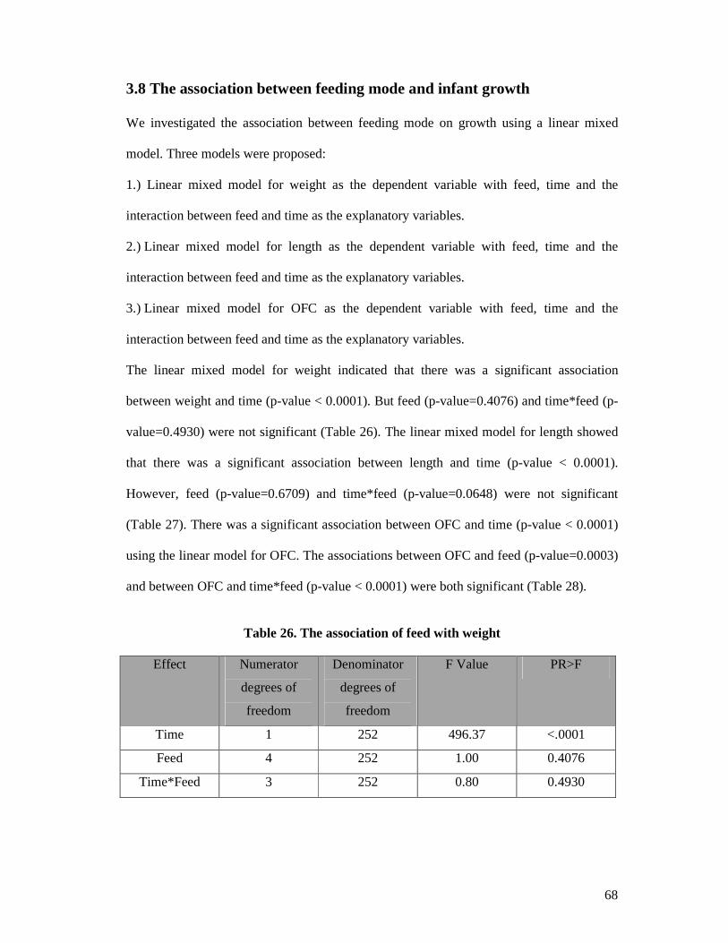
68
3.8 The association between feeding mode and infant growth
We investigated the association between feeding mode on growth using a linear mixed
model. Three models were proposed:
1.) Linear mixed model for weight as the dependent variable with feed, time and the
interaction between feed and time as the explanatory variables.
2.) Linear mixed model for length as the dependent variable with feed, time and the
interaction between feed and time as the explanatory variables.
3.) Linear mixed model for OFC as the dependent variable with feed, time and the
interaction between feed and time as the explanatory variables.
The linear mixed model for weight indicated that there was a significant association
between weight and time (p-value < 0.0001). But feed (p-value=0.4076) and time*feed (p-
value=0.4930) were not significant (Table 26). The linear mixed model for length showed
that there was a significant association between length and time (p-value < 0.0001).
However, feed (p-value=0.6709) and time*feed (p-value=0.0648) were not significant
(Table 27). There was a significant association between OFC and time (p-value < 0.0001)
using the linear model for OFC. The associations between OFC and feed (p-value=0.0003)
and between OFC and time*feed (p-value < 0.0001) were both significant (Table 28).
Table 26. The association of feed with weight
Effect Numerator
degrees of
freedom
Denominator
degrees of
freedom
F Value PR>F
Time 1 252 496.37 <.0001
Feed 4 252 1.00 0.4076
Time*Feed 3 252 0.80 0.4930

69
Table 27. The association of feed with length
The effect of feed on length
Effect Numerator
degrees of
freedom
Denominator
degrees of
freedom
F Value PR>F
Time 1 242 354.70 <.0001
Feed 4 242 0.59 0.6709
Time*Feed 3 243 2.44 0.0648
Table 28. The association of feed with OFC
Effect Numerator
degrees of
freedom
Denominator
degrees of
freedom
F Value PR>F
Time 1 245 422.62 <0.0001
Feed 4 245 5.51 0.0003
Time*Feed 3 245 1.32 <.0001
3.9 The feasibility of each feeding mode
A total of 51 HIV-1 infected women decided to breastfeed their infants at birth. Of which
38 women decided to heat treat their expressed breastmilk (HTEBM) and 13 women opted
to breastfeed their infants directly from the breast (EBM). The majority of HIV-1 infected
women (19 or 50.0 %) chose to exclusively heat-treat their expressed breastmilk based on
the health care worker recommendation. Fourteen (36.8%) women chose to feed their
infants exclusive HTEBM because of its anti-infective and nutritional benefits and 3(7.9%)
women chose it because it was safe and easy to do. Of the 13 HIV-1 infected women that
decided to exclusively breastfeed their infants(EBM not HTEBM), 1(7.7%) mother’s
decision was based on the anti-infective properties of breastmilk, 2(15.4%) mothers
experienced insufficient breastmilk, 3(23.1%) mothers thought that the HTEBM method

70
was too tedious they would rather breastfeed and 2(15.4%) mothers were uncertain of the
use of HTEBM. Twenty-six HIV-1 infected women decided to formula feed their infants
after intensive WHO infant feeding counselling. Ten (38.5%) HIV-1 infected women that
decided to exclusively formula feed their infants at birth were either employed or attending
school. Five (19.2%) women were ill after delivery, 4(15.4%) women based their choice
on the health care worker recommendation, 2(7.7%) women chose to formula feed because
of its anti-infective properties, 2(7.7%) women thought the method was easier to do
compared to either HTEBM or breastfeeding, 1(1.37%) mother reported insufficient
breastmilk and 1(3.8%) mother re-called previously infecting a child through breastmilk
(Table 29).
While in-hospital, 38 HIV-1 infected women fed their infants HTEBM after receiving
counseling and support from the nursing staff at the King Edward VIII hospital. At the 6
week follow-up only 9 HIV-1 infected women were able to continue feeding their infants
HTEBM. As the months passed, the numbers fell to 4 infants on HTEBM at 3 months and
2 infants on HTEBM at 6 months. The decrease in number of infants on HTEBM was
mainly due to mixed feeding.

71
Table 29. Reason for choosing each feeding modality, n = 77. (Results presented as frequency and percentage)
Feed Anti-
infective
Easy
to do
&
Safe
Mother at
school/employed
Health care
worker
recommendation
Maternal
illness
Insufficient
breastmilk
Method
tedious
Previous
child
infected
through
breastmilk
Method
unknown,
uncertain
of use
Afraid of
disclosure
No
Reason
Expressed
Breastmilk
(EBM)
n = 13
1
7.7%
0
0%
0
0%
2
15.4%
0
0%
2
15.4%
3
23.1%
0
0%
2
15.4%
1
7.7%
2
15.38%
Heat-
treated
expressed
breastmilk
(HTEBM)
n = 38
14
36.8%
3
7.9%
0
0%
19
50%
0
0%
0
0%
0
0%
0
0%
0
0%
1
2.6%
1
2.6%
Exclusive
Formula
n = 26
2
7.7%
2
7.7%
10
38.5%
4
15.4%
5
19.2%
1
3.8%
0
0%
1
3.8%
0
0%
0
0%
1
3.8%

72
3.10 Recruitment and follow-up for CTL investigations
Of the 77 infants enrolled in the study 55 infants were assessed for both CTL
responses and clinical outcomes. Delivery information was available for all 55
mother-child pairs. The HIV-1 status of the infants was determined by HIV RNA
PCR at day 1 or HIV DNA PCR at 6 weeks of age. At the time of discharge from
hospital 19 infants were withdrawn from the study: Three infants were transferred to
Inkosi Albert Luthuli Chief (IALCH) hospital for specialised care, 5 infants failed to
meet inclusion criteria (mixed fed in-hospital), 9 infants were lost to follow-up and
2 infants had demised.
At the 6 week follow-up visit, 34 infants were uninfected and 2 infants were HIV-1
infected. After the 6 week follow-up visit, 1 of the 2 HIV-1 infected infants had
demised, 2 uninfected infants had demised and 2 uninfected infants were withdrawn
from the study due to loss to follow-up. An additional uninfected infant tested HIV-
1 positive at the 3 month follow-up visit. At this time point, 2 infants were HIV-1
infected and 29 infants were uninfected. After the 3 month follow-up visit, 1
uninfected infant demised and 2 uninfected infants were lost to follow-up. During
the 6 month follow-up visit, 2 infants were infected and 26 infants were uninfected
.One infected infant demised and 2 uninfected infants were withdrawn from the
study due to loss to follow-up after the 6 month visit. At the end of the study (9
months), 1 infant was HIV-1 infected and 24 infants were uninfected (Figure 14).
The baseline characteristics of the 55 mother-child pairs are included in tables 30a
and 30b. These descriptive characteristics were similar to that reported in tables 4a
and 4b of the full cohort of 77 mother-infant pairs.

73
Figure 15. Participant flow from enrolment to 9 month follow-up (CTL
responses and clinical assessment)
Enrolled
mother-child
pairs
55 (100%)
-3 infants were
withdrawn because of
being transferred
-5 infants were
withdrawn because of
being mixed fed
-9 infants were lost to
follow-up
-2 infants had demised
Infants with known
HIV-1 status, clinical
info and CTL
responses
36(65.5%)
Infants HIV-1
uninfected at 6
weeks
34
Infants HIV-1
infected at 6
weeks
2
Infants HIV-1
infected at 3
mo
2
-1 HIV-1
infected infant
demised after 6
wk follow-up
Infants HIV-1
uninfected at
3mo
29
-2 infants were lost
to follow-up
-2 infant demised
-*1 uninfected
infant tested +ve at
3mo
- 1infant demised
- 2 infants were
lost to follow-up
Infants HIV-1
infected at 6
mo
2
Infants HIV-1
uninfected at
6 mo
26
Infant HIV-
1 infected at
9 mo
1
Infants HIV-
1 uninfected
at 9 mo
24
- 2 infants were
lost to follow-up -1 HIV-1
infected infant
demised before
9 mo follow-up
*

74
Table 30a. Descriptive characteristics of 55 mother-child pairs (sub-study)
Mean 95% CI
Maternal Age 26.80 (25.27, 28.33)
Maternal Parity 2.09 (1.81, 2.38)
Maternal CD4 838.14 (9.68, 1666.59)
Birth Weight (kg) 1.48 (1.43, 1.53)
Birth Length(cm) 40.34 (39.31, 41.37)
OFC(cm) 29.96 (29.96, 30.44)
Apgar 1 min 7.13 (6.57, 7.68)
Apgar 5 Min 8.64 (8.28, 8.99)
Gestation (weeks) 31.47 (31.02, 31.92)

75
Table 30b. Continuation of the descriptive characteristics of 55 mother-child
pairs (sub-study)
Infant Characteristics N (%)
Gender Male 31 (56.4%)
Infant Received NVP No 8 (14.6%)
Yes 46 (83.6%)
Infant Received AZT No 1 (1.8%)
Yes, for one week course 16 (29.1%)
Yes, for 28 day course 37 (67.3%)
Maternal Characteristics
Mode of Delivery NVD 26 (47.3%)
Caesarean 28 (50.9%)
Reason for preterm Delivery Unknown 3 (%)
Spontaneous 33 (5.5%)
Fetal distress 7 (60.00%)
Maternal illness 3 (12.8%)
Abruption 1 (1.8%)
Fetal Distress and abruption 2 (3.6%)
Spontaneous preterm labor + maternal illness (pre-
eclampsia)
2 (3.6%)
Fetal Distress + Maternal illness 1 (1.8%)
Maternal Prophylaxis None 12 (21.8%)
Nevirapine 13 (23.6%)
AZT 12 (21.8%)
Nevirapine & AZT 18 (32.7%)
Maternal history of TB None 52 (94.6%)
Completed Treatment 0
Being Worked up, Not on Treatment 3 (5.45%)
Maternal history of Syphilis Not Confirmed 36 (65.5%)
Confirmed 19 (34.6%)
Maternal history of
Pneumonia
None 54 (98.2%)
Yes, as per mother 1 (1.8%)
Other Chronic Conditions None 52 (94.6%)
Clinical 3 (5.5%)
Employment of Mother Unemployed 37 (67.3%)
Employed 18 (32.7%)
Electricity/Fuel No 1 (1.82%)
Yes 54 (98.2%)
Piped Running Water No 3 (5.5%)
Yes 52 (9.6%)
Education Did not complete primary school (grade 7) 3 (5.5%)
Did not complete secondary school (8-Matric) 21 (38.2%)
Completed secondary education 27 (49.1%)
Completed tertiary education 4 (7.3%)

76
3.11 Immune responses in HIV exposed low birth weight infants
A blood sample was taken from each patient at follow-up visits (6 weeks, 3mo, 6mo
and 9mo) and immune response assays were performed on them. The assays used
included ELISPOT megamatrix (day 1), ELISPOT confirmation (day 2) and
Intracellular Cytokine Staining (ICS) (day 2). Immune response assay results were
available for 25 infants at each time point. Both immunological and clinical data
were available for these infants throughout the study. Three of 25 infants were
diagnosed HIV-1 infected by HIV DNA PCR by 3 months of age. The 3 HIV-1
infected infants had a negative HIV RNA PCR at birth (day1).
At the 6 week follow-up visit only 1 HIV-1 infected infant (FH 001 H) elicited a
HIV-1-specific CTL immune response towards a pool of peptides in the ELISPOT
megamatrix. We were unable to confirm these responses on day 2 with the
ELISPOT confirmation assay. This infant (FH 001 H) was unable to maintain these
responses and therefore we were unable to detect these responses at any of the
consecutive time points. The main feeding modality for this infant was exclusive
HTEBM up to 6 weeks of age followed by exclusive breastmilk and complementary
feeds. At the time of detection of HIV-1 specific CTL responses this infant was fed
on exclusive HTEBM. The second infant diagnosed HIV-1 infected (FH 006 H) at 6
weeks of age demised before the 3 month follow-up visit and we were unable to
detect HIV-1-specific CTL responses at the 6 week follow-up visit. This infant was
fed a combination of HTEBM and EBM. The third infant that was diagnosed HIV-1
infected (FH 005 H) at 3 months of age and was able to elicit a HIV-1-specific CTL
response against pools of peptide in the ELISPOT megamatrix. We were unable to
confirm these responses on day 2 with neither the ELISPOT confirmation nor ICS

77
assay. This infant was also unable to sustain these immune responses up to 6 months
of age and the infant demised prior to the 9 month follow-up. The infant was fed
HTEBM exclusively, until 6 weeks of age followed by breastmilk and
complementary feeds. HIV- 1- specific cytotoxic T lymphocyte activities were
detected against pools of the following viral antigen gag, pol, env and nef in 2
infected infants (FH 002 H and FH 005 H).
We were unable to detect HIV-1-specific CTL response in any of the HIV-1
exposed uninfected infants at any of the study time points. Phytohaemaglutinin A
(PHA) served as the positive control in the ELISPOT megamatrix and a detectable
response was observed each time the assay was performed. Cells were even
stimulated in-vitro prior to the ELISPOT megamatrix and no response was
detectable.
The table below is a summary of the two HIV-1 infected infants WHO staging,
CDC staging, CD4%, CD4+ count and viral load. The 3 infected infants at the time
of HIV diagnosis were all fed exclusive breastmilk. All three mothers had heat
treated their expressed breastmilk while in-hospital but could not sustain this
feeding mode post delivery.
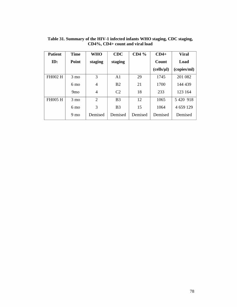
78
Table 31. Summary of the HIV-1 infected infants WHO staging, CDC staging,
CD4%, CD4+ count and viral load
Patient
ID:
Time
Point
WHO
staging
CDC
staging
CD4 % CD4+
Count
(cells/µl)
Viral
Load
(copies/ml)
FH002 H 3 mo
6 mo
9mo
3
4
4
A1
B2
C2
29
21
18
1745
1700
233
201 082
144 439
123 164
FH005 H 3 mo
6 mo
9 mo
2
3
Demised
B3
B3
Demised
12
15
Demised
1065
1064
Demised
5 420 918
4 659 129
Demised

79
CHAPTER 4
DISCUSSION
Despite breastfeeding being a significant mode of postnatal HIV transmission it
remains the main nutritional source and pillar of child survival for the majority of
infants born in Africa. It is therefore, not surprising that considerable research has
centred on making breastfeeding safer in terms of HIV transmission. The flash heat
treatment method (HTEBM) provides a unique opportunity to safely breastfeed
infants but prevent mother-to-child transmission of HIV. This pilot study examined
the feasibility of using HTEBM and its association on clinical outcomes and CTL
responses in HIV exposed low birth weight infants.
In this cohort the risk of an infant acquiring an infection at birth was approximately
2x higher if the maternal CD4 count was less than 500cells/µl compared to those
infants born to an HIV infected women with a maternal CD4 count > 500cells/µl.
This finding was in accordance with previous reports that infants born to women
with lower CD4 counts are more susceptible to morbidity and mortality (Newell et
al., 2004; Marinda et al., 2007).
Studies have shown that HIV infected women are more likely to deliver preterm and
low birth weight infants (Hira et al., 1989; Ryder et al., 1989; St Louis et al., 1993;
Taha et al., 1995). We evaluated the association between maternal CD4 count and
infant growth. In this study, infants born to HIV-1 infected women with advanced
disease (CD4 count 0-200 cells/µl) had a lower birth weight compared to infants
born to HIV-1 infected women with a CD4 count > 350 cells/µl. A similar weight
gain was observed at 36 weeks of age (8.26 vs. 8.15) among infants born to HIV-1

80
infected women with a CD4 count of greater than 350cells/µl and infants born to
HIV-1 infected women with a CD4 count of greater than 500cells/µl. Infants born to
HIV-1 infected women with a CD4 count of greater than 350cells/µl had a higher
mean weight gain during the first weeks of life compared to infants born to HIV-1
infected women with advanced disease (CD4 count < 200cells/µl). However, this
effect diminishes over time.
Several studies have reported on the impact of HIV exposure and infection on infant
growth with contradictory findings. Some studies have shown no observable
differences in growth between HIV exposed uninfected children and HIV
unexposed children. HIV infected children had the poorest growth outcome than
HIV unexposed children and HIV exposed uninfected children (European
collaborative study 2003; Patel et al., 2010). While a study conducted in Malawi
between 2000-2003 showed early stunting among HIV infected and uninfected boys
compared to girls (Taha et al., 2010).
Although exclusive breastfeeding has been well documented to be associated with
reduced morbidity and mortality due to respiratory and diarrhoeal illnesses, We
were not able to show this (Cesar et al., 1999; Oddy et al., 1999; Bahl et al., 2005).
The lack of finding any association between infant feed and infection (defined in
section 3.3) is most likely due to the small sample number of infants in the study.
Recently, studies have shown that HIV infected children that are fed breastmilk had
a higher mean weight and mean length compared with HIV infected children not
receiving any breastmilk (Patel et al., 2010; Taha et al., 2010).

81
We investigated the feasibility of HTEBM before 6 months of age. We decided to
initiate HTEBM in these preterm infants at birth since their mothers were already
expressing breastmilk. The majority of HIV infected women enrolled in this study
chose HTEBM based on the health care worker recommendation. Our study showed
that HTEBM could be initiated and sustained while in-hospital given appropriate
counselling and support. It therefore, appears that HTEBM is feasible to implement
in the hospital setting but it is difficult to sustain post hospital discharge.
The main reason for the lack of sustainability post discharge is that the infants now
require larger volumes of feed and it is difficult for the mother to keep up with the
demands of expressing sufficient milk as well as the time required to flash heat each
batch of expressed milk. However, reports suggest that after 6 months the heat
treatment method becomes feasible once again as the infant now receives additional
complementary feeds (Israel-Ballard et al., 2006b; Mbuya et al., 2010 ). This was
the first study to our knowledge investigating the use of HTEBM in infants less than
6 months of age. We have shown that with good counselling on optimal feeding
choices and adequate support HTEBM can be implemented in the hospital setting.
However, because of the small sample size we had insufficient power to show that
HTEBM conferred a positive benefit on infant growth, morbidity and mortality.
Further, research with sufficient power would be important to elucidate this effect.
We prospectively evaluated the association between early neonatal feeding and CTL
responses in HIV exposed low birth weight infants. This study was undertaken to
assess the capability of the young infant to generate HIV-1 specific CTL responses
with respect to early feeding modality. Cytotoxic T lymphocyte responses have

82
been previously described in HIV exposed term infants (see Table 1). To our
knowledge, this was the first study that investigated CTL responses in HIV exposed
low birth weight infants. HIV-1 specific CTL responses were detected in 2 infected
infants at only a single time point. These responses however, could not be
confirmed and were undetectable at any of the consecutive time points. At the time
of HIV-1 specific CTL detection these infants were fed breastmilk (heated
expressed breastmilk for FH 002 H and unheated breastmilk for FH 005 H). HIV
exposed uninfected infants were unable to generate a HIV-1 specific CTL response
irrespective of feed. During acute HIV infection, CTL responses have been
implicated in the initial containment of the virus. Previously, studies have shown
that some young infants and even the foetus were capable of generating HIV-
specific CTL responses (Table 1). The generation of CTL responses in infancy has
been reported to be variable and develop later the course of infection.
There are a few suggestions for the lack of HIV-1 specific CTL responses in our
cohort. At least 5 other groups investigating CTL responses in HIV exposed term
infants were unable to detect these responses even after virus specific in-vitro
stimulation (Luzuriaga et al., 1991; Buseyne et al., 1993; McFarland et al., 1994;
Luzuriaga et al., 1997; Thobakgale et al., 2007). Exposed uninfected infants in this
study could have completely escaped viral exposure or the small sample size may
have limited our ability to detect responses. The other reason for a lack of CTL
responses in this study could be that low birth weight infants are in fact not able to
mount an adequate immune response as shown in 2 previous non-African studies
(Chatrath, 1997; Das, 1998). Finally, the introduction of enhanced PMTCT
prophylaxis during the course of the study could have had influenced the ability to

83
mount an immune response (outlined in section 2.1). The majority of the mother-
child pairs enrolled in this cohort received both AZT and NVP. Several studies have
shown that exposure to antiretroviral therapy in utero and neonatally are associated
with a decrease in CD4 and CD8 cell counts (McKallip et al., 1995; Francke et al.,
2000; Bunders et al., 2005; Kolber et al., 2008).
LIMITATIONS
A major confounder to the study was the initiation of PMTCT ARV prophylaxis
during the course of the study. The majority of the mother-child pairs enrolled into
the study received prophylactic treatment and we cannot determine to what extent it
has the influenced the CTL immune response. HTEBM was introduced to preterm
infants as their mothers were already expressing breastmilk. However, we are not
certain of the level of prematurity of the immune system and we cannot determine
how this would have influenced the CTL immune response. This was also a
vulnerable population and the drop out rate due to demise or ill health was
significantly higher. Therefore, it was difficult to follow-up patients longitudinally
over a 9 month period. It was also difficult to maintain feeding mode post discharge
despite adequate infant feeding counseling at each follow-up session. Initially one
of our study objectives included investigating CTL immune responses in infants that
were fed EBM (live virus) vs. HTEBM (heat inactivated virus), but due to the
overwhelming success of the implementation of HTEBM at the KEH nursery we
had to combine the two feeding arms (HTEBM and EBM) into one group
(HTEBM).

84
CONCLUSION
In conclusion we have shown that it is feasible for HIV infected mothers to heat
treat their expressed breastmilk during hospital admission. Within the constraints of
a small sample size, we were able to demonstrate in this small cohort of patients that
the clinical outcomes and growth parameters of infants fed HTEBM were similar to
that of infants fed either formula or unheated breastmilk. We were unable to
demonstrate HIV-specific CTL responses in this cohort of infants. Our findings
indicate that additional research is required. However, due to the wide scale
PMTCT programmes in South Africa it will be difficult to establish such research
projects with an ARV naïve cohort. Therefore, we suggest that the scope of this
project would be better replicated in an animal model.

85
CHAPTER 5
APPENDICES
1. Information Document for the Mothers
INFORMATION DOCUMENT
Study title: Role of flash heat-treated breast-milk in improving health outcomes in
HIV-exposed preterm low-birth-weight infants: an operational study.
Hello and congratulations on the birth of your baby! We know that it is a challenge
to have a small baby but the nurse will do their best to support you while you are in
hospital. The Department of Paediatrics is doing research on different feeding
options for premature babies of HIV-positive mothers. Research means the process
of using science to learn the answer to a question. In this study we want to learn
how feeding babies with heated breastmilk compares to feeding babies with non-
heated breastmilk or formula. We are interested in learning which technique
mothers choose to do in the hospital and at home after counselling, and which
technique is most beneficial for the baby.
Invitation to participate: We are inviting you to participate and request your
permission to include your child in this study.
What is involved in the study: If you decide to participate in this study, you will
receive a counselling session with a trained counsellor to discuss the different ways
in which you can feed your baby and the advantages and disadvantages of each
method. The counsellor will also tell and show you how you can “heat-treat” your
breastmilk and feed it to your baby in the way that the World Health Organization
recommends. You can then make a choice to either heat-treat your breastmilk before

86
feeding it to your baby, breastfeed without heat-treating, or formula feed. You will
be able to make all feeding choices yourself. We will ask you some questions
about your decision, and will keep a record of how many times your baby feeds, has
episodes of illness in the hospital, your baby’s growth, and any information we find
about your baby’s HIV status from blood tests.
As part of this study, you may be asked to participate in a discussion group on infant
feeding. If you are asked to be a focus group participant, you have every right to
refuse to participate. Refusing will not affect your status as a study participant or the
care that you and your baby receive. All comments made during these groups will
be recorded and used for research purposes without being linked to your name or
any other personal identification.
When your baby is six weeks old, you will return to our clinic, where we will
examine your baby and you will tell us about your health, that of your baby, and
give us some information on how you have been feeding your baby. We will also
test your baby’s blood for the HIV virus at this time, which is required in South
Africa for any infant with a mother that has HIV, not just those participating in this
study. You will be asked to come into the clinic again when your baby is three
months old and thereafter every three months up to 9 months of age. This is the
standard follow up time for babies who are born prematurely as we need to monitor
their health, and is not part of the study itself. You can also come into the clinic at
any point in between if you think your baby needs medical attention.
87
The study will require that we take two extra blood tests—one within 48 hours of
your baby’s birth, and one at the time that you leave the hospital. We will only
analyze these blood tests if your child’s HIV status changes on the HIV test that is
required by the government. The purpose of these blood tests is to confirm exactly
when an infant becomes HIV positive. These tests will only require a drop of blood
and the blood will be collected by means of a small prick on the heel.
In addition we would request permission to take a small blood sample from your
infant at the clinic visits at 6 weeks, 3, 6, and 9 months. It will be a very small
sample about half a teaspoon of blood. This blood will be used to allow us to do
some tests to determine how the baby’s immune response is working. At discharge
if your blood has not already been tested for CD4 and viral load we will take a
sample from you for these tests. These results are important to allow us to advise
you on your disease management. If these results show that you are in need of
treatment we will refer you to the appropriate clinic.
Risks of participating: There are no risks to participating in this study. While it is
possible that using the flash heat treatment may have a very small effect on a few of
the vitamins and protein in the milk, laboratory studies have shown that the heating
eliminates the HIV virus present in milk. For this reason, some mothers choose to
heat their milk before feeding it to their child.
Benefits of participating: You will receive advice and support in the hospital on
how to best feed your baby. You will specifically receive information on how to
correctly heat-treat milk, which is recommended by the World Health Organization
88
as a way to give your baby the benefits of breastmilk while reducing the chance that
your baby will get HIV from breastmilk. We will be able to closely monitor the
health of your baby and provide the necessary management.
You will be given information on the study while you are involved in the project.
Unfortunately the results of the study may only be available when your child is six
months to a year old so it will not directly benefit your child but it will be very
important to help many other mothers and their babies in the future.
Participation in this study is voluntary. This means that refusal to participate will
not involve any penalty or loss of benefits to which you are otherwise entitled. You
may end your participation in this study at any time without any negative impact on
the care that you and your child receive.
There will be no additional cost to you associated with your participation in
this study. We will provide you with R50 for each clinic visit to assist with your
transport costs.
Confidentiality: Efforts will be made to keep all personal information confidential.
Absolute confidentiality cannot be guaranteed. Personal information may be
disclosed if required by law. Organizations that may inspect and/or copy your
research records for quality assurance and data analysis include groups such as the
Research Ethics Committee and the Medicines Control Council (where appropriate).
All of your information will be labelled with a number code and not your name, and
will be kept in a locked cabinet by the project coordinators.

89
If you or your baby experiences any negative events or unusual symptoms
during the course of the study, please immediately contact Reshmi Dayanand
or Prof Coutsoudis at 031-2604489 or Prof Adhikari on 031-2604345
Contact details of BREC Administrator – for reporting of complaints/ problems:
Ms Patricia Ngwenya, Nelson R Mandela School of Medicine, Private Bag 7,
Congella 4013
Telephone: +27 (0) 31 260 4769
Fax: +27 (0) 31 260 4609
Email: [email protected]
90
2. Consent form for the Mothers (English Version)
INFORMED CONSENT DOCUMENT
You have been asked to participate in a research study Role of flash heat-treated
breast-milk in improving health outcomes in HIV-exposed preterm infants: an
operational study.
You have been informed about the study by Reshmi Dayanand/Nozipho Makanya
and the nurses in the King Edward Hospital Nursery.
You may contact Reshmi Dayanand (073-1402914), Prof Coutsoudis (031-
2604489), or Prof Adhikari (031-2604345) at any time if you have questions about
the research or if your child gets ill or if you are injured as a result of the research.
You may contact the Biomedical Research Office at the Nelson R Mandela School
of Medicine at 031-260 4769 if you have questions about your rights as a research
subject.
Your participation in this research is voluntary, and you will not be penalized or
lose benefits if you refuse to participate or decide to stop.
If you agree to participate, you will be given a signed copy of this document and the
participant information sheet, which is a written summary of the research.
The research study, including the above information, has been described to me
orally. I understand what my involvement in the study means and I voluntarily agree
to participate.
____________________ ____________________
Signature of Participant Date
____________________ _____________________
Signature of Witness Date
(Where applicable)
____________________ _____________________
Signature of Translator Date
(Where applicable)

91
3. Enrolment form
STUDY NUMBER: _____________
MATERNAL HOSPITAL NUMBER:______________
Enrolment Forms Complete at enrolment into care Enter all dates as
dd/mm/yy
Personal Details:
First Name:____________________________ Last Name:____________________________
Date of birth: __/__/__ Age:___(years)
Marital Status:
���� Married ���� Divorced ����Widowed
���� Cohabiting ���� Single
Maternal Hospital Number: ��������������������������������������������
Infant Hospital Number: ��������������������������������������������
Maternal ID: ������������������������ ���������������� �������� ����
Infant ID number (to be completed when birth certificate obtained):__________________________
Infant date of birth:__/__/__
Maternal
Address:_____________________________________________________________________
Maternal contact number:__________________________________
Name of friend/family:__________________ Contact no. of
friend/family:_______________________
Ante-natal clinic
location:_______________________________
Nearest local clinic for
immunizations:_________________________
Date originally counselled:__/__/__ Counselled by: ���� Reshmi ���� Nozipho
Maternal ward at enrolment: ���� O1 ���� O2
���� O3
Infant ward at enrolment: ���� NPN ���� ICU
���� HC
Date enrolled: __/__/__
Baseline Maternal Assessment: (Tick appropriate block)
Maternal parity____
Mode of delivery:
���� caesarean
���� vaginal
���� unknown
Reason for preterm delivery:
���� spontaneous preterm labour
���� fetal distress
���� maternal illness
PMTCT Prophylaxis: (Tick appropriate block)
Which prophylaxis drugs did mum receive?
���� NVP (single dose)
� AZT
� UNKNOWN
CD 4 count: _________________ Date:__/__/__
Clinic location:__________________________________
Maternal Medical History

92
(Tick appropriate block)
Tuberculosis:
���� none
���� on treatment
� being worked up
� not on treatment
� unknown
Syphilis:
���� yes
���� no
���� unknown
Pneumonia:
���� yes
���� no
���� unknown
Other chronic conditions:
___________________________________
Household Questions (Tick appropriate block)
Have you disclosed your HIV status?
���� Yes -> If yes, to whom?
���� partner ���� family
���� Household member (not related) ���� other
����No -> If no, why not? (list
all)_________________________________
Do you have water piped (from a tap) inside
your home?
���� yes
���� no
Do you have electricity at your home?
���� yes ���� no
Do you have a fridge at your home?
���� yes ���� no
Highest level of education
���� did not complete primary school
���� did not complete secondary school
���� Completed secondary education/matric
���� Some tertiary education
���� Completed tertiary education
���� Professional training
• Are you currently employed?
���� yes ���� no
• Main source of household income?
���� family ���� grant ���� other
Ante-natal feeding choice
(recorded on PMTCT
card):_________________________________
Postnatal feeding
choice:_______________________________
Has mother breastfed before?___________________________________________________
Has mother ever expressed breastmilk before?_____________________________________

93
STUDY NUMBER: INFANT HOSPITAL NUMBER:
Baseline Infant Assessment Complete at enrolment into care Enter all DATES as dd/mm/yy
Date of birth: __/__/__
Birth weight :______ (kg) Birth length: ______ (cm) Birth OFC: _______ (cm) APGAR Score: ���� 1min ����
5min Gender: ����M ����F Gestational age:________
Problems in first 24 hrs (Tick appropriate block)
RDS
���� yes ���� no
Hypoglycaemia
���� yes ���� no Neurological
problems
�yes�no
Other:_______________
Respiratory distress at any time: ���� yes ���� no
Ventilation: ���� yes ���� no
Duration:_____day(s)
CPAP: ���� yes ���� no
Duration:_______day(s)
Oxygen: ���� yes ���� no
Duration:______day(s)
Seizures: ���� yes ���� no Feed intolerance: ���� yes ���� no
Duration:_____day(s) TPN given: ���� yes ���� no
Duration:______day(s)
Infections (Tick appropriate block)
• Suspected
sepsis:
���� yes ���� no
(Please tick one)
- proven on
blood
culture ����
- proven on
swab ����
- proven on
CSF ����
- proven on
urine ����
- proven on
ETT/other ����
- Organism
isolated ����
• Suspected
meningitis:
���� yes ���� no
(Please tick one) - proven on
blood
culture ����
- proven on
swab ����
- proven on
CSF ����
- proven on
urine ����
- proven on
ETT/other ����
- Organism
isolated ����
• UTI:
���� yes ���� no
(Please tick one) - proven on
blood
culture ����
- proven on
swab ����
- proven on
CSF ����
- proven on
urine ����
- proven on
ETT/other ����
- Organism
isolated ����
• Pneumonia: ���� yes ���� no
(Please tick one) - proven on blood
culture ����
- proven on swab ����
- proven on
CSF ����
- proven on urine ����
- proven on
ETT/other ����
- Organism isolated ����
Jaundice:
���� yes ���� no
Phototherapy:
���� yes ���� no
Exchange TF:
���� yes ���� no
Tuberculosis:
none ���� work-up pending ���� treatment ���� prophylaxis ����
Apnoea: ���� yes ���� no Hypoglycaemia: ���� yes ���� no
PDA: ���� yes ���� no
Necrotizing enterocolitis: suspected ���� confirmed (XR/biopsy) ���� None ����
PMTCT Prophylaxis
(Tick appropriate block)
Nevirapine: ���� yes ���� no AZT: 7 days ���� 28 days ���� none ����

94
Surgery in neonatal period: ���� yes ���� no
Indication_______________________________________________________________________
Notes:___________________________________________________________________

95
4. In-Hospital Data Form
Date
Amount of feed
Time of Feed
9 12 15 18 21 24 3 6
Type of Feed
(HTEBM/EBM/Formula
Mode of Feeding
(NGT/syringe/cup/breast)
Supplements, fluids, or
fortifiers administered
TPN (concentration &
rate)
Total number of feeds
received by infant
Number of feeds adherent
to post-natal feeding
Infection-related events;
clinical criteria satisfied
Other notes (Relevant
medications, laboratory
results, ventilator settings,
etc)
Description of non-
adherent behavior
Hospital day: Infant ward:

96
Growth
Date Weight (g) Length (cm) OFC (cm) Cranial scan
findings
PCR result
End of week 1
End of week 2
End of week 3
End of week 4
End of week 5
End of week 6/Follow-
up
Details on Discharge
Date of discharge:
Date of death, if
applicable:
Scheduled date of next
follow-up
Medications/supplement
s on discharge
Feeding practice on
discharge:

97
5. Clinical Follow-up Form
STUDY NUMBER:
Morbidity & Feeding data follow-up
Date:__/__/__ Age of infant:_____________
Weight
(kg)___________
Length(cm)___________
__
OFC
(CM)_____________
Infant Morbidities since Hospital admission
Diarrhoeal illness: ���� yes ���� no Respiratory illness: ���� yes ���� no
Other
illness:__________________________
_
Treatment:_______________________
__
Maternal Morbidities since Hospital admission
Maternal
morbidities:___________________________________________________
Maternal ARVs: ���� yes ���� no Clinic where
following_______________
Maternal
CD4:______________________
Maternal viral load
Clinical findings on infant
Poor weight gain: ���� yes ���� no
Tachycardia: ���� yes ���� no
Pallor: ���� yes ���� no
Wasting: ���� yes ���� no
Tachypnea: ���� yes ���� no
Oral thrush: ���� yes ���� no
Eczema: ���� yes ���� no
Scabies: ���� yes ���� no
Lymphadenopathy: ���� yes ���� no
Skin infection: ���� yes ���� no
LRTI: ���� yes ���� no
URTI: ���� yes ���� no
Hepatomegaly: ���� yes ���� no
Splenomegaly: ���� yes ���� no
Other:_____________________________________________________________
_____
Current
problems/assessment:_____________________________________________
Specimen HIV DNA PCR to be sent to virology lab ���� yes ���� no
Extra sample stored ���� yes ���� no
Result: Date of result:
Current feeding practice
What are you currently feeding baby (last 24 hrs)? ���� breastfeeding ���� exclusive breastfeeding

98
���� Expressed breastmilk ���� exclusive expressed breastmilk
���� Heat-treated expressed breastmilk ���� exclusive Heat-treated expressed
breastmilk
���� Formula ���� exclusive Formula
Have you ever mixed fed your
baby?______________________________________________________________
_
What was your feeding practice from your last clinic
visit?______________________
___________________________________________________________________
_____
What prevented you from your original feeding choice (if applicable)?

99
Preparation of Reagents
6. Working FACS Lysing Solution (1:10 Dilution)
50µl 10X FACS Lysing solution (BD Biosciences, San Jose, USA)
450µl Sterile distilled water
Method: 500 µl of the 10 X FACS Lysing solutions was diluted in 450 µl sterile
distilled water. 450 µl of the working FACS Lysing solution was used for each test.
7. Inhibitor Removal Buffer
100% Ethanol
Inhibitor Removal buffer (Roche)
Method: 20ml of 100% ethanol was added to Inhibitor Removal Buffer (IRB) and
mixed by inverting 5-10 times. This is sufficient reconstituted Inhibitor Removal
Buffer for 48 tests.
8. Phosphate Buffered Saline (PBS)
1 Phosphate Buffered Saline (Dulbecco A) Tablet (Oxoid Limited, Hampshire,
England)
100ml Sterile distilled water
Method: One Phosphate Buffered Saline (Dulbecco A) Tablet was dissolved in
100ml of sterile water. The pH of the PBS solution was adjusted to approximately
7.2. The solution was autoclaved for 10 minutes at 115ºC and stored at room
temperature.

100
9. Phosphate Buffers Saline (PBS) + Antibiotics
1 L Phosphate Buffered Saline
1% of 100X Penstrep/Fungizone (P/S/F) (Biowhittaker, Cambrex Bio Science,
Walkersville, USA)
Method: Removed and discarded 10ml of the already prepared PBS. 10ml of the
1% Penstrep/Fungizone (P/S/F) solution was added to the remaining PBS. The
solution was mixed well and stored at 4ºC.
10. R10 Media
500ml RPMI (Biowhittaker, Cambrex Bio Science, Walkersville, USA)
50ml Fetal Calf Serum (FCS) (Delta Bioproducts, Johannesburg, South Africa)
5ml of 200Mm L-glutamine (Biowhittaker, Cambrex Bio Science, Walkersville,
USA)
5ml of 100X Penstrep/Fungizone (P/S/F) (Biowhittaker, Cambrex Bio Science,
Walkersville, USA)
5ml of 1M Hepes Buffer (Biowhittaker, Cambrex Bio Science, Walkersville, USA)
Method: Removed and discarded 65ml of RPMI from the original volume of
500ml. 50ml heat inactivated and filtered FCS, 5ml of 200Mm l-glutamine, 5ml of
100X Penstrep/Fungizone (P/S/F) and 5ml of 1M Hepes Buffer was added to the
remaining RPMI. The media was mixed well and stored at 4ºC.

101
11. 2% Virkon Solution
200g of Virkon Powder (Antec International, Sudbury, Suffolk)
10L Water
Method: 200g of Virkon Powder was dissolved in 10L of water.
12. Blocking Buffer
1L Phosphate Buffer Saline
10ml Fetal Calf Serum (FCS) (Delta Bioproducts, Johannesburg, South Africa)
Method: 10ml of PBS from already prepared 1L bottle was removed and discarded.
10ml of heat inactivated, filtered FCS was added to the remaining PBS. The 1L
solution was mixed well and stored at 4ºC.
13. BCIP (5-bromo-4-chloro-3-indoly/phosphate)
0.015g BCIP (BioRad Laboratories, Hercules, Ca)
1ml DMF (Merck, Wadeville, Gauteng, South Africa)
Method: 0.015g of BCIP powder was dissolved in 1ml DMF. The final
concentration was 15mg/ml. The solution was mixed well and stored at 4ºC.
14. NBT (Nitroblue Tetrazolium Chloride)
0.03g NBT (BioRad Laboratories, Hercules, CA)
700µl DMF (Merck, Wadeville, Gauteng, South Africa)
300µl Sterile distilled water
Method: 0.03g of NBT powder in 700µl DMF and 300µl sterile distilled water was
added. The solution was mixed well and stored at 4ºC.

102
15. 1M Tris Buffer
12.1g TRIS (BioRad Laboratories, Hercules, CA)
1L Sterile distilled water
Method: 12.1g of TRIS powder was dissolved in 1L sterile water with the use of a
magnetic stirring bar. The pH was adjusted to 9.3 with concentrated Hydrogen
Chloride (HCl). The solution was autoclaved for 10 minutes at 115ºC and store at
room temperature.

103
REFERENCES
ABDOLL KARIM, S. S., CHURCHYARD, G. J., ABDOOL KARIM, Q., &
DAWN, S. D. 2009. HIV infection and tuberculosis in South Africa: an urgent need
to escalate the public health response. Lancet, 347:921-33.
AKASHI, K., HAYASHI, S., GONDO, H., MIZUNO, S., HARADA, M.,
TAMURA, K., YAMASAKI, K., SHIBUYA, T., UIKE, N., OKAMURA, T. &
ET AL. 1994. Involvement of interferon-gamma and macrophage colony-
stimulating factor in pathogenesis of haemophagocytic lymphohistiocytosis in
adults. Br J Haematol, 87, 243-50.
APPAY, V. & SAUCE, D. 2008. Immune activation and inflammation in HIV-1
infection: causes and consequences. J Pathol, 214, 231-41.
BAHL, R., FROST, C., KIRKWOOD, B. R., EDMOND, K., MARTINES, J.,
BHANDARI, N. & ARTHUR, P. 2005. Infant feeding patterns and risks of death
and hospitalization in the first half of infancy: multicentre cohort study. Bull World
Health Organ, 83, 418-26.
BAUR, L.A., O'CONNOR, J., PAN, D. A., KRIKTOS, A. M. & STORLIEN,
L. H. 1998. The fatty acid composition of skeletal muscle membrane phospholipid:
its relationship with the type of feeding and glucose levels in young children.
Metabolism, 1998;47(1):106-12.

104
BECQUART, P., PETITJEAN, G., TABAA, Y. A., VALEA, D., HUGUET, M.
F., TUAILLON, E., MEDA, N., VENDRELL, J. P. & VAN DE PERRE, P.
2006. Detection of a large T-cell reservoir able to replicate HIV-1 actively in breast
milk. Aids, 20, 1453-5.
BECQUET, R., BEQUET, L., EKOUEVI, D. K., VIHO, I., SAKAROVITCH,
C., FASSINOU, P., BEDIKOU, G., TIMITE-KONAN, M., DABIS, F. &
LEROY, V. 2007. Two-year morbidity-mortality and alternatives to prolonged
breast-feeding among children born to HIV-infected mothers in Cote d'Ivoire. PLoS
Med, 4, e17.
BENNETT, S. R., CARBONE, F. R., KARAMALIS, F., MILLER, J. F. &
HEATH, W. R. 1997. Induction of a CD8+ cytotoxic T lymphocyte response by
cross-priming requires cognate CD4+ T cell help. J Exp Med, 186, 65-70.
BUNDERS, M., THORNE, C. & NEWELL, M. L. 2005. Maternal and infant
factors and lymphocyte, CD4 and CD8 cell counts in uninfected children of HIV-1-
infected mothers. Aids, 19, 1071-9.
BUSEYNE, F., BLANCHE, S., SCHMITT, D., GRISCELLI, C. & RIVIERE,
Y. 1993. Detection of HIV-specific cell-mediated cytotoxicity in the peripheral
blood from infected children. J Immunol, 150, 3569-81.

105
BUSEYNE, F., BURGARD, M., TEGLAS, J. P., BUI, E., ROUZIOUX, C.,
MAYAUX, M. J., BLANCHE, S. & RIVIERE, Y. 1998. Early HIV-specific
cytotoxic T lymphocytes and disease progression in children born to HIV-infected
mothers. AIDS Res Hum Retroviruses, 14, 1435-44.
CAMPO, J., PEREA, M. A., DEL ROMERO, J., CANO, J., HERNANDO, V.
& BASCONES, A. 2006. Oral transmission of HIV, reality or fiction? An update.
Oral Dis, 12, 219-28.
CESAR, J. A., VICTORA, C. G., BARROS, F. C., SANTOS, I. S. & FLORES,
J. A. 1999. Impact of breast feeding on admission for pneumonia during
postneonatal period in Brazil: nested case-control study. Bmj, 318, 1316-20.
CHANTRY, C. J., ISRAEL-BALLARD, K., MOLDOVEANU, Z., PEERSON,
J., COUTSOUDIS, A., SIBEKO, L. & ABRAMS, B. 2009. Effect of flash-heat
treatment on immunoglobulins in breast milk. J Acquir Immune Defic Syndr, 51,
264-7.
CHATRATH R, SAILI A, JAIN M, DUTTA AK. 1997. Immune status of full-
term small-for-gestational age neonates in India. J Trop Pediatr, 43, 345–8.
CHEYNIER, R., LANGLADE-DEMOYEN, P., MARESCOT, M. R.,
BLANCHE, S., BLONDIN, G., WAIN-HOBSON, S., GRISCELLI, C.,
VILMER, E. & PLATA, F. 1992. Cytotoxic T lymphocyte responses in peripheral
blood of children born to human immundeficiency virus-1-infected mothers. Eur J
Immunol, 22, 2211-7.

106
CONSIDINE, R. V. & CARO, J. F. 1996. Leptin: genes, concepts and clinical
perspective. Horm Res, 46, 249-56.
CONSIDINE, R. V., SINHA, M. K., HEIMAN, M. L., KRIAUCIUNAS, A.,
STEPHENS, T. W., NYCE, M. R., OHANNESIAN, J. P., MARCO, C. C.,
MCKEE, L. J., BAUER, T. L. & ET AL. 1996. Serum immunoreactive-leptin
concentrations in normal-weight and obese humans. N Engl J Med, 334, 292-5.
COOVADIA, H. 2009. Current issues in prevention of mother-to-child
transmission of HIV-1. Curr Opin HIV AIDS, 4, 319-24.
COUTSOUDIS, A., PILLAY, K., KUHN, L., SPOONER, E., TSAI, W. Y. &
COOVADIA, H. M. 2001. Method of feeding and transmission of HIV-1 from
mothers to children by 15 months of age: prospective cohort study from Durban,
South Africa. Aids, 15, 379-87.
COUTSOUDIS, A., PILLAY, K., SPOONER, E., KUHN, L. & COOVADIA,
H. M. 1999. Influence of infant-feeding patterns on early mother-to-child
transmission of HIV-1 in Durban, South Africa: a prospective cohort study. South
African Vitamin A Study Group. Lancet, 354, 471-6.
DAS S, SANYAL S, BANERJEE U, BASU K. 1998. Humoral immunity status in
neonates born to pre-eclamptic toxaemia mothers. J Indian Med Assoc, 96, 77–9.

107
DE MARIA, A., CIRILLO, C. & MORETTA, L. 1994. Occurrence of human
immunodeficiency virus type 1 (HIV-1)-specific cytolytic T cell activity in
apparently uninfected children born to HIV-1-infected mothers. J Infect Dis, 170,
1296-9.
DEVITO, C., BROLIDEN, K., KAUL, R., SVENSSON, L., JOHANSEN, K.,
KIAMA, P., KIMANI, J., LOPALCO, L., PICONI, S., BWAYO, J. J.,
PLUMMER, F., CLERICI, M. & HINKULA, J. 2000a. Mucosal and plasma IgA
from HIV-1-exposed uninfected individuals inhibit HIV-1 transcytosis across
human epithelial cells. J Immunol, 165, 5170-6.
DEVITO, C., HINKULA, J., KAUL, R., LOPALCO, L., BWAYO, J. J.,
PLUMMER, F., CLERICI, M. & BROLIDEN, K. 2000b. Mucosal and plasma
IgA from HIV-exposed seronegative individuals neutralize a primary HIV-1 isolate.
Aids, 14, 1917-20.
DOH. 2010. Republic of South Africa country progress report on the declaration of
commitment on HIV/AIDS. Reporting period: January 2008 - December 2009.
www.unaidsrstesa.org/…/southafrica-2010-country-progress-report-en.pdf
DOHERTY, P. C., ALLAN, W., EICHELBERGER, M. & CARDING, S. R.
1992. Roles of alpha beta and gamma delta T cell subsets in viral immunity. Annu
Rev Immunol, 10, 123-51.

108
DUNCAN, B., EY, J., HOLBERG, C. J., WRIGHT, A. L., MARTINEZ, F. D.
& TAUSSIG, L. M. 1993. Exclusive breast-feeding for at least 4 months protects
against otitis media. Pediatrics, 91, 867-72.
EMBREE, J. E., NJENGA, S., DATTA, P., NAGELKERKE, N. J., NDINYA-
ACHOLA, J. O., MOHAMMED, Z., RAMDAHIN, S., BWAYO, J. J. &
PLUMMER, F. A. 2000. Risk factors for postnatal mother-child transmission of
HIV-1. Aids, 14, 2535-41.
FEENEY, M.E, ROOSEVELT, K.A., TANG, Y., PFAFFEROTT, K. J,
MCINTOSH, K., BURCHETT, S.K., MAO, C., WALKER B.D. & GOULDER
P. J. R. 2003. Comprehensive screening reveals strong and broadly directed human
immunodeficiency virus type-1-specific CD8 responses in perinatally infected
children. Journal of Virology, 77, 7492-7501.
FRANCKE, S., OROSZ, C. G., HAYES, K. A. & MATHES, L. E. 2000. Effect
of zidovudine on the primary cytolytic T-lymphocyte response and T-cell effector
function. Antimicrob Agents Chemother, 44, 1900-5.
HIRA, S. K., KAMANGA, J., BHAT, G. J., MWALE, C., TEMBO, G., LUO,
N. & PERINE, P. L. 1989. Perinatal transmission of HIV-I in Zambia. Bmj, 299,
1250-2.

109
HUANG, S., DUNKLEY-THOMPSON, J., TANG, Y., MACKLIN, E. A.,
STEEL-DUNCAN, J., SINGH-MINOTT, I., RYLAND, E. G., SMIKLE, M.,
WALKER, B. D., CHRISTIE, C. D. & FEENEY, M. E. 2008. Deficiency of
HIV-Gag-specific T cells in early childhood correlates with poor viral containment.
J Immunol, 181, 8103-11.
ILIFF, P. J., PIWOZ, E. G., TAVENGWA, N. V., ZUNGUZA, C. D.,
MARINDA, E. T., NATHOO, K. J., MOULTON, L. H., WARD, B. J. &
HUMPHREY, J. H. 2005. Early exclusive breastfeeding reduces the risk of
postnatal HIV-1 transmission and increases HIV-free survival. Aids, 19, 699-708.
ISRAEL-BALLARD, K., CHANTRY, C., DEWEY, K., LONNERDAL, B.,
SHEPPARD, H., DONOVAN, R., CARLSON, J., SAGE, A. & ABRAMS, B.
2005a. Viral, nutritional, and bacterial safety of flash-heated and pretoria-
pasteurized breast milk to prevent mother-to-child transmission of HIV in resource-
poor countries: a pilot study. J Acquir Immune Defic Syndr, 40, 175-81.
ISRAEL-BALLARD, K., ZIERMANN, R., LEUTENEGGER, C., DI CANZIO,
J., LEUNG, K., STROM, L., ABRAMS, B. & CHANTRY, C. 2005b. TaqMan
RT-PCR and VERSANT HIV-1 RNA 3.0 (bDNA) assay Quantification of HIV-1
RNA viral load in breast milk. J Clin Virol, 34, 253-6.

110
ISRAEL-BALLARD, K., COUTSOUDIS, A., CHANTRY, C. J., STURM, A.
W., KARIM, F., SIBEKO, L. & ABRAMS, B. 2006a. Bacterial safety of flash-
heated and unheated expressed breastmilk during storage. J Trop Pediatr, 52, 399-
405.
ISRAEL-BALLARD, K. A., MATERNOWSKA, M. C., ABRAMS, B. F.,
MORRISON, P., CHITIBURA, L., CHIPATO, T., CHIRENJE, Z. M.,
PADIAN, N. S. & CHANTRY, C. J. 2006b. Acceptability of heat treating breast
milk to prevent mother-to-child transmission of human immunodeficiency virus in
Zimbabwe: a qualitative study. J Hum Lact, 22, 48-60.
ISRAEL-BALLARD, K., DONOVAN, R., CHANTRY, C., COUTSOUDIS, A.,
SHEPPARD, H., SIBEKO, L. & ABRAMS, B. 2007. Flash-heat inactivation of
HIV-1 in human milk: a potential method to reduce postnatal transmission in
developing countries. J Acquir Immune Defic Syndr, 45, 318-23.
ISRAEL-BALLARD, K. A., ABRAMS, B. F., COUTSOUDIS, A., SIBEKO, L.
N., CHERYK, L. A. & CHANTRY, C. J. 2008. Vitamin content of breast milk
from HIV-1-infected mothers before and after flash-heat treatment. J Acquir
Immune Defic Syndr, 48, 444-9.
JANEWAY, A. C., TRAVERS, P., WALPORT, M. & SHLOMCHIK, M. J.
(Ed.)(2008) IMMUNOBIOLOGY: The immune system in health and disease,
Garland Science.

111
JAMIESON, B. D., BUTLER, L. D. & AHMED, R. 1987. Effective clearance of
a persistent viral infection requires cooperation between virus-specific Lyt2+ T cells
and nonspecific bone marrow-derived cells. J Virol, 61, 3930-7.
JEFFERY, B. S. & MERCER, K. G. 2000. Pretoria pasteurisation: a potential
method for the reduction of postnatal mother to child transmission of the human
immunodeficiency virus. J Trop Pediatr, 46, 219-23.
JEFFERY, B. S., SOMA-PILLAY, P., MAKIN, J. & MOOLMAN, G. 2003.
The effect of Pretoria Pasteurization on bacterial contamination of hand-expressed
human breastmilk. J Trop Pediatr, 49, 240-4.
JEFFERY, B. S., WEBBER, L., MOKHONDO, K. R. & ERASMUS, D. 2001.
Determination of the effectiveness of inactivation of human immunodeficiency
virus by Pretoria pasteurization. J Trop Pediatr, 47, 345-9.
JOHN, G. C., NDUATI, R. W., MBORI-NGACHA, D. A., RICHARDSON, B.
A., PANTELEEFF, D., MWATHA, A., OVERBAUGH, J., BWAYO, J.,
NDINYA-ACHOLA, J. O. & KREISS, J. K. 2001. Correlates of mother-to-child
human immunodeficiency virus type 1 (HIV-1) transmission: association with
maternal plasma HIV-1 RNA load, genital HIV-1 DNA shedding, and breast
infections. J Infect Dis, 183, 206-212.

112
JOHN-STEWART, G. C., MBORI-NGACHA, D., PAYNE, B. L.,
FARQUHAR, C., RICHARDSON, B. A., EMERY, S., OTIENO, P., OBIMBO,
E., DONG, T., SLYKER, J., NDUATI, R., OVERBAUGH, J. & ROWLAND-
JONES, S. 2009. HIV-1-specific cytotoxic T lymphocytes and breast milk HIV-1
transmission. J Infect Dis, 199, 889-98.
KOLBER, M. A., SAENZ, M. O., TANNER, T. J., ARHEART, K. L.,
PAHWA, S. & LIU, H. 2008. Intensification of a suppressive HAART regimen
increases CD4 counts and decreases CD8+ T-cell activation. Clin Immunol, 126,
315-21.
KOLETZKO, B. 2001. Fatty acids and early human growth. Am J Clin Nutr, 73,
671-2.
KOLETZKO, B., AGOSTONI, C., CARLSON, S. E., CLANDININ, T.,
HORNSTRA, G., NEURINGER, M., UAUY, R., YAMASHIRO, Y. &
WILLATTS, P. 2001. Long chain polyunsaturated fatty acids (LC-PUFA) and
perinatal development. Acta Paediatr, 90, 460-4.
LAWRENCE, R., & LAWRENCE, R. (5th
Ed.) (1999) Breastfeeding: A guide for
the medical profession, St Louis, Mo: Mosby.

113
LEGRAND, F. A., NIXON, D. F., LOO, C. P., ONO, E., CHAPMAN, J. M.,
MIYAMOTO, M., DIAZ, R. S., SANTOS, A. M., SUCCI, R. C., ABADI, J.,
ROSENBERG, M. G., DE MORAES-PINTO, M. I. & KALLAS, E. G. 2006.
Strong HIV-1-specific T cell responses in HIV-1-exposed uninfected infants and
neonates revealed after regulatory T cell removal. PLoS One, 1, e102.
LEWIS, P., NDUATI, R., KREISS, J. K., JOHN, G. C., RICHARDSON, B. A.,
MBORI-NGACHA, D., NDINYA-ACHOLA, J. & OVERBAUGH, J. 1998.
Cell-free human immunodeficiency virus type 1 in breast milk. J Infect Dis, 177,
34-9.
LOHMAN-PAYNE, B., SLYKER, J. A., RICHARDSON, B. A., FARQUHAR,
C., MAJIWA, M., MALECHE-OBIMBO, E., MBORI-NGACHA, D.,
OVERBAUGH, J., ROWLAND-JONES, S. & JOHN-STEWART, G. 2009.
Infants with late breast milk acquisition of HIV-1 generate interferon-gamma
responses more rapidly than infants with early peripartum acquisition. Clin Exp
Immunol, 156, 511-7.
LOHMAN, B. L., SLYKER, J. A., RICHARDSON, B. A., FARQUHAR, C.,
MABUKA, J. M., CRUDDER, C., DONG, T., OBIMBO, E., MBORI-
NGACHA, D., OVERBAUGH, J., ROWLAND-JONES, S. & JOHN-
STEWART, G. 2005. Longitudinal assessment of human immunodeficiency virus
type 1 (HIV-1)-specific gamma interferon responses during the first year of life in
HIV-1-infected infants. J Virol, 79, 8121-30.

114
LUCAS, A., MORLEY, R., COLE, T. J., LISTER, G. & LEESON-PAYNE, C.
1992. Breast milk and subsequent intelligence quotient in children born preterm.
Lancet, 339, 261-4.
LUNNEY, K. M., ILIFF, P., MUTASA, K., NTOZINI, R., MAGDER, L. S.,
MOULTON, L. H. & HUMPHREY, J. H. Associations between breast milk viral
load, mastitis, exclusive breast-feeding, and postnatal transmission of HIV. Clin
Infect Dis, 50, 762-9.
LUZURIAGA, K., BRYSON, Y., KROGSTAD, P., ROBINSON, J.,
STECHENBERG, B., LAMSON, M., CORT, S. & SULLIVAN, J. L. 1997.
Combination treatment with zidovudine, didanosine, and nevirapine in infants with
human immunodeficiency virus type 1 infection. N Engl J Med, 336, 1343-9.
LUZURIAGA, K., HOLMES, D., HEREEMA, A., WONG, J., PANICALI, D.
L. & SULLIVAN, J. L. 1995. HIV-1-specific cytotoxic T lymphocyte responses in
the first year of life. J Immunol, 154, 433-43.
LUZURIAGA, K., KOUP, R. A., PIKORA, C. A., BRETTLER, D. B. &
SULLIVAN, J. L. 1991. Deficient human immunodeficiency virus type 1-specific
cytotoxic T cell responses in vertically infected children. J Pediatr, 119, 230-6.
MANSOOR, N., ABEL, B., SCRIBA, T. J., HUGHES, J., DE KOCK, M.,
TAMERIS, M., MLENJENI, S., DENATION, L., LITTLE, F.,
GELDERBLOEM, S., HAWKRIDGE, A., BOOM, W. H., KAPLAN, G.,

115
HUSSEY, G. D. & HANEKOM, W. A. 2009. Significantly skewed memory
CD8+ T cell subsets in HIV-1 infected infants during the first year of life. Clin
Immunol, 130, 280-9.
MARINDA, E., HUMPHREY, J. H., ILIFF, P. J., MUTASA, K., NATHOO, K.
J., PIWOZ, E. G., MOULTON, L. H., SALAMA, P. & WARD, B. J. 2007.
Child mortality according to maternal and infant HIV status in Zimbabwe. Pediatr
Infect Dis J, 26, 519-26.
MBUYA, M. N., HUMPHREY, J. H., MAJO, F., CHASEKWA, B., JENKINS,
A., ISRAEL-BALLARD, K., MUTI, M., PAUL, K. H., MADZIMA, R. C.,
MOULTON, L. H. & STOLTZFUS, R. J. Heat treatment of expressed breast milk
is a feasible option for feeding HIV-exposed, uninfected children after 6 months of
age in rural Zimbabwe. J Nutr, 140, 1481-8.
MCFARLAND, E. J., HARDING, P. A., LUCKEY, D., CONWAY, B.,
YOUNG, R. K. & KURITZKES, D. R. 1994. High frequency of Gag- and
envelope-specific cytotoxic T lymphocyte precursors in children with vertically
acquired human immunodeficiency virus type 1 infection. J Infect Dis, 170, 766-74.
MCKALLIP, R. J., NAGARKATTI, M. & NAGARKATTI, P. S. 1995.
Immunotoxicity of AZT: inhibitory effect on thymocyte differentiation and
peripheral T cell responsiveness to gp120 of human immunodeficiency virus.
Toxicol Appl Pharmacol, 131, 53-62.

116
MORRISON, L. A., LUKACHER, A. E., BRACIALE, V. L., FAN, D. P. &
BRACIALE, T. J. 1986. Differences in antigen presentation to MHC class I-and
class II-restricted influenza virus-specific cytolytic T lymphocyte clones. J Exp
Med, 163, 903-21.
MOTT, G. E., JACKSON, E. M., MCMAHAN, C. A. & MCGILL, H. C., JR.
1990. Cholesterol metabolism in adult baboons is influenced by infant diet. J Nutr,
120, 243-51.
NEWELL, M. L., BRAHMBHATT, H. & GHYS, P. D. 2004. Child mortality
and HIV infection in Africa: a review. Aids, 18 Suppl 2, S27-34.
ODDY, W. H., HOLT, P. G., SLY, P. D., READ, A. W., LANDAU, L. I.,
STANLEY, F. J., KENDALL, G. E. & BURTON, P. R. 1999. Association
between breast feeding and asthma in 6 year old children: findings of a prospective
birth cohort study. Bmj, 319, 815-9.
OKADA, Y., YAHATA, G., TAKEUCHI, S., SEIDOH, T. & TANAKA, K.
1989. A correlation between the expression of CD 8 antigen and specific
cytotoxicity of tumor-infiltrating lymphocytes. Jpn J Cancer Res, 80, 249-56.
PARDOLL, D. M. & TOPALIAN, S. L. 1998. The role of CD4+ T cell responses
in antitumor immunity. Curr Opin Immunol, 10, 588-94.

117
PATEL, D., BLAND, R., COOVADIA, H., ROLLINS, N., COUTSOUDIS, A.
& NEWELL, M. L. Breastfeeding, HIV status and weights in South African
children: a comparison of HIV-exposed and unexposed children. Aids, 24, 437-45.
PEETERS, M., TOURE-KANE, C. & NKENGASONG, J. N. 2003. Genetic
diversity of HIV in Africa: impact on diagnosis, treatment, vaccine development
and trials. Aids, 17, 2547-60.
PIKORA, C. A., SULLIVAN, J. L., PANICALI, D. & LUZURIAGA, K. 1997.
Early HIV-1 envelope-specific cytotoxic T lymphocyte responses in vertically
infected infants. J Exp Med, 185, 1153-61.
RICHMAN, D. D. (Ed.) (2003) Human Immunodeficiency Virus, International
Medical Press.
ROWLAND-JONES, S. L., NIXON, D. F., ALDHOUS, M. C., GOTCH, F.,
ARIYOSHI, K., HALLAM, N., KROLL, J. S., FROEBEL, K. &
MCMICHAEL, A. 1993. HIV-specific cytotoxic T-cell activity in an HIV-exposed
but uninfected infant. Lancet, 341, 860-1.
ROWLAND-JONES, S. L., PINHEIRO, S., KAUL, R., HANSASUTA, P.,
GILLESPIE, G., DONG, T., PLUMMER, F. A., BWAYO, J. B., FIDLER, S.,
WEBER, J., MCMICHAEL, A. & APPAY, V. 2001. How important is the
'quality' of the cytotoxic T lymphocyte (CTL) response in protection against HIV
infection? Immunol Lett, 79, 15-20.

118
RYDER, R. W., NSA, W., HASSIG, S. E., BEHETS, F., RAYFIELD, M.,
EKUNGOLA, B., NELSON, A. M., MULENDA, U., FRANCIS, H.,
MWANDAGALIRWA, K. & ET AL. 1989. Perinatal transmission of the human
immunodeficiency virus type 1 to infants of seropositive women in Zaire. N Engl J
Med, 320, 1637-42.
SANDBERG, J. K., FAST, N. M., JORDAN, K. A., FURLAN, S. N.,
BARBOUR, J. D., FENNELLY, G., DOBROSZYCKI, J., SPIEGEL, H. M.,
WIZNIA, A., ROSENBERG, M. G. & NIXON, D. F. 2003. HIV-specific CD8+
T cell function in children with vertically acquired HIV-1 infection is critically
influenced by age and the state of the CD4+ T cell compartment. J Immunol, 170,
4403-10.
SCHUMACHER, W., FRICK, E., KAUSELMANN, M., MAIER-HOYLE, V.,
VAN DER VLIET, R. & BABIEL, R. 2007. Fully automated quantification of
human immunodeficiency virus (HIV) type 1 RNA in human plasma by the
COBAS AmpliPrep/COBAS TaqMan system. J Clin Virol, 38, 304-12.
SCOTT, Z. A., CHADWICK, E. G., GIBSON, L. L., CATALINA, M. D.,
MCMANUS, M. M., YOGEV, R., PALUMBO, P., SULLIVAN, J. L., BRITTO,
P., GAY, H. & LUZURIAGA, K. 2001. Infrequent detection of HIV-1-specific,
but not cytomegalovirus-specific, CD8(+) T cell responses in young HIV-1-infected
infants. J Immunol, 167, 7134-40.
119
SEMRAU, K., GHOSH, M., KANKASA, C., SINKALA, M., KASONDE, P.,
MWIYA, M., THEA, D. M., KUHN, L. and ALDROVANDI, G. M. 2008.
Temporal and lateral dynamics of HIV shedding and elevated sodium in breastmilk
among HIV-positive mothers during the first 4 months of breast-feeding. J Acquir
Immune Defic Syndr, 47, 320-8.
SINGHAL, A., COLE, T. J. & LUCAS, A. 2001. Early nutrition in preterm
infants and later blood pressure: two cohorts after randomised trials. Lancet, 357,
413-9.
SINGHAL, A., FAROOQI, I. S., O'RAHILLY, S., COLE, T. J., FEWTRELL,
M. & LUCAS, A. 2002. Early nutrition and leptin concentrations in later life. Am J
Clin Nutr, 75, 993-9.
ST LOUIS, M. E., KAMENGA, M., BROWN, C., NELSON, A. M.,
MANZILA, T., BATTER, V., BEHETS, F., KABAGABO, U., RYDER, R. W.,
OXTOBY, M. & ET AL. 1993. Risk for perinatal HIV-1 transmission according to
maternal immunologic, virologic, and placental factors. Jama, 269, 2853-9.
TAHA, T., NOUR, S., LI, Q., KUMWENDA, N., KAFULAFULA, G.,
NKHOMA, C. & BROADHEAD, R. The effect of human immunodeficiency
virus and breastfeeding on the nutritional status of African children. 2010. Pediatr
Infect Dis J, 29, 514-8.
TAHA, T. E., DALLABETTA, G. A., CANNER, J. K., CHIPHANGWI, J. D.,
LIOMBA, G., HOOVER, D. R. & MIOTTI, P. G. 1995. The effect of human

120
immunodeficiency virus infection on birthweight, and infant and child mortality in
urban Malawi. Int J Epidemiol, 24, 1022-9.
TEBIT, D. M., NANKYA, I., ARTS, E. J. & GAO, Y. 2007. HIV diversity,
recombination and disease progression: how does fitness "fit" into the puzzle? AIDS
Rev, 9, 75-87.
TERPSTRA, F. G., RECHTMAN, D. J., LEE, M. L., HOEIJ, K. V., BERG, H.,
VAN ENGELENBERG, F. A. & VAN'T WOUT, A. B. 2007. Antimicrobial and
antiviral effect of high-temperature short-time (HTST) pasteurization applied to
human milk. Breastfeed Med, 2, 27-33.
THOBAKGALE, C. F., RAMDUTH, D., REDDY, S., MKHWANAZI, N., DE
PIERRES, C.,MOODLEY, E., MPHATSWE, W., BLANCKENBERG, N.,
CENGIMBO, A., PRENDERGAST, A., TUDOR-WILLIAMS, G., DONG, K.,
JEENA, P., KINDRA, G., BOBAT, R., COOVADIA, H., KIEPIELA, P.,
WALKER, B. D. and GOULDER, P. J. 2007. Human immunodeficiency virus-
specific CD8+T-cell activity is detectable from birth in the majority of in utero-
infected infants. J Virol, 81, 12775-84.
TOWNSEND, A. R. & MCMICHAEL, A. J. 1985. Specificity of cytotoxic T
lymphocytes stimulated with influenza virus. Studies in mice and humans. Prog
Allergy, 36, 10-43.

121
TOWNSEND, C. L., CORTINA-BORJA, M., PECKHAM, C. S., DE RUITER,
A., LYALL, H. & TOOKEY, P. A. 2008. Low rates of mother-to-child
transmission of HIV following effective pregnancy interventions in the United
Kingdom and Ireland, 2000-2006. Aids, 22, 973-81.
TURNER, B. G. & SUMMERS, M. F. 1999. Structural biology of HIV. J Mol
Biol, 285, 1-32.
UNAIDS. 2009. HIV/AIDS for South Africa
www.unaids.org/en/CountryResponses/countries/default.asp.
UNAIDS. 2009. Global impact of HIV/AIDS
www.unaids.org/en/knowledgecentre/HIV/data/../epifactsheet.asp.
WASIK, T. J., BRATOSIEWICZ, J., WIERZBICKI, A., WHITEMAN, V. E.,
RUTSTEIN, R. R., STARR, S. E., DOUGLAS, S. D., KAUFMAN, D., SISON,
A. V., POLANSKY, M., LISCHNER, H. W. & KOZBOR, D. 1999. Protective
role of beta-chemokines associated with HIV-specific Th responses against perinatal
HIV transmission. J Immunol, 162, 4355-64.
WHO. HIV and infant feeding: new evidence and programmatic experience: report
of a technical consultation held on behalf of the Inter-agency Task Team (IATT) on
prevention of HIV infections in pregnant women. Geneva: WHO, 2006.

122
WILLUMSEN, J. F., FILTEAU, S. M., COUTSOUDIS, A., NEWELL, M. L.,
ROLLINS, N. C., COOVADIA, H. M. & TOMKINS, A. M. 2003. Breastmilk
RNA viral load in HIV-infected South African women: effects of subclinical
mastitis and infant feeding. Aids, 17, 407-14.
WILSON, A. C., FORSYTH, J. S., GREENE, S. A., IRVINE, L., HAU, C. &
HOWIE, P. W. 1998. Relation of infant diet to childhood health: seven year follow
up of cohort of children in Dundee infant feeding study. Bmj, 316, 21-5.
WILSON, D., COTTON, M., BEKKER, L., MEYERS, T., VENTER, F. &
MAARTENS, G. (Ed.) (2008) Handbook of HIV medicine, Oxford University
Press Southern Africa
WODARZ, D. & JANSEN V. A. A. 2003. A dynamical perspective of CTL cross-
priming and regulation: implication for cancer immunology. Immunology Letters,
86: 213.
WONG, W. W., HACHEY, D. L., INSULL, W., OPERKUN, A. R. & KLEIN,
P. D. 1993. Effect of dietary cholesterol on cholesterol synthesis in breast-fed and
formula-fed infants J Lipid Res, 34, 1403-11.
ZEICHNER, S. L., & READ, J. (Ed.) (2006) Handbook of Pediatric HIV care,
Cambridge University Press.

123
ZINKERNAGEL, R. M. & DOHERTY, P. C. 1974. Restriction of in vitro T cell-
mediated cytotoxicity in lymphocytic choriomeningitis within a syngeneic or
semiallogeneic system. Nature, 248, 701-2.