neonatal screening programme for increasing early ... · neonatal screening programme for...
TRANSCRIPT

Neonatal screening programme for increasing early postnatal diagnosis of
congenital cytomegalovirus infection in the West Poland Province
Malgorzata Paul1*
, Jerzy Szczapa2, Irena Wojsyk-Banaszak
2, Anna Jaworska
2, Jerzy
Stefaniak1
1 Department and Clinic of Tropical and Parasitic Diseases, University of Medical
Sciences, Poznan, Poland
2 Clinic of Neonatal Infectious Diseases, Department of Neonatology, University of
Medical Sciences, Poznan, Poland
* Corresponding author:
Assoc. Prof. Malgorzata Paul, M.D., Ph.D.
Department and Clinic of Tropical and Parasitic Diseases
University of Medical Sciences
49 Przybyszewskiego Street
60-355 Poznan
Poland
Phone: +48 618691363
Fax: +48 618691699
e-mail: [email protected]

- 2 -
SUMMARY
Congenital infection with cytomegalovirus (CMV) is the main cause of
sensorineural hearing loss and psychomotor impairment which can develop at birth
or later in infant’s life. Because of a lack of nation-wide serological screening for
pregnant women and accepted antiviral therapy during pregnancy in a high
seroprevalence rate population of Poland, we introduced the regional screening
programme for CMV infection in neonates from the Poznan Province to diminish a
risk of the symptomatic disease. The aims of the study were: (i) to determine the
prevalence of specific anti-CMV antibodies in populations of pregnant women and
newborns from the Poznan area, (ii) to increase the early postnatal detection of
congenital CMV infections, and (iii) to evaluate a risk of perinatal or early postnatal
infections with CMV in newborns or infants born to seropositive mothers. Serological
testing was performed in 4.192 live born neonates, using dried blood filter-paper
specimens. The seropositivity rate in the studied population of neonates and
pregnant women was 78.6%. The incidence of perinatal and early postnatal CMV
infections was evaluated to be 3.1% or 1 per 25 neonates born to seropositive
mothers. Congenital CMV infections confirmed by a presence of specific IgM
antibodies were diagnosed in 5 newborns, which represent 1 case per 838
successive deliveries. In a clinical pattern of cytomegalovirus disease respiratory
disorders and low birth weight were most frequently observed, and neurological
signs, hepatitis, haemorrhagies or jaundice were sporadically diagnosed.
Implementation of mass immunodiagnostic screening for congenital CMV infection,
combined with other obligatory neonatal tests for metabolic errors, congenital
malformations and endocrine disorders seems to be a valuable third line prophylactic
strategy to prevent a late development of clinically overt cytomegalovirus disease.
Key words: congenital cytomegaly; cytomegalovirus infection; TORCH; prenatal and
perinatal infections; seroprevalence; neonatal screening; Guthrie cards; filter-paper;
dried blood spots; early postnatal diagnosis; specific IgA, IgM, IgG antibodies;
ELISA; Western blot.

- 3 -
INTRODUCTION
Cytomegalovirus, species specific for humans (HCMV – Human
Cytomegalovirus), is the most frequent cause of congenital viral infections, which
proceeds with a significant risk of developing a symptomatic disease in a foetus, a
newborn, an infant or in an older child [1]. The incidence of congenital CMV infection
detected in newborns differs according to a geographical area, and some
environmental or socio-economical conditions of populations living in particular
regions of the world, and ranges between 0.2 to 2.5% (mean 1%) [2].
A transplacental transmission of CMV can be caused either by a primary
infection of a child’s mother during her pregnancy, and by a reinfection or a late
reactivation of the disease. A secondary infection with the CMV in a pregnant woman
can be related to a reactivation of previously acquired, latent infection or can take
place as a result of a reinfection with a different strain of the virus of a higher
pathogenecity [2]. However, the incidence of congenital cytomegaly in a foetus as a
sequel of a primary maternal infection during pregnancy is significantly much higher
(40-50%) than in a case of a recurrent infection with the virus in a CMV-seropositive
mother (less than 1%) [3].
A primary CMV infection occurs in 0.7 – 4.1% of all pregnant women, with a risk
of a transplacental transmission to a foetus which varies between 24 to 75% (mean
40%). Although most of children with congenital CMV infection born to mothers
infected for the first time during pregnancy do not present any clinical symptoms at
birth, a risk of developing a symptomatic cytomegaloviral disease is much higher
comparing to a reinfection or a reactivation of CMV infection in a mother in her
pregnancy [3-5]. As opposed to rubella or toxoplasmosis, the fact of having specific
anti-CMV antibodies in the peripheral blood during pregnancy does not make an
efficient immunological protection against a possible risk of a materno-foetal
transmission of infection [6]. In countries, where majority of women in a childbearing
age group show a very high percentage of seropositivity, the incidence of congenital
CMV infection is significantly much higher than in areas, where acquired cytomegaly
occurs rarer in a population. There has not been found any relation between a high
seroprevalence in a particular population of pregnant women in a given geographical
area and an incidence of a clinically overt form of CMV infection in newborns [3].

- 4 -
A transplacental transmission of CMV during pregnancy is a consequence of
maternal viraemia, with the same incidence in each of its trimestres. Similarly,
congenital cytomegaly may occur any time during the prenatal period, although
severe neurological symptoms are more often seen in neonates when their mothers
are infected in a second half of pregnancy [4].
CMV infection can also be transmitted in the perinatal period by a direct contact
with secretions of an infected mother (saliva, breast milk, mucus of uterine cervix). It
was reported, that mothers with positive levels of specific anti-CMV IgG antibody,
more often secrete the virus in their milk, and that is why even up to 53% of infants
breast-fed by seropositive mothers can be infected with CMV in the neonatal or
infant’s periods [7].
So far, an actual incidence of pre- and perinatal CMV infections in Poland has
not been precisely evaluated, and reported data were usually estimated on a number
of registered deaths and selected severe cases of symptomatic cytomegaly. Based
on a health minister decree ”about infectious diseases and infections” dated on 6th
of
August 2001, CMV infection in Poland is a subject to a duty of a strict registration.
On the ground of monthly epidemiological reports of the National Institute of Hygiene
(National Research Centre of Public Health) and the Chief Sanitary Inspectorate
(Department of Epidemiology) in Warsaw (Poland) about the incidence of infectious
diseases and intoxications in 2005, there were registered only 8 cases of congenital
cytomegaly in infants below 2 years of life, which were reported by physicians to
regional administration units for control of epidemics and hygiene promotion. In
2006, the number of reported cases of congenital CMV infections was not
significantly higher - 16 cases, including 15 who were hospitalized, and the biggest
number of cases with a documentary evidence came from Kujawsko-Pomorskie,
Malopolskie and Mazowieckie Districts. There is a noteworthy disproportion between
an expected number of congenital CMV infections, which results from a high rate of
seropositivity among Polish women in a procreative age, and sporadically
documented reports about new cases of the disease based on clinical observations
of family doctors, neonatologists and paediatricians.
There is no obligatory systematical serological testing for CMV-specific
antibodies in a pregnant women population in Poland yet and rarely performed tests

- 5 -
in obstetric clinics are not usually consulted by specialists of infectious diseases.
Taking under a consideration a significant risk of congenital cytomegaly in a country
with a high level of seroprevalence, the University Centre in Poznan (Poland)
decided to conduct a pilote regional screening programme for newborns with a
purpose of decreasing a risk of a patent form of the CMV infection. In the age of
obligatory screening tests which are routinely performed in neonates using a few
drops of the blood taken on a filter-paper card (Guthrie card), a detection of CMV
infection combined to other tests for more than 20 different metabolic disorders and
other congenital malformations, including TORCH infections, seems to be a modern
way of a third line prevention strategy that is actually recommended all over the world
[8].
The aims of the new immunodiagnostic study were: (i) to determine the
prevalence of specific anti-CMV antibodies in populations of pregnant women and
newborns from the West Poland Province, (ii) to increase the early postnatal
detection of congenital CMV infections in liveborn neonates by application of less
invasive mass neonatal screening of a high diagnostic sensitivity, and (iii) to
evaluate a risk of perinatal or early postnatal infections with CMV in newborns or
infants born to seropositive mothers.
MATERIALS AND METHODS
1. Evaluation of a seropositivity rate in a sample of newborns and pregnant
women populations from the West Poland Province.
All neonates born on March and April 2000 in obstetric departments of the
University Hospital of Gynaecology and Obstetrics in Poznan (Poland) and in
maternity wards of the 8 selected regional hospitals from the West Poland Province
were included into the pilote study (Kalisz, Kolo, Ostrow Wielkopolski, Poznan, Pila,
Szamotuly, Srem, Trzcianka). As maternal immunoglobulin G antibody is passively
transmitted through the placenta to a foetus, analysis of a total level of CMV-specific
antibodies in neonatal filter-paper spots collected at birth was therefore considered
as an equivalent to the seropositivity rate in a pregnant women population at delivery
time.

- 6 -
Peripheral capillary blood was collected from newborns in neonatal wards during
their first 3 days of life or by nurses at home using a low invasive heel puncture, and
then was absorbed on filter-paper cards (Schleicher & Schuell No. 903, Dassel,
Germany) together with serological screening for congenital toxoplasmosis which
was performed by the Poznan Centre [9, 10]. Neonatal Guthrie cards contained a
date of sampling, newborns’ identification data (name, sex, address of domicile),
some information about delivery and birth (gestational age, delivery mode, date of
birth, birth weight), and then in the laboratory they were marked with a subsequent
number of serological analysis as well as an individual code of the hospital where a
child was born. Neonatal filter-paper cards were delivered to the Laboratory of
Parasitology by an ordinary mail or by a messenger at least once a week. All filter-
paper samples were examined within a few days after being delivered to the
laboratory. Before serological analysis, dried blood specimens were kept at +4ºC; in
this temperature specific antibodies are stable for at least 12 months. After
serological screening, neonatal Guthrie cards were stored at -20 ºC for 2 years to be
available for eventual re-testing when necessary. Advantages of the filter-paper
method that decided about its wider use for neonatal screening were: (i) low
invasiveness of the heel-stick puncture, (ii) identification data written directly on the
analysed sample at the newborn’s bed, (iii) easy storage and transport conditions,
(iv) long activity of specific antibodies on dried blood spots, and (v) protection against
a possible bacterial contamination.
Analysis of a total level of CMV-specific IgA, IgM and IgG antibodies in neonatal
filter-paper samples was performed by using a non-commercial immunoenzymatic
test elaborated in the Department, based on a classic Enzyme-Linked
ImmunoSorbent Assay (ELISA). Briefly, the 3.2-mm filter-paper blood spots from
newborns were incubated in flat-bottomed microwells (NUNC, Roskilde Denmark)
pre-coated overnight with recent and late antigens of AD-169 strain of the CMV virus
from a cell culture in human MRC-5 fibroblasts (Chemicon, Temecula, USA) on a
rocking table at 100 rpm (Jouan, Saint-Herblain, France) for 90 min at room
temperature (RT), and then removed. During the next reaction step, specific anti-
CMV immunoglobulins eluated from the dried blood spots were bound to a rabbit
secondary antibody against human IgA, IgM and IgG immunoglobulins labelled with

- 7 -
alkaline phosphatase (Dako, Glostrup, Denmark). P-nitrophenyl phosphate (40 mg
tablets, Sigma) dissolved in diethanolamine-MgCl2 buffer (Sigma) was applied as
substrate solution and the reaction was then stopped by the addition of 1 M NaOH.
The optical density (OD) was measured with an automatic spectrophotometer Dynex
MRX (Dynatech Laboratories, Chantilly, USA) at 405 nm. The results were
expressed as percent of optical density (OD%) and calculated as the OD of the
analyzed filter-paper sample divided by the OD of the positive standard (Institute
Virion, Ruschlikon, Switzerland) attached to each reaction run. The cut-off value was
determined by serological analysis of 110 CMV-negative filter-paper spots from
healthy newborns, and was calculated to be 0.2. When a result of the filter-paper
screening test exceeded 70% of the OD of the positive control sample, the neonate
and the mother required a detailled verification analysis of serum samples using
commercially available ELISA kits (CMV IgM and CMV IgG, Dialab Diagnostic,
Vienna, Austria) for a final confirmation of CMV infection. The pilote screening assay
was adapted for a diagnosis of both - congenital and perinatal (or early postnatal)
CMV infections in newborns [11].
2. Mass neonatal testing for CMV-specific IgM antibody eluted from dried
blood samples.
All neonates born between February 2004 and May 2005 in maternity clinics of
the University Hospital of Gynaecology and Obstetrics in Poznan (Poland) were
included into the regional screening study for congenital cytomegalovirus infection.
The new screening test detected neonatal anti-CMV IgM antibody directed against
the recent and late antigens of the CMV virus in the peripheral blood of newborns
absorbed on filter-papers during the first days of life. We have strongly
recommended to collect the blood samples as soon as possible after the birth,
preferably during the first day of the age, for increasing diagnosis of neonates
infected in the early prenatal period with a potential risk of having the weak
immunological response of specific IgM antibody after birth. The universal Guthrie
cards were used, which were widely accepted by the World Health Organisation for
neonatal screening of metabolic errors, congenital endocrine disorders, genetic
diseases and prenatally acquired infections including TORCH (Fig. 1). The

- 8 -
procedure of a detection of actively synthesized anti-CMV IgM antibody in eluates
from 3.2-mm filter-paper blood spots was very similar to that detecting a total level of
CMV-specific antibodies described in the pilote study, exept use of secondary rabbit
anti-human IgM antibody specific for µ chain and conjugated with alkaline
phosphatase (Dako, Glostrup, Denmark). CMV-positive and negative standard sera
were applied as controls to every assay run in four separate wells (Institute Virion,
Ruschlikon, Switzerland). A result of the IgM screening test was considered as
positive when optical density (OD) of the analyzed filter-paper sample was higher
than a mean OD value of a negative control sample plus 3 standard deviations (SD)
[11].
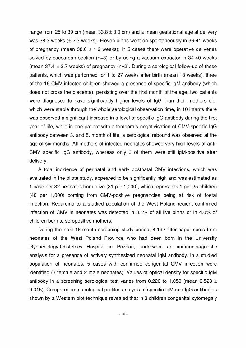
Congenital CMV infection was finally confirmed by (i) a detection of actively
synthesized specific IgM antibody (which is not able to cross the placenta) in the
peripheral blood of a neonate after the 7th
day of his life in traditional serological
techniques (ELISA) and/or by (ii) finding neonatal anti-CMV IgG antibody of different
antigenic specificity or produced in a higher concentration than maternal
immunoglobulins and/or actively synthesized neonatal IgM antibody shown by
comparative analysis of the neonate and the mother sera using the Western blot
technique (Blot CMV IgM i Blot CMV IgG, Test-Line, Brno, Czech Republic). Analysis
of immunological profiles of CMV-specific antibodies in serum samples of the
mothers of congenitally infected newborns was very useful to differentiate between a
primary CMV infection acquired during pregnancy and a secondary infection resulting
from a reinfection or a reactivation of a latent infection. Detection of specific IgM
antibodies against recent viral antigens of 52, 65 or 28 kDa and a lack of specific IgG
against a late CMV antygen of 150 kDa with a detection of some other bands of
specific IgG antibodies were typical for a primary CMV infection. On the other hand,
detection of a various combination of specific IgM antibodies with a whole spectrum
of intensive bands of anti-CMV IgG antibodies was specific for a recurrent maternal
infection during pregnancy (Fig. 2).
3. Clinical assessment.
In the neonates with confirmed CMV infections a paediatric examination,
including estimation of a clinical course, a degree of a development of the disease,

- 9 -
and an intensity of the infection was performed. Clinical examination with an
assessment of growth and psychomotor development was repeated regularly every
2 months during the first year of life, and then every 4 months. Ophthalmic
assessment with eye fundus examination using direct and indirect ophthalmoscopy
was firstly done before the end of the neonatal period, and then in 3 months
intervals. Transfontanel ultrasonography of the head was repeated every 2 months,
usually up to the end of the first year of life. Skull radiography in sutural and sagittal
projections and the evoqued hearing potentials done in the neonatal period were
completed with the computerized tomography scan of the brain between the first and
the 3rd
year of life [12]. Serological follow-up testing for CMV-specific IgM and IgG
antibodies was recommended every 4 months during the first 2 years of life, and
then twice a year in older children.
RESULTS
An evaluation of a current seropositivity of CMV infection in a studied population
was performed based on analysis of a total level of specific IgA, IgM and IgG
antibodies in 513 successively born neonates from the West Poland Province.
Seroprevalence of CMV in a population of neonates born alive and in their mothers
appeared to be very high and reached 78.6% (403 seropositive Guthrie cards).
Seventy-four of 513 patients (14.4%) with the highest titres of specific anti-CMV
immunoglobulins underwent a further clinical and serological observation; 16 patients
(21.6%) were finally confirmed to have perinatal or postnatal CMV infection with a
mild or asymptomatic course (male to female ratio: 1:1). The results of the filter-
paper screening test which detected specific IgA, IgM, and IgG antibodies in 16
neonates infected with CMV are presented in Table I. One pair of monozygotic twins,
who were born prematurely, revealed a very similar clinical pattern and an
immunological profile of specific antibodies (patients No. 3 and 4, Table I). Most
children infected with CMV did not present any clinical symptoms at birth (n=10); only
in 6 cases some reversible signs of prematurity, intrauterine hypotrophy and/or
adaptative breathing disorders were observed. The number of premature births
made 1/4 of all CMV infected cases. Birth weight of the 16 CMV infected neonates
was 1,000 to 3,870 g (mean 3,170.0 ± 698.9 g), head circumference oscillated in a
- 10 -
range from 25 to 39 cm (mean 33.8 ± 3.0 cm) and a mean gestational age at delivery
was 38.3 weeks (± 2.3 weeks). Eleven births went on spontaneously in 36-41 weeks
of pregnancy (mean 38.6 ± 1.9 weeks); in 5 cases there were operative deliveries
solved by caesarean section (n=3) or by using a vacuum extractor in 34-40 weeks
(mean 37.4 ± 2.7 weeks) of pregnancy (n=2). During a serological follow-up of these
patients, which was performed for 1 to 27 weeks after birth (mean 18 weeks), three
of the 16 CMV infected children showed a presence of specific IgM antibody (which
does not cross the placenta), persisting over the first month of the age, two patients
were diagnosed to have significantly higher levels of IgG than their mothers did,
which were stable through the whole serological observation time, in 10 infants there
was observed a significant increase in a level of specific IgG antibody during the first
year of life, while in one patient with a temporary negativisation of CMV-specific IgG
antibody between 3. and 5. month of life, a serological rebound was observed at the
age of six months. All mothers of infected neonates showed very high levels of anti-
CMV specific IgG antibody, whereas only 3 of them were still IgM-positive after
delivery.
A total incidence of perinatal and early postnatal CMV infections, which was
evaluated in the pilote study, appeared to be significantly high and was estimated as
1 case per 32 neonates born alive (31 per 1,000), which represents 1 per 25 children
(40 per 1,000) coming from CMV-positive pregnancies being at risk of foetal
infection. Regarding to a studied population of the West Poland region, confirmed
infection of CMV in neonates was detected in 3.1% of all live births or in 4.0% of
children born to seropositive mothers.
During the next 16-month screening study period, 4,192 filter-paper spots from
neonates of the West Poland Province who had been born in the University
Gynaecology-Obstetrics Hospital in Poznan, underwent an immunodiagnostic
analysis for a presence of actively synthesized neonatal IgM antibody. In a studied
population of neonates, 5 cases with confirmed congenital CMV infection were
identified (3 female and 2 male neonates). Values of optical density for specific IgM
antibody in a screening serological test varies from 0.226 to 1.050 (mean 0.523 ±
0.315). Compared immunological profiles analysis of specific IgM and IgG antibodies
shown by a Western blot technique revealed that in 3 children congenital cytomegaly

- 11 -
was a consequence of a reactivation of CMV infection that had been undergone
previously, while in the remaining two neonates the congenital disease was a sequel
of a primary viral infection acquired by a child’s mother during her pregnancy (Fig. 2).
Furthermore, 38 filter-paper samples collected from neonates in the first days of life
during their stay at hospital wards required re-testing for verifying analysis because
of doubtful results of the screening IgM test (0.91%).
Data referring to delivery and some clinical features of the 5 neonates with
confirmed congenital CMV infection are shown in Table II. In three children, clinically
overt form of CMV infection was diagnosed (characteristics of preterm birth and/or
small gestational age, jaundice, thrombocytopenic purpura, pneumonia, neurological
disorders) but in the remaining two children – only subclinical CMV infection
recognized by a specific antibody response to the virus was observed.
As a number of foetal losses related to CMV remains unknown, the incidence of
congenital CMV infection was estimated as 1 case per 838 liveborn neonates (1.2
per 1,000), which is 1 per 659 deliveries of seropositive pregnant women being at
risk of infection (1.5 per 1,000), with a reference to a current level of seroprevalence
in a pregnant women population from the Poznan Province. Congenital CMV
infection was finally confirmed by showing actively synthesized IgM antibody and/or
by a presence of specific IgG synthesized in a higher concentration than
immunoglobulins in a mother’s peripheral blood, using compared immunological
profiles analysis of the neonate and the mother sera by a referal immunoblotting
technique (Fig. 2).
DISCUSSION
Cytomegaly remains still the most frequently occurring congenital disease of a
viral origin, which can cause hearing impairment or psychomotor retardation. Majority
of neonates with congenital CMV infection do not show any clinical signs at birth and
only 10% of patients have some clinical symptoms of a different degree of intensity
(low birth weight, microcephaly, intracranial calcifications, retinochoroiditis,
hepatosplenomegaly, petechie, skin rash) or they present some discrete
abnormalities in laboratory tests alone (hyperbilirubinaemia, thrombocytopenia,
increased activity of liver enzymes or acute phase proteins). The clinical symptoms

- 12 -
observed in a course of cytomegaloviral disease during the neonatal period are not
specific and can also occur in many other prenatally acquired infections, which are
described by a TORCH acronym, as well as in some genetic disorders. Diagnostic
tests confirming or excluding congenital CMV infection should be performed during
the first 3 weeks of life to make a differential diagnosis between the most severe
intrauterine infection and perinatal infection or infection acquired in the early
postnatal period. Neonatal screeening tests which use peripheral blood samples
collected on filter-papers during the first days of life fulfil the expected criteria
completely.
A diagnostic sensitivity of commercially available serological techniques for a
diagnosis of CMV infection differs considerably regarding to an individual experience
of a laboratory performing an analysis and an applied immunological test, and is
usually not more than 20-75 [1]. That is the reason why studies on improving
performance of immunodiagnostic methods, which serve an early detection of pre-
and perinatal CMV infections are actually gaining bigger and bigger importance. The
first serological screening programme for neonates from the Poznan area was based
on a non-commercial test elaborated in the department, which was able to detect not
only a classical pair of IgM and IgG antibodies but also a presence of anti-CMV IgA
immunoglobulin. This original technique for a combined detection of 3 classes of
specific anti-CMV antibodies with very high diagnostic sensitivity, based on a single
blood spot determination procedure, has not been used in the world yet. Detecting
specific IgM antibody against CMV in the peripheral blood absorbed on neonatal
filter-papers appeared to be similarly innovatory. For a detection of prenatal CMV
infections some routine microbiological techniques of a cell culture of the virus from
saliva or urine, collected in the first day of life of a neonate, are usually performed
[13-16]. Blood samples absorbed on filter-papers have only been used in some
populational studies of newborns in Italy and Brazil to detect the specific
immunological response against CMV or fragments of nucleic acids of the virus [17-
20]. Detection of cytomegalovirus DNA extracted from neonatal filter-paper cards
collected at birth was being considered as a competitive method to serological
neonatal screening but it seems to play a bigger role in individual patients at risk of
infection, like among newborns with sensorineural hearing loss, retinal lesions and

- 13 -
neurological impairment or in cases with some congenital malformations, more than
in screening populational tests [19, 20]. Most of the tests for congenital CMV
performed in neonates are considered for selected cases, who are strongly
suspected of having infection from a TORCH group, so the mass nation-wide or
regional screening programmes are performed sporadically. In Finland, a screening
test for CMV was performed in neonates who were born prematurely before 34
weeks of gestation. In this selected group, the authors got a high percentage of
serologically confirmed congenital CMV infection which reached 4.8% [14]. Similarly,
Barbi and colleagues (1998) reported that a prevalence of congenital CMV infection
was 10 times higher in neonates from a risk group presenting some symptoms of
intrauterine infection (5%), than in a total group of children who were born
successively (0.47%). Molecular biology techniques were recommended to make a
final confirmation of suspected cases of congenital CMV infection, detected by a
traditional culture of the virus isolated from saliva [18]. Moreover, analysis of specific
IgM antibody in eluates from neonatal Guthrie cards by ELISA seemed to have
diagnostic sensitivity and specificity comparable to this, which is shown by a PCR
technique adapted to a detection of DNA in the blood absorbed on filter-papers [21].
A rational decision about a necessity of embracing a whole population of
neonates by screening tests should be preceded by epidemiological analysis of the
incidence of a particular disease with a congenital etiology and its potential risk on
the evaluated geographical region to select the most optimal preventive strategy for
a particular area of the word. That is a reason why the neonatal screening
programme, as an effective method for a prevention of clinically overt congenital
cytomegaly was decided to be implemented in the West Poland Province in a high
seroprevalence population of women in a procreative age with a small risk of primary
CMV infections during pregnancy.
On the basis of the pilote seroepidemiological study performed by the Poznan
Centre in a sample of 513 neonates from the West Poland Province, we have found
a high incidence of positive levels of specific anti-CMV antibodies in a population of
women who give birth that was 78.6%. The Poznan area was described as a region
of a high seropositivity for CMV in pregnant women and a significant risk of
transplacental transmission of the infection to a foetus. So there was a justifiable

- 14 -
assumption, that a risk of congenital CMV infection in our country is proportionally
much higher than it had been suspected to be before, based on sporadically noted
severe cases of symptomatic cytomegaly. The current incidence of congenital CMV
infection, evaluated on the representative number over 4,000 neonates from the
Poznan area as 1 per 838 children born alive (1.2 per 1,000) finally confirmed these
suggestions. Among 5 children who were prenatally infected with CMV and
diagnosed using a filter-paper test detecting specific IgM antibody, only two were
clinically suspected to have TORCH infection. The remaining 3 neonates were only
identified on the basis of a serological IgM screening test, despite they were not
suspected to have a congenital disease while being at neonatal wards.
An acquired form of CMV infection in pregnant women is usually asymptomatic
in more than 90% of postnatally infected cases, that is why it can be only detected
accidentally on the basis of a documented seroconversion of specific IgG antibody. A
symptomatic form of acquired primary cytomegaly is rare, and it usually resembles
mononucleosis-like syndrome with fever, lymphadenopathy, malaise, headache,
muscle pain and features of upper respiratory tract infection. Because a past CMV
infection does not leave a stable persistent immunity, just like most of other
infectious diseases or viral infections, in women with a positive level of specific IgG
antibody there is a potential risk of a secondary reactivation of the disease with a
further development of congenital infection in a foetus or a neonate. These patients
require regularly serological testing for specific antibodies during their pregnancies
and a more intense clinical observation by obstetricians. In the presented study,
among 5 congenitally infected newborns, transplacental transmission of the virus
during a late reactivation of the previously acquired asymptomatic maternal infection
was usually detected (60%), and the primary CMV infection during pregnancy was
less frequently diagnosed.
A differentiation between congenital CMV infection and the infection acquired in
the early postnatal period can create many diagnostic difficulties. So, it is very
advisable to keep children born to IgM-positive mothers under regular virological,
serological, and molecular control during the first year of life. Identification of the 16
neonates with very high levels of anti-CMV antibodies, who were born to seropositive
mothers, suggests rather subclinical perinatal infection or that acquired in the early

- 15 -
postnatal period than congenital infection. A spontaneous delivery and/or breast-
feeding in most of the observed cases (69%), were significantly promoted this way of
the viral transmission. Jim et al. reported that a high level of specific IgG antibody
against CMV in a pregnant woman is thought to be a main risk factor of developing
cytomegaly in the neonatal or infant’s period [22]. According to Miron and co-authors
(2005), among children with small birth weight born to CMV-positive mothers, 5.7%
of them had been infected between 3. and 7. week of life [23]. Similar study
conducted at the University in Toronto showed that 6.2% of children born to
seropositive mothers got infected by CMV in the age of 7-11 weeks [24]. Among
significant risk factors promoting postnatal CMV infection there were mentioned:
spontaneous delivery, pregnancy terminated before 34 weeks of gestational age,
and long-term or begun in the neonatal period breast-feeding [25]. Schanler (2005)
described that postnatal infection with CMV is typical for premature neonates of
about 1. month of the age and usually has a form of latent infection, while
symptomatic cytomegaloviral disease, which resembles a severe generalized form
imitating sepsis occurs sporadically [26].
Cytomegaly should be more often considered in a differential diagnosis of
congenital infections which occur in the neonatal period and in the early infancy. The
lack of typical clinical symptoms at birth is not an efficient argument for excluding
neither congenital disorders nor TORCH infections. In an absence of the persistent
life time immunity after CMV infection acquired before pregnancy, and a significant
risk of a secondary infection or a reactivation, and a former lack of immunization
programmes as well as safe antiviral treatment for pregnant women, a combination
of the serological screening programme for congenital CMV infection with other
neonatal tests performed at birth, like phenylketonuria, congenital hypothyroidism
and toxoplasmosis, can become a valuable method of choice for a serological
detection of this severe disease in the early postnatal period.
CONCLUSIONS
1. The incidence of congenital and perinatal CMV infections in the West Poland
Province appeared to be significantly much higher than it has been suspected

- 16 -
before on a basis of registered by the regional sanitary-epidemiological units
symptomatic cases of the cytomegalovirus disease.
2. Neonates born to seropositive mothers with high levels of CMV-specific
antibodies at delivery require a regular microbiological and serological follow-up
during the first year of life, because of a significant risk of perinatal or postnatal
infections with CMV.
3. Implementation of mass serological testing for congenital CMV infection,
combined with other obligatory neonatal tests for metabolic errors, congenital
malformations and endocrine disorders seems to be a valuable third line
prophylactic strategy to prevent a late development of the clinically overt
cytomegalovirus disease.
ACKNOWLEDGEMENTS
We wish to thank the Heads of the Neonatal and Obstetric Departments from the
district hospitals of the West Poland Province (Kalisz, Kolo, Ostrow Wielkopolski,
Poznan, Pila, Szamotuly, Srem, Trzcianka) as well as the midwifes and nurses for
their excellent help in the realization of this study.
The neonatal screening programme for congenital CMV infection was supported
by the Ministry of the Research, Science and High Education in Warsaw, Poland
(grant no. 3 PO5E 13022).

- 17 -
REFERENCES
1. Stagno S. Cytomegalovirus. In: Infectious Diseases of the Fetus and the
Newborn Infant. Remington J.S. and Klein J.O., eds. Philadelphia: W.B.
Saunders, 5th
ed., 2001: 389-424.
2. Demmler G.J. Infectious Diseases Society of America and Centers for Disease
Control. Summary of a workshop on surveillance for congenital cytomegalovirus
disease. Rev. Infect. Dis. 1991; 13: 315-329.
3. Stagno S., Pass R.F., Dworsky M.E. and al. Congenital cytomegalovirus
infection: the relative importance of primary and recurrent maternal infection. N.
Engl. J. Med. 1982; 306: 945-949.
4. Yow M.D., Williamson D.W., Leeds L.J. and al. Epidemiologic characteristics of
cytomegalovirus infection in mothers and their infants. Am. J. Obstet. Gynecol.
1988; 158: 1189-1195.
5. Ornay A. Fetal effects of primary and non-primary cytomegalovirus infection in
pregnancy: are we close to prevention? Isr. Med. Assoc. J. 2007; 9: 398-401.
6. Ergun U.G., Bakaris S., Ucmak H., Uzbek A. Fatal congenital cytomegalovirus
infection following recurrent maternal infection after a 7-year interval. Saudi
Med. J. 2007; 28: 264-267.
7. Dworsky M.E., Lakeman A.D., Stagno S. Cytomegalovirus transmission within a
family. Pediatr. Infect. Dis., 1984; 236-238.
8. Rhead W.J., Irons M. The call from the newborn screening laboratory:
frustration in the afternoon. Pediatr. Clin. N. Am. 2004; 51: 803-818.
9. Paul M., Petersen E., Pawlowski Z.S., Szczapa J. Neonatal screening for
congenital toxoplasmosis in the Poznan region of Poland by analysis of
Toxoplasma gondii - specific IgM antibodies eluted from filter-paper blood
spots. Pediatr. Infect. Dis. J. 2000; 19: 30-36.
10. Paul M., Petersen E., Szczapa J. Prevalence of congenital Toxoplasma gondii
infection among newborns from the Poznan region of Poland: validation of a
new combined enzyme immunoassay for Toxoplasma gondii - specific
immunoglobulin A and immunoglobulin M antibodies. J. Clin. Microbiol. 2001;
39: 1912-1916.

- 18 -
11. Paul M., Szczapa J., Wojsyk-Banaszak I., Jaworska A., Stefaniak J. Screening
of newborns for early postnatal detection of congenital CMV infection in the
Wielkopolska Province. [Polish]. Post. Neonat. 2006; 1:65-73.
12. Wojsyk-Banaszak I., Szczapa J., Paul M. Screening hearing assessment in
newborns with congenital CMV infections [Polish]. Post. Neonat. 2001; 1: 64-
66.
13. Walcarek K.B., Warren W., Smith R.J. and al. neonatal screening for congenital
cytomegalovirus infection by detection of virus in saliva. J. Infect. Dis. 1993;
167: 1433-1436.
14. Panhani S., Heinonen K.M. Screening for congenital cytomegalovirus infection
among preterm infants born before the 34th
gestational week in Finland. Scand.
J. Infect. Dis. 1994; 26: 375-378.
15. Casteels A., Naessens A., Gordts F. and al. Neonatal screening for congenital
cytomegalovirus infections. J. Piernat. Med. 1999; 27: 116-121.
16. Schlesinger Y., Reich D., Eidelman A.I. and al. Congenital cytomegalovirus
infection in Israel: screening in different subpopulations. Isr. Med. Assoc. J.
2005; 7: 237-240.
17. Neto E.C., Anele E., Rubim R. and al. High prevalence of congenital
toxoplasmosis in Brazil estimated in a 3-year prospective neonatal screening
study. Int. J. Epidemiol. 2000; 29: 941-947.
18. Barbi M., Binda S., Primache V., Clerici D. Congenital cytomegalovirus infection
in a northern Italian region. NEOCMV Group. Eur. J. Epidemiol. 1998; 14: 791-
796.
19. Barbi M., Binda S., Caroppo S., Primache V. Neonatal screening for congenital
cytomegalovirus infection and hearing loss. J. Clin. Virol. 2006; 35: 206-209.
20. Binda S., Caroppo S., Dido P. and al. Modification of CMV DNA detection from
dried blond spots for diagnosing congenital CMV infection. J. Clin. Virol. 2004;
30: 276-279.
21. Sivakumar R., Singh N., Singh S. Nested polymerase chain reaction in the
diagnosis of congenital cytomegalovirus infection. Indian J. Pediatr. 2001; 68:
1043-1046.

- 19 -
22. Jim W.T., Shu C.H., Chiu N.C. and al. Transmission of cytomegalovirus from
mothers to preterm infants by breast milk. Pediatr. Infect. Dis. J. 2004; 23: 848-
851.
23. Miron D., Brosilow S., Felszer K. and al. Incidence and clinical manifestations of
breast milk-acquired cytomegalovirus infection in low birth weight infants. J.
Perinatol. 2005; 25: 299-303.
24. Doctor S., Friedman S., Dunn M.S. and al. Cytomegalovirus transmission to
extremely low-birth weight infants through breast milk. Acta Paediatr. 2005; 94:
53-58.
25. Mussi-Pinhata M.M., Yamamoto A.Y., do Carmo Rego M.A. and al. Perinatal or
early-postnatal cytomegalovirus infection in preterm infants under 34 weeks
gestation born to CMV-seropositive mothers within a high-seroprevalence
population. J. Pediatr. 2004; 145: 685-688.
26. Schanler R.J. CMV acquisition in premature infants fed human milk: reason to
worry? J. Perinatol. 2005; 25: 297-298.

- 20 -
Fig. 1. A sample of the universal filter-paper card (Guthrie card) used for
serological screening programmes in newborns.
- 21 -
IgG IgM IgG IgM MW kDa
MN MN MN MN
Fig. 2. Comparative immunological profiles analysis of CMV-specific IgG and
IgM antibodies in serum samples of 2 mother/neonate pairs shown by a
Western blot technique: A) congenital CMV infection in a neonate caused by a
recurrent infection of the child’s mother during pregnancy; B) congenital CMV
infection in a neonate caused by a primary maternal infection. MW: molecular
weight, M: serum of a mother, N: serum of a neonate. Single arrows show anti-CMV
IgM antibodies actively synthesized by infected newborns. Double arrows show
typical bands of antibodies highly specific for CMV infection.
Table I. Results of combined serological screening for CMV-specific IgA, IgM
and IgG antibodies in 16 newborns with perinatal or early postnatal infection
with CMV.
A B
150
65
52
28

- 22 -
I.D. IgA/IgM/IgG ELISA [OD]
OD% [+] 70%
I.D.
IgA/IgM/IgG ELISA [OD]
OD% [+] 70%
1. 0.717 70.04 9. 0.68 114.44 2. 0.789 77.07 10. 0.592 80.65 3. 0.968 98.78 11. 0.563 76.70 4. 1.019 103.98 12. 0.675 91.96 5. 0.755 77.04 13. 0.555 75.61 6. 0.819 83.57 14. 0.631 85.97 7. 0.664 87.54 15. 0.669 82.80 8. 0.662 87.28 16. 0.737 91.21
All results were performed on filter-paper spots. OD: optical density; OD%: percent of
optical density calculated as the OD of the analyzed sample divided by the OD of the
positive control; [+]: positive result > 70%.
Table II. Clinical parametres of the 5 neonates born with congenital CMV
infection recognized by serological screening detecting specific IgM antibody
in the peripheral blood absorbed on filter-papers.
I.D.
Sex Gestational
age at birth
[weeks]
Birth weight
[g]
Delivery
mode
Agar
scores
[1/3/5 min.]
Clinical signs detected
in the neonatal period

- 23 -
1. F 41 4100 C 9/10/10 Adaptative respiratory
disorders
2. F 36 2060 C 1 Intrauterine hypotrophy
Pneumonia
Respiratory distress
Muscular hypotonia
Seizures
Anaemia
3. M 36 1940 C 4/8 Pneumonia
Respiratory distress
Petechiae
Intraventricular
haemorrhagy
Periventricular brain
leucomalation
Hypothyroidism
Thrombocytopenia
Jaundice
Hepatitis
Hypertransaminasaemia
4. M 28 1380 S 8/8/9 Respiratory distress
Jaundice
5. F 35 2990 F 9/10/10 Unilateral hydronephrosis
S: spontaneous delivery; F: forceps/vacuum extractor; C: cesarean section; F:
female; M: male.