the applications of gold nanoparticle-initialed ... · nano review open access the applications of...
TRANSCRIPT
Liu et al. Nanoscale Research Letters (2016) 11:460 DOI 10.1186/s11671-016-1686-0
NANO REVIEW Open Access
The Applications of Gold Nanoparticle-Initialed Chemiluminescence in BiomedicalDetection
Zezhong Liu*, Furong Zhao, Shandian Gao, Junjun Shao and Huiyun Chang*Abstract
Chemiluminescence technique as a novel detection method has gained much attention in recent years owning tothe merits of high sensitivity, wider linear ranges, and low background signal. Similarly, nanotechnology especiallyfor gold nanoparticles has emerged as detection tools due to their unique physical and chemical properties. Recently,it has become increasingly popular to couple gold nanoparticles with chemiluminescence technique in biologicalagents’ detection. In this review, we describe the superiority of both chemiluminescence and gold nanoparticles andconclude the different applications of gold nanoparticle-initialed chemiluminescence in biomedical detection.
Keywords: Chemiluminescence, Gold nanoparticles, Detection, Biomedicine
ReviewIntroductionDetection of biological agents plays an indispensable rolein biomedicine [1]. In clinical diagnosis, developinghighly sensitive and cost-effective detection methods isin high demand for the reason that some clinical sam-ples have very low concentration that usually cannot bedetected. Since the advent of radioimmunoassay (RIA)[2], the trace materials could be detected. From then,detection technologies have gained much attention anda few techniques were developed, including fluoroimmu-noassay (FIA) and enzyme immunoassay (EIA). The FIAand EIA were regularly used but sometimes cannot meetthe clinical demand due to their low sensitivities. TheRIA is characterized by great sensitivity but has somedrawbacks, including health hazard, short half-life, andenvironment contamination [3].In the late 1970s, chemiluminescence immunoassay
(CLIA) was first introduced [4]. The mechanism ofchemiluminescence (CL) is that some molecules can getthe energy from the chemical reaction and be excited tothe electronically excited state; the energy is disposed ofin the form of light along with the molecules return to
* Correspondence: [email protected]; [email protected] Key Laboratory of Veterinary Etiological Biology, NationalFoot-and-Mouth Diseases Reference Laboratory, Lanzhou Veterinary ResearchInstitute, Chinese Academy of Agricultural Sciences, Lanzhou 730046, China
© 2016 The Author(s). Open Access This articleInternational License (http://creativecommons.oreproduction in any medium, provided you givthe Creative Commons license, and indicate if
ground state [5]. It does not need photoluminescenceand the energy comes from chemical interaction, whichleads to low background signal [6, 7]. In addition, it alsohas some merits including ultrasensitivity, high signal-to-noise ratio, low cost, and wide linear dynamic ranges[8, 9]. All of those advantages make CL to be a powerfuldetection method.Nanobiotechnology is the product of biology and
nanotechnology, which develops rapidly and shows asignificant future promise [10]. Almost all the areas ofbiomedicine such as bioimaging [11, 12], drug delivery[13, 14], oncotherapy [15, 16], and clinical diagnosis [17]have the shadow of nanomaterials. Among the nanoma-terials including magnetic nanoparticles, quantum dots,graphene oxide, carbon nanotubes, and gold nanoparti-cles (AuNPs), the AuNP is the most widely used.Nanotechnology, especially for AuNPs, which offers a
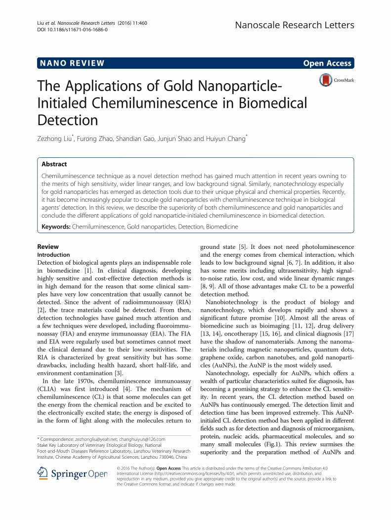
wealth of particular characteristics suited for diagnosis, hasbecoming a promising strategy to enhance the CL sensitiv-ity. In recent years, the CL detection method based onAuNPs has continuously emerged. The detection limit anddetection time has been improved extremely. This AuNP-initialed CL detection method has been applied in differentfields such as for detection and diagnosis of microorganism,protein, nucleic acids, pharmaceutical molecules, and somany small molecules (Fig.1). This review surmises thesuperiority and the preparation method of AuNPs and
is distributed under the terms of the Creative Commons Attribution 4.0rg/licenses/by/4.0/), which permits unrestricted use, distribution, ande appropriate credit to the original author(s) and the source, provide a link tochanges were made.
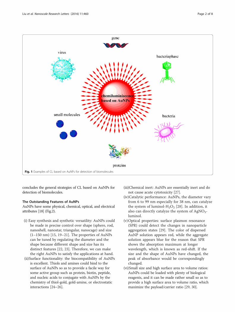
Fig. 1 Examples of CL based on AuNPs for detection of biomolecules
Liu et al. Nanoscale Research Letters (2016) 11:460 Page 2 of 8
concludes the general strategies of CL based on AuNPs fordetection of biomolecules.
The Outstanding Features of AuNPsAuNPs have some physical, chemical, optical, and electricalattributes [18] (Fig.2).
(i) Easy synthesis and synthetic versatility: AuNPs couldbe made in precise control over shape (sphere, rod,nanoshell, nanostar, triangular, nanocage) and size(1–150 nm) [15, 19–21]. The properties of AuNPscan be tuned by regulating the diameter and theshape because different shape and size has itsdistinct features [22, 23]. Therefore, we can makethe right AuNPs to satisfy the applications at hand.
(ii)Surface functionality: the biocompatibility of AuNPsis excellent. Thiols and amines could bind to thesurface of AuNPs so as to provide a facile way forsome active group such as protein, biotin, peptide,and nucleic acids to conjugate with AuNPs by thechemistry of thiol-gold, gold-amine, or electrostaticinteractions [24–26].
(iii)Chemical inert: AuNPs are essentially inert and donot cause acute cytotoxicity [27].
(iv)Catalytic performance: AuNPs, the diameter varyfrom 6 to 99 nm especially for 38 nm, can catalyzethe system of luminol-H2O2 [28]. In addition, italso can directly catalyze the system of AgNO3-luminol.
(v)Optical properties: surface plasmon resonance(SPR) could detect the changes in nanoparticleaggregation states [29]. The color of dispersedAuNP solution appears red, while the aggregatesolution appears blue for the reason that SPRshows the absorption maximum at longerwavelength, which is known as red-shift. If thesize and the shape of AuNPs have changed, thepeak of absorbance would be correspondinglychanged.
(vi)Small size and high surface area to volume ratios:AuNPs could be loaded with plenty of biologicalreagents, and it can be made rather small so as toprovide a high surface area to volume ratio, whichmaximize the payload/carrier ratio [29, 30].
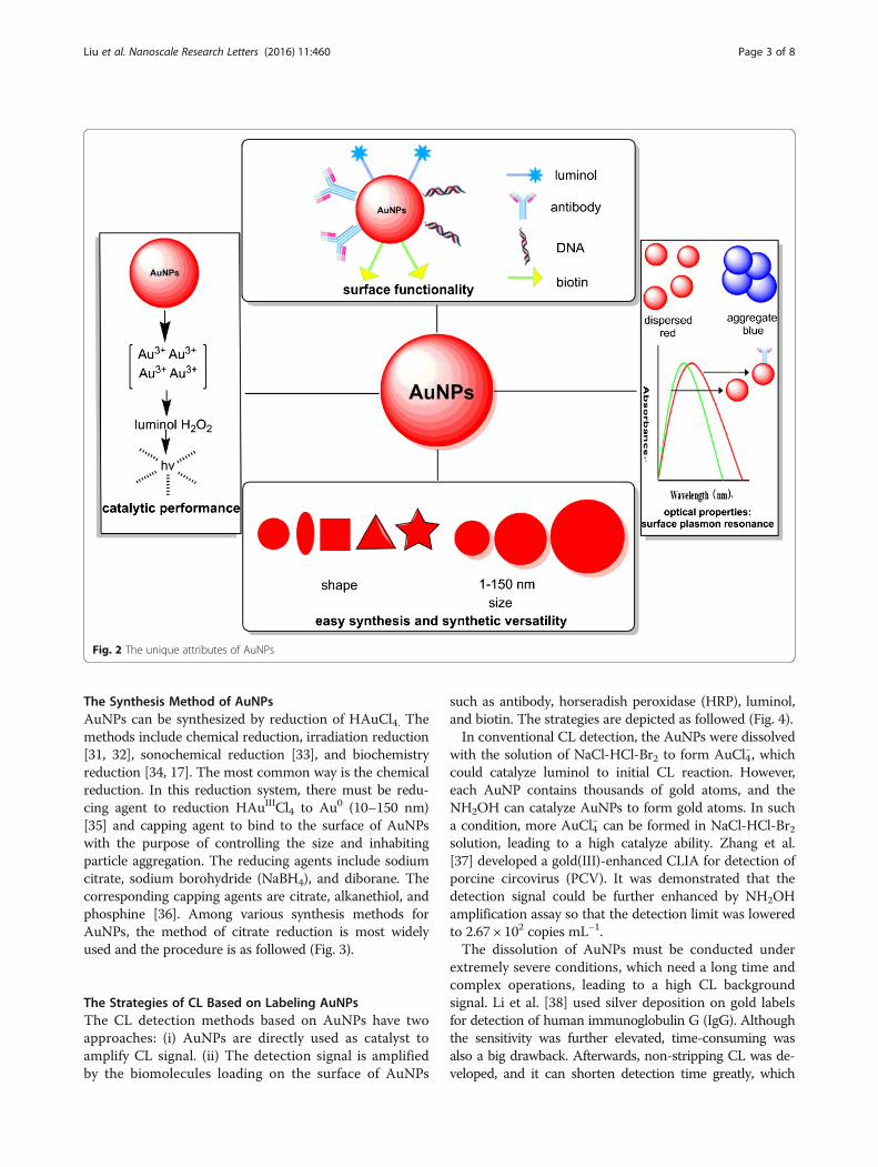
Fig. 2 The unique attributes of AuNPs
Liu et al. Nanoscale Research Letters (2016) 11:460 Page 3 of 8
The Synthesis Method of AuNPsAuNPs can be synthesized by reduction of HAuCl4. Themethods include chemical reduction, irradiation reduction[31, 32], sonochemical reduction [33], and biochemistryreduction [34, 17]. The most common way is the chemicalreduction. In this reduction system, there must be redu-cing agent to reduction HAuIIICl4 to Au0 (10–150 nm)[35] and capping agent to bind to the surface of AuNPswith the purpose of controlling the size and inhabitingparticle aggregation. The reducing agents include sodiumcitrate, sodium borohydride (NaBH4), and diborane. Thecorresponding capping agents are citrate, alkanethiol, andphosphine [36]. Among various synthesis methods forAuNPs, the method of citrate reduction is most widelyused and the procedure is as followed (Fig. 3).
The Strategies of CL Based on Labeling AuNPsThe CL detection methods based on AuNPs have twoapproaches: (i) AuNPs are directly used as catalyst toamplify CL signal. (ii) The detection signal is amplifiedby the biomolecules loading on the surface of AuNPs
such as antibody, horseradish peroxidase (HRP), luminol,and biotin. The strategies are depicted as followed (Fig. 4).In conventional CL detection, the AuNPs were dissolved
with the solution of NaCl-HCl-Br2 to form AuCl4−, which
could catalyze luminol to initial CL reaction. However,each AuNP contains thousands of gold atoms, and theNH2OH can catalyze AuNPs to form gold atoms. In sucha condition, more AuCl4
− can be formed in NaCl-HCl-Br2solution, leading to a high catalyze ability. Zhang et al.[37] developed a gold(III)-enhanced CLIA for detection ofporcine circovirus (PCV). It was demonstrated that thedetection signal could be further enhanced by NH2OHamplification assay so that the detection limit was loweredto 2.67 × 10² copies mL−1.The dissolution of AuNPs must be conducted under
extremely severe conditions, which need a long time andcomplex operations, leading to a high CL backgroundsignal. Li et al. [38] used silver deposition on gold labelsfor detection of human immunoglobulin G (IgG). Althoughthe sensitivity was further elevated, time-consuming wasalso a big drawback. Afterwards, non-stripping CL was de-veloped, and it can shorten detection time greatly, which
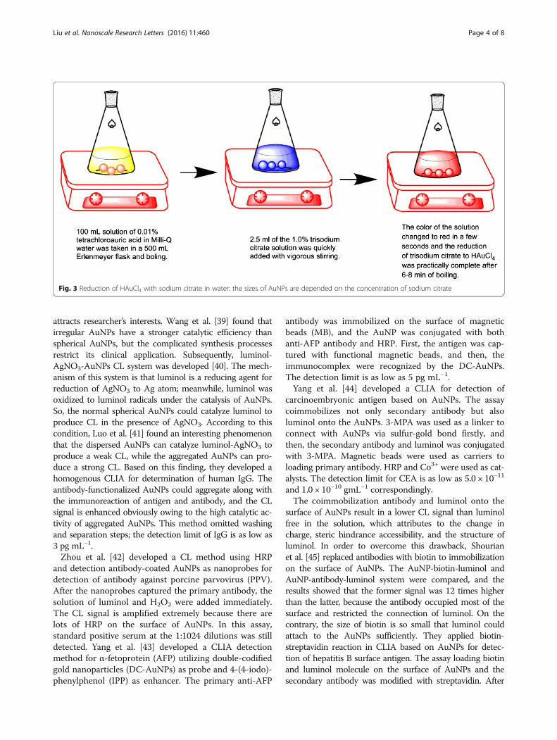
Fig. 3 Reduction of HAuCl4 with sodium citrate in water: the sizes of AuNPs are depended on the concentration of sodium citrate
Liu et al. Nanoscale Research Letters (2016) 11:460 Page 4 of 8
attracts researcher’s interests. Wang et al. [39] found thatirregular AuNPs have a stronger catalytic efficiency thanspherical AuNPs, but the complicated synthesis processesrestrict its clinical application. Subsequently, luminol-AgNO3-AuNPs CL system was developed [40]. The mech-anism of this system is that luminol is a reducing agent forreduction of AgNO3 to Ag atom; meanwhile, luminol wasoxidized to luminol radicals under the catalysis of AuNPs.So, the normal spherical AuNPs could catalyze luminol toproduce CL in the presence of AgNO3. According to thiscondition, Luo et al. [41] found an interesting phenomenonthat the dispersed AuNPs can catalyze luminol-AgNO3 toproduce a weak CL, while the aggregated AuNPs can pro-duce a strong CL. Based on this finding, they developed ahomogenous CLIA for determination of human IgG. Theantibody-functionalized AuNPs could aggregate along withthe immunoreaction of antigen and antibody, and the CLsignal is enhanced obviously owing to the high catalytic ac-tivity of aggregated AuNPs. This method omitted washingand separation steps; the detection limit of IgG is as low as3 pg mL−1.Zhou et al. [42] developed a CL method using HRP
and detection antibody-coated AuNPs as nanoprobes fordetection of antibody against porcine parvovirus (PPV).After the nanoprobes captured the primary antibody, thesolution of luminol and H2O2 were added immediately.The CL signal is amplified extremely because there arelots of HRP on the surface of AuNPs. In this assay,standard positive serum at the 1:1024 dilutions was stilldetected. Yang et al. [43] developed a CLIA detectionmethod for α-fetoprotein (AFP) utilizing double-codifiedgold nanoparticles (DC-AuNPs) as probe and 4-(4-iodo)-phenylphenol (IPP) as enhancer. The primary anti-AFP
antibody was immobilized on the surface of magneticbeads (MB), and the AuNP was conjugated with bothanti-AFP antibody and HRP. First, the antigen was cap-tured with functional magnetic beads, and then, theimmunocomplex were recognized by the DC-AuNPs.The detection limit is as low as 5 pg mL−1.Yang et al. [44] developed a CLIA for detection of
carcinoembryonic antigen based on AuNPs. The assaycoimmobilizes not only secondary antibody but alsoluminol onto the AuNPs. 3-MPA was used as a linker toconnect with AuNPs via sulfur-gold bond firstly, andthen, the secondary antibody and luminol was conjugatedwith 3-MPA. Magnetic beads were used as carriers toloading primary antibody. HRP and Co3+ were used as cat-alysts. The detection limit for CEA is as low as 5.0 × 10⁻¹¹
and 1.0 × 10⁻¹⁰ gmL−1 correspondingly.The coimmobilization antibody and luminol onto the
surface of AuNPs result in a lower CL signal than luminolfree in the solution, which attributes to the change incharge, steric hindrance accessibility, and the structure ofluminol. In order to overcome this drawback, Shourianet al. [45] replaced antibodies with biotin to immobilizationon the surface of AuNPs. The AuNP-biotin-luminol andAuNP-antibody-luminol system were compared, and theresults showed that the former signal was 12 times higherthan the latter, because the antibody occupied most of thesurface and restricted the connection of luminol. On thecontrary, the size of biotin is so small that luminol couldattach to the AuNPs sufficiently. They applied biotin-streptavidin reaction in CLIA based on AuNPs for detec-tion of hepatitis B surface antigen. The assay loading biotinand luminol molecule on the surface of AuNPs and thesecondary antibody was modified with streptavidin. After
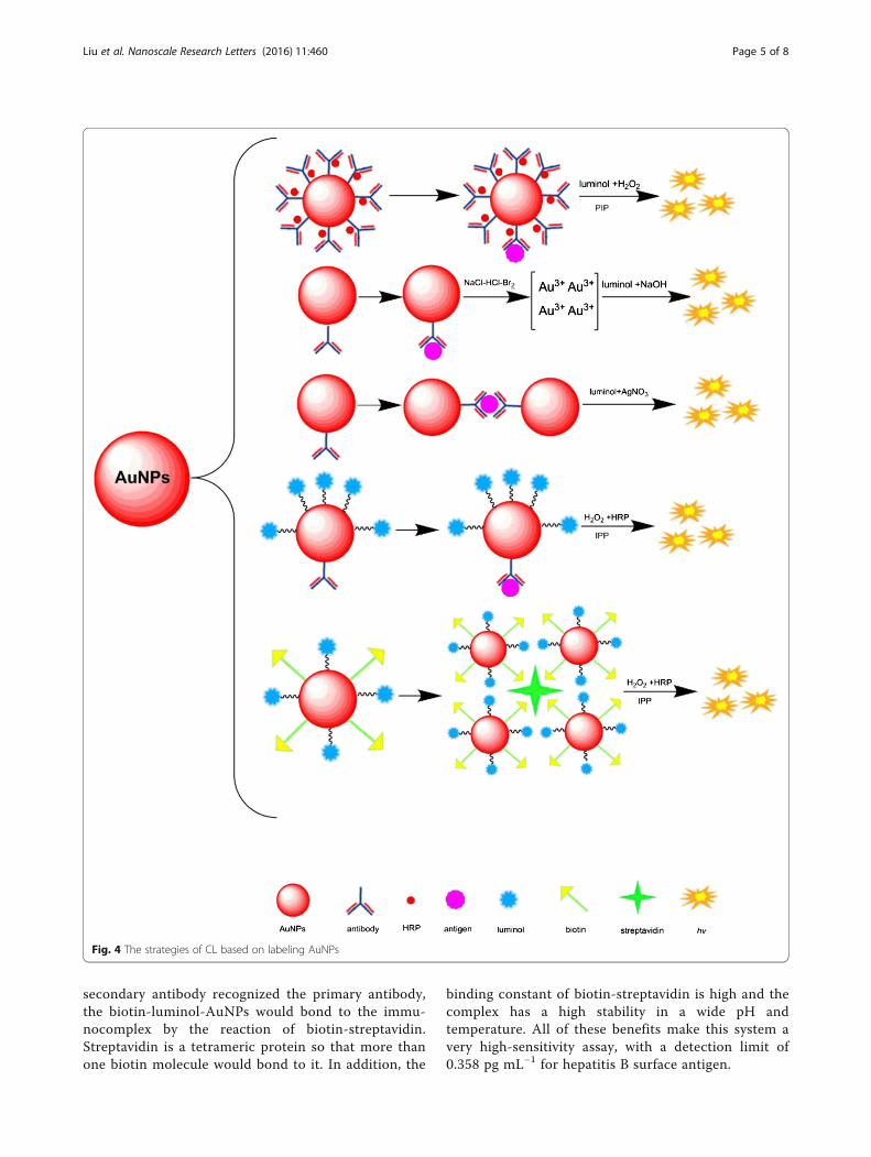
Fig. 4 The strategies of CL based on labeling AuNPs
Liu et al. Nanoscale Research Letters (2016) 11:460 Page 5 of 8
secondary antibody recognized the primary antibody,the biotin-luminol-AuNPs would bond to the immu-nocomplex by the reaction of biotin-streptavidin.Streptavidin is a tetrameric protein so that more thanone biotin molecule would bond to it. In addition, the
binding constant of biotin-streptavidin is high and thecomplex has a high stability in a wide pH andtemperature. All of these benefits make this system avery high-sensitivity assay, with a detection limit of0.358 pg mL−1 for hepatitis B surface antigen.
Liu et al. Nanoscale Research Letters (2016) 11:460 Page 6 of 8
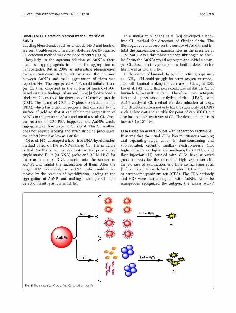
Label-Free CL Detection Method by the Catalytic ofAuNPsLabeling biomolecules such as antibody, HRP, and luminolare very troublesome. Therefore, label-free AuNP-initialedCL detection method was developed recently (Fig. 5).Regularly, in the aqueous solution of AuNPs, there
must be capping agents to inhibit the aggregation ofnanoparticles. But in 2004, an interesting phenomenonthat a certain concentration salt can screen the repulsionbetween AuNPs and make aggregation of them wasreported [46]. The aggregated AuNPs could initial a stron-ger CL than dispersed in the system of luminol-H2O2.Based on these findings, Islam and Kang [47] developed alabel-free CL method for detection of C-reactive protein(CRP). The ligand of CRP is O-phosphorylethanolamine(PEA), which has a distinct property that can stick to thesurface of gold so that it can inhibit the aggregation ofAuNPs in the presence of salt and initial a weak CL. Oncethe reaction of CRP-PEA happened, the AuNPs wouldaggregate and show a strong CL signal. This CL methoddoes not require labeling and strict stripping procedures;the detect limit is as low as 1.88 fM.Qi et al. [48] developed a label-free DNA hybridization
method based on the AuNP-initialed CL. The principleis that AuNPs could not aggregate in the presence ofsingle-strand DNA (ss-DNA) probe and 0.5 M NaCl forthe reason that ss-DNA absorb onto the surface ofAuNPs and inhibit the aggregation of them. After thetarget DNA was added, the ss-DNA probe would be re-moved by the reaction of hybridization, leading to theaggregation of AuNPs and making a stronger CL. Thedetection limit is as low as 1.1 fM.
Fig. 5 The strategies of label-free CL based on AuNPs
In a similar vein, Zhang et al. [49] developed a label-free CL method for detection of fibrillar fibrin. Thefibrinogen could absorb on the surface of AuNPs and in-hibit the aggregation of nanoparticles in the presence of1 M NaCl. After thrombins catalyze fibrinogen to fibril-lar fibrin, the AuNPs would aggregate and initial a stron-ger CL. Based on this principle, the limit of detection forfibrin was as low as 1 fM.In the system of luminol-H2O2, some active groups such
as −NH2, −SH could struggle for active oxygen intermedi-ates with luminol, making the decrease of CL signal [28].Liu et al. [50] found that L-cys could also inhibit the CL ofluminol-H2O2-AuNP system. Therefore, they integratelaminated paper-based analytica device (LPAD) withAuNP-catalyzed CL method for determination of L-cys.This detection system not only has the superiority of LAPDsuch as low cost and suitable for point of care (POC) butalso has the high sensitivity of CL. The detection limit is aslow as 8.2 × 10⁻¹⁰ M.
CLIA Based on AuNPs Couple with Separation TechniqueIt seems that the usual CLIA has multifarious washingand separating steps, which is time-consuming andsophisticated. Recently, capillary electrophoresis (CE),high-performance liquid chromatography (HPLC), andflow injection (FI) coupled with CLIA have attractedgreat interests for the merits of high separation effi-ciency, ease of automation, and time-saving. Jiang et al.[51] combined CE with AuNP-amplified CL to detectionof carcinoembryonic antigen (CEA). The CEA antibodyand HRP were also conjugated with AuNPs. After thenanoprobes recognized the antigen, the excess AuNP
Liu et al. Nanoscale Research Letters (2016) 11:460 Page 7 of 8
conjugates were separated by the CE, and the CL wasactivated by the p-iodophenol (PIP)-enhanced luminol-H2O2-HRP system. The detection process could befinished within 5 min, and the detection limit is as lowas 0.034 ng mL−1. Liu et al. [52] proposed a novelprotocol for detection of protein by means of aptamer-functionalized AuNPs and CE-CL. Taking thrombin asa model, they linked thrombin-binding aptamer to theAuNPs by the SH–Au covalent bond and blocked thespare binding side with blocker DNA. After CE sepa-rated, luminol and H2O2 were added. The detectionlimit is down to 13.5 fmol L−1.Li et al. [53] found that some reductive compounds
such as monoamine neurotransmitters and their metab-olites have an inhibitory effect on the luminol-AgNO3-AuNPs. These reductive compounds have a stronger re-ductive ability than luminol, and competed with lumi-nol for AgNO3, making that the generation of luminolradicals is decreased and leading to a weak CL signal.By taking advantage of this phenomenon, they devel-oped a CL method for simultaneous determination ofmonoamine neurotransmitters and their metabolites ina mouse brain microdialysate. HPLC was applied in thissystem, and the method was simple, sensitive, and fast.Hao and Ma [54] combined FI system with AuNP-
enhanced CL to detection of carcinoembryonic antigen(CEA). They loaded the secondary antibody onto thesurface of AuNPs and utilized the method of dissolvingAuNPs into HNO3-HCl solution. The detection limit isas low as 20 pg mL−1.
ConclusionsCL as the newest labeling technique has been widelyapplied in biomedical diagnosis during the two decade.Nanodiagnostic techniques especially for AuNPs havegained much attention by the researchers. Couplingboth techniques to detection of biological agents is atrend in the last several years. Continuous efforts forlabeling biomarkers (e.g. proteins, genes, and chemilu-minescent agent) onto the surface of AuNPs are madeso as to improve the sensitivity of CL. The labelingstrategies are improving constantly, and the detectionlimit is becoming lower. In addition, the separationtechnique and the label-free strategies are also devel-oped quickly because it can simplify the operation andsave more time. All of these efforts are to developrapid, sensitive, automatic, and point of care detectionmethods. Currently, some new and readily contagiousviruses such as Zika virus, Ebola virus, and avian influ-enza virus, which could pose a great disaster for hu-man, have not been detected in a quick and sensitiveway. If the CL based on AuNP detection method couldbe developed for those virus detection, the significancefor public health would be important. In the next
20 years, the CL based on AuNP detection methodwould become the most widely used detection methodin clinical medicine and clinical veterinary science.
AcknowledgementsThe authors thank the Stake Key Laboratory of Veterinary Etiological Biology,National Foot-and-Mouth Diseases Reference Laboratory, Lanzhou VeterinaryResearch Institute, Chinese Academy of Agricultural Sciences.
Authors’ ContributionsThe paper was written by ZZL. All revisions were discussed by FRZ, SDG, JJS,and HYC. All authors read and approved the final manuscript.
Competing InterestsThe authors declare that they have no competing interests.
Received: 3 July 2016 Accepted: 11 October 2016
References1. Saha K, Agasti SS, Kim C, Li X, Rotello VM (2012) Gold nanoparticles in chemical
and biological sensing. Chem Rev 112:2739–27792. Berson SA, Yallow RS (1961) Immunochemical distinction between insulins
with identical amino-acid sequences. Nature 191:1392–13933. Abo El Sooud K (2003) Influence of albendazole on the disposition kinetics
and milk antimicrobial equivalent activity of enrofloxacin in lactating goats.Pharmacol Res 48:389–395
4. Halmann M, Velan B, Sery T (1977) Rapid identification and quantitation ofsmall numbers of microorganisms by a chemiluminescent immunoreaction.Appl Environ Microbiol 34:473–477
5. Li N, Liu DQ, Cui H (2014) Metal-nanoparticle-involved chemiluminescenceand its applications in bioassays. Anal Bioanal Chem 406:5561–5571
6. Sorouraddin MH, Iranifam M, Imani-Nabiyyi A (2009) A novel captoprilchemiluminescence system for determination of copper(ii) in human hairand cereal flours. J Fluoresc 19:575–581
7. Dodeigne C, Thunus L, Lejeune R (2000) Chemiluminescence as a diagnostictool. A review Talanta 51:415–439
8. Wang C, Wu J, Zong C, Xu J, Ju HX (2012) Chemiluminescent immunoassayand its applications. Chinese J Anal Chem 40:3–10
9. Mirasoli M, Michelini E (2014) Analytical bioluminescence and chemiluminescence.Anal Bioanal Chem 406:5529–5530
10. Nie L, Liu F, Ma P, Xiao X (2014) Applications of gold nanoparticles inoptical biosensors. J Biomed Nanotechnol 10:2700–2721
11. Brambilla D, Nicolas J, Le Droumaguet B, Andrieux K, Marsaud V, CouraudPO, Couvreur P (2010) Design of fluorescently tagged poly(alkylcyanoacrylate) nanoparticles for human brain endothelial cell imaging.Chem Commun 46:2602–2604
12. Craig GA, Allen PJ, Mason MD (2010) Synthesis, characterization, andfunctionalization of gold nanoparticles for cancer imaging. Methods MolBiol 624:177–193
13. Parveen S, Misra R, Sahoo SK (2012) Nanoparticles: a boon to drug delivery,therapeutics, diagnostics and imaging. Nanomed-Nanotechnol 8:147–166
14. Rana S, Bajaj A, Mout R, Rotello VM (2012) Monolayer coated goldnanoparticles for delivery applications. Adv Drug Deliv Rev 64:200–216
15. Her S, Jaffray DA, Allen C (2015) Gold nanoparticles for applications incancer radiotherapy: mechanisms and recent advancements. Adv DrugDeliv Rev. doi:10.1016/j.addr.2015.12.012
16. Chang MY, Shiau AL, Chen YH, Chang CJ, Chen HH, Wu CL (2008) Increasedapoptotic potential and dose-enhancing effect of gold nanoparticles incombination with single-dose clinical electron beams on tumor-bearingmice. Cancer Sci 99:1479–1484
17. Azzazy HM, Mansour MM, Samir TM, Franco R (2012) Gold nanoparticles inthe clinical laboratory: principles of preparation and applications. Clin ChemLab Med 50:193–209
18. Rosi NL, Mirkin CA (2005) Nanostructures in biodiagnostics. Chem Rev 105:1547–1562
19. Zhao PX, Li N, Astruc D (2013) State of the art in gold nanoparticle synthesis.Coord Chem Rev 257:638–665
20. Huang X, Neretina S, El-Sayed MA (2009) Gold nanorods: from synthesis andproperties to biological and biomedical applications. Adv Mater 21:4880–4910
Liu et al. Nanoscale Research Letters (2016) 11:460 Page 8 of 8
21. Lee J, Chatterjee DK, Lee MH, Krishnan S (2014) Gold nanoparticles in breastcancer treatment: promise and potential pitfalls. Cancer Lett 347:46–53
22. Zhang X (2015) Gold nanoparticles: recent advances in the biomedicalapplications. Cell Biochem Biophys 72:771–775
23. Chen S, Ingram RS, Hostetler MJ, Pietron JJ, Murray RW, Schaaff TG, KhouryJT, Alvarez MM, Whetten RL (1998) Gold nanoelectrodes of varied size:transition to molecule-like charging. Science 280:2098–2101
24. Zayats M, Baron R, Popov I, Willner I (2005) Biocatalytic growth of Aunanoparticles: from mechanistic aspects to biosensors design. Nano Lett 5:21–25
25. Radwan SH, Azzazy HM (2009) Gold nanoparticles for molecular diagnostics.Expert Rev Mol Diagn 9:511–524
26. Baptista P, Pereira E, Eaton P, Doria G, Miranda A, Gomes I, Quaresma P,Franco R (2008) Gold nanoparticles for the development of clinicaldiagnosis methods. Anal Bioanal Chem 391:943–950
27. Connor EE, Mwamuka J, Gole A, Murphy CJ, Wyatt MD (2005) Goldnanoparticles are taken up by human cells but do not cause acutecytotoxicity. Small 1:325–327
28. Zhang ZF, Cui H, Lai CZ, Liu LJ (2005) Gold nanoparticle-catalyzed luminolchemiluminescence and its analytical applications. Anal Chem 77:3324–3329
29. Daniel MC, Astruc D (2004) Gold nanoparticles: assembly, supramolecularchemistry, quantum-size-related properties, and applications towardbiology, catalysis, and nanotechnology. Chem Rev 104:293–346
30. Upadhyayula VK (2012) Functionalized gold nanoparticle supported sensorymechanisms applied in detection of chemical and biological threat agents:a review. Anal Chim Acta 715:1–18
31. Gachard E, Remita H, Khatouri J, Keita B, Nadjo L, Belloni J (1998) Radiation-induced and chemical formation of gold clusters. New J Chem 22:1257–1265
32. Esumi K, Matsuhisa K, Torigoe K (1995) Preparation of rodlike gold particles byUV irradiation using cationic micelles as a template. Langmuir 11:3285–3287
33. Mizukoshi Y, Fujimoto T, Nagata Y, Oshima R, Maeda Y (2000) Characterizationand catalytic activity of core-shell structured gold/palladium bimetallicnanoparticles synthesized by the sonochemical method. J Phys Chem B 104:6028–6032
34. He SY, Guo ZR, Zhang Y, Zhang S, Gu JWN (2007) Biosynthesis of goldnanoparticles using the bacteria rhodopseudomonas capsulata. Mater Lett61:3984–3987
35. Frens G (1973) Controlled nucleation for regulation of particle-size inmonodisperse gold suspensions. Nature Phys Sci 241:20–22
36. Ghosh P, Han G, De M, Kim CK, Rotello VM (2008) Gold nanoparticles indelivery applications. Adv Drug Deliv Rev 60:1307–1315
37. Zhang H, Li W, Sheng Z, Han H, He Q (2010) Ultrasensitive detection ofporcine circovirus type 2 using gold(iii) enhanced chemiluminescenceimmunoassay. Analyst 135:1680–1685
38. Li ZP, Liu CH, Fan YS, Wang YC, Duan XR (2006) A chemiluminescentmetalloimmunoassay based on silver deposition on colloidal gold labels.Anal Biochem 359:247–252
39. Wang ZP, Hu JQ, Jin Y, Yao X, Li JH (2006) In situ amplified chemiluminescentdetection of DNA and immunoassay of IgG using special-shaped goldnanoparticles as label. Clin Chem 52:1958–1961
40. Cui H, Guo JZ, Li N, Liu LJ (2008) Gold nanoparticle triggered chemilumineseencebetween luminol and AgNO3. J Phys Chem C 112:11319–11323
41. Luo J, Cui X, Liu W, Li B (2014) Highly sensitive homogenous chemiluminescenceimmunoassay using gold nanoparticles as label. Spectrochim Acta A Mol BiomolSpectrosc 131:243–248
42. Zhou Y, Zhou T, Zhou R, Hu Y (2014) Chemiluminescence immunoassay forthe rapid and sensitive detection of antibody against porcine parvovirus byusing horseradish peroxidase/detection antibody-coated gold nanoparticlesas nanoprobes. Luminescence 29:338–343
43. Yang XY, Guo YS, Bi S, Zhang SS (2009) Ultrasensitive enhancedchemiluminescence enzyme immunoassay for the determination ofalpha-fetoprotein amplified by double-codified gold nanoparticleslabels. Biosens Bioelectron 24:2707–2711
44. Yang XY, Guo YS, Wang AG (2010) Luminol/antibody labeled goldnanoparticles for chemiluminescence immunoassay of carcinoembryonicantigen. Anal Chim Acta 666:91–96
45. Shourian M, Ghourchian H, Boutorabi M (2015) Ultra-sensitive immunosensorfor detection of hepatitis b surface antigen using multi-functionalized goldnanoparticles. Anal Chim Acta 895:1–11
46. Li HX, Rothberg LJ (2004) DNA sequence detection using selectivefluorescence quenching of tagged oligonucleotide probes by goldnanoparticles. Anal Chem 76:5414–5417
47. Islam MS, Kang SH (2011) Chemiluminescence detection of label-free C-reactiveprotein based on catalytic activity of gold nanoparticles. Talanta 84:752–758
48. Qi YY, Li BX, Zhang ZJ (2009) Label-free and homogeneous DNAhybridization detection using gold nanoparticles-based chemiluminescencesystem. Biosens Bioelectron 24:3581–3586
49. Zhang YF, Liu JF, Liu T, Li HB, Xue QW, Li R, Wang L, Yue QL, Wang SH (2016)Label-free, sensitivity detection of fibrillar fibrin using gold nanoparticle-basedchemiluminescence system. Biosens Bioelectron 77:111–115
50. Liu W, Luo J, Guo YM, Kou J, Li BX, Zhang ZJ (2014) Nanoparticle coatedpaper-based chemiluminescence device for the determination of l-cysteine.Talanta 120:336–341
51. Jiang J, Zhao SL, Huang Y, Qin GX, Ye FG (2013) Highly sensitive immunoassay ofcarcinoembryonic antigen by capillary electrophoresis with gold nanoparticlesamplified chemiluminescence detection. J Chromatogr 1282:161–166
52. Liu YM, Liu YY, Zhou M, Huang KJ, Cao JT, Wang H, Chen YH (2014)Chemiluminescence detection of protein in capillary electrophoresis usingaptamer-functionalized gold nanoparticles as biosensing platform. J Chromatogr1340:128–133
53. Li N, Guo JZ, Liu B, Yu YQ, Cui H, Mao LQ, Lin YQ (2009) Determination ofmonoamine neurotransmitters and their metabolites in a mouse brainmicrodialysate by coupling high-performance liquid chromatography withgold nanoparticle-initiated chemiluminescence. Anal Chim Acta 645:48–55
54. Hao MJ, Ma ZF (2012) An ultrasensitive chemiluminescence biosensor forcarcinoembryonic antigen based on autocatalytic enlargement of immunogoldnanoprobes. Sensors 12:17320–17329
Submit your manuscript to a journal and benefi t from:
7 Convenient online submission
7 Rigorous peer review
7 Immediate publication on acceptance
7 Open access: articles freely available online
7 High visibility within the fi eld
7 Retaining the copyright to your article
Submit your next manuscript at 7 springeropen.com