the adorable adoro–a case report
TRANSCRIPT

European Scientific Journal June 2014 edition vol.10, No.18 ISSN: 1857 – 7881 (Print) e - ISSN 1857- 7431
398
THE ADORABLE ADORO–A CASE REPORT
Dr. Niladri Maiti, MDS MDS, Conservative Dentistry & Endodontics, Guru Nanak Institute of
Dental Science and Research, Kolkata India Prof. Dr. Utpal Kumar Das, MDS
HOD, Department of Conservative Dentistry & Endodontics, Guru Nanak Institute of Dental Science and Research, Kolkata India
Abstract Veneer restorations are well suited for conservative and aesthetic
improvement of the anterior dentition.Indirect composite resin veneers present optimal aesthetics and durability.Indirect composite resin veneers utilize the advantages of both direct and indirect techniques in reconstruction of restorations with improved physical properties.The objective of this case report is to utilize the advantage of Indirect composite resin like ADORO in treating discoloured anterior tooth due to old restoration.
Keywords: Indirect composite veneer,ADORO,Lumamat Introduction
The advent of indirect composite resin materials and the ongoing advancement of adhesive technology have generated the development of several conservative aesthetic techniques for correction of a variety of generalized colour defects.These defects include tetracycline stains,fluorosis,hypoplasia,hypo-calcification,aging,pulpal necrosis and morphological defects due to caries,trauma and genetic factors.[Black,1982].Among the clinical procedures employed is the use of Adoro (Ivoclar Vivadent) composite resin as an indirectly-fabricated laboratory-processed laminate veneers.The material’s advantageous properties areattributable to the high content of inorganic fillers in the nanoscale range. In addition, thematrix incorporates a newly developed aromatic aliphatic urethane dimethacrylate, whosetoughness is superior to that of the monomers utilized thus far.
To address the challenges presented by the direct composite resin veneers,indirect composite resin veneer systems have been developed,which allow the restorations to be processed in the laboratory or chair-side.When subjected to heat in combination with increased exposure to visible-spectrum

European Scientific Journal June 2014 edition vol.10, No.18 ISSN: 1857 – 7881 (Print) e - ISSN 1857- 7431
399
light,vaccum or pressure,these types of restorations exhibit greater conversion of the resin through increased polymerization.This conversion may result in the improvement of the materials physical properties,such as wear-resistance, hardness,elimination of shrinkage,colour stability and biocompatibility[Villela ,1994]. Case report Initial patient evaluation An 22-year old female patient reported to the Department of Conservative Dentistry and Endodontics with the chief complaint of old discoloured restorations in both maxillary central incisors.(Fig.no.1)
Medical/dental history was obtained and initial radiographic and clinical examination was performed (Fig.no.2).
The discoloration disrupted the dominance [Lombardi,1973] of the central incisors and the harmony of the smile. [Rufenachi,1990]
A thorough visual assessment was performed to evaluate the occlusion,the morphologic,histologic and optical characteristics as well as the polychromy of the sound adjacent teeth. Pre-operative Aesthetic Considerations
Shade Selection- Shade was selected prior to isolation of the teeth to eliminate shading
variations that can occur as a consequence of dehydration of the teeth,which results in an elevated value(lighter hue).Prior to shade selection,the teeth were cleaned with a prophy cup and a slurry of pumice with 4% chlorhexidine.It was found to be a B1 shade of Vita Shade Guide.
Selection Of The Restorative Composite Resins- In this case different Adoro (Ivoclar Vivadent) composite resin was
selected to compose the body of the restoration and translucency of the incisal edge. Clinical procedure 1)Isolation was accomplished by means of a cheek and lip retractor, bilaterally placed cotton rolls and gauze. 2)A slurry of pumice and 4% chlorhexidine was used in a prophylaxis cup to clean the external aspects of the central incisors .The teeth were thoroughly rinsed for 30 seconds and dried with compressed air. 3)All dentin and enamel surfaces were treated with 35% phosphoric acid etching gel for approximately 15 seconds and a dentin-enamel adhesive was applied as per manufacturer’s instructions. 4)Tooth preparation was performed as it for a porcelain veneer with a veneer preparation system.To avoid random reduction of the facial tooth

European Scientific Journal June 2014 edition vol.10, No.18 ISSN: 1857 – 7881 (Print) e - ISSN 1857- 7431
400
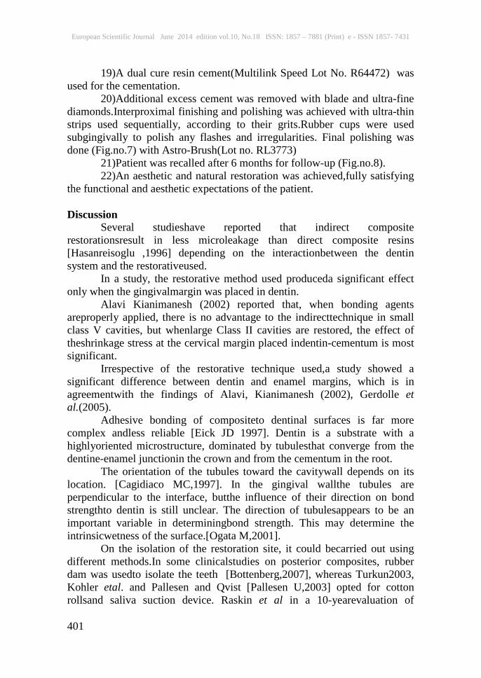
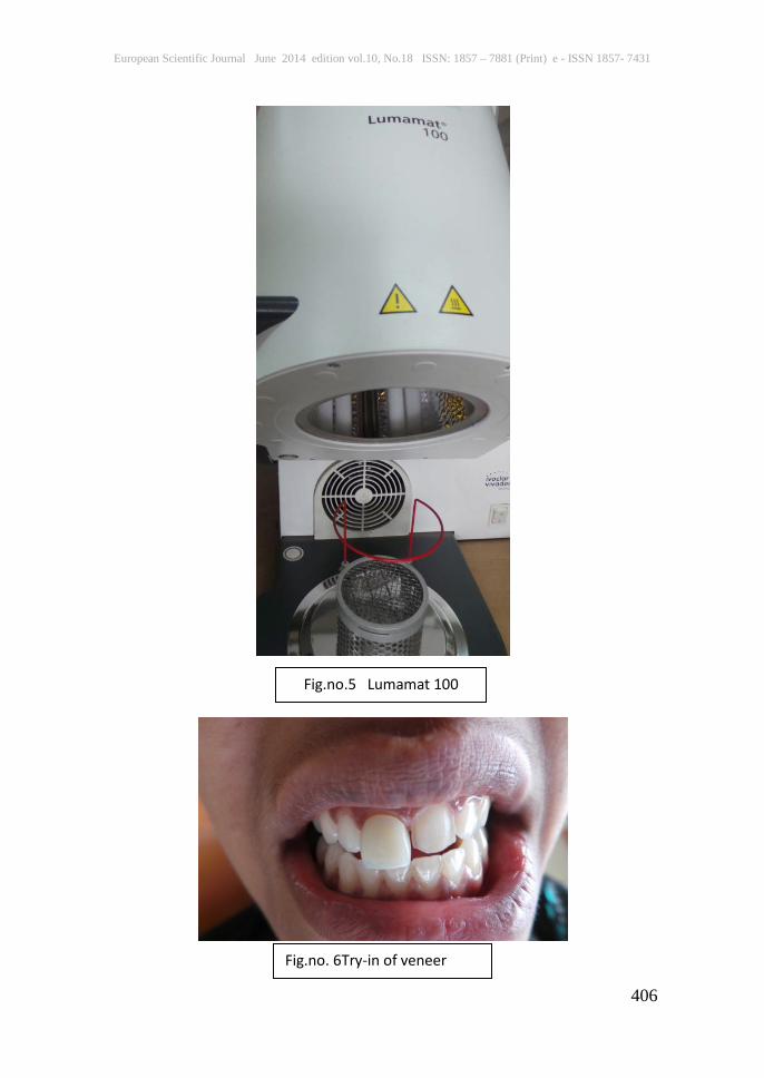
structure,a depth cutter contained in the system was used to determine the initial preparation depth -0.5 mm.After facial reduction is completed,the degree of discoloration must be evaluated to determine whether additional reduction of tooth structure is required to provide additional space for the restorative materials (Fig.no.3). 5)A chamfer finish line which should be within enamel whenever possible to ensure an adequate seal of the veneer,was placed into the interproximal embrasures without breaking contact and confined in enamel along the incisal edge.This preparation is designed to protect the resin veneer,preventing it from chipping during excursive movements of the mandible or mastication. 6)The cervical chamfer was modified into a butt shoulder to provide more thickness to the restorative material at the gingival margin. 7)The entire preparation was further finished with aluminium oxide discs and polished with rubber cups.This step is especially important to prevent adherence of existing composite restorations in the prepared tooth to the resin veneer that will be built up over it. 8)The prepared tooth was cleaned in a rubbing motion with a cotton pellet moistened with 4% chlorhexidine.Prophylaxis cups are not recommended for cleaning at this stage,as they might provoke bleeding of the gingival. 9)Impression of both upper and lower arch was made using light body and heavy body putty material and finally the cast was poured using die stone. 10) After setting,the cast was trimmed. 11)Die hardener was applied followed by separator liquid. 12) Buid up of laminate was performed using various ADORO indirect composite resins.(Fig.no.4) 13)It was then pre cured and processed in Lumamat.(Fig.no.5) 14)After the complete curing phase,the veneer was allowed to cool down and checked in patient’s mouth for optimal fit.(Fig.no.6) 15)The veneers were polished with various abrasives. 16)The internal aspect of the veneer was cleaned,acidified with 35% phosphoric acid gel, and after rinsing and drying, silanated with MONOBOND –S for 60 seconds. 17)The prepared tooth was etched with 37% phosphoric acid gel(N-Etch, Lot No.R76751) for 15 seconds.The etchant was rinsed thoroughly and the surfaces were lightly air-dried to avoid desiccation. 18)The bonding agent(Tetric-N Bond) was applied to the prepared tooth and to the inner surface of the veneer,according to the manufacturer’s recommendations.
European Scientific Journal June 2014 edition vol.10, No.18 ISSN: 1857 – 7881 (Print) e - ISSN 1857- 7431
401
19)A dual cure resin cement(Multilink Speed Lot No. R64472) was used for the cementation. 20)Additional excess cement was removed with blade and ultra-fine diamonds.Interproximal finishing and polishing was achieved with ultra-thin strips used sequentially, according to their grits.Rubber cups were used subgingivally to polish any flashes and irregularities. Final polishing was done (Fig.no.7) with Astro-Brush(Lot no. RL3773) 21)Patient was recalled after 6 months for follow-up (Fig.no.8). 22)An aesthetic and natural restoration was achieved,fully satisfying the functional and aesthetic expectations of the patient. Discussion
Several studieshave reported that indirect composite restorationsresult in less microleakage than direct composite resins [Hasanreisoglu ,1996] depending on the interactionbetween the dentin system and the restorativeused.
In a study, the restorative method used produceda significant effect only when the gingivalmargin was placed in dentin.
Alavi Kianimanesh (2002) reported that, when bonding agents areproperly applied, there is no advantage to the indirecttechnique in small class V cavities, but whenlarge Class II cavities are restored, the effect of theshrinkage stress at the cervical margin placed indentin-cementum is most significant.
Irrespective of the restorative technique used,a study showed a significant difference between dentin and enamel margins, which is in agreementwith the findings of Alavi, Kianimanesh (2002), Gerdolle et al.(2005).
Adhesive bonding of compositeto dentinal surfaces is far more complex andless reliable [Eick JD 1997]. Dentin is a substrate with a highlyoriented microstructure, dominated by tubulesthat converge from the dentine-enamel junctionin the crown and from the cementum in the root.
The orientation of the tubules toward the cavitywall depends on its location. [Cagidiaco MC,1997]. In the gingival wallthe tubules are perpendicular to the interface, butthe influence of their direction on bond strengthto dentin is still unclear. The direction of tubulesappears to be an important variable in determiningbond strength. This may determine the intrinsicwetness of the surface.[Ogata M,2001].
On the isolation of the restoration site, it could becarried out using different methods.In some clinicalstudies on posterior composites, rubber dam was usedto isolate the teeth [Bottenberg,2007], whereas Turkun2003, Kohler etal. and Pallesen and Qvist [Pallesen U,2003] opted for cotton rollsand saliva suction device. Raskin et al in a 10-yearevaluation of

European Scientific Journal June 2014 edition vol.10, No.18 ISSN: 1857 – 7881 (Print) e - ISSN 1857- 7431
402
posterior composites, did not observesignificant differences between these two isolationmethods.
According to Mitra et al.[ Mitra SB,2003] , the nanofilled compositewas shown to have equivalent — if not higher —mechanical properties than the hybrid composite, sincethe nanocomposite showed high translucency, highpolish and polish retention similar to those ofmicrofilled compositein a study by Loguercio et al. in 2007,the nanofilled and microfilled composites showed thebest surface appearance after 12 months. Conclusion
The indirect composite resin veneer technique is practical and reliable in treating most of the single tooth discolorations.It allows the clinician to artistically treat aesthetically compromised dentition by using restorative material that can be fabricated intraorally,heat-treated to enhance its physical properties, and bonded with resin cements that provide improved shade matching properties.The indirect veneer is a viable treatment modality for clinician who enjoy free-hand bonding and the artistry that is associated with it,for it permits the operator to create natural restorations that meet the aesthetic and functional expectations of the patient in a single appointment. References: Black JB,Esthetic restoration of tetracycline-stained teeth.J Am Dent Assoc 1982;104:846-852. Sockwell CL,Heymann HO Brunson,WD,Additional Conservative and Esthetic Treatments. The Art and Science of Operative Dentistry, St.Louis, MO; Mosby,1985. Pollack BF,Blitzer MH,Esthetic veneering materials and techniques.Gen Dent 1983;31:483-488. Christensen GJ.Veneering of teeth:State of the art.Dent Clin North Am 1985;29:373-391. Chalkley Y.Treatment of a single stained anterior tooth utilizing visible light-cured opaquer resin and microfill composite resin.Iowa Dental J 1984;70:37-39. Vaidyanathan J, Vaidyanathan TK, Wang Y, Viswanadhan T. Thermoanalytical characterization of visible light cure dental composite. J Oral Rehab 1992;19:49-64. Villela LC,Carvalho JRF,Araujo MAJ.A modified veneering technique using composite resin.Revista da APCD 1994;48:1535-1537. McCabe JF,Kagi S.Mechanical properties of a composite inlay material following post-curing.Br Dent J 1991;171:246-248. Covey DA,Tahaney SR,Davenport JM.Mechanical properties of heat-treated composite resin restorative materials.J Prosthet Dent 1992;68:458-461.

European Scientific Journal June 2014 edition vol.10, No.18 ISSN: 1857 – 7881 (Print) e - ISSN 1857- 7431
403
Wendt SLJ,Leinfelder KF.Clinical evaluation of a heat-treated resin composite inlay:Three-year results.Am J Dent 1992;5:258-262. Lombardi RE.The principles of visual perception and their clinical application to denture esthetics.J Prosthot Dent 1973;29:358. Levin EI.Dental esthetics and the golden proportion.J Prosthet Dent 1978;40:244. Goulub J,Unity and Variety.Essential Ingredients of a Smile Design;Editorial in Current Opinion in Cosmetic Dentistry,2nd ed,Philadelphia,PA:Current Science,1994. Rufenachi CR.Fundamentals of Esthetics,Chicago,IL:Quintessence,1990. Hasanreisoglu U, Sonmez H, Uctasli S, Wilson HJ.Microleakage of direct and indirect inlay/onlay systems. J Oral Rehabil 1996;23(1):66-71. Milleding P. Microleakage of indirect composite inlays.An in vitro comparison with the direct technique. Acta Odontol Scand 1992;50(5):295-301. Alavi AA, Kianimanesh N. Microleakage of direct and indirect composite restorations with three dentin bonding agents. Oper Dent 2002;27(1):19-24. Gemalmaz D, Ozcan M, Yoruc AB, Alkumru HN. Marginal adaptation of a sintered ceramic inlay system before and after cementation. J Oral Rehabil 1997;24(9):646-51 Eick JD, Gwinnett AJ, Pashley DH, Robinson SJ. Current concepts on adhesion to dentin. Crit Rev Oral Biol Med 1997;8(3):306-35. Pashley DH, Carvalho RM. Dentine permeability and dentine adhesion. J Dent 1997;25(5):355-72. Cagidiaco MC, Ferrari M, Vichi A, Davidson CL. Mapping of tubule and intertubule surface areas available for bonding in Class V and Class II preparations. J Dent 1997;25(5):379-89. Ogata M, Okuda M, Nakajima M, Pereira PN, Sano H, Tagami J. Influence of the direction of tubules on bond strength to dentin. Oper Dent 2001;26(1):27-35 Bottenberg P, Alaerts M, Keulemans F. A prospectiverandomised clinical trial of one bis-GMA-based and two ormocer-based composite restorative systems in class IIcavities: three-year results. J Dent 2007; 35: 163-171. Manhart J, Neuerer P, Scheibenbogen-Fuchsbrunner A,Hickel R. Three-year clinical evaluation of direct and indirect composite restorations in posterior teeth. JProsthet Dent 2000; 84: 289-296. da Rosa Rodolpho PA, Cenci MS, Donassollo TA, LoguercioAD, Demarco FF. A clinical evaluation of posterior composite restorations: 17-year findings. J Dent 2006; 34:427-435. Turkun ŞL. Clinical evaluation of a self-etching and a onebottleadhesive system at two years. J Dent 2003; 31: 527-534.

European Scientific Journal June 2014 edition vol.10, No.18 ISSN: 1857 – 7881 (Print) e - ISSN 1857- 7431
404
Köhler B, Rasmusson CG, Odman P. A five-year clinicalevaluation of Class II composite resin restorations. J Dent2000; 28: 111-116. Pallesen U, Qvist V. Composite resin fillings and inlays.An 11-year evaluation. Clin Oral Invest 2003; 7: 71-79. Raskin A, Setcos JC, Vreven J, Wilson NHF. Influence ofthe isolation method on the 10-year clinical behavior ofposterior resin composite restorations. Clin Oral Invest2000; 4: 148-152. Mitra SB, Wu D, Holmes BN. An application ofnanotechnology in advanced dental materials. J Am DentAssoc 2003; 134: 1382-1390. Loguercio AD, Lorini E, Weiss RV, Tori AP, Picinatto CC,Ribeiro NR, Reis A. A 12-month clinical evaluation ofcomposite resins in class III restorations. J Adhes Dent2007; 9: 57-64. Pictures
Fig.no. 1 Pre-operative view

European Scientific Journal June 2014 edition vol.10, No.18 ISSN: 1857 – 7881 (Print) e - ISSN 1857- 7431
405
Fig.no.3 Tooth preparation
Fig.no.4 ADORO Indirect composite materials
Fig.no.2 Pre-operative IOPAR
European Scientific Journal June 2014 edition vol.10, No.18 ISSN: 1857 – 7881 (Print) e - ISSN 1857- 7431
406
Fig.no. 6Try-in of veneer
Fig.no.5 Lumamat 100

European Scientific Journal June 2014 edition vol.10, No.18 ISSN: 1857 – 7881 (Print) e - ISSN 1857- 7431
407
Fig.no. 7 Post-operative view
Fig.no.8 6 months follow-up