the acquisition of novel n-glycosylation sites in conserved proteins
TRANSCRIPT

Kim and Hahn BMC Bioinformatics (2015) 16:29 DOI 10.1186/s12859-015-0468-5
RESEARCH ARTICLE Open Access
The acquisition of novel N-glycosylation sites inconserved proteins during human evolutionDong Seon Kim and Yoonsoo Hahn*
Abstract
Background: N-linked protein glycosylation plays an important role in various biological processes, includingprotein folding and trafficking, and cell adhesion and signaling. The acquisition of a novel N-glycosylation site mayhave significant effect on protein structure and function, and therefore, on the phenotype.
Results: We analyzed the human glycoproteome data set (2,534 N-glycosylation sites in 1,027 proteins) andidentified 112 novel N-glycosylation sites in 91 proteins that arose in the human lineage since the last commonancestor of Euarchonta (primates and treeshrews). Three of them, Asn-196 in adipocyte plasma membrane-associatedprotein (APMAP), Asn-91 in cluster of differentiation 166 (CD166/ALCAM), and Asn-76 in thyroglobulin, arehuman-specific. Molecular evolutionary analysis suggested that these sites were under positive selection duringhuman evolution. Notably, the Asn-76 of thyroglobulin might be involved in the increased production ofthyroid hormones in humans, especially thyroxine (T4), because the removal of the glycan moiety from thissite was reported to result in a significant decrease in T4 production.
Conclusions: We propose that the novel N-glycosylation sites described in this study may be useful candidatesfor functional analyses to identify innovative genetic modifications for beneficial phenotypes acquired in thehuman lineage.
Keywords: N-glycosylation, Evolution, Glycoproteome, Human
BackgroundN-linked glycosylation of the Asn residue in the consen-sus motif Asn-X-Ser/Thr, where X is any amino acid exceptPro, is one of the most well-studied protein posttransla-tional modifications (PTMs) [1]. N-glycosylation, whichmainly occurs in secreted or cell membrane proteins, playsimportant roles in protein folding, quality control, andtrafficking [2], as well as cell adhesion and signalling [3,4].The emergence of a new N-glycosylation site may alterprotein function either positively or negatively. Forexample, missense mutations in factor VIII created novelN-glycosylation sites that cause severe hemophilia A [5].Similarly, a missense mutation in the interferon γ recep-tor 2 induces novel N-glycosylation, which results in aMendelian susceptibility to mycobacterial disease [6,7].The abolishment of N-glycosylation sites often causesdisrupted protein folding, trafficking, or activity; thus,
* Correspondence: [email protected] of Life Science, Research Center for Biomolecules andBiosystems, Chung-Ang University, 84 Heukseok-ro, Dongjak-gu, Seoul156-756, Korea
© 2015 Kim and Hahn; licensee BioMed CentrCommons Attribution License (http://creativecreproduction in any medium, provided the orDedication waiver (http://creativecommons.orunless otherwise stated.
proper N-glycosylation is crucial for normal proteinfunction [8,9]. A proteome-wide analysis of nonsynon-ymous single-nucleotide variations in the N-glycosylationmotifs of human genes showed that more than 1,000human proteins had either lost or gained N-glycosylationsites due to missense substitutions, some of which may beimplicated in diseases [10].The gain of new N-glycosylation sites during evolution
may affect the structure and molecular function of pro-teins; when these novel modifications confer beneficialtraits, they will be fixed during evolution. Previously, weidentified a large variety of genetic changes that couldhave been involved in the acquisition of human traits, in-cluding gene inactivation [11,12], exon evolution [13,14],and gains of phosphorylation or ubiquitylation [15,16].Therefore, it would be of great interest to collect informa-tion on novel N-glycosylation sites that arose during hu-man evolution, as the sites might have been involved inthe development of some human phenotypes.In order to study associations between the acquisition
of an N-glycosylation site and its phenotypic outcome, a
al. This is an Open Access article distributed under the terms of the Creativeommons.org/licenses/by/4.0), which permits unrestricted use, distribution, andiginal work is properly credited. The Creative Commons Public Domaing/publicdomain/zero/1.0/) applies to the data made available in this article,

Figure 1 Overall procedure for identifying gains of novelN-glycosylation sites during human evolution. Computationalscreening and manual inspection were employed to identify theacquisition of novel N-glycosylation sites in human proteinsduring human evolution.
Kim and Hahn BMC Bioinformatics (2015) 16:29 Page 2 of 12
large amount of N-glycosylation site data and mamma-lian orthologous protein sequence data are required.Recent developments and advances in various high-throughput proteomics techniques for N-glycoproteomeidentification using immunoaffinity subtraction, hydrazidechemistry, and mass spectrometry have made it possibleto access massive amounts of N-glycosylation site datafrom human proteomes [17-19]. These data are availableat the UniProt database (http://www.uniprot.org), whichis a universal protein sequence database, as well as somespecialized PTM databases such as PHOSIDA (http://www.phosida.com/) [20].Since human genome sequences were completed [21,22],
a large amount of nucleotide and protein sequence datahave become available not only from humans but alsofrom many other organisms. Comparative sequence data,including alignments of mammalian orthologous proteinsequences, are available at the University of CaliforniaSanta Cruz (UCSC) Genome Browser Database (http://genome.ucsc.edu) [23].In this study, a bioinformatics method was devised to
identify novel N-glycosylated Asn residues that are lo-cated in the consensus motif Asn-X-Ser/Thr and aroseduring human evolution after the Euarchonta lineage di-verged from the Glires lineage. Both a comprehensive lit-erature survey and extensive data mining were conductedto examine the possible functional implications of novelN-glycosylation sites, especially in cases of human-specificgains.
ResultsIdentification of novel N-glycosylation sites acquiredduring human evolution and determination of the timingof acquisitionWe developed a bioinformatics procedure to identify theacquisition by proteins of the N-glycosylation motifAsn-X-Ser/Thr, where the Asn residue was experimen-tally verified to be N-glycosylated, during human evolu-tion (Figure 1). A novel N-glycosylation site can arise bythe emergence of not only an Asn residue but also a Seror Thr residue to form the consensus motif [24]. Theoverall procedure devised in this study is similar to thatused to identify novel ubiquitylation sites in a previousstudy [16]. Initially, there were 2,534 experimentally verifiedhuman N-glycosylation sites from 1,027 proteins in theUniProt database, and 57,289 orthologous protein sequencealignments from 62 mammalian species, including spe-cies from Euarchonta, Glires, Laurasiatheria, Afrotheria,Xenarthra, Marsupialia, and Monotremata, extracted fromthe UCSC “multiz100way” data [25] (see Additional file 1for the list of mammalian species). These data were ana-lyzed to collect N-glycosylation sites in human proteinsthat newly appeared during the evolution from the com-mon ancestor, Euarchonta (primates and treeshrews); as
the result, 112 novel N-glycosylation sites from 91 pro-teins were identified. A summary of the results are pre-sented in Additional file 2, and detailed alignments areprovided in Additional file 3. Of the 91 proteins, one pro-tein (CFH) had acquired four N-glycosylation sites (Nos.28 to 31 in Additional file 2 and Additional file 3; two pro-teins (PTPRC and PTPRJ) had acquired three sites each(Nos. 85 to 87, and 88 to 90, respectively); 14 proteins hadacquired two sites each; and the remaining 74 proteinshad acquired one site each. Figure 2 shows the number ofthe N-glycosylation sites that are shared by each of theEuarchonta clades along the human lineage: humans,three; ancestor of humans and chimpanzees, two; Africangreat apes, 10; great apes, four; apes, 12; catarrhines, 16;simians, 45; primates, 15; and euarchonts, 5.Most of the novel N-glycosylation sites were gener-
ated by the emergence of an Asn residue in an existingX-X-Ser/Thr motif. However, in some cases, the emer-gence of a Ser or a Thr residue in an Asn-X-X sequencecreated a novel N-glycosylation site: for example, a changefrom Asn-Glu-Ile to Asn-Glu-Thr generated a novelN-glycosylation site at Asn-911 in complement factor H(CFH) in apes (No. 30 in Additional file 3).Of the 112 novel N-glycosylation sites, three sites in three
proteins were human specific (Table 1 and Figure 3); there-fore, these Asn residues subject to the N-glycosylationevolved and were fixed in human proteins after the

Figure 2 Timing of acquisition and numbers of novel N-glycosylation sites in the human lineage. Numbers of novel N-glycosylation sitesacquired in the human lineage of the mammalian phylogenetic tree are shown. The number of sites acquired is shown on each branch wherethe N-glycosylation site consensus motif emerged in the ancestor of the corresponding clade.
Kim and Hahn BMC Bioinformatics (2015) 16:29 Page 3 of 12
divergence of humans and chimpanzees. The residues areAsn-196 in adipocyte plasma membrane-associated pro-tein (APMAP), Asn-91 in cluster of differentiation 166(CD166), and Asn-76 in thyroglobulin.
Human-specific N-glycosylation site Asn-196 in APMAPThe human APMAP (also known as C20orf3) has twoN-glycosylation sites: Asn-160 and Asn-196, the latter ofwhich is human specific: almost all the other mammalsexamined have a Lys residue at this position (Figure 3A).Full-length protein and coding sequences of APMAPorthologs were determined from human, chimpanzee,gorilla, orangutan, gibbon, and rhesus macaque genomes(see Methods section for details). Multiple alignment ofthese proteins showed that human APMAP protein hastwo human-specific amino acid changes, Val-100 andAsn-196, where all the other five primates have Ile and Lys,respectively (Additional file 4). The Asn-196 is a human-specific N-glycosylation site.To test if the APMAP protein has been under positive
selection during human evolution, the ratios of nonsy-nonymous to synonymous rates (dN/dS, ω) across differ-ent branches and sites of the selected primate phylogenywere estimated [26-28]. First, we used “branch models”,M0 (one ω ratio for all branches), free ratio (one ω ratiofor each branch), and two ratio (ω1 for the humanbranch and ω0 for other branches) models (Table 2 and
Table 1 Proteins with human-specific N-glycosylation sites
Noa Gene UniProt ID Position Sequence
5 ALCAM CD166_HUMAN 91 DDVPEYKD
8 APMAP APMAP_HUMAN 196 LSSETPIEGK
105 TG THYG_HUMAN 76 DGRSCWCVaThe number corresponds to that in Additional files 1 and 2.bThe N-glycosylation motif is in bold.
Additional file 4). The likelihood ratio test (LRT) com-paring M0 (one ratio) and free ratio model was not sig-nificant. However, the LRT comparing M0 and two ratiomodel was highly significant (P = 0.006943), suggestingthe human APMAP has evolved at different rate com-pared to other primates. The estimated dN and dS ratesfor the human branch using two ratio model were0.0024 and 0.0000 (see Additional file 4 for details), re-spectively, indicating possible accelerated nonsynonymoussubstitution during human evolution. Next, we used“branch-site models”, model A (ω ratio is left to vary) andnull model A (ω ratio is fixed to 1), to infer positively se-lected sites in human APMAP. The two aforementionedhuman-specific amino acid positions, Val-100 and Asn-196, were detected to be under positive selection withoverall probability of 0.828 and 0.953, respectively, usingthe Bayes empirical Bayes (BEB) test [29]. However, theLRT comparing model A and null model A was not sig-nificant. Although it is statistically insignificant, the acqui-sition of Asn-196 and its subsequent N-glycosylationmight have a significant effect on the structure and func-tion of APMAP in humans.APMAP is an adipocyte plasma membrane-associated
protein, which is induced during adipocyte differenti-ation [30]. It is ubiquitously expressed in human embry-onic and adult tissues, with the highest levels in liver,placenta, and kidney [31]. APMAP may exhibit calcium-
b Protein
RLNLSENYTLSI CD166 antigen
NMSFVNDLTV Adipocyte plasma membrane-associated protein
GANGSEVLGSRQ Thyroglobulin
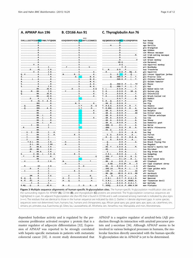
Figure 3 Multiple sequence alignments of human-specific N-glycosylation sites. The human-specific N-glycosylation modification sites andthe surrounding regions for APMAP (A), CD166 (B), and thyroglobulin (C) proteins are presented. The N-glycosylation consensus sequences arehighlighted in cyan. An adjacent N-glycosylation site (Asn-95) that is found in CD166 and is well conserved among mammals is indicated by plus signs(+++). The residues that are identical to those in the human sequence are indicated by dots (.). Dashes (−) denote alignment gaps. In some species,sequences were not determined. hum, humans; hac, humans and chimpanzees; aga, African great apes; gra, great apes; ape, apes; cat, catarrhines; sim,simians; pri, primates; eua, Euarchonta; gli, Glires; lau, Laurasiatheria; afr, Afrotheria; xen, Xenarthra; mar, Marsupialia; and mon, Monotremata.
Kim and Hahn BMC Bioinformatics (2015) 16:29 Page 4 of 12
dependent hydrolase activity and is regulated by the per-oxisome proliferator activated receptor γ protein that is amaster regulator of adipocyte differentiation [32]. Expres-sion of APMAP was reported to be strongly correlatedwith hepatic-specific metastasis in patients with metastaticcolorectal cancer [33]. A recent study demonstrated that
APMAP is a negative regulator of amyloid-beta (Aβ) pro-duction through its interaction with amyloid precursor pro-tein and γ-secretase [34]. Although APMAP seems to beinvolved in various biological processes in humans, the mo-lecular function directly associated with the human-specificN-glycosylation site in APMAP is yet to be determined.

Table 2 Molecular evolutionary analysis of APMAP, CD166, and thyroglobulin
Protein Model ln L ω 2Δl P value Positively selected sitesa
APMAP Branchmodels
M0 (one ratio) −2042.0968 ω0 = 0.07385
Free ratio −2035.7572 See Additional file 4 (M0 vs Free ratio)12.6792
0.1234
Two ratio −2038.4530 ω0 = 0.05733,ω1 = 999.000
(M0 vs Two ratio)7.2876
0.006943**
Branch-sitemodels
Model A −2038.4530 ω0 = 0.05732, ω1 = 1,ω2 = 999.000
Val-100, Asn-196
Null model A −2039.0253 ω0 = 0.05746,ω1 = 1, ω2 = 1
(Model A vs Nullmodel A) 1.1446
0.2847
CD166 Branchmodels
M0 (one ratio) −2713.8934 ω0 = 0.09913
Free ratio −2709.9951 See Additional file 5 (M0 vs Free ratio)7.7966
0.4536
Two ratio −2713.8904 ω0 = 0.09991,ω1 = 0.09134
(M0 vs Two ratio)0.0060
0.9383
Branch-sitemodels
Model A −2713.8934 ω0 = 0.09913,ω1 = 1, ω2 = 1
Asn-91
Null model A −2713.8934 ω0 = 0.09913,ω1 = 1, ω2 = 1
(Model A vs Nullmodel A) 0.000
1.000
Thyroglobulin Branchmodels
M0 (one ratio) −15128.0999 ω0 = 0.35639
Free ratio −15121.3529 See Additional file 6 (M0 vs Free ratio)13.4941
0.09595
Two ratio −15124.3983 ω0 = 0.33454,ω1 = 0.78473
(M0 vs Two ratio)7.4033
0.006511**
Branch-sitemodels
Model A −15103.5548 ω0 = 0.000, ω1 = 1,ω2 = 3.59382
Asn-76, Ser-633, Ser-734, Asn-775,Met-911, Ser-913, Gly-1061, Ser-1140,Thr-1204, Met-1242, Thr-1498, Arg-1646,His-1669, Arg-1691, Asp-1795, His-2486,Arg-2530, Asn-2616, Leu-2632, Glu-2702,Thr-2727, Thr-2765
Null model A −15104.1308 ω0 = 0.000, ω1 = 1,ω2 = 1
(Model A vs Nullmodel A) 1.1520
0.2831
aHuman-specific N-glycosylation sites are in bold. See Additional files 4, 5, and 6 for details.**P < 0.05.
Kim and Hahn BMC Bioinformatics (2015) 16:29 Page 5 of 12
Human-specific N-glycosylation site Asn-91 in CD166The human CD166, which is also known as activatedleukocyte cell adhesion molecule (ALCAM), has 10 N-glycosylation sites. The residue Asn-91 is found in humansbut not in other euarchonts; thus, this residue evolved afterthe divergence of humans and chimpanzees (Figure 3B).Most other mammals have a Ser residue at this pos-ition. However, some Glires and Laurasiatheria spe-cies, especially whales and ruminants, independentlyacquired a consensus sequence for N-glycosylation atthis position.Multiple alignment of full-length CD166 proteins from
human, chimpanzee, gorilla, orangutan, gibbon, and rhe-sus macaque genomes revealed that the Asn-91 is onlyresidue that differs between humans and chimpanzees(Additional file 5). Although the Asn-91 is human-specific,all the LRTs (M0 versus free ratio model, M0 versus two
ratio model, and model A versus null model A) were insig-nificant (Table 2 and Additional file 5), implying that therehas been no statistically noticeable positive selection onthe human CD166. Interestingly, the human-specific N-glycosylation site Asn-91 was still inferred to be underpositive selection with overall probability of 0.613, sug-gesting the acquisition of this site and its N-glycosylationmight have an effect on the function of human CD166proteins.CD166 binds to the T-cell differentiation antigen CD6
and may play a role in the binding of T and B cells to ac-tivated leukocytes, as well as in interactions betweencells of the nervous system [35]. CD166 is composed offive extracellular immunoglobulin (Ig)-like domains: twoIg-like V-type domains and three Ig-like C2-type domains.The human-specific N-glycosylation site Asn-91 is locatedin the first Ig-like V-type domain, which mediates the
Kim and Hahn BMC Bioinformatics (2015) 16:29 Page 6 of 12
CD166–CD6 interaction [35-37]. Most functional studieson CD166 have focused on its cancer-related functionssuch as invasion, migration, and adhesion [37,38]. How-ever, recent studies show that CD166 is also involved inaxon growth in neuronal cells such as retinal ganglioncells and dorsal root ganglion cells [39,40]. Therefore, it ispossible that the gain of the Asn-91 N-glycosylation site inCD166 might be involved in the evolution of novel pheno-types in nervous system development, as well as in im-mune response and cell adhesion processes, which mustbe validated experimentally.
Human-specific N-glycosylation site Asn-76 inthyroglobulinThyroglobulin is the precursor of the thyroid hormonesT4 and triiodothyronine (T3), both of which regulatemetabolism in humans [41-43]. The human thyroglobulinhas 17 N-glycosylation sites; Asn-76, which becomes Asn-57in the mature form of thyroglobulin, is a human-specificN-glycosylation site: most other mammals have Aspor Glu at this position (Figure 3C). Interestingly, multiplealignment of thyroglobulin orthologs from humans, chim-panzees, gorillas, orangutans, gibbons, and rhesus ma-caques revealed additional 20 amino acid positions withhuman-specific substitution (Additional file 6).To test if the thyroglobulin has been under positive se-
lection during human evolution, ω ratios across differentbranches and sites of the selected primate phylogenywere estimated (Table 2 and Additional file 6). The LRTcomparing M0 and free ratio model was not significant.However, the LRT comparing M0 and two ratio modelwas highly significant (P = 0.006511), implying the hu-man thyroglobulin has evolved at different rate com-pared to other primates. The estimated ω ratio for thehuman branch (ω1) using two ratio model was 0.78473,while ω0 for other branches was 0.33454 (see Additionalfile 6 for details), suggesting a slightly accelerated nonsy-nonymous substitution during human evolution.Inference of positively selected sites in human thyro-
globulin using model A showed that the 21 aforementionedamino acid positions might have been under positive selec-tion although the LRT comparing model A and null modelA was not significant. In spite of statistical insignificance,the acquisition of novel N-glycosylation site Asn-76,together with other 20 human-specific amino acid substi-tutions, might have a significant effect on the thyroid hor-mone metabolism in humans.The thyroglobulin protein precursor itself has no bio-
logical function but serves as a chemical platform for thy-roid hormone production. When the two N-glycosylationsites in its N-terminal region, including the human-specific Asn-76, were deglycosylated by peptide-N4-(N-acetyl-β-glucosaminyl)-asparagine amidase, T4 productiondecreased by half compared to that seen with the normal
protein [44]. Therefore, proper N-glycosylation modifications,including those at the human-specific site, in thyroglobu-lin are crucial for normal T4 production and the controlof metabolism. It has been suggested that humans andchimpanzees differ with respect to their thyroid hor-mone metabolism [45]. Interestingly, compared to chim-panzees, humans have a higher T4 plasma concentration,which may be implicated in the origins of humanintelligence [46].
Novel N-glycosylation sites shared by other animalsOf the 112 novel N-glycosylation sites in human proteins,109 sites were shared by other animals. For example,12 N-glycosylation sites were shared by all apes, indicatingthat this site appeared in the common ancestor of apes.The Asn-480 of pappalysin-1 (also known as pregnancy-associated plasma protein-A), which is N-glycosylated inhumans [47], is shared by all apes examined (Figure 4A);in contrast, all the other mammals, even Marsupialia andMonotremata species, have an Asp residue at this pos-ition. Pappalysin-1 has metalloproteinase activity and spe-cifically cleaves insulin-like growth factor-binding proteins[48,49]; it is present at high concentration in maternalblood during pregnancy and is essential for normal fetaldevelopment [50]. The serum pappalysin-1 concentrationfrequently increases in patients with severe sepsis and ap-pears to be associated with sepsis-related myocardial dys-function [51]. However, there is no comprehensive studyof whether the gain of Asn-480 in apes is associated withthese phenotypes.There are 12 N-glycosylation sites that might have arisen
in the common ancestor of apes. One is Asn-113 in thethyrotropin receptor, or thyroid-stimulating hormonereceptor (TSHR) (Figure 4B). The THSR responds tothyroid-stimulating hormone (also known as thyrotropin)and stimulates the production of T4 and T3 in the thyroidgland [52]. Human THSR has six N-glycosylation sites:Asn-113 is specific to apes, and the other five are con-served in other mammals. However, a mutated TSHR inwhich N-glycosylation at Asn-113 had been disrupted hadthe same expression level and function as the wildtypeTSHR; thus, Asn-113 N-glycosylation may not be im-portant for TSHR function [53]. Therefore, gain of N-glycosylation at Asn-113 may be neutral or have afunction yet to be determined.The UDP-glucuronosyltransferase 1–9 (UD19) Asn-344
is one of 45 N-glycosylation sites that are shared by sim-ians (apes and monkeys; Figure 4C). Nonsimian mammalshave a Lys residue at this position. UD19, which is alsoknown as UDP glucuronosyltransferase 1 family, polypep-tide A9, is involved in the conjugation and eliminationof toxic xenobiotic and endogenous compounds [54,55].Unglycosylation of UD19 resulted in the inhibition ofproper protein folding and the impairment of glucuronidation

Figure 4 Multiple sequence alignments of N-glycosylation sites that arose during human evolution. The N-glycosylation sites and thesurrounding regions for pappalysin-1 (A), TSHR (B), and UD19 (C) proteins are presented. See Figure 3 for further details.
Kim and Hahn BMC Bioinformatics (2015) 16:29 Page 7 of 12
activity; thus, N-glycosylation plays a role in folding thehuman UD19 protein [56]. UD19 is one of nine func-tional isoforms produced by the alternative utilizationof the first nine exons in the UGT1A gene locus [57].Because Asn-344 is located in the common exon 5, notonly UD19 but also eight other isoforms of UDP glucuro-nosyltransferase 1 enzyme have this novel N-glycosylationsite [56].
DiscussionPreviously, it has been suggested that the gain of novelprotein PTM sites such as ubiquitylation sites may beassociated with the acquisition of novel phenotypes dur-ing human evolution by modulating the activity or net-work of proteins [16]. It is also highly probable thatgains of novel N-glycosylation sites may result in func-tional modification of proteins and phenotypic changes

Kim and Hahn BMC Bioinformatics (2015) 16:29 Page 8 of 12
in an organism. In this study, 1,027 human glycoproteinscontaining experimentally verified N-glycosylation sitesand their orthologous mammalian proteins were system-atically compared. As a result, 112 novel N-glycosylationsites were identified in 91 proteins that newly appearedduring human evolution after the Euarchonta lineage di-verged from the Glires lineage. It must be noted thatmost of these novel N-glycosylation sites were obtainedby high-throughput mass spectrometry. The presence ofthese modifications must be further scrutinized by con-ventional molecular biology techniques.Not all the novel N-glycosylation sites described in this
study may have resulted in functional innovation. Someof them might have appeared as a result of random gen-etic drift and be functionally neutral. Nevertheless, someof them could have conferred selective advantage duringhuman evolution and be fixed in the human genome.One such example identified in this study is the novelN-glycosylation site in UD19, which is involved in theelimination of potentially toxic xenobiotics and endogen-ous compounds. UD19 acquired the novel N-glycosylationsite Asn-344 during the evolution of the common an-cestor of apes and monkeys (see Figure 4C). Whenthe N-glycosylation at Asn-344 is abolished, folding isinhibited in UD19, and its glucuronidase activity is re-duced [54]. Therefore, glycosylation at Asn-344 is re-quired for proper folding and activity of UD19. It ispossible that ancestral simian primates required betterdefense mechanisms against toxic compounds introducedinto their systems by environmental or dietary shifts. Theacquisition of a new N-glycosylation site in UD19 mighthave conferred improved xenobiotics metabolism to apesand monkeys, although there is no direct evidence for thishypothesis.The three human-specific N-glycosylation sites are
particularly interesting (see Table 1 and Figure 3). Theresidue Asn-196 in APMAP is the first of the threehuman-specific N-glycosylation sites, which was inferredto be positively selected with an extremely high prob-ability in humans (see Table 2 and Additional file 4). Thehuman APMAP has been reported to be involved in avariety of biological processes including adipocyte differ-entiation, hepatic-specific metastasis in cancer, and in-hibition of Aβ production [32-34]. The fact that APMAPis implicated in adipocyte differentiation is particularlyinteresting because humans and great apes exhibit largedifferences in adipose tissue and fatty acid storage, andthese differences may be associated with the develop-ment of subcutaneous fat and even in brain development[58,59]. Therefore, the molecular functional study ofhuman-specific sequence changes in proteins such asAPMAP, which are associated with adipose tissue andlipid metabolism, may reveal the molecular mechanismsfor the evolution of these traits.
The human CD166 protein has two Ig-like V-type do-mains and three Ig-like C2-type domains (Figure 5A)and functions as a cell adhesion molecule. The human-specific N-glycosylation site Asn-91, which was inferredto be positively selected (see Table 2 and Additional file5), is located within the first Ig-like V-type domain,which is responsible for protein–protein interactions[35-37]. The addition of a bulky glycan moiety to thisdomain may change its structural profile and thus affectcell–cell adhesion activity or ligand specificity. The mostinteresting function of CD166 is its involvement in axongrowth in neuronal cells [39,40]. Of the 583 amino acidresidues in CD166, only the residue at position 91 differsbetween humans and chimpanzees; therefore, the emer-gence of Asn-91 and its N-glycosylation might be associatedwith evolution of human-specific phenotypes, probablyin the nervous system, which must be determinedexperimentally.The human thyroglobulin, which serves as a precursor
molecule for the thyroid hormones T4 and T3, has ahuman-specific N-glycosylation site Asn-76. The humanthyroglobulin contains 11 thyroglobulin type-1 domains,which are involved in the control of proteolytic degrad-ation [60]. The human-specific Asn-76 is located withinthe first thyroglobulin type-1 domain (Figure 5B). TheAsn-76 was inferred to be positively selected during hu-man evolution, along with the other 20 positions (seeTable 2 and Additional file 6). Removal of the glycan groupfrom the Asn-76 reduced thyroid hormone production, es-pecially T4 production [44]; thus, the gain of Asn-76 andits N-glycosylation, together with the other 20 putativelyadaptive amino acid changes, may be implicated in the in-creased T4 concentration present in humans as comparedto chimpanzees. It is possible that the additional glycanmoiety may confer increased resistance to the proteolyticdegradation of thyroglobulin proteins and thus lead to in-creased thyroid hormone production. It is very interestingthat the T4 concentration in humans is higher than that inchimpanzees [45], as elevated T4 production may havecaused the modification of human physiology in responseto selection pressures in a specific environment: specific-ally, it has been proposed that an altered thyroid hor-mone metabolism might have been beneficial for earlyhumans in the savannah environment, as they practicedpersistence hunting and thus had large energy require-ments [46].Losses of N-glycosylation sites during human evolu-
tion are also very interesting. Some human diseases havebeen reported to be caused by the loss of N-glycosylationsites [61]. To find cases where ancestrally conservedN-glycosylation sites were lost during human evolution, alarge amount of N-glycosylation data collected fromanimals distantly related to humans is required. The N-glycoproteome data obtained from mouse tissues and

Figure 5 Schematic domain organizations of CD166 (A) and thyroglobulin (B). The N-glycosylation sites are indicated with lollipops, andhuman-specific sites are indicated in red. The domain organizations are derived from the UniProt database; the accession numbers are[Swiss-Prot:Q13740] (CD166) and [Swiss-Prot:P01216] (thyroglobulin).
Kim and Hahn BMC Bioinformatics (2015) 16:29 Page 9 of 12
plasma using high-throughput mass spectrometry wouldbe an ideal dataset for this analysis [62]. With a simplemodification, the procedure described in this study couldbe used to analyze these data for the identification ofN-glycosylation sites that were lost during human evolu-tion and their possible phenotypic implications.
ConclusionsWe have devised and applied a bioinformatics methodto identify the acquisition of N-glycosylation sites duringhuman evolution. We propose that the acquisition of novelN-glycosylation sites may play a role in the development oflineage-specific phenotypes during evolution. Thus, thecases identified in this study may provide a useful resourcefor molecular functional analyses in search of human traitsacquired during evolution.
MethodsHuman N-glycosylation site dataThe N-glycosylation sites in human proteins were ob-tained from the UniProt database (as of 13 November,2013). The feature table of the UniProt records wasscanned to collect entries with experimentally identifiedN-glycosylation sites. Specifically, the lines starting with“FT” followed by the “CARBOHYD” tag were examinedfor whether they contained a term “N-linked (GlcNAc…)”,which would indicate that the protein was N-glycosylated.Sites without experimental evidence, labeled as “potential”,“by similarity”, “partial”, or “probable”, were excluded. Asa result, 2,534 N-glycosylation sites from 1,027 humanproteins were obtained.
Mammalian orthologous proteinsMammalian orthologs of the human glycosylated pro-teins were obtained from the UCSC Genome BrowserDatabase (http://genome.ucsc.edu). The “CDS FASTA
alignment from multiple alignments” data, derived fromthe “multiz100way” alignment data prepared from 100vertebrate genomes [25], were downloaded using the TableBrowser tool of the UCSC Genome Browser. Protein se-quences from 62 mammalian species were extracted fromthese alignment datasets. The selected mammalian spe-cies include humans, 12 other Euarchonta species (chim-panzees, gorillas, orangutans, gibbons, rhesus macaques,crab-eating macaques, baboons, green monkeys, marmo-sets, squirrel monkeys, bushbabies, and treeshrews), 13Glires species (lesser Egyptian jerboas, prairie voles, Chinesehamsters, golden hamsters, mice, rats, naked mole-rats, guinea pigs, chinchillas, brush-tailed rats, rabbits,and pikas), 25 Laurasiatheria species (pigs, alpacas, Bactriancamels, dolphins, killer whales, Tibetan antelopes, cows,sheep, goats, horses, white rhinoceroses, cats, dogs, fer-rets, pandas, Pacific walruses, Weddell seals, black flying-foxes, megabats, David’s myotis bats, microbats, big brownbats, hedgehogs, shrews, and star-nosed moles), sixAfrotheria species (elephants, cape elephant shrews,manatees, cape golden moles, tenrecs, and aardvarks),one Xenarthra species (armadillos), three Marsupialiaspecies (opossums, Tasmanian devils, and wallabies), andone Monotremata species (platypuses). Additional file 1contains detailed information on species and genomeassemblies.
Computational screening for candidate novelN-glycosylation sites in human proteinsThe total number of experimentally identified N-glycosylationsites collected from human proteins was 2,534. To identifymammalian proteins that were orthologous to each of thehuman N-glycosylated proteins, the “multiz100way” align-ment data containing 57,289 alignment sets were analyzed(see Figure 1 for the overall procedure). There were 1,027orthologous protein datasets comprising 2,534 human
Kim and Hahn BMC Bioinformatics (2015) 16:29 Page 10 of 12
N-glycosylation sites. From each dataset, sequences of 62mammalian species were extracted and realigned usingMUSCLE (http://www.drive5.com/muscle) [63]. Then,each modification site in the alignment was analyzed,and cases where more than 30% of non-Euarchonta spe-cies had an N-glycosylation motif, which might representancestrally conserved sites, were discarded; cases whereonly a small number of sequences were aligned were alsodiscarded. A total of 184 sites in 130 protein alignmentswere retained after this computational screening step andsubjected to in-depth semimanual inspection.
Manual inspection to select novel N-glycosylation sites inhuman proteinsAs the final step, extensive manual inspection and cur-ation on the 184 candidate sites was carried out to iden-tify highly plausible cases of gains of N-glycosylationsites in the human lineage. Datasets showing the follow-ing conditions were filtered out: cases where the humansequence of UniProt database was different from that ofUCSC in three or more amino acid sequence residues be-cause of a possible paralogous relationship; cases wherethe N-glycosylated site was different from the consensusAsn-X-Ser/Thr; cases where the human N-glycosylationoccurred only in a rare variant or mutant allele; or caseswhere the chimpanzee protein sequence was not included.In each dataset, sequences containing many gaps in align-ment were removed from the dataset to retain only highquality sequences.As the final result, 112 novel N-glycosylation sites in
91 human proteins were identified. Then, multiple alignmentswere constructed to determine when the N-glycosylationmotifs first appeared. The possible functional conse-quences of the novel N-glycosylation site were thenassessed by comprehensive literature survey and se-quence analysis.
Molecular evolutionary analysisFull-length protein and coding sequences of APMAP,CD166, and thyroglobulin were collected from humans,chimpanzees, gorillas, orangutans, gibbons, and rhesusmacaques. Human cDNA RefSeq sequences were ob-tained from the National Center for Biotechnology In-formation (NCBI) (http://www.ncbi.nlm.nih.gov/refseq):accession numbers are [NCBI:NM_020531] (APMAP),[NCBI:NM_001627] (CD166), and [NCBI:NM_003235](thyroglobulin). For chimpanzee, gorilla, orangutan, gib-bon, and rhesus macaque orthologs, genome assemblies(panTro4, gorGor3, ponAbe2, nomLeu3, and rheMac3,respectively) were searched using each of human cDNAsequences at the UCSC Genome Browser Database.Exons, which were predicted from genomic segments,were assembled into a virtual cDNA and then conceptu-ally translated to get a protein sequence. Some exons,
which were missing in the current genome assembly,were obtained by assembling whole genome shotgunreads by searching the NCBI Sequence Read Archive(SRA) with SRA-BLAST server (http://www.ncbi.nlm.nih.gov/sra).The ratio of nonsynonymous to synonymous substi-
tution rates (dN/dS, ω) was estimated by a likelihoodmethod implemented in the codeml program of thePAML package (version 4.8a) [64]. To detect possibleaccelerated evolution in human proteins, we employed“branch models” that allow the ω ratio to vary amongbranches in phylogeny [27]; M0 (one ω ratio for all line-ages), free ratio (one ω ratio for each branch), and tworatio (ω1 for the human branch and ω0 for the otherbranches). To infer positively selected sites in humanproteins, we used “branch-site models” that allow the ωratio to vary among both sites and lineages [26,28]; modelA (ω is left to vary) and null model A (ω is fixed to 1). Tocompare the fit of nested models, the likelihood ratio test(LRT) was performed [27]. P values were obtained usingthe “chi2” program in the PAML package. Protein andcoding sequences, tree files, control files, and major resultfiles for APMAP, CD166, and thyroglobulin are providedin Additional files 4, 5, and 6, respectively.
Additional files
Additional file 1: List of mammalian species and genome assemblies.
Additional file 2: List of novel N-glycosylation sites.
Additional file 3: Sequence alignments of novel N-glycosylation sites.
Additional file 4: Molecular evolutionary analysis of APMAP.
Additional file 5: Molecular evolutionary analysis of CD166.
Additional file 6: Molecular evolutionary analysis of thyroglobulin.
AbbreviationsAβ: Amyloid-beta; APMAP: Adipocyte plasma membrane-associatedprotein; CD166: Cluster of differentiation 166; CD6: Cluster of differentiation 6;PTM: Posttranslational modification; T3: Triiodothyronine; T4: Thyroxine;TSHR: Thyroid-stimulating hormone receptor; UD19: UDP-glucuronosyltransferase 1–9.
Competing interestsThe authors declare that they have no competing interests.
Authors’ contributionsYH conceived of the study. DSK and YH conducted the analysis. DSK and YHprepared the manuscript. Both authors read and approved the final manuscript.
AcknowledgementsThis work was supported by the Basic Science Research Program throughthe National Research Foundation of Korea (NRF) funded by the Ministry ofEducation, Science and Technology (2012R1A1B3001513), Republic of Korea.
Received: 1 October 2014 Accepted: 15 January 2015
References1. Schwarz F, Aebi M. Mechanisms and principles of N-linked protein
glycosylation. Curr Opin Struct Biol. 2011;21(5):576–82.2. Helenius A, Aebi M. Intracellular functions of N-linked glycans. Science.
2001;291(5512):2364–9.

Kim and Hahn BMC Bioinformatics (2015) 16:29 Page 11 of 12
3. Dennis JW, Nabi IR, Demetriou M. Metabolism, cell surface organization, anddisease. Cell. 2009;139(7):1229–41.
4. Scott H, Panin VM. The role of protein N-glycosylation in neural transmission.Glycobiology. 2014;24(5):407–17.
5. Aly AM, Higuchi M, Kasper CK, Kazazian Jr HH, Antonarakis SE, Hoyer LW.Hemophilia A due to mutations that create new N-glycosylation sites.Proc Natl Acad Sci U S A. 1992;89(11):4933–7.
6. Vogt G, Chapgier A, Yang K, Chuzhanova N, Feinberg J, Fieschi C, et al.Gains of glycosylation comprise an unexpectedly large group of pathogenicmutations. Nat Genet. 2005;37(7):692–700.
7. Vogt G, Vogt B, Chuzhanova N, Julenius K, Cooper DN, Casanova JL.Gain-of-glycosylation mutations. Curr Opin Genet Dev. 2007;17(3):245–51.
8. Winterpacht A, Hilbert K, Stelzer C, Schweikardt T, Decker H, Segerer H, et al.A novel mutation in FGFR-3 disrupts a putative N-glycosylation site andresults in hypochondroplasia. Physiol Genomics. 2000;2(1):9–12.
9. Wujek P, Kida E, Walus M, Wisniewski KE, Golabek AA. N-glycosylation iscrucial for folding, trafficking, and stability of human tripeptidyl-peptidase I.J Biol Chem. 2004;279(13):12827–39.
10. Mazumder R, Morampudi KS, Motwani M, Vasudevan S, Goldman R.Proteome-wide analysis of single-nucleotide variations in the N-glycosylation sequon of human genes. PLoS One. 2012;7(5):e36212.
11. Hahn Y, Lee B. Identification of nine human-specific frameshift mutationsby comparative analysis of the human and the chimpanzee genomesequences. Bioinformatics. 2005;21 Suppl 1:i186–194.
12. Kim DS, Wang Y, Oh HJ, Lee K, Hahn Y. Frequent loss and alteration of theMOXD2 gene in catarrhines and whales: a possible connection with theevolution of olfaction. PLoS One. 2014;9(8):e104085.
13. Kim DS, Hahn Y. Identification of human-specific transcript variants inducedby DNA insertions in the human genome. Bioinformatics. 2011;27(1):14–21.
14. Kim DS, Hahn Y. Human-specific protein isoforms produced by novel splicesites in the human genome after the human-chimpanzee divergence.BMC Bioinformatics. 2012;13:299.
15. Kim DS, Hahn Y. Identification of novel phosphorylation modification sitesin human proteins that originated after the human-chimpanzee divergence.Bioinformatics. 2011;27(18):2494–501.
16. Kim DS, Hahn Y. Gains of ubiquitylation sites in highly conserved proteins inthe human lineage. BMC Bioinformatics. 2012;13:306.
17. Chen R, Jiang X, Sun D, Han G, Wang F, Ye M, et al. Glycoproteomicsanalysis of human liver tissue by combination of multiple enzyme digestionand hydrazide chemistry. J Proteome Res. 2009;8(2):651–61.
18. Liu T, Qian WJ, Gritsenko MA, Camp 2nd DG, Monroe ME, Moore RJ, et al.Human plasma N-glycoproteome analysis by immunoaffinity subtraction,hydrazide chemistry, and mass spectrometry. J Proteome Res.2005;4(6):2070–80.
19. Wollscheid B, Bausch-Fluck D, Henderson C, O'Brien R, Bibel M, Schiess R,et al. Mass-spectrometric identification and relative quantification ofN-linked cell surface glycoproteins. Nat Biotechnol. 2009;27(4):378–86.
20. Gnad F, Gunawardena J, Mann M. PHOSIDA 2011: the posttranslationalmodification database. Nucleic Acids Res. 2011;39(Database issue):D253–260.
21. International Human Genome Sequencing Consortium. Finishing theeuchromatic sequence of the human genome. Nature. 2004;431(7011):931–45.
22. Venter JC, Adams MD, Myers EW, Li PW, Mural RJ, Sutton GG, et al. Thesequence of the human genome. Science. 2001;291(5507):1304–51.
23. Mangan ME, Williams JM, Kuhn RM, Lathe 3rd WC. The UCSC GenomeBrowser: What every molecular biologist should know. Curr Protoc Mol Biol.2014;107:19 19 11–36.
24. Williams R, Ma X, Schott RK, Mohammad N, Ho CY, Li CF, et al. Encodingasymmetry of the N-glycosylation motif facilitates glycoprotein evolution.PLoS One. 2014;9(1):e86088.
25. Blanchette M, Kent WJ, Riemer C, Elnitski L, Smit AF, Roskin KM, et al.Aligning multiple genomic sequences with the threaded blockset aligner.Genome Res. 2004;14(4):708–15.
26. Dasmeh P, Serohijos AW, Kepp KP, Shakhnovich EI. Positively selected sitesin cetacean myoglobins contribute to protein stability. PLoS Comput Biol.2013;9(3):e1002929.
27. Yang Z. Likelihood ratio tests for detecting positive selection andapplication to primate lysozyme evolution. Mol Biol Evol. 1998;15(5):568–73.
28. Yang Z, dos Reis M. Statistical properties of the branch-site test of positiveselection. Mol Biol Evol. 2011;28(3):1217–28.
29. Yang Z, Wong WS, Nielsen R. Bayes empirical Bayes inference of amino acidsites under positive selection. Mol Biol Evol. 2005;22(4):1107–18.
30. Albrektsen T, Richter HE, Clausen JT, Fleckner J. Identification of a novelintegral plasma membrane protein induced during adipocytedifferentiation. Biochem J. 2001;359(Pt 2):393–402.
31. Ilhan A, Gartner W, Nabokikh A, Daneva T, Majdic O, Cohen G, et al.Localization and characterization of the novel protein encoded by C20orf3.Biochem J. 2008;414(3):485–95.
32. Bogner-Strauss JG, Prokesch A, Sanchez-Cabo F, Rieder D, Hackl H, Duszka K,et al. Reconstruction of gene association network reveals a transmembraneprotein required for adipogenesis and targeted by PPARγ. Cell Mol Life Sci.2010;67(23):4049–64.
33. Mekenkamp LJ, Haan JC, Koopman M, Vink-Borger ME, Israeli D, Teerenstra S,et al. Chromosome 20p11 gains are associated with liver-specific metastasis inpatients with colorectal cancer. Gut. 2013;62(1):94–101.
34. Mosser S, Alattia JR, Dimitrov M, Matz A, Pascual J, Schneider BL, et al. Theadipocyte differentiation protein APMAP is an endogenous suppressor ofAβ production in the brain. Hum Mol Genet. 2015;24(2):371–82.
35. Bowen MA, Patel DD, Li X, Modrell B, Malacko AR, Wang WC, et al. Cloning,mapping, and characterization of activated leukocyte-cell adhesion molecule(ALCAM), a CD6 ligand. J Exp Med. 1995;181(6):2213–20.
36. Bowen MA, Bajorath J, D'Egidio M, Whitney GS, Palmer D, Kobarg J, et al.Characterization of mouse ALCAM (CD166): the CD6-binding domain isconserved in different homologs and mediates cross-species binding.Eur J Immunol. 1997;27(6):1469–78.
37. Weidle UH, Eggle D, Klostermann S, Swart GW. ALCAM/CD166: cancer-related issues. Cancer Genomics Proteomics. 2010;7(5):231–43.
38. Jannie KM, Stipp CS, Weiner JA. ALCAM regulates motility, invasiveness,and adherens junction formation in uveal melanoma cells. PLoS One.2012;7(6):e39330.
39. Thelen K, Jaehrling S, Spatz JP, Pollerberg GE. Depending on itsnano-spacing, ALCAM promotes cell attachment and axon growth.PLoS One. 2012;7(12):e40493.
40. Thelen K, Maier B, Faber M, Albrecht C, Fischer P, Pollerberg GE. Translationof the cell adhesion molecule ALCAM in axonal growth cones - regulationand functional importance. J Cell Sci. 2012;125(Pt 4):1003–14.
41. Malthiery Y, Lissitzky S. Primary structure of human thyroglobulin deducedfrom the sequence of its 8448-base complementary DNA. Eur J Biochem.1987;165(3):491–8.
42. van de Graaf SA, Ris-Stalpers C, Pauws E, Mendive FM, Targovnik HM,de Vijlder JJ. Up to date with human thyroglobulin. J Endocrinol.2001;170(2):307–21.
43. Yen PM. Physiological and molecular basis of thyroid hormone action.Physiol Rev. 2001;81(3):1097–142.
44. Mallet B, Lejeune PJ, Baudry N, Niccoli P, Carayon P, Franc JL. N-glycansmodulate in vivo and in vitro thyroid hormone synthesis. Studyat the N-terminal domain of thyroglobulin. J Biol Chem.1995;270(50):29881–8.
45. Gagneux P, Amess B, Diaz S, Moore S, Patel T, Dillmann W, et al. Proteomiccomparison of human and great ape blood plasma reveals conservedglycosylation and differences in thyroid hormone metabolism. Am J PhysAnthropol. 2001;115(2):99–109.
46. Previc FH. Thyroid hormone production in chimpanzees and humans:implications for the origins of human intelligence. Am J Phys Anthropol.2002;118(4):402–3.
47. Overgaard MT, Sorensen ES, Stachowiak D, Boldt HB, Kristensen L,Sottrup-Jensen L, et al. Complex of pregnancy-associated plasmaprotein-A and the proform of eosinophil major basic protein. Disulfidestructure and carbohydrate attachment. J Biol Chem. 2003;278(4):2106–17.
48. Laursen LS, Overgaard MT, Soe R, Boldt HB, Sottrup-Jensen L, Giudice LC,et al. Pregnancy-associated plasma protein-A (PAPP-A) cleaves insulin-likegrowth factor binding protein (IGFBP)-5 independent of IGF: implicationsfor the mechanism of IGFBP-4 proteolysis by PAPP-A. FEBS Lett.2001;504(1–2):36–40.
49. Lawrence JB, Oxvig C, Overgaard MT, Sottrup-Jensen L, Gleich GJ, Hays LG,et al. The insulin-like growth factor (IGF)-dependent IGF binding protein-4protease secreted by human fibroblasts is pregnancy-associated plasmaprotein-A. Proc Natl Acad Sci U S A. 1999;96(6):3149–53.
50. Kalousova M, Muravska A, Zima T. Pregnancy-associated plasma protein A(PAPP-A) and preeclampsia. Adv Clin Chem. 2014;63:169–209.
51. Zhang Z, Dai H, Yu Y, Yang J, Chen J, Wu L. Elevated pregnancy-associatedplasma protein A predicts myocardial dysfunction and death in severesepsis. Ann Clin Biochem. 2014;51(Pt 1):22–9.

Kim and Hahn BMC Bioinformatics (2015) 16:29 Page 12 of 12
52. Farid NR, Szkudlinski MW. Minireview: structural and functional evolution ofthe thyrotropin receptor. Endocrinology. 2004;145(9):4048–57.
53. Nagayama Y, Nishihara E, Namba H, Yamashita S, Niwa M. Identification ofthe sites of asparagine-linked glycosylation on the human thyrotropinreceptor and studies on their role in receptor function and expression.J Pharmacol Exp Ther. 2000;295(1):404–9.
54. Burchell B, Coughtrie MW. UDP-glucuronosyltransferases. Pharmacol Ther.1989;43(2):261–89.
55. Rowland A, Miners JO, Mackenzie PI. The UDP-glucuronosyltransferases: theirrole in drug metabolism and detoxification. Int J Biochem Cell Biol.2013;45(6):1121–32.
56. Nakajima M, Koga T, Sakai H, Yamanaka H, Fujiwara R, Yokoi T. N-glycosylation plays a role in protein folding of human UGT1A9. BiochemPharmacol. 2010;79(8):1165–72.
57. Gong QH, Cho JW, Huang T, Potter C, Gholami N, Basu NK, et al. ThirteenUDP-glucuronosyltransferase genes are encoded at the human UGT1 genecomplex locus. Pharmacogenetics. 2001;11(4):357–68.
58. Cunnane SC, Crawford MA. Survival of the fattest: fat babies were the key toevolution of the large human brain. Comp Biochem Physiol A Mol IntegrPhysiol. 2003;136(1):17–26.
59. Varki A, Altheide TK. Comparing the human and chimpanzee genomes:searching for needles in a haystack. Genome Res. 2005;15(12):1746–58.
60. Mihelic M, Turk D. Two decades of thyroglobulin type-1 domain research.Biol Chem. 2007;388(11):1123–30.
61. Li S, Iakoucheva LM, Mooney SD, Radivojac P. Loss of post-translationalmodification sites in disease. Pac Symp Biocomput. 2010;15:337–47.
62. Zielinska DF, Gnad F, Wisniewski JR, Mann M. Precision mapping of anin vivo N-glycoproteome reveals rigid topological and sequence constraints.Cell. 2010;141(5):897–907.
63. Edgar RC. MUSCLE: multiple sequence alignment with high accuracy andhigh throughput. Nucleic Acids Res. 2004;32(5):1792–7.
64. Yang Z. PAML 4: phylogenetic analysis by maximum likelihood. Mol BiolEvol. 2007;24(8):1586–91.
Submit your next manuscript to BioMed Centraland take full advantage of:
• Convenient online submission
• Thorough peer review
• No space constraints or color figure charges
• Immediate publication on acceptance
• Inclusion in PubMed, CAS, Scopus and Google Scholar
• Research which is freely available for redistribution
Submit your manuscript at www.biomedcentral.com/submit