the 1994 distinguished materials scientist lecture the
TRANSCRIPT

Bull. Mater. Sci., Vol. 17, No. 7, December 1994, pp. 1199 1213. ~', Printed in India.
The 1994 Distinguished Materials Scientist Lecture
The Materials Research Society of India
Heparin bonding: Then and now*
M S V A L I A T H A N
Professor of Cardiac Surgery and Director, Sree Chitra Tirunal Institute, Trivandrum 695011, India Also Honorary Professor, Jawaharlal Nehru Centre for Advanced Scientific Research, Bangalore 560012, India
*Distinguished Matefials Sdentist Award Lecture presented a t theMRSIMeet in~ Hyderabad on February9, 1994.
Born in 1934 in Kerala, M S Valiathan graduated from the Trivandrum Medical College in 1956. He received his postgraduate surgical training in the University of Liverpool Hospitals and became a Fellow of the Royal Colleges of Surgeons of England and Edinburgh in 1960 and a Master of Surgery from the University of Liverpool two years later.
After serving on the faculty of the Postgraduate Medical Institute, Chandigarh, Valiathan specialized in cardiac surgery at the Johns Hopkins and Georgetown University Hospitals in the United States and received the Fellowship of the Canadian Royal College in cardiovascular and thoracic surgery. Prior to his appointment as Professor of Cardiac Surgery and Director of the Sree Chitra Tirunal Medical Centre in 1974, he served as a visiting professor in biomedical engineering at the Indian Institute of Technology, Madras. Valiathan's work has been mainly in the field of cardiac surgery which he organized and brought to high professional standards at the Sree Chitra Tirunal Institute. Apart from the major commitment to cardiac surgery, he and his team carried out original studies on a tropical cardiomyopathy--endomyo- cardial fibrosis--and pioneered the concept of its geochemical aetiology. His other interest has been the development of biomaterials and medical devices and the promotion of a medical devices industry in the country. He has published a book and over 100 papers relating to cardiac surgery, endomyocardial fibrosis and biomaterials and contributed chapters to several books.
1199
1200 M S VaIiathan
He is the recipient of many honours for his scientific and professional work, including the Hunterian Professorship of the Royal College of Surgeons of England; Fellowships of the American College of Cardiology, National Academy of Medical Sciences, Indian Academy of Sciences, Indian National Science Academy, National Academy of Sciences, and Indian National Academy of Engineering; Kerala State Award in Science and Technology; R D Birla National Award in Medical Sciences; Om Prakash Bhasin National Award in Medical Sciences; Dhanwantari Prize of the Indian National Science Academy; Basanti Devi Amir Chand Prize of the Indian Council of Medical Research; and the Padma Bhushan. He served as a member of the Science Advisory Committee to the Indian Cabinet and as the President of the Association of Indian Universities. He is an honorary consultant in cardiac surgery to the Armed Forces.
It was under Valiathan's leadership that the Sree Chitra Tirunal Medical Centre made rapid strides and became an institute of national importance which is unique for the convergence of medical science and technology in its programmes. This novel approach to medical technology has yielded products for patient care such as the blood bag, oxygenator, hydrocephalus shunt, vascular graft and a tilting disc heart valve, and triggered the growth of a medical devices industry in India. After 20 years at Sree Chitra, Valiathan joined the Manipal Academy of Higher Education as Vice-Chancellor in 1994.
Abstract. Blood remains fluid so long as it flows in the cardiovascular system; it clots in other situations. While this phenomenon, vascular homeostasis, has been studied for a century, the development of artificial surfaces that induce minimal or no clotting became important only with the growth of cardiovascular surgery. The advent of the graphite-benzal konium-heparin surface which employed the ionic bonding of heparin was a milestone in the effort to develop non-clotting surfaces. The technique of ionic bonding was followed over the years by the grafting of heparin molecule to surfaces and most recently, by the covalent bonding of heparin. The covalent bonding of heparin preserves the non-clotting property of prosthetic surfaces for long periods and holds promise for numerous applications in cardiovascular surgery and other branches of medicine. The introduction of covalent bonding and similar approaches will greatly improve the biocompatibility and durability of the present generation of biomedical devices.
Keywords. Heparin bonding; graphite-benzal konium; blood compatibility; Carmeda surface.
1. A personal apologia
I would like to begin by expressing my gratefulness to the Mate r ia l s Research Society of Ind ia for choos ing me as the Dis t inguished Mate r i a l s Scientist for 1993. I t is a great h o n o u r which I shall cherish as a surgeon whose profess ional work was in ter twined with tha t of mater ia l s scientists for m a n y years. I t is no less a p p r o p r i a t e
on this occas ion to acknowledge the debt of surgery to mater ia l s science for the gift of innumerab le devices which have l ightened the burden of h u m a n suffering.
One is often told that surgery and scientific research are incompat ib le ; tha t handcra f t and b ra inwork are inharmonious . It is even al leged tha t excellence in one mus t exclude
qual i ty in the other. The ant iqui ty of this f a l l a c y - - f o r tha t is what it i s - - i s borne
out by a voice from the past: 'The last pa r t of surgery, namely opera t ions , const i tu tes a reflection on the heal ing art. I t is a taci t admiss ion of the inadequacy of surgery. It is like an a rmed savage t rying to get by force wha t a civilised m a n would get by
s t ra tagem' . Tha t was John Hunter , the father of scientific surgery, speak ing to us
from the 18th century. As a surgical neophy te in 1960, I was therefore in i l lus t r ious c o m p a n y for seeking a role for research in surgery and regard ing a su rgeon as an inves t iga tor par excellence.
I unde r took exper imenta l studies for the first t ime on the p rob l e m of c i r rhot ic ascites in D r G i b b o n ' s l a b o r a t o r y in Ph i lade lph ia more than 30 years ago when
por ta l hyper tens ion and its compl ica t ions l oomed large in general surgery (La londe

Heparin bondino: Then and now 1201
et al 1964). ! liked the philosophy and method of experiment and learnt to appreciate Claude Bernard's profound statement, 'In the search for truth by the experimental method, feeling always takes the lead: it begets the a priori idea or intuition; reasoning develops the idea and deduces its logical consequences. But if feeling must be clarified by reason, reason must be guided by experiment'. Dr Gibbon's personal example was no less overwhelming because he had developed the heart-lung machine and performed the first successful open heart operation on heart-lung bypass after an experimental odyssey that spanned no less than two decades. When I chose cardiac surgery as my life-work after warming both hands in the fire of general surgery at the Postgraduate Medical Institute in Chandigarh, the imprint of experiment was still fresh on my mind. Appropriately enough, I learnt my first lessons in cardiac surgery in the Hunterian laboratory of the Johns Hopkins Hospital in Baltimore. Little did I realize then that my introduction to blood-compatible materials by Gott would bear upon my general approach to cardiac surgery and upon the course of my life.
2. Materials and blood compatibility
Prior to 1960, there was scarcely any progress in the development of materials with a blood-compatible surface. Not that efforts had not been made. Alexis Carrell had indeed noted in the early years of the century that paraffin coating would prolong the clotting time of blood in glass test tubes; later on, other observers had observed that silicone coating too would produce a similar effect. So long as the search for materials with a blood-compatible surface was confined to the physiological laboratory, it remained essentially a low-key affair. Once the experimental need was met by paraffin- or silicone-coated tubes, physiologists turned their attention to vascular homeostasis, which is another name for blood remaining fluid indefinitely within the cardiovascular system. Do the electronegative charge of the lining of blood vessels and the similar charge of the blood elements hold the key to vascular homeostasis? In that case, how does one explain the marked clot-inducing tendency of a glass surface, which carries a negative charge? If bioelectrical factors are not preeminent, do the naturally occurring anticoagulants, including heparan sulphate in the endo- thelium, ensure the fluidity of blood? Since clotting is more prone to occur in the sluggish veins than the pulsatile arteries, does stasis have a role in vascular homeostasis? So wondered the physiologists, whose studies remained a far cry from the development of new materials or surfaces. The picture changed dramatically in the 1950s when cardiovascular surgery made its triumphant advent. The need to replace damaged heart valves and diseased arteries became insistent and demanded materials with far greater, and more durable, blood compatibility than what siliconized materials could provide. In the heroic fifties, intrepid surgeons like Hufnagel, Debakey and Vorhees pressed into use methyl methacrylate, polyester, nylon and other materials that had been newly developed for industrial applications. To everyone's surprise, these early cardiovascular implants functioned reasonably well. It did not take long for the pioneers to realize that the relative success of the first-generation implants had less to do with the blood compatibility of their surface than with their placement in the environment of high-velocity flow. A conduit made of polyester, for example, would remain patent indefinitely as a substitute in the aortic position, but would induce a clot within hours as a replacement of the inferior vena cava.

1202 M S Valiathan
It was in this unpromising context that Gott made the serendipitous observation that heparin would bind to rigid materials and make their surface clot-repellent under given conditions (Gott et al 196t). His observation became a trail-blazer because no fewer than 20 surfaces with various degrees of antithrombogenicity were developed in the following two decades.
3. The GBH surface
In an attempt to reduce clot formation on artificial heart valves, Gott evaluated a number of materials and coatings by using an ingenious method. He placed small plastic rings constructed of many different polymers in the canine vena cava and found that most of the rings were fully or partly oc61uded by clots at the conclusion of the study. This was not surprising because the vena cava is a highly thrombogenic zone. What was surprising was the fact that methyl methacrylate rings coated with graphite stayed open for as long as 14 days. This was initially attributed to the known properties of graphite such as smoothness, inertness, conductivity and negative zeta potential. But subsequent studies revealed that the clot-repellent property of graphite was related to its ability to bind heparin when rinsed with a cationic surface-active agent (Gott et al 1965). The heparin bonding which took place in the laboratory was not the result of a planned exercise in surface engineering but the unintended outcome of a routine laboratory practice.
Every surgical laboratory has its rituals and protocols that are scrupulously followed in the performance of experiments. In Gott's laboratory where I worked, the graphite coating of plastic rings and other experimental components followed a strict protocol. Many colloidal graphite solutions are commercially available as industrial lubricants, and they consist of graphite particles of various sizes, a liquid plastic binder, and various hydrocarbon compounds that act as diluents and a vehicle for graphite (Gott et a11968b). Colloidal graphite binds most satisfactorily to polymers that can be etched slightly by the solvent carrying the graphite particles. Our practice was to soak the rings, leaflets and other components in a colloidal graphite solution and dry them in a hot-air oven. This was followed by their immersion for several hours in a solution of benzalkonium chloride, a common hospital disinfectant that is also surface-active. Just before implantation in the animal, the devices were rinsed in heparin. The technique for applying the graphite coating on plastics and metals was simple except that it could be messy!
The new clot-repellent surface was given the self-explanatory term GBH (graphite- benzalkonium-heparin) surface. Figure 1 illustrates the proposed chemical arrangement of heparin and benzalkonium molecules on a graphite surface. Thanks to the adsorptive property of graphite, it firmly binds the cationic detergent benzalkonium chloride which has surface-active properties. In turn, the positively charged quaternary ammonium radical on the cationic detergent binds heparin, which is a polysaccharide containing three negatively charged sulphate groups per unit. The GBH surface is sufficiently stable to withstand the systemic challenge of protamine (Whiffen et al 1964) and retains 20% of the initial heparin activity even after 3 months in the venous circulation (Whiffen and Beckler 1966).
The clot-repellent effect of the GBH surface was further evaluated in a series of experiments where GBH-coated and uncoated leaflets of various plastic films were implanted in the canine right atrium (Valiathan et al 1966). The plastic films included

Heparin bondin9." Then and now 1203
s ~ c.o. coo- e.o;', c .o . coo" "1
~oFO J--o t-J-o L o 1
i Y ? ! SO;" eio;" so;" Jx ' , 2o
0 H-C-H
i÷
H--C-H
H-( --N
H-C--H
H-C-H
H-C-H
H-( -H
H-C-H
H-C-H
H-C--H
H-C-H
H-C-H
0 H--C-H
H--C-H I
H-C-H
CI-- H-C-H
H-C-H
H-C-H
H - C - H
H-C-H
H-C-H
H-I :-H
H-C'-H
0 H-C-H
c~--~-- -¢Hs
H-g,--H I
H-C-H I
H'-g-H
cl - I H"-C--H
I H"C-H
I H"C-H
I H-C--H
H~:-H M--~--N
I N-~-H |
H-C-H H~C-H
B e n z a l k o n i u r n c h l o r i d e
Figure !. Scheme of G B H surface.
those of Teflon, Lexan, Tedlar, vinyl, Silastic and conductive vinyl. These studies showed that the plain plastic leaflets caused clotting in every instance (figure 2) whereas 17 out of 24 GBH-coated leaflets remained free from clot or thrombus (figure 3).
While the clot-repellent property of the GBH surface was no longer in doubt, we were aware of its drawbacks. In the first place, the heparin bonding was ionic and could not last indefinitely. Secondly, the graphite coating made blood conduits opaque and the visualization of air bubbles impossible. The surgeon could not therefore rule out the possibility of air embolism. Thirdly, the graphite blocked the diffusion of solutes if the GBH surface was to be used for dialysis. Lastly, the GBH coating could be applied only on metals or rigid plastics and could crack on the application of a clamp. Notwithstanding these defects, the GBH surface found applications in the Gott-Dagget valve, an experimental double-chambered artificial heart (Topaz et al 1967), and a technique for thoracic aortic bypass (Valiathan et al 1968). A brief description of this technique is not inappropriate because it continues to be in vogue.
Aneurysms are bulbous dilatations of arteries. When the arterial wall weakens owing to congenital defects, atherosclerosis or infection, the intraluminal pressure operates relentlessly according to the law of Laplace and produces a gradual, but progressive, increase in the diameter of the swelling and eventual rupture. If the lesion involves the aorta or the arteries of the brain the rupture is invariably fatal. Aneurysms of the aorta in the chest are not only vulnerable to fatal rupture; they also pose great difficulties in surgical resection. This is because the temporary interruption of blood

1204 M S Valiathan
Figure 2. Uncoated plastic leaflet engulfed in thrombus.
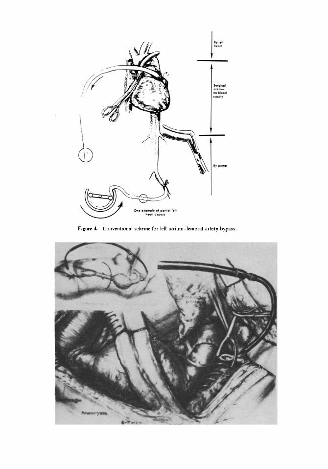
flow, which is inevitable during surgery on the thoracic aorta, carries the extra danger of depriving the spinal cord of blood supply and causing paraplegia. To prevent this complieation, the technique in the early sixties employed a temporary left heart bypass, complete with the arrest of coagulation in the patient with heparin, insertion of a temporary shunt between the left atrium of the heart anti the femoral artery, and the use of an external pump (figure 4). Not only was this cumbersome, it also caused troublesome generalized bleeding due to circulating heparin. Obviously a temporary shunt from the aorta above the level of the aneursym to a downstream location would have done the job just as well. If the need could be met locally without causing a major circulatory disturbance, so much better would the patient also be. To smoothen one's way, it is unnecessary to carpet a thorny path all the way; wearing shoes will do. But the localized approach had a catch: the temporary shunt should not clot during two or three hours when the aneurysm was being removed and the aorta reconstituted with a prosthetic graft. The GBH surface raised new hope, and we soon developed a GBH-coated shunt in the laboratory at the Hopkins for thoracic aortic bypass, which was subsequently used in patients with great success (Valiathan 1969) (figures 5, 6, 7).

Heparin bondin(¢: Then and now 1205
4. Heparin bonding--after GBH
For all its drawbacks, the GBH surface excited much interest in heparin bonding. The structure of the heparin molecule made it an admirable plaything in the hands of chemists. Heparin is a polysaccharide, the bulk of the chain consisting of alternating units of uronic acid and glucosamine, bound in !-4 linkages (figure 8). Most of the amino groups of the glucosamine residues are sulphated, only a minor proportion being acetylated. A variable fraction of the heparin chains are linked to serine or small peptides: in most tissues studied, it occurs primarily as single polysaccharide chains with molecular weight ranging from 5000 to 15,000. In addition to the single- chain type, a macromolecular form of heparin with much higher molecular weight also exists in tissues such as rat skin. Heparan sulphate, closely related to heparin, consists of a family of molecules in which deacetylation has occurred to a much less degree during biosynthesis. While heparin is stored in the granules of the mast cell, heparan sulphate is an ubiquitous component of the surface of many, if not all, cell types and is present in the form of a proteoglycan. Obviously this proteoglycan is different from heparin proteoglycan. Despite similarities in their polysaccharide components, they are distinct with respect to their core proteins. The presence of heparan sulphate on the endothelium perhaps contributes in no small measure to the fluidity of blood in the cardiovascular system. It is, so to say, a natural analogue of a heparinized surface.
Figure 3. GBH-coated plastic leaflet with no thrombus.
l | y leill h l a r t
i
)<7
Surgical o t e o ~ no b lood supply
By pump
One examale of part iol left heart bypass
Figure 4. Conventional scheme for left atrium-femoral artery bypass.

Figure 6. Shunt in position. Aneurysmal aorta being divided between clamps.
Figure 7. Aneurysm resection is over. Aorta being reconstituted by a polyester graft.
Figure 5. GBH-coated shunt being introduced into the left subclavian artery. Note the large aortic aneurysm. (Inset shows the scheme of bypass in the patient.)

1208 M S Valiathan
CH2OSO; CM2OSO; COO" CH~OSO; CHzOSO; ) - - o J - - o J - - o , - - o o
OH ON O 014 OH O OH OH O OH --0 O - J ~ ' - ~ J l - O 0 O--
C O* ~CH,
Figure 8. The repeating units in heparin,
Polymer
T Chloromethylation J-~/~ -/-~ CI CHzO CH 3 I
Polystyrene bonded CHzCl to polymer by cobalt irradiation
Quaternizationl with C6Hs N (CH3)2 Dimethylaniline
or pyridine
~ C Heparinization T C H3 ~[RO$O~" ] H3
l ~ I ~+ 4- CH z_ N-CsH. CH z- N C6H s I = I CH~ el- CH 3 CI-
[,,OSO Figure 9. A scheme for heparinizing a nongraphited surface via chloromethylation and quaternization.
Since GBH demonstrated that negatively charged heparin reacts with positively charged quaternary ammonium salts, chemists devised methods to bond quaternary groups directly to a number of polymers prior to heparinization. Another method was to include a minor proportion ofa quaternizable monomer such as vinyl pyridine in a polymer of interest and subsequently treat it for quaternization and heparinization (Leininger et al 1966). These and other techniques of surface modification yielded a large number of heparinized surfaces, which were studied for blood compatibility by placing the modified rings in the canine vena cava (Gottet al 1968a). Among the plethora of surfaces, investigators claimed to find antithrombogenicity in their respective creations, but lost all memory of it the next year! Perhaps the only surface to graduate from the experimental class was that developed by the Battelle Laboratories and applied subsequently in the celebrated Gott shunt (Gottet al 1966; Grade et al 1969). The Battelle surface utilized a cationic surfactant, tridodecylmethyl ammonium chloride (TDMAC), to bond heparin to the underlying polymer and eliminated the graphite layer (figure 9). This was the scenario a decade after GBH made its appearance.

Heparin bonding: Then and now t209
5. Heparin bonding now
A decade, in my view, is too short a period for a biomedical idea to grow and fructify. This is because the ideas are far ahead of available technology, if not the understanding of contemporary scientists. Gibbon conceived heart-lung bypass, which had to wait for 20 years for realization. In 1955 Melrose introduced the concept of potassium cardioplegia, which was promptly discarded; it returned to change the face of cardiac surgery more than 20 years later. When one looks at Souttar's mitral valvotomy, Lillehei's technique for conserving the chordal mechanism during mitral-valve replacement, and many other examples, one is struck by the recurrence of the 20-year interval for the maturation of biomedical ideas. When I see the recent interest in trans- myocardial puncture by Nd-YAG laser for myocardial revascularization, I tend to think that the time has come for the idea of the 'snake heart' operation which Prof. Sen advanced in the late sixties. Efforts in heparinizing polymer surfaces waxed in the late sixties and early seventies, but waned thereafter for 20 years, called the 'years of frustration' for work on blood-material interactions (Andrade et al 1981). Two decades later, heparin bonding has returned to occupy the centre stage of research in biomaterials.
The biological effect of heparin is primarily shown through its interaction with antithrombin. Thanks to the heavily sulphated residues, heparin binds to antithrombin with great affinity and accelerates the inactivation of coagulation factors, only a small fraction of the heparin molecule complexing with antithrombin. Since the earlier attempts to attach heparin by ionic bonding and surface grafting did not produce a stable linkage, efforts continued to couple heparin covalently through the carboxyl, hydroxyl and amino functions of the molecule. The resultant surfaces failed to show significant resistance to clotting, presumably due to the surface binding of the functional groups on heparin. However, Larm's description of what he called the 'end-point attachment of heparin' signalled a major departure in the method of heparin bonding (Larm et al 1983). Here, the chemical coupling took place at the terminal aldehyde group of reduced heparin without detriment to the anti-thrombin binding site of the heparin molecule. Reduced heparin is in the low-molecular-weight range of the heterogeneous family of heparins. Bound to prosthetic surfaces according to this method, heparin is stable and not leached on contact with blood or saline. Strongly anticoagulant, the orientation of the end-point-attached heparin on a surface resembles the distribution ofheparan sulphate proteoglycan on cell surfaces (figures 10 and 11). The thromboresistant effect of end-point-attachment of heparin, which has the proprietary name 'Carmeda bioactive surface', is far higher than the thrombin-inhibiting capacity of ionically bound heparin. The Carmeda surface is claimed to have a shelf life of 2 years.
The impact of the new bioactive surface of heparin on surgical practice has been considerable. In the first place, it has reduced the requirement for heparin and its neutralization by protamine during prolonged heart-lung bypass. Secondly, it has proved to be far gentler to blood than untreated surfaces in so far as it activates neutrophils and C3 complement to a much less degree (Nilsson et a! 1990) and reduces platelet count even less (Segesser et al 1990). The promise of biocompatibility of the Carmeda surface has been such that it has even been applied on implantable lenses to reduce the adherence and growth of cells.
The heart-lung bypass systems, including centrifugal pumps, which feature the

1210 M S Valiathan
H e p a r i n S u l p h a t e
Core P r o t e i n
Plasma Membrane
Figure 10. A diagram to show the distribution of heparin and heparinoid molecules on cell surface.
Heparln A c t i v e Binding Sequence
I Spacer Module
~/::"~" Blood contac t surface'"'::~'z~z':~
Figure 11. The new technique for creating heparin-active binding sequence. Note the similarity to figure 10.
new bioactive surface of heparin are costly, as new medical devices always are. Their cost-benefit analysis is a subject of ongoing debate in Western countries. But, to paraphrase Fermi, where do we, in India, come in? Over the past decade our group in Trivandrum developed and transferred for production a series of blood-contacting devices including the blood bag, oxygenator, cardiotomy reservoir and the heart valve. Soon a vascular graft will join the list. Two things common to them are the need for blood contact in clinical use and the need for import at high cost. Without exception, they are based on materials that have stood the test of time in surgical use. As such their biocompatibility and degree of medical safety are perfectly acceptable by contemporary standards. But in medical technology, as indeed in other branches of technology, a given product or process is no more than a stage in a never-ending process of evolution. Endowed with a heparinized surface, each of our devices will improve in function and pave the way for a new generation of advanced
Heparin bonding: Then and now 1211
devices. The blood bag (Pal et al 1988), for example, is made of sheets extruded from a PVC compound that differs little from the material used in other commercially available blood bags. If the blood-contacting surface of the bag is heparinized covalently, the preservation of blood elements, including platelets and serum proteins, would substantially improve and this would lengthen the safe storage period of blood. This is no small thing because the cellular components are literally thrown away at present after three weeks of storage, not to speak of the need to check the level of potassium before administering the plasma.
The oxygenator is the central unit of the heart-lung bypass system, which includes lengthy conduits of PVC tubes, a cardiotomy reservoir to receive blood from the heart chambers, and a pump (Venkatesan et al 1981, 1983; Valiathan 1985; Nazer et al 1991). The oxygenator is basically a disposable device which removes carbon dioxide from the venous blood and adds oxygen to it. It is perfectly efficient in terms of gas exchange during heart-lung bypass even though it can hardly compare with the lungs in surface area or blood compatibility. Since our oxygenator employs the bubble principle, which involves the bubbling of oxygen through a column of venous blood and the subsequent removal of excess bubbles by cascading the oxygenated blood over a defoaming substrate, its blood-contacting surface is quite large. Added to this are the surfaces of the cardiotomy reservoir and the lengthy conduits, which altogether must permit five litres of blood to pass through the system every minute. It is a tribute to the toughness and self-regenerative capacity of our tissues that the damage suffered by the cellular elements and serum proteins of blood after 3-4 hours of heart-lung bypass remains modest and reversible. One might say that the heart- lung bypass succeeds more by nature's healing processes than by our success in surface engineering! But even the modest damage to blood can cause morbidity, complications and prolongation of recovery. If the surface of the extracorporeal system had a stable, heparinized surface, heart-lung bypass for several hours could be far less traumatic to blood. This has been demonstrated by the Carmeda experience.
The artificial heart valve poses a different problem (Valiathan et al 1978; Valiathan and Bhuvaneshwar 1982; Bhuvaneshwar et al 1983a, 1991; Lal et a! 1984; Valiathan 1993). Here is a device which consists of a housing made of Haynes 25 alloy, a tilting disc of ultra-high-molecular-weight polyethylene, and a sewing ring of polyester cloth. Unlike the blood bag and oxygenator, it is a permanent cardiac implant which must open and close 100,000 times a day for many years without mechanical failure. A problem that continues to bedevil the mechanical valves is the formation of clots on the non-moving components of the valve and the obligation for the patients to take anticoagulant medications. The ring of plastic fabric which is sewn to fix the valve to the margins of the excised valve provides the most vulnerable site for the formation of clots. It is entirely possible that a heparinized surface on the sewing ring may further reduce the already low rate of thromboembolism and make mechanical valves safer.
Vascular grafts are seamless, crimped tubes woven or knitted from polyester yarn (Bhuvaneshwar et al 1981, 1983b). They function very well as replacement for arteries larger than 10 mm in diameter and save the lives and limbs of thousands of patients all over the world every year. Unfortunately their long-term patency rate drops sharply as the diameter of the artery becomes smaller and they are poor substitutes indeed for vessels as small as the carotid or the coronary arteries. Vascular surgery has failed to breach the 10mm barrier despite much effort. The effect of heparin
1212 M S Valiathan
bonding of the graft on the long-term patency of small-diameter grafts is therefore of great interest.
These are by no means the only examples of the clinical promise of heparinized surfaces. As we learn more of the biological role of heparins and the technology for heparin bonding improves, heparin-bonded surfaces will cast their mantle on not only cardiovascular but other devices as well. After all the distribution of heparan sulphate is not confined to the cardiovascular system.
6. Conclusion
The story of heparin bonding is typical of what is happening in biomaterials science. The long search for inert materials is giving way to the development of bioactive surfaces thanks to the realization that no surface in the body is inert. The change in direction from inertness to bioactivity is unmistakable.
The early hunters for a blood-compatible surface were exact men who wished to copy nature. But wherever they went, vibrant vessels vanished, filmy leaflets of heart valves died, the shining intima ceased to exist, and the grace and splendour of nature's handiwork were transformed into painted pipes, one-way check valves and plastic fabrics, which were studied and described in the greatest detail. The varied designs and the magic of self-renewal disappeared during their experiments, which turned the living celebration of nature into a mixture of chemical processes and cunning devices. When the point of disenchantment was close, the call has come for a return to nature. For none can deny that the attachment of heparin or albumin or cells to surfaces is no more than a humble attempt to clothe non-living materials in natural garments. To the extent the effort succeeds, we shall have triumphed in making materials biocompatible and medical devices safe.
References
Andradc J D, Coleman D L and Didesheim P 1981 Trans. Am. Soc. Artif. Intern. Organs 27 659 Bhuvaneshwar G S, Muraleedharan C V, Lal AV, Ramani A V and Valiathan M S 1991a Trans. Indian
Ceram. Soc. 50 87 Bhuvaneshwar G S, Muraleedharan C V, Ramani A V and Valiathan M S 1991b Bull. Mater. Sci. 14 1363 Bhuvaneshwar G S, Ramani A V and Valiathan M S 1983a Bull. Mater. Sci. 5 111 Bhuvaneshwar G S, Valiathan M S, Lal A V and Kartha C C 1983b Indian J. Med. Res. 78 556 Bhuvaneshwar G S, Venkatesan V S, Pattankar V L, Kartha C C, Lal A V and Valiathan M S 1981
Indian J. Med. Res. 74 580 Gott V L, Koepke D E, Dagget R L, Zimmerstoff W and Young W P 1961 Surgery 50 382 Gott V L, Leininger R L, Falb R D, Ameli M M and Valiathan M S 1968a Surgery 63 60 Gott V L, Whiffen J D and Dutton R C 1965 Science 142 1297 Gott V L, Whiffen J D, Leininger R I, Falb R D and Valiathan M S 1966 Bibl. Haematol. 29 863 Gott V L, Whiffen J D and Valiathan M S 1968b Ann. N Y Acad. Sci. 146 21 Grode G A, Anderson S J, Grotta H M and Falb R D 1969 Trans. Am. Soc. Artif. Intern. Organs 15 1 Lal A V, Chandran M, Bhuvaneshwar G S and Valiathan M S 1984 Indian let. J. 61 328 Lalonde J B, Valiathan M S and Ballinger W F 1964 JAMA 187 117 Larm O, Larsson R and Olsson P 1983 Biomater. Meal. Dev. Artif. Organs 11 161 Leininger R I, Epstein M M, Falb R D and Grode A 1966 Trans. Am. Soc. Artif. Intern. Organs 12 151 Nazer Y, Vijayakumar H, Nagesh D S, Vijayan S and Valiathan M S 1991 Indian J. Thorac. Cardiovasc.
Surg. 7 138 Nilsson L, Storm K E, Thelin S, Bagge L, Hultman J, Thorelius J and Nilsson U 1990 Artif. Organs 14 46

H e p a r i n bonding . T h e n and now 1213
Pal S N, Kalyanakrishnan V, Jayaprakash P A and Ramani A V 1988 Indian J. Technol. 26 45 Segesser L K, Lachat M, Leskosek B and Turna M 1990 Perfusion 5 26 Topaz S R, Ameli M M, Morovati S S, Valiathan M S, Brown B G and Gott V L 1967 Trans. Am. Soc.
Artif. Intern. Organs 13 294 Valiathan M S 1969 Ann. Royal Coll. Sur 9. England 45 131 Valiathan M S 1985 Curt. Sei. 54 964 Valiathan M S 1993 Indian J. Thorac. Cardiovasc. Sur 9. 9 8 Valiathan M S and Bhuvaneshwar G S 1982 Indian Heart J. 34 387 Valiathan M S, Bhuvaneshwar G S and Venkatesan V S 1978 Indian J. Suro. 40 283 Valiathan M S, Weldon C S, Bender H W, Topaz S R and Gott V L 1968 J. Surg. Res. $197 Valiathan M S, Whiffen J D, Weldon C S and Gott V L 1966 Trans. Am. Soc. Artif. lntern. Organs 12 174 Venkatesan V S, Lal A V, Bhuvaneshwar G S and Valiathan M S 1981 J. Extracorp. Teeh. 13 209 Venkatesan V S, Lal A V and Valiathan M S 1983 Bull. Mater. Sei. 5 97 Whiffen J D and Beckler D C 1966 J. Thorac. Cardiovasc. Surg. 52 121 Whiffen J D, Young W P and Gott V L 1964 J. Thorae. Cardiovase. Surg. 48 317