teratology in zebrafish embryos: a tool for risk...
TRANSCRIPT
1
Teratology in ZebrafishEmbryos: A Tool for Risk
Assessment
Nadeem Ali
Master of Science Programme in Veterinary Medicinefor International Students
Faculty of Veterinary Medicine and Animal ScienceSwedish University of Agricultural Sciences
Uppsala 2007

2
Report - Master of Science Programme in Veterinary Medicinefor International StudentsFaculty of Veterinary Medicine and Animal ScienceSwedish University of Agricultural SciencesReport no. 65ISSN 1403-2201

3
Teratology in ZebrafishEmbryos: A Tool for Risk
Assessment
Nadeem Ali
Division of Pathology, Pharmacology and ToxicologyDepartment of Department of Biomedical Sciences and Veterinary Public Health
Faculty of Veterinary Medicine and Animal Science
Swedish University of Agricultural SciencesUppsala 2007
4
The present thesis is a partial fulfilment of the requirements for aMaster of Science Degree in Veterinary Medicine for InternationalStudents at the Swedish University of Agricultural Sciences (SLU), inthe field of aquatic toxicology.
Nadeem Ali,
Division of Pathology, Pharmacology and ToxicologyDepartment of Department of Department of Biomedical Sciences and Veterinary PublicHealthFaculty of Veterinary Medicine and Animal ScienceSwedish University of Agricultural Sciences (SLU)P.O. Box 7028, SE- 750 07 Uppsala, SwedenPrint: SLU Service/Repro, Uppsala 2007

5
To my family
6
Table of contents
Abbreviations 8
Acknowledgements 9
Abstract 11
Background 12
Historical background of teratology 12
Mechanism of teratogenesis 13
Susceptibility of teratogenesis with age 14
Early embryonic development 15
Organogenesis and early differentiation 15
Late embryonic development 15
Laboratory animals for teratogens testing 16
Mammals 16
Amphibians 17
Birds 17
Fish 18
Teratogens 18
Recreational and social teratogens 18
Pharmaceutical teratogens 19
Industrial and environmental teratogens 19
Agricultural teratogens 19
Metabolic and infectious diseases 19
Model substances for the study 20
Retinoic acid 20
Lithium 21
Ethanol 21
6- Aminonicotinamide 22
Ochratoxin A 22
Arsenic 23
References 24

7
Aim of the study 31
Research paper 32
Teratology in zebrafish embryos: a tool for riskassessment
32
Abstract 32
Introduction 33
Material and method 36
Test material 36 Animals 36 Test design 37 Exposure and embryo developmental test 37 Statistics End points
3839
Results 40
Retinoic acid 40 Lithium hydroxide 43 Ethanol 45 6- Aminonicotinamide 47 Ochratoxin A 50 Sodium arsenate 53 LOEC and NOEC values table 55
Discussion 56
References 61

8
Abbreviations
RA Retinoic acid6-AN AminonicotinamideOA Ochratoxin AFAS Foetal alcohol syndromeCNS Central nervous systemDNA Deoxyribonucleic acid,T-RNA Transfer Ribonucleic acidANOVA Analysis of varianceNOEC No Observed Effect ConcentrationLOEC Lowest Observed Effect ConcentrationHPF Hours post fertilizationDMSO Dimethyl SulfoxideM Molar

9
Acknowledgements"In the name of God, most Gracious, most Compassionate".
The study was conducted at the Division of Pathology, Pharmacology andToxicology, the Department of Biomedical Sciences and Veterinary PublicHealth, Swedish University of Agricultural Sciences (SLU), Uppsala,Sweden.I am pleased to tender my humble gratitude for all the people who were withme from the commencement of study till its termination. I would like to paymy respect to all those researchers whose work helped me to build up myresearch work.
Firstly I would like to express my thanks for the Prof. Dr. Leif Norrgren,who accepted me as a Master student and kindly supervised me during thewhole study period. You were so kind to me during whole period that Ireally don’t have words to say thanks to you and to express my gratitude foryou. Just for you sir:"I would thank you from the bottom of my heart, but for you my heart hasno bottom." "My peers accept me and respect me, and that's enough."
Gunnar Carlsson, my assistant supervisor, you have been very patient andhelpful in answering all my questions and helping me in research work.Your resolute and calculative approach in coming up with solutions withhaving a multiple orientation with various things simultaneously is veryimpressive. Örn Stefan and Anna Norman it was really nice working withyou and I really enjoyed the time I have spend working with you. I wouldlike to say thanks to the whole department for their kind help which I gotfrom them time to time during my stay here.
I express my gratitude and high regards for Dr. Karin Östensson, Directorof International Master of Science programme, for giving me an opportunityto participate in the programme. I give my sincere thank to Marie Sundbergfor her excellent help and assistance given to me during my stay in Sweden.As well as I would like to take the opportunity to give my sincere regards tothe practical help I use to get from Lundqusit Anne-Sofie.
Javed Iqbal Saleemi, Shamraiz Shehzad, Toafeer Mehdi, Imran Hussan,Fahad Gulraiz, Mehboob Danish, Mir Mazhar, Muhammad Imran and AbidHussain are some of my best friends with whom I have spent a lot of wondermoments during my university years at Faisalabad, I would love to recall

10
these moments always. I am really blessed and fortunate to have friends likeyou in my life. I wish you all the success in your lives. I am very lucky tohave some very good friends during my stay here at Uppsala and I have a lotmemories with them, especially Muhammad Shahid Khan, MuhammadOmer Khan, Muhammad Shakeel, Naveed Nazir and co. May GOD blessyou all the success in your lives.
Femi , Hitesh , Shahiduzamman, Guldasta, Anthony, Victor and Girogi, itwas really very nice time with all of you guys, wish you all the best for yourfutures. It will be injustice if I will not say thanks to Joliot Bravo, thank youBravo without your help it would have been more stiff task for me toachieve what I have achieved today.
My highest regards for my whole family, I will never ever forget theirsacrifices, love and devotion to me and the comfort I get from them. I am indearth of vocabulary to express my feelings towards you all.

11
AbstractTeratology, study of abnormal prenatal development, as a descriptivescience has starts with written language. The modern experimentalteratology era started in the early quarter of 20th centaury. Since thethalidomide catastrophe in early 1960s regulatory agencies launchedrequirements for new drugs to be thoroughly tested on animals priorto human use. One of the major concerns in the teratological studiesis the mechanism of teratogenesis; it is very difficult to know theexact mechanism of teratogenesis. However there are many proposedmechanism of teratogenesis by Wilson 1973. Teratogens induce oneor multiple unique pathogenic responses in the developing embryos.Susceptibility f teratogenesis varies with age and therefore can bedivided into three developmental periods: early embryonicdevelopment, organogenesis and early differentiation, and lateembryonic development.Animal based studies provide the initial guideline if a chemical ordrug may present a teratogenic risk. A variety of laboratory animalsfrom different classes of animals are being used for the teratologicalstudies. Rat, rabbit, mice, hamster, and non human primates are themost prevalent laboratory animal species of the mammal class.Xenopus laevis of the amphibian class has been used and suggested asa model for mammalian teratogenicity. From the bird class chicken,duck and quail have been used most often in laboratory studies.Zebrafish, Japanese medaka and fathead minnow are the mostcommonly used laboratory fish species, promoted by OECD forfuture testing of chemical toxicity. Teratogens can be classified asrecreational and social teratogens, pharmaceutical teratogens,industrial and environmental teratogens, agricultural teratogens, andmetabolic and infectious diseases. In the present study modelsubstances were selected from the different classes of teratogens. Theselected substances were; retinoic acid, lithium, ethanol, 6-aminonicotinamide, ochratoxin A and arsenic, .Key words: teratology; teratogenesis; retinoic acid; lithium; ethanol;6-aminonicotinamide; ochratoxin A ; arsenic; zebrafish

12
Background
Historical background of teratologyTeratology is the study of abnormal prenatal development and congenitalmalformations, which may be caused by exposure to chemicals or physicalfactors. Teratology as a descriptive science starts with written language, a marblesculpture from southern Turkey dating back to 6500 B.C., depicts conjoined twins(Warknay,1983), and Egyptian wall paintings of human malformed conditionssuch as cleft palate and achondroplasia have been dated as early as 5000 yearsago. The Babylonians Greek and Romans believed that abnormal infants werereflections of stellar events and such were considered to be portents of future. TheLatin word monstrum from monstrare (to show) or monere (to warn) is derivedfrom this perceived ability of malformed infants to foretell the future. In turn rootof teratogenesis is from Greek word teraton meaning “wonder” and by derivation“monster” (Francis, 1994).
Modern experimental teratology as a science was born in 1920s and 1930s, whenthe birth of malformed piglets from sows fed an experimental diet high in fat ordeficient in vitamin A elicited shocking teratogenic effects (Hale, 1933;Schardein 1993). Subsequent evidence about teratology came to light over thenext two decades; correlation of particular birth defects of children with maternalRubella infection in 1941 (Gregg, 1941) and with environmental mercurycontamination in 1956 (Igata, 1993); malformed rat born following the inclusionof a growth inhibiting amino acid mimic in their mother’s diet (Murphy et al.,1956) and malformed children born following failed Aminoptern inducedabortions (Thiersh, 1956).
Thalidomide represents the 1st case of a substance producing minimal toxicity inthe adult but considerable toxicity in the foetus (MC Bride, 1961; Lenz, 1966).Since the Thalidomide catastrophe regulatory agencies launched requirements fornew drugs to be thoroughly tested on animals prior to approval for marketing(Bailey et. al., 2005; Rowan, 1984).
The confounding nature of results from experiences based on animal studies hasrequired principles to be elaborated and revised (Wilson, 1997; Finnell, 1999).Many variables have been found to interfere with interspecies and animal humancomparisons (Nielsen et al., 2001; Palmer, 1986), these must be considered whendesigning developmental and reproductive toxicology studies. They can besummarized as;
I. Susceptibility to teratogenesis depends on the genotype of the conceptusand how it interacts with the environment (Schardein, 1993).
II. Susceptibility to a teratogenic agent varies with the developmental stage atwhich the exposure occurs (Wilson, 1972).
III. Teratogenic agents act in specific ways on developing cells and tissues toinitiate abnormal embryogenesis (Wilson, 1973).

13
IV. Access of an adverse environmental agent to developing tissues depends onthe nature of the agent (Polifka and Friedman, 1999).
V. The manifestations of deviant development increase in degree as dosageincrease from the co-effect to the lethal level (Brent, 1995).
VI. Manifestation of deviant development includes death, malformation andgrowth retardation. (Wilson ,1972).
Animal based studies of developmental toxicology provide the initialinformation’s on whether a drug or chemical constitute a teratogenic risksubstance. Typically, a range of doses administered via the most appropriate routeis given to pregnant animals during the period of embryonic organogenesis andthe outcomes are compared to untreated control animals. The most prevalentspecies used are mice, rats, hamster, primates and rabbits but no one species is anideal experimental animal because they are not fulfilling the “Ideal” criteria suchas producing large litters after a short gestation, inexpensive maintenance and aninability and unwillingness to’ bite, scratch , kick, howl or squeal’ (Wilson,1975). It is also acknowledged that the laboratory handling of animals can inducephysiological stress responses and cause alterations in behaviour and both thesefactors can affect teratogenicity results. Participation in the teratology researchitself is so inherently stressful that this can never be excluded (Balcombe et al.,2004).
During the past 30-40 years a lot of money has been spent on teratological testing,but still scientist are looking for alternative laboratory species which can producelarge litter after a short gestation, easy and inexpensive handling, inability andunwillingness to’ bite, kick, scratch , howl or squeal’ (Wilson, 1975).
Mechanism of Teratogenesis
Mechanism of teratogenesis falls into two broad categories based on the etiologyof the congenital malformation:
a. Errors in the genetic programming based on deviations in thegenotype of the embryo or the low probability for errors of a normalgenotype.
b. Environmental agents or factors that interact with the embryoduring the period of development, e.g., drugs, chemicals, radiation,infections or mechanical factors.
Etiology of malformations includes both genetic and environmental factors, butthere is another large category labelled as unknown, e.g., polygenic,multifactorial, spontaneous errors of development and synergistic interactions ofteratogens.To induce malformations, teratogens must cross the placenta or reach thedeveloping embryo through some other route, which makes phenomenon ofteratogenesis applicable to all organisms including those in which embryonicdevelopment occurs outside the mother.

14
A number of suggested mechanisms involved in teratogenesis has been proposedby Wilson (1973);
1. Mutation: Changes in the nucleotide sequence in DNA. However,mutation is unlikely to play a role in the production of malformationsfollowing in utero exposure. Mutagens usually produce cytotoxic effectswhich are related to the cell destruction not to the genetic changes thatpersist and effect embryonic development for many cell cycles in thedeveloping embryo.
2. Chromosomal aberrations: alterations in the amount of DNA. However,chromosomal aberrations play little or no role when induced in somaticcells of the developing embryo but may cause cell death and retardationof differentiation.
3. Altered nucleic acid synthesis and function: a disturbance in translation,transcription or DNA synthesis.
4. Mitotic interference: a disturbance in the cell cycle.5. Lack of precursors, substrates and co-enzymes for biosynthesis: a general
or specific nutritional deficiency.6. Altered energy source: interference with the citric acid cycle or terminal
electron transport system.7. Enzyme inhibition: limited or specific enzyme inhibition.8. Osmolar imbalance: alterations in the fluid pressures, viscosities and
osmotic pressures.9. Altered membrane characteristics: a disruption in membrane transport
and permeability.10. Other mechanisms: an extensive list of possible mechanisms for which
there is a little scientific support.
In the developing embryo many of the above mentioned cellular insults can causeunique pathogenic responses (Tyl, 2000) such as:
1. Reduced cell proliferation.2. Cell death.3. Altered cell-cell interactions4. Reduced biosynthesis.5. Mechanical disruption of developing structures.6. Inhibitions of morphogenetic movements.
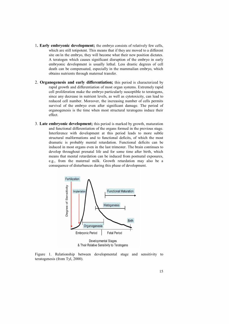
Susceptibility of teratogenesis with age
Susceptibility to teratogenesis varies with the developmental stage (F1) (Tyl,2000). It is generally accepted that every teratogen acts at specific times ofdevelopment, and that these vary, within limits, between agents. The limits aredetermined by the developmental sequences of the embryo. The stages ofgestation can be divided, at first approximation, into 3 developmental periods:Early embryonic development, organogenesis, and late embryonic development.
15
1. Early embryonic development; the embryo consists of relatively few cells,which are still totipotent. This means that if they are moved to a differentsite on/in the embryo, they will become what their new position dictates.A teratogen which causes significant disruption of the embryo in earlyembryonic development is usually lethal. Less drastic degrees of celldeath can be compensated, especially in the mammalian embryo, whichobtains nutrients through maternal transfer.
2. Organogenesis and early differentiation; this period is characterized byrapid growth and differentiation of most organ systems. Extremely rapidcell proliferation make the embryo particularly susceptible to teratogens,since any decrease in nutrient levels, as well as cytotoxicity, can lead toreduced cell number. Moreover, the increasing number of cells permitssurvival of the embryo even after significant damage. The period oforganogenesis is the time when most structural teratogens induce theireffect.
3. Late embryonic development; this period is marked by growth, maturationand functional differentiation of the organs formed in the previous stage.Interference with development at this period leads to more subtlestructural malformations and to functional deficits, of which the mostdramatic is probably mental retardation. Functional deficits can beinduced in most organs even in the last trimester. The brain continues todevelop throughout prenatal life and for some time after birth, whichmeans that mental retardation can be induced from postnatal exposures,e.g., from the maternal milk. Growth retardation may also be aconsequence of disturbances during this phase of development.
Figure 1. Relationship between developmental stage and sensitivity toteratogenesis (from Tyl, 2000).
16
Laboratory animals for teratogen testingAnimal-based studies of developmental toxicology provide the initial informationon whether a drug or chemical may present a teratogenic risk. Typically, a rangeof doses administered via the most appropriate route is given to pregnant animalsduring the period of embryonic organogenesis, and the outcomes compared tocontrol untreated animals. The most prevalent species used are mice, rats andrabbits. The usual sample size is 20 individuals per dose, and the dose range isselected so that the highest dose causes signs of toxicity, the lowest causes nodiscernable effect in the foetus, and at least one intermediate dose.
Considering, physiological and biochemical differences between animal speciesused in teratology research and humans, means that it is impossible for a singlespecies to have all desirable properties. Desired characteristics for the idealteratology animal have been proposed in a ‘wish list’, but no species comes closeto fulfilling the criteria. For example, no species absorbs, metabolises andeliminates test substances like a human nor possesses the same placental transferproperties; no one species has the same pre-term developmental and metabolicpatterns as humans; and, even if it were possible to meet all of these standards,the animal would be unlikely to meet other ‘ideal’ criteria such as producinglarge litters after a short gestation, inexpensive maintenance, and an inability andunwillingness to ‘bite, scratch, kick, howl or squeal’ (Wilson, 1975).
Mammals
Rodents have become the most commonly used group for evaluating potentialhuman teratogens. Proponents of animal use, while admitting that it can only givean approximation of effects in humans, praise the rat model since for many yearsall human teratogens identified exhibited teratogenesis in rats (Tuchmann-Duplessis et al., 1972). There are, however, important exceptions, such as withthe prostaglandin E1 analogue misoprostol: treatment of humans with this drugfor peptic ulcer disease or to initiate labour has a strong association with foetalmalformations known as Moebius syndrome (Pastuszak et al., 1998), but is notteratogenic in the rat even up to 10 times the human dose (Klasco and Heitland,2003). There are many examples of positive results in rodents that have little orno effect in humans (‘false positives’), especially at normal exposures andtherapeutic dose levels. Notable examples include glucocorticoids andbenzodiazepines, which induce oral clefts in rats, mice and rabbits, but not inhumans (Baxter and Fraser, 1950; Buresh and Urban, 1970; Czeizel 1987;Fainstat, 1954; Fraser and Sajoo, 1995; Miller and Becker, 1975; Pinsky andDiGeorge, 1965; Rosenberg et al., 1983; Shepard 1994; Shiono and Mills, 1984;Walker 1971; Wilson et al., 1970). Also aspirin, which causes cardiacmalformations in several species of animals, such as the rat and the rhesusmonkey, is harmless in man (Beall and Klein, 1977; Klein et al., 1981; Slone etal., 1976; Werler et al., 1989; Wilson et al., 1977).Because of the inherent problems and inadequacies of teratology testing andresearch with the five groups of animals most commonly used (mouse, rat, rabbit,
17
hamster, and monkey), scientists have tried to incorporate other species into theirexperiments in an attempt to find something approaching that elusive ‘idealanimal’. Dogs (more specifically beagles) were tested with an array of knownteratogenic compounds, but deemed unsuitable due to poor sensitivity; in anycase, it is known that many drugs are metabolised differently in the dog, and thereare particular problems with extrapolation from dogs to humans with reference tosteroids (Schardein, 1993). By using cats there was some promising concordancewith several compounds, though in common with dogs they are known tometabolise a significant number of drugs differently, some uniquely, and therewere discordant results compared to other species with compounds such ashydantoins, thalidomide, and especially the anti-leukaemia drug and abortifacientaminopterin, which is highly teratogenic in humans but not at all in cats(Schardein, 1993). Pigs were found to be as insensitive as dogs, ferrets did notlive up to early expectations, and non-human primates, despite their closephylogenetic relationship to humans, have been particularly disappointing as apredictive model (Schardein, 1993). Over 100 teratogenic agents classified as‘possible’ or ‘probable’ have been tested in nonhuman primates, and the vastmajority showed a high level of discordance; of the known human teratogenstested, only about half were found also to be teratogenic in one or more primatespecies (Bailey et. al., 2005 ).
Amphibians
Amphibians have proven to be useful tools in both classical and modernembryology. The African clawed frog, Xenopus laevis, has also been suggested asa model for mammalian teratogenicity. Amphibian development is also used toidentify overall aquatic toxicity and the chemicals used for such studies tend to behighly relevant to ecological problems. Xenobiotics known to affect developmentin amphibians include methyl mercury (5 to 30 ppb), high levels of selenium, thefungicides dichlone and chloranil and benzo (a) pyrene.
Birds
Birds have proven important in all aspects of developmental biology andtoxicology. Chick embryos are excellent experimental subjects for embryologicinvestigations, since they are easily obtained, can be maintained outside the eggfor at least 24 hours, and are large enough for easy manipulation. The effect ofDDT in thinning eggshells (Ratcliffe, 1967) and of PCBs in causingmalformations in birds, have been documented repeatedly. Recently, seleniumemerged to induce developmental malformations in birds (Ratti et al., 2006).Chicken, duck and quail have been used most often in laboratory studies, butcorrelations between pollutants in eggs and reproductive problems in wild birdshave been carried out in numerous species ranging from gulls to cormorants andospreys. It is quite clear that there is as much variability among avian as amongmammalian species in response to reproductive and development toxicants.Chlorinated hydrocarbon insecticides, PCBs, and selenium, many or mostorganophosphate insecticides (Ops) are potent teratogens in birds. The avian

18
teratogenicity of organophosphates is in striking contrast to their inactivity inmammalian development. Lead on the other hand, appears to be less toxic toavian than to mammalian development.
Fish
Development in fish has been studied because of the economic importance ofmany species and because fish are used as indicators in connection withenvironmental assessment. Zebrafish, Japanese medaka, goldfish, rainbow troutand fathead minnow are the most common laboratory species, which also are keyspecies promoted by OECD for future testing of chemical toxicity.
The zebrafish, Danio rerio, has been used extensively as a model species fordevelopmental biology. This species is small in size (3-5cm in length), easilyobtainable, inexpensive, readily maintainable, under appropriate conditions willprovide a large number of non-adherent and transparent eggs (Laale, 1997). Onefemale lays approximately 50 to 200 eggs per day. The morphological andmolecular basis of tissues and organ development in zebrafish are in generaleither identical or similar to other vertebrates including men (Chen and Fishman,1996; Granato and Nusslein-volhard, 1996). There is extensive similarity betweenthe zebrafish and human genomes so many human developmental and diseasegenes have counterparts in the zebrafish. The zebrafish genome is 1700 millionbase pairs in length, about half the size of the human genome. Most humangenes have homologues to zebrafish and the functional domain of the proteinsuch as ATP binding domain of kinases are almost 100% identical betweenhomologous genes, although the similarity over the entire protein is about 60%(Langheinrich, 2003). As protein function largely resides in functional domainswhere drugs often binds, the zebrafish model is highly valid model for studyingdrug effect in human. Solvents, oil dispersants, pesticides and metals are knownto affect fish development deleteriously.
Teratogens
A teratogen is any medication, chemical, infectious disease, or environmentalagent that might interfere with the normal development of a foetus and result inthe loss of a pregnancy, a birth defect, or a pregnancy complication. There are avariety of teratogens that are relatively common. Some examples are listed, butthere are other teratogens which are not on this list.
Recreational and Social teratogens
Two thirds of all infant mortalities are due to alterations in foetal development ordevelopment during the first year of life. Often times, recreational drugssignificantly reduce foetal and post natal growth and are known to significantlyincrease infant mortality, such as alcohol (Gilbert, 1997; O'Rahilly, 1992),cocaine (Schardein, 1993), cigarettes (Klaassen, 1996). These teratogens usually

19
disrupt foetal development before the mother knows she is pregnant and has achance to change her lifestyle.
Pharmaceutical teratogens
Most embryonic organs and the central nervous system are extremely sensitive tothe teratogenic affects of pharmaceuticals in the early development. Often timesthese drugs causes embryonic malformations before the woman knows she ispregnant. Thalidomide (Schardein, 1993), Diethylstilbestrol (Schardein, 1993),Retinoic Acid (Schardein, 1993), Valproic Acid (Smith, 1982; Schardein, 1993),Warfarin (Smith, 1982; Schardein, 1993), chemotherapy (Brent, 1986), lithium(Gilbert, 2003; Stachel et al., 2003), nicotinic acid (Johnson and McColl, 1995)etc are some examples of Pharmaceutical teratogens.
Industrial and Environmental teratogens
In the modern era where rapidly growing industries are fulfilling the needs ofgrowing population and has great economic importance, on the other hand theseindustries are releasing a huge amount of waste products in the environment.These waste products are not only polluting the environment but also leave severedetrimental effects on the quality of life. Among these a lot of waste productshave teratogenic affects on the population such as organic solvents, chemicals,arsenic (Wlodarczyk et al., 1996), cadmium and lead (Weiss et al., 1986),anaesthetic gases, organic mercury(Harada, 1986) etc.
Agricultural teratogens
Studies have determined that insecticides (organochlorine insecticides) mayinterfere with fertility and reproduction by mimicking estrogen-like compounds.In some avian species, steroid metabolism is altered, making it impossible totransport calcium to the developing egg shell. Insecticides have also been found toaccumulate and concentrate in the yolk sac of developing fish. Rodent studies ofDDT showed reduced testicular size in males and estrogenic effects in femalerats. Other teratogenic insecticides include dieldrin (reduction in fertility,increased mortality, delayed ossification and increased supernumerary ribs) andKepone (reduction in sperm count and reduced motility).Herbicides, 2, 3, 7, 8-tetrachloro-dibenzo-p-dioxin (TCDD) were used in 2, 4, 5-T teratological studiesresulting in cleft palate and congenital renal abnormalities.
Metabolic and Infectious Diseases
The TORCH complex is a group of similar malformations induced by microbialteratogens. These microbes affect 1-5% of all live births and are among theleading causes of neonatal morbidity and mortality. These organisms are Syphilis,Cytomegalovirus, rubella (South et al., 1986), cytomegalovirus (Reynolds et al.,1986), genital herpes, toxoplasmosis (Larsen, 1986), Fifth disease, and

20
chickenpox. Some metabolic disorders also induce malformations in the embryose.g. Diabetes, hyperthermia (high fever). General symptoms include prematurebirth, growth retardation, neurological abnormalities, and damage of the eye,liver, heart and ear as well as bone lesions. Microcephaly, hydrocephaly, seizuresand psychomotor retardation accompany these malformations.
On the basis of study documentation in animals and human and risk assessmentto human teratogens are classified into different categories as shown in the table(A).
These categories are in agreement with FDA regulations as described in Millstein(1980): Table (A)
Category A No demonstrated risk; possibility of foetal harm is remote.
Category B Either: no evidence of foetal risk in extrapolation from animaldata plus no information from human studies, or: evidence ofadverse effects in animal studies that could not be confirmed inhumanstudies.
Category C Either: evidence of adverse effects in animal studies combinedwith no studies in humans, or: studies in animals and humans arenot available. Drugs only given to pregnant women when thepossible benefit outweighs the risks.
Category D Positive evidence of foetal risk. Drugs may be given to pregnantwomen when the benefits are acceptable despite the risk.
Category E Known not to affect animal reproduction but no human data.
Category X Animal or human studies or human experience have revealedfoetal abnormalities. Risk of use in pregnant women outweighsany possible benefits.
Model substances for the experiment
Retinoic acid (RA)
RA is the acidified form of Vitamin A. It is a drug commonly used to treat acnevulgaris and keratosis pilaris. In human foetus 13-cis-retinoic acid results in acharacteristic pattern of anomalies, including absent or defective ears, absent orsmall jaws, cleft palate, aortic arch abnormalities, and abnormalities of the centralnervous system. Similar anomalies are observed in other mammals. In mice, forexample, embryonic exposure to retinoic acid results in axial truncation andcauses a dramatic reduction in the sizes of the first and second pharyngeal arches,
21
which normally form the jaw, ear, and other facial bones (Gilbert, 2003; Ligas,2000).
RA disrupts development by altering the expression of Hox genes, causing the re-specification of the anterior–posterior axis and inhibition of neural crest cellmigration from the cranial region of the neural tube (Gilbert, 2003). RA cannotbind directly to genes, so in order to affect gene expression, the RA moleculeneeds to bind to specific RA receptors (RAR). After binding, the receptor becomesan active transcription factor. The RA-bound RARs have at least two modes ofaction, one of which is to bind to their DNA enhancer sequences and activateparticular genes that are not usually activated in these cells. These genes includecertain homeotic genes that specify the anterior-posterior position along the bodyaxis. In this way, they can cause homeotic transformations, generally convertinganterior structures into more posterior structures (Gilbert, 2003). In the embryo,there is a gradient of retinoic acid from the anterior end to the posterior end.Excess RA results in a posterior region having a higher than normal level of RA,and so more anterior Hox genes are expressed in typically posterior regions(Ligas, 2000).
Lithium
Lithium, in the form of lithium carbonate, is regularly prescribed to people withmanic depressive (Bipolar) disorder. It takes the peaks and the valleys out of theemotional swings that a person with disease suffers, and is thought to do sothrough its control of receptors for the neurotransmitter glutamate. Since late1800s embryologist have known from studies on non human embryos that lithiumcauses severe developmental defects. The attribute teratogenic effects areincreased risk of heart defects especially Ebstein’s anomaly which affects thetricuspid valve and seriously compromises heart ability to function effectively.Spina bifida and floppy infant syndrome are also reported defects.
In studies on non human embryos, the developmental defects caused by lithiumare clear. As early as 1982 Herbst studied that echinoderm embryos exposed tolithium salts undergo exogastrulation. More recently it has been shown that bothXenopus and zebrafish embryos exposed to lithium during cleavage stages aredorsalized, showing severe defects in the posterior region (Gilbert, 2003; Stachelet al., 2003). These teratogenic defects are caused by lithium’s inhibition ofglycogen synthase kinase in the Wnt/β catenin or by depletion of inositoltriphosphate in the PIP (phosphoinositol pathway) cycle as the dorsal-ventral axisis being established (Klein and Melton, 1996; Stachel et al., 1993; Baraban,1994). In the zebra fish, later exposure (at the eleventh cleavage throughgastrulation) causes anterior defects especially of the eyes, in some cases causinga lack of eyes altogether (Stachel et al., 1993).
Ethanol
Ethanol, widely consumed as a recreational drug, has long been stronglyassociated with teratogenesis as FAS (Jones et. al., 1973). FAS present up to onein three children of alcoholic mothers. FAS manifestations include growth

22
deficiency, CNS problems, characteristic facial features and organ malformations(Streissguth et al., 1995; Gilbert, 1997; Cartwright and Smith, 1995; Smith,1997; Sulik et al., 1988). Each year, up to 40000 babies are born with somedegree of alcoholic related damage (Sokol et. al., 2003).
Ethanol is able to permeate the placenta and enter foetal circulatory system,thereby causing developmental abnormalities. Ethanol impairs placental bloodflow to the foetus by constricting blood vessels which induce hypoxia and foetalmalnutrition. Ethanol rapidly crosses the placenta and blood brain barrier of thefoetus. There are many proposed mechanism of action for ethanol such as alteredneural crest cell migration, increased neural crest cell death or general cell deathby superoxide radial lysis of cells or mitochondrial cell dysfunction. Ethanol mayinhibit growth factor regulating cell proliferation and survival effects on Glialcells effects on development of neurotransmitter systems.
In zebrafish ethanol is responsible for abnormal migration of prechordal platecells that ultimately causes cyclopia and other deformities (Blader and Strähle,1998). The prechordal plate cell expressed genes like goosecoid and Islet 1, whichcontrol cell differentiation in the anterior region of the embryo (Blader andSträhle, 1998). Sulik (1988) studied that ethanol appears to cause abnormal celldeath. Ethanol achieves apoptosis by activating the cell’s self destructionmachineries (Sulik et al., 1988). Therefore in addition to ectopic prechordalplates specific gene expression, ethanol induced apoptosis appears to contribute tothe observed deformities in zebrafish embryos (Sulik et al., 1988).
6-Aminonicotinamide (6-AN)6-Aminonicotinamide is a potent niacin antagonist (Johnson and McColl, 1995)having carcinostatic and teratogenic effects.6-AN is a reported teratogen inlaboratory animals and induce cleft lip, stunted growth and hind limb defects inmice (Pinsky and Fraser ,1960) and similar malformations were observed inrabbit . Nicotinamide is transferred actively across the placenta (Hill and Longo,1980; Kaminetzky et al., 1974) and into breast milk (Deodhar et al.,1964).Abnormal neural tube closure defects and other abnormalities wereobserved in chick when eggwhite was replaced with a solution containing 20 mgof nicotinic acid (Hansborough, 1974).
6-AN induce teratogenesis by inhibiting nicotinamide adenine dinucleotidedependent reactions during ATP synthesis. (Wilson et al., 1975; Dietrich et al.,1958).
Ochratoxin A (OA)OA is an important food borne mycotoxin and is a potent teratogenic agentproduced by several species of aspergillus and pencillium. OA is a reportedteratogen in rats (Brown et. al.,1976; Mayura et al.,1982), mice (Hayes etal.,1974), Hamsters (Hood et al.,1976) and chicken (Gilani et al.,1975)

23
OA inhibits protein synthesis by competition with the amino acid phenylalanine(Phe) in the phenylalanyl-tRNA synthetase-catalyzed reaction (Bunge et al.,1978; Creppy et al., 1979).At post transcriptional level with OA having a directeffect on the translation step in the protein synthesis. This involves thecompetitive inhibition of phenylalanine-tRNA synthetase, so that aminoacylationand peptide elongation are stopped (Creppy et. al., 1979; WHO series28).
Manolova et al., (1990) described that one possible way of OA induction ofteratogenesis is chromosomal aberrations by conducting positive in vitroexperiment on human lymphocytes.
OA-DNA adducts formation in different parts of the foetus e.g. liver, kidney andother tissues, is a suggested mechanism of teratogenesis for OA by many scientist(Pfohl-Leszkowicz et al., 1991, 1993; Grosse et al., 1995, 1997; Castegnaro et al.,1998; Pfohl-Leszkowicz et al., 1998).
Arsenic
Arsenic, a metal pollutant found naturally in groundwater and unnaturally inmine waste sites and agricultural runoff, has been considered toxic to humans forseveral millennia. Arsenic is teratogenic, and it has been shown to cross themammalian placenta, affecting developing embryos whose mothers undergoexposure (Wlodarczyk et al., 1996).
Arsenic has been found to cause high mortality, miscarriage, stillbirth,developmental retardation and birth defects in humans (Shalat et al., 1996);neural tube, ocular and jaw defects (Stump et al., 1999) and behaviouralretardation and delayed ear detachment (Rodriguez et al., 2002) in rodentembryos; and everted internal organs and small eyes in chicks (Shalat et al.,1996). Neural tube defects have been identified as a common defect amongmammals, possibly reflecting the fact that arsenic accumulates in theneuroepithelium of developing fetuses (Shalat et al., 1996). Inorganic arsenic, ofwhich sodium arsenate and arsenic trioxide are two kinds, may cause neural tubedefects by repressing cell replication through microtubule organization or byinhibiting cell shape changes necessary to neural tube formation (Shalat et al.,1996). Wlodarczyk and co-workers (1996) suggest that arsenic damaging of DNAis responsible for inhibition of cell propagation, thus delaying and preventing anormal neural tube closure.

24
References
1. Baily, J. 2005.The Future of Teratology research is in vitro. BiogenicAmines,vol.19, 97-145.
2. Balcombe, J., Barnard, N. & Sandusky, C. 2004. Laboratory routinescause animal stress. Contemporary Topics in Laboratory Animal Science(in press).
3. Baraban , J.M. 1994. Toward a crystal-clear view of lithium's site ofaction. Proc. Natl. Acad. Sci., USA 91, 5738-5739.
4. Baxter, H. & Fraser, F.C. 1950. The production of congenital defects inthe offspring of femalemice treated with cortisone. McGill Med. J. 19,245-249.
5. Beall, J.R. & Klein, M.F. 1977. Enhancement of aspirin-inducedteratogenicity by food restrictionin rats. Toxicol. Appl. Pharmacol. 39,489-495.
6. Blader, P. & Strähle, U. 1998. Ethanol Impairs Migration of PrechordalPlate in the Zebrafish Embryo. Developmental Biology 201, 185-201.
7. Brent, R.L. 1986. Teratogen Update: Environmentally Induced BirthDefect Risks, ed. J.L. Sever and R.L. Brent. New York: Alan R. Liss,Inc.
8. Brent, R.L. 1995. The application and principles of toxicology andteratology in evaluating the risksof new drugs for treatment of drugaddiction in women of reproductive age. NIDA Res. Monogr.149, 130-184.
9. Brown, M.H., Szczech, G.M. & Purmalis, B.P. 1976. Teratogenic andtoxic effects of ochratoxin A in rats. Toxicol Appl Pharmacol. 373, 31-338.
10. Bunge, I., Dirheimer, G. & Röschenthaler, R. 1978. In vivo and in vitroinhibition of protein synthesis in Bacillus stearothermophilus byochratoxin A. Biochem Biophys Res Commun. 83, 398-405.
11. Buresh, J.J. & Urban, T.J. 1970. The teratogenic effect of the steroidnucleus in the rat. J. Dent. Res. 43, 548-554.
12. Castegnaro, M., Mohr, U., Pfohl-Leszkowicz, A., Estève, J., Steinmann,J., Tillmann, T., Michelon, J. & Bartsch, H. 1998. Sex- and strain-specific induction of renal tumors by ochratoxin A in rats correlates with

25
DNA adduction. Int. J. Cancer, 77, 70-75.
13. Cartwright, M.M. & Smith, S.M. 1995. Increased cell death and reducedneural crest cell numbers in ethanol-exposed embryos: Partial Basis forthe Fetal Alcohol Syndrome Phenotype. Alcohol Clin Exp Res. 19, 378-86.
14. Chen, J.N. & Fishman, M.C. 1996. Zebrafish tinman homologdemarcates the heart field and initiates myocardial differentiation.Development 122, 3809-3816.
15. Creppy, E.E., Lugnier, A.A., Fasiolo, F., Heller, K., Röschenthaler, R. &Dirheimer, G. 1979. In vitro inhibition of yeast phenylalanyl-tRNAsynthetase by ochratoxin A. Chem Biol Interact. 24, 257–261.
16. Czeizel, A. 1987. Lack of evidence of teratogenicity of benzodiazepinedrugs in Hungary. Reprod. Toxicol. 1, 183-188.
17. Dietrich, L.S., Friedland, I.M. & Kaplan L.A. 1958. Pyridine nucleotidemetabolism: mechanism of action of the niacin antagonist, 6-aminonicotinamide. J Biol Chem. 233, 946-968.
18. Fainstat, T. 1954. Cortisone-induced congenital cleft palate in rabbits.Endocrinology 55, 502-508.
19. Finnell, R.H. 1999. Teratology: General considerations and principles. J.Allergy Clin. Immunol.103 , S337–S342 (review).
20. Francis ,B.M. 1994.Toxic Substances in the Environment.Chapter 9.Developmental Toxicology John Wiley & Sons, Inc., New York, 199-299.
21. Fraser, F.C. & Sajoo, A. 1995. Teratogenic potential of corticosteroids inhumans. Teratology 51, 45-46.
22. Gilani, S.H., Bancroft, J. & Reily, M. 1978. Teratogenicity of ochratoxinA in chick embryos. Toxicol Appl Pharmacol .46, 543-546.
23. Gilbert, S. 2003. Developmental Biology, 7th edition. SinauerAssociates Inc. Massachusetts: 345-347, 353.
24. Gilbert, S.F. 1997. Developmental Biology. Sinauer Associates, Inc.Massachusetts. 5th ed.
25. Granato, M. & Nüsslein-Volhard, C. 1996. Fishing for genes controllingdevelopment. Curr. Opin. Genet. Dev. 6, 461-468.

26
26. Gregg, N. McA. 1941. Congenital cataract following German measles inthe mother, Trans.Ophthalmol. Soc. 3, 35-46.
27. Grosse, Y., Baudrimont, I., Castegnaro, M., Betbeder, A.M., Creppy,E.E., Dirheimer, G. & Pfohl-Leszkowicz, A. 1995. Formation ofochratoxin A metabolites and DNA-adducts in monkey kidney cells.Chem.-Biol. Interactions 95, 175-187.
28. Grosse, Y., Chekir-Ghedira, L., Huc, A., Obrecht-Pflumio, S.,Dirheimer, G., Bacha, H. & Pfohl-Leszkowicz, A. 1997. Retinol,ascorbic acid and alpha-tocopherol prevent DNA adduct formation inmice treated with the mycotoxins ochratoxin A and zearalenone. CancerLett. 114, 225.229.
29. Hale, F. 1933. Pigs born without eyeballs. J. Hered 24, 105-110.
30. Hansborough, L.A. 1974. Effect of increased nicotinic acid in the egg onthe development of the chick embryo. Growth 11,177-184.
31. Harada, M. 1986. Teratogen Update: Environmentally Induced BirthDefect Risks, ed. J.L. Sever and R.L. Brent. New York: Alan R. Liss,Inc.
32. Hayes, A.W., Hood, R.D. & Lee, H.L. 1974. Teratogenic effects ofochratoxin A in mice. Teratology 9, 93-97.
33. Herbst, C. 1892.Expt. Unterstuchungen uber den Einfluss desveranderten chemischen Zusammensetzung des umgebenden Mediumauf die Entwicklung der Tiere. Zeitchr . F. Wiss. Zool 55, 446-518.
34. Hood, R.D., Naughton, M.J. & Hayes, A.W. 1976. Prenatal effects ofOchratoxin A in hamsters. Teratology 13, 11-14.
35. Igata, A. 1993. Epidemiological and clinical features of Minamatadisease. Environ. Res. 63, 157–169.
36. Johnson, W.J. & McColl, J.C. 1995. 6-Aminonicotinamide a potentnicotinamide antagonist. Science 122, 834.
37. Jones, K.L., Smith, D. W., Ulleland, C. N. & Streissguth, A. P. 1973.Pattern of malformation in off-spring of chronic alcoholic mothers.Lancet 1, 1267-1271.
38. Kaminetzky, H.A., Bai, j., Greenwald, E. & Caterini, H. 1974. Drug-related menstrual aberrations. Obstet Gynecol 44, 713-719.
39. Klaassen, C. 1996. Casarett and Doull's Toxicology: The Basic ScienceOf Poisons. New York, McGraw-Hill.

27
40. Klasco, R.K. & Heitland, G. 2003. REPRORISK® System.MICROMEDEX, Greenwood Village, Colorado (Edition expires12/2003).
41. Klein, P.S. & Melton, D.A. 1996. A molecular mechanism for the effectof lithium on development. Proc. Natl. Acad. Sci. USA. 93, 8455-9.
42. Klein, K.L., Scott, W. J. & Wilson, J. G. 1981. Aspirin-inducedteratogenesis: a unique pattern of cell death and subsequent polydactylyin the rat. J. Exper. Zool. 216, 107-112.
43. Laale, H.W. 1977. The biology and use of zebrafish, Brachydanio rerio,in fisheries research: A literature review. J. Fish. Biol. 10, 121-173.
44. Langheinrich ,U. 2003. Bioessays. 25, 904-912.
45. Larsen, J.W. 1986. Teratogen Update: Environmentally Induced BirthDefect Risks, ed. J.L. Sever and R.L. Brent. New York: Alan R. Liss,Inc.
46. Lenz, W. 1966. Malformations caused by drugs in pregnancy. Am. J.Dis. Child. 112, 99-106.
47. Ligas, A. 2000. Testing the effects of retinoic acid on tail formation ofdeveloping zebrafish embryos. Accessed March 13, 2004.
http://www.swarthmore.edu/NatSci/sgilber1/DB_lab/Fish/ZF_RA.html
48. Manolova, Y., Manolov, G., Parvanova., L., Petkova-Bocharova, T.,Castegnaro, M. & Chernozemsky, I.N. 1990. Induction of characteristicchromosomal aberrations, particularly x-trisomy, incultured humanlymphocytes treated by ochratoxin A; a mycotoxin implicated in Balkanendemic nephropathy. Mutat. Res. 231, 143-9.
49. Mayura, K., Reddy, R.V., Hayes, A.W. & Berndt, W.O. 1982.Embryocidal, fetotoxic and teratogenic effects of ochratoxin A in rats.Toxicology 25, 175-185.
50. McBride, W.G. 1961. Thalidomide and congenital abnormalities. Lancet2, 1358.
51. Miller, R.P. & Becker, B.A. 1975. Teratogenicity of oral diazepam anddiphenylhydantoin in mice. Toxicol. Appl. Pharmacol. 32, 53-61.
52. Nagel, R. 1993. Fish and environmental chemicals - a critical evaluationof tests.In: Braunbeck, Segner und Hanke (Edit.) Fish in ecotoxicologyand ecophysiology, VCH Verlagsgesellschaft, 147-158.

28
53. O'Rahilly, R. 1992. Human Embryology & Teratology. New York,Wiley-Liss.
54. Pastuszak, A.L., Schuler, L., Speck-Martins, C.E., Coelho, K.E.,Cordello, S.M., Vargas, F.,Brunoni, D., Schwarz, I.V., Larrandaburu,M., Safattle, H., Meloni, V.F. & Koren, G. 1998. Use of misoprostolduring pregnancy and Mobius’ syndrome in infants. New Engl. J. Med.25, 1881.1885.
55. Pfohl-Leszkowicz, A., Chakor, K., Creppy, E. & Dirheimer, G. 1991.DNA adduct formation in mice treated with ochratoxin A. In:Castegnaro, M., Plestina, R., Dirheimer, G., Chernozemsky, I.N. &Bartsch, H., eds, Mycotoxins, Endemic Nephropathy and Urinary TractTumours (IARC Scientific Publications No. 115), Lyon: IARCPress,245-253.
56. Pfohl-Leszkowicz, A., Grosse, Y., Castegnaro, M., Nicolov, I.G.,Chernozemsky, I.N., Bartsch, H., Betbeder, A.M., Creppy, E.E. &Dirheimer, G. 1993. Ochratoxin A-related DNA adducts in urinary tracttumours of Bulgarian subjects. In: Castegnaro, M., Plestina, R.,Dirheimer, G., Chernozemsky, I.N. & Bartsch, H., eds, Mycotoxins,Endemic Nephropathy and Urinary Tract Tumours (IARC ScientificPublications No. 115), Lyon: IARCPress, 141-148.
57. Pfohl-Leszkowicz, A., Pinelli, E., Bartsch, H., Mohr, U. & Castegnaro,M. 1998. Sex- and strain-specific expression of cytochrome P450s inochratoxin A-induced genotoxicity and carcinogenicity in rats. Mol.Carcinog. 23, 76-85.
58. Pinsky, L. & Fraser, F.C. 1960.Congenital malformations after two hourinactivation of nicotinamide in pregnant mice. British Medical Journal2, 195-197.
59. Pinsky, L. & Fraser, F.C. 1960. Production of skeletal malformations inthe offspring of pregnant mice treated with 6aminonicotinamide.Biologia Neonatorum 1, lU6-112.
60. Pinsky, L. & DiGeorge, A.M. 1965. Cleft palate in the mouse: ateratogenic index of glucocorticoidpotency. Science 147, 402-403.
61. Polifka, J.E. & Friedman, J.M. 1999. Clinical teratology: identifyingteratogenic risks in humans. Clin. Genet. 56, 409-420.
62. Ratcliffe, D.A. (1967). Decrease in egg shell weight in certain birds ofprey. Nature 215, 208-10.
29
63. Ratti, J.T., Moser, A.M., Garton, E.O. & Miller, R. 2006. SeleniumLevels in Bird Eggs and Effects on Avian Reproduction. Journal ofWildlife Management 70, 572-578.
64. Reynolds, D.W., Stagno, S. & Alford, C.A. 1986. Teratogen Update:Environmentally Induced Birth Defect Risks, ed. J.L. Sever and R.Brent. 1986, New York: Alan R. Liss, Inc.
65. Rodríguez, V.M., Carrizales, L., Mendoza, M.S., Fajardo, O.R. &Giordano, M. 2002. Effects of sodium arsenate exposure on developmentand behavior in the rat. Neurotoxicology and Teratology 24, 743-750.
66. Rosenberg, L., Mitchell, A.A., Parsells, J.L., Pashayan, H., Luvik, C. &Shapiro, S. 1983. Lack of relation of oral clefts to diazepam use duringpregnancy. New Engl. J. Med. 309, 1282-1285.
67. Rowan, A.N. 1984. Of mice, models and men. State University of NewYork Press, Albany.
68. Schardein, J.L. 1993. Chemically Induced Birth Defects, 2nd edn. rev.Marcel Dekker, New York.
69. Schardein, J.L. 1985. Chemically induced birth defects. In: Drug andChemical Toxicol. Series, vol.2. Marcel Dekker, New York. (Cited inACGIH, 1991).
70. Shalat, S.L., Walker, D.B., Finnell, R.H. 1996. Role of arsenic as areproductive toxin with particular attention to neural tube defects.Journal of Toxicology and Environmental Health 48, 253-272.
71. Shepard, T.H. 1994. Catalog of Teratogenic Agents, 7th edn. JohnsHopkins University Press,Baltimore, MD, USA.
72. Shiono, P.H. & Mills, J.L. 1984. Oral clefts and diazepam use duringpregnancy. New Engl. J. Med. 311, 919-920.
73. Slone, D., Siskind, V., Heinonen, O.P., Monson, R.R., Kaufman, D.W.& Shapiro, S. 1976.Aspirin and congenital malformations, Lancet 1,1373-1375.
74. Smith, S. 1997. Alcohol-induced cell death in the embryo. AlcoholHealth and Research World. 21, 287-297.
75. Sokol, R.J., Delaney-Black, V., Nordstrom, B. 2003. Fetal alcoholspectrum disorder. JAMA.290, 2996-2999.

30
76. South, M.A. & Sever, J.L. 1986. Teratogen Update: EnvironmentallyInduced Birth Defect Risks, ed. J.L. Sever and R.L. Brent. New York:Alan R. Liss, Inc.
77. Stachel, S.E., Grunwald, D.G. & Myers, P.Z. 1993. Lithium perturbationand goosecoid expression identify a dorsal specification pathway in thepregastrula zebrafish. Development 117, 1261-1274.
78. Streissguth, A.P. & Novick, N.J. 1995. Identifying Clients With PossibleFoetal Alcohol Syndrome in the Treatment Setting. Treatment Today 7,14-15.
79. Stump, D.G., Holson J.F., Fleeman T.L., Nemec M.D. & Farr C.H.1999. Comparative effects of single intraperitoneal or oral doses ofsodium arsenate or arsenic trioxide during in utero development.Teratology 60, 283-291.
80. Sulik, K.K., Cook, C.S. & Webster, W.S. 1988. Teratogens andcraniofacial malformations: relationships to cell death. Development.103, 213-32.
81. Tuchmann-Duplessis, H., David, G. & Hagel, P. 1972. IllustratedHuman Embryology, Translated by Hurley, L.S. Masson, Paris.
82. Tyl, R.W. 2000. Developmental Toxicology. Chapter 53(1167-1201) inGeneral and Applied Toxicology, 2nded. vol.2, B. Ballantyne,T.Marrs.Syversen(Eds.), Grove,s Dictionaries Inc., NY.
83. Walker, B.E. 1971. Induction of cleft palate in rats with anti-inflammatory drugs. Teratology 4, 39-42.
84. Weiss, B. & Doherty, R.A. 1986. Teratogen Update: EnvironmentallyInduced birth Defect Risks, ed. J.L. Sever and R.L. Brent. New York:Alan R. Liss, Inc.
85. Werler, M.M., Mitchell, A.A. & Shapiro, S. 1989. The relation ofaspirin use during the first trimester of pregnancy to congenital cardiacdefects. New Engl. J. Med. 321, 1639-1642.
86. Wilson, J.G., Fradkin, R. & Schumacher, H.J. 1970. Influence of drugpretreatment on the effectiveness of known teratogenic agents.Teratology 3, 210-211 (abstract).

31
Aim of the study
The aim of the present study was to assess the potential of thezebrafish as a laboratory animal model for teratogen testing. Potentialhuman teratogens i.e. Retinoic Acid , Lithium Hydroxide, Ethanol,Arsenic Oxide, Ochratoxin A and 6–Aminonicotinamide were usedon zebrafish embryos. Results of the study were compared with theresults of other laboratory animals
32
RESEARCH PAPER
Teratology in zebrafish embryos: Atool for risk assessmentNadeem Ali*, Gunnar Carlsson and Leif Norrgren
Department of Biomedical Sciences and Veterinary Public Health,Divisionof Pathology, Pharmacology and Toxicology, Faculty of VeterinaryMedicine and Animal Science, Swedish University of Agricultural Sciences(SLU), P.O. Box 7028, SE- 750 07 Uppsala, Sweden*Corresponding author email: [email protected]
Abstract
Teratology, the study of abnormal prenatal development and congenitalmalformations induced by exogenous chemical and physical agents, is one of thedebating areas of medical research in the quest for the eradication of preventablebirth defects. Identification of agents with teratogenic potential from the hugeamount of drugs that human being come into contact within their every dayenvironment is crucial because they are compromising the quality of life formillions of people world wide. Since the thalidomide catastrophe in 1961regulatory agencies have instigated requirements for new drugs to be thoroughlytested on animals prior to approval for marketing. Different laboratory animalsmainly rat, mice, rabbit, primates are being used for chemicals teratogenicity testsbut none of these laboratory animal models fulfil the criteria for idealexperimental model. In the recent times zebrafish has emerged as a prominentdevelopmental study model.
Fertilized eggs of zebrafish were used to test teratogenicity of chemicals.Zebrafish embryos were cultured in 96 -well plates, with a range of differentconcentrations of the chemicals, a method described by Schulte and Nagel (1994).The following parameters of the development of Danio rerio were observed atevery 24hpf until 144hpf: coagulation of eggs, hatching, movement, circulation,heart rate, pigmentation, development of eyes, fins and tail, and yolk sac oedema.Six chemicals from different teratogenic classes (retinoic acid, lithium hydroxide,Ochratoxin A, 6-aminonicotinamide, sodium arsenate and ethanol) were selectedfor the teratogenic testing in the zebrafish embryos. The developmental defectsinduced by these chemicals in zebrafish embryos were studied. The results werecompared with other laboratory animal’s available data about the substances. Allchemicals except sodium arsenate induced severe developmental defects amongzebrafish embryos.
33
Introduction
Teratology, the study of abnormal prenatal development and congenitalmalformations induced by exogenous chemical or physical agents, is a growingarea of medical research in the quest for the eradication of preventable birthdefects. Birth defects are known to occur in huge numbers roughly seven to tenpercent of all children require extensive medical care to diagnose or treat a birthdefect, this compromise the quality of life for millions of people world wide(Dicke, 1989). Identification of agents with teratogenic potential from the hugeamount of drugs and chemicals that human being come into contact with in theireveryday environment in crucial. It is estimated that 10% of all birth defects arecaused by a prenatal exposure or teratogen (O'Rahilly, 1996). A good knowledgeabout these hazardous substances would enable medical professionals and would-be mothers to minimize foetal exposure to them. This will help in achieving thelaudable goal of abolishing teratogen- induced malformations.
Since Thalidomide catastrophe in 1961 (Mc Bride, 1961; Lenz, 1966) regulatoryagencies such as United States Food and Drug Administration (FDA) instigatedrequirements for new drugs to be thoroughly tested on animals prior to approvalfor marketing. Currently the burden of identifying of human teratogens has restedsquarely on the shoulders of animal-based research. Animal-based studies ofdevelopmental toxicology provide the initial guideline whether a drug or chemicalmay present a teratogenic risk during pregnancy. The most prevalent species usedare mice, rats, rabbits, hamsters and primates, but no one species fulfilling theideal teratology animal criteria such as “producing large litters after a shortgestation, in expansive maintenance and inability and unwillingness to ‘bit,scratch, kick, howl, squeal’”. Scientist are still exploring new alternative forteratogen testing. Recently zebrafish (Danio rerio) has emerged as a competitivelaboratory animal model species and has been used as a model species fordevelopmental biology.
The zebrafish (Danio rerio) (Hamilton-Buchanan, 1922; formally Brachydaniorerio) is a small cyprinid found in tributaries and branches of the Ganga river inSouth-East Asia (Eaton and Farley, 1974). This species measures 3 to 5 cm as anadult and thrives both in soft and hard water. At 26°C zebrafish grow quickly andreaches maturity within 3 months (Skidmore, 1967).
Zebrafish is easily obtainable, inexpensive and readily maintainable. Underappropriate conditions it provides a large number of non-adherent andtransparent eggs i.e. approximately 50 to 200 eggs per female per day (Laale,1977). Zebrafish embryonic development has been described in numerous studiesand has been used as model in numerous fields of molecular genetics, vertebratedevelopmental biology, neurobiology and transgenic research (Roosen-Runge,1938; Thomas and Waterman, 1978; Kimmel et at., 1988; Hisaoka and Battle,1958; Laale, et al., 1977; Sander and Baumann, 1983; Nagel, 1988; Kimmel etal., 1995; Nusslein-Volhard, 1994; Westerfield, 1995; Lele and Krone, 1996;Goolish et al., 1999; Wixon, 2000). From the last decade of 20th century and start

34
of 21st century studies showed that the zebrafish has become one of the mostimportant models for vertebrate developmental biology (Ekker and Akimenko,1991; Nusslen- Volhard, 1994; Westerfield, 1995).
There are numerous advantages for the use of zebrafish as a toxicological modelspecies (Spitsbergen and Kent, 2003; Teraoka et al., 2003) as well as for otherdisciplines. This is evident by the number of publications about zebrafish studiesin the recent past. In early 1990s there was less than one hundred zebrafishpublications annually submitted. This rose to around about thousands at the turnof the century and now the average is around 3500 per year.
In contrast to other vertebrate models the zebrafish model has great benefits withregards to their sizes, husbandry and early morphology. Zebrafish has highfecundity, small size (3 to 5cm), short generation time, rapid development,external fertilization, translucent embryos and is easy to maintain at laboratorylevel (Laale, 1997). This greatly reduces housing space and husbandry cost.Zebrafish has also been utilized as a laboratory species for quite some time, so theoptimum breeding and maintenance conditions have been well documented(Westerfield, 1995). In contrast to larger laboratory species the minute size oflarval and adult zebrafish minimizes costs through low quantities of dosingsolutions (Only nanogram quantities of experimental chemicals, drugs andpollutants) and thereby creates limit volumes of waste for disposal. Small size ofzebrafish minimize the quantity of labware and chemicals both for treating andmaintaining live fish and for performing various assay (low quantities of reagentsand histological assessments, small amount of embedding materials andmicroscope slides) (Hill et al.,2002). Small embryos allow reasonable sample sizeand several experimental replicates at one time. The zebrafish embryos can grow,develop and survive for several days in a single well of a 384-well plate and 96-well plate through the absorption of yolk and can be visually assessed formalformations (Nagel, 1993; Mac Rae and Peterson, 2003). The zebrafish embryois completely transparent so it facilitates visual multi-parameter observation andanalysis at a time. In one screen the cardiovascular system, central nervoussystem and skin can be assessed with a dissection microscope (Peterson et al.,2000). Zebrafish embryo’s entire body plan develop by in 24 hours and all theprecursor cell and the tissues of the brain, eyes, heart and musculature can beeasily visualized using light microscope. The zebrafish completes embryogenesisin the first 72 hours and most of the internal organs including the cardiovascularsystem, gut, liver, and kidney develop rapidly in the first 24 to 48 hours (Stainierand Fishman, 1994). Unlike rodents, embryological development can becontinually followed in live individuals rather than in harvested embryos andfoetuses. In addition, zebrafish embryos that are malformed, missing organs, ordisplaying organ dysfunction, can usually survive substantially past the time inwhich those organs start to function in healthy individuals. For example, mutantzebrafish such as silent heart, still heart, and slow mo (Chen et al., 1996), andtoxicant-exposed embryos with heart abnormalities (Antkiewicz et al., in press;Incardona et al., 2004) survive well beyond 24hpf when the heart normally beginsto beat (Kimmel et al., 1995). This is in contrast to rodent embryos withmalformed hearts that tend to die in utero. An important advantage of zebrafish

35
model is that the morphological and molecular basis of tissues and organdevelopment are in general either identical or similar to other vertebratesincluding men (Chen and Fishman, 1996; Granato and Nusslein-volhard, 1996).There is extensive similarity between the zebrafish and human genomes so manyhuman developmental and disease genes have counterparts in the zebrafish. Thezebrafish genome is 1700 million base pairs in length, about half the size of thehuman genome. Most human genes have homologues to zebrafish and thefunctional domain of the proteins such as ATP binding domain of kinases arealmost 100% identical between homologous genes, although the similarity overthe entire protein is about 60 % (Langheinrich, 2003).
In the present study, the teratogenicity of six chemicals was studied by usingfertilized eggs of the zebrafish (Danio rerio). These chemicals were selected fromdifferent categories of potential human teratogens with different mechanisms ofaction to induce teratogenesis. Ethanol, widely consumed as a recreational drug,has long been strongly associated with teratogenesis as foetal alcohol syndrome(FAS) (Jones et. al., 1973). FAS is present up to one in three children of alcoholicmothers. FAS manifestations include growth deficiency, CNS problems,characteristic facial features and organ malformations (Streissguth et al., 1995;Gilbert, 1997; Cartwright and Smith, 1995; Smith, 1997; Sulik et al., 1988). Toinduce malformations ethanol alters the mechanism of cell migration in thedeveloping foetus (Blader and Strähle, 1998) and in some times cell death (Suliket al., 1988). Retinoic acid (RA) is the acidified form of Vitamin A. It is a drugcommonly used to treat acne vulgaris and keratosis pilaris. In human foetus 13-cis-retinoic acid results in a characteristic pattern of anomalies, including absentor defective ears, absent or small jaws, cleft palate, aortic arch abnormalities, andabnormalities of the central nervous system. RA disrupts development by alteringthe expression of Hox genes, causing the re-specification of the anterior–posterioraxis and inhibition of neural crest cell migration from the cranial region of theneural tube (Gilbert, 2003). Lithium, medicine for manic depressive (Bipolar)disorder, causes severe developmental defects the attribute teratogenic effects areincreased risk of heart defects especially an Ebstein’s anomaly which affects thetricuspid valve and seriously compromises heart ability to function effectively.Spina bifida and floppy infant syndrome are also reported defects. Theseteratogenic defects are caused by lithium’s inhibition of glycogen synthase kinasein the Wnt/β catenin and by depletion of inositol triphosphate in the PIP(phosphoinositol pathway) cycle as the dorsal-ventral axis is being established(Klein and Melton, 1996; Stachel et al., 1993; Baraban, 1994). Arsenic, a metalpollutant, cause high mortality, miscarriage, stillbirth, developmental retardationand birth defects in humans that include neural tube, ocular and jaw defects(Stump et al., 1999; Shalat et al., 1996). Wlodarczyk et. al. (1996) suggested thatarsenic damaging of DNA is responsible for inhibition of cell propagation, thusdelaying and preventing a normal neural tube closure. Ochratoxin A (OA) ,a foodborne mycotoxin, is a reported teratogen in rats (Brown et. al.,1976; Mayura etal.,1982), mice (Hayes et al.,1974), Hamsters (Hood et. al.,1976) and chicken(Gilani et al.,1975). The mechanism of teratogenesis of OA is to inhibit proteinsynthesis by competition with the amino acid phenylalanine (Phe) in thephenylalanyl-tRNA synthetase-catalyzed reaction (Bunge et al., 1978; Creppy et

36
al., 1979). 6 -Aminonicotinamide is a potent niacin antagonist (Johnson andMcColl, 1995) having carcinostatic and teratogenic effects.6-AN is a reportedteratogen in laboratory animals and induce cleft lip, stunted growth and hind limbdefects in mice (Pinsky and Fraser, 1960). 6-AN induce teratogenesis byinhibiting nicotinamide adenine dinucleotide dependent reactions during ATPsynthesis (Wilson et al., 1975).
The aim of the present study was to observe the potential of zebrafish as alaboratory animal species for teratogen testing by using the above mentionedhuman potential teratogens. Results of the study were compared with the resultsof other laboratory animal species.
Materials and method
Test material
Six different chemicals with known potential human teratogenicity were used forthe study, these chemical were bought from the respective companies as shown inthe table (a). Experiments were performed at the Department of BiomedicalSciences and Veterinary Public Health, Division of Pathology, Pharmacology andToxicology, Swedish University of Agricultural Sciences, Uppsala, Sweden.
Animals
Fertilized eggs of zebrafish (Danio rerio; cyprinidae, Hamilton-buchanan 1822)were used. Adult zebrafish (Danio rerio), originally bought from a local importer(akvarium kalhäll, Jakobsberg, Sweden), were adapted in the fish laboratory atthe Department of Biomedical Sciences and Veterinary Public Health, Division ofPathology, Pharmacology and Toxicology, Swedish University of Agricultural
Chemical Trade name Company Chemicalformula
Molecularweight
Retinoic acid Tretinoin® F. Hoffmann-LaRoche Ltd,Switzerland
C20H28O2 300.42
Lithium hydroxide LiOH 98%® E.Merck,Germany LiOH 23.95
Ochratoxin A Ochratoxin A® Fluka AG,chem.Switzerland
C20H18ClNO6 403.81
Ethanol 96% C2H5OH 46.07
6-Aminonicotinamide
6-Aminonicotinamide, purum,
Fluka AG, chem.Switzerland
≥99.0% (NT) ®
C6 H7 N3 O 137.14
Arsenic Sodium arsenatedibasic heptahydrate ≥98.0%
Sigma-Aldrich Na2HAsO4 ·7H2O
312.01
37
Sciences, Uppsala , Sweden. The fish were kept in the laboratory at a temperatureof 26±1°C and 12:12 hours day/night regimen. The fish were fed twice a day withsera micron (Sera ®, Germany), Artemia nauplii and powdered freeze-dried redgrubs (Nutrafin ®, Taiwan). ). The fish were kept in constantly aerated,standardised water (CaCl2x2H2O (117.6 mg/litre), MgSO4x7H2O (49.3 mg/litre),NaHCO3 (25.9 mg/litre) and KCl (2.3 mg/litre)) (ISO 7346-1, 1996).
Test design
On the day before the experiment, phenotypic zebrafish females (5-10) and males(10-20) from the main aquarium were transferred to a breeding funnel with 15Lstandardised water. A net cage with 4mm holes was kept in the breeding funnel tokeep adult zebrafish away from the eggs. In the morning, once the light wasturned on, the fish begun to mate. The eggs were collected after 30 minutes fromthe bottom tap of the breeding funnel and rinsed twice in standardised water.Fertilized eggs were selected with a dissecting microscope. After egg collection,the adult zebrafish were returned to the main aquarium.
Exposure and embryo developmental test
Fertilized eggs that had reached the four cell stage were exposed to a range ofconcentrations of potential human teratogenic substances.
Retinoic acid (10-9 M, 10-8M, 10-7.8M, 10-7.6M, 10-7.4M, 10-7.2M, 10-7M)
Lithium hydroxide (1x10⁻²M,2x10-2M, 3x10⁻²M, 4x10⁻²M, 8x10⁻²M, 12x10-2M,16x10-2M)
Ochratoxin A (0.5x10⁻⁷M, 1x10⁻⁷M, 2x10⁻⁷M, 3x10⁻⁷M, 6x10⁻⁷M, 12x10-7M,25x10-7M)
6Aminonicotinamide (0.5x10⁻⁶M, 1x10⁻⁶M, 3.5x10⁻⁶M, 7x10⁻⁶M, 14x10-6M,22x10-6M, 29x10-6M)
Sodium arsenate (1.5x10⁻³M,3x10-3M,6x10-3M,9x10-3M,12x10-3M,15x10-3M)
Ethanol (0.5%, 1%, 1.25%, 1.5%, 1.75%, 2%, 2.5%)
This method was described by Schulte and Nagel (1994). Selected fertilized eggsthat had reached the four cell stage were transferred into the individual u–shapedwells of 96 well styrene plate (costar, coring incorporated, USA) by the use of apipette filled with rearing media. DMSO was used as a solvent for RA and OA;all other substances were water soluble. Each well contained 250μl rearing mediawhich was not changed during the study. 24 eggs were used per concentrationgroup and the study was done twice for each substance. The styrene plates werecovered with plastic sheets to avoid evaporation and were incubated at a constanttemperature of 26ºC. Embryos were examined under dissecting microscope after
38
every 24 hours for 6 days, after which the embryos were euthanized with MS-222(200mg/liter) solution. Microscopic examination was performed by use of a lightsource equipped with a fiber optic cable to prevent temperature stress of theembryos.
Statistics
Heart rate was analysed statistically using one way ANOVA followed byDunnett's post hoc test. Hatching and coagulation were analysed by fisher exacttest. All other ordinal parameters were analysed by using Kruskal-Wallis testfollowed by Mann-Whitney test, where each group was compared with respectivecontrol group. No observed effect concentration (NOEC) and lowest observedeffect concentration (LOEC) were calculated. The significance level was set at0.95 (P<0.05). The analyses were made by using Statview 5.0.1 (SAS instituteInc.) and MINITAB release 14 (Minitab Inc.). Sidan:32.
39
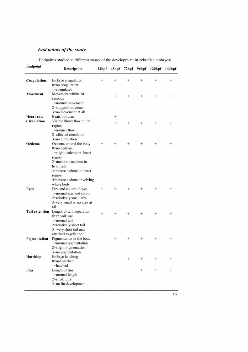
End points of the study
Endpoints studied at different stages of the development in zebrafish embryos;Endpoint Description 24hpf 48hpf 72hpf 96hpf 120hpf 144hpf
Coagulation Embryo coagulation + + + + + +0=no coagulation1=coagulated
Movement Movement within 30seconds + + + + + +
1=normal movement.2=sluggish movement3=no movement at all
Heart rate Beats/minutes +Circulation Visible blood flow in tail
region + + + + +
1=normal flow2=affected circulation3=no circulation
Oedema Oedema around the body + + + + + +0=no oedema1=slight oedema in heartregion2=moderate oedema inheart rate3=severe oedema in heartregion4=severe oedema involvingwhole body.
Eyes Size and colour of eyes + + + + + +1=normal size and colour2=relatively small size3=very small or no eyes atall.
Tail extension Length of tail, separationfrom yolk sac + + + + + +
1=normal tail2=relatively short tail3= very short tail andattached to yolk sac
Pigmentation Pigmentation in the body + + + + +1=normal pigmentation2=slight pigmentation3=no pigmentation
Hatching Embryo hatching0=not hatched + + + +
1=hatchedFins Length of fins + + +
1=normal length2=small fins3=no fin development

40
Results
Embryos were continuously monitored till 144hpf. Readings of the selected endpoints were taken after every 24 hours for 6 days. For the statistical analysis datataken by 48hpf and 144hpf were selected because most of the studied end pointswere developed by 48hpf and heart rate was easy to count, and 144hpf was thestudy termination point.
Retinoic acid
Zebrafish embryos treated with RA at concentrations of 10-8M and 10-9 Mpresented no apparent developmental abnormalities. By approximately 48hpf, theembryos displayed no evidence of truncation and had apparently normaldevelopment. Normal development was observed 2 to 3 days after treatments. Bywhich most of embryos had hatched. No difference in heart rate was observedwith respect to control group (Table I). The interior and posterior regions ofembryos developed similarly to that of control. These embryos survived untileuthanized.
The embryos treated with higher concentrations of RA10-7.8M and 10-7.6M wereseverely affected. These embryos presented truncation of the interior and posterioraxis by 48hpf. Heart rate of these embryos was significantly different from thecontrol group. The yolk sacs were on average noticeably larger than those of thecontrol embryos. Embryos treated with 10-7.4M, 10-7.2M, 10-7M had severe underdevelopment and larger oedema, no movement, no circulation, small eyes, shorttail. Most of the embryos treated with these concentrations of RA died by the 3rd
day after treatment (Table I).
Table text(* = significant different)(- = no statistically significant data)(HR = Heart rate beats/minute)(HPF = hours post fertilization)(Median values for all ordinal parameters and mean values for heart rate)(Statistical analysis was done for the results when at least 5 embryos were aliveout of 24 individuals)

41
Concentration Time Coagulation Movement Circulation Oedema Eyes Tail Pigmentation Hatching HR(mean)
Retinoic acid 48hpfControl 0.0 1.0 1.0 0.0 1.0 1.0 1.0 0.0 138.510-9 M 0.0 1.0 1.0 0.0 1.0 1.0 1.0 0.0 134.510-8M 0.0 1.0 1.0 0.0 1.0 1.0 1.0 0.0 135.510-7.8M 0.0 1.0 1.0 1.0* 1.0 1.0 1.0 0.0 125.5*
10-7.6M 0.0 1.0 2.0* 1.0* 1.0 1.0 1.0 0.0 117.5*10-7.4M 0.0 2.5* 3.0* 3.0* 2.0* 2.0* 2.0* 0.0 -10-7.2M 0.0 3.0* 3.0* 3.0* 3.0* 2.0* 2.0* 0.0 -
10-7M 0.0 3.0* 3.0* 4.0* 3.0* 2.0* 2.0* 0.0 -
Concentration Time Coagulation Movement Circulation Oedema Eyes Tail Pigmentation Hatching FinsRetinoic acid 144hpfControl 0.0 1.0 1.0 0.0 1.0 1.0 1.0 1.0 1.010-9 M 0.0 1.0 1.0 0.0 1.0 1.0 1.0 1.0 1.010-8M 0.0 1.0 1.0 0.0 1.0 1.0 1.0 1.0 1.010-7.8M 0.5* 2.5* 1.0 2.0* 1.5* 1.0 1.0 1.0 1.010-7.6M 0.5* 3.0* 2.5* 2.5* 2.0* 2.0* 1.0 1.0 1.5*10-7.4M 0.5* 3.0* 3.0* 3.0* 3.0* 2.0* 2.0* 1.0 3.0*10-7.2M 1.0* 3.0* 3.0* 3.0* 3.0* 2.0* 2.0* 1.0 3.0*10-7M 1.0* - - - - - - - -
Table (I); Statistical analysed studied end point results for RA by 48hfp and 144hpf.

42
A
B
Figure (A) 10-7.4 M RA treated embryos by 144hpf. No development of eyes, veryshort tail, yolk sac oedema and not hatched (B) 10-7.8M RA treated embryos by144hpf. Microphthalmia and yolk sac oedema.

43
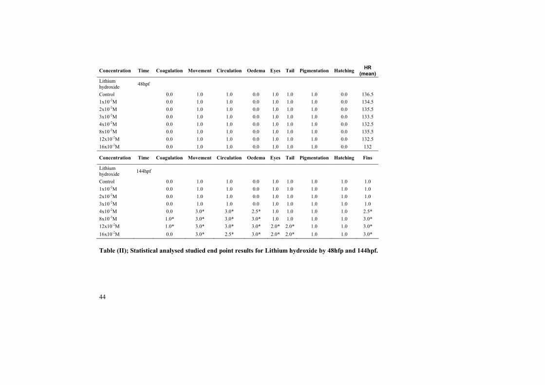
Lithium hydroxide
Zebrafish embryos treated with LiOH at the concentration (1x10⁻²M, 2x10⁻²M,3x10-2M, 4x10⁻²M, 8x10⁻²M, 12x10-2M and 16x10-2M) showed normaldevelopment and presented no apparent developmental abnormalities by 48hpf.By approximately 144hpf, embryos treated with 1x10⁻²M, 2x10⁻²M and 3x10⁻²Mcontinued normal development as to that of control group. Most of the embryostreated with 4x10⁻²M ,8x10-2M and12x10-2M coagulated by day six and nocoagulated embryos showed affected movement, no circulation and severetruncation of interior and posterior axis. Most of the embryos exhibited scoliosis.Coagulation rate was less among the embryos treated with 16x10-2M but they haddeveloped severe developmental abnormalities by 144hpf i.e. no movement, nocirculation, small eyes, short tail, under developed fins and scoliosis (Table II).
Figure. 12x10-2M Lithium hydroxide treated embryo after 144hfp.Microphthalmia, yolk sac oedema and relatively short tail.
44
Table (II); Statistical analysed studied end point results for Lithium hydroxide by 48hfp and 144hpf.
Concentration Time Coagulation Movement Circulation Oedema Eyes Tail Pigmentation Hatching HR(mean)
Lithiumhydroxide 48hpf
Control 0.0 1.0 1.0 0.0 1.0 1.0 1.0 0.0 136.51x10⁻²M 0.0 1.0 1.0 0.0 1.0 1.0 1.0 0.0 134.52x10⁻²M 0.0 1.0 1.0 0.0 1.0 1.0 1.0 0.0 135.53x10⁻²M 0.0 1.0 1.0 0.0 1.0 1.0 1.0 0.0 133.54x10⁻²M 0.0 1.0 1.0 0.0 1.0 1.0 1.0 0.0 132.58x10⁻²M 0.0 1.0 1.0 0.0 1.0 1.0 1.0 0.0 135.512x10⁻²M 0.0 1.0 1.0 0.0 1.0 1.0 1.0 0.0 132.516x10⁻²M 0.0 1.0 1.0 0.0 1.0 1.0 1.0 0.0 132
Concentration Time Coagulation Movement Circulation Oedema Eyes Tail Pigmentation Hatching Fins
Lithiumhydroxide 144hpf
Control 0.0 1.0 1.0 0.0 1.0 1.0 1.0 1.0 1.01x10⁻²M 0.0 1.0 1.0 0.0 1.0 1.0 1.0 1.0 1.02x10⁻²M 0.0 1.0 1.0 0.0 1.0 1.0 1.0 1.0 1.03x10⁻²M 0.0 1.0 1.0 0.0 1.0 1.0 1.0 1.0 1.04x10⁻²M 0.0 3.0* 3.0* 2.5* 1.0 1.0 1.0 1.0 2.5*8x10-2M 1.0* 3.0* 3.0* 3.0* 1.0 1.0 1.0 1.0 3.0*12x10-2M 1.0* 3.0* 3.0* 3.0* 2.0* 2.0* 1.0 1.0 3.0*16x10-2M 0.0 3.0* 2.5* 3.0* 2.0* 2.0* 1.0 1.0 3.0*
45
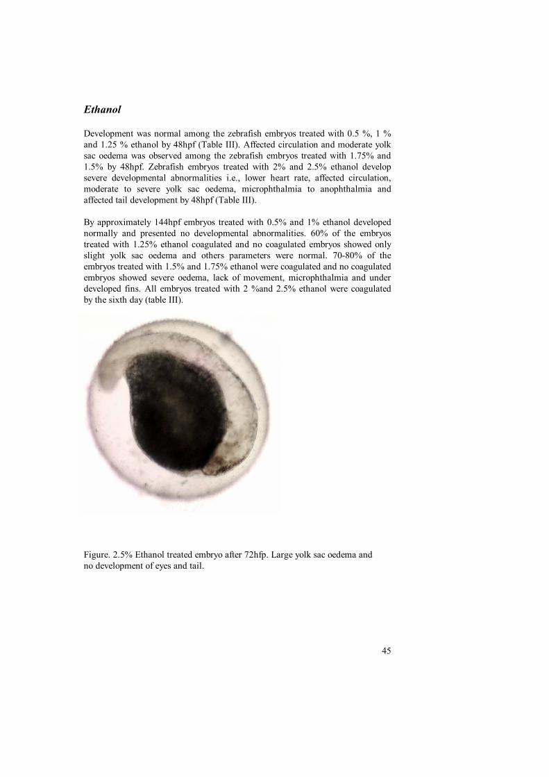
Ethanol
Development was normal among the zebrafish embryos treated with 0.5 %, 1 %and 1.25 % ethanol by 48hpf (Table III). Affected circulation and moderate yolksac oedema was observed among the zebrafish embryos treated with 1.75% and1.5% by 48hpf. Zebrafish embryos treated with 2% and 2.5% ethanol developsevere developmental abnormalities i.e., lower heart rate, affected circulation,moderate to severe yolk sac oedema, microphthalmia to anophthalmia andaffected tail development by 48hpf (Table III).
By approximately 144hpf embryos treated with 0.5% and 1% ethanol developednormally and presented no developmental abnormalities. 60% of the embryostreated with 1.25% ethanol coagulated and no coagulated embryos showed onlyslight yolk sac oedema and others parameters were normal. 70-80% of theembryos treated with 1.5% and 1.75% ethanol were coagulated and no coagulatedembryos showed severe oedema, lack of movement, microphthalmia and underdeveloped fins. All embryos treated with 2 %and 2.5% ethanol were coagulatedby the sixth day (table III).
Figure. 2.5% Ethanol treated embryo after 72hfp. Large yolk sac oedema andno development of eyes and tail.
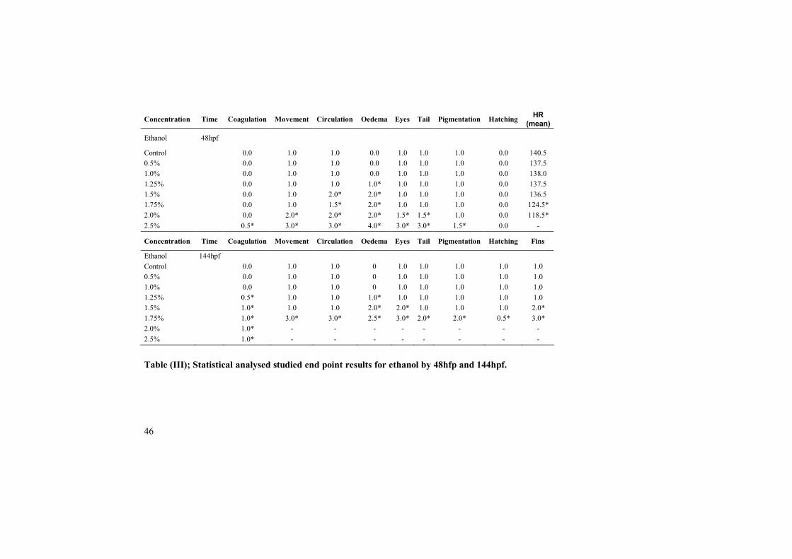
46
Table (III); Statistical analysed studied end point results for ethanol by 48hfp and 144hpf.
Concentration Time Coagulation Movement Circulation Oedema Eyes Tail Pigmentation Hatching HR(mean)
Ethanol 48hpf
Control 0.0 1.0 1.0 0.0 1.0 1.0 1.0 0.0 140.50.5% 0.0 1.0 1.0 0.0 1.0 1.0 1.0 0.0 137.51.0% 0.0 1.0 1.0 0.0 1.0 1.0 1.0 0.0 138.01.25% 0.0 1.0 1.0 1.0* 1.0 1.0 1.0 0.0 137.51.5% 0.0 1.0 2.0* 2.0* 1.0 1.0 1.0 0.0 136.51.75% 0.0 1.0 1.5* 2.0* 1.0 1.0 1.0 0.0 124.5*2.0% 0.0 2.0* 2.0* 2.0* 1.5* 1.5* 1.0 0.0 118.5*2.5% 0.5* 3.0* 3.0* 4.0* 3.0* 3.0* 1.5* 0.0 -
Concentration Time Coagulation Movement Circulation Oedema Eyes Tail Pigmentation Hatching Fins
Ethanol 144hpfControl 0.0 1.0 1.0 0 1.0 1.0 1.0 1.0 1.00.5% 0.0 1.0 1.0 0 1.0 1.0 1.0 1.0 1.01.0% 0.0 1.0 1.0 0 1.0 1.0 1.0 1.0 1.01.25% 0.5* 1.0 1.0 1.0* 1.0 1.0 1.0 1.0 1.01.5% 1.0* 1.0 1.0 2.0* 2.0* 1.0 1.0 1.0 2.0*1.75% 1.0* 3.0* 3.0* 2.5* 3.0* 2.0* 2.0* 0.5* 3.0*2.0% 1.0* - - - - - - - -2.5% 1.0* - - - - - - - -
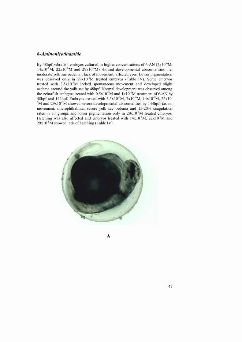
47
6-Aminonicotinamide
By 48hpf zebrafish embryos cultured in higher concentrations of 6-AN (7x10⁻⁶M,14x10-6M, 22x10-6M and 29x10-6M) showed developmental abnormalities, i.e.moderate yolk sac oedema , lack of movement, affected eyes. Lower pigmentationwas observed only in 29x10-6M treated embryos (Table IV). Some embryostreated with 3.5x10⁻⁶M lacked spontaneous movement and developed slightoedema around the yolk sac by 48hpf. Normal development was observed amongthe zebrafish embryos treated with 0.5x10⁻⁶M and 1x10⁻⁶M treatment of 6-AN by48hpf and 144hpf. Embryos treated with 3.5x10⁻⁶M, 7x10⁻⁶M, 14x10-6M, 22x10-
6M and 29x10-6M showed severe developmental abnormalities by 144hpf, i.e. nomovement, microphthalmia, severe yolk sac oedema and 15-20% coagulationrates in all groups and lower pigmentation only in 29x10-6M treated embryos.Hatching was also affected and embryos treated with 14x10-6M, 22x10-6M and29x10-6M showed lack of hatching (Table IV).
A
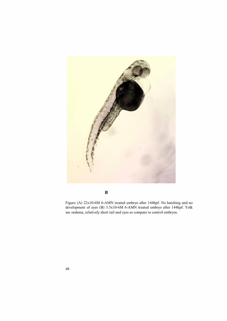
48
B
Figure (A) 22x10-6M 6-AMN treated embryo after 144hpf. No hatching and nodevelopment of eyes (B) 3.5x10⁻⁶M 6-AMN treated embryo after 144hpf. Yolksac oedema, relatively short tail and eyes as compare to control embryos.
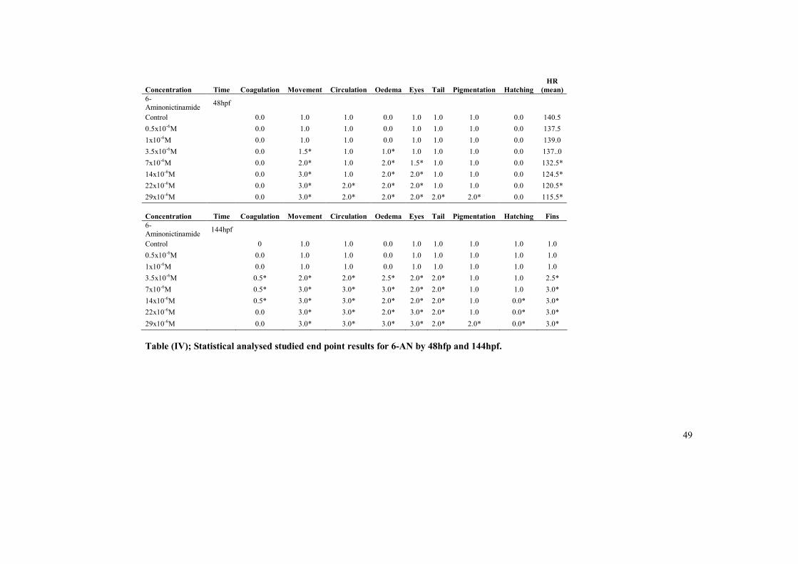
49
Table (IV); Statistical analysed studied end point results for 6-AN by 48hfp and 144hpf.
Concentration Time Coagulation Movement Circulation Oedema Eyes Tail Pigmentation HatchingHR
(mean)6-Aminonictinamide 48hpf
Control 0.0 1.0 1.0 0.0 1.0 1.0 1.0 0.0 140.50.5x10-6M 0.0 1.0 1.0 0.0 1.0 1.0 1.0 0.0 137.51x10⁻⁶M 0.0 1.0 1.0 0.0 1.0 1.0 1.0 0.0 139.03.5x10-6M 0.0 1.5* 1.0 1.0* 1.0 1.0 1.0 0.0 137..07x10-6M 0.0 2.0* 1.0 2.0* 1.5* 1.0 1.0 0.0 132.5*14x10-6M 0.0 3.0* 1.0 2.0* 2.0* 1.0 1.0 0.0 124.5*22x10-6M 0.0 3.0* 2.0* 2.0* 2.0* 1.0 1.0 0.0 120.5*29x10-6M 0.0 3.0* 2.0* 2.0* 2.0* 2.0* 2.0* 0.0 115.5*
Concentration Time Coagulation Movement Circulation Oedema Eyes Tail Pigmentation Hatching Fins6-Aminonictinamide 144hpf
Control 0 1.0 1.0 0.0 1.0 1.0 1.0 1.0 1.00.5x10-6M 0.0 1.0 1.0 0.0 1.0 1.0 1.0 1.0 1.01x10⁻⁶M 0.0 1.0 1.0 0.0 1.0 1.0 1.0 1.0 1.03.5x10-6M 0.5* 2.0* 2.0* 2.5* 2.0* 2.0* 1.0 1.0 2.5*7x10-6M 0.5* 3.0* 3.0* 3.0* 2.0* 2.0* 1.0 1.0 3.0*14x10-6M 0.5* 3.0* 3.0* 2.0* 2.0* 2.0* 1.0 0.0* 3.0*22x10-6M 0.0 3.0* 3.0* 2.0* 3.0* 2.0* 1.0 0.0* 3.0*29x10-6M 0.0 3.0* 3.0* 3.0* 3.0* 2.0* 2.0* 0.0* 3.0*

50
Ochratoxin A
At higher concentrations of OA (6x10⁻⁷M, 12x10-7M and 25x10-7M) by 48hpfembryos showed developmental abnormalities, i.e. oedema, affected circulationand significantly lower heart rate in 12x10-7M and 25x10-7M treated embryos.Embryos treated with lower concentrations 0.5x10⁻⁷M, 1x10⁻⁷M, 2x10⁻⁷M and3x10-7M continued to develop normal as that of the control group and noabnormalities were observed 48hpf (Table V).By 144hpf embryos treated with 0.5x10⁻⁷M and 1x10⁻⁷M concentration of OAexhibited no developmental abnormalities and continued normal development.All of the embryos treated with 12x10-7M and 25x10-7M were coagulated. 60%coagulation was observed among the zebrafish embryos treated with 6x10⁻⁷M andno coagulated embryos exhibited developmental abnormalities, i.e. lack ofmovement, severe yolk sac oedema, affected circulation, under developed fins andaffected eyes. Embryos treated with 2x10⁻⁷M and 3x10⁻⁷M showed somedevelopmental abnormalities, i.e. slight oedema and coagulation. In 3x10⁻⁷Mtreated embryos observed developmental abnormalities were not significantlydifferent from the control embryos (Table V).
A

51
BFigure (A & B) 6x10⁻⁷M Ochratoxin A treated embryo after 144hpf.Development of large yolk sac oedema.

52
Table (V); Statistical analysed studied end point results for OTA by 48hfp and 144hpf
Concentration Time Coagulation Movement Circulation Oedema Eyes Tail Pigmentation Hatching HR(mean)
Ochratoxin A 48hpfControl 0.0 1.0 1.0 0.0 1.0 1.0 1.0 0.0 141.00.5x10-7M 0.0 1.0 1.0 0.0 1.0 1.0 1.0 0.0 144.51x10⁻⁷M 0.0 1.0 1.0 0.0 1.0 1.0 1.0 0.0 142.52x10⁻⁷M 0.0 1.0 1.0 0.0 1.0 1.0 1.0 0.0 135.03x10⁻⁷M 0.0 1.0 1.0 0.0 1.0 1.0 1.0 0.0 138.56x10⁻⁷M 0.0 1.0 1.5* 0.0 1.0 1.0 1.0 0.0 136.512x10⁻⁷M 0.0 1.0 2.5* 1.0* 1.0 1.0 1.0 0.0 130.0*25x10-7M 0.0 1.0 3.0* 2.0* 1.0 1.0 2.0* 0.0 125.5*
Concentration Time Coagulation Movement Circulation Oedema Eyes Tail Pigmentation Hatching Fins
Ochratoxin A 144hpfControl 0.0 1.0 1.0 0.0 1.0 1.0 1.0 1.0 1.00.5x10-7M 0.0 1.0 1.0 0.0 1.0 1.0 1.0 1.0 1.01x10⁻⁷M 0.0 1.0 1.0 0.0 1.0 1.0 1.0 1.0 1.02x10⁻⁷M 0.5* 1.0 1.5* 1.5* 1.0 1.0 1.0 1.0 1.03x10⁻⁷M 0.0 1.0 1.0 0.0 1.0 1.0 1.0 1.0 1.06x10⁻⁷M 1.0* 3.0* 3.0* 3.0* 1.5* 1.0 1.0 1.0 2.0*12x10-7M 1.0* - - - - - - - -25x10-7M 1.0* - - - - - - - -

53
Sodium arsenate
Zebrafish embryos provide no evidence of phenotypic abnormal developmentwhen cultured in sodium arsenate rearing media even as high as 16x10-3Msolution by 48hpf. After day six of sodium arsenate treatment all exposed embryosdeveloped normally as that of control group (Table VI).
12x10-3M Sodium arsenate treated embryo after 144hpf. No developmental abnormalities, all studied parameters are normal

54
Table (VI); Statistical analysed studied end point results for sodium arsenate by 48hfp and 144hpf
Concentration Time Coagulation Movement Circulation Oedema Eyes Tail Pigmentation Hatching HR(mean)
Sodium arsenate 48hpf
Control 0.0 1.0 1.0 0.0 1.0 1.0 1.0 0.0 141.51.5x10-3M 0.0 1.0 1.0 0.0 1.0 1.0 1.0 0.0 138.53x10⁻³M 0.0 1.0 1.0 0.0 1.0 1.0 1.0 0.0 140.06x10⁻³M 0.0 1.0 1.0 0.0 1.0 1.0 1.0 0.0 137.59x10⁻³M 0.0 1.0 1.0 0.0 1.0 1.0 1.0 0.0 139.512x10⁻³M 0.0 1.0 1.0 0.0 1.0 1.0 1.0 0.0 138.515x10⁻³M 0.0 1.0 1.0 0.0 1.0 1.0 1.0 0.0 137.0Concentration Time Coagulation Movement Circulation Oedema Eyes Tail Pigmentation Hatching Fins
Sodium arsenate 144hpf
Control 0.0 1.0 1.0 0.0 1.0 1.0 1.0 1.0 1.01.5x10-3M 0.0 1.0 1.0 0.0 1.0 1.0 1.0 1.0 1.03x10⁻³M 0.0 1.0 1.0 0.0 1.0 1.0 1.0 1.0 1.06x10⁻³M 0.0 1.0 1.0 0.0 1.0 1.0 1.0 1.0 1.09x10⁻³M 0.0 1.0 1.0 0.0 1.0 1.0 1.0 1.0 1.012x10⁻³M 0.0 1.0 1.0 0.0 1.0 1.0 1.0 1.0 1.015x10⁻³M 0.0 1.0 1.0 0.0 1.0 1.0 1.0 1.0 1.0

55
LOEC and NOEC values table
Table (A) summary of the LOEC (lowest observed effect concentration) andNOEC (no observed effect concentration) data after 48hpf and 144hpf for selectedendpoints.NO* Not observed
Substance End point NOEC48hpf(Molar)
LOEC48hpf(Molar)
NOEC144hpf(Molar)
LOEC144hpf (Molar)
Retinoic acid Coagulated eggs 10-7 NO 10-8 10-7.8
Movement 10-7.4 10-7.6 10-8 10-7.8
Circulation 10-7.8 10-7.6 10-7.8 10-7.6
Extension of tail 10-7.6 10-7.4 10-7.8 10-7.6
Oedema 10-8 10-7.8 10-8 10-7.8
Heart rate 10-8 10-7.8 NO NO Eye 10-7.4 10-7.6 10-7.8 10-7.6
Lithium hydroxide Coagulated eggs 16x10-2 NO 4x10⁻² 8x10⁻²
Movement 16x10-2 NO 4x10⁻² 8x10⁻²
Circulation 16x10-2 NO 4x10⁻² 8x10⁻²
Extension of tail 16x10-2 NO 8x10⁻² 12x10-2
Oedema 16x10-2 NO 3x10⁻² 4x10⁻²
Eyes 16x10-2 NO 8x10⁻² 12x10-2
Ethanol Coagulated eggs 2% 2.5% 1% 1.25% Movement 1.75% 2% 1.5% 1.75% Circulation 1.25% 1.5% 1.25% 1.5% Extension of tail 1.75% 2% 1.5% 1.75% Oedema 1% 1.25% 1% 1.25% Eyes 1.75% 2% 1.25% 1.5% Pigmentation 2% 2.5% 1.5% 1.75%
6-Aminonicotinamide
Coagulated eggs NO NO 1x10⁻⁶ 3.5x10⁻⁶
Movement 1x10⁻⁶ 3.5x10⁻⁶ 1x10⁻⁶ 3.5x10⁻⁶
Circulation 14x10-6 22x10-6 1x10⁻⁶ 3.5x10⁻⁶
Extension of tail 22x10-6 29x10-6 1x10⁻⁶ 3.5x10⁻⁶
Oedema 1x10⁻⁶ 3.5x10⁻⁶ 1x10⁻⁶ 3.5x10⁻⁶
Eyes 3.5x10⁻⁶ 7x10⁻⁶ 1x10⁻⁶ 3.5x10⁻⁶
Hatching NO NO 7x10⁻⁶ 14x10-6
Ochratoxin A Coagulated eggs 25x10-7 NO 1x10⁻⁷ 2x10⁻⁷
Movement 25x10-7 NO 3x10⁻⁷ 6x10⁻⁷
Circulation 1x10⁻⁷ 2x10⁻⁷ 1x10⁻⁷ 2x10⁻⁷
Extension of tail 25x10-7 NO 6x10⁻⁷ 12x10-7
Oedema 6x10⁻⁷ 12x10-7 1x10⁻⁷ 2x10⁻⁷
Eyes 25x10-7 NO 3x10⁻⁷ 6x10⁻⁷
Pigmentation 12x10-7 25x10-7 6x10⁻⁷ NO Heart rate 3x10⁻⁷ 6x10⁻⁷ NO NO

56
Discussion
Identification of environmental agents that cause damage to unborn children isabsolutely imperative. Armed with the knowledge of those drugs, chemicals andfoodstuffs that human beings are routinely exposed to that are truly teratogenic,exposures can be avoided, reduced or limited, and the incidence of tragicteratogen-induced birth defects drastically lowered. The burden of this goalcurrently rests heavily upon animal-based testing, but none of the animal modelfulfilling the ideal teratology animal model criteria such as producing large littersafter a short gestation, inexpensive maintenance, and an inability andunwillingness to ‘bite, scratch, kick, howl or squeal’ (Wilson, 1975), and also dueto some ethically limitations it is not possible to perform extensive teratologicaltesting on routine basis. Thus, a need exists for assays that retain the high valueof in vivo toxicity testing, but can be rapidly performed on a large number ofchemicals. Since the thalidomide catastrophe scientists are exploring newalternative for teratological testing (McBride 1961; Lenz 1966). In the recenttimes zebrafish emerge as a competitive laboratory animal model. In the presentstudy zebrafish embryos were used to test the teratogenicity of some potentialhuman teratogens. Chemicals with known potential teratogenicity to human wereselected from different categories of chemicals to study the potential of zebrafishmodel for teratological studies. Comparisons of the results were done with otherlaboratory animal species as shown in the Table (B).
Species Substances
Retinoicacid
Lithiumhydroxide
OchratoxinA
Ethanol 6-Amino nicotinamide
Arsenicoxide
+/+-/- +%
Mouse + +- + + + + 5/1/0 83%Rat + +- + + + + 5/1/0 83%Rabbit + - + + + 4/0/1 80%Hamster + + + + 4/0/0 100%Primate + - + +- 2/1/1 50%Pig +- + + 2/1/0 50%Zebrafish + + + + + - 5/0/1 83%
Table (B)“+” positive teratogenic results“-” negative teratogenic results“+-”unknown teratogenic results
Retinoic acid is expressed in a gradient along the anterior-posterior axis of thezebrafish. Different Hox genes are expressed along the anterior-posterior axisaccording to the concentration of RA present in that region, embryos showedtruncation at anterior and posterior axis as shown in the results because thisexogenous retinoic acid altered the natural gradient, causing ectopic expression ofHox genes. Regions that would normally have a lower concentration of retinoicacid now had a higher concentration and hence expressed genes similar to those

57
expressed in the hindbrain and trunk region of the embryo, where the largestconcentration of retinoic acid is usually found. The magnitude of the effects oftreatment with exogenous retinoic acid appeared to be concentration dependentEmbryos showed lack of movement, lower heart rate, microphthalmia toanophthalmia, short tail and yolk sac oedema which may be indicative of theirstunted growth and a less that normal level of metabolization of the yolk sacnutrients. The developmental abnormalities caused by RA among the zebrafishembryos were very much obvious at early stages (48hpf) and later stages (144hpf)of development on the anterior (craniofacial) and posterior (tail) trunk. Thephenotypic developmental abnormalities of anterior and posterior trunk of thezebrafish embryos are similar to those observed in human i.e. defective ears,absent or small jaws, cleft palate, aortic arch abnormalities and abnormalities ofthe central nervous system, and in mice i.e. axial truncation, reduction in the sizeof first and second pharyngeal arches which normally form the jaws, ears andother facial bones (Gilbert, 2003; Ligas, 2000; Koren G, 1994). Abnormalities ofthe CNS in the zebrafish embryos can be correlated to the rapid or reducedmovement of the embryos (Behra et al., 2004; Bretaud et al., 2004; Granato et al.,1996; Lam et al., 2005; Mckinley et al., 2005; Tiedeken et al., 2005). In thepresent discussion comparison of the anterior trunk abnormalities of the zebrafishembryos are made with the phenotypic craniofacial abnormalities of the other labanimal models, coagulation with low birth rate, still birth and high infantmortality and in general anterior and posterior trunk malformations with stuntedgrowth of the new born.
Lithium hydroxide treatment affected the anterior-posterior morphogenesis ofzebrafish embryos such that higher concentrations of this substance lead toincreasingly greater inhibition of anterior and posterior development. Lithium-induced teratogenesis has been shown to perturb the development of ventralstructures in premidblastular zebrafish, when the blastula is still formingmorphogenetic gradients of proteins in the Wnt signalling pathway (such as b-catenin) and continuing to transcribe maternal mRNAs. In these early embryos,lithium teratogenesis leads to the expression of bustled and radialized phenotypes,which exhibit hyperdorsalization (Stachel et al., 1993). In the sphere-stageembryos, morphogenetic gradients have already been established, so the effect oflithium in this case probably occurs downstream of these events. The phenotypeof LiOH cultured embryos higher then 3x10⁻²M exhibited severe yolk sac oedema,microphthalmia, short tail, scoliosis, under developed fins and lack of movement.Embryos cultured in 8x10⁻²M and higher concentration of LiOH showed highmortality. Similar developmental malformations induced by lithium are observedin other vertebrate species e.g. in mice i.e. reduction in the number of new born,vertebral defects, cleft palate, craniofacial abnormalities (Chenroff andKavloc,1982; Szabo,1970; Smithberg and Dixit,1982; RTECS,1995a), in rat i.e.malformations of eyes, external ears, cleft palate, reduction in the number of livebirth(Wright et al., 1970; Marathe and Thomas,1986), in pig i.e. reduction in thelive piglets numbers, still birth and high mortality among the live piglets(Kelleyet al., 1978), and in human i.e. Ebstein’s anomaly ; a congenital cardiacabnormality, still birth and other developmental defects in the Apgar Score (The

58
Apgar score is a standardised way of checking the health of a newborn baby)“Appearance, Pulse, Grimace, Activity, Respiration” (RTECS, 1995b).
Blader and Strähle (1998) have studied that embryonic development of zebrafishis affected by ethanol in a manner similar to higher vertebrates. Phenotypicabnormalities caused by ethanol were observed among the zebrafish embryoscultured higher than 1% ethanol solution as shown in the Table (A) by LOEC andNOEC values. The effected range of ethanol in the present study is significantlydifferent with Blader and Strähle´s (1998) ethanol concentration 2.4 percent.There was high mortality rate among the zebrafish embryos cultured higher then1.5% and this is may be due to apoptosis by activating the cell’s self destructionmachineries as suggested by Sulik et al. (1988). Microphthalmia andanophthalmia was observed in embryos treated with 2% and 2.5% ethanol,supporting the fact that ethanol does affect eyes development among zebrafishembryos (Blader and Strähle, 1998). Ethanol did cause defective development inthe posterior structures in zebrafish embryos. Although the mechanisms behindthe observed deformities of the trunk and the tail are not known, it can beconjectured that the impairment of epiboly may be responsible. Blader andSträhle (1998) noted that epiboly was marginally impaired by the treatment, withthe advance of the blastoderm margin lagging behind untreated controls by 10 to15% toward the end of gastrulation. Epiboly establishes the posterior area of theembryo including the trunk region and the tail bud and, therefore, it seemsreasonable that impairing the last 10-15% of epiboly may cause defective trunkand tail (Gilbert, 2003). Lack of pigmentation was observed among some theembryos treated with higher concentration of ethanol. The mechanism for thislack of pigmentation is not known but it is thought that ethanol might causedeformities in the spinal cord from which neural crest cells destined to becomemelanocytes arise (Gilbert, 2003). These observed developmental abnormalitiesare similar with abnormalities observed in human, chicken and rat i.e. metalretardation, behavioural abnormalities, craniofacial malformation, heart andneural crest defects (Gilbert, 1997; Cartwright et al., 1995; Smith, 1997; Sulik etal., 1988; Koren G, 1994).
6-AN 3.5x10⁻⁶M rearing solution and higher concentrations caused highmortality, low hatching rate, lack of movement, affected circulation,microphthalmia, short to no developed fins and short tail. It can be conjecturedthat 6-AN inhibition of nicotinamide adenine dinucleotide dependent reactionsduring ATP synthesis is responsible for these malformations. When 6-ANdecreases the pentose pathway then the arterioles partially lose their ability toregulate the blood flow and blood resistance increases then all the normalmechanisms and metabolisms of the body effected and this may in turn producemalformations in the developing embryos (Wilson et al., 1975; Dietrich et al.,1958). This malformations pattern is similar with that observed in mice i.e.decrease litter size, CNS problems (Matschke et al., 1997), cleft palate, hind limbdefects (Pinsky et al., 1960), in rat i.e., stunted growth, foetal death, craniofacialabnormalities, CNS problems (TJADAB, 1975), in rabbit i.e. stunted growth,cardio vascular problems, CNS problems and craniofacial including eyes and earsdefects (TJADAB, 1983).

59
Zebrafish embryos cultured higher then 1x10⁻⁷ rearing media of OA developmalformations. By day six these embryos presented high mortality and affectedcirculation and spontaneous movement, yolk sac oedema, microphthalmia andunder developed fins. These malformations may be due to chromosomalaberrations because it can cause cell death, retardation of differentiation indeveloping embryos (Wilson, 1973; Manolova et al., 1990). Another possible wayof explanation for these malformations is OA-DNA adducts in the zebrafishembryos body tissues (Pfohl-Leszkowicz et al., 1991, 1993; Grosse et al., 1995,1997; Castegnaro et al., 1998; Pfohl-Leszkowicz et al., 1998). These observedmalformation pattern is similar with that observed in rat i.e. high mortality,decrease in the number of new born, decrease foetal weight, celosome withoedema and CNS abnormalities (More´ and Galtier, 1974; Kinhara et al., 1984),and in Mice i.e. high mortality craniofacial malformation, microphthalmia,anophthalmia, malformed tail and limbs (Singh and Hood, 1985; Arora etal.,1981).
No arsenic related mortalities or deformities were observed in any of thetreatments performed. There were no notable mortalities or deformations wereobserved at any experimental concentrations among the zebrafish embryos at48hpf and 144hpf. These results are opposing to the other lab animal resultsbecause arsenic is a proven teratogenic substance as studies have shown thatarsenic can cross the mammalian placenta, affecting developing embryos whosemothers undergo exposure (Wlodarczyk et al., 1996). One possible reason for thearsenic’s apparent lack of effect on zebrafish development is that neuralpatterning defects in vertebrates, such as those associated with arsenic exposure,are notoriously difficult to detect from morphological observation alone(Furutani-Seiki et al., 1996). The neural tube forms by the process of apoptoticcavitations in fish, as compared to neural groove invagination in mammals, andas a result, makes gross morphological observation of neural tube defects muchharder to recognize. Arsenic exists in trivalent and pentavalent ionic forms, andis often given as either sodium arsenate (As(V)) or arsenic trioxide (As(III)), withsometimes opposing results. Some studies have suggested that As(V) is less toxicto fish than As(III), an identical result to findings in higher vertebrates(Dabrowski 1975). This may explain the absence of abnormalities as in this studyAs(V) was used for the experiment. But this also opposes the phenomena thatonly the trivalent ion of arsenic has direct biological ramifications: As(V) must beconverted to As(III) to be active (Peterková and Puzanová 1974). Kalter (1968)noted that the effect of teratogens in organisms such as fish and amphibians isprimarily dependent upon the stage at which the embryo is exposed to thechemical. Response to arsenic is also species dependent and therefore cannot begeneralized based on broader classifications.
Results and the above discussion show that the zebrafish embryo toxicity test hasprovided positive evidences about the teratogens testing. This test is simple tohandle, easy to understand, economical and easily repeatable. In the present studythe percentage of positive results is high (83%) as shown in the Table (B) andcomparable to other laboratory animal species. But there are a lot of advantageswith this animal model, i.e. this specie is easily obtainable, inexpensive, has high

60
fecundity, a small size (3 to 5cm), short generation time, rapid development,external fertilization, translucent embryos and is easy to maintain at laboratorylevel (Laale, 1977). Under appropriate conditions one female lays approximately50 to 200 eggs per day (Laale, 1977), so the sample size can be large enough asdesirable. The zebrafish comes close to being the ideal model organism forvertebrate developmental studies because it appears to combine the best featuresof all the other models. Like most of the amphibians, zebrafish embryos developexternally and can be viewed and manipulated at all stages. However, zebrafishdevelopment is more rapid than most of the amphibians and the organization ofthe embryo are simpler and (like worms and fruit flies) the embryo is transparent.Like the mouse, the zebrafish is amenable to genetic analysis and has a similargeneration interval (2-3 months). However, zebrafish are smaller than mice andthey produce more offspring in a shorter time. A female zebrafish can lay up to200 eggs per day, while a mouse may produce a litter of up to 15 embryos in 21days.
Most of the proof-of-concept studies performed so far have shown that knownteratogens (e.g. Valproic acid, cadmium and tetrachlorodibenzo-para-dioxin etc)in humans can be recapitulated in the zebrafish (Rubinstein, 2006). It stillremains to be proved, however, whether drugs with unknown toxicitycharacteristics will cause effects in zebrafish embryos. Studies with wider rangeof compounds with unknown toxicity characteristics will allow toxicologists topredict with reasonable assurance that such effects are likely in humans.Furthermore, it may be difficult to correlate the concentration and absorption ofthese compounds by zebrafish embryos in the media required to cause toxiceffects in zebrafish to the plasma concentration of compound that would cause thesame effect in humans. Determination of the actual amount of compoundabsorbed by zebrafish as well as an understanding of compound metabolism inzebrafish will be important to make useful correlations. Potential differences inthe physiology of embryos should also be considered. Based on the results of thispreliminary study, it can be said that in future with more extensive research andwith wider range of compounds zebrafish assay can be integrated intoteratological and drug toxicity screening programmes on a commercial level forthe drugs and chemicals before human use. This work has been a necessary firststep toward developing zebrafish as a model to predict toxicity of compounds inhumans.

61
References
1. Antkiewicz, D.S., Burns, G.C., Carney, S.A., Peterson, R.E. &Heideman, W. 2005. Heart malformation is an early response toTCDD in embryonic zebrafish. Toxicol. Sci. 84, 1-14.
2. Arora, R.G. & Frolen, H. 1981. Interference of mycotoxins withprenatal development of the mouse. II. Ochratoxin A inducedteratogenic effects in relation to the dose and stage of gestation. ActaVet Scand 22: 535-552.
3. Baily, J. 2005. The Future of Teratology research is in vitro, BiogenicAmines, vol.19, no.2. 97-145.
4. Baraban , J.M. 1994. Toward a crystal-clear view of lithium's site ofaction. Proc. Natl. Acad. Sci., USA 91. 5738–5739.
5. Behra, M., Etard, C., Cousin, X. & Strahle, U. 2004. The use ofzebrafish mutants to identify secondary target effects of acetylcholineesterase inhibitors. Toxicol. Sci. 77, 325-333.
6. Blader, P. & Strähle, U. 1998. Ethanol Impairs Migration ofPrechordal Plate in the Zebrafish Embryo. Developmental Biology201, 185-201.
7. Brent, R.L. 1995. The application and principles of toxicology andteratology in evaluating the risksof new drugs for treatment of drugaddiction in women of reproductive age. NIDA Res. Monogr.149, 130-184.
8. Bretaud, S., Lee, S. & Guo, S. 2004. Sensitivity of zebrafish toenvironmental toxins implicated in Parkinson’s disease. Neurotoxicol.Teratol. 26, 857-864.
9. Brown, M.H., Szczech, G.M. & Purmalis, B.P. 1976. Teratogenic andtoxic effects of ochratoxin A in rats. Toxicol Appl Pharmacol. 37,331-338.
10. Bunge, I., Dirheimer, G. & Röschenthaler, R. 1978. In vivo and invitro inhibition of protein synthesis in Bacillus stearothermophilus byochratoxin A. Biochem Biophys Res Commun, 83, 398-405.
11. Castegnaro, M., Mohr, U., Pfohl-Leszkowicz, A., Estève, J.,Steinmann, J., Tillmann, T., Michelon, J. & Bartsch, H. 1998. Sex-and strain-specific induction of renal tumors by ochratoxin A in ratscorrelates with DNA adduction. Int. J. Cancer 77, 70-75.

62
12. Cartwright, M.M. & Smith, S.M. 1995. Increased cell death andreduced neural crest cell numbers in ethanol-exposed embryos: PartialBasis for the Fetal Alcohol Syndrome Phenotype. Alcohol Clin ExpRes. 19, 378-86.
13. Chen, J.N., Haffter, P., Odenthal, J., Vogelsang, E., Brand, M., vanEeden, F.J., Furutani-Seiki, M., Granato, M., Hammerschmidt, M. &Heisenberg, C.P., et al. 1996. Mutations affecting the cardiovascularsystem and other internal organs in zebrafish. Development 123, 293-302.
14. Chen, J.N. & Fishman, M.C. 1996. Zebrafish tinman homologdemarcates the heart field and initiates myocardial differentiation.Development 122, 3809-3816.
15. Chernoff, N. & Kavlock, R.J. 1982.An in vivo teratology screenutilizing pregnant mice. J. Toxicol. Environ. Health 10, 541-550.
16. Creppy E.E., Lugnier, A.A., Fasiolo, F., Heller, K., Röschenthaler, R.& Dirheimer, G. 1979. In vitro inhibition of yeast phenylalanyl-tRNAsynthetase by ochratoxin A. Chem Biol Interact. 24, 257-261.
17. Eaton, R.C. & Farley, R.D. 1974. Spawning cycle and egg productionof zebrafish, Brachydanio rerio, in thelaboratory. Copeia 1, 195-209.
18. Ekker, M. & Akimenko, M.A. 1991. Le poisson zèbre (Danio rerio),unmodèle en biologie du development. Medicine/sciences 7, 533-560.
19. Furutani-Seiki, M., Jiang, Y.J., Brand, M., Heisenberg, C.P., Houart,C., Beuchle, D., van Eeden, M. Granato, F.J., Haffter, P.,Hammerschmidt, M., Kane, D.A., Kelsh, R.N., Mullins, M.C.,Odenthal, J. & Nusslein-Volhard, C. 1996. Neural degenerationmutants in the zebrafish, Danio rerio. Development 123, 229-239.
20. Gilani, S.H., Bancroft, J. & Reily, M. 1978. Teratogenicity ofochratoxin A in chick embryos. Toxicol Appl Pharmacol 46, 543-546.
21. Gilbert, S. 2003. Developmental Biology,7th edition. SinauerAssociates Inc. Massachusetts. 345-347, 353.
22. Gilbert, S.F. 1997. Developmental Biology. Sinauer Associates, Inc.Massachusetts. 5th ed.
23. Goolish, E.M., Okutake, K. & Lesure, S. 1999. Growth andSurvivorship of larval zebrafish Danio rerio on processed diets. N. Am.J. Aquacult. 61, 189-198.

63
24. Granato, M. & Nüsslein-Volhard, C. 1996. Fishing for genescontrolling development. Curr. Opin. Genet. Dev. 6, 461-468.
25. Granato, M., Van Eeden F.J.M. & Schach, U. et al. 1996. Genescontrolling and mediating locomotion behavior of the zebrafishembryo and larva. Development 23, 399-413.
26. Grosse, Y., Baudrimont, I., Castegnaro, M., Betbeder, A.M., Creppy,E.E., Dirheimer, G. & Pfohl-Leszkowicz, A. 1995. Formation ofochratoxin A metabolites and DNA-adducts in monkey kidney cells.Chem.-Biol. Interactions, 95, 175-187.
27. Grosse, Y., Chekir-Ghedira, L., Huc, A., Obrecht-Pflumio, S.,Dirheimer, G., Bacha, H., & Pfohl-Leszkowicz, A. 1997. Retinol,ascorbic acid and alpha-tocopherol prevent DNA adduct formation inmice treated with the mycotoxins ochratoxin A and zearalenone.Cancer Lett., 114, 225-229.
28. Incardona, J.P., Collier, T.K. & Scholz, N.L. 2004. Defects in cardiacfunction precede morphological abnormalities in fish embryos exposedto polycyclic aromatic hydrocarbons. Toxicol. Appl. Pharmacol. 196,191–205
29. International Organization for Standardization (ISO) .1996. Waterquality-Determination of the acute lethal toxicity of substances to afreshwater fish [Brachydanio rerio Hamilton-Buchanan (Teleostei,Cyprinidae)] Part 1: Static method. ISO 7346–1:1996.
30. ISO .1999 .Water quality—Determination of toxicity to embryos andlarvae of freshwater fish. Semi-static method. ISO 12890:1999.
31. Johnson, W.J. & McColl, J.C. 1995. 6-Aminonicotinamide -a potentnicotinamide antagonist. Science 122, 834.
32. Jones, K.L., Smith, D.W., Ulleland, C.N. & Streissguth, A.P. 1973.Pattern of malformation in off-spring of chronic alcoholic mothers.Lancet 1, 1267-1271.
33. Kelley, K.W., McGlone, J.J. & Froseth, J.A. 1978. Lithium toxicity inpregnant swine. Proc. Soc. Exper. Biol. Med. 158, 123-127.
34. Kihara , T., Nakagawa, K., Yamamoto, Y. & Tanimura, T. 1984.Behavioural teratological study of rat offspring exposed to ochratoxinA in utero by using cross-fostering. (abstract). Teratology, 30, 10A.
35. Kimmel, C.B. 1989. Genetics and early development of zebrafish.Trends Genet. 5, 283-288.

64
36. Kimmel, C.B., Ballard, W.W., Kimmel, S.R., Ullmann, B. &Schilling, T.T. 1995. Stages of embryonic development of thezebrafish. Dev. Dyn. 203, 253-310.
37. Kimmel, C.B., Sepich, D.S. & Trevarrow, B. 1988. Development andsegmentation in zebrafish. Develop. Suppl. 104, 197-207.
38. Klein, P.S. & .Melton, D.A. 1996. A molecular mechanism for theeffect of lithium on development. Proc. Natl. Acad. Sci. USA. 93,8455-9.
39. Koren, G. 1994. Maternal-Fetal toxicology, second edition. NY.,Marcel Dekker inc.
40. Laale, H.W. 1977. The biology and use of zebrafish, Brachydaniorerio, in fisheries research: A literature review. J. Fish. Biol. 10, 121-173.
41. Lam, C.S., Korzh, V. & Strahle, U. 2005. Zebrafish embryos aresusceptible to the dopaminergic neurotoxin MPTP. Eur. J. Neurosci.21, 1758-1762.
42. Langheinrich ,U. 2003. Bioessays. 25, 904-912.
43. Lele, Z. & Krone, P.H. 1996. The zebrafish as a model system indevelopmental, toxicological and transgenic research. Biotech. Adv.14, 57-72.
44. Lenz, W. 1966. Malformations caused by drugs in pregnancy, Am. J.Dis. Child. 112, 99-106.
45. Ligas, A. 2000. Testing the effects of retinoic acid on tail formation ofdeveloping zebrafish embryos. Accessed March 13, 2004.http://www.swarthmore.edu/NatSci/sgilber1/DB_lab/Fish/ZF_RA.html
46. MacRae, C.A. & Peterson, R.T. 2003. Zebrafish-based small moleculediscovery. Chem. Biol. 10, 901-908.
47. Manolova,Y., Manolov, G., Parvanova, L., Petkova-Bocharova, T.,Castegnaro, M. & Chernozemsky, I.N. 1990. Induction ofcharacteristic chromosomal aberrations, particularly x-trisomy,incultured human lymphocytes treated by ochratoxin A; a mycotoxinimplicated in Balkan endemic nephropathy. Mutat. Res. 231, 143-9.
48. Marathe, M.R. & Thomas, G.P. 1986. Embryotoxicity andteratogenicity of lithium carbonate in Wistar rats. Toxicol. Lett. 34,115-120.
65
49. Matschke, G.H. & Fe.gerston, K.A. 1997. Teratogenic effect of 60-aminonicotinamide in mice. J Toxicol Environ Health. 4, 735-43.
50. Mayura, K., Reddy, R.V., Hayes, A.W. & Berndt, W.O. 1982.Embryocidal, fetotoxic and teratogenic effects of ochratoxin A in rats.Toxicology 25, 175-185.
51. McBride, W.G. 1961.Thalidomide and congenital abnormalities.Lancet 2, 1358.
52. Mckinley, E.T., Baranowski, T.C., Blavo, D.O., Cato, C., Doan, T,N,& Rubinstein, A.L. 2005. Neuroprotection of MPTP-induced toxicityin zebrafish dopaminergic neurons. Brain Res. Mol. 141, 128-137.
53. Meinelt, T. & Nagel, R. 1996. Testing acute toxicity in the embryo ofzebrafish ( Brachydanio rerio ) – an alternative to the fish acutetoxicity test. Proceedings, 2nd World Congress – Alternatives &animal use in the life sciences.Utrecht, The Netherlands.
54. More, J. & Galtier, P. 1974. Toxicité de l'ochratoxine A. I. Effetembryotoxique et tératogène chezle rat. Ann. Réch. Vet. 5, 167-178.
55. Murphy, M.L. & Karnofsky, D.A. 1956. Effect of azaserine and othergrowth-inhibiting agentson foetal development of the rat. Cancer 9,955-962.
56. Nagel, R. 1988. Umwelt chemikalien und Fische –Beiträge zu einerBewertung. Habilitationschrift. Mainz: Johannes Gutenberg–Universität.
57. Nagel, R. 1993. Fish and environmental chemicals - a criticalevaluation of tests.In: Braunbeck, Segner und Hanke (Edit.) Fish inecotoxicology and ecophysiology, VCH Verlagsgesellschaft, 147-158.
58. Nüsslein-Volhard, C. 1994. Of flies and fishes. Science 266, 572-576.
59. Peterson, R.T., Link, B.A., Dowling, J.E., & Schreiber, S.L. 2000.Small molecule developmental screens reveal the logic and timing ofvertebrate development. Proc. Natl. Acad. Sci. U.S.A. 97, 12965-12969.
60. Pfohl-Leszkowicz, A., Chakor, K., Creppy, E. & Dirheimer, G. 1991.DNA adduct formation in mice treated with ochratoxin A. In:Castegnaro, M., Plestina, R., Dirheimer, G., Chernozemsky, I.N. &Bartsch, H., eds, Mycotoxins, Endemic Nephropathy and UrinaryTract Tumours (IARC Scientific Publications No. 115), Lyon:IARCPress, 245-253.

66
61. Pfohl-Leszkowicz, A., Grosse, Y., Castegnaro, M., Nicolov, I.G.,Chernozemsky, I.N., Bartsch, H., Betbeder, A.M., Creppy, E.E. &Dirheimer, G. 1993. Ochratoxin A-related DNA adducts in urinarytract tumours of Bulgarian subjects. In: Castegnaro, M., Plestina, R.,Dirheimer, G., Chernozemsky, I.N. & Bartsch, H., eds, Mycotoxins,Endemic Nephropathy and Urinary Tract Tumours (IARC ScientificPublications No. 115), Lyon: IARCPress, 141-148.
62. Pfohl-Leszkowicz, A., Pinelli, E., Bartsch, H., Mohr, U. &Castegnaro, M. 1998. Sex- and strain-specific expression ofcytochrome P450s in ochratoxin A-induced genotoxicity andcarcinogenicity in rats. Mol. Carcinog. 23, 76-85.
63. Pinsky, L. & Fraser, F.C. 1960. Congenital malformations after twohour inactivation of nicotinamide in pregnant mice. British MedicalJournal 2, 195-197.
64. Pinsky, L. & Frasfx, F.C. 1959. Production of skeletal malformationsin the offspring of pregnant mice treated with 6aminonicotinamide.Biologia Neonatorum 1, lU6-112.
65. Roosen-Runge, E.C. 1938. On the early development – bipolardifferentiation and cleavage of the zebra fish, Brachydanio rerio. Biol.Bull. 75, 119-133.
66. Rowan, A.N. 1984. Of mice, models and men. State University ofNew York Press, Albany.
67. RTECS (Registry of Toxic Effects of Chemical Substances). 1995a.Lithium chloride. Online file retrieved 4/5/95. National Institute ofOccupational Safety and Health, Cincinnati, OH.
68. RTECS (Registry of Toxic Effects of Chemical Substances). 1995b.Lithium carbonate. Online file retrieved 4/5/95. National Institute ofOccupational Safety and Health, Cincinnati, OH.
69. Rubinstein, A.M. 2006. Zebrafish assays for drug toxicity screening.Expert opin. Drug Metab. Toxicol. 2, 231-40.
70. Sander, K. & Baumann, M. 1983. Auslösung von embryonalenFehlbildungen beim Zebrabärbling. Biologiein unserer Zeit 13, 87-94.
71. Shalat, S.L., Walker, D.B. & Finnell, R.H. 1996. Role of arsenic as areproductive toxin with particular attention to neural tube defects.Journal of Toxicology and Environmental Health 48, 253-272.
72. Singh, J. & Hood, R.D. 1985. Maternal protein deprivation enhancesthe teratogenicity of ochratoxin A in mice. Teratology 32, 381-388.

67
73. Skidmore, J.F. 1965. Resistance to zinc sulphate of the zebrafish(Brachydainio rerio Hamilton-Buchanan) at different phases of its lifehistory. Ann. Appl. Biol. 56, 47-53.
74. Smith, S. 1997. Alcohol-induced cell death in the embryo. AlcoholHealth and Research World. 21, 287-297.
75. Smithberg, M. & Dixit, P.K. 1982. Teratogenic effects of lithium inmice. Teratology 26, 239-246.
76. Spitsbergen, J.M. & Kent, M.L. 2003. The state of the art of thezebrafish model for toxicology and toxicologic pathology research–advantages and current limitations. Toxicol Pathol 31 (Suppl.), 62-87.
77. Stachel, S.E., Grunwald, D.G. & Myers, P.Z. 1993. Lithiumperturbation and goosecoid expression identify a dorsal specificationpathway in the pregastrula zebrafish. Development 117, 1261-1274.
78. Stainier, D.Y.R., Lee, R.K. & Fishman, M.C. 1993. Cardiovasculardevelopment in the zebrafish. I. Myocardial fate map and heart tubeformation. Development 119, 31-40.
79. Streissguth, A.P. & Novick, N.J. 1995. Identifying Clients WithPossible Foetal Alcohol Syndrome in the Treatment Setting.Treatment Today, vol. 7, no. 3, 14-15.
80. Stump, D.G., Holson J.F., Fleeman T.L., Nemec M.D., & Farr C.H.1999. Comparative effects of single intraperitoneal or oral doses ofsodium arsenate or arsenic trioxide during in utero development.Teratology 60, 283–291.
81. Sulik, K.K., Cook, C.S. & Webster, W.S. 1988. Teratogens andcraniofacial malformations: relationships to cell death. Development103, 213-32.
82. Szabo, K.T. 1970. Teratogenic effect of lithium carbonate in the foetalmouse. Nature 225, 73-75.
83. Teraoka, H., Dong, W. & Hiraga, T. 2003a. Zebrafish as a novelexperimental model for developmental toxicology. Congenit Anom(Kyoto) 43, 123-132.
84. Thomas, R.G. & Waterman, R.E. 1978. Gastrulation in the teleost,Brachydanio rerio. Scan. Electr. Microscopy 11, 531-540.

68
85. TJADAB Teratology, The International Journal of AbnormalDevelopment.(Alan R. Liss,Inc.,41E.11th st., NewYork, NY10003)V.1-1968-Volume (issue)/page/year:12,233,1975.
86. TJADAB Teratology, The International Journal of AbnormalDevelopment. (Alan R. Liss,Inc.,41E.11th st., New York,NY10003)V.1-1968-Volume (issue)/page/year: 28,20A,1983.
87. Tiedeken, J.A., Ramsdell, J.S. & Ramsdell, A.F. 2005. Developmentaltoxicity of domoic acid in zebrafish. Neurotoxicol. Teratol. 27, 711-717.
88. Westerfield, M. 1995. The zebrafish book: a guide for the laboratoryuse of zebrafish (Brachydanio rerio), 3rd edition. USA-Eugene:University of Oregon Press, Institute of Neuroscience.
89. Wilson, J.G., Soctt, W.J. & Ritter, E.J. 1975. Inhibition of ATPsynthesis associated with 6-aminonicotinamide (6-AN) teratogenesisin rat embryos. Teratology 12, 233-8.
90. Wilson, J.G. 1975. Reproduction and teratogenesis: current methodsand suggested improvements. J. Assoc. Off. Anal. Chem. 58, 657-667.
91. Wilson, J.G. 1973. Environment and Birth Defects. New York:Academic. 305.
92. Wixon, J. 2000. Danio rerio, the zebrafish. Yeast 17, 225-231.
93. Wlodarczyk, B.A., Bennett, G.D., Calvin, J.A. & Finnell, R.H. 1996.Arsenic-induced neural tube defects in mice: alterations in cell cyclegene expression. Reproductive Toxicology 10, 447-454.
94. Wright, T.L., Hoffman, L.H. & Davies, J. 1970. Lithiumteratogenicity. Lancet 1970.