technical specification - farmadismofarmadismo.com/cd2/electromedicina/ecografia/sa r3...
TRANSCRIPT

Technical Specification
V1.01
TECHNICAL SPECIFICATION – SONOACE R3 | 1
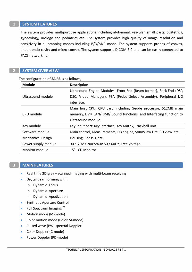
1 SYSTEM FEATURES
The system provides multipurpose applications including abdominal, vascular, small parts, obstetrics,
gynecology, urology and pediatrics etc. The system provides high quality of image resolution and
sensitivity in all scanning modes including B/D/M/C mode. The system supports probes of convex,
linear, endo-cavity and micro-convex. The system supports DICOM 3.0 and can be easily connected to
PACS networking.
2 SYSTEM OVERVIEW
The configuration of SA R3 is as follows,
Module Description
Ultrasound module
Ultrasound Engine Modules: Front-End (Beam-former), Back-End (DSP,
DSC, Video Manager), PSA (Probe Select Assembly), Peripheral I/O
interface.
CPU module
Main host CPU: CPU card including Geode processor, 512MB main
memory, DVI/ LAN/ USB/ Sound functions, and Interfacing function to
Ultrasound module
Key module Key Input part: Key Interface, Key Matrix, Trackball unit
Software module Main control, Measurements, DB engine, SonoView Lite, 3D view, etc.
Mechanical Design Housing, Chassis, etc.
Power supply module 90~120V / 200~240V 50 / 60Hz, Free Voltage
Monitor module 15” LCD Monitor
3 MAIN FEATURES
Real time 2D gray – scanned imaging with multi-beam receiving
Digital Beamforming with:
o Dynamic Focus
o Dynamic Aperture
o Dynamic Apodization
Synthetic Aperture Control
Full Spectrum ImagingTM
Motion mode (M-mode)
Color motion mode (Color M-mode)
Pulsed wave (PW) spectral Doppler
Color Doppler (C-mode)
Power Doppler (PD-mode)

TECHNICAL SPECIFICATION – SONOACE R3 | 2
Tissue Harmonic Imaging
Pulse Inversion Harmonic Imaging
Extreme High Dynamic Range
Trapezoidal Imaging
Combined modes
o 2D/M, 2D/PWD, 2D/CD, 2D/PD, 2D/CD/PW, 2D/PD/PW, 2D/CD/Color M
Cine for 511 frames
15” monitor with non-interlaced display
Freehand 3D
3D Multi Planar Imaging
2 Active Probe Ports (Optional)
QuickScanTM
DICOM 3.0 compatible Image filing : SonoView pro
SonoView Image management
Various Measurement Packages
Applications
o General, Abdomen, Obstetrics, Fetal Heart, Gynecology, Renal, Urology, Breast, Small Parts
o Vascular, Pediatric, MSK, Cardiac, Neonatal
HPRF
Standby mode (Hibernation mode)
Post Gain Control
Peripheral output device support
Language support : English, Italian, German, French, Spanish, Chinese and Russian
4 SCANHEADS
Curved Array: CN2-8, C2-4/20, CN4-9, EC4-9
Linear Array : LN5-12/40, L5-12/60, LE5-12
Safety Class : BF
4.1. CN2-8
Application: Abdomen, OB, Gynecology, Renal, Fetal Heart
Center frequency: 3.5MHz
Radius of curvature: 60mm
FOV: 60°
Number of elements : 80
Biopsy guide available
Veterinary supported: Abdomen, OB
TECHNICAL SPECIFICATION – SONOACE R3 | 3
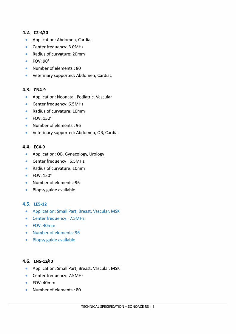
4.2. C2-4/20
Application: Abdomen, Cardiac
Center frequency: 3.0MHz
Radius of curvature: 20mm
FOV: 90°
Number of elements : 80
Veterinary supported: Abdomen, Cardiac
4.3. CN4-9
Application: Neonatal, Pediatric, Vascular
Center frequency: 6.5MHz
Radius of curvature: 10mm
FOV: 150°
Number of elements : 96
Veterinary supported: Abdomen, OB, Cardiac
4.4. EC4-9
Application: OB, Gynecology, Urology
Center frequency : 6.5MHz
Radius of curvature: 10mm
FOV: 150°
Number of elements: 96
Biopsy guide available
4.5. LE5-12
Application: Small Part, Breast, Vascular, MSK
Center frequency : 7.5MHz
FOV: 40mm
Number of elements: 96
Biopsy guide available
4.6. LN5-12/40
Application: Small Part, Breast, Vascular, MSK
Center frequency: 7.5MHz
FOV: 40mm
Number of elements : 80
TECHNICAL SPECIFICATION – SONOACE R3 | 4
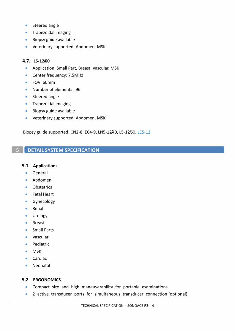
Steered angle
Trapezoidal imaging
Biopsy guide available
Veterinary supported: Abdomen, MSK
4.7. L5-12/60
Application: Small Part, Breast, Vascular, MSK
Center frequency: 7.5MHz
FOV: 60mm
Number of elements : 96
Steered angle
Trapezoidal imaging
Biopsy guide available
Veterinary supported: Abdomen, MSK
Biopsy guide supported: CN2-8, EC4-9, LN5-12/40, L5-12/60, LE5-12
5.1 Applications
General
Abdomen
Obstetrics
Fetal Heart
Gynecology
Renal
Urology
Breast
Small Parts
Vascular
Pediatric
MSK
Cardiac
Neonatal
5.2 ERGONOMICS
Compact size and high maneuverability for portable examinations
2 active transducer ports for simultaneous transducer connection (optional)
5 DETAIL SYSTEM SPECIFICATION
TECHNICAL SPECIFICATION – SONOACE R3 | 5
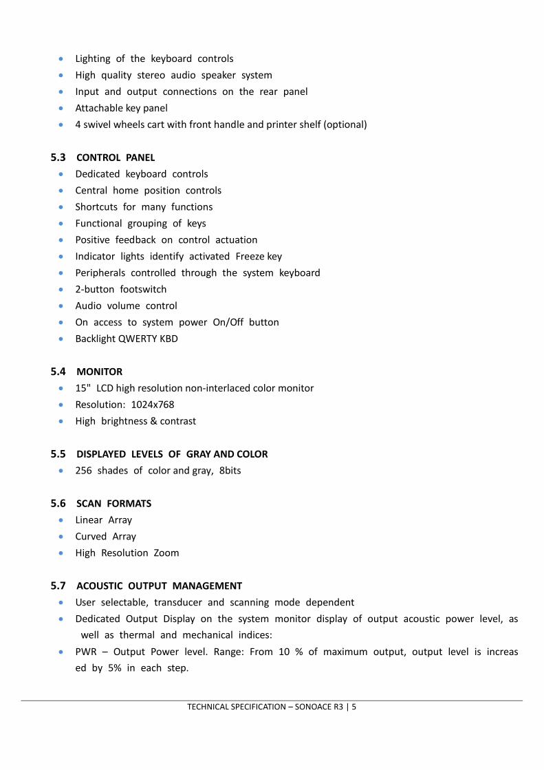
Lighting of the keyboard controls
High quality stereo audio speaker system
Input and output connections on the rear panel
Attachable key panel
4 swivel wheels cart with front handle and printer shelf (optional)
5.3 CONTROL PANEL
Dedicated keyboard controls
Central home position controls
Shortcuts for many functions
Functional grouping of keys
Positive feedback on control actuation
Indicator lights identify activated Freeze key
Peripherals controlled through the system keyboard
2-button footswitch
Audio volume control
On access to system power On/Off button
Backlight QWERTY KBD
5.4 MONITOR
15" LCD high resolution non-interlaced color monitor
Resolution: 1024x768
High brightness & contrast
5.5 DISPLAYED LEVELS OF GRAY AND COLOR
256 shades of color and gray, 8bits
5.6 SCAN FORMATS
Linear Array
Curved Array
High Resolution Zoom
5.7 ACOUSTIC OUTPUT MANAGEMENT
User selectable, transducer and scanning mode dependent
Dedicated Output Display on the system monitor display of output acoustic power level, as
well as thermal and mechanical indices:
PWR – Output Power level. Range: From 10 % of maximum output, output level is increas
ed by 5% in each step.
TECHNICAL SPECIFICATION – SONOACE R3 | 6
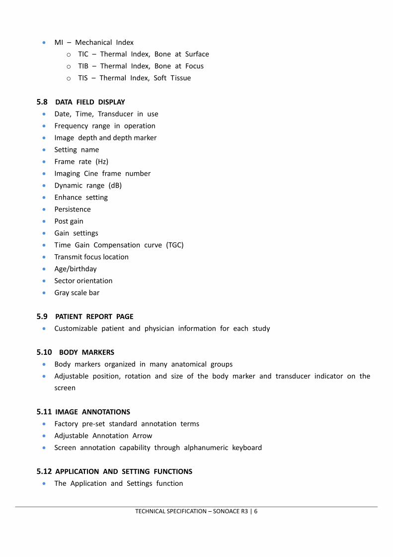
MI – Mechanical Index
o TIC – Thermal Index, Bone at Surface
o TIB – Thermal Index, Bone at Focus
o TIS – Thermal Index, Soft Tissue
5.8 DATA FIELD DISPLAY
Date, Time, Transducer in use
Frequency range in operation
Image depth and depth marker
Setting name
Frame rate (Hz)
Imaging Cine frame number
Dynamic range (dB)
Enhance setting
Persistence
Post gain
Gain settings
Time Gain Compensation curve (TGC)
Transmit focus location
Age/birthday
Sector orientation
Gray scale bar
5.9 PATIENT REPORT PAGE
Customizable patient and physician information for each study
5.10 BODY MARKERS
Body markers organized in many anatomical groups
Adjustable position, rotation and size of the body marker and transducer indicator on the
screen
5.11 IMAGE ANNOTATIONS
Factory pre-set standard annotation terms
Adjustable Annotation Arrow
Screen annotation capability through alphanumeric keyboard
5.12 APPLICATION AND SETTING FUNCTIONS
The Application and Settings function

TECHNICAL SPECIFICATION – SONOACE R3 | 7
Dedicated Application key
Dedicated Settings key
Settings-specific programs
Direct access to Settings and Applications during the examination
Default Program set-up for each Category
Backup storage and retrieval of the Programs and Applications through a external DVD-RW,
CD-RW and USB Flash Memory
Factory pre-set Programs and Applications protected from alteration and deletion
5.13 TRANSMIT FOCAL ZONE ENHANCEMENT
User-selectable position and number of transmit focal zone settings through a toggle switch
5.14 DISPLAY DYNAMIC RANGE
User selectable in 2dB increments
5.15 FRAME RATE
Max. above 280 FPS
5.16 INVERT OPTIONS
Up/down
Right/left
5.17 DEPTH SELECTION
Range: from 2 to 30 cm
5.18 TIME GAIN COMPENSATION
6 slide-pot controls
Reassigned on HRZ, Depth and U/D Invert adjustments
5.19 IMAGE PROCESSING PARAMETERS
2D Gain
Edge Enhance (10 steps)
Persistence
Real time 2D Filter
Dynamic Range (up to 180dB)
o High dynamic --> "soft gray" image
o Low dynamic --> "hard gray" image
Reject level
TECHNICAL SPECIFICATION – SONOACE R3 | 8
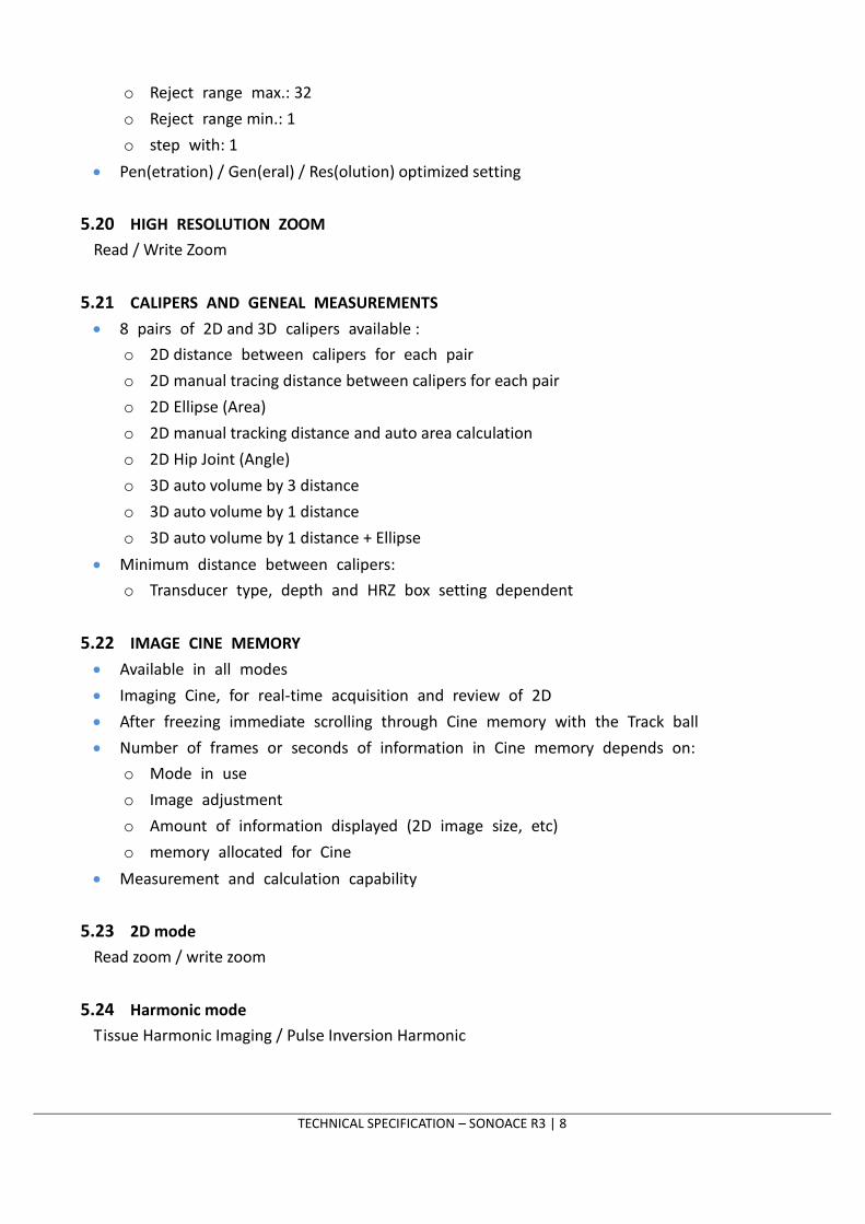
o Reject range max.: 32
o Reject range min.: 1
o step with: 1
Pen(etration) / Gen(eral) / Res(olution) optimized setting
5.20 HIGH RESOLUTION ZOOM
Read / Write Zoom
5.21 CALIPERS AND GENEAL MEASUREMENTS
8 pairs of 2D and 3D calipers available :
o 2D distance between calipers for each pair
o 2D manual tracing distance between calipers for each pair
o 2D Ellipse (Area)
o 2D manual tracking distance and auto area calculation
o 2D Hip Joint (Angle)
o 3D auto volume by 3 distance
o 3D auto volume by 1 distance
o 3D auto volume by 1 distance + Ellipse
Minimum distance between calipers:
o Transducer type, depth and HRZ box setting dependent
5.22 IMAGE CINE MEMORY
Available in all modes
Imaging Cine, for real-time acquisition and review of 2D
After freezing immediate scrolling through Cine memory with the Track ball
Number of frames or seconds of information in Cine memory depends on:
o Mode in use
o Image adjustment
o Amount of information displayed (2D image size, etc)
o memory allocated for Cine
Measurement and calculation capability
5.23 2D mode
Read zoom / write zoom
5.24 Harmonic mode
Tissue Harmonic Imaging / Pulse Inversion Harmonic
TECHNICAL SPECIFICATION – SONOACE R3 | 9
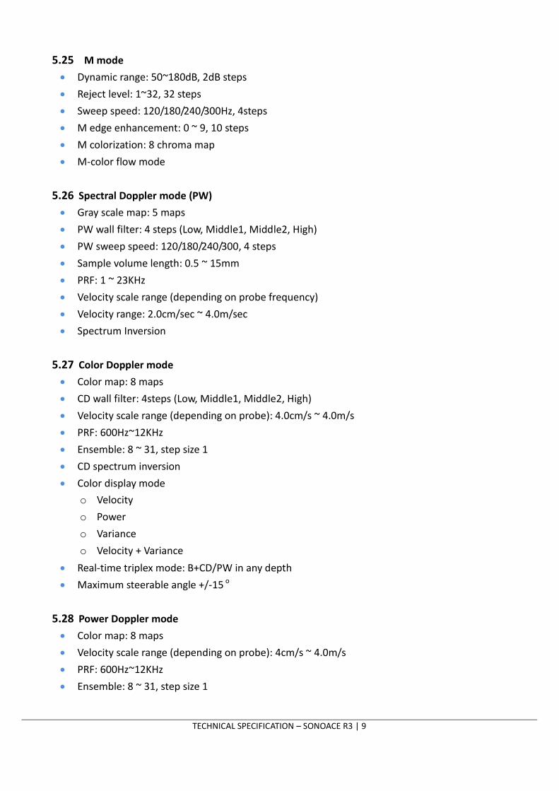
5.25 M mode
Dynamic range: 50~180dB, 2dB steps
Reject level: 1~32, 32 steps
Sweep speed: 120/180/240/300Hz, 4steps
M edge enhancement: 0 ~ 9, 10 steps
M colorization: 8 chroma map
M-color flow mode
5.26 Spectral Doppler mode (PW)
Gray scale map: 5 maps
PW wall filter: 4 steps (Low, Middle1, Middle2, High)
PW sweep speed: 120/180/240/300, 4 steps
Sample volume length: 0.5 ~ 15mm
PRF: 1 ~ 23KHz
Velocity scale range (depending on probe frequency)
Velocity range: 2.0cm/sec ~ 4.0m/sec
Spectrum Inversion
5.27 Color Doppler mode
Color map: 8 maps
CD wall filter: 4steps (Low, Middle1, Middle2, High)
Velocity scale range (depending on probe): 4.0cm/s ~ 4.0m/s
PRF: 600Hz~12KHz
Ensemble: 8 ~ 31, step size 1
CD spectrum inversion
Color display mode
o Velocity
o Power
o Variance
o Velocity + Variance
Real-time triplex mode: B+CD/PW in any depth
Maximum steerable angle +/-15 o
5.28 Power Doppler mode
Color map: 8 maps
Velocity scale range (depending on probe): 4cm/s ~ 4.0m/s
PRF: 600Hz~12KHz
Ensemble: 8 ~ 31, step size 1

TECHNICAL SPECIFICATION – SONOACE R3 | 10
PD wall filter: 4steps (Low, Middle1, Middle2, High)
Geode processor
Hard drive: 160 GB (SATA type)
RAM size:512MB
DVD-RW, USB, LAN capability
Stored Format: BMP, JPEG, TIF, DICOM
Exported Format: BMP, JPEG
Function Description
Measurement
2D mode: distance, angle, area, ellipse, circumference, volume
PW Spectral Doppler: velocity, pressure, acceleration
M mode: time, slope, distance
OB measurements
Fetal Biometry :
GS,CRL,YS,BPD,OFD,HC,APD,TAD,MAD,AC,FTA,FL,SL,TTD, APTD, APTDxTTD
Fetal Long Bones : HUM,ULNA,TIB,RAD,FIB,CLAV,VERT
Fetal Cranium : CEREB, OOD, IOD, CM, NF, Lvent, NT
Fetal Others : FOOT, EAR, MP
AFI
Volume Flow [B/Doppler]
Umbilical Artery [Doppler]
Mid Cereb Artery [Doppler]
Left Uterine Artery [Doppler]
Right Uterine Artery [Doppler]
Left Fetal Carotids [Doppler]
Right Fetal Carotids [Doppler]
Fetal Aorta [Doppler]
Ductus Venous [Doppler]
Fetal HR
Ratio : FL/BPD,CI(BPD/OFD),HC/AC,FL/AC,FL/HC,FL/FOOT
Observations : Fetal Description ,Fetal Heart, Fetal Brain, Fetal
Abdomen,
7 MEASUREMENT PACKAGE
6 ULTRASOUND PC MODULE

TECHNICAL SPECIFICATION – SONOACE R3 | 11
Biophysical Profile, Maternal Survey
Comment
Obstetric Biometry table list
FW
Campbell,Hadlock,Hadlock1,Hadlock2,Hadlock3,Hadlock4,
Hansmann,Merz,Osaka,Shepard,Tokyo1,Tokyo2,Shinozuka1,
Shinozuka2
FW-
Growt
h
Hadlock, Osaka, Tokyo, Doubilet, Brenner, Williams
GS GA Table Hansmann, Hellman, Korean, Nyberg, Tokyo
Growth Table None
CRL GA Table
Hadlock, Hansmann, Korean, Nelson, Osaka,
Robinson, Tokyo, Rempen
Growth Table Hansmann, Korean, Osaka, Tokyo, ASUM(SCW)
YS GA Table None
Growth Table None
BPD
GA Table
Campbell, Chitty(o-i),Chitty(o-o),
Hadlock, Hansmann, Jeanty, Korean, Kurtz, Merz,
Osaka, Sabbagha, Tokyo, Bessis
Growth Table
Chitty(o-i),Chitty(o-o),
Hadlock, Hansmann, Korean, Merz, Osaka,
Tokyo, ASUM(SCW),CFEF
OFD GA Table Hansmann, Korean, Merz
Growth Table Hansmann, Korean, ASUM(SCW), Merz
HC
GA Table Campbell, Chitty(m),Chitty(d), Hadlock,
Hansmann, Korean, Merz
Growth Table Chitty(m),Chitty(d),Hadlock, Hansmann,Korean,
Merz, CFEF, ASUM(SCW)
APD GA Table Hansmann, Bessis
Growth Table Hansmann
TAD GA Table None
Growth Table CFEF
MAD GA Table Eik-NesSH
Growth Table Eik-NesSH
AC
GA Table Campbell, Hadlock, Hansmann, Korean, Merz,
Tokyo
Growth Table
Campbell, Chitty(m), Chitty(d), Hadlock,
Hansmann, Jeanty, Korean, Merz, Tokyo,
ASUM(SCW),CFEF

TECHNICAL SPECIFICATION – SONOACE R3 | 12
FTA GA Table Osaka
Growth Table Osaka
FL
GA Table Campbell, Chitty, Hadlock, Hansmann, Hohler,
Jeanty, Korean, Merz, Osaka, Tokyo, Bessis
Growth Table Campbell, Chitty, Hadlock, Hansmann, Jeanty,
Korean,Merz,Osaka,Tokyo,ASUM(SCW),CFEF
SL GA Table None
Growth Table None
TTD GA Table Hansmann
Growth Table Hansmann
APTD GA Table Hansmann
Growth Table Hansmann
APTDx
TTD
GA Table Shinozuka
Growth Table Shinozuka
HUM GA Table Jeanty, Korean, Merz, Osaka
Growth Table Jeanty, Korean, Merz, Osaka, ASUM(SCW)
ULNA GA Table Jeanty
Growth Table Jeanty, Merz
TIB GA Table Jeanty, Merz
Growth Table Jeanty, Merz
RAD GA Table None
Growth Table Merz
FIB GA Table None
Growth Table None
CLAV GA Table Yarkoni
Growth Table Yarkoni
LV GA Table Tokyo
Growth Table None
CEREB GA Table Chitty, Hill
Growth Table Goldstein
OOD GA Table Jeanty
Growth Table None
IOD
GA Table None
Growth Table None
FOOT
GA Table None
Growth Table None
EAR GA Table None
TECHNICAL SPECIFICATION – SONOACE R3 | 13
Growth Table None
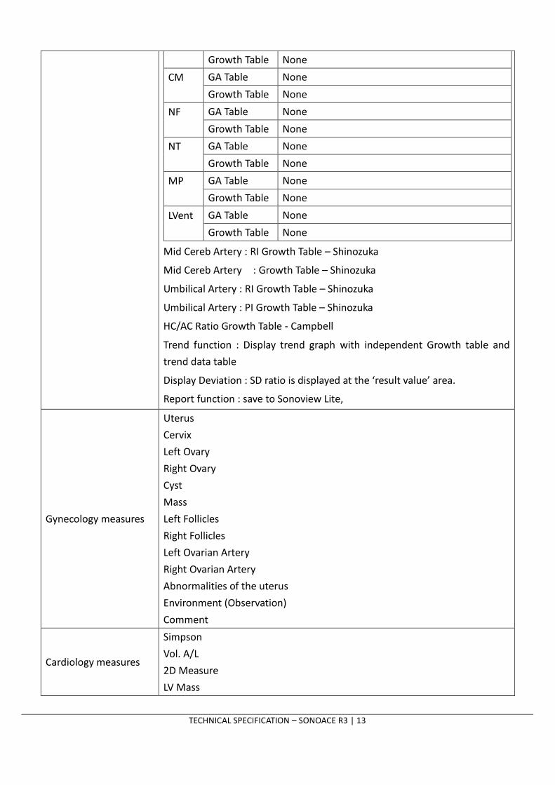
CM
GA Table None
Growth Table None
NF
GA Table None
Growth Table None
NT
GA Table None
Growth Table None
MP
GA Table None
Growth Table None
LVent
GA Table None
Growth Table None
Mid Cereb Artery : RI Growth Table – Shinozuka
Mid Cereb Artery : Growth Table – Shinozuka
Umbilical Artery : RI Growth Table – Shinozuka
Umbilical Artery : PI Growth Table – Shinozuka
HC/AC Ratio Growth Table - Campbell
Trend function : Display trend graph with independent Growth table and
trend data table
Display Deviation : SD ratio is displayed at the ‘result value’ area.
Report function : save to Sonoview Lite,
Gynecology measures
Uterus
Cervix
Left Ovary
Right Ovary
Cyst
Mass
Left Follicles
Right Follicles
Left Ovarian Artery
Right Ovarian Artery
Abnormalities of the uterus
Environment (Observation)
Comment
Cardiology measures
Simpson
Vol. A/L
2D Measure
LV Mass

TECHNICAL SPECIFICATION – SONOACE R3 | 14
Left Ventricle(M)
Ao/LA(B)
Ao/LA(M)
Mitral Valve(M)
LVOT Doppler [B/Doppler]
Mitral Valve Inflow [Doppler]
Mitral Valve Regurg [Doppler]
Aortic Valve Systolic [Doppler]
Aortic Valve Regurg [Color/Doppler]
Tricuspid Value Inflow [Doppler]
Tricuspid Valve Regurg [Doppler]
Pulmonary Valve Inflow [Doppler]
Pulmonary Valve Regurg [Color/Doppler]
Pulmonic Veins [Doppler]
Hepatic Veins [Doppler]
Qpulm:Qsys [B/Doppler]
Heart Rate [M/Doppler]
Comment
Urology
General [Doppler]
Prostate Volume
T-Zone Prostate Volume
Bladder Volume
Left Kidney Volume
Right Kidney Volume
Residual Volume
Observations : Digital Rectal Exam
Transrectal US Prostate
Transrectal US Seminal Vesicles
Comment
Vascular
Indication
General
Volume Flow [B/Doppler]
Rt. Subclavian / Lt. Subclavian [B/Doppler]
Rt. Prox CCA / Lt. Prox CCA [B/Doppler]
Rt. Mid CCA / Lt. Mid CCA [B/Doppler]

TECHNICAL SPECIFICATION – SONOACE R3 | 15
Rt. Distal CCA / Lt. Distal CCA [B/Doppler]
Rt. Bulb / Lt. Bulb [B/Doppler]
Rt. Prox ICA / Lt. Prox ICA [B/Doppler]
Rt. Mid ICA / Lt. Mid ICA [B/Doppler]
Rt. Distal ICA / Lt. Distal ICA [B/Doppler]
Rt. ECA / Lt. ECA [B/Doppler]
Rt. Vertebral / Lt. Vertebral [B/Doppler]
ICA/CCA Ratio
A/B Ratio
Vertebral [B/Doppler]
HR [M/Doppler]
Comment
Fetal Echo
2D Echo [B]
CTAR [B]
Fetal M-mode [M]
Main Pulmonary Artery [D]
Ductua Arteriosus [D]
Inferior Vena Cava [D]
Ductus Venosus [D]
Ascending Aorta [D]
Descending Aorta [D]
Mitral Valve Inflow [D]
Mitral Valve Regurg [D]
Tricuspid Valve Inflow [D]
Tricuspid Valve Regurg [D]
PLI (Preload Index) [D]
Fetal Heart
Environment : 4Chamber,3Vessel,LOT,ROT,AorticArch,CardRhythm
Comment
Report
Open Line Transfer : Only for English Windows
Export Function : Save Report Content to Excel / Text Format
Print : Print Contents to Local Printer
On-board printing device control
SonoView Lite
DICOM 3.0
8 DOCUMENTATION CAPABILITIES

TECHNICAL SPECIFICATION – SONOACE R3 | 16
Device Description
BW Video Page Printer Sony UP-897MD
Sony UP-D897 (USB interface)
Mitsubishi P-91
Mitsubishi P-93W
Color Video Page Printer Sony UP-20
Sony UP-21MD
Sony UP-D23MD (USB interface)
Sony UP-D25MD
External Monitor Recommended Specification
- Display type: LCD Monitor
- Resolution: 1024x768
- Input signal: DVI (or with DVI to RGB gender)
Foot Switch 2-pedal medical foot control (USB interface)
Switch functions among the following items.
Left: Dual, Freeze, Update, Store
Right: Dual, Freeze, Update, Store
External USB MO Drive Backup for Sonoview Lite
Fujitsu DynaMO 1300U2B or later version
External USB Flash Backup for Sonoview Lite
Imation iFLASH USB 2.0 1GB
Imation USB Swing Blue 1G
CD-RW Backup for Sonoview Lite
MyBox(External USB Case) + LG CDRW 52x
LiteOn LTR-52327SX CD-RW
LiteON CD-RW/DVD-ROM (SOHC-5232KX)
LiteON CD-RW (SOHR-5239SX)
Samsung SE-S084D, LG GP-08NU20
InkJet Printer Printing for measurement report or Sonoview Lite
HP DeskJet 5650
HP DeskJet 5940
HP DeskJet 6540
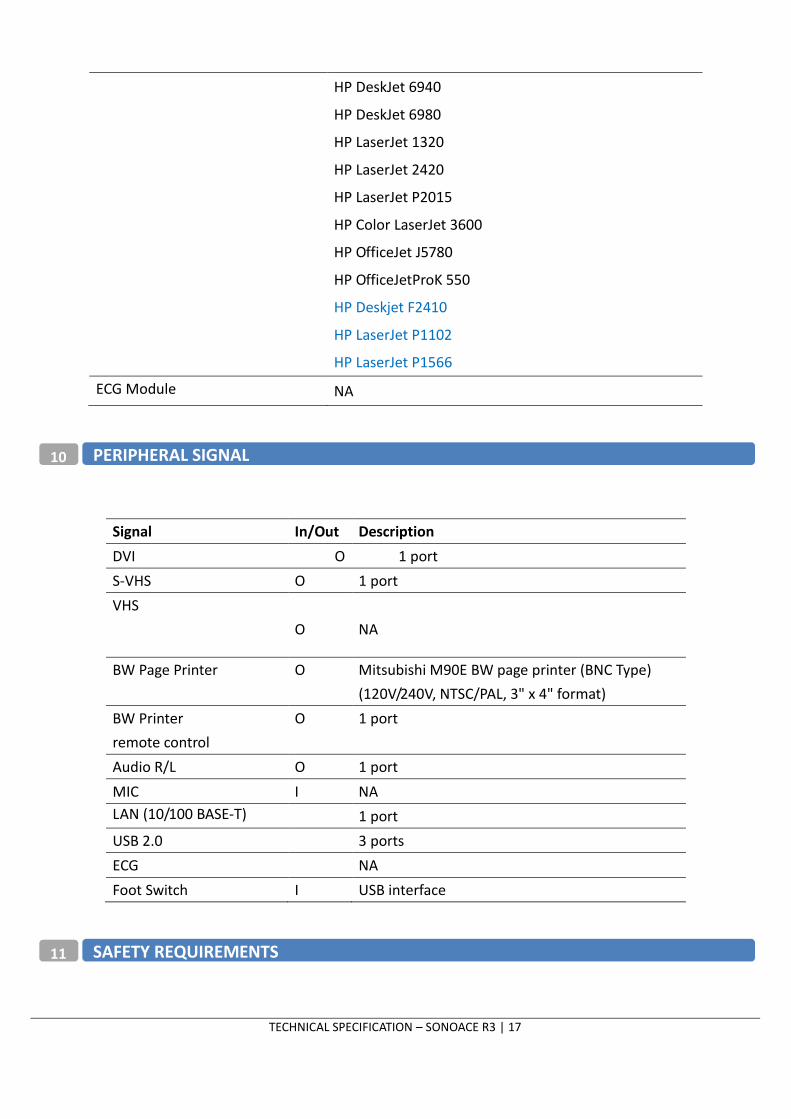
9 OPTIONAL DEVICES
TECHNICAL SPECIFICATION – SONOACE R3 | 17
HP DeskJet 6940
HP DeskJet 6980
HP LaserJet 1320
HP LaserJet 2420
HP LaserJet P2015
HP Color LaserJet 3600
HP OfficeJet J5780
HP OfficeJetProK 550
HP Deskjet F2410
HP LaserJet P1102
HP LaserJet P1566
ECG Module NA
Signal In/Out Description
DVI O 1 port
S-VHS O 1 port
VHS
O NA
BW Page Printer O Mitsubishi M90E BW page printer (BNC Type)
(120V/240V, NTSC/PAL, 3" x 4" format)
BW Printer
remote control
O 1 port
Audio R/L O 1 port
MIC I NA
LAN (10/100 BASE-T) 1 port
USB 2.0 3 ports
ECG NA
Foot Switch I USB interface
10 PERIPHERAL SIGNAL
11 SAFETY REQUIREMENTS
TECHNICAL SPECIFICATION – SONOACE R3 | 18
IEC/EN 60601-1 Medical Electrical Equipment, Part1, General Requirements for safety.
IEC/EN 60601-1-1 Safety requirements for medical electrical system.
IEC/EN 60601-1-2 Electromagnetic compatibility-Requirements and tests.
IEC/EN 60601-1-4 / ISO 14971 Programable electrical medical system
IEC/EN 60601-2-37 Particular requirements for the safety
IEC 61157 Declaration of acoustic output parameters.
EN/ISO 10993-1 Biological evaluation of medical devices.
UL 60601-1 Medical Electrical Equipment, Part1, General Requirements for safety.
CAN/CSA22.2,601.1 Medical Electrical Equipment, Part1, General Requirements for Safety
AIUM/NEMA UD-2 Acoustic Output Measurement Standard for Diagnostic Ultrasound Equipment
AIUM/NEMA UD-3 Standards for Real-Time Display of Thermal and Mechanical Acoustic Output
Indices on Diagnostic Ultrasound Equipment.
Power consumption : 90W
Heat dissipation : 490 BTU/h
Size: 375mm x 402mm x 188mm(H x W x D)
Weight: 8.5kg (approx)
100-120V/50~60Hz
200-240V/50~60Hz
Ambient temperature: 10°C–35°C (50°F–104°F)
Relative humidity: Up to 90% non-condensing
For more information on specification, please contact Product Marketing Team.
Phone : +82-2-2194-1094
Fax : +82-2-2194-1129
E-mail : [email protected]
12 POWER AND PHYSICAL SPECIFICATIONS
13 OPERATION ENVIRONMENT