technical note: analysis of microrna function using 1 ... · technical note: analysis of microrna...
TRANSCRIPT
1
Technical Note: Analysis of m
icroRN
A function using miR
CU
RY LN
ATM m
icroRN
A Inhibitors
Technical note: Analysis of microRNA function using miRCURY LNA™ microRNA Inhibitors
IntroductionThe study of microRNAs has come a long way since the discovery of these small RNAs some fifteen years ago. We now know that microRNAs are involved in a number of important biological processes such as embryonic development and cell differentiation and they have been linked to several important diseases such as cancer and heart disease. In addition, microRNAs have been found in a large number of different organisms including single cell alga, plants, vertebrates and their associated viruses.
However, out of the more than 9000 microRNAs currently annotated in miRBase (v.12.0), only a fraction have been characterized in terms of their biological role. Clearly, a great challenge ahead will be to elucidate the function of these microRNAs and to identify their mRNA targets.
One of the most powerful and straight forward ways of determining the function of a microRNA is by performing inhibition experiments. In such experiments, the phenotypic changes of cells transfected with antisense oligonucleotides are closely monitored to elucidate the biological role of the targeted microRNA. In addition to their role in characterizing microRNAs of unknown function, inhibition experiments can also be used for validating bioinformatically predicted mRNA targets.
Although a very powerful method, a number of technical challenges have to be overcome in order to get quality microRNA inhibition data. First, the silencing technology used must be effective under physiological conditions. Second, the oligonucleotide must be optimally delivered to the cells and, third, the effect of the inhibition has to be monitored in an efficient way. Here, we discuss these challenges and outline guidelinesfor the design of knockdown experiments using miRCURY LNA™ microRNA Inhibitors.
Experimental design In the early stages of planning a microRNA inhibition experiment, it is important to ensure that the microRNA is actually expressed in the cells. If the endogenous microRNA is not expressed in the cell line, then you are unlikely to observe any effect of silencing that microRNA.
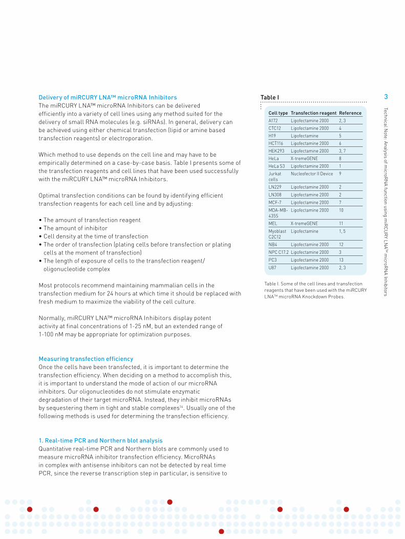
Figure I
18
16
14
12
10
8
6
4
2
0
5 nM
miR
CURY™ 1M
M
miR
CURY™
miR
CURY™ 2M
M
miR
CURY™ 4M
M
20 nM
Mismatch discrimination ability ofmiRCURY LNA™ Knockdown Probes
100 nM
Fold
Up-
regu
latio
n FL
(RLU
) / R
L(R
LU)
Figure I. Mismatch discrimination of miRCURY LNA™ microRNA Inhibitors. The pMIR-21 luciferase reporter is upregulated compared to the no-oligonucleotide control when HeLa cells are transfected with perfectly matched and mismatched (MM) antisense inhibitors targeting hsa-mir-21. There is a sharp drop in luciferase expression when comparing the perfectly matched microRNA inhibitor to the single (1MM) and double (2MM) mismatched inhibitors, indicating that miRCURY LNA™ microRNA inhibitors are very specific and can distinguish between single nucleotide mismatches. Luciferase expression levels were measured using Dual-Glo Luciferase 24 h after transfection. FL, firefly luciferase signal; RL, Renilla luciferase signal; RLU, relative light units.

2
Technical Note: Analysis of m
icroRN
A function using miR
CU
RY LN
ATM m
icroRN
A Inhibitors
Another consideration that should be addressed early in the planning stages is whether to knock down just a few microRNAs or to perform high throughput screens. In addition to individual antisense inhibitors, Exiqon offers the miRCURY LNA™ microRNA Inhibitor Library, which contains over 900 antisense oligonucleotides and has been specifically developed for high throughput screens. Other considerations include which method to use for the delivery of the antisense inhibitors and how to determine the effect of the silencing. These topics will be discussed in greater detail below.
An important aspect of any silencing experiment is the use of proper controls. It is always reassuring to verify that the observed phenotype is not reproduced when using a negative control (scrambled) LNA™ microRNA inhibitor. In addition, a custom designed mismatch control LNA™ microRNA inhibitor can be used to assess the specificity of the silencing (Figure I).
Designing efficient inhibitorsThere are a number of challenges in designing efficient LNA™ microRNA inhibitors. Mature microRNAs are only around 20 nucleotides in length, which means that even full length traditional antisense inhibitors (such as 2’-O-Me) will have poor affinity for their microRNA targets. This is especially problematic for AT-rich target sequences as the melting temperature of such duplexes will be relatively low. The use of full length oligonucleotides is further complicated by the fact that many microRNAs have autocomplementary end sequences, which may result in extensive self-annealing of the microRNAinhibitors.
By incorporating Locked Nucleic Acid (LNA™) monomers into the microRNA inhibitors, the affinity of the inhibitors for their target microRNAs is greatly increased and the resistance to enzymatic degradation is improved. This means that shorter oligonucleotides can be designed, thus avoiding the problem of using full length sequences.
We have taken advantage of this to develop new sophisticated inhibitordesign algorithms that calculate the most efficient inhibitors by adjusting the length and the LNA™ spiking pattern of the oligonucleotides. This has allowed us to design second generation antisense inhibitors with uniform and very high potency by normalizing their Tm and by minimizing problems of autocomplementarity (Figure II). Furthermore, this means that all our microRNA inhibitors have high silencing efficiency at low concentrations, which reduces the risk of negative side-effects.
50
40
30
20
10
0
Produ
ct A
Produ
ct B
2’-OM
e
miR
CURY LNA™
1 nM
5 nM
20 nM
microRNA inhibitor F
old
up
reg
ula
tio
n o
f m
icro
RN
A r
ep
ort
er
ge
ne
exp
ress
ion
Figure II. miRCURY LNA™ microRNA inhibitors are more efficient than competing technologies at inhibiting microRNAs. The expression of the pMIR-21 luciferase reporter is upregulated compared to the no-oligonucleotide control when MCF7 cells are transfected with antisense inhibitors directed against hsa-mir-21. This effect is significantly stronger in cells transfected with LNA™ oligonucleotides than cells transfected with DNA (Product A and B) or 2’-O-Me oligonucleotides. Measurements were taken 60 h after transfection.
Figure II
3
Technical Note: Analysis of m
icroRN
A function using miR
CU
RY LN
ATM m
icroRN
A Inhibitors
Delivery of miRCURY LNA™ microRNA InhibitorsThe miRCURY LNA™ microRNA Inhibitors can be delivered efficiently into a variety of cell lines using any method suited for the delivery of small RNA molecules (e.g. siRNAs). In general, delivery can be achieved using either chemical transfection (lipid or amine based transfection reagents) or electroporation.
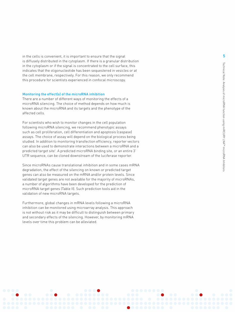
Which method to use depends on the cell line and may have to be empirically determined on a case-by-case basis. Table I presents some of the transfection reagents and cell lines that have been used successfully with the miRCURY LNA™ microRNA Inhibitors.
Optimal transfection conditions can be found by identifying efficient transfection reagents for each cell line and by adjusting:
• The amount of transfection reagent• The amount of inhibitor• Cell density at the time of transfection• The order of transfection (plating cells before transfection or plating
cells at the moment of transfection)• The length of exposure of cells to the transfection reagent/
oligonucleotide complex
Most protocols recommend maintaining mammalian cells in the transfection medium for 24 hours at which time it should be replaced with fresh medium to maximize the viability of the cell culture.
Normally, miRCURY LNA™ microRNA Inhibitors display potent activity at final concentrations of 1-25 nM, but an extended range of 1-100 nM may be appropriate for optimization purposes.
Measuring transfection efficiencyOnce the cells have been transfected, it is important to determine the transfection efficiency. When deciding on a method to accomplish this, it is important to understand the mode of action of our microRNA inhibitors. Our oligonucleotides do not stimulate enzymatic degradation of their target microRNA. Instead, they inhibit microRNAs by sequestering them in tight and stable complexes14. Usually one of the following methods is used for determining the transfection efficiency.
1. Real-time PCR and Northern blot analysisQuantitative real-time PCR and Northern blots are commonly used to measure microRNA inhibitor transfection efficiency. MicroRNAs in complex with antisense inhibitors can not be detected by real time PCR, since the reverse transcription step in particular, is sensitive to
Table I
Cell type Transfection reagent Reference
A172 Lipofectamine 2000 2, 3
CTC12 Lipofectamine 2000 4
H19 Lipofectamine 5
HCT116 Lipofectamine 2000 6
HEK293 Lipofectamine 2000 3, 7
HeLa X-tremeGENE 8
HeLa S3 Lipofectamine 2000 1
Jurkat cells
Nucleofector II Device 9
LN229 Lipofectamine 2000 2
LN308 Lipofectamine 2000 2
MCF-7 Lipofectamine 2000 7
MDA-MB- 435S
Lipofectamine 2000 10
MEL X-tremeGENE 11
Myoblast C2C12
Lipofectamine 1, 5
NB4 Lipofectamine 2000 12
NPC C17.2 Lipofectamine 2000 3
PC3 Lipofectamine 2000 13
U87 Lipofectamine 2000 2, 3
Table I. Some of the cell lines and transfection reagents that have been used with the miRCURY LNATM microRNA Knockdown Probes.
4
Technical Note: Analysis of m
icroRN
A function using miR
CU
RY LN
ATM m
icroRN
A Inhibitors
antisense compounds targeting the microRNA template. Similarly, strong complexes between miRCURY LNA™ inhibitors and microRNAsare not dissociated even in highly denaturing gels15. Such complexes can therefore be visualized in Northern blots as bands that migrate at a higher molecular weight than unbound mature microRNAs. However, we do not recommend these methods to estimate transfection efficiency. When transfection is inefficient it is very often because the microRNAinhibitoris retained on the outside of the cell membrane or is sequestered in internal vesicles, physically separated from their microRNA targets in the cytoplasm. However, during cell lysis these oligonucleotides are released and will interact with microRNAs in the lysate. Real-time PCR and Northern blot analysis therefore, can not distinguish between biologically relevant complexes that formed in the cytoplasm of transfected cells and complexes that formed following cell lysis.
2. Western blot analysisFor microRNAs with well characterized mRNA targets, we instead recommend monitoring transfection efficiency by measuring target mRNA encoded protein levels by Western blotting.
3. MicroRNA reporter assaysIf suitable antibodies are not available, we instead recommend using a microRNA reporter assay. An example of such an assay could be a plasmid encoded Renilla luciferase gene in which a target site complementary to the studied microRNA has been introduced into the 3’-UTR of the reporter. Consequently, the expression of the luciferase protein from this plasmid will be repressed when this particular microRNA is present in the cell. However, antisense-mediated inhibition of the microRNA results in a derepression of the reporter and an up-regulated luciferase expression. Thus, a reporter system needs to be constructed for each microRNA in the study and the background expression of the reporter depends on the expression level of the studied microRNA in the cell.
In addition to the microRNA reporter vector, a control vector constitutively expressing a different reporter molecule, e.g., firefly luciferase or beta-galactosidase should be used for normalization of transfection efficiency.
4. Fluorescently labeled microRNA inhibitorsA very convenient way of measuring transfection efficiency is by using fluorescently labeled miRCURY LNA™ microRNA Inhibitors. Flow cytometry can be used to sort the cells transfected with these oligo-nucleotides. While direct visualization of the LNA™ microRNA Inhibitors
5
Technical Note: Analysis of m
icroRN
A function using miR
CU
RY LN
ATM m
icroRN
A Inhibitors
in the cells is convenient, it is important to ensure that the signal is diffusely distributed in the cytoplasm. If there is a granular distribution in the cytoplasm or if the signal is concentrated to the cell surface, this indicates that the oligonucleotide has been sequestered in vesicles or at the cell membrane, respectively. For this reason, we only recommend this procedure for scientists experienced in confocal microscopy.
Monitoring the effect(s) of the microRNA inhibitionThere are a number of different ways of monitoring the effects of a microRNA silencing. The choice of method depends on how much is known about the microRNA and its targets and the phenotype of the affected cells.
For scientists who wish to monitor changes in the cell population following microRNA silencing, we recommend phenotypic assays such as cell proliferation, cell differentiation and apoptosis (caspase) assays. The choice of assay will depend on the biological process being studied. In addition to monitoring transfection efficiency, reporter vectors can also be used to demonstrate interactions between a microRNA and a predicted target site1. A predicted microRNA binding site, or an entire 3’ UTR sequence, can be cloned downstream of the luciferase reporter.
Since microRNAs cause translational inhibition and in some cases mRNA degradation, the effect of the silencing on known or predicted target genes can also be measured on the mRNA and/or protein levels. Since validated target genes are not available for the majority of microRNAs, a number of algorithms have been developed for the prediction of microRNA target genes (Table II). Such prediction tools aid in the validation of new microRNA targets.
Furthermore, global changes in mRNA levels following a microRNA inhibition can be monitored using microarray analysis. This approach is not without risk as it may be difficult to distinguish between primary and secondary effects of the silencing. However, by monitoring mRNA levels over time this problem can be alleviated.

6
Technical Note: Analysis of m
icroRN
A function using miR
CU
RY LN
ATM m
icroRN
A Inhibitors
Table II
Method Type of Method Reference Method Availability
Data Availa-bility
Resource
Stark et al. Complementary (Stark et al., 2003)
Online search Yes http://www.russell.embl.de/microRNAs
miRanda Complementary (John et al., 2004)
Download Yes http://www.microrna.org
miRanda MiRBase
LipofectamineComplementary
(Enright et al., 2003)
Online search Yes http://microrna.sanger.ac.uk
Target Scan Seed Complementary
(Lewis et al., 2005)
Online search Yes http://www.targetscan.org
DIANA microT Thermodynamics (Kirakidou et al., 2004)
Download Yes http://diana.cslab.ece.ntua.gr/
PicTar Thermodynamics (Krek et al., 2005)
N/A Yes http://pictar.mdc-berlin.de/
RNAHybrid Thermodynamics & Statistical model
(Rehmsmeier et al., 2004)
Download Yes http://bibiserv.techfak.uni-bielefeld.de/rnahybrid
miRGen++ Bayesian Inference (Huang et al., 2007b)
Mathlab Code Yes http://www.psi.toronto.edu/ genmir
MiTarget Support Vector Machine
(Kim et al., 2006)
Online search Yes http://cbit.snu.c.kr/~miTarget
MiTarget2 Support Vector Machine
(Wang and El Naqa, 2008)
Online search Yes http://mirdb.org
TarBase Experimentally Validated Targets
(Sethupathy et al., 2006)
N/A Yes http://diana.cslab.ece.ntua.gr/tarbase/
ConclusionsMicroRNAs have the potential to inhibit multiple target genes or potentially an entire pathway, which make them attractive targets for antisense inhibition studies. Our miRCURY LNA™ microRNA Inhibitors offer a convenient and powerful way of silencing microRNAs andelucidating their function. However, whether you are inhibiting single microRNAs or using the miRCURY LNA™ microRNA InhibitorLibrary for high throughput screens, careful planning of the experimental set-up is crucial for the outcome of the experiment.
Also, it is important to bear in mind that networks of several different microRNAs may act cooperatively in the translational regulation of individual mRNAs. Therefore, simultaneous disruption of multiple microRNAs may be required to elicit a biological phenotype. This can only be achieved using miRCURY LNA™ microRNA Inhibitors, as only they have the high potency needed for the cotransfection of several oligonucleotides at non-cytotoxic concentrations.
Table II. Methods and resources for microRNA target prediction. Reprinted from Enright in microRNA Research-Fundamentals, Reviews and Perspectives.
7
Technical Note: Analysis of m
icroRN
A function using miR
CU
RY LN
ATM m
icroRN
A Inhibitors
Scientific contributorsAnna Karina Busch, Niels M. Frandsen, Hazel Pinheiro, Johan WahlinExiqon A/S, Vedbæk, Denmark.
References
1. The microRNA miR-181 targets the homeobox protein Hox-A11 during mammalian myoblast differentiation. Naguibneva et al. Nat. Cell Biol. 2006, 8: 278-84.
2. MicroRNA-21 is an antiapoptotic factor in human glioblastoma cells. Chan et al. Cancer Res. 2005, 65: 6029-33.
3. MicroRNA-21 knockdown disrupts glioma growth in vivo and displays synergistic cytotoxicity with neural precursor cell delivered S-TRAIL in human gliomas. Corsten et al. Cancer Res. 2007, 67: 8994-9000.
4. MicroRNAs regulate the expression of the alternative splicing factor nPTB during muscle development. Boutz et al. Genes Dev. 2007, 21: 71-84.
5. An LNATM-based loss-of-function assay for microRNAs. Naguibneva et al. Biomed. Pharmacother. 2006, 60: 633-8.
6. Transcripts targeted by the microRNA-16 family cooperatively regulate cell cycle progression. Linsley et al. Mol. Cell Biol. 2007, 27: 2240-52.
7. Programmed cell death 4 (PDCD4) is an important functional target of the microRNA miR-21 in breast cancer cells. Frankel et al. 2008, 283: 1026-33.
8. X-tremeGene siRNA transfection reagent: A powerful tool for antisense inhibition of microRNA in human cells with miRCURY LNATM Knockdown Probes. Watzele et al. Biochemica, 2006, 2: 28-30.
9. Suppression of microRNA-silencing pathway by HIV-1 during virus replication. Triboulet et al. Science, 2007, 315: 1579-82.
10. miR-200b mediates post-transcriptional repression of ZFHX1B. Christoffersen et al. RNA, 2007, 13: 1172 – 1178.
11. MicroRNA expression dynamics during murine and human erythroid differentiation. Zhan et al. Exp. Hematol. 2007, 35: 1015-25.
12. A minicircuitry comprised of microRNA-223 and transcription factors NFI-A and C/EBPalpha regulates human granulopoiesis. Fazi et al. Cell, 2005, 123: 819-31.
13. miR-221 and miR-222 expression affects the proliferation potential of human prostate carcinoma cell lines by targeting p27Kip1. Galardi et al. J. Biol. Chem. 2007, 282:23716-24.
14. Antagonism of microRNA-122 in mice by systemically administered LNA-antimiR leads to up-regulation of a large set of predicted target mRNAs in the liver. Elmén et al. Nucleic Acids Res. 2008, 306: 1153-62.
15. miR-122 targeting with LNA/2’-O-methyl oligonucleotide mixmers, peptide nucleic acids (PNA), and PNA-peptide conjugates. Fabani & Gait. RNA. 2008, 14: 336-46.
www.exiqon.com