tc-99m dtpa dynamic renal scintigraphy in the …
TRANSCRIPT

Tc-99m DTPA DYNAMIC RENAL SCINTIGRAPHY IN THE
EVALUATION OF RENAL TRANSPLANTS: THE
IMPORTANCE OF PERFUSION INDEX IN THE DIAGNOSIS
OF RENAL ALLOGRAFT REJECTION
Pages with reference to book, From 139 To 148
Qaisar Hussain Siraj, Asma Inam-ur-Rehman, Amin Waqar, Syed Azhar Ahmed ( Nuclear Medical Centre, Armed Forces
Institute of Pathology, Rawalpindi. )
Mukhtar Hamid Shah, Rauf Iftilthar Ahmed, Mohammad Sadiq, Rehan Burney ( D.N.S.R.P. (P.A.E.C), Combined Military
Hospital, Rawalpindi. )
Abstract
Sixty patients with transplanted kidneys underwent dynamic renal scintigraphy using Technetium99m
DTPA. The Perfusion Index, in particular was found to be valuable in differentiating between the two
major renal transplant complications of acute tubular necrosis and acute rejection. In addition, other
complications like outflow obstruction, pararenal fluid collections, leakage, etc., were readily
diagnosed with a high degree of accuracy (JPMA 38: 139,1988).
INTRODUCTION
Since the time of the first successful clinical renal transplant in Chicago1, tremendous progress has
been made in the technique. However, major potential complications jeopardise the function of the
transplanted kidney as well as the life of the recipient. Since therapy differs for the various underlying
conditions, an early definitive diagnosis and, if possible, prognosis must be provided to ensure the
health of the transplant patient. Serum creatinine, blood urea nitrogen, and creatinine clearance are
helpful, but lack specificity. The radionuclide studies are useful in the evaluation of renal transplants
and can provide diagnostic criteria for prerenal, renal, and post-renal causes of acute post-transplant
renal failure, as well as for prognosis2. Prerenal conditions such as renal artery stenosis, renal
conditions such as acute rejection (AR) or acute tubular necrosis (ATN), and postrenal conditions such
as obstruction or leakage, which are the most common causes of acute post-transplant renal failure can
be differentiated. As a result of the current increase in renal transplant surgery for end term renal failure
in Pakistan, a sizeable number of patients are now being referred to the clinical nuclear medicine
services for the radionuclide evaluation of the transplants. We present here, our experience of
radionuclide studies in the evaluation of renal transplants at the Nuclear Medical Centre, Armed Forces
Institute of Pathology, Rawalpindi.
MATERIALS AND METHODS
Patients receiving renal transplants locally as well as a few who had received transplants abroad and
later came to us for assessment were evaluated. From April 1986 to May 1987, 98 radionuclide renal
transplant studies were performed on sixty patients. Four of these patients had received transplants
from cadavers and the rest from live related donors. There were nine females and fifty one males aged
17 to 54 years (mean age = 34.7 ± 9.5 yr). Graft survival in these patients ranged from one day to six
years. The study was carried out using a modern large field of view gamma camera fitted with a low
energy general purpose parallel hole collimator and linked to a dedicated On-line computer (Nova 4C).
The patient was positioned under the camera so as to include the aortic bifurcation, the renal transplant,

and the bladder. Tc-99m DTPA was injected in a bolus dose of 6 mCi. The study was recorded on the
computer, using a frame rate of 1 frame per second for the first 30 sec followed by 30 sec frames for
the next 30 minutes. A 64x64 matrix was used, using word mode. In some patients images at 1-2 hr or
later after injection were also recorded. Regions of interest over the iliac artery (distal to the transplant),
the transplant (excluding any portions overlying the artery), and a background area were defined from
the computer derived data, for analysis (Figure 1).

Activity-time curves for these regions were generated after subtraction of the background activity.
Visual interpretation of the analogue images were correlated with the computer generated data and
physiologic parameters like renal perfusion, selpctive renal uptake of the radio-pharmaceutical, transit
time through the kidney, and clearance time of the radioactivity from the kidney, were evaluated.
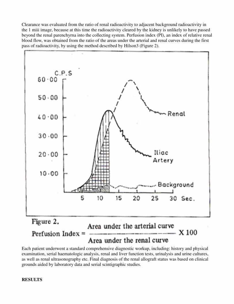
Clearance was evaluated from the ratio of renal radioactivity to adjacent background radioactivity in
the 1 miii image, because at this time the radioactivity cleared by the kidney is unlikely to have passed
beyond the renal parenchyrna into the collecting system. Perfusion index (PI), an index of relative renal
blood flow, was obtained from the ratio of the areas under the arterial and renal curves during the first
pass of radioactivity, by using the method described by Hilson3 (Figure 2).
Each patient underwent a standard comprehensive diagnostic workup, including: history and physical
examination, serial haematologic analysis, renal and liver function tests, urinalysis and urine cultures,
as well as renal ultrasonography etc. Final diagnosis of the renal allograft status was based on clinical
grounds aided by laboratory data and serial scintigraphic studies.
RESULTS

On the basis of all the available data including the radionudide studies, the patients were divided into 5
groups. Those with normally functioning transplants (group 1), with acute tubular necrosis (group 2),
with rejection (group 3),. with urinary tract infections (group 4), and those with outflow problems
(group 5). Figure 3 shows the range of P1 values in various groups. The PI values (mean and s.e.m) for
groups 1-5 were 1383±26, 184±49, 341±103, 141±27 and 149±17 respectively. There was no statistical
difference in perfusion indices in patients with stable graft function and those suffering from acute
tubular necrosis, ureteral obstruction or nephropathy. There was however, a significant increase (P<
0.001) in PI in patients with acute rejection.
Figure 3 shows the difference in perfusion in a kidney with AR as compared to a normally functioning
transplant. All of the patients in Group 1 showed a renogram curve peaking at 3 to 6 min with steep
slopes (Figure 4a),

good selective accumulation of the radioactivity in the transplanted kidney at 2 min, and rapid
clearance of the background activity (Figure 5).

In 8 patients with elevated BUN/Creatinine levels, scintigrdphic assessment showed normally
functioning transplants. Rapid return of BUN levels to normal were subsequently seen in 6 patients
while in 2 patients error in BUN results was correctly predicted. ATN was diagnosed on the basis of the
scintigraphic findings and was confirmed clinically by exclusion i.e. all other possible complications

were ruled out; and by the subsequent clinical and scintigraphic course of progressive improvement in
the renal function. Thirty one scintigraphic studies demonstrated 19 episodes of ATN in 18 patients.
The mean time since transplant in this group was 16 ± 16 days. The renal scintigraphs of the patients
with ATN showed mildly reduced to good perfusion, poor clearance of background activity, varying
degrees of delayed and diminished selective uptake of the radiotracer, and late appearance or non-
appearance of activity into the collecting system (Figure 6).

The renogram curves ranged from those with delayed and blunted peaks with poor slopes to almost flat
curves (Figure 4b). The severity of the scan and renographic appearances were seen to be in proportion
to the clinical degree of the ATN status. Often a fairly good renal image at 2 to 5 min. was seen
representing the blood pool rather than selective uptake, followed by fading of the renal image with

rising background levels. Acute rejection was diagnosed scintigraphically on the basis of reduced
perfusion with a rise in the perfusion index, when a baseline PI was available, or by a high perfusion
index, together with commensurate reduction in both perfusion and clearance. The diagnosis was con-
firmed by the clinical picture, ultrasound appearances when available, subsequent response to therapy,
as well as serial scintigraphic studies. Fifteen scintigraphic studies documented 8 episodes of rejection
in 7 patients. The renal scintigraphs obtained in patients in rejection showed moderately reduced to
poor perfusion, with commensurately reduced clearance and selective uptake. Often renal concentration
of the tracer was sufficient to permit visualization of the renal calyces and pelvis, and varying amounts
of tracer depending upon the degree of rejection appeared in the urinary bladder. (Figure 7).

The renogram curves ranged from low amplitude curves with diminished slopes to almost flat curves.
However, wherever a peak could be defined, no delay in time to peak was noticeable. The renogram
curves of the patients with moderate to severe rejection showed sharper peaks and steeper slopes as
compared to the renogram curves of the patients with ATN, for the same degree of diminution in renal
function. The mean time since transplant in this group of patients was 60±55 days indicating its
generally late occurrence. Figure 4 (c) shows a representative renogram of a patient with acute
rejection. Obstruction was documented on scintigraphy in 6 cases. Four showed mild to moderate
degrees of obstruction with delayed renogram peaks, slow drainage of the tracer from the transplant,
and ureteric dilatation. One patient manifested severe obstruction 10 wk. after the transplant operation,
with a rising renogram curve. The obstruction was relieved by a resection of the stenotic distal, ureter
and reimplantation. After the second operation he developed ATN which was diagnosed on scintigraphy
5 days postoperatively. Another patient showed partial obstruction to outflow of tracer from the
transplant with a large photodeficient area inferior to the transplant on the 7th postoperative day.
Ultrasound studies showed a large fluid collection causing the obstruction. Significant urinary tract
infection (UTI) was diagnosed in 6 cases on the basis of urinalysis. One patient was in AR, while a
second had ATN. On recovery from ATN, UTI still persisted. The renogram curves in the
uncomplicated cases were generally normal, although sluggish outflow was seen in two cases.
Perfusion and selective uptake were normal. One patient showed complete renal artery occlusion. No
perfusion or selective uptake was seen in the transplant. A subsequent injection of Tc-99m stannous
colloid also showed absence of perfusion with no tracer uptake in the transplant (Figure 8).

This confirmed the diagnosis of renal artery occlusion and excluded the possibility of severe rejection.
Four patients in this series showed persistently poor perfusion and uptake in a portion of the
transplanted kidney representing confirmed operative trauma to a branch of the renal artery (Figure 9).
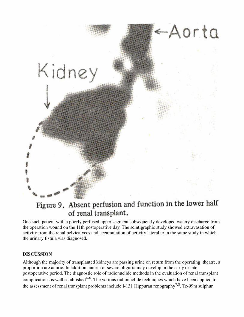
One such patient with a poorly perfused upper segment subsequently developed watery discharge from
the operation wound on the 11th postoperative day. The scintigraphic study showed extravasation of
activity from the renal pelvicalyces and accumulation of activity lateral to in the same study in which
the urinary fistula was diagnosed.
DISCUSSION
Although the majority of transplanted kidneys are passing urine on return from the operating theatre, a
proportion are anuric. In addition, anuria or severe oliguria may develop in the early or late
postoperative period. The diagnostic role of radionuclide methods in the evaluation of renal transplant
complications is well established4-6. The various radionuclide techniques which have been applied to
the assessment of renal transplant problems include I-131 Hippuran renography7,8, Tc-99m sulphur
colloid studies9, In-111 labeled leukocytes10 or platelets11, Gallium citrate12, radioiodinated
fibrinogen12, and Tc-99m DTPA studies3,13-15. The Tc-99m DTPA studies have the apparent
advantages of decreased study time, shorter biological half life, and improved anatomical detail of the
collecting system, perirenal area and vasculture16. Additional parameters obtained from the DTPA
study provide valuable diagnostic criteria in differentiating between the two common post transplant
complication of ATN and rejection15,17,18. The improved anatomical detail provides better assessment
of the excretory dynamics in assessing post renal obstruction and leakage, and clearer visualization of
extrarenal collection19,20. The majority of the transplanted kidneys show an element of ATN. This may
manifest itself as severe oliguria from the time of return from the operating theatre, or its onset may be
delayed. The duration of oliguria from ATN has been reported from 2-31 days but longer periods of
ATM are compatible with good graft function21. The findings of Tc-99m DTPA studies are diagnostic3.
Generally ATN is characterized by relatively normal blood flow whereas rejection is characterized by
poor blood flow; and this difference in blood flow can be demonstrated by the proper bolus injection of
a technetium labelled radiopharmaceutical. In our series the kidneys with ATN were seen to be
moderately well perfused with the maximum PI in each ATM episode between 117-275 in 17 out of 19
cases, with little or no subsequent selective parenchymal accumulation of tracer and poor clearance.

The mechanism of relatively preserved renal perfusion in the face of decreased clearance in patients
with ATM is unknown. Ischaemic damage to renal tubular cells may cause some cells to slough into the
tubular lumen and secondarily cause obstruction22; this process seems to be the major cause of
oliguria23. Ischaemic damage also increases the permeability of the tubular basement membrane,
allowing molecules that are filtered to diffuse back into the vascular compartment22.
Serial imaging in patients with ATM was seen to demonstrate continuing perfusion with improvement
in P1 and gradual return of function. Indeed long term follow-up of patients in ATM by Kjellstrand et
al. demonstrated that they did not differ prognostically from patients who experienced immediate renal
function24. Acute rejection crises continUe to pose a serious threat to patients receiving renal allografts,
and are among the most difficult problems for those concerned with the management of such patients.
Correct diagnosis of the transplant status is essential for initiation of the appropriate treatment, as
undertreatmènt may result in the loss of the graft and overtreatment may endanger the life of the patient
due to complications of immunosuppression. The diagnosis of acute rejection may, however, be
hampered by a paucity of definitive clinical signs or symptoms, as well as by a lack of definitive
biochemical or radiological diagnostic modalities capable of detecting early acute rejection and
differentiating it from the many other conditions that can adversely affect renal function25. Classic
signs and symptoms of acute rejection;including decreased renal function, graft tenderness, fever,
oliguria, and/or hypertension are frequently absent in most cases, and frequently occur in the course of
graft ureteral obstruction, pyelonepltritis, or drug induced renal dysfunction26. Serum creatinine, blood
urea nitrogen, and creatinine clearance are helpful, but lack specificity. The differentiation of acute
tubular necrosis from rejection has been difficult and often uncertain even with the combination of
clinical and laboratory information. It is generally agreed that damage to the microcirculation of the
kidney occurs early in the course of acute rejection27,28. These studies have shown that obstruction and
disruption of peritubular capillaries and venules occur in association with infiltration of the allograft by
lymphoblasts, and are one of the earliest manifestations of acute rejection. Further progression of AR
produces fibrinoid necrosis of the walls of arterioles and small arteries. The deposition of fibrin and
platelets on the damaged intima leads in turn to further obstruction of the cortical microcirculation and
renal ischaemia and necrosis. The observation that microcirculatory changes precede functional de-
rangement during AR is the basis for the usefulness of radionuclide renal studies in the evaluation of
graft dysfunction after renal transplantation3,13. The identification of a decreased blood flow provides a
valid point that can be easily measured in any nuclear medicine laboratory. The Tc—99m DTPA study
shows a rise in perfusion index, corresponding to the fall in renal blood flow which has been shown to
occur as one of the earliest changes in rejection29. Further refinement of this technique has established
that selective analysis of cortical perfusion will enhance the accuracy of Tc—99m DTPA studies for the
early detection of AR and in differentiating AR from non-immunological causes of renal allograft
dysfunction. Anaise et al. have reported an accuracy of 94% in diagnosing AR using the cortex PI15. In
our series the maximum P1 recorded during each rejection episode ranged between 234 to over 500,
with lower values recorded in the very early stages and in the recovery phase. The decrease in renal
function associated with rejection was evident on scintigraphy by reduced parenchymal accumulation
and excretion of tracer. However, for the same degree of reduced blood flow, rejection was seen to
generally exhibit a much better parenchymal accumulation than in ATN. This was reflected also in the
renogram curve which for the same degree of renal dysfunction displayed better curve patterns with the
significant earlier time to peak in cases of rejection as compared to cases of ATN. In patients with renal
transplants, one of the most difficult periods clinically is between 4 days and 3 weeks when ATN and
rejection may both occur30. Serial radionuclide studies over several days to wegks proved to be
particularly helpful in distinguishing ATN from rejection. ATN usually improves with time after the
ischaemic insult and thus continuing perfusion with improvement in P1 was always seen, despite
relatively poor parenchymal accumulation. In rejection the diminution of perfusion was persistent over
a longer time period and often progressive. A positive response to therapy was heralded by a falling PI.
In summary, our data support the hypothesis that decreased clearance with relatively preserved

perfusion in Tc—99m DTPA studies is sufficiently specific to be useful in differentiating ATN from the
other causes of decreased renal clearance17. Acute renal failure attributable to postrenal conditions such
as obstruction of the ureters or urinary extravasation can be readily diagnosed by scintillation camera
studies. Obstruction is heralded by a delayed radionuclide uptake and peak count rate, with progressive
accumulation of the radiopharmaceutical in the excretory system. The administration of furosemide
(lasix) may alter the accumulation pattern of the radiopharmaceutical, and hence be useful for
differentiating confusing conditions. Lower tract obstruction from haematoma, lymphocele, distal
ureter necrosis, or occlusion of the ureteroneocystomy can be identified by the prolonged retention and
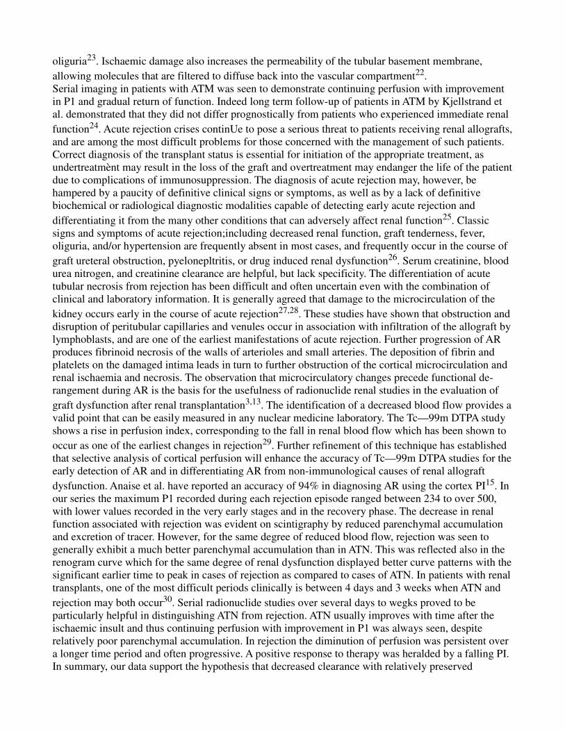
excessive pelvic accumulation of the tracer. It is worth noting that obstruction of a transplanted kidney
frequently presents with only subtle scan changes, i.e., a slight delay in excretion or a slight excess of
pelvic activity. This is probably due to the fact that the obstructions we seek to detect are acute, often
present for only hours and at most days. The more dramatic hallmarks such as massive pelvicalyceal
distension, loss of cortex, or megaloureter do not have time to develop31 (Figure 11).
We conclude that the simple and non-invasive Tc-99m DTPA dynamic renal scintigraphy is an accurate,
reliable, repeatable, and the single most important investigation for the assessment of the renal
transplant status. Visual assessment of the scintigraphs aided by quantitative parameters like the
perfusion index and the quantitative analysis of the renogram curves provide a wealth of data enabling
us to accurately assess and differentiate between the major transplant complications.
REFERENCES
1. Lawler, R.H., West, J.W., McNulty, P.H., Clancy, EJ. and Murphy, R.P. Homotranspiantation of the
kidney in the human; a preliminary report. JAMA., 1950; 144 : 844.
2. Schlegel, J.U. and Lang, E.K. Computed radionucide urogram for assessing acute renal failure. AJR.,
1980; 134:1029.
3. Hilson, A. 1. W., Maisey, M.N., Brown, C.B. et al. Dynamic renal transplant imaging with Tc-99m
DTPA (Sn) supplemented by a transplant perfusion index in the management of renal transplants. J.
Nucl. Med., 1978; 19 : 994.
4. Rosenthall, L., Mangel, R., Lisbona, R. and Lacourciere, Y. Diagnostic applications of radio-
pertechnetate and radiohippurate imaging in post-renal transplant complications. Radiology, 1974;1i1:
347.
5. Dubovsky, E.V., Logic, J.R., Dietelm, A.G., Balch, C.M. and Tauxe, W.N. Comprehensive evaluation
of renal function in the transplanted kidney. J.Nucl. Med., 1975; 16: 1115.
6. Weiss, E.R., Blahd, W.H., Winston, MA,, Harten bower, D.L., Koppel, M. and Thomas, P.B.
Scintillation camera in the evaluation of renal transplants. J. Nucl. Med., 1970; 11: 69.
7. Collins, J.J. Jr., Pizak, L. F. Jr., Tamvakopoulus, S.K. and Wilson, E,R. Serial renograms after kidney
transplantation in man. Surg. Forum, l963;14 :217.
8. Blaufox, M,D. and MenU, J.P. Evaluation of renal transplant function by iodohippurate sodium 1
-13l.JAMA.,1967;202 :575.
9. Frick, MY., Loken, M.K., Goldberg, M.E. and Simmons, RI. Use of 99m Tc-Sulfur colloid in
evaluation of renal transplant complications. J. Nucl. Med., 1976; 17 :181.
10. Frick, MY., Henke, C.E., Forstorm, L.A., Simmons, R. A., McCullough, J. and Loken, M.K. Use of
111 In-labelled leukocytes in evaluation of renal transplant rejection. Clin. Nucl.Med., l979;4 : 24.
11. Smith, N., Chandler, S., Hawker, R. J., Hawker, L. M. and Barnes, A.D. Indium-labelled autologus
platelet as diagnostic aid after renal transplantation. Lancet , 1979; 2 : 1241.
12. George, E.A., Codd, J.E., Newton, W.T., Henry, RE. and Donati, R.M. Comparative evaluation of
renal transplant rejecton with radioiodinated fibrinogen 99mTc-sulfur colloid and 67 Gacitrate. J. Nucl.
Med., 1976; 17:175.
13. Preston, D.F. and Luke, R.G. Radionucide evaluation of renal transplants. JJ4ucL Mcci, 1979; 20 :
1095.
14. Kiepfer, R.F., Kirchner, P.T. and Gerber, R.F. Clinical application of kidney to aortic blood flow
index. J. Nucl. Med., 1976; 17: 537.
15. Anaise, D., Oster, Z.H., Atkins, H.L., et al. Cortex perfusion index: a sensitive detector of acute
rejection crises in transplanted kidneys. J. NucI. Med., 1986; 27: 1697.
16. Staab, E.V., Jones, T.M., Partain, C.L., et aL A comparison of 99mTc-DTPA and 1-131 OIH in the
evaluation of renal transplant function. (Abstr). 3. Nucl., Med., 1976; 17 : 537.
17. Shanahan, W.S. M., Kllngensmith,W.C. and Well, R. 99m-Tc DTPA renal studies for acute tubular
necrosis; specificity of dissociation between perfusion and clearance. AJR., 1981; 136 : 249.
18. Singh, A. and Cohen, W.N. Renal allograft rejection; sonography and scintigraphy. MR., 1980;135:
73.
19. Bingham, J.B., Hilson, A.J.W. and MaAey, M.N. The appearances of renal transplant lymphocoeles
during dynamic renal scintigraphy. Br. J. Radiol., l978;51 : 342.
20. Spigos, D. G., Tan ,W,, Pavel, D.G., Mozes, M., Jonasson, 0. and Capek, V. Diagnosis of urine
extravasation after renal transplantation. MR., 1977; 129 : 409.
21. Oliver, DO, In Kidney transplantation; principles and practice. Edited by Morris, P. J. New York,
Grune and Straton, 1979, p. 58.
22. Donahue, J.F., Venkatachalam, M.A., Bernard B. B. and Levinsky, N.G. Tubular leakage and
obstruction after renal isehaemia; structural- functional correlations. Kidney Int., 1978; 13 : 208.

23. Arendshorst, Wi., Finn, W.F. and Gottschalk, C.W. Micropuncture study of acute renal failure
following temporary renal ischaemia in the rat. Kidney Int., 1976; 10 (Suppl. 6): 5100.
24. Kjellstrand, C.M., Casali, R.E., Simmons, R.L., Shideman, J.R., Buselmeier, T.J and Najarian, J.S.
Etiology and prognosis in actue post-transplant renal failure. AJM.,1976;61 : 190.
25. Winchester, J.F., Gelfarid, M.C., Foegh, M.L., Heifrich, G.B. and Schreiner, G.E. Early indicators
of renal aliograft rejection. Kidney Int, 1983; Suppl. 14: S-34.
26. Williams, G.M. in kidney transplantation; principles and practice. Edited by Morris, P.J. New York,
Grune and Stratton, 1 984, p. 335.
27. Tourville, D.R., Kim, D.U., Viscu, R., et al. Anticipation of renal transplant failure by
postanastomosis biopsy and immunofluorescence. Transplant. Proc., 1977; 9 : 91.
28. Porter, K.A. Morphological aspects of renal homograft rejection. Br. Med. Bull., 1965; 21:171.
29. Hollenberg, N.K., Retik, AS., Rosen, S.M., Murray, JR., and Merril, J.P. The role of vaso-
constriction in the ischaemia of renal ailograft rejection. Transplantation, 1968; 6 : 59.
30. Stazl, T.E. In Experience in renal transplantation. Philadelphia, Saunders, 1964,p. 142.
31. Strauss, H.W., Kirchener, P.T. and Wagner, H.N. Jr. Diseases of the kidney. Edited by Early, L. E.
and Gottchallc, C.W. Boston, Little Brown, 1979,p. 160.