tannic acid inhibits staphylococcus aureus surface ...iai.asm.org/content/81/2/496.full.pdf ·...
TRANSCRIPT
Tannic Acid Inhibits Staphylococcus aureus Surface Colonization in anIsaA-Dependent Manner
David E. Payne,a Nicholas R. Martin,a Katherine R. Parzych,a,b Alex H. Rickard,c Adam Underwood,c Blaise R. Bolesa
Department of Molecular, Cellular, and Developmental Biology,a Life Sciences Institute,b Department of Epidemiology,c University of Michigan, Ann Arbor,Michigan, USA
Staphylococcus aureus is a human commensal and pathogen that is capable of forming biofilms on a variety of host tissues andimplanted medical devices. Biofilm-associated infections resist antimicrobial chemotherapy and attack from the host immunesystem, making these infections particularly difficult to treat. In order to gain insight into environmental conditions that influ-ence S. aureus biofilm development, we screened a library of small molecules for the ability to inhibit S. aureus biofilm forma-tion. This led to the finding that the polyphenolic compound tannic acid inhibits S. aureus biofilm formation in multiple biofilmmodels without inhibiting bacterial growth. We present evidence that tannic acid inhibits S. aureus biofilm formation via amechanism dependent upon the putative transglycosylase IsaA. Tannic acid did not inhibit biofilm formation of an isaA mutant.Overexpression of wild-type IsaA inhibited biofilm formation, whereas overexpression of a catalytically dead IsaA had no effect.Tannin-containing drinks like tea have been found to reduce methicillin-resistant S. aureus nasal colonization. We found thatblack tea inhibited S. aureus biofilm development and that an isaA mutant resisted this inhibition. Antibiofilm activity was elim-inated from tea when milk was added to precipitate the tannic acid. Finally, we developed a rodent model for S. aureus throatcolonization and found that tea consumption reduced S. aureus throat colonization via an isaA-dependent mechanism. Thesefindings provide insight into a molecular mechanism by which commonly consumed polyphenolic compounds, such as tannins,influence S. aureus surface colonization.
Staphylococcus aureus is a Gram-positive bacterium that existsboth as a commensal, commonly colonizing humans, and as a
deadly pathogen, possessing the ability to cause a multitude ofinfections (1–3). The ability of S. aureus to colonize surfaces con-tributes to its lifestyle as both a commensal and a pathogen (4).When colonizing a surface, S. aureus forms a structured commu-nity called a biofilm, in which cells are encased in a polymericmatrix. Although the exact composition of this matrix variesgreatly from strain to strain and between different growth condi-tions, its components include extracellular DNA, polysaccharides,proteins, and amyloid fibers (5, 6). The variability among biofilmsformed by S. aureus contributes to its ability to colonize humansand cause many different kinds of biofilm-associated infections,including osteomyelitis (7), endocarditis (8), and implanted de-vice infections (9).
Management of biofilm infections is extremely difficult due totheir inherent resistance both to antimicrobial chemotherapiesand to the host immune response (4, 10).
New approaches are needed to overcome the challenge of an-timicrobial resistance. Enzymatic disruption of the biofilm matrixand alteration of gene expression to induce biofilm disassemblyare currently among the alternatives being investigated (6). Inaddition, much research has focused on understanding the envi-ronmental conditions and bacterial molecular mechanisms thatinfluence S. aureus biofilm development. Several environmentalfactors, such as glucose, osmolarity, ethanol, hemoglobin, tem-perature, and anaerobiosis, have been reported to affect biofilmformation and disassembly (6, 11–13).
Beyond these examples, little is known about the contributionof other environmental conditions and the molecular mecha-nisms that respond to them to control biofilm development. It ishoped that a deeper understanding of these environmental cueswill lead to innovative treatments for S. aureus biofilm infections.
Therefore, we set out to look for novel environmental factors thatcould influence S. aureus biofilm development by screening asmall chemical library.
This approach led to the finding that tannic acid inhibits S.aureus biofilm formation without inhibiting cell growth. Furtheranalysis revealed increased levels of the protein IsaA (immuno-dominant staphylococcal antigen A) in culture supernatants whenstrains were grown in the presence of tannic acid. IsaA is expressedduring infections and is antigenic, as high titers of antibodyagainst IsaA are readily found in individuals that have had an S.aureus infectious disease (14). IsaA is a putative lytic transglyco-sylase and has been shown by zymography to cleave peptidoglycan(15). Lytic transglycosylases are a unique class of lysozyme-likeenzymes that catalyze cleavage of the �-1,4-glycosidic bond be-tween N-acetylmuramic acid (MurNAc) and N-acetylgluco-samine (GlcNAc) (16).
MATERIALS AND METHODSStrains and plasmids. The bacterial strains and plasmids used in thisstudy are described in Table 1. Strains of Escherichia coli were grown inLuria-Bertani broth or Luria agar plates. For selection of chromosomalmarkers or maintenance of plasmids, E. coli antibiotic concentrations
Received 22 August 2012 Returned for modification 30 September 2012Accepted 24 November 2012
Published ahead of print 3 December 2012
Editor: S. M. Payne
Address correspondence to Blaise R. Boles, [email protected].
Supplemental material for this article may be found at http://dx.doi.org/10.1128/IAI.00877-12.
Copyright © 2013, American Society for Microbiology. All Rights Reserved.
doi:10.1128/IAI.00877-12
496 iai.asm.org Infection and Immunity p. 496–504 February 2013 Volume 81 Number 2
on June 4, 2018 by guesthttp://iai.asm
.org/D
ownloaded from

were 100 �g/ml for ampicillin and 10 �g/ml for chloramphenicol. Exceptwhere noted, S. aureus strains were grown in tryptic soy broth (TSB) ortryptic soy agar (TSA). For selection of chromosomal markers or mainte-nance of plasmids, S. aureus antibiotic concentrations were 10 �g/ml forerythromycin and 10 �g/ml for chloramphenicol. All reagents were pur-chased from Fisher Scientific (Pittsburg, PA) or Sigma (St. Louis, MO),unless otherwise indicated. pKP1 was created by PCR amplifying the isaAopen reading frame from strain SH1000 using primers o82 (5=-ATGCGG
TACCCTTGCACTACGACATTCAAATTC-3=) and o83 (5=-ATGCGAATTCCTCTCCCCAATTTCTATGGG-3=) and ligating this fragment intothe multiple-cloning site of pALC2073. pKP1-IsaA.EQ was created byPCR amplifying the pKP1 vector with overlapping primers o172 (5=-TCATCGCTCGTCAATCA-3=) and o173 (5=-TGACCATTTGATTGACGAG-3=), both of which contain the desired point mutation. The PCR prod-uct was treated with DpnI to remove the template plasmid andtransformed. Mutated plasmids were verified by DNA sequencing.
Growth assays. SH1000 cultures were grown in TSBg (66% TSB with0.2% glucose). The optical density at 600 nm (OD600) was measured every30 min, and ODs from early and late log phase were used to calculatedoubling times. Doubling times presented are averages from three biolog-ical replicates. To calculate population density in stationary phase, cul-tures were grown for 24 h, washed in phosphate-buffered saline (PBS),bath sonicated for 4 min, serially diluted, and plated on TSA. The num-bers of CFU were counted on the following day. CFU counts presented areaverages from four biological replicates.
Biofilm assays. Microtiter plate biofilms were grown as previouslydescribed (11). Briefly, late-log-phase S. aureus cultures were diluted1:200 in a final volume of 200 �l 66% TSB in wells of a 96-well microtiterplate (164688; Nunc). Glucose was added to a final concentration of 0.2%to induce biofilm formation. Plates were incubated overnight at 37°C withshaking at 200 rpm. After incubation, medium was removed by pipettingand wells were gently washed with 150 �l sterile water. One hundredmicroliters of 0.1% crystal violet was added, and the mixture was allowedto sit for 10 min. Crystal violet was removed by pipetting, and wells wereagain washed with 150 �l sterile water. Plates were air dried and photo-graphed. To quantitate crystal violet stain, 150 �l of 40 mM HCl in etha-nol was added to each well, the contents were pipetted to mix, and theplates were allowed to sit for 5 min. The contents of the wells were againmixed, 100 �l of stain was moved to a new plate, and the absorbance at 595nm was measured. All microtiter plate quantitations with multigroupcomparisons were analyzed by analysis of variance and found to have Pvalues of �0.05. Data sets were analyzed post hoc by a Dunnett’s test, andthe P values of these tests are listed in the applicable figure legends.
Drip-flow biofilms were set up and grown as described previously (5),with the growth medium being 2% tryptic soy broth (0.6 g/liter) with0.2% glucose (2 g/liter). After 5 days of growth, coupons were removedwith sterile tweezers and biofilm cells were harvested into 10 ml sterilePBS. Samples were bath sonicated for 10 min, serially diluted, and platedin plate count agar. Colonies were counted on the following morning.
Flow-cell biofilms were grown in 2% tryptic soy broth (0.6 g/liter) with0.2% glucose (2 g/liter) supplemented with tannic acid, as indicated in thefigure legends. Confocal scanning laser microscopy and image analysiswere performed as described previously (11). Biofilms were treated with330 nM Syto9 (LIVE/DEAD BacLight bacterial viability kit; Invitrogen,Carlsbad, CA) 15 min prior to visualization.
Cotton rat oropharynx colonization model. To assess the ability of S.aureus to colonize the throat, a cotton rat throat colonization model wasdeveloped on the basis of previous studies done in mice studying Strepto-coccus pyogenes throat colonization (25). Animal work was carried out instrict accordance with the recommendations in the Guide for the Care andUse of Laboratory Animals of the National Research Council (26). Theprotocol was approved by the Committee on Use and Care of Animals(UCUCA) of the University of Michigan (permit number 10394). Allefforts were made to minimize pain and discomfort during the procedure.Female cotton rats were obtained from Harlan Laboratories and housed at3 per cage in a room kept at 23°C � 2°C with 50 to 60% relative humidityand a 12-h light and 12-h dark cycle. Rats were given tap water and rodentchow ad libitum and were acclimated to the laboratory environment for aminimum of 6 days before inoculation. S. aureus (strain BB2146 orBB2497) was grown overnight in TSB, harvested by centrifugation,washed, and resuspended in PBS. Female cotton rats were anesthetized,and a 100-�l aliquot containing 1 � 105 CFU was instilled into the throatof anesthetized animals via gavage. Throat swab samples for culture were
TABLE 1 Strains and plasmidsa
Strain orplasmid Relevant genotype Resistance
Reference orsource
S. aureus strainsBB1209 SH1000/pALC2073 Cm 17BB2146 SH1000 spectinomycin
resistantSpec 13
BB2183 isaA::Tetr Tet 15BB2184 isaA::Tetr/pSK5630 Cm 15BB2185 isaA::Tetr/pMEL4 Cm 15BB2242 SH1000/pKP1 Cm This workBB2333 SH1000/pKP1.IsaA.EQ Cm This workBB2515 Dripper isolate None This workBB2518 Dripper isolate None This workBB2519 Dripper isolate None This workBB2520 Dripper isolate None This workBB2521 Dripper isolate None This workBB2522 Dripper isolate None This workBB2523 Dripper isolate None This workBB2524 Dripper isolate None This workBB2527 Dripper isolate None This workBB2528 Dripper isolate None This workBB2529 Dripper isolate None This workBB2585 Rat isolate Spec This workBB2586 Rat isolate Spec This workBB2587 Rat isolate Spec This workBB2588 Rat isolate Spec This workBB2589 Rat isolate Spec This workBB2590 Rat isolate Spec This workBB204 Newman 18BB206 RN6390 19BB207 RN6911 20BB248 FRI1169 21BB607 Blood isolate 5BB608 Blood isolate 5BB609 Blood isolate This workBB610 Bone isolate This workBB611 Bone isolate This workBB612 Bone isolate This workBB687 MW2 22BB1263 LAC 23BB707 Nasal isolate This workBB759 UAMS 24
PlasmidspSK5630 Cm 15pMEL4 isaA under the control of
native promoterCm 15
pALC2073 Cm 17pKP1 isaA under the control of
Tet promoterCm This work
pKP1.IsaA.EQ E183Q-isaA under thecontrol of Tetpromoter
Cm This work
a Cm, chloramphenicol; Spec, spectinomycin; Tet, tetracycline.
Tannic Acid Inhibits S. aureus Biofilms via IsaA
February 2013 Volume 81 Number 2 iai.asm.org 497
on June 4, 2018 by guesthttp://iai.asm
.org/D
ownloaded from

taken using an alginate swab inserted into the oropharynx of anesthetizedrats at the indicated time points. The swab was streaked onto mannitol saltagar containing spectinomycin at 100 �g/ml, and these plates were incu-bated for 24 h at 37°C. The growth of S. aureus colonies on these plates wasinterpreted as the animal being colonized. Tea was given to the indicatedanimals at 2, 5, and 8 days after the initial S. aureus colonization by slowlydelivering 200 �l of room temperature tea (prepared as described below)into the anesthetized animal’s throat via gavage. Control animals weregiven the same volume of water. Animals were held upright during gavageand monitored closely to avoid pulmonary aspiration.
Protein gel and Western blot analyses. Cultures were grown over-night at 37°C with shaking in 66% TSB with 0.2% glucose. Cultures werenormalized by the OD600, and cells were removed by centrifugation. Cul-ture supernatants were concentrated by trichloroacetic acid (TCA) pre-cipitation, boiled for 10 min in SDS running buffer, run on a 14% poly-acrylamide gel, and stained with Coomassie. Western blotting wasperformed by boiling normalized culture supernatants for 10 min in SDSrunning buffer and separating on a 14% polyacrylamide gel. Proteins weretransferred to a polyvinylidene difluoride membrane and probed with apolyclonal anti-IsaA antibody. The IsaA antibody was generated in rabbitsusing the peptide DQLNAAPIKDGAYD, which corresponds to aminoacids 48 to 61 of the IsaA protein.
Tea. Black tea was brewed by adding 100 ml boiling water to one bag ofTwinings English breakfast tea and steeped for 7 min. Tea was cooled toroom temperature and filter sterilized. Milk was made from powder to aconcentration of 100 mg/ml (approximately the same concentration usedto make milk from powder for consumption). To precipitate tannins fromtea, 5 �l milk was mixed with 25 �l tea and 20 �l water. The mixture wasallowed to sit at room temperature for 1 h before use.
RESULTSTannic acid inhibits S. aureus biofilm formation in multiplebiofilm models. To identify chemicals and environmental condi-tions that influence S. aureus biofilm formation, we screened acollection of compounds contained in Biolog plates PM1 to PM20for the ability to inhibit surface colonization by strain SH1000.Briefly, the contents of each well of the Biolog plates were resus-pended in 102 �l distilled water, and 50 �l of this resuspensionwas added to a microtiter plate assay and screened for the ability toinhibit biofilm formation. Of the 1,920 conditions tested, 41 in-hibited S. aureus biofilm formation (for a complete list, see TableS1 in the supplemental material). Among these compounds wastannic acid, a common component in teas and other plant-derivedfoods. Because S. aureus colonizes the oropharynx and oral cavity(2, 27, 28) and is likely to encounter this compound during colo-nization, we focused our efforts on tannic acid. We first confirmedand expanded the result from the screen, showing that tannic acidinhibited S. aureus biofilm formation at low micromolar concen-trations in a concentration-dependent manner (Fig. 1). Tannicacid inhibited surface colonization in several biofilm model sys-tems (Fig. 1B and C).
To ensure that tannic acid’s biofilm-inhibitory activity was notspecific to SH1000, we tested its effect on biofilms of 15 otherstrains in microtiter plate assays (see Fig. S1 in the supplementalmaterial). These strains included both clinical isolates and estab-lished lab strains. Because many of these strains did not growrobust biofilms in the tryptic soy broth used for SH1000, we alsotested these 15 strains in a peptone-based medium that promotesamyloid fiber formation in the biofilms (5). The overwhelmingmajority of these strains formed less robust biofilms in the pres-ence of tannic acid, suggesting that this effect is broadly applicable.
Tannic acid is a mix of plant-derived polyphenolic com-
pounds, and two of the most abundant and consistently isolatedcomponents of commercially available tannic acid are gallic acidand pentagalloyl glucose (29). Therefore, we also tested the abilityof these compounds to inhibit S. aureus biofilm formation. Gallicacid failed to inhibit S. aureus biofilm formation, whereaspentagalloyl glucose inhibited S. aureus biofilm formation at con-centrations similar to those observed with tannic acid (Fig. 1D).
Tannic acid at 20 �M does not inhibit growth of S. aureus.Because tannic acid is known to have antimicrobial activity, weinvestigated whether it affected S. aureus growth at the concentra-tions where we see antibiofilm activity. We grew cultures in 66%TSBg supplemented with up to 20 �M tannic acid and observedgrowth (Table 2). In log phase, the doubling times of culturesgrown with and without tannic acid were not statistically signifi-cantly different. We also allowed the cultures to grow to stationaryphase and counted the numbers of CFU in each culture. There wasno statistically significant difference between final CFU counts.
S. aureus supernatants display increased levels of IsaA in thepresence of tannic acid. To begin elucidating how tannic acidexerts its antibiofilm effect, we grew planktonic cultures of S. au-reus in the presence of various concentrations of tannic acid. Weexamined changes in the extracellular protein profile of culturesupernatants by SDS-PAGE. One band, migrating at approxi-mately 26 kDa, became more pronounced with increasing con-centrations of tannic acid (Fig. 2A). This protein band was excisedand identified by mass spectrometric analysis to be immunodom-inant staphylococcal antigen A (IsaA). IsaA is a putative lytictransglycosylase that has previously been implicated in cleavingpeptidoglycan (15). A polyclonal antibody was generated againstIsaA, and subsequent Western blot analysis confirmed that in-creased levels of IsaA were present in culture supernatants supple-mented with tannic acid (Fig. 2B).
An isaA mutant resists tannic acid-mediated biofilm inhibi-tion. Since tannic acid affected IsaA abundance, we asked whetherIsaA has a role in tannic acid biofilm inhibition. An isogenic isaAmutant was assessed for its ability to form biofilms in the presenceof tannic acid (Fig. 3A and B). In contrast to what we observedwith the wild type, increasing the tannic acid concentration up to20 �M had no effect on the ability of an isaA mutant to form abiofilm. However, complementation of the isaA mutant by ex-pressing isaA from its native promoter on a plasmid restored thesusceptibility of this strain to the antibiofilm effects of tannic acid.Taken together these results suggest that the antibiofilm effects oftannic acid are dependent upon the presence of IsaA.
IsaA expression prevents S. aureus biofilm formation. Sincetannic acid increases the level of IsaA found in culture superna-tants and results in reduced biofilm formation, we hypothesizedthat overexpression of IsaA would inhibit biofilm formation. Totest this hypothesis, we cloned the isaA gene behind an induciblepromoter and assessed biofilm formation. Induction of IsaA ex-pression did not interfere with growth (data not shown) and re-sulted in no biofilm formation, whereas noninducing conditionsor the empty vector allowed biofilm formation (Fig. 4A). Theoverexpression vector pALC2073 that we used is known to beleaky (30), and therefore, IsaA levels in the absence of inducerwere higher than those in the empty vector controls (Fig. 4B). Thisincrease in IsaA abundance under the noninducing conditions didnot affect biofilm formation. Taken together, these results suggestthat it is possible to increase IsaA levels somewhat without having
Payne et al.
498 iai.asm.org Infection and Immunity
on June 4, 2018 by guesthttp://iai.asm
.org/D
ownloaded from

an effect but that once IsaA levels hit a certain threshold, biofilmformation is inhibited.
The putative transglycosylase active site is necessary forIsaA’s antibiofilm activity. To investigate if the putative transgly-cosylase activity was required for the antibiofilm activity of IsaA,we constructed a mutant with a point mutation in the conservedtransglycosylase active site that would be expected to abolish ac-
tivity. Family 1 lytic transglycosylases, including IsaA, share a con-served E-S motif, with the glutamyl residue being essential forcatalysis (31, 32). In Salmonella enterica, the peptidoglycan-di-gesting activity of two lytic transglycosylases was dramatically re-duced by replacing the conserved glutamyl residue with a glu-tamine (33). We made the analogous mutation in IsaA’sconserved active site (E183Q) and expressed it from the plasmidpKP1.IsaA.EQ. Western blot analysis revealed that this constructproduced a protein whose size was consistent with the size of thewild-type protein (Fig. 4B). However, overexpression ofpKP1.IsaA.EQ did not result in biofilm inhibition (Fig. 4A), sug-gesting that the IsaA putative transglycosylase activity is responsi-ble for the protein’s antibiofilm effects.
Spontaneous mutants that resist tannic acid biofilm inhibi-tion fail to produce IsaA. Although tannic acid inhibits biofilmformation in drip reactors grown for 3 days (Fig. 1), we noticed
FIG 1 Tannic acid inhibits biofilm formation in S. aureus. (A) S. aureus does not form a biofilm in a microtiter plate assay when grown with micromolarconcentrations of tannic acid (TA). Biofilm formation was induced by supplementing the growth medium with 0.2% glucose. Error bars represent standarddeviations. *, P � 0.005 compared to the control with glucose and without tannic acid. (B) S. aureus biofilm formation in a flow cell is dramatically reduced bytreatment with 20 �M tannic acid. Shown are three-dimensional image reconstructions of a z series created with Velocity software. Confocal laser scanningmicroscopy images are representative of three separate experiments, and each side of a grid square represents 15 �m. (C) S. aureus forms significantly less biofilmin drip reactors when grown with 20 �M tannic acid. Drip-reactor biofilms were grown for 3 days and photographed before harvesting. Arrowhead indicatesvisible biofilm. Cells were harvested from four replicate drip reactors, and the numbers of CFU were counted. Error bars represent standard deviations. *, P �0.01 compared to untreated control. (D) Two major components of commercial tannic acid are compared for antibiofilm activity. Pentagalloyl glucose (PGG)significantly inhibits biofilm formation. Gallic acid (GA) causes no significant inhibition. Error bars represent standard deviations. *, P � 0.01 compared to thecontrol with glucose and without tannic acid.
TABLE 2 Growth rate and final culture density when grown in thepresence of tannic acid
TA concn(�M)
Doubling time(min)
No. of CFU (109)/mlafter 24 h
0 35.6 � 0.4 1.35 � 0.775 34.6 � 2.6 1.4 � 0.6620 36.2 � 0.8 1.65 � 1.03
Tannic Acid Inhibits S. aureus Biofilms via IsaA
February 2013 Volume 81 Number 2 iai.asm.org 499
on June 4, 2018 by guesthttp://iai.asm
.org/D
ownloaded from

that extending the drip biofilm growth period to 5 days allowed asignificant biofilm to form in the presence of tannic acid. Thistannic acid-resistant biofilm was broken up by sonication andplated onto nutrient agar to isolate single colonies. In testing 11isolates, we found that 3 resisted tannic acid biofilm inhibition(Fig. 5A). Western blot analysis of culture supernatants revealedthat the three tannic acid-resistant isolates also failed to produceIsaA (Fig. 5B), further strengthening the link between IsaA andtannic acid-mediated biofilm inhibition.
Tea inhibits S. aureus biofilm formation in an isaA-depen-dent manner. Tannic acid is a variable mixture of plant-derivedpolyphenols, consisting primarily of gallotannins (29), that hashistorically been used to precipitate proteins from solution(34). Tannins are an abundant component of vascular planttissue and help protect plants against bacterial and fungal in-fection (35). In addition, tannins are thought to be partiallyresponsible for the astringent taste of red wines and tea (36,37). We therefore wondered if a tannin-containing drink, suchas tea, would directly affect biofilms by the same isaA-depen-dent mechanism as tannic acid. We added various concentra-tions of brewed black tea to a biofilm formation assay. In thewild-type background, very low concentrations of brewed tea(1:500 dilution of brewed tea) significantly inhibited biofilmformation (Fig. 6A). However, an isaA mutant formed a bio-film in the presence of tea at any concentration tested, and thisphenotype could be complemented by expression of isaA fromits native promoter on a plasmid (Fig. 6C).
Since tannins and proteins readily coprecipitate (38), we testedwhether the addition of milk (protein) to tea would affect theantibiofilm properties of tea. Milk was added to freshly brewedblack tea at a concentration of 5 mg/ml. The mixture was vortexedbriefly and allowed to sit at room temperature for 1 h before it wasdirectly added to microtiter plate biofilm assays. Unlike tea alone,the tea-milk mixture failed to inhibit biofilm formation (Fig. 6B).
This lack of biofilm inhibition corresponded to the removal ofpolyphenols from the tea (39, 40).
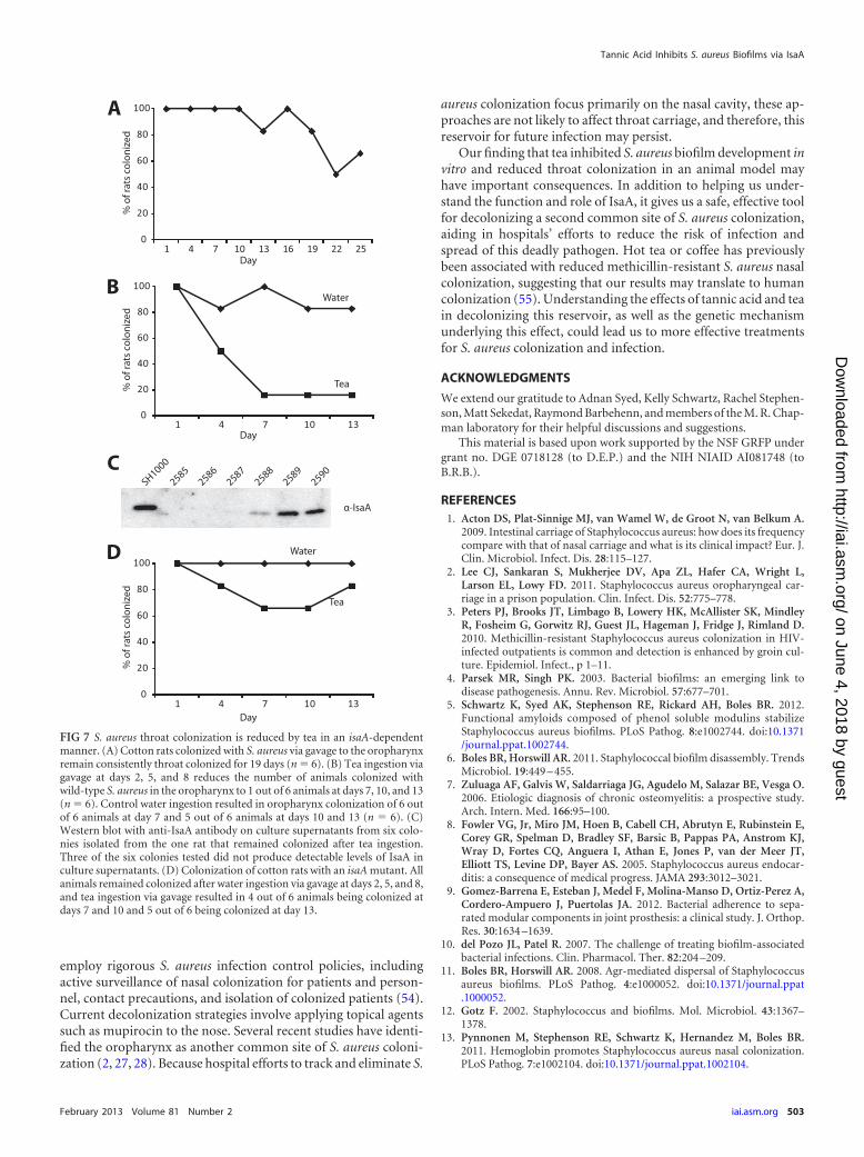
Tea inhibits S. aureus throat colonization in an isaA-depen-dent manner. Emerging evidence suggests that, in addition to thenasopharynx, S. aureus commonly colonizes the oropharynx andoral cavity (2, 27, 28). Since tea is a commonly consumed beveragethat inhibits surface colonization in our in vitro models, we testedif tea could impact S. aureus colonization in a rodent model. Thecotton rat has previously been used to study S. aureus nasal colo-nization (41), so we first assessed if S. aureus would also colonizethe cotton rat oropharynx. Oral inoculation with 1 � 105 S. aureusCFU resulted in reproducible oropharynx colonization, with 5 outof 6 animals displaying detectable oropharynx colonization over aperiod of 19 days (Fig. 7A).
To determine if tea ingestion influenced S. aureus oropharynxcolonization, 200 �l of black tea was administered via gavage tocolonized rats at 2, 5, and 8 days after initial colonization. Teaingestion reduced the number of animals colonized with wild-
FIG 2 Tannic acid increases levels of IsaA in S. aureus culture supernatants.(A) Western blot of TCA-precipitated supernatants from overnight cultures ofS. aureus grown in 66% TSB supplemented with 0.2% glucose and with theindicated concentrations of tannic acid. When cultures are grown with higherconcentrations of tannic acid, a band (indicated with an arrowhead) appearswith an apparent molecular mass slightly below 26 kDa. The band was excised,and the protein was identified by mass spectrometry as IsaA. (B) Western blotof unprecipitated supernatants from overnight cultures probed with poly-clonal anti-IsaA antibody. IsaA levels in culture supernatants increase whenthe culture is grown with tannic acid.
FIG 3 isaA is necessary for tannic acid-induced biofilm inhibition. (A) StrainSH1000 (BB386), a mutant isaA::Tetr strain (�isaA; BB2183), an isaA::Tetr
strain with an empty vector (�isaA � EV; BB2184), and a mutant isaA::Tetr
strain with an isaA complement (�isaA � isaA; BB2185) were assayed in amicrotiter plate for tannic acid-induced biofilm inhibition. Strains lackingfunctional isaA were resistant to inhibition. *, P �� 0.001 compared to isogenicuntreated control. (B) SH1000 (BB386), a mutant isaA::Tetr strain with anempty vector (BB2184), and a mutant isaA::Tetr strain with an isaA comple-ment (BB2185) were assayed in a flow cell for tannic acid-induced biofilminhibition. The isaA mutant was resistant to inhibition.
Payne et al.
500 iai.asm.org Infection and Immunity
on June 4, 2018 by guesthttp://iai.asm
.org/D
ownloaded from

type S. aureus in the oropharynx, with 5 out of 6 animals not beingcolonized after tea ingestion versus 1 out of 6 animals in a watergavage control group not being colonized (Fig. 7B). In addition,we tested six colonies from the rat that remained colonized with S.aureus after tea ingestion for IsaA production. Three of the sixisolates produced no detectable IsaA, and one produced dramat-ically lower levels of IsaA than the wild type (Fig. 7C). Finally, anisaA mutant appeared to maintain higher levels of colonizationupon tea ingestion. Four out of six animals colonized with an isaAmutant versus one out of six colonized with the isogenic wild-typeparent remained colonized after tea ingestion (Fig. 7D and B).These results suggest that consumption of polyphenolic com-pounds, like those in tea, may reduce S. aureus oropharynx colo-nization in an isaA-dependent manner.
DISCUSSION
The ability of S. aureus to colonize surfaces and form biofilmscontributes to its success as a commensal and pathogen. S. aureuslives as a commensal attached to surfaces such as the skin, naso-pharynx, and oropharynx (2). As a pathogen, S. aureus can attachto internal tissues such as bone, heart valves, or implanted medicaldevices (7–9). Colonization by S. aureus increases the incidence ofinfection, and biofilm infections represent a serious clinical situ-ation, based on their recalcitrance to antibiotics, their persistence,
and the propensity of organisms to detach and colonize new sites(6). Relatively little is known regarding how natural products thatare common in the human diet can influence S. aureus coloniza-tion and biofilm development. Therefore, understanding how S.aureus responds to natural products and different environmentalconditions is an important issue that warrants further investiga-tion.
In this work we demonstrate that the polyphenolic compoundtannic acid can inhibit S. aureus surface colonization in a multi-tude of biofilm models (Fig. 1). Analysis of liquid culture super-natants revealed increased levels of the protein IsaA when strainswere grown in the presence of tannic acid (Fig. 2). An isaA mutantwas not susceptible to the biofilm inhibition effects of tannic acid,and this phenotype was complemented by expressing isaA underthe control of its native promoter in trans (Fig. 3). Expression ofIsaA from an inducible promoter inhibited biofilm formation,and this was dependent upon a catalytic residue at the putativeIsaA transglycosylase active site (Fig. 4). After prolonged incuba-tion in drip-flow biofilms, isolates that were resistant to the anti-biofilm effect of tannic acid appeared, and these isolates failed toproduce IsaA (Fig. 5). Black tea, a common source of tannic acidin the human diet, inhibited biofilm formation in vitro in an isaA-dependent manner (Fig. 6). Finally, we developed an animalmodel for S. aureus throat colonization and found that tea re-duced throat colonization in an isaA-dependent manner (Fig. 7).
Tannic acid has long been known to have antibacterial prop-erties (42), bacteria are known to actively modulate gene expres-sion in response to tannins (43, 44), and recently, it has been
FIG 4 Induced expression of IsaA inhibits biofilm formation. (A) SH1000harboring empty vector (BB1209), isaA (BB2242), or E183Q-isaA (BB2333)under a tetracycline-inducible promoter was grown with 0.2% glucose and 250ng/ml anhydrotetracycline (aTet). No biofilm formed when IsaA was overex-pressed. A biofilm formed when E183Q-IsaA was overexpressed. Error barsrepresent standard deviations. WT, wild type; *, P �� 0.001 compared to theisogenic control with glucose and without anhydrotetracycline. (B) Wild-typeIsaA and E183Q-IsaA are overexpressed when induced with 250 ng/ml anhy-drotetracycline. The Western blot is of SH1000 harboring strains with anempty vector (BB1209), isaA (BB2242), or E183Q-isaA (BB2333) under a tet-racycline-inducible promoter, with and without anhydrotetracycline induc-tion, probed with polyclonal anti-IsaA antibody.
FIG 5 Biofilm resistance to tannic acid is coincident with a reduction in IsaAexpression. Strains derived from 11 S. aureus colonies isolated from a tannicacid-treated biofilm were tested for resistance to tannic acid-induced biofilminhibition, as well as for IsaA production. (A) Three of 11 strains (BB2519,BB2520, and BB2524) are robustly resistant to tannic acid-induced biofilminhibition in a microtiter plate biofilm assay. (B) The same 3 strains also do nothave detectable IsaA in their culture supernatants. The Western blot is ofculture supernatants from the 11 strains isolated from a tannic acid-resistantbiofilm (along with wild type) probed with polyclonal anti-IsaA antibody.
Tannic Acid Inhibits S. aureus Biofilms via IsaA
February 2013 Volume 81 Number 2 iai.asm.org 501
on June 4, 2018 by guesthttp://iai.asm
.org/D
ownloaded from

suggested to have antibiofilm properties (45). Pentagalloyl glu-cose (one of the major components of commercial tannic acid)and ellagic acid (another plant-derived polyphenolic compound)have also been shown to inhibit biofilm formation in S. aureus (46,47). To the best of our knowledge, no genetic mechanism for theantibiofilm properties of tannic acid or related polyphenols in S.aureus has been proposed. In this report, we show that tannic acidcauses an increase in extracellular IsaA levels and that increasedlevels of IsaA are able to inhibit biofilm formation in S. aureus. Atthis time, the molecular mechanism leading to increased extracel-lular IsaA levels is not known. Several possible mechanisms thatwould lead to this outcome are under investigation, includingdifferential regulation of isaA and altered protein stability.
Lytic transglycosylases have been extensively studied in E. coli,where they have been shown to cleave peptidoglycan at the �-1,4-glycosidic bond between N-acetylmuramic acid (MurNAc) andN-acetylglucosamine (GlcNAc) (16). By virtue of their ability tocleave the polysaccharide backbone of the peptidoglycan layer,lytic transglycosylases are thought to play a role in synthesis anddegradation of the peptidoglycan. It has been proposed that lytic
transglycosylases play important roles in cellular elongation, sep-tation, recycling of muropeptides, and pore formation (48).
To the best of our knowledge, this is the first report describinga specific function of the lytic transglycosylase IsaA in S. aureus.The mechanism by which IsaA leads to biofilm inhibition remainsunclear, but the evidence that we present in this paper demon-strates that this activity depends on IsaA’s catalytic function as alytic transglycosylase. There are several ways in which cleavage ofpeptidoglycan could lead to a reduction in biofilm formation. Forexample, peptidoglycan cleavage could change the composition ofproteins and teichoic acids displayed on the cell wall, cleavingaway factors necessary for surface colonization. Alternatively,peptidoglycan cleavage could release a signaling molecule (49),leading to modulation of biofilm-related gene expression. Work isunder way to elucidate how IsaA activity leads to biofilm inhibi-tion.
S. aureus nasal colonization is a significant risk factor for sev-eral infections, including bacteremia, postoperative infections,and diabetic foot ulcer infections, and contributes to the spread ofthis pathogen in hospital environments (50–53). Many hospitals
FIG 6 Black tea inhibits biofilm formation in S. aureus. (A) Black tea inhibits biofilm formation in a dose-dependent manner. Biofilm formation was inducedin a microtiter plate biofilm assay by supplementing the medium with 0.2% glucose. Error bars represent standard deviations. *, P �� 0.001 compared to thecontrol with glucose and without tea. (B) When black tea is mixed with milk, the tea loses its biofilm-inhibitory effect. Error bars represent standard deviations.*, P �� 0.001 compared to the control with glucose, without tea, and without milk. (C) isaA is necessary for tea-induced biofilm inhibition. Strain SH1000(BB386), a mutant isaA::Tetr strain (BB2183), a mutant isaA::Tetr mutant strain with an empty vector (BB2184), and a mutant isaA::Tetr mutant strain with anisaA complement (BB2185) were assayed in microtiter plates for tea-induced biofilm inhibition. Strains lacking functional isaA were resistant to inhibition. Errorbars represent standard deviations. *, P �� 0.001 compared to an isogenic control with glucose and without tea.
Payne et al.
502 iai.asm.org Infection and Immunity
on June 4, 2018 by guesthttp://iai.asm
.org/D
ownloaded from
employ rigorous S. aureus infection control policies, includingactive surveillance of nasal colonization for patients and person-nel, contact precautions, and isolation of colonized patients (54).Current decolonization strategies involve applying topical agentssuch as mupirocin to the nose. Several recent studies have identi-fied the oropharynx as another common site of S. aureus coloni-zation (2, 27, 28). Because hospital efforts to track and eliminate S.
aureus colonization focus primarily on the nasal cavity, these ap-proaches are not likely to affect throat carriage, and therefore, thisreservoir for future infection may persist.
Our finding that tea inhibited S. aureus biofilm development invitro and reduced throat colonization in an animal model mayhave important consequences. In addition to helping us under-stand the function and role of IsaA, it gives us a safe, effective toolfor decolonizing a second common site of S. aureus colonization,aiding in hospitals’ efforts to reduce the risk of infection andspread of this deadly pathogen. Hot tea or coffee has previouslybeen associated with reduced methicillin-resistant S. aureus nasalcolonization, suggesting that our results may translate to humancolonization (55). Understanding the effects of tannic acid and teain decolonizing this reservoir, as well as the genetic mechanismunderlying this effect, could lead us to more effective treatmentsfor S. aureus colonization and infection.
ACKNOWLEDGMENTS
We extend our gratitude to Adnan Syed, Kelly Schwartz, Rachel Stephen-son, Matt Sekedat, Raymond Barbehenn, and members of the M. R. Chap-man laboratory for their helpful discussions and suggestions.
This material is based upon work supported by the NSF GRFP undergrant no. DGE 0718128 (to D.E.P.) and the NIH NIAID AI081748 (toB.R.B.).
REFERENCES1. Acton DS, Plat-Sinnige MJ, van Wamel W, de Groot N, van Belkum A.
2009. Intestinal carriage of Staphylococcus aureus: how does its frequencycompare with that of nasal carriage and what is its clinical impact? Eur. J.Clin. Microbiol. Infect. Dis. 28:115–127.
2. Lee CJ, Sankaran S, Mukherjee DV, Apa ZL, Hafer CA, Wright L,Larson EL, Lowy FD. 2011. Staphylococcus aureus oropharyngeal car-riage in a prison population. Clin. Infect. Dis. 52:775–778.
3. Peters PJ, Brooks JT, Limbago B, Lowery HK, McAllister SK, MindleyR, Fosheim G, Gorwitz RJ, Guest JL, Hageman J, Fridge J, Rimland D.2010. Methicillin-resistant Staphylococcus aureus colonization in HIV-infected outpatients is common and detection is enhanced by groin cul-ture. Epidemiol. Infect., p 1–11.
4. Parsek MR, Singh PK. 2003. Bacterial biofilms: an emerging link todisease pathogenesis. Annu. Rev. Microbiol. 57:677–701.
5. Schwartz K, Syed AK, Stephenson RE, Rickard AH, Boles BR. 2012.Functional amyloids composed of phenol soluble modulins stabilizeStaphylococcus aureus biofilms. PLoS Pathog. 8:e1002744. doi:10.1371/journal.ppat.1002744.
6. Boles BR, Horswill AR. 2011. Staphylococcal biofilm disassembly. TrendsMicrobiol. 19:449 – 455.
7. Zuluaga AF, Galvis W, Saldarriaga JG, Agudelo M, Salazar BE, Vesga O.2006. Etiologic diagnosis of chronic osteomyelitis: a prospective study.Arch. Intern. Med. 166:95–100.
8. Fowler VG, Jr, Miro JM, Hoen B, Cabell CH, Abrutyn E, Rubinstein E,Corey GR, Spelman D, Bradley SF, Barsic B, Pappas PA, Anstrom KJ,Wray D, Fortes CQ, Anguera I, Athan E, Jones P, van der Meer JT,Elliott TS, Levine DP, Bayer AS. 2005. Staphylococcus aureus endocar-ditis: a consequence of medical progress. JAMA 293:3012–3021.
9. Gomez-Barrena E, Esteban J, Medel F, Molina-Manso D, Ortiz-Perez A,Cordero-Ampuero J, Puertolas JA. 2012. Bacterial adherence to sepa-rated modular components in joint prosthesis: a clinical study. J. Orthop.Res. 30:1634 –1639.
10. del Pozo JL, Patel R. 2007. The challenge of treating biofilm-associatedbacterial infections. Clin. Pharmacol. Ther. 82:204 –209.
11. Boles BR, Horswill AR. 2008. Agr-mediated dispersal of Staphylococcusaureus biofilms. PLoS Pathog. 4:e1000052. doi:10.1371/journal.ppat.1000052.
12. Gotz F. 2002. Staphylococcus and biofilms. Mol. Microbiol. 43:1367–1378.
13. Pynnonen M, Stephenson RE, Schwartz K, Hernandez M, Boles BR.2011. Hemoglobin promotes Staphylococcus aureus nasal colonization.PLoS Pathog. 7:e1002104. doi:10.1371/journal.ppat.1002104.
FIG 7 S. aureus throat colonization is reduced by tea in an isaA-dependentmanner. (A) Cotton rats colonized with S. aureus via gavage to the oropharynxremain consistently throat colonized for 19 days (n � 6). (B) Tea ingestion viagavage at days 2, 5, and 8 reduces the number of animals colonized withwild-type S. aureus in the oropharynx to 1 out of 6 animals at days 7, 10, and 13(n � 6). Control water ingestion resulted in oropharynx colonization of 6 outof 6 animals at day 7 and 5 out of 6 animals at days 10 and 13 (n � 6). (C)Western blot with anti-IsaA antibody on culture supernatants from six colo-nies isolated from the one rat that remained colonized after tea ingestion.Three of the six colonies tested did not produce detectable levels of IsaA inculture supernatants. (D) Colonization of cotton rats with an isaA mutant. Allanimals remained colonized after water ingestion via gavage at days 2, 5, and 8,and tea ingestion via gavage resulted in 4 out of 6 animals being colonized atdays 7 and 10 and 5 out of 6 being colonized at day 13.
Tannic Acid Inhibits S. aureus Biofilms via IsaA
February 2013 Volume 81 Number 2 iai.asm.org 503
on June 4, 2018 by guesthttp://iai.asm
.org/D
ownloaded from

14. Lorenz U, Ohlsen K, Karch H, Hecker M, Thiede A, Hacker J. 2000.Human antibody response during sepsis against targets expressed bymethicillin resistant Staphylococcus aureus. FEMS Immunol. Med. Mi-crobiol. 29:145–153.
15. Stapleton MR, Horsburgh MJ, Hayhurst EJ, Wright L, Jonsson IM,Tarkowski A, Kokai-Kun JF, Mond JJ, Foster SJ. 2007. Characterizationof IsaA and SceD, two putative lytic transglycosylases of Staphylococcusaureus. J. Bacteriol. 189:7316 –7325.
16. Holtje JV, Mirelman D, Sharon N, Schwarz U. 1975. Novel type ofmurein transglycosylase in Escherichia coli. J. Bacteriol. 124:1067–1076.
17. Bateman BT, Donegan NP, Jarry TM, Palma M, Cheung AL. 2001.Evaluation of a tetracycline-inducible promoter in Staphylococcus aureusin vitro and in vivo and its application in demonstrating the role of sigB inmicrocolony formation. Infect. Immun. 69:7851–7857.
18. Duthie ES, Lorenz LL. 1952. Staphylococcal coagulase; mode of actionand antigenicity. J. Gen. Microbiol. 6:95–107.
19. Peng HL, Novick RP, Kreiswirth B, Kornblum J, Schlievert P. 1988.Cloning, characterization, and sequencing of an accessory gene regulator(agr) in Staphylococcus aureus. J. Bacteriol. 170:4365– 4372.
20. Novick RP, Ross HF, Projan SJ, Kornblum J, Kreiswirth B, MoghazehS. 1993. Synthesis of staphylococcal virulence factors is controlled by aregulatory RNA molecule. EMBO J. 12:3967–3975.
21. Reiser RF, Hinzman SJ, Bergdoll MS. 1987. Production of toxic shocksyndrome toxin 1 by Staphylococcus aureus restricted to endogenous airin tampons. J. Clin. Microbiol. 25:1450 –1452.
22. CDC. 1999. Four pediatric deaths from community-acquired methicillin-resistant Staphylococcus aureus—Minnesota and North Dakota, 1997-1999. MMWR Morb. Mortal. Wkly. Rep. 48:707–710.
23. Voyich JM, Braughton KR, Sturdevant DE, Whitney AR, Said-Salim B,Porcella SF, Long RD, Dorward DW, Gardner DJ, Kreiswirth BN,Musser JM, DeLeo FR. 2005. Insights into mechanisms used by Staphy-lococcus aureus to avoid destruction by human neutrophils. J. Immunol.175:3907–3919.
24. Gillaspy AF, Hickmon SG, Skinner RA, Thomas JR, Nelson CL, Smelt-zer MS. 1995. Role of the accessory gene regulator (agr) in pathogenesis ofstaphylococcal osteomyelitis. Infect. Immun. 63:3373–3380.
25. Husmann LK, Yung DL, Hollingshead SK, Scott JR. 1997. Role ofputative virulence factors of Streptococcus pyogenes in mouse models oflong-term throat colonization and pneumonia. Infect. Immun. 65:1422–1430.
26. National Research Council. 1996. Guide for the care and use of laboratoryanimals. National Academies Press, Washington, DC.
27. Nurjadi D, Lependu J, Kremsner PG, Zanger P. 2012. Staphylococcusaureus throat carriage is associated with ABO-/secretor status. J. Infect.65:310 –317.
28. Ohara-Nemoto Y, Haraga H, Kimura S, Nemoto TK. 2008. Occurrenceof staphylococci in the oral cavities of healthy adults and nasal oral traf-ficking of the bacteria. J. Med. Microbiol. 57:95–99.
29. Salminen J-P, Karonen M. 2011. Chemical ecology of tannins and otherphenolics: we need a change in approach. Funct. Ecol. 25:325–338.
30. Corrigan RM, Foster TJ. 2009. An improved tetracycline-inducible ex-pression vector for Staphylococcus aureus. Plasmid 61:126 –129.
31. Blackburn NT, Clarke AJ. 2001. Identification of four families of pepti-doglycan lytic transglycosylases. J. Mol. Evol. 52:78 – 84.
32. Thunnissen AM, Dijkstra AJ, Kalk KH, Rozeboom HJ, Engel H, KeckW, Dijkstra BW. 1994. Doughnut-shaped structure of a bacterial mura-midase revealed by X-ray crystallography. Nature 367:750 –753.
33. Monteiro C, Fang X, Ahmad I, Gomelsky M, Romling U. 2011. Regu-lation of biofilm components in Salmonella enterica serovar Typhimu-rium by lytic transglycosylases involved in cell wall turnover. J. Bacteriol.193:6443– 6451.
34. Haslam E, Lilley TH, Cai Y, Martin R, Magnolato D. 1989. Traditionalherbal medicines—the role of polyphenols. Planta Med. 55:1– 8.
35. Scalbert A. 1991. Antimicrobial properties of tannins. Phytochemistry30:3875–3883.
36. Bandyopadhyay P, Ghosh AK, Ghosh C. 2012. Recent developments onpolyphenol-protein interactions: effects on tea and coffee taste, antioxi-dant properties and the digestive system. Food Funct. 3:592– 605.
37. Vidal S, Francis L, Noble A, Kwiatkowski M, Cheynier V, Waters E.2004. Taste and mouth-feel properties of different types of tannin-likepolyphenolic compounds and anthocyanins in wine. Anal. Chim. Acta513:57– 65.
38. Thomas AW, Frieden A. 1923. The gelatin-tannin reaction. Ind. Eng.Chem. 15:839 – 841.
39. Anderson RA, Polansky MM. 2002. Tea enhances insulin activity. J.Agric. Food Chem. 50:7182–7186.
40. Morton JF. 1979. Tea with milk. Science 204:909.41. Kokai-Kun JF. 2008. The cotton rat as a model for Staphylococcus aureus
nasal colonization in humans: cotton rat S. aureus nasal colonizationmodel. Methods Mol. Biol. 431:241–254.
42. Henis Y, Volcani R, Tagari H. 1964. Effect of water extracts of carobpods, tannic acid, and their derivatives on morphology and growth ofmicroorganisms. Appl. Microbiol. 12:204 –209.
43. Quan S, Koldewey P, Tapley T, Kirsch N, Ruane KM, Pfizenmaier J, ShiR, Hofmann S, Foit L, Ren G, Jakob U, Xu Z, Cygler M, Bardwell JC.2011. Genetic selection designed to stabilize proteins uncovers a chaper-one called Spy. Nat. Struct. Mol. Biol. 18:262–269.
44. Zoetendal EG, Smith AH, Sundset MA, Mackie RI. 2008. The BaeSRtwo-component regulatory system mediates resistance to condensed tan-nins in Escherichia coli. Appl. Environ. Microbiol. 74:535–539.
45. Chusri S, Phatthalung PN, Voravuthikunchai SP. 2012. Anti-biofilmactivity of Quercus infectoria G. Olivier against methicillin-resistantStaphylococcus aureus. Lett. Appl. Microbiol. 54:511–517.
46. Lin MH, Chang FR, Hua MY, Wu YC, Liu ST. 2011. Inhibitory effectsof 1,2,3,4,6-penta-O-galloyl-beta-D-glucopyranose on biofilm formationby Staphylococcus aureus. Antimicrob. Agents Chemother. 55:1021–1027.
47. Quave CL, Estevez-Carmona M, Compadre CM, Hobby G, Hendrick-son H, Beenken KE, Smeltzer MS. 2012. Ellagic acid derivatives fromRubus ulmifolius inhibit Staphylococcus aureus biofilm formation andimprove response to antibiotics. PLoS One 7:e28737. doi:10.1371/journal.pone.0028737.
48. Scheurwater E, Reid CW, Clarke AJ. 2008. Lytic transglycosylases: bac-terial space-making autolysins. Int. J. Biochem. Cell Biol. 40:586 –591.
49. Shah IM, Laaberki MH, Popham DL, Dworkin J. 2008. A eukaryotic-likeSer/Thr kinase signals bacteria to exit dormancy in response to pepti-doglycan fragments. Cell 135:486 – 496.
50. Munoz P, Hortal J, Giannella M, Barrio JM, Rodriguez-Creixems M,Perez MJ, Rincon C, Bouza E. 2008. Nasal carriage of S. aureus increasesthe risk of surgical site infection after major heart surgery. J. Hosp. Infect.68:25–31.
51. Stanaway S, Johnson D, Moulik P, Gill G. 2007. Methicillin-resistantStaphylococcus aureus (MRSA) isolation from diabetic foot ulcers corre-lates with nasal MRSA carriage. Diabetes Res. Clin. Pract. 75:47–50.
52. von Eiff C, Becker K, Machka K, Stammer H, Peters G. 2001. Nasalcarriage as a source of Staphylococcus aureus bacteremia. Study Group. N.Engl. J. Med. 344:11–16.
53. Wertheim HF, Vos MC, Ott A, van Belkum A, Voss A, Kluytmans JA,van Keulen PH, Vandenbroucke-Grauls CM, Meester MH, VerbrughHA. 2004. Risk and outcome of nosocomial Staphylococcus aureus bac-teraemia in nasal carriers versus non-carriers. Lancet 364:703–705.
54. Lee BY, Bailey RR, Smith KJ, Muder RR, Strotmeyer ES, Lewis GJ,Ufberg PJ, Song Y, Harrison LH. 2010. Universal methicillin-resistantStaphylococcus aureus (MRSA) surveillance for adults at hospital admis-sion: an economic model and analysis. Infect. Control Hosp. Epidemiol.31:598 – 606.
55. Matheson EM, Mainous AG, III, Everett CJ, King DE. 2011. Tea andcoffee consumption and MRSA nasal carriage. Ann. Fam. Med. 9:299 –304.
Payne et al.
504 iai.asm.org Infection and Immunity
on June 4, 2018 by guesthttp://iai.asm
.org/D
ownloaded from