bdellovibrio bacteriovorus inhibits staphylococcus aureus biofilm
TRANSCRIPT
Bdellovibrio bacteriovorus InhibitsStaphylococcus aureus Biofilm Formationand Invasion into Human Epithelial CellsAjay K. Monnappa1*, Mohammed Dwidar1*, Jeong Kon Seo2, Jin-Hoe Hur2 & Robert J. Mitchell1
1School of Life Sciences, Ulsan National Institute of Science and Technology, 2UNIST Central Research Facility, Ulsan NationalInstitute of Science and Technology.
Bdellovibrio bacteriovorus HD100 is a predatory bacterium that attacks many Gram-negative humanpathogens. A serious drawback of this strain, however, is its ineffectiveness against Gram-positive strains,such as the human pathogen Staphylococcus aureus. Here we demonstrate that the extracellular proteasesproduced by a host-independent B. bacteriovorus (HIB) effectively degrade/inhibit the formation of S.aureus biofilms and reduce its virulence. A 10% addition of HIB supernatant caused a 75% or greaterreduction in S. aureus biofilm formation as well as 75% dispersal of pre-formed biofilms. LC-MS-MSanalyses identified various B. bacteriovorus proteases within the supernatant, including the serine proteasesBd2269 and Bd2321. Tests with AEBSF confirmed that serine proteases were active in the supernatant andthat they impacted S. aureus biofilm formation. The supernatant also possessed a slight DNAse activity.Furthermore, treatment of planktonic S. aureus with the supernatant diminished its ability to invadeMCF-10a epithelial cells by 5-fold but did not affect the MCF-10a viability. In conclusion, this studyillustrates the hitherto unknown ability of B. bacteriovorus to disperse Gram-positive pathogenic biofilmsand mitigate their virulence.
Multidrug resistance in human pathogens is a growing concern as the number of patients infected withthese bacteria is increasing1–3. This resistive facet is further exacerbated as many chronic human diseasesare biofilm associated4. This poses a serious threat to human health since bacteria present within biofilms
are naturally more resistant to antibiotic treatments5, even without the requisite genetic markers for antibioticresistance.
Recent studies have shown that Bdellovibrio bacteriovorus and other similar organisms, collectively referred toas BALOs (Bdellovibrio-and-like-organisms)6,7, predate upon Gram-negative human pathogens8,9, includingmultidrug resistant Acinetobacter baumannii1. B. bacteriovorus was also shown to be a very promising tool forcombating biofilms10–12. This was attributed to the ability of this bacterium to penetrate deeply inside preybiofilms and effectively destroy them; a characteristic which distinguishes them from other biological tools suchas bacteriophages and protists12. A major limitation of BALOs, however, is their inability to attack or predate uponGram-positive strains8,10,13, a category that comprises numerous human pathogens14,15, including Staphylococcusaureus, one of the most frequent nosocomial infection-associated multidrug resistant pathogens isolated frompatients16,17.
S. aureus commonly colonizes the skin or within the nasal passage of humans18, but is also able to form biofilmson a variety of abiotic surfaces, including medical equipment, catheters, implants and prosthetics19–21. Biofilmsformed by S. aureus, as with all bacteria, are composed of bacterial cells embedded within a matrix called theextracellular polymeric substances (EPS)22. This EPS matrix is formed using extracellular DNA, polysaccharidesand proteins and anchors the cells to the surface, making it difficult to eradicate the organism once it establishesitself. To address this need with S. aureus, several groups have evaluated the use of hydrolytic enzymes, includingproteases and DNases, to degrade the EPS and, thus, remove and disperse the unwanted biofilms16,18,23.
As a predator, B. bacteriovorus HD100 produces numerous hydrolytic enzymes that are needed for it toeffectively hydrolyze its prey’s macromolecules, including a cache of 150 proteases/peptidases24 and numerousother hydrolases. With this extensive arsenal in its genome, B. bacteriovorus HD100 is thought to have the highestnumber of protease genes per unit genome of all reported bacterial strains24. Whereas the production of theseproteins is expected to occur during its intraperiplasmic stage of predation inside the prey cell, several studiesfound that cultures of host independent (HI) mutants of B. bacteriovorus possess a strong extracellular protease
OPEN
SUBJECT AREAS:PATHOGENS
APPLIED MICROBIOLOGY
ANTIMICROBIAL RESISTANCE
INFECTION
Received4 November 2013
Accepted2 January 2014
Published22 January 2014
Correspondence andrequests for materials
should be addressed toR.J.M. (esgott@unist.
ac.kr)
* These authorscontributed equally to
this work.
SCIENTIFIC REPORTS | 4 : 3811 | DOI: 10.1038/srep03811 1
activity25,26. Consequently, this study aimed at evaluating and util-izing the strong hydrolytic arsenal of B. bacteriovorus against S.aureus biofilms.
ResultsHost-independent Bdellovibrio bacteriovorus HD100 release pro-teases into the media. A host-independent mutant of B. bacterio-vorus (HIB) isolated in our lab was cultivated axenically in PYEmedia. This strain was selected since similarly developed host-independent isolates from other groups are known to secreteproteolytic enzymes25. When the cell-free supernatant from ourisolate at mid-log phase (OD600 0.5) was likewise assayed, wefound that it harboured a proteolytic activity corresponding to 13.76 7.4 ng/ml proteinase K.
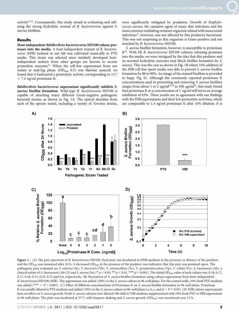
Bdellovibrio bacteriovorus supernatant significantly inhibits S.aureus biofilm formation. Wild-type B. bacteriovorus HD100 iscapable of attacking many different Gram-negative pathogenicbacterial strains, as shown in Fig. 1A. The optical densities fromeach of the species tested, including a variety of Yersinia strains,
were significantly mitigated by predation. Growth of Staphylo-coccus aureus, the causative agent of many skin infections and themost common multidrug resistant organism related with nosocomialinfections27, however, was not affected by this predatory bacterium.This was not surprising as this organism is Gram-positive and notattacked by B. bacteriovorus HD100.
S. aureus biofilm formation, however, is susceptible to proteinaseK28. With HI B. bacteriovorus HD100 cultures releasing proteasesinto the media, we were intrigued by the idea that this predator andits secreted hydrolytic enzymes may block biofilm formation by S.aureus. This was the case as shown in Fig. 1B where 10% addition ofthe HIB cell-free spent media was able to prevent S. aureus biofilmformation by 80 to 90%. An image of the stained biofilms is providedin Supp. Fig. S1. Although the commonly reported proteinase Kconcentrations used in preventing and removing S. aureus biofilmsranges from about 1 or 2 mg/ml28,29 to 100 mg/ml30, this study foundthat proteinase K at a concentration of 1 ng/ml still led to an averageinhibition of 63%. These results are in agreement with our findingswith the HIB supernatants and their low proteolytic activities, whichare comparable to 1.4 ng/ml proteinase K after 10% dilution. It is
Figure 1 | (A) The prey spectrum of B. bacteriovorus HD100. Each prey was incubated in DNB medium in the presence or absence of the predator
and the OD600 was measured after 24 h. A decreased OD600 in the presence of the predator was indicative that this prey was predated upon. The
pathogenic prey evaluated are S. enterica (Se), Y. bercovieri (Yb), Y. enterocolitica (Ye), Y. pseudotuberculosis (Yp), Y. rohdei (Yr), A. baumannii (Ab), a
clinical isolate of A. baumannii (Ab-CI) and S. aureus (Sa) (* p , 0.05, ** p , 0.01, *** p , 0.001). The initial OD600 value of each culture was: 0.18, 0.17,
0.17, 0.18, 0.15, 0.25, 0.25 and 0.4, respectively. (B) Prevention of S. aureus biofilm formation using culture supernatant from host-independent
B. bacteriovorus HD100 (HIB). The supernatant was added (10%) to the S. aureus culture in 96-well plates. For the control wells, 10% fresh PYE medium
was added (*** 5 P , 0.001). (C) Effect of different concentrations of Proteinase K on S. aureus-biofilm formation in 96 well plates. Proteinase
K was serially diluted in PYE medium and added (10%) to the S. aureus culture in 96-well plates (a, b, c, and d 5 P , 0.05). (D) HIB culture supernatants
have no effect on S. aureus growth. Fresh S. aureus cultures were diluted 100-fold in TSB medium supplemented with 10% fresh PYE or HIB supernatant
in 96 well plates. The plate was incubated at 37uC with frequent shaking and S. aureus growth (OD600) was monitored over 12 h.
www.nature.com/scientificreports
SCIENTIFIC REPORTS | 4 : 3811 | DOI: 10.1038/srep03811 2
worthy to note, though, that even with the significant loss in S.aureus’ biofilm formation, the HIB spent media had no obvious effecton the growth of this pathogen (Fig. 1D) showing that the anti-biofilm activity did not results from this supernatant being toxic tothis S. aureus.
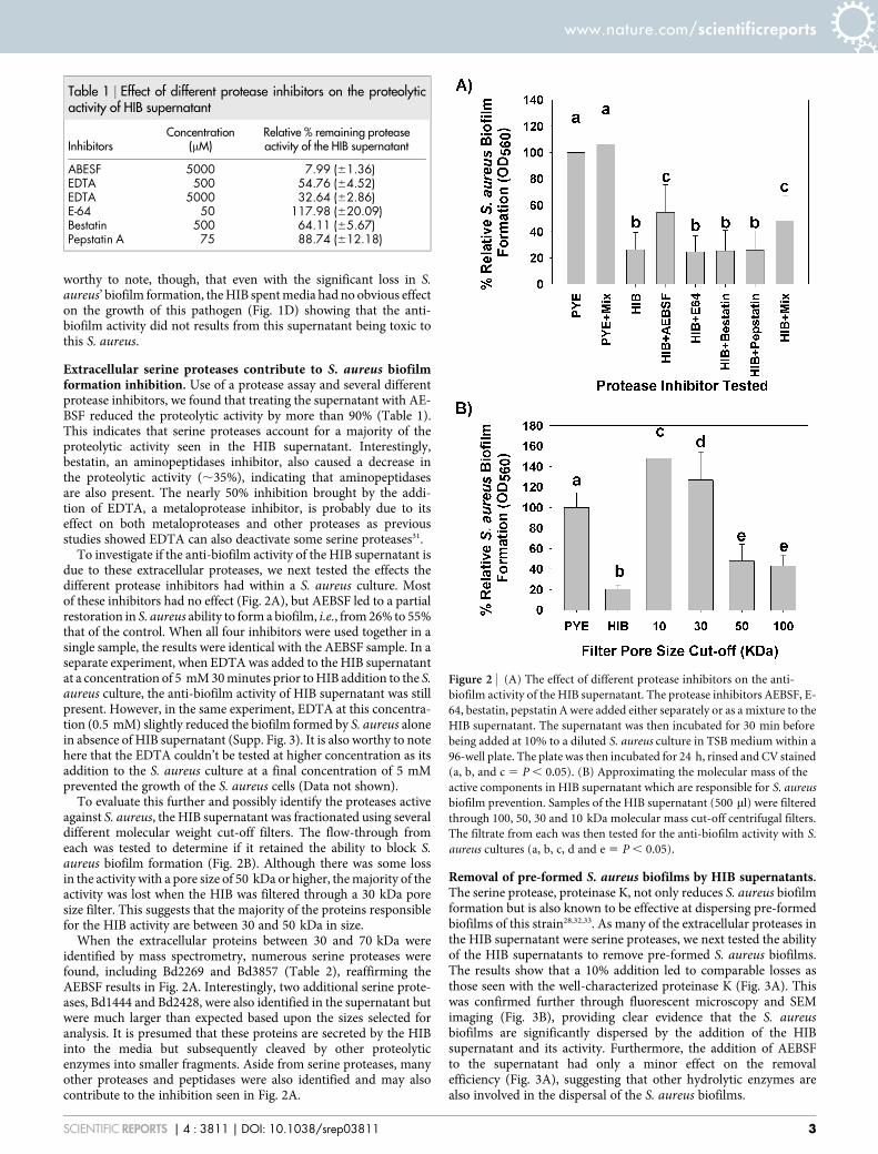
Extracellular serine proteases contribute to S. aureus biofilmformation inhibition. Use of a protease assay and several differentprotease inhibitors, we found that treating the supernatant with AE-BSF reduced the proteolytic activity by more than 90% (Table 1).This indicates that serine proteases account for a majority of theproteolytic activity seen in the HIB supernatant. Interestingly,bestatin, an aminopeptidases inhibitor, also caused a decrease inthe proteolytic activity (,35%), indicating that aminopeptidasesare also present. The nearly 50% inhibition brought by the addi-tion of EDTA, a metaloprotease inhibitor, is probably due to itseffect on both metaloproteases and other proteases as previousstudies showed EDTA can also deactivate some serine proteases31.
To investigate if the anti-biofilm activity of the HIB supernatant isdue to these extracellular proteases, we next tested the effects thedifferent protease inhibitors had within a S. aureus culture. Mostof these inhibitors had no effect (Fig. 2A), but AEBSF led to a partialrestoration in S. aureus ability to form a biofilm, i.e., from 26% to 55%that of the control. When all four inhibitors were used together in asingle sample, the results were identical with the AEBSF sample. In aseparate experiment, when EDTA was added to the HIB supernatantat a concentration of 5 mM 30 minutes prior to HIB addition to the S.aureus culture, the anti-biofilm activity of HIB supernatant was stillpresent. However, in the same experiment, EDTA at this concentra-tion (0.5 mM) slightly reduced the biofilm formed by S. aureus alonein absence of HIB supernatant (Supp. Fig. 3). It is also worthy to notehere that the EDTA couldn’t be tested at higher concentration as itsaddition to the S. aureus culture at a final concentration of 5 mMprevented the growth of the S. aureus cells (Data not shown).
To evaluate this further and possibly identify the proteases activeagainst S. aureus, the HIB supernatant was fractionated using severaldifferent molecular weight cut-off filters. The flow-through fromeach was tested to determine if it retained the ability to block S.aureus biofilm formation (Fig. 2B). Although there was some lossin the activity with a pore size of 50 kDa or higher, the majority of theactivity was lost when the HIB was filtered through a 30 kDa poresize filter. This suggests that the majority of the proteins responsiblefor the HIB activity are between 30 and 50 kDa in size.
When the extracellular proteins between 30 and 70 kDa wereidentified by mass spectrometry, numerous serine proteases werefound, including Bd2269 and Bd3857 (Table 2), reaffirming theAEBSF results in Fig. 2A. Interestingly, two additional serine prote-ases, Bd1444 and Bd2428, were also identified in the supernatant butwere much larger than expected based upon the sizes selected foranalysis. It is presumed that these proteins are secreted by the HIBinto the media but subsequently cleaved by other proteolyticenzymes into smaller fragments. Aside from serine proteases, manyother proteases and peptidases were also identified and may alsocontribute to the inhibition seen in Fig. 2A.
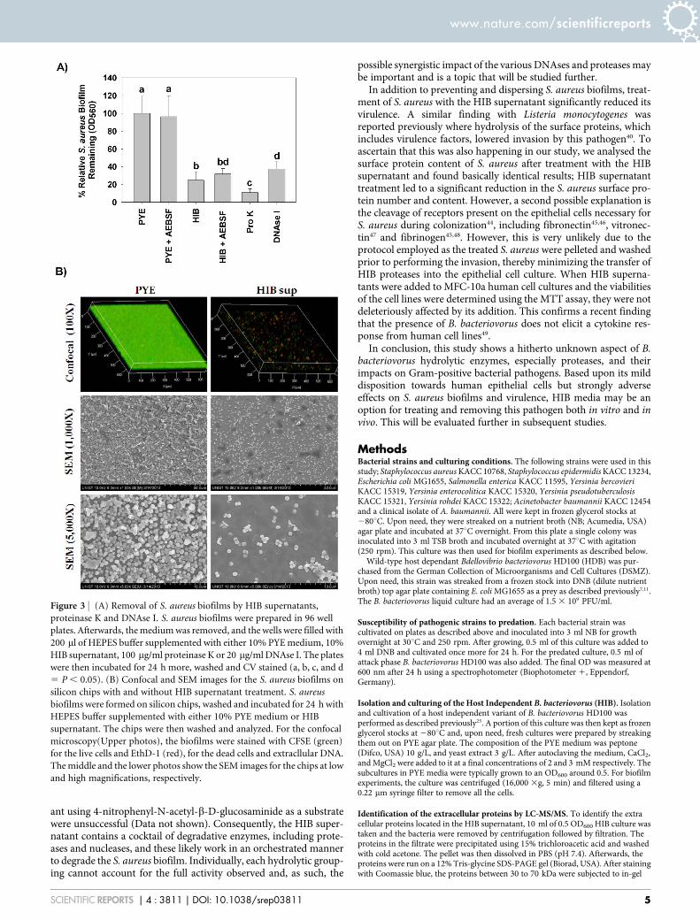
Removal of pre-formed S. aureus biofilms by HIB supernatants.The serine protease, proteinase K, not only reduces S. aureus biofilmformation but is also known to be effective at dispersing pre-formedbiofilms of this strain28,32,33. As many of the extracellular proteases inthe HIB supernatant were serine proteases, we next tested the abilityof the HIB supernatants to remove pre-formed S. aureus biofilms.The results show that a 10% addition led to comparable losses asthose seen with the well-characterized proteinase K (Fig. 3A). Thiswas confirmed further through fluorescent microscopy and SEMimaging (Fig. 3B), providing clear evidence that the S. aureusbiofilms are significantly dispersed by the addition of the HIBsupernatant and its activity. Furthermore, the addition of AEBSFto the supernatant had only a minor effect on the removalefficiency (Fig. 3A), suggesting that other hydrolytic enzymes arealso involved in the dispersal of the S. aureus biofilms.
Table 1 | Effect of different protease inhibitors on the proteolyticactivity of HIB supernatant
InhibitorsConcentration
(mM)Relative % remaining proteaseactivity of the HIB supernatant
ABESF 5000 7.99 (61.36)EDTA 500 54.76 (64.52)EDTA 5000 32.64 (62.86)E-64 50 117.98 (620.09)Bestatin 500 64.11 (65.67)Pepstatin A 75 88.74 (612.18)
Figure 2 | (A) The effect of different protease inhibitors on the anti-
biofilm activity of the HIB supernatant. The protease inhibitors AEBSF, E-
64, bestatin, pepstatin A were added either separately or as a mixture to the
HIB supernatant. The supernatant was then incubated for 30 min before
being added at 10% to a diluted S. aureus culture in TSB medium within a
96-well plate. The plate was then incubated for 24 h, rinsed and CV stained
(a, b, and c 5 P , 0.05). (B) Approximating the molecular mass of the
active components in HIB supernatant which are responsible for S. aureus
biofilm prevention. Samples of the HIB supernatant (500 ml) were filtered
through 100, 50, 30 and 10 kDa molecular mass cut-off centrifugal filters.
The filtrate from each was then tested for the anti-biofilm activity with S.
aureus cultures (a, b, c, d and e 5 P , 0.05).
www.nature.com/scientificreports
SCIENTIFIC REPORTS | 4 : 3811 | DOI: 10.1038/srep03811 3
As our results show that DNAse I can also disperse the S. aureusbiofilms (Fig. 3A), a finding corroborated by other groups16, wetested the HIB supernatant for DNAse activity. Supp. Fig. S3 showsthat the supernatant also possesses some DNAse activity. Measuringit quantitatively found it to be equivalent to ,0.07 6 0.03 mg/mlDNAse I. This activity, however, is too low to affect S. aureus biofilmsalone as shown in Supp. Fig. S4, where DNAse I concentrationsbelow ,6 mg/ml didn’t have a significant effect on the pre-formedS. aureus biofilms. Likewise, attempts to detect b-D-glucosaminidaseactivities within the HIB supernatant were unsuccessful (Data notshown).
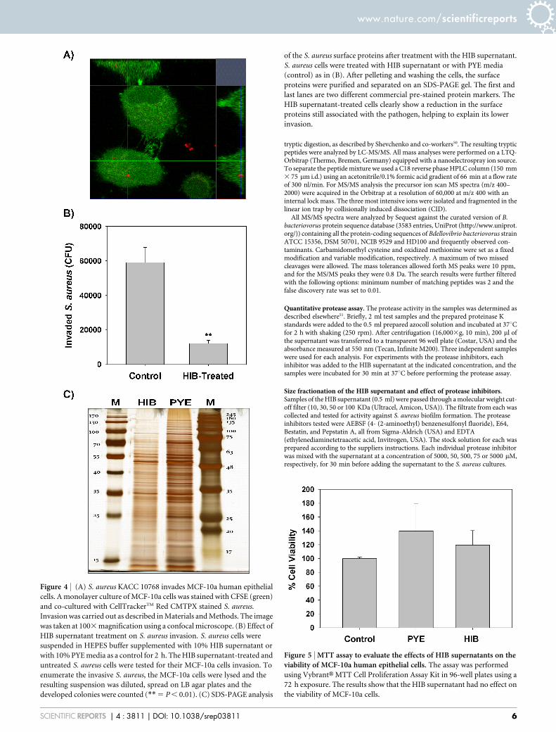
Treatment of S. aureus with the HIB supernatant reduces itsvirulence and invasion. Treatment of S. aureus with the HIB super-natant not only reduced its ability to form or maintain biofilms butalso its ability to invade human epithelial cells (Fig. 4). Fig. 4A is aconfocal image showing one of the MCF-10a cells harbouring twointernalized S. aureus bacteria. Treatment of the S. aureus culturewith 10% HIB supernatant for 2 h reduced the number of invasionevents by nearly 5-fold when compared with the untreated bacterialculture (Fig. 4B). An SDS-PAGE analysis shows that the treatmentsignificantly reduced the presence of S. aureus surface proteins(Fig. 4C), a group that includes assorted virulence factors35,36. Fur-thermore, when the HIB supernatant itself was tested for its effect onthe MCF-10a epithelial cells using an MTT assay, no toxicity wasdetected (Fig. 5) as the epithelial cell viability was quite similar in thecontrol, HIB-treated and PYE-treated wells.
DiscussionIt has been proposed that B. bacteriovorus HD 100 can be used as aprobiotic bacterium owing to its ability to predate upon gram nega-tive bacterial strains and their biofilms, including those composed ofknown human pathogens8,37,38. A major limitation, however, is theinability of this predator to attack gram positive strains, a groupingthat includes the major nosocomial pathogen S. aureus. Although B.bacteriovorus does not predate upon this strain or affect its viability,studies have not considered B. bacteriovorus potential impact on thebiofilm stability or virulence of S. aureus.
This study found that host independent (HI) mutant of B. bacter-iovorus produces significant amount of proteolytic enzymes in itsculturing medium. This agrees well with the studies done by other
groups who also identified proteolytic enzymes secreted by otherindependently isolated HI variants of B. bacteriovorus25,26. One ofthese studies used 2-D gel electrophoresis to identify the proteinspresent approximately 100 protein spots39. Six of the proteases iden-tified in their study were also found in the supernatant of our HIBvariant, including serine proteases Bd1962 and Bd2269 and the car-boxypeptidase Bd0306. The identification of these proteins withinboth of these studies through different techniques corroborates theirsecretion by this strain and suggests that this may be a common traitof all HIB mutants. As the biofilm EPS matrix is composed of severaldifferent macromolecules, including proteins, it is not surprising thatresearchers have sought to use proteases to disperse pre-formedbiofilms established by Staphylococcus aureus23 and other bacteria40,41
and fungi42, as well as those composed of multiple strains or species43.A recent study likewise reported that the Esp serine protease fromStaphylococcus epidermidis is capable of both inhibiting and remov-ing S. aureus biofilms18.
The results presented here demonstrate that the HIB supernatantpossesses a sufficient hydrolytic activity to significantly inhibit ordisperse S. aureus biofilms. Furthermore, as shown in Supp. Fig.S5, the supernatant was also effective in dispersing Staphylococcusepidermidis biofilms. This is in contrast to a recent report that the S.epidermidis Esp serine protease disperses S. aureus biofilms, but isapparently not effective against its own biofilms under similar con-ditions as colonization of patients by S. epidermidis correlates withthe absence of S. aureus18. Our results show that the HIB superna-tants are effective against both of these pathogens.
Whereas a significant proportion of the anti-biofilm activity seenfor the HIB supernatant comes from the serine proteases based onthe experiments with the serine protease inhibitor AEBSF, our dataalso suggests that additional effectors are secreted as the activity isnot completely blocked by AEBSF. This was particularly true in thebiofilm dispersal experiments (Fig. 3). It is clear that other proteasesmay also be contributing as treatment with AEBSF did not comple-tely block the proteolytic activity (Table 1). Furthermore, DNAsesare also present within the supernatants and were found to have anequivalent activity as ,0.07 6 0.03 mg/ml DNAse I. A previous studyalso found that S. aureus biofilms can be partially inhibited, althoughnot dispersed, by the action of the Dispersin B enzyme16, which worksby degrading the PNAG (poly-N-acetylglucosamine) polymer pro-duced in the biofilm matrix of Staphylococci34. However, ourattempts to find b -D-glucosaminidase activity in the HIB supernat-
Table 2 | Proteases and peptidases identified by mass spectrometry within the HIB supernatant. The size range used for the analysis wasapproximately between 30 and 70 kDa
Gene Uniprot Accession Protein Name Size (kDa) Subcellular Localization
ProteasesBd2269 Q6MKV8 Serine protease, subtilase family 56.6 ExtracellularBd2675 Q6MJU3 Putative membrane protein with protease subunit 33.5 UnknownBd2321 Q6MKR4 Subtilisin-like serine protease 74.9 UnknownBd2428 Q6MKG5 Serine protease 114.9 ExtracellularBd1444 Q6MN19 Serine protease, subtilase family 111.5 UnknownBd2692 Q6MJS6 Protease 53 ExtracellularBd2627 Q6MJY9 Periplasmic protease 32.3 UnknownBd2535 Q6MK75 Putative serine protease 28.5 UnknownBd3857 Q6MGR5 Alkaline serine protease subtilase family 43.2 ExtracellularBd0449 Q6MQL6 Putative protease 57.2 UnknownBd2675 Q6MJU3 Putative membrane protein with protease subunit 33.5 UnknownPeptidasesBd0306 Q6MQZ5 Carboxypeptidase 34 UnknownBd1962 Q6MLP5 Putative V8-like Glu-specific endopeptidase 31.3 UnknownBd3622 Q6MHC8 Aminopeptidase 45.6 Extracellularcpt Q6MIC9 Carboxypeptidase N/Adcp Q6MII6 Peptidyl-dipeptidase N/Apip Q6MHR0 Proline iminopeptidase N/ABd1518 Q6MMV5 Aminopeptidase 78 Unknown
www.nature.com/scientificreports
SCIENTIFIC REPORTS | 4 : 3811 | DOI: 10.1038/srep03811 4
ant using 4-nitrophenyl-N-acetyl-b-D-glucosaminide as a substratewere unsuccessful (Data not shown). Consequently, the HIB super-natant contains a cocktail of degradative enzymes, including prote-ases and nucleases, and these likely work in an orchestrated mannerto degrade the S. aureus biofilm. Individually, each hydrolytic group-ing cannot account for the full activity observed and, as such, the
possible synergistic impact of the various DNAses and proteases maybe important and is a topic that will be studied further.
In addition to preventing and dispersing S. aureus biofilms, treat-ment of S. aureus with the HIB supernatant significantly reduced itsvirulence. A similar finding with Listeria monocytogenes wasreported previously where hydrolysis of the surface proteins, whichincludes virulence factors, lowered invasion by this pathogen40. Toascertain that this was also happening in our study, we analysed thesurface protein content of S. aureus after treatment with the HIBsupernatant and found basically identical results; HIB supernatanttreatment led to a significant reduction in the S. aureus surface pro-tein number and content. However, a second possible explanation isthe cleavage of receptors present on the epithelial cells necessary forS. aureus during colonization44, including fibronectin45,46, vitronec-tin47 and fibrinogen45,48. However, this is very unlikely due to theprotocol employed as the treated S. aureus were pelleted and washedprior to performing the invasion, thereby minimizing the transfer ofHIB proteases into the epithelial cell culture. When HIB superna-tants were added to MFC-10a human cell cultures and the viabilitiesof the cell lines were determined using the MTT assay, they were notdeleteriously affected by its addition. This confirms a recent findingthat the presence of B. bacteriovorus does not elicit a cytokine res-ponse from human cell lines49.
In conclusion, this study shows a hitherto unknown aspect of B.bacteriovorus hydrolytic enzymes, especially proteases, and theirimpacts on Gram-positive bacterial pathogens. Based upon its milddisposition towards human epithelial cells but strongly adverseeffects on S. aureus biofilms and virulence, HIB media may be anoption for treating and removing this pathogen both in vitro and invivo. This will be evaluated further in subsequent studies.
MethodsBacterial strains and culturing conditions. The following strains were used in thisstudy; Staphylococcus aureus KACC 10768, Staphylococcus epidermidis KACC 13234,Escherichia coli MG1655, Salmonella enterica KACC 11595, Yersinia bercovieriKACC 15319, Yersinia enterocolitica KACC 15320, Yersinia pseudotuberculosisKACC 15321, Yersinia rohdei KACC 15322; Acinetobacter baumannii KACC 12454and a clinical isolate of A. baumannii. All were kept in frozen glycerol stocks at280uC. Upon need, they were streaked on a nutrient broth (NB; Acumedia, USA)agar plate and incubated at 37uC overnight. From this plate a single colony wasinoculated into 3 ml TSB broth and incubated overnight at 37uC with agitation(250 rpm). This culture was then used for biofilm experiments as described below.
Wild-type host dependant Bdellovibrio bacteriovorus HD100 (HDB) was pur-chased from the German Collection of Microorganisms and Cell Cultures (DSMZ).Upon need, this strain was streaked from a frozen stock into DNB (dilute nutrientbroth) top agar plate containing E. coli MG1655 as a prey as described previously7,11.The B. bacteriovorus liquid culture had an average of 1.5 3 109 PFU/ml.
Susceptibility of pathogenic strains to predation. Each bacterial strain wascultivated on plates as described above and inoculated into 3 ml NB for growthovernight at 30uC and 250 rpm. After growing, 0.5 ml of this culture was added to4 ml DNB and cultivated once more for 24 h. For the predated culture, 0.5 ml ofattack phase B. bacteriovorus HD100 was also added. The final OD was measured at600 nm after 24 h using a spectrophotometer (Biophotometer 1, Eppendorf,Germany).
Isolation and culturing of the Host Independent B. bacteriovorus (HIB). Isolationand cultivation of a host independent variant of B. bacteriovorus HD100 wasperformed as described previously25. A portion of this culture was then kept as frozenglycerol stocks at 280uC and, upon need, fresh cultures were prepared by streakingthem out on PYE agar plate. The composition of the PYE medium was peptone(Difco, USA) 10 g/L, and yeast extract 3 g/L. After autoclaving the medium, CaCl2,and MgCl2 were added to it at a final concentrations of 2 and 3 mM respectively. Thesubcultures in PYE media were typically grown to an OD600 around 0.5. For biofilmexperiments, the culture was centrifuged (16,000 3g, 5 min) and filtered using a0.22 mm syringe filter to remove all the cells.
Identification of the extracellular proteins by LC-MS/MS. To identify the extracellular proteins located in the HIB supernatant, 10 ml of 0.5 OD600 HIB culture wastaken and the bacteria were removed by centrifugation followed by filtration. Theproteins in the filtrate were precipitated using 15% trichloroacetic acid and washedwith cold acetone. The pellet was then dissolved in PBS (pH 7.4). Afterwards, theproteins were run on a 12% Tris-glycine SDS-PAGE gel (Biorad, USA). After stainingwith Coomassie blue, the proteins between 30 to 70 kDa were subjected to in-gel
Figure 3 | (A) Removal of S. aureus biofilms by HIB supernatants,
proteinase K and DNAse I. S. aureus biofilms were prepared in 96 well
plates. Afterwards, the medium was removed, and the wells were filled with
200 ml of HEPES buffer supplemented with either 10% PYE medium, 10%
HIB supernatant, 100 mg/ml proteinase K or 20 mg/ml DNAse I. The plates
were then incubated for 24 h more, washed and CV stained (a, b, c, and d
5 P , 0.05). (B) Confocal and SEM images for the S. aureus biofilms on
silicon chips with and without HIB supernatant treatment. S. aureus
biofilms were formed on silicon chips, washed and incubated for 24 h with
HEPES buffer supplemented with either 10% PYE medium or HIB
supernatant. The chips were then washed and analyzed. For the confocal
microscopy(Upper photos), the biofilms were stained with CFSE (green)
for the live cells and EthD-1 (red), for the dead cells and extracllular DNA.
The middle and the lower photos show the SEM images for the chips at low
and high magnifications, respectively.
www.nature.com/scientificreports
SCIENTIFIC REPORTS | 4 : 3811 | DOI: 10.1038/srep03811 5
tryptic digestion, as described by Shevchenko and co-workers50. The resulting trypticpeptides were analyzed by LC-MS/MS. All mass analyses were performed on a LTQ-Orbitrap (Thermo, Bremen, Germany) equipped with a nanoelectrospray ion source.To separate the peptide mixture we used a C18 reverse phase HPLC column (150 mm3 75 mm i.d.) using an acetonitrile/0.1% formic acid gradient of 66 min at a flow rateof 300 nl/min. For MS/MS analysis the precursor ion scan MS spectra (m/z 400–2000) were acquired in the Orbitrap at a resolution of 60,000 at m/z 400 with aninternal lock mass. The three most intensive ions were isolated and fragmented in thelinear ion trap by collisionally induced dissociation (CID).
All MS/MS spectra were analyzed by Sequest against the curated version of B.bacteriovorus protein sequence database (3583 entries, UniProt (http://www.uniprot.org/)) containing all the protein-coding sequences of Bdellovibrio bacteriovorus strainATCC 15356, DSM 50701, NCIB 9529 and HD100 and frequently observed con-taminants. Carbamidomethyl cysteine and oxidized methionine were set as a fixedmodification and variable modification, respectively. A maximum of two missedcleavages were allowed. The mass tolerances allowed forth MS peaks were 10 ppm,and for the MS/MS peaks they were 0.8 Da. The search results were further filteredwith the following options: minimum number of matching peptides was 2 and thefalse discovery rate was set to 0.01.
Quantitative protease assay. The protease activity in the samples was determined asdescribed elsewhere51. Briefly, 2 ml test samples and the prepared proteinase Kstandards were added to the 0.5 ml prepared azocoll solution and incubated at 37uCfor 2 h with shaking (250 rpm). After centrifugation (16,0003g, 10 min), 200 ml ofthe supernatant was transferred to a transparent 96 well plate (Costar, USA) and theabsorbance measured at 550 nm (Tecan, Infinite M200). Three independent sampleswere used for each analysis. For experiments with the protease inhibitors, eachinhibitor was added to the HIB supernatant at the indicated concentration, and thesamples were incubated for 30 min at 37uC before performing the protease assay.
Size fractionation of the HIB supernatant and effect of protease inhibitors.Samples of the HIB supernatant (0.5 ml) were passed through a molecular weight cut-off filter (10, 30, 50 or 100 KDa (Ultracel, Amicon, USA)). The filtrate from each wascollected and tested for activity against S. aureus biofilm formation. The proteaseinhibitors tested were AEBSF (4- (2-aminoethyl) benzenesulfonyl fluoride), E64,Bestatin, and Pepstatin A, all from Sigma-Aldrich (USA) and EDTA(ethylenediaminetetraacetic acid, Invitrogen, USA). The stock solution for each wasprepared according to the suppliers instructions. Each individual protease inhibitorwas mixed with the supernatant at a concentration of 5000, 50, 500, 75 or 5000 mM,respectively, for 30 min before adding the supernatant to the S. aureus cultures.
Figure 5 | MTT assay to evaluate the effects of HIB supernatants on theviability of MCF-10a human epithelial cells. The assay was performed
using VybrantH MTT Cell Proliferation Assay Kit in 96-well plates using a
72 h exposure. The results show that the HIB supernatant had no effect on
the viability of MCF-10a cells.
Figure 4 | (A) S. aureus KACC 10768 invades MCF-10a human epithelial
cells. A monolayer culture of MCF-10a cells was stained with CFSE (green)
and co-cultured with CellTrackerTM Red CMTPX stained S. aureus.
Invasion was carried out as described in Materials and Methods. The image
was taken at 1003 magnification using a confocal microscope. (B) Effect of
HIB supernatant treatment on S. aureus invasion. S. aureus cells were
suspended in HEPES buffer supplemented with 10% HIB supernatant or
with 10% PYE media as a control for 2 h. The HIB supernatant-treated and
untreated S. aureus cells were tested for their MCF-10a cells invasion. To
enumerate the invasive S. aureus, the MCF-10a cells were lysed and the
resulting suspension was diluted, spread on LB agar plates and the
developed colonies were counted (**5 P , 0.01). (C) SDS-PAGE analysis
of the S. aureus surface proteins after treatment with the HIB supernatant.
S. aureus cells were treated with HIB supernatant or with PYE media
(control) as in (B). After pelleting and washing the cells, the surface
proteins were purified and separated on an SDS-PAGE gel. The first and
last lanes are two different commercial pre-stained protein markers. The
HIB supernatant-treated cells clearly show a reduction in the surface
proteins still associated with the pathogen, helping to explain its lower
invasion.
www.nature.com/scientificreports
SCIENTIFIC REPORTS | 4 : 3811 | DOI: 10.1038/srep03811 6
Qualitative and quantitative DNAse activity assay. The DNAse activity was firstqualitatively assayed using the pUC19 vector. For this, equal volumes of the HIBsupernatant (or fresh PYE medium for the control) and the plasmid solution weremixed so that the final concentration of the plasmid was 20 ng/ml. After 2 h ofincubation at 37uC, the samples were analysed by gel electrophoresis within a 1.2%agarose gel.
For the quantitative DNAse activity measurements, the assay was performed asdescribed previously52, albeit with some modifications. Briefly, double-strandedgenomic DNA extracted from E. coli MG1655 was added to 20 ml of HIB supernatantor a DNAse I standard (prepared in PYE medium) so that the final DNA concen-tration was 100 mg/ml. The reaction mixtures were incubated for 2 h at 37uC. ThePicoGreen dsDNA quantitation kit was then used to measure the concentration of thedsDNA remaining in the samples. The DNAse activity of the supernatant was thendetermined relative with the DNAse I standards.
b-N-acetylglucosaminidase assay to test for glycanase activity within the HIBsupernatant. This assay was performed according to the protocol described byKaplan et al (2003)53. The substrate 4-nitrophenyl-N-acetyl-b-D-glucosaminide, waspurchased from Sigma-Aldrich (USA). The reaction was carried out in 1.5 ml tubes.Samples (100 ml) of the HIB supernatant or fresh PYE medium, as a control, wereadded to 900 ml of the reaction mixture (1 ml final volume). Aliquots (200 ml) weretaken over an 18 h time period and the absorbance at OD405 was measured to detectany activity by p-nitrophenolate release.
Biofilm prevention and removal assays. To generate the biofilms, an overnightculture of S. aureus in trypticase soy broth (TSB) medium was diluted 100 fold intofresh TSB broth. To each well within a 96-well plate (Costar, USA), 180 ml of thisculture was added, after which, 20 ml (10%) of either PYE media, HIB supernatant orother additives were added. The plate was incubated for 24 h at 37uC without shaking.After washing carefully with distilled water and drying, 250 ml of 0.1% crystal violet(CV) was added to each well. The plate was incubated for 20 minutes at roomtemperature, washed and dried. Subsequently, 250 ml of ethanol supplemented with2% acetic acid was added to each well for 30 min. The absorbance of each well wasthen measured at 560 nm using a Glomax Multi1 (Promega, USA).
For biofilm removal tests with S. aureus or S. epidermidis, their biofilms wereprepared as described above. The medium was removed and the wells filled with200 ml HEPES buffer (with 2 mM CaCl2 and 3 mM MgCl2) supplemented with either10% PYE medium, 10% HIB supernatant, 100 mg/ml proteinase K (Invitrogen, USA),20 mg/ml DNAse I (Invitrogen, USA) or other additives. The plates were then incu-bated for 24 h at 37uC, washed and then stained with CV. The absorbance at 560 nmwas measured as described above.
Statistical analysis of the biofilm experiments. Each experiment was done usingthree independent S. aureus cultures using at least four wells for each (total of 12 wellsfor each experimental condition). To minimize the error due to plate-to-platehandling variations, the results for each plate were analyzed relative to the controlwells for the given plate and the results from different days were then averagedtogether. Statistical analysis was performed using Graphpad Prism program (version5.01). For comparing two groups, the student t-test was used and statisticalsignificance was designated on the graphs using asterisks (*, **, or ***) for P values ,
0.05, 0.01, and 0.001, respectively. For comparing more than two sets of data, theANOVA test was performed, followed by Tukey test. In this case, the statisticallysignificantly different groups, at P , 0.05, were designated on the graphs usingdifferent letters (a, b, c, and d).
Biofilm formation on silicon chips. The silicon chips used in this study were 10 mm3 10 mm chips cut from silicon wafers (Shin-Etsu, Japan). The chips and theirbiofilms were prepared as described previously10.
Confocal and scanning electron microscopy. To visualize the S. aureus biofilmsformed on the silicon chips, the bacterial cells were stained by immersing the chip for30 min in HEPES buffer containing CellTraceTM CFSE (1 mg/ml), and ethidiumhomodimer-1 (EthD-1, 0.004 mM) (Life Technologies, USA). These dyes stain livecells and dead cell/extracellular DNA, respectively. The chips were then washedgently to remove any excess dye before imaging. Confocal microscopy was performedusing an LSM700 confocal microscope (Carl Zeiss) operated by ZEN 2009 software.Scanning electron microscopy imaging of these biofilms was performed as describedpreviously10. The images shown are representatives for the results obtained from threedifferent chips that were prepared and analysed independently for each case.
Visualization and enumeration of invasive S. aureus. MCF-10a cell monolayer wasprepared in an 8-well chambered cover glass (NuncH Lab-TekH II, Thermo-Scientific)by inoculating 7.5 3 104 cells/well in DMEM/F12 media. After growth for 36 h, theMCF-10a cells were stained with CFSE by replacing the media with 0.5 ml PBScontaining 5 mg/ml CFSE and incubating for 10 minutes at 37uC in a CO2 incubator.The cells were washed twice with PBS and then 0.5 ml of DMEM/F12 media wasadded to each well. The stained cells were again incubated for 1 h at 37uC.
An overnight culture of S. aureus was centrifuged at 16,0003g for 2 minutes andresuspended at an OD600 of 1.0 in HEPES buffer supplemented with 1 mg/mlCellTracker Red CMTPX (Invitrogen, USA). After incubation for 20 minutes in ashaking incubator at 37uC, the labelled bacteria were centrifuged as above and washedthree times with HEPES buffer. The cell pellet was suspended in 1 ml HEPES buffer
and diluted 15100 into DMEM/F12 media. For the invasion, the culture media in thewell was replaced with 0.5 ml of the prepared S. aureus solution and the plate incu-bated for 1 h at 37uC. Afterwards the media was replaced with 1 ml fresh DMEM/F12media containing 100 unit/ml penicillin G and 100 mg/ml streptomycin (penicillin-streptomycin mixture, Invitrogen, USA) and 50 mg/ml gentamicin (Sigma, USA) for15 minutes. This mixture of antibiotics was used to ensure all extracellular bacteria,i.e., those that did not invade, were killed before plating and colony enumeration asdescribed below. The medium was then replaced with antibiotic-free DMEM/F12media and the culture was imaged immediately using confocal microscopy.
To enumerate the invaded bacteria, the MCF-10a cells were lysed by incubatingthem at room temperature for 10 min in 1 ml PBS containing 0.2% triton X-100(Sigma-Aldrich, USA.) followed by manual disruption. The resultant cell lysate wasserially diluted and plated out on LB agar plates to allow the colonies to grow at 37uCfor 24 h.
S. aureus surface protein extraction and processing. Three different colonies of S.aureus were grown overnight in TSB broth. S. aureus cells from each tube were thensuspended at an OD600 of 1.0 in 5 ml HEPES buffer supplemented with either HIBsupernatant (10%) or PYE (10%) as a control. The six tubes were then incubated for2 h at 37uC. Afterwards, the cells in each tube were centrifuged, and washed 3 timeswith PBS. The proteins were isolated and run out on an SDS-PAGE gel according tothe protocol published previously40. For SDS-PAGE analysis, two commercial pre-stained protein markers were used - PageRuler, (Thermo Scientific, USA) and Wide-View (Wako Chemical, Japan).
Assessing the impact of the HIB supernatant on MCF-10a viability. Viabilityassays were performed using the VybrantH MTT Cell Proliferation Assay Kit(Invitrogen, USA) in 96 well plates (Microtest, Falcon, USA). For this, each well wasseeded with 2500 MCF-10a cells in DMEM/F12 media and incubated at 37uC inside aCO2 incubator for 24 h. Afterwards, the medium was replaced with 100 ml of freshculture medium supplemented with 10% of the test samples and then the plates wereincubated again for 72 h at 37uC. The medium was then removed and 100 ml of freshphenol red-free media supplemented with 10 ml of the 12 mM MTT stock solutionwas added to each well. After 4 h incubation at 37uC, the MTT media were removedand 100 ml of DMSO was added. The contents inside each well were mixed and theplate was incubated again at 37uC for 10 minutes. The absorbance of each well wasthen measured at 540 nm.
1. Kadouri, D. E., To, K., Shanks, R. M. Q. & Doi, Y. Predatory Bacteria: A potentialally against multidrug-resistant Gram-negative pathogens. PLoS One 8, e63397;DOI:10.1371/journal.pone. 0063397 (2013).
2. Casely-Hayford, M. A., Kerr, N. O., Smith, E., Gibbons, S. & Searcey, M.Antitumour antibiotics with potent activity against multidrug resistant (MDR)Staphylococcus aureus: a new approach to targeting resistant bacteria. Med Chem1, 619–627 (2005).
3. Khan, R. et al. Antimicrobial activity of five herbal extracts against multi drugresistant (MDR) strains of bacteria and fungus of clinical origin. Molecules 14,586–597 (2009).
4. Wood, T. K., Hong, S. H. & Ma, Q. Engineering biofilm formation and dispersal.Trends Biotechnol 29, 87–94 (2011).
5. Hetrick, E. M., Shin, J. H., Paul, H. S. & Schoenfisch, M. H. Anti-biofilm efficacy ofnitric oxide-releasing silica nanoparticles. Biomaterials 30, 2782–2789 (2009).
6. Lu, F. & Cai, J. The protective effect of Bdellovibrio-and-like organisms (BALO) ontilapia fish fillets against Salmonella enterica ssp enterica serovar Typhimurium.Lett Appl Microbiol 51, 625–631 (2010).
7. Monnappa, A. K., Dwidar, M. & Mitchell, R. J. Application of bacterial predationto mitigate recombinant bacterial populations and their DNA. Soil Biol Biochem57, 427–435 (2013).
8. Dashiff, A., Junka, R. A., Libera, M. & Kadouri, D. E. Predation of humanpathogens by the predatory bacteria Micavibrio aeruginosavorus and Bdellovibriobacteriovorus. J Appl Microbiol 110, 431–444 (2011).
9. Dashiff, A. & Kadouri, D. E. Predation of oral pathogens by Bdellovibriobacteriovorus 109J. Mol Oral Microbiol 26, 19–34 (2011).
10. Dwidar, M., Hong, S., Cha, M., Jang, J. & Mitchell, R. J. Combined application ofbacterial predation and carbon dioxide aerosols to effectively remove biofilms.Biofouling 28, 671–680 (2012).
11. Dwidar, M., Leung, B. M., Yaguchi, T., Takayama, S. & Mitchell, R. J. Patterningbacterial communities on epithelial cells. PloS One 8, e67165 (2013).
12. Kadouri, D. & O’Toole, G. A. Susceptibility of biofilms to Bdellovibriobacteriovorus attack. Appl Environ Microbiol 71, 4044–4051 (2005).
13. Stolp, H. & Starr, M. P. Bdellovibrio Bacteriovorus Gen. Et Sp. N., a predatory,ectoparasitic, and bacteriolytic microorganism. Anton Van Lee J M S 29, 217–248(1963).
14. Nitsche-Schmitz, D. P., Rohde, M. & Chhatwal, G. S. Invasion mechanisms ofGram-positive pathogenic cocci. Thromb Haemostasis 98, 488–496 (2007).
15. Tchatalbachev, S., Ghai, R., Hossain, H. & Chakraborty, T. Gram-positivepathogenic bacteria induce a common early response in human monocytes. BMCMicrobiol 10, 275; DOI:10.1186/1471-2180-10-275 (2010).
16. Izano, E. A., Amarante, M. A., Kher, W. B. & Kaplan, J. B. Differential roles ofpoly-N-acetylglucosamine surface polysaccharide and extracellular DNA in
www.nature.com/scientificreports
SCIENTIFIC REPORTS | 4 : 3811 | DOI: 10.1038/srep03811 7
Staphylococcus aureus and Staphylococcus epidermidis biofilms. Appl EnvironMicrobiol 74, 470–476 (2008).
17. Valaperta, R. et al. Staphylococcus aureus nosocomial infections: the role of a rapidand low-cost characterization for the establishment of a surveillance system. NewMicrobiol 33, 223–232 (2010).
18. Iwase, T. et al. Staphylococcus epidermidis Esp inhibits Staphylococcus aureusbiofilm formation and nasal colonization. Nature 465, 346–U100 (2010).
19. Gold, H. S. & Karchmer, A. W. Catheter-associated Staphylococcus aureusbacteremia. Hosp Pract (1995) 31, 133–137, 142–134, 150 (1996).
20. Munckhof, W. J. Intravenous catheter-associated Staphylococcus aureusbacteraemia: a common problem that can be prevented. Intern Med J 35, 315–318(2005).
21. Prabhakara, R. et al. Suppression of the Inflammatory Immune Response Preventsthe Development of Chronic Biofilm Infection Due to Methicillin-ResistantStaphylococcus aureus. Infect Immun 79, 5010–5018 (2011).
22. Flemming, H. C., Neu, T. R. & Wozniak, D. J. The EPS matrix: The ‘‘House ofbiofilm cells’’ J Bacteriol 189, 7945–7947 (2007).
23. Lauderdale, K. J., Malone, C. L., Boles, B. R., Morcuende, J. & Horswill, A. R.Biofilm dispersal of community-associated methicillin-resistant Staphylococcusaureus on orthopedic implant material. J Orthop Res 28, 55–61 (2010).
24. Rendulic, S. et al. A predator unmasked: Life cycle of Bdellovibrio bacteriovorusfrom a genomic perspective. Science 303, 689–692 (2004).
25. Dori-Bachash, M., Dassa, B., Pietrokovski, S. & Jurkevitch, E. Proteome-basedcomparative analyses of growth stages reveal new cell cycle-dependent functionsin the predatory bacterium Bdellovibrio bacteriovorus. Appl Environ Microbiol 74,7152–7162 (2008).
26. Engelking, H. M. & Seidler, R. J. The involvement of extracellular enzymes in themetabolism of Bdellovibrio. Archiv fur Mikrobiologie 95, 293–304 (1974).
27. Groll, A. H. Nosocomial Infections and multidrug-resistant organisms inGermany: Epidemiological data from KISS (The Hospital Infection SurveillanceSystem). Dtsch Arztebl Int 108, 320–320 (2011).
28. Park, J. H. et al. Extracellular protease in Actinomycetes culture supernatantsinhibits and detaches Staphylococcus aureus biofilm formation. Biotechnol Lett 34,655–661 (2012).
29. Boles, B. R. & Horswill, A. R. agr-mediated dispersal of Staphylococcus aureusbiofilms. PLoS Pathog 4, e1000052; DOI: 10.1371/journal.ppat.1000052 (2008).
30. Seidl, K. et al. Staphylococcus aureus CcpA affects biofilm formation. InfectImmun 76, 2044–2050 (2008).
31. Syngkon, A. et al. Studies on a novel serine protease of a DeltahapADeltaprtVVibrio cholerae O1 strain and its role in hemorrhagic response in the rabbit ilealloop model. PloS One 5, (2010).
32. Toledo-Arana, A. et al. Staphylococcus aureus develops an alternative, ica-independent biofilm in the absence of the arlRS two-component system.J Bacteriol 187, 5318–5329 (2005).
33. Chaignon, P. et al. Susceptibility of staphylococcal biofilms to enzymatictreatments depends on their chemical composition. Appl Microbiol Biot 75,125–132 (2007).
34. Gokcen, A., Vilcinskas, A. & Wiesner, J. Methods to identify enzymes that degradethe main extracellular polysaccharide component of Staphylococcus epidermidisbiofilms. Virulence 4, 260–270 (2013).
35. Malachowa, N. et al. Characterization of a Staphylococcus aureus surface virulencefactor that promotes resistance to oxidative killing and infectious endocarditis.Infect Immun 79, 342–352 (2011).
36. Sharp, L. et al. Cloning of immunogenic surface-associated proteins fromStaphylococcus aureus. J Dent Res 77, 731–731 (1998).
37. Dwidar, M., Monnappa, A. K. & Mitchell, R. J. The dual probiotic and antibioticnature of Bdellovibrio bacteriovorus. BMB Rep 45, 71–78 (2012).
38. Sockett, R. E. & Lambert, C. Bdellovibrio as therapeutic agents: a predatoryrenaissance? Nat Rev Microbiol 2, 669–675 (2004).
39. Dori-Bachash, M., Dassa, B., Pietrokovski, S. & Jurkevitch, E. proteome-basedcomparative analyses of growth stages reveal new cell cycle-dependent functionsin the predatory bacterium Bdellovibrio bacteriovorus. Appl Environ Microb 74,7152–7162 (2008).
40. Longhi, C. et al. Protease treatment affects both invasion ability and biofilmformation in Listeria monocytogenes. Microb Pathogenesis 45, 45–52 (2008).
41. Molobela, I. P., Cloete, T. E. & Beukes, M. Protease and amylase enzymes forbiofilm removal and degradation of extracellular polymeric substances (EPS)
produced by Pseudomonas fluorescens bacteria. Afr J Microbiol Res 4, 1515–1524(2010).
42. Al-Fattani, M. A. & Douglas, L. J. Biofilm matrix of Candida albicans and Candidatropicalis: chemical composition and role in drug resistance. J Med Microbiol 55,999–1008 (2006).
43. Marcato-Romain, C. E., Pechaud, Y., Paul, E., Girbal-Neuhauser, E. & Dossat-Letisse, V. Removal of microbial multi-species biofilms from the paper industryby enzymatic treatments. Biofouling 28, 305–314 (2012).
44. Sugimoto, S. et al. Staphylococcus epidermidis esp degrades specific proteinsassociated with Staphylococcus aureus biofilm formation and host-pathogeninteraction. J Bacteriol 195, 1645–1655 (2013).
45. Heilmann, C., Hartleib, J., Hussain, M. S. & Peters, G. The multifunctionalStaphylococcus aureus autolysin Aaa mediates adherence to immobilizedfibrinogen and fibronectin. Infect Immun 73, 4793–4802 (2005).
46. Proctor, R. A., Mosher, D. F. & Olbrantz, P. J. Fibronectin binding toStaphylococcus aureus. J Biol Chem 257, 4788–4794 (1982).
47. Chhatwal, G. S., Preissner, K. T., Mullerberghaus, G. & Blobel, H. Specific bindingof the human S-protein (vitronectin) to Streptococci, Staphylococcus aureus, andEscherichia coli. Infect Immun 55, 1878–1883 (1987).
48. Cheung, A. L., Krishnan, M., Jaffe, E. A. & Fischetti, V. A. Fibrinogen acts as abridging molecule in the adherence of Staphylococcus aureus to cultured humanendothelial-cells. J Clin Invest 87, 2236–2245 (1991).
49. Shanks, R. M. Q. et al. An eye to a kill: Using predatory bacteria to control Gram-negative pathogens associated with ocular infections. PLoS One 8, e66723; DOI:10.1371/journal. pone.0066723 (2013).
50. Shevchenko, A., Tomas, H., Havlis, J., Olsen, J. V. & Mann, M. In-gel digestion formass spectrometric characterization of proteins and proteomes. Nat Protoc 1,2856–2860 (2006).
51. Jiang, N., Tan, N. S., Ho, B. & Ding, J. L. Azocoll protease activity assay. ProtocolExchange DOI:10.1038/nprot.2007.484, (2007).
52. Sumby, P. et al. Extracellular deoxyribonuclease made by group A Streptococcusassists pathogenesis by enhancing evasion of the innate immune response. P NatlAcad Sci USA 102, 1679–1684 (2005).
53. Kaplan, J. B., Ragunath, C., Ramasubbu, N. & Fine, D. H. Detachment ofActinobacillus actinomycetemcomitans biofilm cells by an endogenous beta-hexosaminidase activity. J Bacteriol 185, 4693–4698 (2003).
AcknowledgmentsThe authors are thankful for Ms. Sun-Yi Lee in the UCRF division in Ulsan NationalInstitute of Science and Technology for her help with the SEM imaging. This research wassupported by the Basic Science Research Program through the National ResearchFoundation of Korea (NRF) funded by the Ministry of Education (2013R1A1A2008590).
Author contributionsA.K.M., M.D. and R.J.M. conceived and performed the experiments, J.K.S. performed theLC-MS/MS experiments and analysis, J.-H.H., A.K.M. and M.D. performed themicroscopic imaging and analyses. A.K.M., M.D., J.K.S. and R.J.M. wrote the mainmanuscript text and A.K.M., M.D. and R.J.M. prepared the figures and tables. All authorsreviewed the manuscript.
Additional informationSupplementary information accompanies this paper at http://www.nature.com/scientificreports
Competing financial interests: The authors declare no competing financial interests.
How to cite this article: Monnappa, A.K., Dwidar, M., Seo, J.K., Hur, J.-H. & Mitchell, R.J.Bdellovibrio bacteriovorus Inhibits Staphylococcus aureus Biofilm Formation and Invasioninto Human Epithelial Cells. Sci. Rep. 4, 3811; DOI:10.1038/srep03811 (2014).
This work is licensed under a Creative Commons Attribution-NonCommercial-NoDerivs 3.0 Unported license. To view a copy of this license,
visit http://creativecommons.org/licenses/by-nc-nd/3.0
www.nature.com/scientificreports
SCIENTIFIC REPORTS | 4 : 3811 | DOI: 10.1038/srep03811 8