tandemheart to reduce infarct size trial - avidal tandemheart to reduce infarct... · rapid and...
TRANSCRIPT

TandemHeart to Reduce Infarct Size Trial
Percutaneous Left Ventricular Bypass for Infarct Reduction in STEMI
Rapid and efficacious primary PCI has been correlated with improved outcomes in
STEMI patients, but reperfusion injury limits the degree of myocardial salvage that may be
attained. Most STEMI patients now survive the initial acute ischemic event but are later
impacted by reduced myocardial function and eventual heart failure. Thus, the current
practice of medicine has addressed acute survival of STEMI but shifted the final patient
outcome into the future.
Studies have shown that unloading the left ventricle prior to reperfusion results in a
significant improvement in myocardial salvage. However, the positive effects of ventricular
unloading have yet to be realized in STEMI, due to the emphasis on speed to reperfusion
and door-to-balloon time as the standard of care. With ventricular unloading historically
delivered through surgical cardiopulmonary bypass, this therapy was not compatible
with the emergent need for reperfusion in STEMI patients. Recent advancements in the
capability to provide ventricular support with a percutaneous device have for the first time
enabled the potential application of unloading as a therapy for STEMI patients.
The TandemHeart to Reduce Infarct Size (TRIS) Trial aims to combine the beneficial effects
of rapid reperfusion with the benefits of myocardial unloading to minimize infarct size
after STEMI and to improve long-term clinical outcomes. The TandemHeart approach to
ventricular unloading has been shown to offer a level of unloading that is unique among
the available percutaneous support platforms.
ExEcutivE Summary
T
R
I
S
01
T
R
I
A
L
Myocardial infarction (MI) or acute myocardial infarction
(AMI), commonly known as a heart attack, results from the
interruption of blood supply to a part of the heart, causing
heart cells to die. This is most commonly due to occlusion
of a coronary artery following the rupture of a vulnerable
atherosclerotic plaque. The resulting ischemia (restriction
in blood supply) and the ensuing oxygen shortage, if left
untreated for a sufficient period of time, can cause damage
or death (infarction) of heart muscle tissue (myocardium).1
An MI requires immediate medical attention. Treatment
attempts to salvage as much myocardium as possible
and to prevent further complications. Most cases of
myocardial infarction with ST elevation on ECG (STEMI)
are due to complete occlusion of the coronary artery and
are treated with reperfusion therapy, such as percutaneous
coronary intervention (PCI).2 When performed rapidly by an
experienced team, primary PCI restores flow in the culprit
artery in more than 95% of patients.3 Multiple clinical studies
have shown that shorter “door-to-balloon” intervals correlate
to improved myocardial salvage and better long-term clinical
outcomes.4,5 As a result, ACC/AHA guidelines recommend
a “door-to-balloon” interval of no more than 90 minutes.6
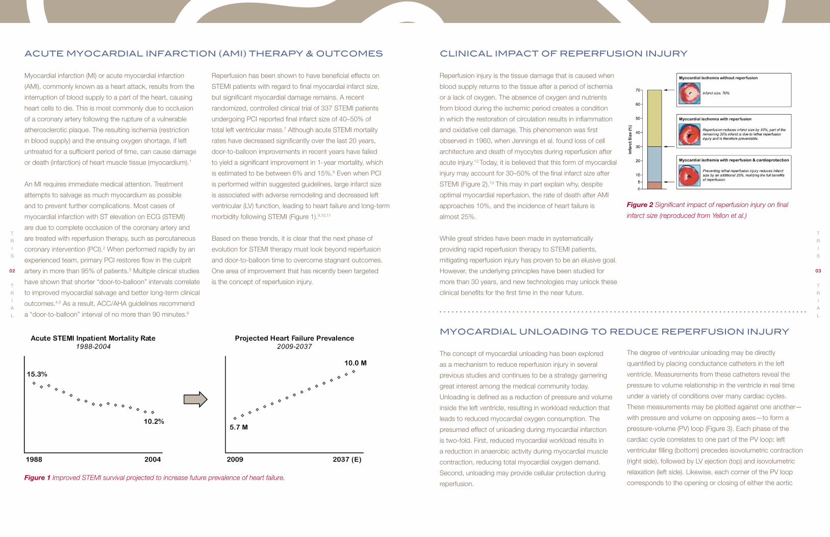
Reperfusion has been shown to have beneficial effects on
STEMI patients with regard to final myocardial infarct size,
but significant myocardial damage remains. A recent
randomized, controlled clinical trial of 337 STEMI patients
undergoing PCI reported final infarct size of 40–50% of
total left ventricular mass.7 Although acute STEMI mortality
rates have decreased significantly over the last 20 years,
door-to-balloon improvements in recent years have failed
to yield a significant improvement in 1-year mortality, which
is estimated to be between 6% and 15%.8 Even when PCI
is performed within suggested guidelines, large infarct size
is associated with adverse remodeling and decreased left
ventricular (LV) function, leading to heart failure and long-term
morbidity following STEMI (Figure 1).9,10,11
Based on these trends, it is clear that the next phase of
evolution for STEMI therapy must look beyond reperfusion
and door-to-balloon time to overcome stagnant outcomes.
One area of improvement that has recently been targeted
is the concept of reperfusion injury.
acutE myocardial infarction (ami) thErapy & outcomES clinical impact of rEpErfuSion injury
myocardial unloading to rEducE rEpErfuSion injury
Reperfusion injury is the tissue damage that is caused when
blood supply returns to the tissue after a period of ischemia
or a lack of oxygen. The absence of oxygen and nutrients
from blood during the ischemic period creates a condition
in which the restoration of circulation results in inflammation
and oxidative cell damage. This phenomenon was first
observed in 1960, when Jennings et al. found loss of cell
architecture and death of myocytes during reperfusion after
acute injury.12 Today, it is believed that this form of myocardial
injury may account for 30–50% of the final infarct size after
STEMI (Figure 2).13 This may in part explain why, despite
optimal myocardial reperfusion, the rate of death after AMI
approaches 10%, and the incidence of heart failure is
almost 25%.
While great strides have been made in systematically
providing rapid reperfusion therapy to STEMI patients,
mitigating reperfusion injury has proven to be an elusive goal.
However, the underlying principles have been studied for
more than 30 years, and new technologies may unlock these
clinical benefits for the first time in the near future.
Figure 2 Significant impact of reperfusion injury on final
infarct size (reproduced from Yellon et al.)
The concept of myocardial unloading has been explored
as a mechanism to reduce reperfusion injury in several
previous studies and continues to be a strategy garnering
great interest among the medical community today.
Unloading is defined as a reduction of pressure and volume
inside the left ventricle, resulting in workload reduction that
leads to reduced myocardial oxygen consumption. The
presumed effect of unloading during myocardial infarction
is two-fold. First, reduced myocardial workload results in
a reduction in anaerobic activity during myocardial muscle
contraction, reducing total myocardial oxygen demand.
Second, unloading may provide cellular protection during
reperfusion.
The degree of ventricular unloading may be directly
quantified by placing conductance catheters in the left
ventricle. Measurements from these catheters reveal the
pressure to volume relationship in the ventricle in real time
under a variety of conditions over many cardiac cycles.
These measurements may be plotted against one another—
with pressure and volume on opposing axes—to form a
pressure-volume (PV) loop (Figure 3). Each phase of the
cardiac cycle correlates to one part of the PV loop: left
ventricular filling (bottom) precedes isovolumetric contraction
(right side), followed by LV ejection (top) and isovolumetric
relaxation (left side). Likewise, each corner of the PV loop
corresponds to the opening or closing of either the aortic Figure 1 Improved STEMI survival projected to increase future prevalence of heart failure.
T
R
I
S
02
T
R
I
A
L
T
R
I
S
03
T
R
I
A
L

or mitral valve. The area inside the PV loop is equivalent to
myocardial stroke work, and a smaller area inside the loop
indicates less work being done by the left ventricle, with a
corollary decrease in myocardial oxygen demand.
In STEMI patients, lethal reperfusion injury is theorized to
result in additional cell death when blood flow is restored,
which results in larger infarct size and impaired ventricular
function. However, if myocardial oxygen demand is reduced
below the level of supply, muscle contraction can occur
aerobically. Reperfusion therapies, such as primary PCI, may
be implemented more safely, without the dynamic stress of
multiple myocardial cells demanding and sharing a limited
supply of oxygen. Based on these foundational principles,
multiple studies have evaluated the effectiveness of unloading
on myocardial salvage.
Figure 3 (A) Left ventricular pressure and volume over a single cardiac cycle. (B) Left ventricular pressure-volume (PV) loop
with one revolution corresponding to one cardiac cycle. (C) Smaller PV loop indicates ventricular workload reduction.
From 1979 to 1994, multiple animal studies investigated the
potential benefits of unloading the left ventricle on infarct
size after AMI.14,15,16,17,18,19 These studies generally involved
the creation of an artificial AMI followed by unloading of the
ventricle and vessel reperfusion. Histochemical analyses
post-necropsy were then performed to determine infarct
size. While these studies demonstrated the effectiveness of
LV unloading on the reduction of infarct size, the protocols
and surgical techniques used would not be applicable to
the STEMI population commonly treated with percutaneous
therapy alone.
initial invEStigationS of unloading in StEmi
From 2005 to 2008, three separate animal studies demon-
strated that unloading of the left ventricle with Intra-Aortic
Balloon Counterpulsation (IABC) prior to reperfusion reduces
infarct size and improves myocardial salvage.20,21,22 Based on
the results of these pre-clinical animal studies, a multicenter
randomized controlled trial was commenced in 2009:
Counterpulsation to Reduce Infarct Size Pre-PCI Acute
Myocardial Infarction (CRISP AMI).
The purpose of the CRISP AMI trial was to test the
hypothesis that left ventricular unloading via IABC delivered
prior to PCI would reduce myocardial infarct size in patients
with large anterior STEMI, compared to treatment with PCI
alone. The study enrolled 337 patients across 30 hospital
sites in 9 countries between June 2009 and February 2011,
with 176 patients randomized to the PCI-only control group
and 161 patients randomized to the IABC+PCI treatment
group. Unfortunately, the strategy of routine IABC use prior
to PCI in STEMI did not result in a statistically significant
reduction in myocardial infarct size, which was the study’s
primary endpoint. Additionally, none of the secondary
endpoints—microvascular obstruction, LV ejection fraction,
myocardial salvage index—indicated significant benefit in
the treatment group.23
Multiple explanations for the failure of CRISP AMI were
hypothesized. One explanation was that the beneficial
effects of unloading with IABC were offset by the additional
time required to insert the intra-aortic balloon. However,
the overall ischemic time from reported symptom onset to
first device was approximately 3 hours for all patients with
only a 10-minute difference between the two groups.
Another explanation offered that the potential protective
effect of LV unloading occurred too late in the course of
the MI to salvage significant myocardium. Several previous
studies found that only a small degree of myocardial salvage
occurs with ischemic times beyond 2 hours.24,25
One additional explanation for the failure of CRISP AMI is
that IABC may not have provided enough left ventricular
unloading to produce a treatment effect. In 2003, an animal
study was conducted to test whether the degree and timing
of unloading would impact myocardial salvage.26 Twenty-six
sheep were divided into four groups: (1) reperfusion with
no unloading (control group), (2) partial unloading initiated
after reperfusion, (3) full unloading initiated after reperfusion,
and (4) full unloading initiated before reperfusion.
The results showed significant infarct reduction in all
treatment groups, compared to the control, but each
treatment group showed more infarct reduction than the
previous one. Thus, the degree of unloading has a highly
linear correlation with myocardial salvage, and the maximum
benefit is only realized when a high degree of unloading is
delivered prior to reperfusion (Figure 4).
Based on these findings, the degree of unloading offered by
IABC is unlikely to produce the incremental clinical benefits
that have been noted in smaller animal studies. In fact, further
review of the original animal studies attempting therapeutic
unloading in AMI indicates that significant myocardial salvage
is achieved only when the LV is unloaded by at least 70%,
with no significant benefit when LV unloading is less than
50%.15,16,17,18,19,20 Thus, in order to extend the benefit of
unloading to a human STEMI population, a circulatory device
must approach full cardiac support levels.
Figure 4. Infarct size sequentially reduced with higher
degrees of unloading (reproduced from Meyns et al.).
A B C
67.2%
54.0%
41.6%
18.1%
0.0%
20.0%
40.0%
60.0%
80.0%
No Unloading Partial UnloadingAfter Reperfusion
Full UnloadingAfter Reperfusion
Full UnloadingBefore Reperfusion
Infarct Size (% of area at risk)
T
R
I
S
04
T
R
I
A
L
T
R
I
S
05
T
R
I
A
L

The TandemHeart system is a unique circulatory support
platform that enables a high degree of ventricular unloading.
The system comprises three parts: (1) the TandemHeart
Transseptal and Arterial Cannulae, (2) the TandemHeart
Centrifugal Pump, and (3) the Escort Controller (Figure 5).
The TandemHeart system is designed to access the left
atrium of the heart via a transseptal puncture, so that
oxygenated blood may be withdrawn from this chamber and
returned to the femoral artery. This creates an extracorporeal
blood circuit that bypasses the left ventricle, with the
TandemHeart pump operating in parallel to the native heart
(Figure 6).
During TandemHeart support, the PV loop shifts to the left
and down, indicating that the TandemHeart is reducing both
volume and pressure in the ventricle, resulting in reduced
myocardial stroke work. Overall, the TandemHeart system
combines a strong centrifugal pump and left atrial cannula-
tion to enable up to 90% reduction of left ventricular stroke
work and myocardial oxygen demand (Figure 7).27
Several alternative percutaneous circulatory support
platforms are commercially available. One platform utilizes a
small intraluminal axial pump placed across the aortic valve
directly into the left ventricle. This creates an intracorporeal
circuit that operates in series with the native left ventricle,
in contrast to the TandemHeart parallel circuit. As a result,
while this platform may be delivered via a single arterial
access point and does not require a transseptal puncture,
the largest diameter version (21Fr) only offers a maximum 45%
ventricular unloading, half the capability of TandemHeart.
Another alternative support platform withdraws deoxygenated
blood from the venous circulation through the femoral vein
to an extracorporeal pump, which then pushes the blood
through an oxygenator before returning to the femoral
artery. Unfortunately, this form of percutaneous cardiopul-
monary bypass (pCPB), otherwise known as veno-arterial
tandEmhEart unloading capability
Figure 5 Components of the TandemHeart Circulatory
Support System: Cannulae, Pump, and Controller.
Figure 6 The TandemHeart system utilizes transseptal
access of the left atrium to enable percutaneous LV bypass.
Figure 7 TandemHeart support level measured via
PV loop analysis in a large animal model.
extracorporeal membrane oxygenation (VA ECMO), does not
unload the left ventricle. In fact, the use of pCPB increases
pressure and volume in the LV, which subsequently increases
myocardial strain and oxygen demand.
Based on the aforementioned animal studies, these
alternative platforms may not provide the necessary degree
of unloading (>70%) to impact myocardial infarct size
in a human STEMI patient population. Therefore,
CardiacAssist commissioned pre-clinical animal studies
with the TandemHeart platform to test whether its unique
unloading capability would translate to a high degree of
myocardial salvage, indicating the potential for positive
long-term outcomes in a future human STEMI trial.
tandEmhEart prE-clinical animal StudiES
To consider the role of TandemHeart unloading in infarct
reduction, we theorized that if the myocardium is reperfused
while stroke work and myocardial oxygen consumption is
low, then fewer myocytes would die and total myocardial
infarct size would be reduced. Pre-clinical animal tests were
performed with 50-kg pigs divided into two groups. In both
groups, each animal’s left anterior descending (LAD) artery
—which supplies most of the blood flow to the left ventricle—
was occluded using an angioplasty balloon for 120 minutes
to simulate STEMI. After this point, the two groups received
alternative treatments.
In the control group, the angioplasty balloon was deflated,
removing the occlusion and allowing the myocardium to
be reperfused. After an additional 120 minutes, each animal
was sacrificed and its heart was harvested for analysis. In
the TandemHeart group, instead of receiving reperfusion
after 120 minutes of occlusion, TandemHeart support was
initiated. This support was continued for 30 minutes while
the occlusion remained, resulting in a total LAD occlusion
time of 150 minutes, compared to 120 minutes in the
control group. After 30 minutes of support, the balloon was
deflated while circulatory support continued throughout the
next 120 minutes, followed by sacrifice and harvesting for
further analysis (Figure 8).
TandemHeartLeft Atrial Cannulation
0
20
40
60
80
100
120
140
0 20 40 60 80 100 120 140 160
Pressure (m
mHg)
Volume (ml)
Native Left Ventricle
LV with TandemHeart
Figure 8 Experimental design for TandemHeart in STEMI pre-clinical animal study.
LAD Closed
LAD Closed
LAD Open
LAD Open
Reperfuse
Reperfuse
0 30 60 90 120 150 240180 210 270
TandemHeart Support
Harvest
Harvest
Occlude
Occlude
(minutes)
Treatment Group
Control Group
T
R
I
S
06
T
R
I
A
L
T
R
I
S
07
T
R
I
A
L

In contrast, TandemHeart-supported animals exhibited a
PV loop shift downward and to the left while on support,
indicating a high degree of pressure and volume reduction
(unloading) with a corresponding reduction in myocardial
oxygen demand. Additionally, after successful reperfusion,
the treatment group’s PV loops shifted back to their baseline
position, indicating myocardial salvage and a return to normal
pumping action for the LV (Figure 11).
The positive functional results exhibited in PV loop analysis
at different phases throughout the animal studies were
later confirmed via histochemical staining of the harvested
myocardial tissue, which was used to determine final infarct
size.TandemHeart unloading prior to reperfusion enabled
approximately a 50% decrease in myocardial infarct size
compared to the control group (Control: 53% infarct;
TandemHeart: 27% infarct), with a greater degree of
myocardial salvage in each progressive section of the LV
(Figure 12).
Pressure-volume loop data was obtained via conductance
catheters during each of the significant experimental intervals
across both groups. This data was used to calculate and
compare left ventricular stroke work. As expected, animals
in the TandemHeart group exhibited a high and statistically
significant degree of left ventricular unloading (Figure 9).
In both the control group and the TandemHeart group,
baseline PV loops were similar in size and shape. Likewise,
during LAD occlusion, each group’s PV loop shifted rightward
by a similar degree, indicating both diastolic and systolic
volume increase in the left ventricle. However, after this point,
the PV loops for each group exhibited marked differences.
Even after successful reperfusion, PV loops of animals in
the control group shifted far to the right, indicating significant
myocardial damage resulting in LV dilation and volume
overload. This rightward PV loop shift also indicates decreased
pumping capability of the heart and may eventually result in
end organ hypoperfusion, which is a pattern commonly seen in
heart failure patients with dilated cardiomyopathy (Figure 10).
These results were confirmed to be statistically significant
by repeating the protocol with multiple animals. Additionally,
the degree of unloading as defined by LV stroke work was
determined to be highly correlated to percent myocardial
salvage (Figure 13).
Because reperfusion injury is now theorized to be attenuated
by the activation of several cascades of pro-survival
signaling pathways—specifically the reperfusion injury
salvage kinase (RISK) pathway—this activity was also
assessed. Two prominent measurable components of this
molecular cardioprotection pathway are the AKT and ERK
kinase cascades.28 The pre-clinical animal study results
showed evidence of these cell survival catalysts only in
the TandemHeart group (Figure 14).
Figure 13 Myocardial salvage was found to be closely
correlated to the degree of unloading (R=0.82).
Figure 9 TandemHeart support significantly reduces
left ventricular stroke work.
*
TandemHeart Support
Off On
LV Stroke Work (m
L*mmHg
)
3500
3000
2500
2000
1500
1000
500
0
Figure 10 Control group animals exhibited significant
myocardial damage despite successful reperfusion.
BaselineOcclusion
Post‐Reperfusion
LV Volume (mL)50 75 100 125 150
0
75
100
LV Pressure (m
mHg
)
Figure 11 TandemHeart group animals exhibited a return to
normal left ventricular function after unloading & reperfusion.
50 75 100 125 1500
75
100
LV Pressure (m
mHg
)
LV Volume (mL)
Occlusion
On Support
Baseline & Post‐Reperfusion
Figure 12 TandemHeart group exhibited 50% reduction in myocardial infarct size versus control group.
Base Apex
Control
TandemHeart
Control TandemHeart
% M
yocardial Infarct Size
80
60
50
40
30
20
10
0
70
*
T
R
I
S
08
T
R
I
A
L
T
R
I
S
09
T
R
I
A
L

Based on the uniformly positive results from these pre-clinical
animal studies, CardiacAssist proposed a pivotal human
clinical trial to the U.S. Food and Drug Administration (FDA)
to investigate the use of TandemHeart to reduce infarct size
in STEMI. Investigational Device Exemption (IDE) approval
was received in December 2012, with hospital site
recruitment and patient enrollment set to begin in 2013.
The TandemHeart to Reduce Infarct Size (TRIS) Trial is a
prospective, randomized, controlled trial to demonstrate
superiority of PCI incorporating the TandemHeart system
versus standard PCI treatment with respect to both safety
and effectiveness. Subjects will be randomly assigned in a
1:1 ratio to treatment with TandemHeart support with PCI or
to PCI treatment alone. This study is designed to evaluate
the use of the TandemHeart system prior to revascularization
(as compared to standard PCI therapy) to limit infarct size in
subjects with an acute anterior AMI with an ST elevation of
> 2mm in two or more contiguous anterior leads.
The TandemHeart system will be used for 12 hours and
then weaned from use within the subsequent 12 hours
(for a total of up to 24 hours of use). This system offers
a unique solution for these subjects, as it is designed for
percutaneous access to provide rapid ventricular unloading
and increased systemic support. Specifically, the objective of
this study is to demonstrate the ability of the TandemHeart
system to limit myocardial reperfusion injury and infarct size
in a human STEMI patient population.
The primary endpoint for the TRIS Trial is reduction in
myocardial salvage index (MSI), which measures the
effectiveness of interventions that aim to reduce final
infarct size. MSI has previously been determined to be
a valid and reliable surrogate for mortality in clinical
trials testing the efficacy of reperfusion therapies in AMI.
Secondary endpoints include 1-year mortality and the rates
of cardiac re-hospitalization and implantable cardioverter
defibrillator (ICD) placement within the first 365 days after
treatment.30
For this study, a combined method of angiographic
assessment and cardiovascular magnetic resonance
(CMR) imaging performed 3–5 days after the infarct
(reviewed by a core lab) will be used to determine the
MSI. The literature and available data suggest an 11-point
improvement in MSI corresponds to a 5% absolute
reduction in infarct size. We can approximate that the
5 percentage point reduction in infarct size (used to guide
prior AMI studies), corresponding to a 3.1% increase in EF,
would be associated with an approximate 10% decrease
in one-year mortality.31,32 As a result, the TRIS Trial has the
potential to show real clinical value and create a paradigm
shift in STEMI care in the current era.
For more information regarding the TRIS Trial, or to be
considered as a participating hospital site, please contact
CardiacAssist at cardiacassist.com/contact.
tandEmhEart to rEducE infarct SizE (triS) trial
Figure 14 Biochemical markers for cellular protection and survival were exhibited only in the TandemHeart group.
p‐ERK
t‐ERK
p‐AKT
t‐AKT
GAPDH
Sham Control TandemHeart
Control TandemHeartSham
4.03.53.02.52.01.51.00.50.0
* Ɨ
pERK
/GAP
DHFold‐Change vs. Sham
Control TandemHeartSham
4.03.53.02.52.01.51.00.50.0
* Ɨ
pAKT/G
APDH
Fold‐Change vs. Sham
T
R
I
S
10
T
R
I
A
L
T
R
I
S
11
T
R
I
A
L

1Thygesen K et al. Third universal definition of myocardial
infarction. Circulation. 2012 Oct 16;126(16):2020-35.
2Roe MT, et al. Treatments, trends, and outcomes of acute
myocardial infarction and percutaneous coronary intervention.
J Am Coll Cardiol. 2010 Jul 20;56(4):254-63.
3Keeley EC, et al. Primary angioplasty versus intravenous
thrombolytic therapy for acute myocardial infarction: a
quantitative review of 23 randomised trials. Lancet. 2003
Jan 4;361(9351):13-20.
4Cannon CP, et al. Relationship of symptom-onset-to-balloon
time and door-to-balloon time with mortality in patients under-
going angioplasty for acute myocardial infarction. JAMA. 2000
Jun14;283(22):2941-7.
5De Luca G, et al. Time-to-treatment significantly affects the
extent of ST-segment resolution and myocardial blush in patients
with acute myocardial infarction treated by primary angioplasty.
Eur Heart J. 2004 Jun;25(12):1009-13.
6O’Gara PT, et al. 2013 ACCF/AHA Guideline for the Management
of ST-Elevation Myocardial Infarction: Executive Summary. J Am
Coll Cardiol. 2013;61(4):485-510.
7Patel MR, et al. Intra-aortic balloon counterpulsation and infarct
size in patients with acute anterior myocardial infarction without
shock: the CRISP AMI randomized trial. JAMA.
2011 Sep 28;306(12):1329-37.
8Roger VL, et al. Heart disease and stroke statistics—2012 update:
a report from the American Heart Association. Circulation. 2012
Jan 3;125(1):e2-e220.
9Wu E et al. Infarct size by contrast enhanced cardiac magnetic
resonance is a stronger predictor of outcomes than left ventrical
ejection fraction or end-systolic volume index: prospective cohort
study. Heart. 2008 Jun;94(6):730-6.
10Ezekowitz JA, et al. Predicting chronic left ventricular dysfunction
90 days after ST-segment elevation myocardial infarction: An
Assessment of Pexelizumab in Acute Myocardial Infarction
(APEX-AMI) Substudy. Am Heart J. 2010 Aug;160(2):272-8.
11Mohaved MR et al. Trends in the Age Adjusted Mortality from
Acute ST Segment Elevation Myocardial Infarction in the United
States (1988–2004) Based on Race, Gender, Infarct Location and
Comorbidities. Amer J Cardiol. 2009 Oct 15;104(8):1030-4.
12Jennings RB, et al. Myocardial necrosis induced by temporary
occlusion of a coronary artery in the dog. Arch Pathol.
1960;70:68-78.
13Yellon DM, et al. Myocardial reperfusion injury. N Engl J Med.
2007;357:1121-35.
14Pennock JL, et al. Reduction of myocardial infarct size:
comparison between left atrial and left ventricular bypass.
Circulation. 1979 Feb;59(2):275-9.
15Takanashi Y, et al. Reduction of myocardial infarct size in swine: a
comparative study of intraaortic balloon pumping and transapical
left ventricular bypass. Ann Thorac Surg. 1981 Nov;32(5):475-85.
16Catinella FP, et al. Left atrium-to-femoral artery bypass: effective-
ness in reduction of acute experimental myocardial infarction.
J Thorac Cardiovasc Surg. 1983 Dec;86(6):887-96.
17Allen BS, et al. Reperfusion conditions: critical importance of total
ventricular decompression during regional reperfusion. J Thorac
Cardiovasc Surg. 1986 Sep;92(3 Pt 2):605-12.
18Axelrod HI, et al. A comparison of methods for limiting myocardial
infarct expansion during acute reperfusion—primary role of
unloading. Circulation. 1987 Nov;76(5 Pt 2):V28-32.
19Fonger JD, et al. Enhanced preservation of acutely ischemic
myocardium with transseptal left ventricular assist.
Ann Thorac Surg. 1994 Mar;57(3):570-5.
20Achour H, et al. Mechanical left ventricular unloading prior to
reperfusion reduces infarct size in a canine infarction model.
Catheter Cardiovasc Interv. 2005 Feb;64(2):182-92.
21LeDoux JF, et al. Left ventricular unloading with intra-aortic
counter pulsation prior to reperfusion reduces myocardial release
of endothelin-1 and decreases infarction size in a porcine ischemia-
reperfusion model. Catheter Cardiovasc Interv.
2008 Oct 1;72(4):513-21.
22Azevedo CF, et al. The effect of intra-aortic balloon counterpulsa-
tion on left ventricular functional recovery early after acute myo-
cardial infarction: a randomized experimental magnetic resonance
imaging study. Eur Heart J. 2005;26(12):1235-1241.
23Patel MR, et al. Intra-aortic balloon counterpulsation and
infarct size in patients with acute anterior myocardial infarction
without shock: the CRISP AMI randomized trial. JAMA. 2011
Sep 28;306(12):1329-37.
24Francone M, et al. Impact of primary coronary angioplasty delay
on myocardial salvage, infarct size, and microvascular damage
in patients with ST-segment elevation myocardial infarction:
insight from cardiovascular magnetic resonance. J Am Coll Cardiol.
2009 Dec 1;54(23):2145-53.
25Eitel I, et al. Prognostic significance and determinants of
myocardial salvage assessed by cardiovascular magnetic resonance
in acute reperfused myocardial infarction. J Am Coll Cardiol. 2010
Jun 1;55(22):2470-9.
26Meyns B, et al. Left ventricular support by catheter-mounted axial
flow pump reduces infarct size. J Am Coll Cardiol. 2003 Apr 2;41(7):
1087-95.
27Kapur NK, et al. From Door to Balloon to Door to Unload: Shifting
the Paradigm of Cardioprotective Therapy for Ischemia Reperfusion
Injury in Acute Myocardial Infarction. Circulation. 2012;126:A16199.
footnotES
28Yellon DM, et al. Myocardial reperfusion injury. N Engl J Med.
2007 Sep 13;357(11):1121-35.
29Wang QD, et al. Pharmacological possibilities for protection
against myocardial reperfusion injury. Cardiovasc Res. 2002
Jul;55(1):25-37.
30Ndrepepa G, et al. Prognostic value of myocardial salvage
achieved by reperfusion therapy in patients with acute myocardial
infarction. J Nucl Med. 2004 May;45(5):725-9.
31Wu E, et al. Infarct size by contrast enhanced cardiac magnetic
resonance is a stronger predictor of outcomes than left ventricular
ejection fraction or end-systolic volume index: prospective cohort
study. Heart. 2008 Jun;94(6):730-6.
32Stone GW, et al. AMIHOT-II Trial Investigators. Effect of
supersaturated oxygen delivery on infarct size after percutaneous
coronary intervention in acute myocardial infarction. Circulation
Cardiovascular Interventions. 2009;2:366-75.
T
R
I
S
12
T
R
I
A
L
T
R
I
S
13
T
R
I
A
L

7100-0100 Rev 1 | 07.26.2013
cardiacassist.com