systemic inflammation in a melanoma patient treated with immune
TRANSCRIPT

CASE REPORT Open Access
Systemic inflammation in a melanomapatient treated with immune checkpointinhibitors—an autopsy studyViktor H. Koelzer1,2, Sacha I. Rothschild3, Deborah Zihler4, Andreas Wicki3, Berenika Willi5, Niels Willi1,Michèle Voegeli4, Gieri Cathomas1, Alfred Zippelius3 and Kirsten D. Mertz1*
Abstract
Background: Immune checkpoint inhibitors targeting cytotoxic T-lymphocyte-associated protein 4 (CTLA-4) andprogrammed cell death protein 1 (PD-1) have been recently approved for treatment of patients with metastaticmelanoma and non-small cell lung cancer (NSCLC). Despite important clinical benefits, these therapies areassociated with a diverse spectrum of immune-related adverse events (irAEs) that are typically transient, butoccasionally severe or even fatal.
Case presentation: This autopsy case illustrates that clinically overt irAEs may represent only a fraction of the totalspectrum of immune-related organ pathology in patients treated with immune checkpoint inhibitors. We report acomprehensive analysis of systemic irAE pathology based on the autopsy of a 35-year-old female patient withmetastatic melanoma treated first with ipilimumab and then nivolumab. The clinical course was characterized by amixed tumor response with regression of skin and lung metastases and fatal progression of metastatic disease inthe small bowel, peritoneum and brain. During therapy with ipilimumab, radiographic features of immune-relatedpneumonitis were noted. The autopsy examination established a sarcoid-like granulomatous reaction of the lung,pulmonary fibrosis and diffuse alveolar damage. Importantly, a clinically unapparent but histologically strikingsystemic inflammation involving the heart, central nervous system, liver and bone marrow was identified. Severeimmune-related end-organ damage due to lymphocytic myocarditis was found.
Conclusions: Autopsy studies are an important measure of quality control and may identify clinically unapparentirAEs in patients treated with immunotherapy. Pathologists and clinicians need to be aware of the broad spectrumof irAEs for timely management of treatment-related morbidity.
Keywords: Melanoma, Immunotherapy, Immune checkpoint inhibitors, Antibody, Ipilimumab, Nivolumab,Autoimmunity, Autopsy, Anti-tumor T cell response
BackgroundFour years after the approval of the first checkpoint inhibi-tor ipilimumab (anti-CTLA-4) for advanced melanoma in2011, cancer immunotherapy is now considered one of thepillars of cancer therapy [1]. Immune checkpoint inhibitorsinteracting with the PD-1/PD-L1 axis were recently ap-proved by the Food and Drug Administration (FDA) basedon successful large randomized controlled clinical trials [2]
of patients with metastatic melanoma [3, 4], non-small celllung cancer (NSCLC) [5, 6] and renal cell cancer [7]. Thereis a broad activity in different cancer types including DNAmismatch repair deficient colorectal cancer [8], ovariancancer [9] and treatment-refractory Hodgkin lymphoma[10]. Durable responses with survival plateaus have beenreported. As a consequence, the number of patients treatedwith immunotherapy is expected to increase. Both patholo-gists and clinicians therefore need to be increasingly awareof the unique spectrum of tissue reactions associated withimmune checkpoint inhibitor therapy to guide patientmanagement in daily practice.
* Correspondence: [email protected] of Pathology, Cantonal Hospital Baselland, Mühlemattstrasse 11,CH-4410 Liestal, SwitzerlandFull list of author information is available at the end of the article
© 2016 Koelzer et al. Open Access This article is distributed under the terms of the Creative Commons Attribution 4.0International License (http://creativecommons.org/licenses/by/4.0/), which permits unrestricted use, distribution, andreproduction in any medium, provided you give appropriate credit to the original author(s) and the source, provide a link tothe Creative Commons license, and indicate if changes were made. The Creative Commons Public Domain Dedication waiver(http://creativecommons.org/publicdomain/zero/1.0/) applies to the data made available in this article, unless otherwise stated.
Koelzer et al. Journal for ImmunoTherapy of Cancer (2016) 4:13 DOI 10.1186/s40425-016-0117-1
Efficacious cancer treatment with checkpoint inhibitorscan cause systemic immune activation that may poten-tially lead to tissue damage. Common adverse reactionsaffect the skin, gastrointestinal tract, liver, endocrineorgans and lungs, ranging from clinically unapparent tosevere immune-mediated organ damage [11]. The severityof irAEs clearly correlates with the dose and length ofanti-CTLA-4 and anti-PD-1 treatment [12]. In particular,combination therapy with several immune checkpointinhibitors may cause more adverse drug reactions thanmonotherapy [13]. Interestingly, a weak correlation of theseverity of irAEs with treatment response has also beendescribed [14]. Consequently, irAEs may be more com-mon in long term survivors. Several case reports have pre-viously illustrated the diverse clinical spectrum of irAEsincluding diffuse alveolar damage and immune mediatedpneumonitis [15], myocarditis [16], arthritis [17], severeskin toxicity [11], hypophysitis and meningoencephalitis[18]. Due to the strong immune activation by checkpointinhibition, it may be assumed that less severe adverse drugreactions accompany overt irAEs in patients treated withimmunomodulators and may contribute to long termtreatment-related organ damage. Even though analyses ofsystemic organ pathologies based on autopsy studiesfollowing treatment with immune checkpoint inhibitorsare an important measure of quality control, postmortemstudies are currently lacking in the literature. Here we re-port a comprehensive analysis of systemic irAE pathologybased on the autopsy of a 35-year-old female patient withmetastatic melanoma sequentially treated with ipilimumaband nivolumab (Fig. 1).
Case presentationIn August 2012, the patient presented with a malignantmelanoma arising from a congenital nevus in the rightdorsum of the foot which had been diagnosed followingexcisional biopsy at a local primary care physician (Breslowthickness 1.7 mm, Clark Level IV) (Fig. 2a). A wide excisionof the lesion with adequate safety margins was performed
and the patient was lost to follow up. In August 2013, oneyear after the primary excision, a local recurrence ofmalignant melanoma was detected (diameter 1.55 mm,infiltration depth 1.55 mm). Histopathological examinationrevealed an in-transit metastasis (diameter 3 mm) in thesubcutaneous tissue which focally reached the deep surgicalmargin (Fig. 2b). Re-excision with adequate safety marginsand a sentinel lymph node dissection was performed, iden-tifying melanoma micrometastases in two out of fourlymph nodes examined (Fig. 2c). Following a positive preg-nancy test, active surveillance was maintained.One year after the first recurrence and 4 months after
delivery, the patient presented to her dermatologist for afollow up examination. A positron-emission tomographywas performed revealing enlarged and enhancing rightinguinal lymph nodes with soft tissue extension. Threeweeks later, multiple skin metastases on the right leg weredetected and confirmed as melanoma by punch biopsy(Fig. 2d). Molecular analysis of one skin metastasis wasperformed at an outside institution. No potentially target-able BRAF, NRAS or c-KIT mutations were reported.First line therapy with dacarbazine every 3 weeks for
5 cycles was initiated in November 2014. The patientexperienced disease progression under dacarbazine treat-ment with increasing size and number of skin, nodal andsoft tissue metastases as well as newly detected metasta-ses in both lungs (Fig. 3a). As dacarbazine can causehematopoietic depression with severe leukocytopeniaand thrombocytopenia, the differential blood countswere closely monitored. After the third cycle of dacarba-zine, hematological studies showed significantly reducedneutrophil counts. No other hematological abnormalitieswere detected. Liver, renal and thyroid function wasnormal (see Additional file 1: Table S1 for laboratorystudies).Following completion of the fifth cycle of dacarbazine,
the patient was treated with ipilimumab at 3 mg/kg every 3weeks for four cycles from February to April of 2015.Radiotherapy to the soft tissues and nodes in the left
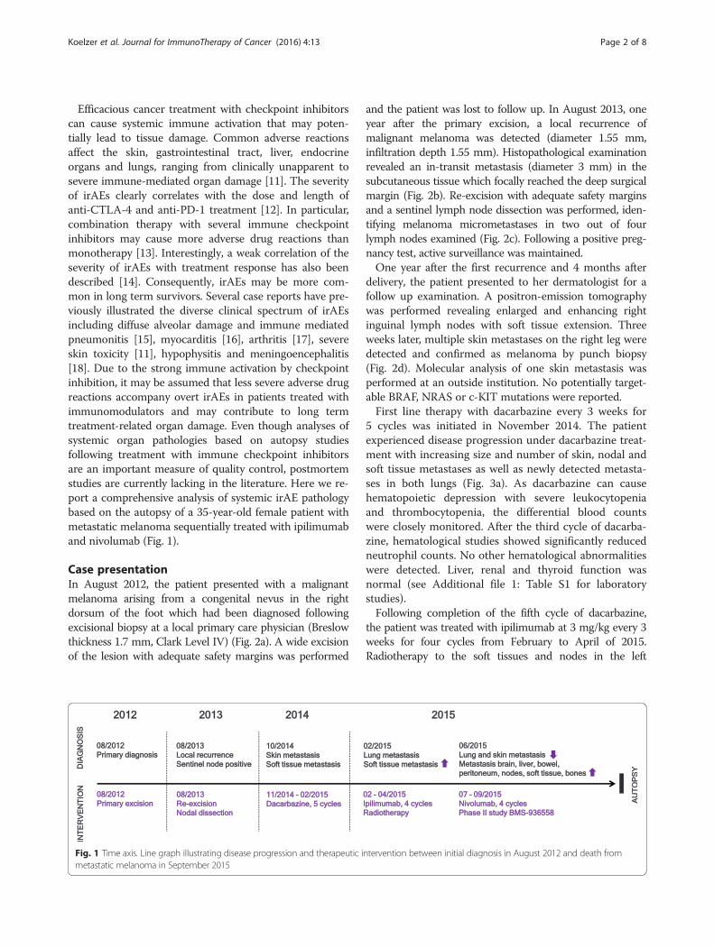
Fig. 1 Time axis. Line graph illustrating disease progression and therapeutic intervention between initial diagnosis in August 2012 and death frommetastatic melanoma in September 2015
Koelzer et al. Journal for ImmunoTherapy of Cancer (2016) 4:13 Page 2 of 8

inguinal region was administered (60Gy, 30 fractions,March to April 2015). After completing the fourth cycle ofipilimumab in the end of April 2015, computed tomog-raphy showed evidence of a mixed tumor response with re-gression of pulmonary and skin metastases. Theappearance of bilateral pulmonary ground glass opacitieswas noted (Fig. 3b). The patient was closely monitored. Asthere was no evidence of reduced pulmonary function, notreatment was given. Differential blood counts, liver, renaland thyroid function tests were within normal range [Add-itional file 1: Table S1].In June 2015, radiographic follow-up identified new
metastatic lesions in the liver, the abdominal wall, the pelvicperitoneum, uterus and ovaries, spine and pelvic bones. On
the first of July, 2015, Nivolumab therapy was initiated at3 mg/kg every 2 weeks for four cycles as part of a Phase IIstudy (BMS-936558). Repeated diffuse bleeding from meta-static lesions in the abdominal cavity and macrohematuriaaffecting the HB-level (Additional file 1: Table S1) requiredred blood cell transfusions. Multiple brain metastases weredetected by computed tomography in the beginning ofSeptember of 2015. The patient suffered from intensenausea, head and neck pain and was treated with intraven-ous opioids and corticoids. C-reactive protein levels wereelevated (250 mg/L) without fever. Laboratory values weresignificant for anemia, neutrophilia, lymphopenia, reactivethrombocytosis, moderately elevated aspartate aminotrans-ferase (GOT), elevated thyroid stimulating hormone (TSH)
a b c
d e f
1 mm 200 µm 500 µm
200 µm 200 µm 100 µmCD8
Fig. 2 Morphological progression from initial diagnosis. a Melanoma ex naevo (08/2012) b local recurrence (08/2013) with deep in transitmetastasis and c sentinel node metastasis d skin metastasis (10/2014) e dedifferentiated melanoma at autopsy f intratumoral CD8-positive T-cellinfiltrates at autopsy as detected by immunohistochemistry; scale bars as indicated
a b c
d e f
100 µm
200 µm 100 µm 200 µmEVG CD8 PAS
Fig. 3 Lung damage patterns. a CT Thorax: Pulmonary metastasis before therapy with Ipilimumab (02/2015) b CT Thorax: pulmonary metastasisregression, ground glass opacities after Ipilimumab (04/2015) c sarcoid-like reaction d elastica stain showing epithelioid granulomas surroundedby fibrotic rings e CD8-positive T-cell infiltrates surrounding giant cell granulomas as detected by immunohistochemistry f diffuse alveolardamage; scale bars as indicated
Koelzer et al. Journal for ImmunoTherapy of Cancer (2016) 4:13 Page 3 of 8
and lactate dehydrogenase (LDH) values. Peritoneal tapswere performed showing bloody ascites and elevated leuco-cyte counts, consistent with spontaneous bacterial periton-itis. Rapid progression of intraabdominal disease led toileus and renal failure. The patient deceased under bestsupportive care. An autopsy was performed.
Autopsy findingsPartially necrotic, amelanotic melanoma metastases weredetected in brain, liver, soft tissues, small bowel, pelvic peri-toneum, uterus and ovaries (Fig. 2e). Complete regressionof the pulmonary metastases, osseous metastases and skinlesions was documented. A prominent intratumoral cyto-toxic T-cell infiltrate with up to 100 CD8+ T-cells/mm2
(Fig. 2f), frequent expression of PD1 and cytotoxic granule-associated RNA binding protein (TIA-1) (Additional file 5:Figure S1) as well as a prominent histiocytic infiltrate andtumor necrosis were noted in the majority of lesionsexamined.Histopathologic examination of the lungs revealed two
pathogenetically distinct tissue reaction patterns. First,we observed panlobular histiocytic granulomas withgiant cells (Fig. 3c), perifocal interstitial lymphocyticinfiltrates and fibrotic rings (Fig. 3d) in both lungs withan interlobular, peribronchiolar, and subpleural distribu-tion. The perifocal lymphocytic infiltrate was rich inCD8-positive T-cells (Fig. 3e) with frequent expressionof TIA-1 and PD-1. Central necrosis was absent. No in-crease in eosinophils or mast cells was detected. Second,an acute and multifocal pattern of diffuse alveolar dam-age with formation of hyaline membranes was observedin all pulmonary lobes (Fig. 3f ). No residual melanomacells were detected by S100 immunohistochemistry. The
patient history was non-significant for allergies or occu-pational exposure to dust or silica. PAS and silver stainsfor fungi as well as Ziehl-Neelsen stains for mycobacteriawere negative. Tissue based polymerase chain reaction(PCR) analysis for mycobacteria, francisella tularensis,bartonella henselae, CMV, HSV, VZV, EBV, mucor andaspergillus were negative. Autoimmune and infectiousdisease serology and tissue testing was negative (seeAdditional file 2: Table S2).Autopsy analysis of the heart demonstrated an age-
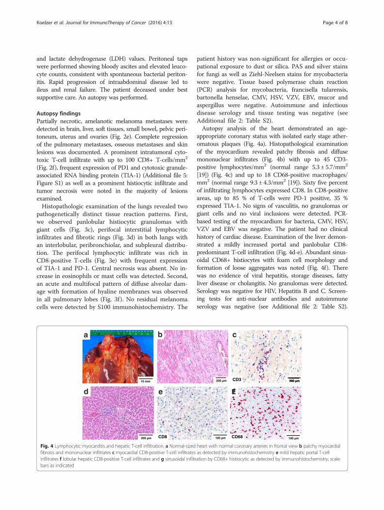
appropriate coronary status with isolated early stage ather-omatous plaques (Fig. 4a). Histopathological examinationof the myocardium revealed patchy fibrosis and diffusemononuclear infiltrates (Fig. 4b) with up to 45 CD3-positive lymphocytes/mm2 (normal range 5.3 ± 5.7/mm2
[19]) (Fig. 4c) and up to 18 CD68-positive macrophages/mm2 (normal range 9.3 ± 4.3/mm2 [19]). Sixty five percentof infiltrating lymphocytes expressed CD8. In CD8-positiveareas, up to 85 % of T-cells were PD-1 positive, 35 %expressed TIA-1. No signs of vasculitis, no granulomas orgiant cells and no viral inclusions were detected. PCR-based testing of the myocardium for bacteria, CMV, HSV,VZV and EBV was negative. The patient had no clinicalhistory of cardiac disease. Examination of the liver demon-strated a mildly increased portal and panlobular CD8-predominant T-cell infiltration (Fig. 4d-e). Abundant sinus-oidal CD68+ histiocytes with foam cell morphology andformation of loose aggregates was noted (Fig. 4f). Therewas no evidence of viral hepatitis, storage diseases, fattyliver disease or cholangitis. No granulomas were detected.Serology was negative for HIV, Hepatitis B and C. Screen-ing tests for anti-nuclear antibodies and autoimmuneserology was negative (see Additional file 2: Table S2).
a b c
d Ef
200 µm100 µm
200 µm 100 µm
CD3
CD68
200 µm
e
100 µmCD8
10 mm
Fig. 4 Lymphocytic myocarditis and hepatic T-cell infiltration. a Normal-sized heart with normal coronary arteries in frontal view b patchy myocardialfibrosis and mononuclear infiltrates c myocardial CD8-positive T-cell infiltrates as detected by immunohistochemistry e mild hepatic portal T-cellinfiltrates f lobular hepatic CD8-positive T-cell infiltrates and g sinusoidal infiltration by CD68+ histiocytic as detected by immunohistochemistry; scalebars as indicated
Koelzer et al. Journal for ImmunoTherapy of Cancer (2016) 4:13 Page 4 of 8

There was no history of a preexisting liver disease. A list ofstains performed and staining protocols can be found in(Additional file 3: Table S3 and Additional file 4: Table S4).Histopathologic examination of the brain was pertinent
for severe aseptic lymphocytic meningitis with extensionof mononuclear infiltrates into the brain parenchyma(Fig. 5a). T-cell subtyping revealed that the lymphocyticinfiltrate predominantly consisted of CD8+ T-cells withup to 180 CD8 + T-cells/mm2 detected in the meningesand up to 15 CD8+ T-cells/ mm2 detected in the periven-tricular brain parenchyma (Fig. 5b). Up to 45 % of T-cellsexpressed PD-1, up to 60 % showed reactivity for TIA-1(see Additional file 5: Figure S1) with a strong concomi-tant CD68+ histiocytic infiltrate (Fig. 5c). There was noevidence of viral inclusions, vasculitis or ischemic lesions.The bone marrow was hypercellular with evidence ofdiffuse lymphocytosis (Fig. 5d). Up to 15 % of all nucleatedcells in the bone marrow were CD3+ lymphocytes (Fig. 5e)and up to 10 % were CD79+ B-cells. Interestingly, onlyapproximately 20 % of T-lymphocytes expressed CD8(Fig. 5f). Analysis of endocrine organs including adrenalsand thyroid glands did not reveal any abnormalities.
DiscussionImmune checkpoint inhibition directed against PD-1 andCTLA-4 has the potential to activate effector T-cellsagainst a wide spectrum of tumor- and self-antigens. Thepresent case demonstrates the importance of systematicpostmortem studies to identify relevant safety findings inthis setting. We illustrate that clinically overt irAEs maybe accompanied by a wide spectrum of unsuspected auto-immune pathologies that require timely treatment. Bothpathologists and clinicians need to be increasingly awareof the unique spectrum of immune related adverse drug
experiences for optimal patient management in dailypractice.In patients with metastatic disease, a significant fraction
of tumor-antigen specific effector and memory T-cellsmay be detectable in the tumor and peripheral circulation,yet may be constrained by tumor-induced immune sup-pression mechanisms. Targeting the PD-1/PD-L1 andCTLA-4 signaling axis by immunomodulatory antibodiescan induce a significant anti-tumoral immune response.However, therapeutic response to ipilimumab is accom-panied by clinically detectable irAEs in up to 72 % ofpatients with grade 3–4 irAEs in 24 % and lethal outcomein 0.86 % of cases [11]. Onset of irAEs occurs on average10 weeks after the onset of treatment and correlates withdosage but can occur as late as 2 years after initializationof treatment [11, 15]. In the landmark CheckMate 037trial of nivolumab in advanced melanoma, irAEs havebeen observed at a similar frequency of 68 % with lesscommon grade 3–4 events (9 %) [20]. This rate of grade 3or 4 toxicity is similar to that seen with many chemothera-peutic agents or targeted therapies [21]. The conductedlarge clinical trials include a systematic assessment ofirAEs under anti-CTLA-4 and anti-PD-1 blockade. How-ever, these analyses are primarily based on clinical, labora-tory and radiographic evidence with few cases reportingthe analysis of tissue biopsies. Systematic autopsy studiesof patients treated with immune checkpoint inhibitors areso far lacking in the literature.This comprehensive autopsy study of a young patient
treated sequentially with ipilimumab and nivolumab dem-onstrates that clinically or radiographically apparent organdysfunction may represent only a small part of treatment-related unfavorable medical occurrences in a given case.In particular, a sarcoid-like pulmonary reaction, features
100 µm
a b c
200 µm CD8 CD68200 µm
100 µm
d e f
200 µm CD3 CD8200 µm
Fig. 5 Meningoencephalitis and bone marrow lymphocytosis. a Meningeal lymphocytosis with b a predominantly CD8+ T-cell infiltrate as demonstratedby immunohistochemistry and c extension into the brain parenchyma; d Hypercellular bone marrow with e an increase in CD3+ and f CD8+T-lymphocytes as detected by immunohistochemistry; scale bars as indicated
Koelzer et al. Journal for ImmunoTherapy of Cancer (2016) 4:13 Page 5 of 8

of diffuse alveolar damage, aseptic meningoencephalitisand myocarditis with myocardial damage were discoveredat autopsy as concomitant findings of a clinically sus-pected immune-related pneumonitis. These findings wereclassified as irAEs based on the close temporal associa-tions between immune checkpoint inhibitor therapy andhistopathologic findings with a significant increase of CD8+, TIA1+ and PD-1+ T-cells in the affected organs. Lesssignificant findings included hepatic lymphocytic infiltra-tion and bone marrow lymphocytosis.As the patient received both ipilimumab and nivolumab,
a definite association of a particular damage pattern toone of the two agents is not possible in the present case.However, the temporal association of the CT-graphic find-ing of ground glass opacities after ipilimumab therapymay favor an assignment of these pulmonary findings tothis agent. Indeed, CTLA4-related pneumonitis duringcancer immunotherapy has been previously described as arare event, but was diagnosed based on radiographicassessment only [22]. We found a pattern of multifocaldiffuse alveolar damage with hyaline membranes under-lying this radiographic finding. In addition, a pulmonarysarcoid-like granulomatosis was identified. Previous CT-scans and medical history was insignificant for sarcoidosis,and post-mortem tissue based analysis ruled out an infec-tious etiology. Interestingly, a similar occurrence followingCTLA-4 blockade has been described by other authors[11, 23]. These findings may indicate that combinedcheckpoint blockade may also cause superimposed histo-pathological damage patterns as a correlate of distinctimmunological effects. Indeed, combination therapy leadsto divergent gene expression changes in T-cells andmonocyte populations that may underlie these specificirAEs [24]. Immune checkpoint inhibitor therapy has beenlinked to the uncontrolled release of cytokines in the formof a cytokine storm [25]. While the typical symptoms of acytokine storm such as high fever, vasodilation, peripheraledema and distributive shock were not detected in thepresent case, cytokine release may have contributed to thedevelopment of irAEs. In particular, interleukin-2 (IL-2)secretion by activated T-cells is thought to play a role inthe pathogenesis of sarcoidosis [26] and may be a mech-anism linking sarcoid-like granulomatous reactions toimmunotherapy. Indeed, the disease activity of a preexist-ing sarcoidosis may also be increased by high dose IL-2treatment of neoplasia [27] and human immunodeficiencyvirus (HIV) infection [28]. Larger case series would bedesirable to further investigate the mechanistic linkbetween immune checkpoint inhibition and the develop-ment of sarcoid-like irAEs.Cardiac autopsy findings demonstrated a lymphocytic
myocarditis with patchy fibrosis in the absence of clinical,serological or tissue-based evidence for an infectious eti-ology. A non-infectious myocarditis in patients treated with
nivolumab [3, 29] and ipilimumab [4] has been previouslydescribed, but biopsy studies are rare. Here we demonstratea pattern of diffuse lymphocytic infiltrates with a strongpredominance of CD8+/PD-1+/TIA1+ cytotoxic T-cellsand concomitant diffuse CD68+ histiocytic infiltrates.These findings are reminiscent of a previous report includ-ing analysis of a myocardial biopsy from a patient treatedwith anti-PD-1 antibody (pembrolizumab) suggesting asimilar pathogenetic mechanism [16]. It is important tonote the potential pathophysiologic and clinical implica-tions of concurrent lung and heart toxicity which may bemore severe and prone for critical events than either alone.Brain autopsy revealed a severe aseptic lymphocytic
meningoencephalitis driven by CD8+/PD-1+ cytotoxic T-cells. Aseptic meningitis has been reported in a patienttreated with ipilimumab [18] but histologic analysis has sofar been lacking. A range of neurological and endocrineadverse events has been associated with immune check-point inhibitors. Most frequently, autoimmune hypophysi-tis and thyroidits is encountered with anti-CTLA-4treatment [11]. In the present case, no tissue based, radio-graphic or laboratory evidence of hypophysis or thyroiddysfunction was present.Other findings included a bone marrow lymphocy-
tosis and hepatic mononuclear and histiocytic infil-trates. Hepatic toxicity and elevation of liver enzymeshas been described as a mostly low-grade irAE in pa-tients treated with ipilimumab [11] and nivolumab[20]. In a biopsy study, a pan-lobular hepatitis withprominent sinusoidal histiocytic infiltrates and centralvein endothelialitis has been previously suggested as ahistologic clue to ipilimumab-associated hepatitis [30].A bone marrow lymphocytosis has not been previ-ously identified, but may be a concomitant feature ofgeneralized T-cell activation due to immune check-point blockade. In the present case, no significanthematological abnormalities in the peripheral bloodcounts were detected.
ConclusionOur results contribute to a better understanding of theatypical immune toxicity associated with checkpoint in-hibition by anti-CTLA-4 and anti-PD-1 antibodies. A dee-per knowledge of these immune-related adverse eventsand its multidisciplinary management will help to reducemorbidity and therapy interruptions. Future perspectivesinclude the concurrent administration of antibodies tar-geting CTLA-4 and PD-1. Our data underline that carefulmonitoring is of particular importance in this setting toidentify potentially harmful immune pathology.
ConsentWritten informed consent was given by the legal guard-ians of the patient for publication of this case report and
Koelzer et al. Journal for ImmunoTherapy of Cancer (2016) 4:13 Page 6 of 8

any accompanying images. A declaration of no objectionwas obtained from the local ethics committee (Ethik-kommission Nordwest- und Zentralschweiz (EKNZ); filedesignation: UBE 15–106). A copy of the written consentis available for review by the Editor-in-Chief of thisjournal.
Additional files
Additional file 1: Table S1. Laboratory studies. Data on differentialblood counts, renal function, liver function tests and thyroid functionduring each treatment cycle (dacarbazine, ipilimumab, nivolumab) areprovided. (DOCX 16 kb)
Additional file 2: Table S2. Autoimmune serology. Table showing thescreening results of autoimmune serology including systemic antibodies,anti-neutrophil cytoplasmic antibodies (ANCA) and anti-neuronalantibodies. (DOCX 16.8 kb)
Additional file 3: Table S3. Immunohistochemical stains. Systematicoverview of the immunohistochemical stains performed for each organand tumor specimen. (DOCX 21.3 kb)
Additional file 4: Table S4. Antibodies and staining protocols. All stainswere performed on a Leica BOND III / max autostainer platform (LeicaBioystems, Muttenz, Switzerland). Information on antibody clones,pretreatment and staining protocols is provided. (DOCX 18 kb)
Additional file 5: Figure S1. Expression of cytotoxic granule-associatedRNA binding protein (TIA-1) and programmed cell death protein 1 (PD-1,nivolumab). Frequent expression of PD-1 and TIA-1 in tumor infiltrating T-cells(A, B) and in lymphocytic infiltrates in the peripheral organs including themeninges (C, D); scale bars as indicated. (PPTX 4.8 mb)
AbbreviationsANCA: anti-neutrophil cytoplasmic antibodies; CD: cluster of differentiation;CMV: cytomegalovirus; CTLA-4: cytotoxic T-lymphocyte-associated protein 4;EBV: Epstein-Barr virus; FDA: Food and Drug Administration; HSV: herpes simplexvirus; irAEs: immune-related adverse events; NSCLC: non-small cell lung cancer(NSCLC); PCR: polymerase chain reaction; PD-1: programmed cell death protein 1;TIA-1: cytotoxic granule-associated RNA binding protein; VZV: varicella-zoster virus.
Competing interestsAZ received research funding from Roche Glycart, consultant fees and travelgrants from Roche, Bristol-Myers Squibb (BMS) and Merck, Sharp and Dohme(MSD). SIR serves as an advisor for Roche, BMS and MSD, is principal investigatorsfor clinical trials led by Roche, BMS and MSD and received research funding andtravel grants from Roche. AW serves as an advisor for Roche, Merck, BMS, Novartis,Lilly, Bayer, Amgen and AstraZeneca. All other authors report no financial andnon-financial competing interests.
Authors’ contributionsVHK performed the autopsy, the histopathological analysis, participated indesign and coordination of the study and drafted the manuscript. KDMsupervised the autopsy and histopathological analysis, oversaw design andcoordination of the study and drafted the manuscript. SIR treated the patientat the University Hospital Basel, helped with rendering the diagnosis,critically reviewed the manuscript and helped with revision of the paper. DZtreated the patient at Kantonsspital Baselland, helped with rendering thediagnosis and helped with revision of the paper. AW treated the patient atthe University Hospital Basel, helped with rendering the diagnosis, andcritically reviewed the manuscript and the revision of the paper. BWreviewed and interpreted the radiographic images, helped with renderingthe diagnosis and critically reviewed the manuscript. AZ treated the patientat the University Hospital Basel, helped with rendering the diagnosis andcritically reviewed the manuscript. MV treated the patient at KantonsspitalBaselland, helped with rendering the diagnosis and critically reviewed themanuscript. GC helped to analyze the histological sections, studied immuneinfiltrates by immunostaining and reviewed the manuscript. NW helped toanalyze the histological sections, studied immune infiltrates by
immunostaining, critically discussed the case with VHK and KDM andreviewed the manuscript. All authors read and approved the finalmanuscript.
AcknowledgmentsWe thank Dr. Sara Brittingham, MD for helpful discussions of the manuscript.
Author details1Institute of Pathology, Cantonal Hospital Baselland, Mühlemattstrasse 11,CH-4410 Liestal, Switzerland. 2Translational Research Unit (TRU), Institute ofPathology, University of Bern, Murtenstrasse 31, CH-3010 Bern, Switzerland.3Division of Medical Oncology, University Hospital Basel, Petersgraben 4,CH-4031 Basel, Switzerland. 4Department of Medical Oncology, CantonalHospital Baselland, Mühlemattstrasse 11, CH-4410 Liestal, Switzerland.5Institute of Radiology, Cantonal Hospital Baselland, Mühlemattstrasse 11,CH-4410 Liestal, Switzerland.
Received: 22 December 2015 Accepted: 9 February 2016
References1. Mellman I, Coukos G, Dranoff G. Cancer immunotherapy comes of age.
Nature. 2011;480(7378):480–9. doi:10.1038/nature10673.2. Topalian SL, Hodi FS, Brahmer JR, Gettinger SN, Smith DC, McDermott DF,
et al. Safety, activity, and immune correlates of anti-PD-1 antibody in cancer.N Engl J Med. 2012;366(26):2443–54. doi:10.1056/NEJMoa1200690.
3. Robert C, Long GV, Brady B, Dutriaux C, Maio M, Mortier L, et al. Nivolumabin previously untreated melanoma without BRAF mutation. N Engl J Med.2015;372(4):320–30. doi:10.1056/NEJMoa1412082.
4. Hodi FS, O'Day SJ, McDermott DF, Weber RW, Sosman JA, Haanen JB, et al.Improved survival with ipilimumab in patients with metastatic melanoma. NEngl J Med. 2010;363(8):711–23. doi:10.1056/NEJMoa1003466.
5. Borghaei H, Paz-Ares L, Horn L, Spigel DR, Steins M, Ready NE, et al.Nivolumab versus Docetaxel in Advanced Nonsquamous Non-Small-CellLung Cancer. N Engl J Med. 2015;373(17):1627–39. doi:10.1056/NEJMoa1507643.
6. Brahmer J, Reckamp KL, Baas P, Crino L, Eberhardt WE, Poddubskaya E, et al.Nivolumab versus Docetaxel in Advanced Squamous-Cell Non-Small-CellLung Cancer. N Engl J Med. 2015;373(2):123–35. doi:10.1056/NEJMoa1504627.
7. Motzer RJ, Escudier B, McDermott DF, George S, Hammers HJ, Srinivas S,et al. Nivolumab versus Everolimus in Advanced Renal-Cell Carcinoma. NEngl J Med. 2015;373(19):1803–13. doi:10.1056/NEJMoa1510665.
8. Le DT, Uram JN, Wang H, Bartlett BR, Kemberling H, Eyring AD, et al. PD-1Blockade in Tumors with Mismatch-Repair Deficiency. N Engl J Med. 2015;372(26):2509–20. doi:10.1056/NEJMoa1500596.
9. Silen W, Machen TE, Forte JG. Acid–base balance in amphibian gastricmucosa. Am J Physiol. 1975;229(3):721–30.
10. Villasboas JC, Ansell SM. Nivolumab for the treatment of classical Hodgkinlymphoma after failure of autologous stem cell transplant and Brentuximab.Expert Rev Anticancer Ther. 2015. doi:10.1586/14737140.2016.1121812
11. Bertrand A, Kostine M, Barnetche T, Truchetet ME, Schaeverbeke T. Immunerelated adverse events associated with anti-CTLA-4 antibodies: systematic reviewand meta-analysis. BMC Med. 2015;13:211. doi:10.1186/s12916-015-0455-8.
12. Postow M, Wolchok J. Toxicities associated with checkpoint inhibitorimmunotherapy. In: UpToDate, Post TW (Ed), UpToDate, Waltham, MA(Accessed on December 15, 2015)
13. Tsai KK, Daud AI. Nivolumab plus ipilimumab in the treatment of advancedmelanoma. J Hematol Oncol. 2015;8(1):123. doi:10.1186/s13045-015-0219-0.
14. Freeman-Keller M, Kim Y, Cronin H, Richards A, Gibney G, Weber J.Nivolumab in Resected and Unresectable Metastatic Melanoma:Characteristics of Immune-Related Adverse Events and Association withOutcomes. Clin Cancer Res. 2015. doi:10.1158/1078-0432.CCR-15-1136.
15. Nishino M, Sholl LM, Hodi FS, Hatabu H, Ramaiya NH. Anti-PD-1-RelatedPneumonitis during Cancer Immunotherapy. N Engl J Med.2015;373(3):288–90. doi:10.1056/NEJMc1505197.
16. Laubli H, Balmelli C, Bossard M, Pfister O, Glatz K, Zippelius A. Acute heartfailure due to autoimmune myocarditis under pembrolizumab treatment formetastatic melanoma. J Immunother Cancer. 2015;3:11.doi:10.1186/s40425-015-0057-1.
Koelzer et al. Journal for ImmunoTherapy of Cancer (2016) 4:13 Page 7 of 8

17. de Velasco G, Bermas B, Choueiri TK. Auto-immune arthropathy anduveitis as complications from PD-1 inhibitor. Arthritis Rheumatol. 2015.doi:10.1002/art.39406.
18. Bot I, Blank CU, Boogerd W, Brandsma D. Neurological immune-relatedadverse events of ipilimumab. Pract Neurol. 2013;13(4):278–80.doi:10.1136/practneurol-2012-000447.
19. Bocchi EA, Tanigawa RY, Brandao SM, Cruz F, Issa V, Ayub-Ferreira S, et al.Immunohistochemical quantification of inflammatory cells inendomyocardial biopsy fragments after heart transplantation: a newpotential method to improve the diagnosis of rejection after hearttransplantation. Transplant Proc. 2014;46(5):1489–96.doi:10.1016/j.transproceed.2013.12.062.
20. Weber JS, D'Angelo SP, Minor D, Hodi FS, Gutzmer R, Neyns B, et al.Nivolumab versus chemotherapy in patients with advanced melanoma whoprogressed after anti-CTLA-4 treatment (CheckMate 037): a randomised,controlled, open-label, phase 3 trial. Lancet Oncol. 2015;16(4):375–84.doi:10.1016/S1470-2045(15)70076-8.
21. Postow MA, Callahan MK, Wolchok JD. Immune Checkpoint Blockade in CancerTherapy. J Clin Oncol. 2015;33(17):1974–82. doi:10.1200/JCO.2014.59.4358.
22. Bashey A, Medina B, Corringham S, Pasek M, Carrier E, Vrooman L, et al.CTLA4 blockade with ipilimumab to treat relapse of malignancy afterallogeneic hematopoietic cell transplantation. Blood. 2009;113(7):1581–8.doi:10.1182/blood-2008-07-168468.
23. Berthod G, Lazor R, Letovanec I, Romano E, Noirez L, Mazza Stalder J, et al.Pulmonary sarcoid-like granulomatosis induced by ipilimumab. J Clin Oncol.2012;30(17):e156–9. doi:10.1200/JCO.2011.39.3298.
24. Das R, Verma R, Sznol M, Boddupalli CS, Gettinger SN, Kluger H, et al.Combination therapy with anti-CTLA-4 and anti-PD-1 leads to distinctimmunologic changes in vivo. J Immunol. 2015;194(3):950–9.doi:10.4049/jimmunol.1401686.
25. Bakacs T, Mehrishi JN, Moss RW. Ipilimumab (Yervoy) and the TGN1412catastrophe. Immunobiology. 2012;217(6):583–9. doi:10.1016/j.imbio.2011.07.005.
26. Ziegenhagen MW, Muller-Quernheim J. The cytokine network in sarcoidosisand its clinical relevance. J Intern Med. 2003;253(1):18–30.
27. Logan TF, Bensadoun ES. Increased disease activity in a patient withsarcoidosis after high dose interleukin 2 treatment for metastatic renalcancer. Thorax. 2005;60(7):610–1. doi:10.1136/thx.2004.024018.
28. Blanche P, Gombert B, Rollot F, Salmon D, Sicard D. Sarcoidosis in a patientwith acquired immunodeficiency syndrome treated with interleukin-2. ClinInfect Dis. 2000;31(6):1493–4. doi:10.1086/317475.
29. Ansell SM, Lesokhin AM, Borrello I, Halwani A, Scott EC, Gutierrez M, et al.PD-1 blockade with nivolumab in relapsed or refractory Hodgkin'slymphoma. N Engl J Med. 2015;372(4):311–9. doi:10.1056/NEJMoa1411087.
30. Johncilla M, Misdraji J, Pratt DS, Agoston AT, Lauwers GY, Srivastava A, et al.Ipilimumab-associated Hepatitis: Clinicopathologic Characterization in aSeries of 11 Cases. Am J Surg Pathol. 2015;39(8):1075–84. doi:10.1097/PAS.0000000000000453.
• We accept pre-submission inquiries
• Our selector tool helps you to find the most relevant journal
• We provide round the clock customer support
• Convenient online submission
• Thorough peer review
• Inclusion in PubMed and all major indexing services
• Maximum visibility for your research
Submit your manuscript atwww.biomedcentral.com/submit
Submit your next manuscript to BioMed Central and we will help you at every step:
Koelzer et al. Journal for ImmunoTherapy of Cancer (2016) 4:13 Page 8 of 8