synthesis of diamond-like phase from graphite by ultrafast
TRANSCRIPT

1Scientific RepoRts | 5:11812 | DOi: 10.1038/srep11812
www.nature.com/scientificreports
Synthesis of diamond-like phase from graphite by ultrafast laser driven dynamical compressionFrancisco C. B. Maia1, Ricardo E. Samad2, Jefferson Bettini3, Raul O. Freitas1, Nilson D. Vieira Junior2 & Narcizo M. Souza-Neto1
Rapid variations of the environmental energy caused by ultrashort laser pulses have induced phase transitions in carbon allotropes, therefore bringing the promise of revealing new carbon phases. Here, by exposing polycrystalline graphite to 25 fs laser pulses at 4 J/cm2 fluence under standard air atmosphere, we demonstrated the synthesis of translucent micrometer-sized structures carrying diamond-like and onion-like carbon phases. Texturized domains of the diamond phase were also identified. Concerning different synthesized carbon forms, pulse superposition and singularities of the thermodynamical process, we pinpoint the synthesis mechanism by the laser-induced subsequent products energetically evolving to attain the diamond-like phase.
In recent years carbon allotropes, such as nanodiamonds, have shown promising new applications in many fields due to its physical, chemical and surface characteristics. Their high electron mobility, field electron emission and magnetic properties1 make them important players in carbon based electronics2,3. Their tribological and mechanical1,4,5 properties give rise to harder coatings1,5, which are biocompatible and can provide improved biological prosthesis joints6 with decreased wear. This biocompatibility allied to their biosensing, optical and nanoprobing functionalities7,8 provide drug delivery and cellular labeling capacity7. This wide range of novel applications has fostered the active search for new and more efficient synthesis and production methods of carbon allotropes.
In this pursuit, it has been shown that dynamical compression is a way to steer carbon allotropes, through rapid energy variations, to phase transitions. As examples, transition from graphite to diamond headed by shock compression experiments occurs through the fast martensitic mechanism9; in a setup designed for quenching shock-compressed samples at a fast cooling rate, the n-diamond phase10 has been created, and in regions of shock impacted meteorites nanodiamonds have been formed11. These observa-tions brought to light the importance of carbon phase transformations in non-equilibrium states, spur-ring studies in a faster time scale using ultrashort pulses, which trigger shockwaves12,13 carrying extreme temperature and pressure in the matter. Experiments employing high peak power ultrashort laser pulses with durations around 100 femtoseconds irradiating Highly Oriented Pyrolytic Graphite (HOPG), usu-ally under vacuum14–16 or in the graphite/liquid interface17, have originated sp3-bounded lattices on the graphite14,18 and provided preliminary evidences of diamond formation on the substrates14–17,19,20. Ultrashort laser shots raise promises for synthesizing known21 and still theoretically predicted22,23 sp3 carbon structures. Here, we have taken advantage of moderate energy ultrashort laser pulses to gener-ate shockwaves and induce the formation of a diamond-like phase from the sp2 carbon polycrystalline graphite precursor.
In the present work, laser shots irradiating graphite under air dramatically modified the surface and created micrometer scale translucent structures where diamond-like crystallites were found coexisting with onion-like phases and quasi-amorphous nanometer sized graphite. Such materials were recovered
1Laboratório Nacional de Luz Síncrotron (LNLS), Campinas, São Paulo 13083-970, Brazil. 2Instituto de Pesquisas Energéticas e Nucleares (IPEN-CNEN/SP), São Paulo 05508-000, Brazil. 3Laboratório Nacional de Nanotecnologia (LNNano), Campinas, São Paulo 13083-970, Brazil. Correspondence and requests for materials should be addressed to F.C.B.M. (email: [email protected]) or N.M.S.N. (email: [email protected])
received: 06 January 2015
accepted: 08 June 2015
Published: 07 July 2015
OPEN
www.nature.com/scientificreports/
2Scientific RepoRts | 5:11812 | DOi: 10.1038/srep11812
after the laser irradiation process and analyzed by micro Raman spectroscopy, Scanning Electron Microscopy (SEM) and High Resolution Electron Microscopy (HREM). Considering the ultrafast optical excitation, ablation regime and the distinct laser-synthesized carbon structures, we propose an indirect mechanism in which subsequent laser-induced transformations lead to the synthesis of the diamond-like phase.
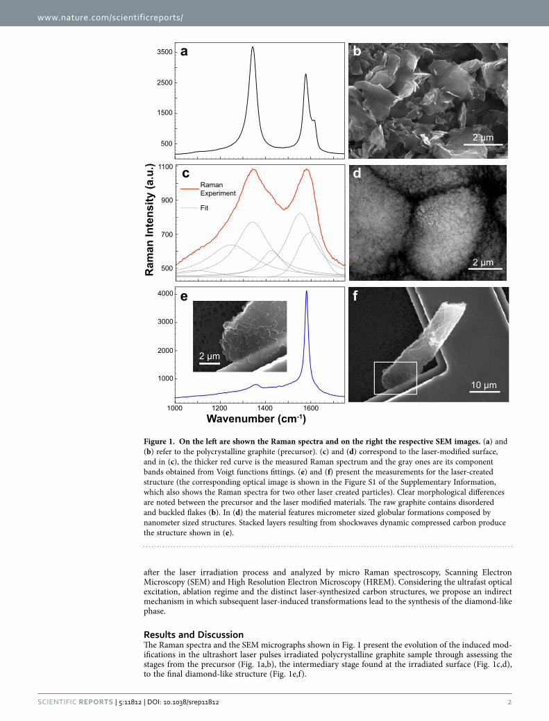
Results and DiscussionThe Raman spectra and the SEM micrographs shown in Fig. 1 present the evolution of the induced mod-ifications in the ultrashort laser pulses irradiated polycrystalline graphite sample through assessing the stages from the precursor (Fig. 1a,b), the intermediary stage found at the irradiated surface (Fig. 1c,d), to the final diamond-like structure (Fig. 1e,f).
1000
1000
500
1500
2500
3500
2000
3000
4000
1200 1400 1600
a
e
b
d
2 µm
2 µm
10 µm
f
Ram
an In
tens
ity (a
.u.)
Wavenumber (cm-1)
500
700
900
1100
RamanExperiment
Fit
c
2 µm
Figure 1. On the left are shown the Raman spectra and on the right the respective SEM images. (a) and (b) refer to the polycrystalline graphite (precursor). (c) and (d) correspond to the laser-modified surface, and in (c), the thicker red curve is the measured Raman spectrum and the gray ones are its component bands obtained from Voigt functions fittings. (e) and (f) present the measurements for the laser-created structure (the corresponding optical image is shown in the Figure S1 of the Supplementary Information, which also shows the Raman spectra for two other laser created particles). Clear morphological differences are noted between the precursor and the laser modified materials. The raw graphite contains disordered and buckled flakes (b). In (d) the material features micrometer sized globular formations composed by nanometer sized structures. Stacked layers resulting from shockwaves dynamic compressed carbon produce the structure shown in (e).
www.nature.com/scientificreports/
3Scientific RepoRts | 5:11812 | DOi: 10.1038/srep11812
In the Fig. 1a the pristine graphite exhibits its characteristic Raman modes at 1343 and 1615 cm−1 (related to structural defects bands D1 and D2, respectively), and at 1579 cm−1 originating from the stretching of the sp2-bounded carbon atoms forming the well-known hexagonal structure (G-band)24. After irradiation, notable changes were observed in the Raman spectrum of the laser modified surface, shown in Fig. 1c, with respect to the precursor (Fig. 1a). While the D1 band appears at 1340 cm−1, the displacement of the G band to 1595 cm−1 (Fig. 1c) confirms the laser-induced pressure, since similar shifts to higher wavenumbers were verified for HOPG upon room-temperature compression25. Additionally, Raman resonances, which do not manifest in the precursor (Fig. 1a), emerge at 1087, 1245, 1425 and 1552 cm−1. Analogous spectra have been reported for an experiment that quenched Carbon Black from high-pressure high-temperature environment26 (15 GPa and 1700 °C for 15 min) and also in shocked meteorites27. In the former work, the new vibrations were declared unexplained Raman modes, while in the latter they were attributed to a new carbon phase. Considering previous works26–28 and our experi-mental observations, we suggest that these vibrations are evidence of the creation of sp3 carbon phases by the ultrafast laser excitation, as theoretical models29 have predicted sp3 lattices with rather specific Raman resonances.
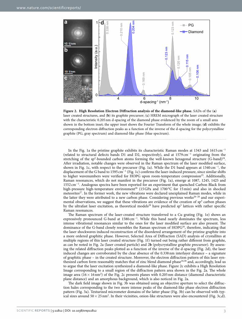
The Raman spectrum of the laser-created structure transferred to a Cu grating (Fig. 1e) shows an expressively pronounced G-band at 1580 cm−1. While this band nearly dominates the spectrum, less intense vibrational resonances similar to the ones for the laser modified surface are also present. The dominance of the G-band closely resembles the Raman spectrum of HOPG26, therefore, indicating that the laser shockwaves induced reconstruction of the disordered arrangement of the pristine graphite into a more ordered graphitic phase. However, Selected Area of Diffraction (SAD) analysis of crystallites at multiple regions of this laser created structure (Fig. 1f) turned out being rather different from graphite, as can be noted in Fig. 2a (laser created particle) and 2b (polycrystalline graphite precursor). By assess-ing the related diffraction peaks plotted as a function of the inverse of the d-spacing (Fig. 2d), the laser induced changes are corroborated by the clear absence of the 0.338 nm interlayer distance – a signature of graphitic phase – in the created structure. Moreover, the electron diffraction pattern of this laser syn-thesized carbon form reasonably matches that of zinc blend diamond phase10,30 and, accordingly, lead us to argue that the laser excitation synthesized a diamond-like phase. Figure 2c exhibits a High Resolution Image corresponding to a small region of the diffraction pattern area shown in the Fig. 2a. The whole image area (16 × 16 nm2) of the Fig. 2c presents planes with 0.205 nm distance (diamond characteristic plane distance) and an amorphous background, which is also noticed in Fig. 2a.
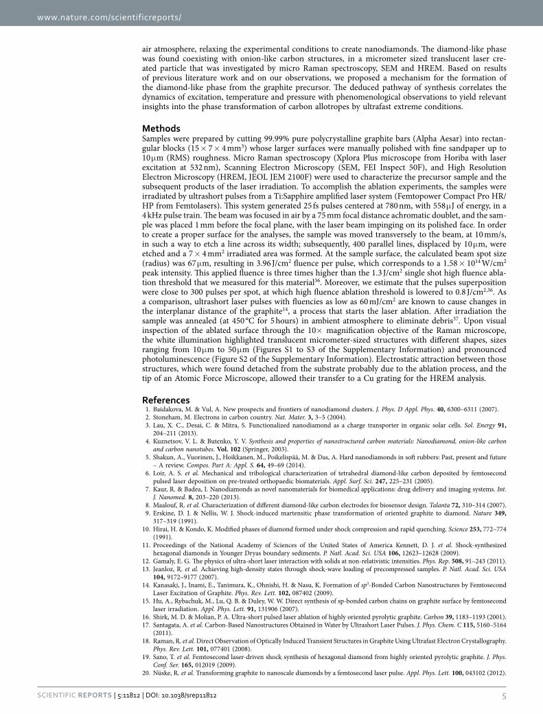
The dark field image shown in Fig. 3b was obtained using an objective aperture to select the diffrac-tion hales corresponding to the two more intense peaks of the diamond-like phase electron diffraction pattern (Fig. 3a). Texturized micrometer domains of the latter phase (Fig. 3b) can be observed with typ-ical sizes around 50 × 25 nm2. In their vicinities, onion-like structures were also encountered (Fig. 3c,d).
40
1
6 8 10In
tens
ity (a
.u.)
d-spacing-1 (nm-1)
0.10
6 nm
0.10
5 nm
0.12
0 nm
0.12
3 nm
0.20
4 nm
0.12
3 nm
0.11
5 nm
0.21
0 nm
0.33
8 nm
0.16
9 nm
Diamond
PGa d
b
c
Figure 2. High Resolution Electron Diffraction analysis of the diamond-like phase. SADs of the (a) laser created structures, and (b) its graphite precursor; (c) HREM micrograph of the laser created structure with the characteristic 0.205 nm d-spacing of the diamond phase evidenced by the zoom of a small area shown in the bottom inset; the upper inset shows the Fourier Transform of the whole image; (d) exhibits the corresponding electron diffraction peaks as a function of the inverse of the d-spacing for the polycrystalline graphite (PG, gray spectrum) and diamond-like phase (blue spectrum).

www.nature.com/scientificreports/
4Scientific RepoRts | 5:11812 | DOi: 10.1038/srep11812
Here we propose that the mechanism for the transformation from graphite to the diamond-like phase follows an indirect pathway that is underpinned on the morphology of the starting material, specific thermodynamical events dictated by the fluence of overlapping ultrashort laser pulses18 and the forma-tion of natural catalysts, as the onion-like structures31 and the laser driven nanometer sized graphite26. Initially, the high densities of free electrons accumulating at the boundaries of the precursor defective graphitic flakes favored the absorption of the ultrashort pulses energy steering to the creation of super-excited states, and ultimately to ablation32. As a consequence of the ultrafast excitation above the high fluence ablation threshold and the following explosive ablation, nonthermal shockwaves first propagate into the material after the electronic relaxation, and then heating and thermal equilibrium take place. This thermodynamical process, which happens at each laser shot, produces cumulative incremental lat-tice distortions14,33 leading to more ordered carbon forms34. Considering the exposure time with pulse superposition, these latter structures behave as transient states whose energetic barrier to phase transi-tion lowers while the crystallinity gradually increases at each shockwave shot. Therefore, the formation of the diamond-like phase is assisted by those intermediary carbon structures which assume the role of nucleation sites more prone to phase transitions35.
ConclusionIn summary, we have demonstrated, for the first time to our knowledge, the synthesis of a diamond-like phase from blasting polycrystalline graphite with spatially overlapping 25 fs ultrashort laser pulses under
a b
c d
Figure 3. Texturized domains of the diamond-like phase and onions-like carbon coexisting at the laser created particle. (a) SAD of the diamond-like phase from a small area on the region of the laser created structure displayed in (b). The highlighted red spot in the frame (a) represent the selected diffraction peaks for construction the patterned domains shown in (b). Onion-like phases also found at the laser created structure are presented in the frames (c) and (d).
www.nature.com/scientificreports/
5Scientific RepoRts | 5:11812 | DOi: 10.1038/srep11812
air atmosphere, relaxing the experimental conditions to create nanodiamonds. The diamond-like phase was found coexisting with onion-like carbon structures, in a micrometer sized translucent laser cre-ated particle that was investigated by micro Raman spectroscopy, SEM and HREM. Based on results of previous literature work and on our observations, we proposed a mechanism for the formation of the diamond-like phase from the graphite precursor. The deduced pathway of synthesis correlates the dynamics of excitation, temperature and pressure with phenomenological observations to yield relevant insights into the phase transformation of carbon allotropes by ultrafast extreme conditions.
MethodsSamples were prepared by cutting 99.99% pure polycrystalline graphite bars (Alpha Aesar) into rectan-gular blocks (15 × 7 × 4 mm3) whose larger surfaces were manually polished with fine sandpaper up to 10 μ m (RMS) roughness. Micro Raman spectroscopy (Xplora Plus microscope from Horiba with laser excitation at 532 nm), Scanning Electron Microscopy (SEM, FEI Inspect 50F), and High Resolution Electron Microscopy (HREM, JEOL JEM 2100F) were used to characterize the precursor sample and the subsequent products of the laser irradiation. To accomplish the ablation experiments, the samples were irradiated by ultrashort pulses from a Ti:Sapphire amplified laser system (Femtopower Compact Pro HR/HP from Femtolasers). This system generated 25 fs pulses centered at 780 nm, with 558 μ J of energy, in a 4 kHz pulse train. The beam was focused in air by a 75 mm focal distance achromatic doublet, and the sam-ple was placed 1 mm before the focal plane, with the laser beam impinging on its polished face. In order to create a proper surface for the analyses, the sample was moved transversely to the beam, at 10 mm/s, in such a way to etch a line across its width; subsequently, 400 parallel lines, displaced by 10 μ m, were etched and a 7 × 4 mm2 irradiated area was formed. At the sample surface, the calculated beam spot size (radius) was 67 μ m, resulting in 3.96 J/cm2 fluence per pulse, which corresponds to a 1.58 × 10¹⁴ W/cm2 peak intensity. This applied fluence is three times higher than the 1.3 J/cm2 single shot high fluence abla-tion threshold that we measured for this material36. Moreover, we estimate that the pulses superposition were close to 300 pulses per spot, at which high fluence ablation threshold is lowered to 0.8 J/cm2,36. As a comparison, ultrashort laser pulses with fluencies as low as 60 mJ/cm2 are known to cause changes in the interplanar distance of the graphite14, a process that starts the laser ablation. After irradiation the sample was annealed (at 450 °C for 5 hours) in ambient atmosphere to eliminate debris37. Upon visual inspection of the ablated surface through the 10× magnification objective of the Raman microscope, the white illumination highlighted translucent micrometer-sized structures with different shapes, sizes ranging from 10 μ m to 50 μ m (Figures S1 to S3 of the Supplementary Information) and pronounced photoluminescence (Figure S2 of the Supplementary Information). Electrostatic attraction between those structures, which were found detached from the substrate probably due to the ablation process, and the tip of an Atomic Force Microscope, allowed their transfer to a Cu grating for the HREM analysis.
References1. Baidakova, M. & Vul, A. New prospects and frontiers of nanodiamond clusters. J. Phys. D Appl. Phys. 40, 6300–6311 (2007).2. Stoneham, M. Electrons in carbon country. Nat. Mater. 3, 3–5 (2004).3. Lau, X. C., Desai, C. & Mitra, S. Functionalized nanodiamond as a charge transporter in organic solar cells. Sol. Energy 91,
204–211 (2013).4. Kuznetsov, V. L. & Butenko, Y. V. Synthesis and properties of nanostructured carbon materials: Nanodiamond, onion-like carbon
and carbon nanotubes. Vol. 102 (Springer, 2003).5. Shakun, A., Vuorinen, J., Hoikkanen, M., Poikelispää, M. & Das, A. Hard nanodiamonds in soft rubbers: Past, present and future
– A review. Compos. Part A: Appl. S. 64, 49–69 (2014).6. Loir, A. S. et al. Mechanical and tribological characterization of tetrahedral diamond-like carbon deposited by femtosecond
pulsed laser deposition on pre-treated orthopaedic biomaterials. Appl. Surf. Sci. 247, 225–231 (2005).7. Kaur, R. & Badea, I. Nanodiamonds as novel nanomaterials for biomedical applications: drug delivery and imaging systems. Int.
J. Nanomed. 8, 203–220 (2013).8. Maalouf, R. et al. Characterization of different diamond-like carbon electrodes for biosensor design. Talanta 72, 310–314 (2007).9. Erskine, D. J. & Nellis, W. J. Shock-induced martensitic phase transformation of oriented graphite to diamond. Nature 349,
317–319 (1991).10. Hirai, H. & Kondo, K. Modified phases of diamond formed under shock compression and rapid quenching. Science 253, 772–774
(1991).11. Proceedings of the National Academy of Sciences of the United States of America Kennett, D. J. et al. Shock-synthesized
hexagonal diamonds in Younger Dryas boundary sediments. P. Natl. Acad. Sci. USA 106, 12623–12628 (2009).12. Gamaly, E. G. The physics of ultra-short laser interaction with solids at non-relativistic intensities. Phys. Rep. 508, 91–243 (2011).13. Jeanloz, R. et al. Achieving high-density states through shock-wave loading of precompressed samples. P. Natl. Acad. Sci. USA
104, 9172–9177 (2007).14. Kanasaki, J., Inami, E., Tanimura, K., Ohnishi, H. & Nasu, K. Formation of sp3-Bonded Carbon Nanostructures by Femtosecond
Laser Excitation of Graphite. Phys. Rev. Lett. 102, 087402 (2009).15. Hu, A., Rybachuk, M., Lu, Q. B. & Duley, W. W. Direct synthesis of sp-bonded carbon chains on graphite surface by femtosecond
laser irradiation. Appl. Phys. Lett. 91, 131906 (2007).16. Shirk, M. D. & Molian, P. A. Ultra-short pulsed laser ablation of highly oriented pyrolytic graphite. Carbon 39, 1183–1193 (2001).17. Santagata, A. et al. Carbon-Based Nanostructures Obtained in Water by Ultrashort Laser Pulses. J. Phys. Chem. C 115, 5160–5164
(2011).18. Raman, R. et al. Direct Observation of Optically Induced Transient Structures in Graphite Using Ultrafast Electron Crystallography.
Phys. Rev. Lett. 101, 077401 (2008).19. Sano, T. et al. Femtosecond laser-driven shock synthesis of hexagonal diamond from highly oriented pyrolytic graphite. J. Phys.
Conf. Ser. 165, 012019 (2009).20. Nuske, R. et al. Transforming graphite to nanoscale diamonds by a femtosecond laser pulse. Appl. Phys. Lett. 100, 043102 (2012).
www.nature.com/scientificreports/
6Scientific RepoRts | 5:11812 | DOi: 10.1038/srep11812
21. Mundy, C. J. et al. Ultrafast transformation of graphite to diamond: an ab initio study of graphite under shock compression. J. Chem. Phys. 128, 184701 (2008).
22. Umemoto, K., Wentzcovitch, R. M., Saito, S. & Miyake, T. Body-Centered Tetragonal C4: A Viable sp3 Carbon Allotrope. Phys. Rev. Lett. 104, 125504 (2010).
23. Niu, H. et al. Families of Superhard Crystalline Carbon Allotropes Constructed via Cold Compression of Graphite and Nanotubes. Phys. Rev. Lett. 108, 135501 (2012).
24. Ferrari, A. C. & Robertson, J. Raman spectroscopy of amorphous, nanostructured, diamond-like carbon, and nanodiamond. Philos. T. Roy. Soc. A 362, 2477–2512 (2004).
25. Wang, Y., Panzik, J. E., Kiefer, B. & Lee, K. K. Crystal structure of graphite under room-temperature compression and decompression. Sci. Rep. 2, 520 (2012).
26. Guillou, C. L., Brunet, F., Irifune, T., Ohfuji, H. & Rouzaud, J.-N. Nanodiamond nucleation below 2273 K at 15 GPa from carbons with different structural organizations. Carbon 45, 636–648 (2007).
27. Ferroir, T. et al. Carbon polymorphism in shocked meteorites: Evidence for new natural ultrahard phases. Earth Planet. Sc. Lett. 290, 150–154 (2010).
28. Amsler, M. et al. Crystal Structure of Cold Compressed Graphite. Phys. Rev. Lett. 108, 065501 (2012).29. Bai, Y. et al. First-principles investigation in the Raman and infrared spectra of sp3 carbon allotropes. Carbon 78, 70–78 (2014).30. Kumar, A. et al. Formation of nanodiamonds at near-ambient conditions via microplasma dissociation of ethanol vapour. Nat.
Commun. 4, 2618 (2013).31. Álvarez-Murga, M. et al. “Compressed Graphite” Formed During C60 to Diamond Transformation as Revealed by Scattering
Computed Tomography. Phys. Rev. Lett. 109, 025502 (2012).32. Gamaly, E. G., Rode, A. V., Luther-Davies, B. & Tikhonchuk, V. T. Ablation of solids by femtosecond lasers: Ablation mechanism
and ablation thresholds for metals and dielectrics. Phys. Plasmas 9, 949–957 (2002).33. Jeschke, H. O., Garcia, M. E. & Bennemann, K. H. Theory for the Ultrafast Ablation of Graphite Films. Phys. Rev. Lett. 87, 015003
(2001).34. Xiao, J., Ouyang, G., Liu, P., Wang, C. X. & Yang, G. W. Reversible nanodiamond-carbon onion phase transformations. Nano
Lett. 14, 3645–3652 (2014).35. Khaliullin, R. Z., Eshet, H., Kuhne, T. D., Behler, J. & Parrinello, M. Nucleation mechanism for the direct graphite-to-diamond
phase transition. Nat. Mater. 10, 693–697 (2011).36. Samad, R. E., Maia, F. C., Souza, N. M., de Rossi, W. & Vieira, N. D. Determination of the Graphite Incubation Parameter in the
Ultrafast Regime using the D-Scan Technique. Paper presented at Frontiers in Optics 2014: Light-Matter Interaction, Tucson, Arizona. doi: 10.1364/LS.2014.LTh4I.6 (2014, October).
37. Osswald, S., Yushin, G., Mochalin, V., Kucheyev, S. O. & Gogotsi, Y. Control of sp2/sp3 carbon ratio and surface chemistry of nanodiamond powders by selective oxidation in air. J. Am. Chem. Soc. 128, 11635–11642 (2006).
AcknowledgmentsWork at LNLS and LNNano are supported by the Brazilian Ministry of Science and Technology. We thank the Brazilian funding agencies CNPq, CAPES and FAPESP for their financial support. We thank the multi-user laboratory (LAMULT) at University of Campinas for the micro Raman spectroscopy measurements.
Author ContributionsR.E.S. and N.D.V. Jr. made the ablation experiments. F.C.B.M. performed Raman measurements. R.O.F. steered the experiments with the Atomic Force Microscope. J.B. accomplished electronic micrographs and diffraction experiments. N.M.S.-N. proposed the experiments and throughout coordinated the work. All authors took part in the discussion and effectively contributed to the conclusions.
Additional InformationSupplementary information accompanies this paper at http://www.nature.com/srepCompeting financial interests: The authors declare no competing financial interests.How to cite this article: Maia, F. C. B. et al. Synthesis of diamond-like phase from graphite by ultrafast laser driven dynamical compression. Sci. Rep. 5, 11812; doi: 10.1038/srep11812 (2015).
This work is licensed under a Creative Commons Attribution 4.0 International License. The images or other third party material in this article are included in the article’s Creative Com-
mons license, unless indicated otherwise in the credit line; if the material is not included under the Creative Commons license, users will need to obtain permission from the license holder to reproduce the material. To view a copy of this license, visit http://creativecommons.org/licenses/by/4.0/