synthesis of biocompatible gelatin-functionalised graphene nanosheets for drug delivery applications
TRANSCRIPT
Synthesis of Biocompatible Gelatin-functionalisedGraphene Nanosheets For Drug Delivery Applications
Guangxin Chen,A Congde Qiao,A,B Yang Wang,A and Jinshui YaoA
ASchool of Materials Science and Engineering, Qilu University of Technology,
Jinan 250353, ChinaBCorresponding author. Email: [email protected]
Gelatin-functionalised graphene nanosheets (gelatin-GNS) with good biocompatibility were successfully synthesisedusing gelatin as a reductant. Factors that affect the reduction of graphene oxide (GO), such as the ratio of gelatin to GO, pH,and temperature, were investigated to establish optimum reaction conditions.We found thatGOwas efficiently reduced by
gelatin at a comparatively low temperature and a stable gelatin-GNS aqueous dispersion was formed. The as-obtainedbiocompatible gelatin-GNS displayed a high methotrexate (MTX) drug loading capacity and a good ability for controlleddrug release. The pH-dependent release behaviour of MTX from MTX@gelatin-GNS showed that the release amount
under acid conditions was much higher than that under neutral conditions, indicating a gelatin-mediated sustained releaseprocess.
Manuscript received: 10 December 2013.
Manuscript accepted: 1 March 2014.Published online: 1 May 2014.
Introduction
One-atom-thick planar sheet of sp2 hybridised carbon atoms
densely packed in a honeycomb crystal lattice, i.e. graphenenanosheets (GNS), were first reported in 2004, and consideredto be the basic structure unit of fullerenes, carbon nanotubes
(CNT), and graphite.[1] GNS havemany novel properties such asunique mechanical properties,[2,3] thermal properties,[4–6]
excellent electrical properties,[7–11] optical properties,[12–15] and
high surface area.[16] These unique features offer great potentialfor many applications in the fields of optoelectronic materialsand devices,[17–20] electrochemical energy storage,[21–23]
nanocomposite materials,[24] and sensors.[25–27] At present,
much attention has been paid to the preparation of graphenenanosheets. It is known that most unique properties of GNS aredue to its single sheet nature. However, GNS are hydrophobic
and tend to aggregate or restack into graphite owing to the Vander Waals interaction and strong p–p stacking. Thus, hindranceof aggregation is critical for the preparation of GNS. Recently,
some biomacromolecules, such as chitosan,[28] glucose,[29]
bovine serum albumin (BSA),[30] dopamine,[31] and protein,[32]
were applied for the preparation of solubleGNSwith advantages
of non-toxicity and environmental friendly properties.Gelatin is a traditional water-soluble biopolymer with dis-
tinctive advantages of non-toxicity, biodegradability, and lowcost, and is commonly used for pharmaceutical and medical
applications. It is a product of partial hydrolysis of collagen. Thegelatin molecular chain is a linear polypeptide that consists ofdifferent amino acids residues. It has been employed as a
reducing reagent for the preparation of nanomaterials becauseof its mild reductive ability. Furthermore, a stable dispersion ofgraphene can be obtained because of hydrophobic interactions
between the non-polar zone of the gelatin chains and graphene.
Therefore, gelatin is a promising biopolymer for the preparationof biocompatible graphene, and it has been used as a reducing
reagent for the synthesis of graphene nanosheets.[33] However,the factors which influence the reduction of graphene oxide(GO), such as the ratio of gelatin to GO, pH, and temperature,
have not been investigated in detail to allow optimisation of thereaction conditions.
At present, nano-scaled drug carriers have emerged as a
bridge linking nanotechnology and advanced drug delivery. Themedicine can be loaded on these nano-scaled materials based ondifferent mechanisms such as embedding, surface absorption,hydrogen bonding, and other types of interactions.[34–37] As a
two-dimensional plane structural material, graphene can pro-vide larger specific surface area than other commonly usedmaterials and forms strong p–p conjugation with drug mole-
cules, therefore is a good candidate for drug loading. Yanget al.[38] investigated the loading and release behaviours ofdoxorubicin hydrochloride (DXR) on GO, and they found that
the weight ratio of the loaded drug to the GO carrier could reach200%. Liu et al.[33] successfully prepared gelatin-GNS using aneasy method, and found that it could be used as an ideal drug
carrier for R6G. However, to date, reports on the application ofgraphene for drug delivery are relatively rare because of its poorsolubility and stability in physiological environments. On thebasis of these observations, a new approach is essential to
develop novel biocompatible graphene with high solubilityand stability for application in drug delivery.
In this paper, gelatin was used to functionalise GO to obtain a
stable gelatin-GNS suspension and optimum reaction conditionswere established by exploring the effects of the ratio of gelatin/GO, pH, and temperature on the reduction of GO. The structure
of gelatin-GNS was characterised by UV–vis spectroscopy,
CSIRO PUBLISHING
Aust. J. Chem. 2014, 67, 1532–1537
http://dx.doi.org/10.1071/CH13678
Journal compilation � CSIRO 2014 www.publish.csiro.au/journals/ajc
Full Paper
X-ray diffraction (XRD), and atomic force microscopy (AFM).
Moreover, the thermal behaviour of the gelatin-GNS wasstudied by thermal gravimetric analysis (TGA), and the rela-tionship between structure and thermal property of gelatin-GNS
was also examined. Because gelatin is a natural, nontoxic, andbiodegradable macromolecule, the present study facilitates thelarge-scale production of reduced graphene oxide, thus broad-ening the application of graphene. In addition, gelatin-GNS was
employed as a carrier to load and release methotrexate (MTX).The results showed that the gelatin-GNS with good stability andbiocompatibility could be selected as an ideal drug carrier for
application in the biomedical field.
Experimental
Materials
Graphite powder, concentrated sulfuric acid, KMnO4, K2S2O8
(99%), NaNO3, P2O5 (99%), NaOH, HCl, H2O2 (30%), andDMSOwere purchased from SinopharmChemical Reagent Co.,Ltd. Gelatin (from bovine skin, average molecular weight
80000) was purchased from Sigma Co., Ltd. Fetal bovine serum(FBS) was purchased from Nanjing Key-Gen Biotech Co., Ltd(Nanjing, China). MTX was purchased from Shanghai ShinePharmaceutical Laboratories Co., Ltd. All aqueous solutions
were prepared using ultrapure water (18MO cm) from aMilli-Qsystem (Millipore). All reagents were used as received.
Preparation of Gelatin-GNS Aqueous Suspension
GO was prepared from purified natural graphite by a modifiedHummersmethod.[39,40] Briefly, concentratedH2SO4was added
into a 500-mL flask filled with graphite, followed by the addi-tion of NaNO3, then solid KMnO4 under stirring while thetemperature of the mixture was kept below 208C. Then, thetemperature was adjusted to 308C. Excess distilled water wasadded to the mixture and the temperature was increased to 808C.Then, 30% H2O2 was added until the colour of the mixture
turned to brilliant yellow. The mixture was first filtered andwashed several times with 8% aqueous HCl to remove metalions and then washed with distilled water to remove the acid.Finally, the filter cake was dried in air, then re-dispersed in
water. The suspended GO sheets were obtained after ultrasonictreatment. Finally, the GO suspension was reduced by gelatinsolution for 24 h at 908C to obtain a gelatin-GNS suspension.
Loading and Release of MTX
For loading of MTX on gelatin-GNS, a certain amount of
gelatin-GNS solution (0.5mgmL�1) was respectively mixedwith MTX solutions with different concentrations (0.025, 0.03,0.04, and 0.05mgmL�1; DMSO acted as co-solvent). The
resulting mixtures were stirred for 24 h at room temperature.After centrifugation at 25782g for 15min, the supernatantcomposed of unbound MTX was collected. The amount of
unbound MTX was determined by measuring the UV absor-bance at 304 nm (characteristic absorbance of MTX), andcompared to a calibration curve recorded under identicalconditions, thus allowing the drug loading efficiency to be
determined. Standard MTX solutions (0.003, 0.004 0.005,0.006, 0.007, and 0.008mgmL�1) were used to construct thecalibration curve for quantitative analysis (see Supplementary
Material). The fluorescence spectra of 0.05mgmL�1 MTX,MTX-loaded gelatin-GNS (MTX@gelatin-GNS), and gelatin-GNS were measured in the wavelength range of 320 to 580 nm
using an excitation wavelength of 304 nm. The release of
MTX from the synthesised gelatin-GNS nanocarrier was
investigated by mixing the MTX@gelatin-GNS complex, withsaturated adsorption of MTX, with phosphate buffer saline(PBS) at the physiological temperature of 378C and pH of 7.4
(physiological pH), pH of 5.4 (endosomal pH of cancer cells),and pH of 2. After shaking for different times (1, 12, and 24 h),the mixture was centrifuged at 25782g for 15min and thesupernatant was collected to determine the amount of released
MTX by measuring the UV absorbance of MTX at 304 nmfollowing the protocol described above.
Characterisation
TheUV–vis spectrophotometer (UV-2550, Shimadzu) was usedto investigate the optical absorption of the GO and gelatin-GNSsuspensions in the wavelength range of 190–800 nm. XRD
analysis was conducted with a BDX3300 X-ray diffractometerequipped with a multichannel detector, and using Cu Ka1
(l¼ 0.15406 nm) monochromatic X-ray beam. All the samples
were measured within a 2y range of 58–608 at a scan rate of18min�1. AFM images of GO and gelatin-GNS were taken in atapping mode using a NanoScope III A (Digital Instrument,USA). Image processing and data analysis were performed
using NanoScope Analysis version 1.40 provided with theinstrument. The coatings for the AFM imaging were preparedby drop-casting a diluted suspension onto a cleaned Si substrate.
TGA was performed on a SDTQ600 (TA Instrument). Thesamples were heated from room temperature to 7008C at aheating rate of 108Cmin�1 under a nitrogen atmosphere. Fluo-
rescence spectra were obtained from a RF-5301PC spectroflu-orometer (Shimadzu).
Results and Discussion
Reduction of GO
Gelatin was not only used as a functionalisation reagent toprevent aggregation, but also as a mild reductant for GO.
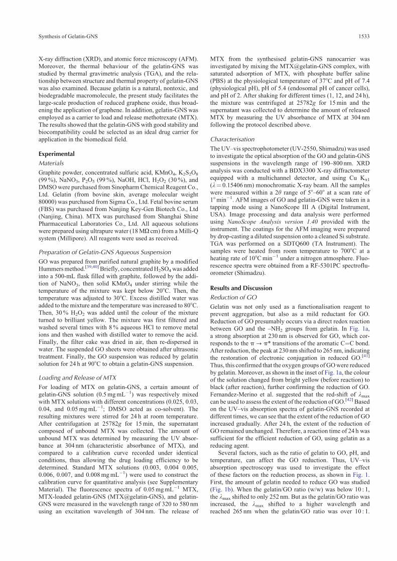
Reduction of GO presumably occurs via a direct redox reactionbetween GO and the –NH2 groups from gelatin. In Fig. 1a,a strong absorption at 230 nm is observed for GO, which cor-
responds to the p-p* transitions of the aromatic C¼C bond.After reduction, the peak at 230 nm shifted to 265 nm, indicatingthe restoration of electronic conjugation in reduced GO.[41]
Thus, this confirmed that the oxygen groups ofGOwere reducedby gelatin. Moreover, as shown in the inset of Fig. 1a, the colourof the solution changed from bright yellow (before reaction) toblack (after reaction), further confirming the reduction of GO.
Fernandez-Merino et al. suggested that the red-shift of lmax
can be used to assess the extent of the reduction of GO.[42] Basedon the UV–vis absorption spectra of gelatin-GNS recorded at
different times, we can see that the extent of the reduction of GOincreased gradually. After 24 h, the extent of the reduction ofGO remained unchanged. Therefore, a reaction time of 24 h was
sufficient for the efficient reduction of GO, using gelatin as areducing agent.
Several factors, such as the ratio of gelatin to GO, pH, andtemperature, can affect the GO reduction. Thus, UV–vis
absorption spectroscopy was used to investigate the effectof these factors on the reduction process, as shown in Fig. 1.First, the amount of gelatin needed to reduce GO was studied
(Fig. 1b). When the gelatin/GO ratio (w/w) was below 10 : 1,the lmax shifted to only 252 nm. But as the gelatin/GO ratio wasincreased, the lmax shifted to a higher wavelength and
reached 265 nm when the gelatin/GO ratio was over 10 : 1.
Synthesis of Gelatin-GNS 1533
This wavelength is often used to assess the extent of completionof the chemical reduction. Thus, the ratio of gelatin/GO gelatinrequired for the reduction of GO is 10 : 1.
Next, GO reduction at different pH values was investigatedand shown in Fig. 1c. When equal volumes of GO and gelatinwere mixed, the pH of the mixture was 5, corresponding to anacid environment. Although the GO was thoroughly rinsed, it
remained acidic. The pH of the solution was controlled byadding HCl or NaOH to the mixture. The lmax values were247, 252, and 265 nm when the GO was reduced at pH 3, 4,
and 5, respectively.When the pH of solution was very low, i.e. 1and 2, the lmax values were 234 and 237 nm, respectively. Thedecrease in the reduction ability of gelatin may be due to the
formation of NH3þ ions, as the pH value at isoelectric point of
gelatin is,5. The lmax also decreased when the pH was over 5,reaching a value of 247 nm in weakly basic conditions (pH¼ 8).
Therefore, the pH was fixed at 5 in this work.Fig. 1d shows the effect of temperature on the GO reduction.
After 24 h, the lmax was 265 nm at 908C, and lmax values were258 and 249 nm when the reaction temperatures were 80 and
708C, respectively. When the reduction proceeded at 60 and508C, the lmax values were 238 and 236 nm, respectively. Thisindicated that GO cannot be effectively reduced by gelatin at
lower temperatures. As a result, a temperature of 908C was usedto achieve optimum reduction in this experiment.
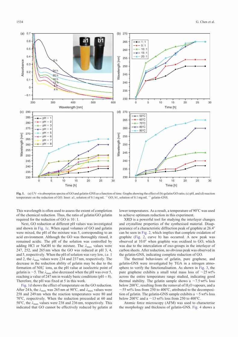
XRD is a powerful tool for studying the interlayer changes
and crystalline properties of the synthesised material. Disap-pearance of a characteristic diffraction peak of graphite at 26.48can be seen in Fig. 2, which implies that complete oxidation ofgraphite (Fig. 2, curve b) has occurred. A new peak was
observed at 10.08 when graphite was oxidised to GO, whichwas due to the intercalation of oxo-groups in the interlayer ofcarbon sheets. After reduction, no obvious peak was observed in
the gelatin-GNS, indicating complete reduction of GO.The thermal behaviours of gelatin, pure graphene, and
gelatin-GNS were investigated by TGA in a nitrogen atmo-
sphere to verify the functionalisation. As shown in Fig. 3, thepure graphene exhibits a small total mass loss of ,25wt%across the entire temperature range studied, indicating good
thermal stability. The gelatin sample shows a ,7.5wt% lossbelow 2008C, resulting from the removal of H2O vapours, and a,55wt% loss from 250 to 4008C, attributed to the decomposi-tion of gelatin. The gelatin-GNS sample exhibits a,5wt% loss
below 2008C and a ,13wt% loss from 250 to 4008C.Atomic force microscopy (AFM) was used to characterise
the morphology and thickness of gelatin-GNS. Fig. 4 shows a
�0.1
200 300
4 h8 h12 h16 h20 h
24 h
0 h
pH � 150�C60�C70�C80�C90�C
pH � 2pH � 3pH � 4pH � 5pH � 6pH � 7pH � 8
1 : 15 : 110 : 115 : 120 : 1
400
Wavelength [nm]
Wav
elen
gth
[nm
]
500 600 0230
235
240
245
250
255
260
265
270
5 10
Time [h]
Time [h] Time [h]
15 20 25 30
0 5 10 15 20 25 300 5 10 15 20 25 30
0
0.1
0.2
0.3
0.4
0.5
Abs
orba
nce
0.6
0.7(a) (b)
Wav
elen
gth
[nm
]
230
235
240
245
250
255
260
265
270(d)
Wav
elen
gth
[nm
]
230
235
240
245
250
255
260
265
270
275
280
285
290(c)
Fig. 1. (a) UV–vis absorption spectra of GO and gelatin-GNS as a function of time. Graphs showing the effect of (b) gelatin/GO ratio, (c) pH, and (d) reaction
temperature on the reduction of GO. Inset: a1, solution of 0.1mgmL�1 GO; b1, solution of 0.1mgmL�1 gelatin-GNS.
1534 G. Chen et al.

representative AFM image and cross-section analysis alongwith the line in the AFM image of gelatin-GNS. Generally,
the GO nanosheet has a thickness of ,0.8 nm, which is largerthan the theoretical value of graphene sheet, owing to theunstripped GO and presence of covalently bound oxo-
groups.[43] Moreover, after the reduction, the thickness of theobtained gelatin-GNS increased to ,5.0–10.0 nm. Althoughmost oxo-groups were removed after reduction, the thickergelatin-GNS could be attributed to the attachment of gelatin.
Furthermore, as could be observed, the functionalisation ofgelatin could be stripped and prevented GNS agglomerationbecause most of the negatively surface charged gelatin-GNS
was kept independent from each other in the dispersion. There-fore, gelatin not only acted as a reducing reagent, but also playedan important role as a stabilising reagent for the stabilisation of
the gelatin-GNS.
Loading and Release of MTX
It is known that many anticancer drugs are aromatic and
hydrophobic that leads to poor water solubility, and this dis-advantage generally hinders their clinical applications. In this
work, gelatin-GNS was used as a nanocarrier for MTX (an
anticancer drug that is commonly used to treat tumours). Theone-atom thickness and two-dimensional plane of GNS afford alarge specific surface area which is more suitable for drug
carriers because both sides of a single sheet are available fordrug binding. The highly p-conjugated structure of GNS canform p–p stacking interactions with the aromatic part of MTX,
as well as the hydrophobic effect between them. In addition, thehydrogen bonding interactions between MTX and gelatin canincrease the binding strength of MTX with the gelatin-GNS.
Fig. 5 shows the loading process of MTX on gelatin-GNS.Before the reaction, the lmax was 304 nm; the lmax valueschanged to 301 nm, 295.5, and 287 nm at different reactiontimes. This is due to the p–p stacking interactions between
gelatin-GNS and MTX, also confirming the successful loadingof MTX. Compared with the UV–vis absorption spectra ofgelatin-GNS, theMTX@gelatin-GNS has no significant absorp-
tion peak at 265 nm, resulting from the overlap of absorptionpeaks of gelatin-GNS and MTX.
The loading capacity of MTX on gelatin-GNSwas measured
byUV spectroscopy andwas determined based on the differencein MTX concentrations between the original MTX solution andthe supernatant solution after loading. The loading of MTX on
gelatin-GNS was studied in different initial MTX concentra-tions with respect to the same concentration of gelatin-GNS(0.5mgmL�1). The saturated loading capacity was 31 mg ofMTX per 100mg of gelatin-GNS. The high loading capacity of
0100 200 300
Temperature [�C]400 500 600 700
c
b
a
20
40
60
Wei
ght [
%]
80
100
Fig. 3. TGA curves of: a, pure graphene; b, gelatin-GNS; and c, gelatin.
22.9 nm
0 1 : Height 10.0 µm
�11.9 nm
[µm]7654321[nm]
�5
0
5
10
15
Fig. 4. Representative AFM image and cross-section analysis of
gelatin-GNS.
0
5000
a
bc
10 15 202θ [degree]
25 30
b
c
10 20 30
2θ [degree]40 50 60
10000
15000
20000
25000
Inte
nsity
30000
35000
40000
45000
Fig. 2. XRD patterns of: a, graphite; b, GO; and c, gelatin-GNS.
Synthesis of Gelatin-GNS 1535
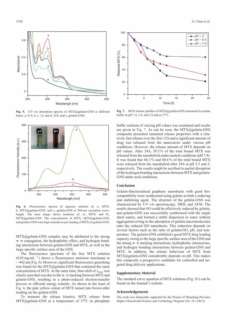
MTX@gelatin-GNS complex may be attributed to the strongp–p conjugation, the hydrophobic effect, and hydrogen bond-ing interactions between gelatin-GNS and MTX, as well as the
large specific surface area of the GNS.The fluorescence spectrum of the free MTX solution
(0.05mgmL�1) shows a fluorescence emission maximum at
,442 nm (Fig. 6). However, significant fluorescence quenchingwas found for the MTX@gelatin-GNS that contained the sameconcentration of MTX. At the same time, blue-shift of lmax was
clearly seen that was due to thep–p stacking betweenMTX andgelatin-GNS, resulting in a photo-induced electron-transferprocess or efficient energy transfer. As shown in the inset of
Fig. 6, the pale yellow colour of MTX turned into brown afterloading on the gelatin-GNS.
To measure the release kinetics, MTX release fromMTX@gelatin-GNS at a temperature of 378C in phosphate
buffer solution of varying pH values was examined and results
are given in Fig. 7. As can be seen, the MTX@gelatin-GNScomposite presented sustained release properties with a rela-tively fast release over the first 12 h and a significant amount of
drug was released from the nanocarrier under various pHconditions. However, the release amount of MTX depends onpH values. After 24 h, 39.5% of the total bound MTX wasreleased from the nanohybrid under neutral conditions (pH 7.4).
It was found that 68.3% and 88.6% of the total bound MTXwere released from the nanohybrid after 24 h at pH 5.3 and 2,respectively. The results might be ascribed to partial disruption
of the hydrogen bonding interactions betweenMTX and gelatin-GNS under acid conditions.
Conclusion
Gelatin-functionalised graphene nanosheets with good bio-
compatibility were synthesised using gelatin as both a reducingand stabilising agent. The structure of the gelatin-GNS wascharacterised by UV–vis spectroscopy, XRD, and AFM. Theresults showed that GO could be effectively reduced by gelatin,
and gelatin-GNS was successfully synthesised with the singlesheet nature, and formed a stable dispersion in water withoutaggregation owing to the adsorption of gelatin macromolecules
onto the reduced GO nanosheets. This reduction depends onseveral factors such as the ratio of gelatin/GO, pH, and tem-perature. The gelatin-GNS exhibited a good MTX drug loading
capacity owing to the large specific surface area of the GNS andthe strong p–p stacking interactions, hydrophobic interactions,and hydrogen bonding interactions between gelatin-GNS andMTX. In addition, the release behaviour of MTX from
MTX@gelatin-GNS considerably depends on pH. This makesthis composite a prospective candidate for controlled and tar-geted drug delivery applications.
Supplementary Material
The standard curve equation of MTX solutions (Fig. S1) can befound on the Journal’s website.
Acknowledgements
The work was financially supported by the Project of Shandong Province
Higher Educational Science and Technology Program (No. J11 LB13).
400
300
200
100
0
b
a
c
Wavelength [nm]
Flu
ores
cenc
e in
tens
ity [a
.u.]
350 400 450 500 550
Fig. 6. Fluorescence spectra of aqueous solution of: a, MTX;
b, MTX@gelatin-GNS; and c, gelatin-GNS at 304-nm excitation wave-
length. The inset image shows solutions of: a1, MTX; and b1,
MTX@gelatin-GNS. The concentration of MTX, MTX@gelatin-GNS,
and gelatin-GNSwere kept constant as per loading ofMTX on gelatin-GNS.
100
80
60
40
20
00 5 10 15 20 25
Ret
aine
d M
TX
[%]
Time [h]
pH � 2pH � 5.4pH � 7.4
Fig. 7. MTX release profiles of MTX@gelatin-GNS measured in acetate
buffer at pH 7.4, 5.4, and 2.0 and at 378C.
0
200 300 400
Wavelengh [nm]
500 600
0.2
0.4
Abs
orba
nce
0.6
a
b
c
d
e
Fig. 5. UV–vis absorption spectra of MTX@gelatin-GNS at different
times: a, 0; b, 6; c, 12; and d, 24 h; and e, gelatin-GNS.
1536 G. Chen et al.

References
[1] K. S. Novoselov, A. K. Geim, S. V. Morozov, D. Jiang, Y. Zhang,
S. V. Dubonos, I. V. Grigorieva, A. A. Firsov, Science 2004, 306, 666.doi:10.1126/SCIENCE.1102896
[2] F. Schedin, A. K. Geim, S. V. Morozov, E. W. Hill, P. Blake,
M. I. Katsnelson, K. S. Novoselov, Nat. Mater. 2007, 6, 652.doi:10.1038/NMAT1967
[3] C. Lee, X. D. Wei, J. W. Kysar, J. Hone, Science 2008, 321, 385.doi:10.1126/SCIENCE.1157996
[4] J. C. Meyer, A. K. Geim, M. I. Katsnelson, K. S. Novoselov, T. J.
Booth, S. Roth, Nature 2007, 446, 60. doi:10.1038/NATURE05545[5] R. Mas-Balleste, C. Gomez-Navarro, J. Gomez-Herrero, F. Zamora,
Nanoscale 2011, 3, 20. doi:10.1039/C0NR00323A[6] R. Z. Ma, T. Sasaki, Adv. Mater. 2010, 22, 5082. doi:10.1002/ADMA.
201001722[7] K. S. Novoselov, A. K. Geim, S. V. Morozov, D. Jiang, M. I.
Katsnelson, I. V. Grigorieva, S. V. Dubonos, A. A. Firsov, Nature
2005, 438, 197. doi:10.1038/NATURE04233[8] K. S. Novoselov, A. K. Geim, S. V. Morozov, D. Jiang, Y. Zhang,
S. V. Dubonos, I. V. Grigorieva, A. A. Firsov, Science 2004, 306, 666.doi:10.1126/SCIENCE.1102896
[9] Y. Zhang, Y. W. Tan, H. L. Stormer, P. Kim, Nature 2005, 438, 201.doi:10.1038/NATURE04235
[10] K. I. Bolotin, K. J. Sikes, Z. Jiang, M. Klima, G. Fudenberg, J. Hone,
P. Kim, H. L. Stormer, Solid State Commun. 2008, 146, 351.doi:10.1016/J.SSC.2008.02.024
[11] J. H. Chen, C. Jang, S. D. Xiao, M. Ishigami, M. S. Fuhrer, Nat.
Nanotechnol. 2008, 3, 206. doi:10.1038/NNANO.2008.58[12] R. R. Nair, P. Blake, A. N. Grigorenko, K. S. Novoselov, T. Stauber,
N. M. R. Peres, A. K. Geim, Science 2008, 320, 1308. doi:10.1126/SCIENCE.1156965
[13] F.Wang,Y.Zhang,C. Tian, C.Girit, A. Zettl,M.Crommie,Y.R. Shen,
Science 2008, 320, 206. doi:10.1126/SCIENCE.1152793[14] K. F. Mak, M. Y. Sfeir, Y. Wu, C. H. Lui, J. A. Misewich, T. F. Heinz,
Phys. Rev. Lett. 2008, 101, 196405. doi:10.1103/PHYSREVLETT.101.196405
[15] P. Avouris, Nano Lett. 2010, 10, 4285. doi:10.1021/NL102824H[16] S. Park, R. S. Ruoff, Nat. Nanotechnol. 2009, 4, 217. doi:10.1038/
NNANO.2009.58[17] M. Lotya, Y. Hernandez, P. J. King, R. J. Smith, V. Nicolosi, L. S.
Karlsson, F. M. Blighe, S. De, Z. M. Wang, I. T. McGovern, G. S.
Duesberg, J. N. Coleman, J. Am. Chem. Soc. 2009, 131, 3611.doi:10.1021/JA807449U
[18] S. De, P. J. King, M. Lotya, A. O’Neill, E. M. Doherty, Y. Hernandez,
G. S. Duesberg, J. N. Coleman, Small 2010, 6, 458. doi:10.1002/SMLL.200901162
[19] H. X. Chang, G. F. Wang, A. Yang, X. M. Tao, X. Q. Liu, Y. D. Shen,
Z. J. Zheng, Adv. Funct. Mater. 2010, 20, 2893. doi:10.1002/ADFM.201000900
[20] S. S. Li, K. H. Tu, C. C. Lin, C. W. Chen, M. Chhowalla, ACS Nano
2010, 4, 3169. doi:10.1021/NN100551J[21] D. A. Dikin, S. Stankovich, E. J. Zimney, R. D. Piner, G. H. B.
Dommett, G. Evmenenko, S. T. Nguyen, R. S. Ruoff, Nature 2007,
448, 457. doi:10.1038/NATURE06016
[22] H. F. Xiang, K. Zhang, G. Ji, J. Y. Lee, C. J. Zou, X. D. Chen, J. S. Wu,
Carbon 2011, 49, 1787. doi:10.1016/J.CARBON.2011.01.002[23] M. M. Hantel, T. Kaspar, R. Nesper, A. Wokaun, R. Kotz, Electro-
chem. Commun. 2011, 13, 90. doi:10.1016/J.ELECOM.2010.11.021[24] H. Kim, A. A. Abdala, C. W. Macosko, Macromolecules 2010, 43,
6515. doi:10.1021/MA100572E[25] C. Shan, H. Yang, J. Song, D. X. Han, A. Ivaska, L. Niu, Anal. Chem.
2009, 81, 2378. doi:10.1021/AC802193C[26] M. Zhou, Y. Zhai, S. J. Dong, Anal. Chem. 2009, 81, 5603.
doi:10.1021/AC900136Z[27] S. Mao, G. Lu, K. Yu, Z. Bo, J. H. Chen, Adv. Mater. 2010, 22, 3521.
doi:10.1002/ADMA.201000520[28] Y. Q. Guo, X. Y. Sun, Y. Liu, W. Wang, H. X. Qiu, J. P. Gao, Carbon
2012, 50, 2513. doi:10.1016/J.CARBON.2012.01.074[29] O. Akhavan, E. Ghaderi, S. Aghayee, Y. Fereydooni, A. Talebi,
J. Mater. Chem. 2012, 22, 13773. doi:10.1039/C2JM31396K[30] J. B. Liu, S. H. Fu, B. Yuan, Y. L. Li, Z. X. Deng, J. Am. Chem. Soc.
2010, 132, 7279. doi:10.1021/JA100938R[31] I. Kaminska, M. R. Das, Y. Coffinier, J. Niedziolka-Jonsson,
J. Sobczak, P. Woisel, J. Lyskawa, M. Opallo, R. Boukherroub,
S. Szunerits, ACS Appl. Mater. Interfaces 2012, 4, 1016. doi:10.1021/AM201664N
[32] C. Gao, B. Book-Newell, J. Irudayarai, Chem. Commun. 2011, 47,
12658. doi:10.1039/C1CC15052A[33] K. P. Liu, J. J. Zhang, F. F. Cheng, T. T. Zheng, C. M.Wang, J. J. Zhu,
J. Mater. Chem. 2011, 21, 12034. doi:10.1039/C1JM10749F[34] K. N. J. Burger, R. W. H. M. Staffhorst, H. C. de Vijlder, M. J.
Velinova, P. H. Bomans, P.M. Frederik, B. de Kruijff,Nat.Med. 2002,
8, 81. doi:10.1038/NM0102-81[35] Z. Liu, X.M. Sun,N. Nakayama-Ratchford, H. J. Dai,ACSNano 2007,
1, 50. doi:10.1021/NN700040T[36] Z. Liu, J. T. Robinson, X. M. Sun, H. J. Dai, J. Am. Chem. Soc. 2008,
130, 10876. doi:10.1021/JA803688X[37] A. N. Koo, H. J. Lee, S. E. Kim, J. H. Chang, C. Park, C. Kim, J. H.
Park, S. C. Lee, Chem. Commun. 2008, 48, 6570. doi:10.1039/B815918A
[38] X. Yang, X. Zhang, Z. Liu, Y. Ma, Y. Huang, Y. Chen, J. Phys. Chem.
C 2008, 112, 17554. doi:10.1021/JP806751K[39] W. S. Hummers, R. E. Offeman, J. Am. Chem. Soc. 1958, 80, 1339.
doi:10.1021/JA01539A017[40] M.Hirata, T. Gotou, S. Horiuchi,M. Fujiwara,M.Ohba,Carbon 2004,
42, 2929.[41] D. Y. Lee, Z. Khatun, J. H. Lee, Y. K. Lee, Biomacromolecules 2011,
12, 336. doi:10.1021/BM101031A[42] M.J. Fernandez-Merino, L. Guardia, J.I. Paredes, S. Villar-Rodil,
P. Solıs-Fernandez, A.Martınez-Alonso, J. M. Tascon, J. Phys. Chem.
C 2010, 114, 6426. doi:10.1021/JP100603H[43] S. Stankovich, D. A. Dikin, R. D. Piner, K. A. Kohlhaas,
A. Kleinhammes, Y. Y. Jia, Y.Wu, S. T. Nguyen, R. S. Ruoff,Carbon
2007, 45, 1558. doi:10.1016/J.CARBON.2007.02.034
Synthesis of Gelatin-GNS 1537