synthesis of analogs of a potential drug for treatment of …€¦ · ii abstract prior work in the...
TRANSCRIPT

Synthesis of Analogs of a Potential Drug for Treatment of Epilepsy
Adrien Fluet-Chouinard
A thesis submitted in partial fulfillment of the requirements for the
Master’s degree in Chemistry
Department of Chemistry and Biomolecular Sciences
Faculty of Sciences
University of Ottawa
© Adrien Fluet-Chouinard, Ottawa, Canada, 2019

ii
Abstract
Prior work in the Durst group had generated more than forty analogs of the potent
anticonvulsant isoxylitone isolated isolated from a medicinal plant Delphinium denudatum Wall.
The nitrile designated as TD532 was the most potent compound generated by A. Saikaley. The
starting material for the synthesis of TD532 is isophorone. The observation that TD532 showed
considerable potential as an anticonvulsant suggested that other cyclohexenones might have
have similar activity. During this project close to fifty derivatives of cyclohex-2-enone, focusing
mainly on 3-arylcylohex-2-enones, were prepared. The synthesis of these compounds is
described and structure activity relationships are discussed. Based on all the available structure
activity data, we have designated the indicated portion of structure A as the pharmacophore
for anticonvulsant and anti-epileptic activity.
The ester designated as TD561 (compound 40) showed excellent potential in both in vitro
and in vivo assays. It has been shown to be a pro-drug of the corresponding acid TD562
(compound 48). These two compounds and the sodium salt of TD562 are currently undergoing
final pre-clinical studies at the Center for Drug Research and Development in Vancouver. Five
analogs, including TD561 are also under investigation by the Epilepsy and Seizure Division of the
US National Institutes of Health.

iii
The compounds 40, 48 and the sodium salt of 48 are the centerpieces of a PCT patent
applied for by OB Pharma (Toronto) in June 2017.

iv
Acknowledgements
I would like to express my sincerest gratitude to Dr. Tony Durst, who was willing to give
me a chance and take me on as an Honour’s student during my undergraduate studies. His
passion for chemistry was so contagious that I decided to pursue my graduate studies in his
research lab, which was a great learning experience. His humanistic perspective on life and
teaching has had a greatly positive impact on my attitude and outlook in general. His
mentorship and unwavering support were greatly appreciated.
I would also like to thank my parents, Raymonde and Gérald, who helped me push myself
through the challenging moments and helped me appreciate the good times. Their
encouragement and assistance in many aspects of my life were very important to my
motivation and ambition.
Whether they directly or indirectly lent a hand during my graduate studies, I have to
acknowledge all of my fellow students that I met and spent time with in and out of the lab.
Thank you for your friendly attitude and for contributing to a great positive learning and helping
atmosphere. I will always cherish the good times we had as part of the Durst lab.
v
Table of Contents Abstract ......................................................................................................................................ii
Acknowledgements ................................................................................................................... iv
List of Figures ........................................................................................................................... vii
List of tables .............................................................................................................................. ix
List of abbreviations ................................................................................................................ xiii
1 Introduction...................................................................................................................... 1
1.1 Epilepsy ..................................................................................................................... 1
1.2 History of anti-epileptic drug development ............................................................. 3
1.3 Mechanism of seizures on neuronal activity and the effect of AEDs ....................... 8
1.4 The Current Epilepsy Drug Pipeline. ....................................................................... 10
1.5. Isoxylitones as potential AEDs ................................................................................ 13
1.6 Objective ................................................................................................................. 16
1.7 Previous work ......................................................................................................... 17
2 Discussion and Results ................................................................................................... 23
2.1 Introduction ............................................................................................................ 23
2.1.1 Bioassays performed ........................................................................................... 25
2.2 Substituents at C3 ................................................................................................... 30
2.3 Analogs containing substituents at C3 and complex EWG at C1 ............................ 34
2.4 Synthesis of 3-(4-carboxymethylphenyl)-5.5-dimethylcyclohex-2-en-1-one,
40(TD561) ……………………………………………………………………………………………………………………………41
2.5 Potential alternate synthetic routes to 23.............................................................. 47
2.6 Synthesis of salt and isopropyl ester analogs of compound 40. ............................ 51
2.7 Synthesis of amide analogs of compound 40 ......................................................... 54
2.8 Synthesis of analogs with different aromatic substituents .................................... 58
2.9 Sulfur and oxygen containing substituents at C3. .................................................. 68
2.10 Additional variations related to compound 40 ....................................................... 70
2.11 Analogs derived from 1,3-cyclohexanedione ......................................................... 71
2.12 Enol-thioether analogs ............................................................................................ 73
2.13 Additional comments concerning the bio-assay data. ........................................... 75
2.14 Biological evaluation of 40 (TD561): The path towards Phase 1 clinical trials and
commercialization ..................................................................................................................... 79

vi
2.15 Conclusions and future work .................................................................................. 82
3 Experimental data .......................................................................................................... 87
References: ........................................................................................................................... 205

vii
List of Figures
FIGURE 1.2.1 STRUCTURE OF THE BARBITURATE AED, PHENOBARBITAL ........................................................................................ 4
FIGURE 1.2.2 STRUCTURES OF ANALOGS AND PREVIOUSLY DISCOVERED AEDS USED AS THEIR BASIS ................................................... 5
FIGURE 1.2.3 STRUCTURES OF CARBAMAZEPINE AND IMIPRAMINE .............................................................................................. 6
FIGURE 1.2.4 COMMON ANTICONVULSANT DRUGS CURRENTLY USED ON THE MARKET ..................................................................... 7
FIGURE 1.4.1 STRUCTURES OF COMPOUNDS CURRENTLY IN CLINICAL TRIALS FOR THEIR POTENTIAL AS AEDS ...................................... 12
FIGURE 1.5.1 STRUCTURES OF E/Z ISOXYLITONE ISOMERS 1A AND 1B ........................................................................................ 14
FIGURE 1.5.2 ISOXYLITONE ANALOGS, 2-5 INCLUDING THE ACID (2) REPORTED BY THE RAMAN GROUP ............................................. 15
FIGURE 1.7.1 SYNTHESIS OF ISOXYLITONE E AND Z, 1A AND 1B ................................................................................................. 18
FIGURE 1.7.2 SERIES OF ESTER ANALOGS .............................................................................................................................. 18
FIGURE 1.7.3 SYNTHESIS OF THE ETHYL ESTER ANALOGS OF ISOXYLITONE. .................................................................................... 19
FIGURE 1.7.4 BIOLOGICAL ACTIVITY DEPENDING ON STERIC EFFECTS ........................................................................................... 20
FIGURE 1.7.5. SYNTHESIS OF 19 (TD532) ........................................................................................................................... 21
FIGURE 1.7.6 COMPARISON OF THE STRUCTURES OF ISOXYLITONE 1 AND ISOPHORONE 8 AND THEIR FUNCTIONAL GROUP AT C3 ............ 22
FIGURE 2.1.1 STRUCTURES OF ISOPHORONE 8 AND THE KEY PHARMACOPHORE 25. ...................................................................... 23
FIGURE 2.1.2 STRUCTURES OF ISOXYLITONE 1 (R=CH3), SULFOXIDE ANALOGS 17 (R=CH3) 18 (R=PH), AND THE ACTIVE NITRILE ANALOGS
19 (TD532). ......................................................................................................................................................... 24
FIGURE 2.1.3 PROPOSED CHANGES TO THE BASIC ENONE STRUCTURE. ........................................................................................ 25
FIGURE 2.2.1 STRUCTURES OF TAUTOMERS OF DIMEDONE ....................................................................................................... 30
FIGURE 2.2.2 STRUCTURE OF ENOL ETHERS OBTAINED FROM DIMEDONE. .................................................................................... 30
FIGURE 2.2.3 MECHANISMS OF INTRODUCTION OF SUBSTITUENTS AT C3 VIA NUCLEOPHILIC ATTACK. ............................................... 31
FIGURE 2.2.4 SYNTHESIS OF 30. ......................................................................................................................................... 32
FIGURE 2.2.5 SYNTHESIS OF 31. ......................................................................................................................................... 32
FIGURE 2.3.1 STRUCTURE OF 19 (TD532) AND RELATED COMPOUNDS. ..................................................................................... 34
FIGURE 2.3.2 SYNTHESIS OF 32E AND 32Z. .......................................................................................................................... 35
FIGURE 2.3.3 SYNTHESIS OF 34E AND 34Z. .......................................................................................................................... 35

viii
FIGURE 2.4.1 . GENERAL CATALYTIC CYCLE FOR SUZUKI-MIYAURA COUPLINGS28 ........................................................................... 41
FIGURE 2.4.2. ONE POT, TWO-STEP, SYNTHESIS OF 40 (TD561). ............................................................................................. 42
FIGURE 2.4.3 1H NMR SPECTRUM OF 40 (TD561) WITH ASSIGNMENTS .................................................................................... 43
FIGURE 2.4.4 13C NMR SPECTRUM OF 40 (TD561) WITH PEAK ASSIGNMENTS ........................................................................... 44
FIGURE 2.4.5 STRUCTURE OF 40 (TD561) ........................................................................................................................... 45
FIGURE 2.4.6 STRUCTURE OF BIPHENYL BY-PRODUCT IMPURITY ................................................................................................. 47
FIGURE 2.5.1 SCHEME FOR AN ALTERNATE SYNTHETIC ROUTE TO 40. ......................................................................................... 48
FIGURE 2.5.2 DECARBOXYLATION OF AN AROMATIC ACID AND ITS COUPLING TO 4-ISOPRPOYLCYCLOHEXANONE ................................. 49
FIGURE 2.5.3 APPLICATION OF JEFFREY CONDITIONS OF HECK REACTION AS A POTENTIAL ROUTE TO 40. ........................................... 49
FIGURE 2.5.4 A POTENTIAL APPROACH TO SYNTHESIS OF 40 (TD561). ...................................................................................... 50
FIGURE 2.5.5 SECOND STEP OF MODIFIED TWO POT BORONIC ACID APPROACH TO 40 (TD561). ..................................................... 51
FIGURE 2.6.1 SYNTHESIS OF SALT ANALOGS OF THE ACID 48. .................................................................................................... 52
FIGURE 2.6.2 PREPARATION OF THE ISOPROPYL ESTER 52. COMPARISON OF LOG P VALUES FOR 40 AND 52. ..................................... 52
FIGURE 2.7.1 AMIDE ANALOGS OF 40. ................................................................................................................................. 55
FIGURE 2.7.2 HYDROXYLATION BY CYP3A4 .......................................................................................................................... 56
FIGURE 2.8.1 SYNTHESIS OF P-NITRO AND AMINE ANALOGS VIA THE BORONIC ACID APPROACH ........................................................ 59
FIGURE 2.8.2 STRUCTURES OF NEW SERIES OF ANALOGS CONTAINING EWG ON THE BENZENE RING ................................................. 60
FIGURE 2.8.3 SYNTHESIS OF ANALOGS 63, 64 AND 66 ............................................................................................................ 61
FIGURE 2.8.4. ALTERNATE SYNTHESIS OF 66. ........................................................................................................................ 62
FIGURE 2.8.5 STRUCTURE OF CIPROFLOXACIN ........................................................................................................................ 63
FIGURE 2.8.6 SYNTHESIS OF FLUORO-SUBSTITUTED AROMATIC COMPOUNDS ............................................................................... 64
FIGURE 2.8.7 STRUCTURES AND CALCULATED LOGP OF TRIFLUOROMETHYL AND SULFONE ANALOGS COMPARED TO 40 ........................ 65
FIGURE 2.8.8 STRUCTURES OF HETEROAROMATIC ANALOGS ..................................................................................................... 67
FIGURE 2.9.1 SYNTHESIS OF SULFUR CONTAINING ANALOGS STARTING FROM DIMEDONE INTERMEDIATES .......................................... 68
FIGURE 2.9.2 SYNTHESIS OF SULFIDE ANALOGUE FROM 1,3-CYCLOHEXANEDIONE ......................................................................... 69
FIGURE 2.9.3 SYNTHESIS OF SULFONE ANALOGUE 80 .............................................................................................................. 69
FIGURE 2.10.1 SYNTHESIS OF PROPIONITRILE ANALOGS OF 40. ................................................................................................. 71
ix
FIGURE 2.11.1 SYNTHESIS OF 1,3-CYCLOHEXANEDIONE ANALOGS VIA BORONIC ACID APPROACH ..................................................... 72
FIGURE 2.11.2 SYNTHESIS OF PROPIONITRILE ANALOGS FROM 1,3-CYCLOHEXANEDIONE ................................................................ 72
FIGURE 2.13.1 STRUCTURES OF COMPOUNDS CURRENTLY BEING STUDIED BY NINDS .................................................................... 78
FIGURE 2.13.2 STRUCTURE FOR CERTAIN ANALOGS CONTAINING A VARIETY OF SUBSTITUENTS ......................................................... 79
FIGURE 2.14.1 STRUCTURAL PROPERTIES OF 40 (TD561) AND ITS CALCULATED LOGP VALUE ......................................................... 80
FIGURE 2.15.1 STRUCTURES OF ISOPHORONE 8 AND 90 .......................................................................................................... 83
FIGURE 2.15.2 POTENTIAL SYNTHESIS OF POTENTIAL ANALOGUE 92 ........................................................................................... 83
FIGURE 2.15.3 STRUCTURES OF 5-ARYL SUBSTITUTED ANALOGS ................................................................................................ 84
FIGURE 2.15.4 STRUCTURE OF OPEN CHAIN ANALOGUE 97 AND COMPARISON WITH 33 ................................................................ 85
FIGURE 2.15.5 STRUCTURES OF PROPOSED OF OPEN CHAINED NEXT TARGET ANALOGS ................................................................... 86
List of tables
TABLE 1.1-1 SIMPLIFIED CLASSIFICATION OF SEIZURES BY BEHAVIORAL AND ELECTROPHYSIOLOGIC DATA .............................................. 2
TABLE 2.1-1 SUMMARY OF REDUCTION OF NEURON FIRING AT 60HZ USING IN VITRO BIOASSAYS ON BRAIN CELL SLICES AS PART OF
SAIKALEY’S WORK ON ANALOGS 17, 18 AND 19 ............................................................................................................ 24
TABLE 2.2-1 SUMMARY OF REDUCTION OF NEURON FIRING AT 60HZ USING IN VITRO BIOASSAYS ON BRAIN CELL SLICES OF ISOOPHORONE
8, AND ANALOGS CONTAINING LARGER CARBON CONTAINING GROUPS AT C3 ...................................................................... 33
TABLE 2.3-1 SUMMARY OF COMPARISONS OF REDUCTION OF NEURONAL ACTIVITY AT 60HZ OF THREE SERIES OF COMPOUNDS TO IDENTIFY
THE EFFECT OF DIFFERENT SIZES AND COMPLEXITIES OF EWG AT C1 .................................................................................. 37
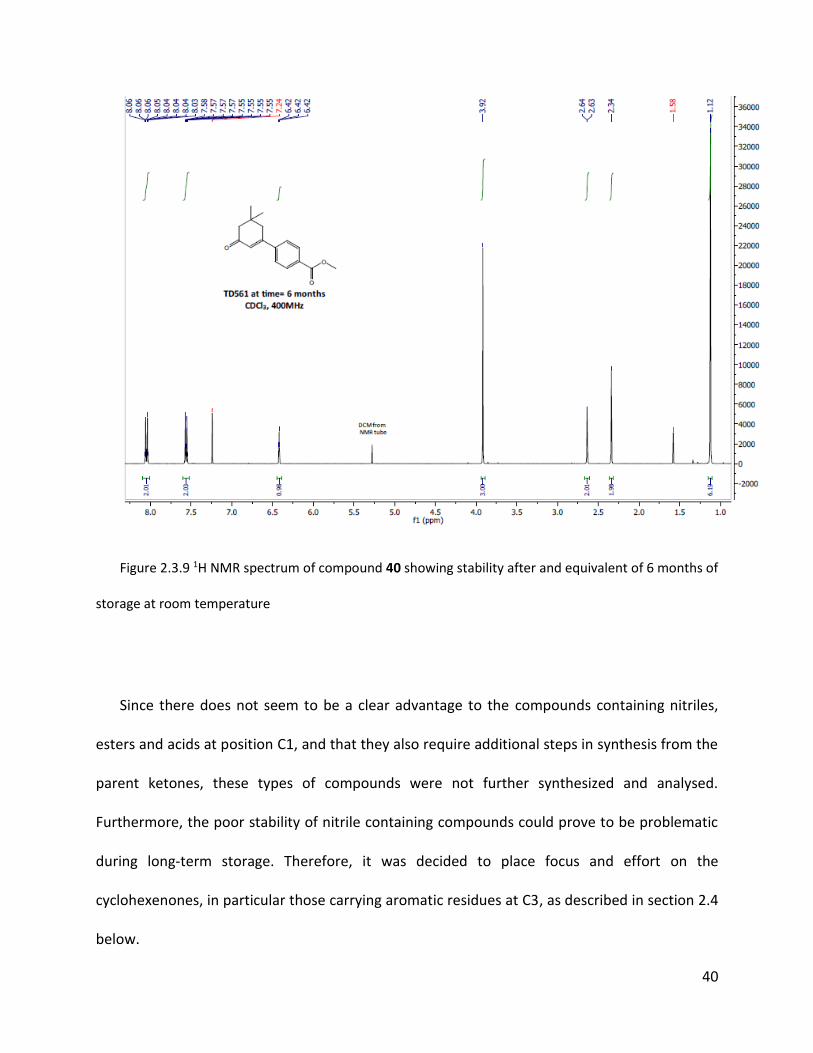
TABLE 2.4-1 SUMMARY OF REDUCTION OF NEURON FIRING AT 20 HZ AND 60 HZ BY 40 USING IN VITRO BIOASSAYS ON BRAIN CELL SLICES
........................................................................................................................................................................... 46
TABLE 2.6-1 SUMMARY OF REDUCTION OF NEURON FIRING AT 60 HZ BY 40 AND ITS SALTS AND ISOPROPYL ESTER USING IN VITRO
BIOASSAYS ON BRAIN CELL SLICES ................................................................................................................................ 53

x
TABLE 2.6-2 SUMMARY OF REDUCTION OF NEURON FIRING AT 20 HZ BY 40 AND ITS SALTS AND ISOPROPYL ESTER USING IN VITRO
BIOASSAYS ON BRAIN CELL SLICES ................................................................................................................................ 53
TABLE 2.7-1 SUMMARY OF REDUCTION OF NEURON FIRING AT 60 HZ BY 40 AND ITS AMIDE ANALOGS USING IN VITRO BIOASSAYS ON BRAIN
CELL SLICES............................................................................................................................................................. 57
TABLE 2.7-2 SUMMARY OF REDUCTION OF NEURON FIRING AT 20 HZ BY 40 AND ITS AMIDE ANALOGS USING IN VITRO BIOASSAYS ON BRAIN
CELL SLICES............................................................................................................................................................. 57
TABLE 2.8-1 SUMMARY OF REDUCTION OF NEURON FIRING AT 60 HZ BY NITRO AND AMINE ANALOGS USING IN VITRO BIOASSAYS ON BRAIN
CELL SLICES............................................................................................................................................................. 59
TABLE 2.8-2 SUMMARY OF REDUCTION OF NEURON FIRING AT 20 HZ BY NITRO AND AMINE ANALOGS USING IN VITRO BIOASSAYS ON BRAIN
CELL SLICES............................................................................................................................................................. 59
TABLE 2.8-3 SUMMARY OF REDUCTION OF NEURON FIRING AT 60 HZ BY BENZYL ETHER, ACID AND ALCOHOL ANALOGS USING IN VITRO
BIOASSAYS ON BRAIN CELL SLICES ................................................................................................................................ 62
TABLE 2.8-4 SUMMARY OF REDUCTION OF NEURON FIRING AT 20 HZ BY BENZYL ETHER, ACID AND ALCOHOL ANALOGS USING IN VITRO
BIOASSAYS ON BRAIN CELL SLICES ................................................................................................................................ 63
TABLE 2.8-5 SUMMARY OF REDUCTION OF NEURON FIRING AT 60 HZ BY FLUORO SUBSTITUTED AROMATIC AND PHENYL ANALOGS USING IN
VITRO BIOASSAYS ON BRAIN CELL SLICES ....................................................................................................................... 64
TABLE 2.8-6 SUMMARY OF REDUCTION OF NEURON FIRING AT 20 HZ BY FLUORO SUBSTITUTED AROMATIC AND PHENYL ANALOGS USING IN
VITRO BIOASSAYS ON BRAIN CELL SLICES ....................................................................................................................... 64
TABLE 2.8-7 SUMMARY OF REDUCTION OF NEURON FIRING AT 60 HZ BY TRIFLUORO AND SULFONE SUBSTITUTED AROMATICS COMPARED
TO 40 USING IN VITRO BIOASSAYS ON BRAIN CELL SLICES ................................................................................................. 66
TABLE 2.8-8 SUMMARY OF REDUCTION OF NEURON FIRING AT 20 HZ BY TRIFLUORO AND SULFONE SUBSTITUTED AROMATICS COMPARED
TO 40 USING IN VITRO BIOASSAYS ON BRAIN CELL SLICES ................................................................................................. 66
TABLE 2.8-9 SUMMARY OF REDUCTION OF NEURON FIRING AT 60 HZ BY HETEROAROMATIC AND PHENYL ANALOGS USING IN VITRO
BIOASSAYS ON BRAIN CELL SLICES ................................................................................................................................ 67
TABLE 2.8-10 SUMMARY OF REDUCTION OF NEURON FIRING AT 20 HZ BY HETEROAROMATIC AND PHENYL ANALOGS USING IN VITRO
BIOASSAYS ON BRAIN CELL SLICES ................................................................................................................................ 67

xi
TABLE 2.9-1 SUMMARY OF REDUCTION OF NEURON FIRING AT 60 HZ BY SULFUR DERIVATIVES 76 AND 79 COMPARED TO ETHYL ETHER 29
USING IN VITRO BIOASSAYS ON BRAIN CELL SLICES .......................................................................................................... 70
TABLE 2.9-2 SUMMARY OF REDUCTION OF NEURON FIRING AT 20 HZ BY SULFUR DERIVATIVES 76 AND 79 COMPARED TO ETHYL ETHER 29
USING IN VITRO BIOASSAYS ON BRAIN CELL SLICES .......................................................................................................... 70
TABLE 2.11-1 SUMMARY OF REDUCTION OF NEURON FIRING AT 60 HZ BY ANALOGS DERIVED FROM 1,3-CYCLOHEXANDIONE USING IN
VITRO BIOASSAYS ON BRAIN CELL SLICES ....................................................................................................................... 73
TABLE 2.11-2 SUMMARY OF REDUCTION OF NEURON FIRING AT 20 HZ BY ANALOGS DERIVED FROM 1,3-CYCLOHEXANDIONE USING IN
VITRO BIOASSAYS ON BRAIN CELL SLICES ....................................................................................................................... 73
TABLE 2.12-1 SUMMARY OF REDUCTION OF NEURON FIRING AT 60 HZ BY ENOL-THIO ANALOG 76 COMPARED TO DIMEDONE 26 AND
ETHOXY ENOL ETHER 29 USING IN VITRO BIOASSAYS ON BRAIN CELL SLICES .......................................................................... 74
TABLE 2.12-2 SUMMARY OF REDUCTION OF NEURON FIRING AT 20 HZ BY ENOL-THIO ANALOG 76 COMPARED TO DIMEDONE 26 AND
ETHOXY ENOL ETHER 29 USING IN VITRO BIOASSAYS ON BRAIN CELL SLICES .......................................................................... 74
TABLE 2.13-1 SUMMARY OF REDUCTION OF NEURON FIRING AT 60 HZ BY COMPOUNDS CURRENTLY BEING STUDIED BY NINDS USING IN
VITRO BIOASSAYS ON BRAIN CELL SLICES ....................................................................................................................... 77
TABLE 2.13-2 SUMMARY OF REDUCTION OF NEURON FIRING AT 20 HZ BY COMPOUNDS CURRENTLY BEING STUDIED BY NINDS USING IN
VITRO BIOASSAYS ON BRAIN CELL SLICES ....................................................................................................................... 77
TABLE 2.13-3 SUMMARY OF REDUCTION OF NEURON FIRING AT 60 HZ BY OTHER COMPOUNDS WITH SIMILAR DATA COMPARED TO THOSE
CURRENTLY BEING STUDIED BY NINDS USING IN VITRO BIOASSAYS ON BRAIN CELL SLICES ....................................................... 78
TABLE 2.13-4 SUMMARY OF REDUCTION OF NEURON FIRING AT 20 HZ BY OTHER COMPOUNDS WITH SIMILAR DATA COMPARED TO THOSE
CURRENTLY BEING STUDIED BY NINDS USING IN VITRO BIOASSAYS ON BRAIN CELL SLICES ....................................................... 78
TABLE 2.15-1 SUMMARY OF REDUCTION OF NEURON FIRING AT 60HZ BY ISOPHORONE 8 AND THE NATURAL PRODUCT 90 DURING IN
VITRO BIOASSAYS ON BRAIN CELL SLICES ....................................................................................................................... 83
TABLE 2.15-2 SUMMARY OF REDUCTION OF NEURON FIRING AT 20HZ BY ISOPHORONE 8 AND THE NATURAL PRODUCT 90 DURING IN
VITRO BIOASSAYS ON BRAIN CELL SLICES ....................................................................................................................... 84
TABLE 2.15-3 SUMMARY OF REDUCTION OF NEURON FIRING AT 60 HZ BY 97 AND 33 DURING IN VITRO BIOASSAYS ON BRAIN CELL SLICES
........................................................................................................................................................................... 85

xii
TABLE 2.15-4 SUMMARY OF REDUCTION OF NEURON FIRING AT 20 HZ BY 97 AND 33 DURING IN VITRO BIOASSAYS ON BRAIN CELL SLICES
........................................................................................................................................................................... 85

xiii
List of abbreviations
°C Degrees Celsius 13C NMR Carbon 13 NMR 1H NMR Proton NMR AED Anti Epileptic Drug CDCl3 Deuterated Chloroform CDRD Center for Drug Research and Development DCM Dichloromethane EtOAc Ethyl Acetate ETSP Epilepsy Therapy Screening Program Eq. Equivalents g Gram GABA γ-Aminobutyric acid Hz Hertz LDA Lithium Diisopropylamine LogP Octanol/Water partition coefficient mg m
milligram Multiplet
M Molar mins Minutes mL Millilitres mmol Millimoles MW Molecular Weight nBuLi nButyllithium NINDS National Institute of Neurological Disorders and Stroke NMR Nuclear Magnetic Resonance p Para substituted Ph Phenyl PhMgBr Phenylmagnesium bromide ppm Parts per million SEM Standard error of the mean

1
1 Introduction
1.1 Epilepsy
Epilepsy is one of the most common neurological diseases in the world.1 There are
approximately 50 million people worldwide affected by recurrent unprovoked seizures
symptomatic of epilepsy.1 Around 60% of affected people are believed to have idiopathic
generalized epilepsy which has no clear identifiable cause but is presumed to have a strong
underlying genetic basis and shows no structural brain malformations or defects.1 Symptomatic
epilepsy however is characterized by having known causes such as brain damage from
problematic births, genetic conditions leading to brain malformations, limited oxygen supply to
the brain related to a stroke, brain tumors and infection. For a patient to be considered to have
epilepsy, they must suffer from at least one unprovoked seizure and present further
predisposition determined by family history or the presence of epileptiform changes detected
by electroencephalogram.1 Whereas normal brain activity is characterized by asynchronous
neuronal firing, these seizures are caused by an abnormal synchronous discharge of cortical
neurons.1 Here are the different types of seizures:

2
Table 1.1-1 Simplified classification of seizures by behavioral and electrophysiologic data
I. Partial (focal seizures) II. Generalized seizures
A. Simple partial seizures with motor,
sensory, psychic, or autonomic symptoms
A. Absence seizures
B. Complex partial seizures B. Tonic-clonic seizures
C. Partial seizures with secondary
generalization
C. Other (myoclonic, tonic, clonic,
atonic)
Partial or focal seizures are caused by focal brain injury that are confined in one area of
the brain and are preceded by diverse sensory experiences or auras.2 These types of seizures
are not always linked to loss of consciousness but can potentially lead to generalized seizures.2
Generalized types of seizures always include loss of consciousness and can cause symptoms
varying from short loss of mental focus and disorientation to falling and constant muscle
contractions. Epilepsy and the associated unprovoked seizures can be successfully controlled
through treatment with anti-epileptic drugs (AEDs) at a relatively low cost. However, there is
evidence of medically resistant epileptic patients whose epileptic seizures persists after the use
of two appropriate AED treatment trials.2 There is also low availability of AED in low and
middle-income countries, preventing appropriate treatment of people suffering from epilepsy.
Therefore, there is still a need for more effective and affordable AED development.1
3
1.2 History of anti-epileptic drug development
Potassium bromide is considered to be the first drug with any documented value for
treating epilepsy, first documented3 by Sir Charles Locock of the Royal Medical and Chirurgical
Society in May of 1857. He commented that he had successfully treated women with hysterical
epilepsy and it was presumed that bromides could dampen sexual excitement which they
believed was the cause of these seizures. The widespread of bromides for the treatment of
epilepsy in conjunction with other agents such as zinc, digitalis and iron began after the
publication of other reports of successful treatment trials. Despite the associated secondary
effects such as dermatological conditions and more severe psychological symptoms potentially
leading to psychosis, bromides remained the foundation of epilepsy treatment for many years.3
In 1912, barbiturates, already widely used as hypnotics and sedatives were given by
clinical assistant Albert Hauptmann to epileptic patients as tranquilizers.4 A reduction of
frequency of seizures was observed, which led to subsequent studies on the subject. Bromides
were then eventually replaced as the most widely used AED by phenobarbital because of its
greater efficiency and the absence of severe secondary effects that were associated with
bromides. Phenobarbital in combination with bromides were then used for treatment by some
physicians. This treatment was discontinued after a few years because it was ineffective in most
patients.3

4
Figure 1.2.1 Structure of the barbiturate AED, phenobarbital
The next advancement in AED development was marked with the beginning of testing of
potential drugs on electrically induced epilepsy in animal models, notably cats, by Drs. Houston
Merritt and Tracy Putnam.5 The discovery of anti-seizure properties of phenytoin can be
attributed to studies of their group using these methods in 1938.5 Patients who were previously
unaffected by the combination therapy of phenobarbital and bromides showed successful
treatment using phenytoin and without the sedative effect of the previous AEDs. This not only
demonstrated that animal models were a cost-effective approach to drug discovery, but also
that the clinical trials on human patients could be reserved for compounds that were shown to
provide the best results in these animal trials. Phenytoin was such a successful treatment
associates with reduced neurological side effects that it was established as the best AED to be
used for partial seizures and is still used in certain situations today. Subsequently, downsides of
using phenytoin such as extensive drug interactions, chronic toxicities and serious delayed
effects such as carcinogenicity and teratogenicity were discovered. More effective analogs of
phenytoin were then rapidly discovered by altering certain functional groups on the basic
chemical moiety, the five and six membered heterocyclic rings of initially described members of
this family of AEDs.3
5
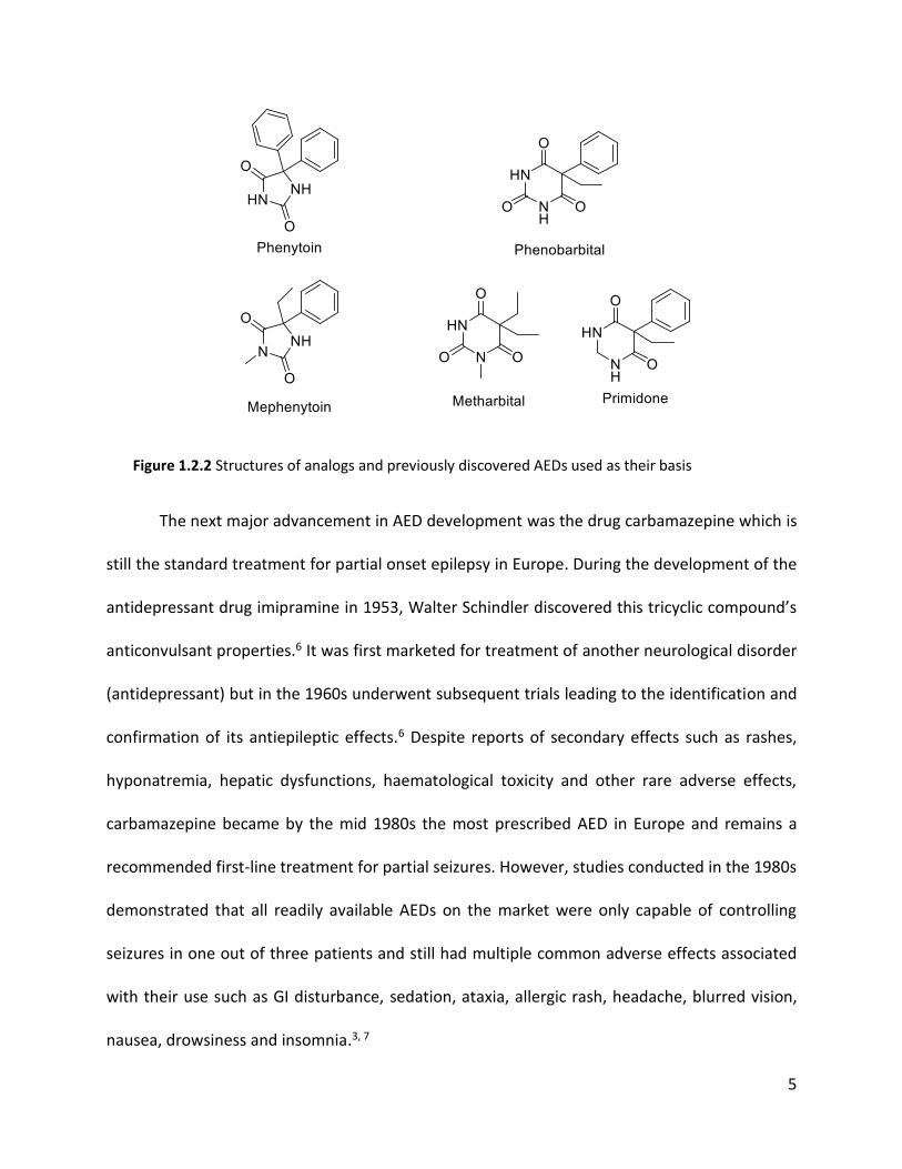
Figure 1.2.2 Structures of analogs and previously discovered AEDs used as their basis
The next major advancement in AED development was the drug carbamazepine which is
still the standard treatment for partial onset epilepsy in Europe. During the development of the
antidepressant drug imipramine in 1953, Walter Schindler discovered this tricyclic compound’s
anticonvulsant properties.6 It was first marketed for treatment of another neurological disorder
(antidepressant) but in the 1960s underwent subsequent trials leading to the identification and
confirmation of its antiepileptic effects.6 Despite reports of secondary effects such as rashes,
hyponatremia, hepatic dysfunctions, haematological toxicity and other rare adverse effects,
carbamazepine became by the mid 1980s the most prescribed AED in Europe and remains a
recommended first-line treatment for partial seizures. However, studies conducted in the 1980s
demonstrated that all readily available AEDs on the market were only capable of controlling
seizures in one out of three patients and still had multiple common adverse effects associated
with their use such as GI disturbance, sedation, ataxia, allergic rash, headache, blurred vision,
nausea, drowsiness and insomnia.3, 7

6
Figure 1.2.3 Structures of Carbamazepine and Imipramine
In 1975, the National Institute of Neurological Disorders and Strokes in the US
established the Anticonvulsant Drug Development Program in order to stimulate the
development of new AEDs. Following the screening of almost 30,000 new chemical compounds
over the years, an increase of licensed AEDs has been observed. Suitable drug candidates were
primarily found by systematic screening against a range of seizure animal models in rats, but
also by structural modification of existing molecules to create new analogs and by target-
oriented design. The deeper understanding of basic mechanisms of epilepsy has greatly
contributed to progress in brain research (see section 1.3 below), which in turn led to
discoveries of neurotransmitters of the brain and to further targeted drug development. These
recent advances in AED development have improved the amount of medically controllable
cases of seizures to about 70%.3

7
Figure 1.2.4 Common anticonvulsant drugs currently used on the market
Current treatment of seizures and epilepsy uses a variety of anticonvulsant drugs, some
of which are combinations of several different types of drugs.3 Many currently used AEDs share
considerable structural similarities. With few exceptions, they contain a nitrogen heterocycle
either fused to or carrying an additional aromatic ring (Figures 1.2.2 to 1.2.4).
Limitations of current AEDs
Other than the common, less severe side effects mentioned previously, AEDs can in rare
cases have potentially life-threatening adverse effects such as systemic reactions resulting in
multi-organ failure, epidermal necrolysis and thrombocytopenia.8 Withdrawal from an AED
medication program should be avoided because it can cause the resurgence of seizures and
status epilepticus, which is a series of seizures without any time to recuperate between the
events. 8

8
It has also been reported that increase in risk of anxiety, depression and suicidality has been
associated with the use of anticonvulsants. In a meta-analysis of clinical trials including 11 AEDs,
the FDA reported that patients taking these drugs had twice the risk of suicidal thoughts and
behaviours than those patients taking a placebo.9
AEDs are only currently used to treat the symptoms of epilepsy and not the cause of the
disease itself. Only if the seizures occur or are expected to occur in a rate that is more disabling
to the individual than the potential side effects of treatment is it advisable to proceed with
treatment using AEDs. Even when blocking seizures, AEDs do not seem to affect the course of
the underlying epilepsy, which is a serious limitation of current AED treatment.10
Importantly, it needs to be emphasized that fully 30% of epilepsy cases are resistant to
currently available AEDs. (See section 1.4)
1.3 Mechanism of seizures on neuronal activity and the effect of AEDs
The action potential is the basic mechanism of neuronal activity and is mediated by a
number or factors involving a network of neurotransmitters in the synaptic region and voltage-
gated ion channels located along the neuron. Action potentials occur due to depolarization of
the neuronal membrane, with membrane depolarization propagating down the axon to
induce neurotransmitter release at the synapse between neurons. Common features of
epileptic seizure activity are neuronal hyperexcitability (stimuli leads to larger increase in
neuronal firing) and neuronal hypersynchronicity (abnormal synchronized neuronal firing
leading to seizures) of multiple neurons. For a seizure to manifest, these features must be

9
present in large populations or networks of neurons. AEDs act on three different classes of
molecular targets to limit epileptic activity. Efficient AED mechanisms affect either ionotropic
glutamate receptors, γ-aminobutyric acid receptors (GABAA and GABAB), voltage-gated sodium
and calcium channel or a combination of these targets.2, 11
GABA is the primary inhibitory neurotransmitter in the brain. Inhibition mediated by GABA
in the brain occurs because of release of GABA from the presynaptic neuron that acts on two
postsynaptic receptors. GABAA receptor activation results in hyperpolarization of the neuron by
increasing chloride transfer inside the neuron and causing rapid inhibitory effect. GABAB
receptor activation results in decrease of calcium entry and causes a slow inhibitory effect.
Epileptic central nervous systems were studied and found to have reduced GABA-mediated
inhibition, which could lead to uncontrolled neuronal activation and subsequent seizures.
Several GABA agonist drugs are potent AEDs and GABA antagonist drugs have been shown to
induce seizures.2, 12, 13
Voltage-gated sodium channels are responsible for depolarisation of the nerve cell
membrane and conduction of action potentials across the surface of neuronal cells. Inhibition
of voltage-gated sodium channels resulting in blockage of sustained, high-frequency, repetitive
firing of neurons has been observed in AEDs effective in the treatment of partial seizures and
certain general seizures associated with epilepsy. The widely used AEDs phenytoin and
carbamazepine inhibit voltage-gated sodium channels and this reduction of sodium current is
thought to be the main mechanism of their therapeutic efficacy. These drugs produce a
characteristic voltage and frequency-dependent reduction in channel conductance, resulting in

10
a limitation of repetitive neuronal firing, with little effect on the unaffected neurons with
normal single action potentials.2, 12, 14, 15
1.4 The Current Epilepsy Drug Pipeline.
A recent review16 by Kaur, Kumar and Medhi entitled ”Antiepileptic drugs in development
pipeline: A recent update” repeated the well known statistics that epilepsy affects as many as
70 million people in the world. In the United States a total of more than 1.5 million people of
which more than 300,000 are younger than fourteen and more than 500,000 are older than
sixty five suffer from various forms of this disease. Rates of epilepsy are even higher in
developing countries. For example, the prevalence of epilepsy is reported at 6–10 per 1000
people which equates to between six and ten million out of a population that now exceeds one
billion. It is therefore not surprising that considerable efforts are being made to develop new
AEDs. Unfortunately many of these new drugs are modifications of older versions which are
known to have rather serious side effects. Despite availability of a large number of AEDs, one-
third of patients still have intolerable and untreatable conditions.16
Based on their analysis these authors suggest that there is an urgent need to create new
opportunities and improve the existing drugs to relieve patients especially those not responding
to the currently approved drugs. Their review covered AEDs that were under development and
in clinical trials during the years 2015 to 2016 for treating onset seizures, refractory, partial
seizures, generalized tonic clonic seizures, and resistant partial onset seizures.14
11
Most of the available AEDs have not shown efficacy in treatment of patients with refractory
epilepsy. It is unlikely that analogs and modification of currently used AEDs will address this
shortcoming. However the possibility exists that members of completely new families might
offer such hope. Also, most currently used AEDs have significant adverse effects nausea, ataxia,
drowsiness and other more rare life-threatening effects. It is plausible that novel families of
AEDs might be efficacious in controlling seizures with more therapeutic efficacy and less
adverse effects.16
The review article16 describes twenty-two compounds that are currently in clinical trials and
at various stages of development. The mechanism of action of most of the compounds under
investigation has been identified. Nine compounds on the list target ion channel inhibition,
mainly sodium or calcium, which is the most commonly recognized target for AEDs. Several
others affect the GABA system. At least three quarters of the compounds on the list contain
one or more nitrogen containing heterocycles a number of which are analogs of currently used
AEDs. Interestingly a number of these compounds have other known application. Included in
this group are Verapramil (hypertension, migraine), Buspirone (anxiolytic), Thalidomide
(hypertension, migraines, leprosy), Pregabalin (neuropathic pain, restless leg syndrome) and
Ganaxalone (anxiolytic).16

12
Figure 1.4.1 Structures of compounds currently in clinical trials for their potential as AEDs
Verapramil may be used for the prevention of migraines and cluster headaches. Buspirone
is an anxiolytic agent and serotonin receptor agonist. Thalidomide, despite its terrible history
for causing significant birth defect in the 1960s is currently used to treat leprosy. Pregabalin is a
medication used to treat neuropathic pain, fibromyalgia, and generalized anxiety disorder. Its
use for epilepsy is as an add-on therapy for partial seizures with or without secondary
generalization in adults. Ganaxolone is an experimental drug which is under development for
potential medical use as an anxiolytic and anticonvulsants.16

13
The U.S. Food and Drug Administration approved in late 2018 Epidiolex (cannabidiol) [CBD]
oral solution for the treatment of seizures associated with two rare and severe forms of
epilepsy, Lennox-Gastaut syndrome and Dravet syndrome, in patients two years of age and
older. This is the first FDA-approved drug that contains a purified drug substance derived from
marijuana.17
It was recently reported by researchers at Toronto Sick Children’s Hospital that a mixture of
THC and CBD showed promise as a treatment for drug resistant epilepsy in children due to
Dravet syndrome.17 This was considered significant since despite advances in the treatment of
epilepsy over the past twenty years no effective treatments have been available for children
who suffer from this condition.17
1.5. Isoxylitones as potential AEDs
Rahman et al.18 from the University of Karachi in Pakistan recently isolated and
identified a potent anticonvulsant agent a medicinal plant Delphinium denudatum Wall growing
in the Himalayan region of Pakistan. The crude ethanolic extracts from which alkaloids had
been removed were used in epileptic animal model studies and shown to exhibit strong

14
anticonvulsant activity in vivo. This fraction was also used for in vitro studies and showed strong
inhibition in epilepsy induced hippocampal neurons in cultured cells. It was further purified and
an isomeric mixture of E/Z isoxylitones, 1, was isolated and identified as the active
compounds.18, 19
Figure 1.5.1 Structures of E/Z isoxylitone isomers 1a and 1b
The first of the Raman group’s two patents18 described the isolation and in vitro and in vivo
evaluation of the isoxylitones as a mixture of E and Z isomers. They also separated the two
isomers and showed that they were easily interconverted by exposure to mild acid including
stomach acidity. The second patent19 reported a small group of analogs including a number of
ketone, ester and acid analogs shown below. Their results showed that the natural product was
more potent as determined by the bioassays described below than any of the derivatives that
they had prepared; with only the acid, 2, showing modest activity. It was clear from the results
reported in the second patent19 that replacing the methyl group in isoxylitone with larger
groups such as ethyl, propyl and phenyl reduced potency; indeed the phenyl derivative, 5, was
essentially inactive. All compounds were bio-assayed as E/Z mixtures.

15
Figure 1.5.2 Isoxylitone analogs, 2-5 including the acid (2) reported by the Raman group
The bioassay tests that were performed on these compounds by the research group at
the University of Karachi18, 19 are the Maximal Electroshock Test (MEST), the subcutaneous
pentylenetetrazol (scPTZ) test, and the electrical kindling model. These assays are commonly
used models to evaluate potential new AEDs. The MEST and scPTZ tests are considered acute
seizure models and are used to determine anticonvulsant activity of compounds whereas the
electrical kindling model is considered a chronic seizure model and it used to identify more
long-term antiepileptic activity. MEST consists of the application of an electric current via ear-
clips or electrodes of fixed intensity and short duration (0.2s) to the animal resulting in tonic-
clonic seizures. In this test, the AED is considered to have the ability to prevent the spread of
seizures if the animal does not fully extend its hind limbs. In the scPTZ test, a convulsive dose of
pentylenetetrazol is administered subcutaneously to induce a clonic seizure. The model is then
observed for a post-injection period of 30 minutes. The MEST gives information about the
potential activity against generalized tonic-clonic seizures whereas scPTZ tests identify the
efficiency of compounds against myoclonic and absences seizures. The MEST and scPTZ tests
are mainly used as preliminary screens for new AEDs because of the varying levels of
predictability of efficacy compared to the more reliable electrical kindling tests.18, 19

16
The electrical kindling model is widely considered an excellent animal model for
determination of antiepileptic efficacy of potential AEDs. This model can be used to study
molecules that may interfere with the generation of epilepsy, which would normally progress to
longer and more intense seizures. The kindling process consists of repeated induction of focal
seizures by electrical discharge over a long period of time to produce a progressive and
permanent increase of epileptic response. Once the rodent experiences an appropriate number
of seizures of adequate intensity, the kindling process is considered completed and the animal
is fully kindled.20
1.6 Objective
The Durst lab was approached by Professor Michael Poulter from the Department of
Pharmacology at Western University in London, Ontario, to synthesize a library of analogs of
isoxylitone. 1. Dr. Poulter is a member of the Ontario Brain Institute; his group have been
studying epilepsy for over 20 years. Poulter was intrigued by the level of activity of the natural
product, its simplicity as a chemical structure and the novelty of these compounds as potential
anti-epileptic compounds. All clinically used AEDs including the first used drug such as phentoin
and the subsequently developed carbamazepine and imipramine and their analogs contain
nitrogen heterocycles. These compounds are ineffective in treating almost 30% of epilepsy
cases (refractory epilepsy). Additionally, significant unwanted side effects are associated with
all of the classic anti-epilepsy drugs.

17
There was a distinct possibility that isoxylitone might become the lead structure of a new
family of anti-epileptic compounds and that these would not have the unwanted side effects
associated with typical AEDs. Dr. Poulter felt confident enough to start a small company which
he named OB Pharma. The goal of this company was to not only investigate the potential of the
isoxylitone molecule but also generate a library of analogs some of which would hopefully be
more potent that isoxylitone itself and become potential drug candidates.
1.7 Previous work
A previous graduate student in the Durst lab, Amanda Saikaley, started working on this
project during her M. Sc. prior to the publication of the second Raman group patent. The first
part of her work consisted of synthesizing the isomeric mixture of the lead compound
isoxylitone E/Z (1a and 1b). They chose not to follow the synthesis of the Pakistani researchers
and developed the synthesis shown below in Fig 1.6.1. Reaction of ethyl acetoacetate 6, first
with sodium hydride and then nBuLi generated its highly reactive dianion 7 which was
condensed with isophorone, 8. The initial adduct 9 was dehydrated and then saponified to the
beta-keto ester 10 which readily lost CO2 to afford isoxylitone 1. The success of this synthesis
was confirmed by NMR analysis.21

18
Figure 1.7.1 Synthesis of isoxylitone E and Z, 1a and 1b
Saikaley prepared more than thirty analogs in an effort to improve potency of the lead
structure. A series of vinylic esters (11-14) closely resembling the structure of isoxilitone 1 were
produced by condensing the anions derived from a number of acetates with isophorone 8;
these were also obtained as E/Z mixtures.21
Figure 1.7.2 Series of ester analogs
The ethyl ester analogue 12 was proven to be equipotent to the parent isoxylitone
based on the voltage sensitive dye imaging (VSDI) bioassay and on the kindling model assay
carried out in the Poulter laboratories (see Section 2.1.1). It was possible to isolate a pure

19
sample of the Z acid (2Z) and then esterify it to give pure Z ethyl ester (12Z). The pure Z acid
was shown to be less active than the mixture. While this was interesting, it was of relatively
little importance since these compounds underwent ready isomerization between the E and the
Z isomers even under mildly acidic conditions comparable to stomach acidity.
Figure 1.7.3 Synthesis of the ethyl ester analogs of isoxylitone.
It was also noted that the activity of isoxiltone type compounds was highly sensitive to
steric effects of the substituents attached to the carbonyl carbon. Replacement of the methyl
ketone by ethyl (15), propyl and phenyl (16) ketones successively reduced the desired biological
activity with the latter compound being essentially inactive. This observation verified the
conclusions arrived at by the Raman group in their second patent.19 A similar observation was
made in going from the methyl (17) to the phenyl (18) sulfoxide.

20
Figure 1.7.4 Biological activity depending on steric effects
The nitrile 19, prepared by reaction of isophorone 8 with α-lithiopropionitrile 20 turned out
to be the most potent compound reported in the Saikaley thesis. It too existed as a mixture of E
and Z isomers. Indeed, initial dehydration of the adduct obtained from the condensation of α-
lithiopropionitrile and isophorone yielded a mixture of five dienes including 19E and 19Z. It was
later discovered that heating of this mixture with p-toluene sulfonic acid in toluene resulted in
the formation of essentially only E and Z 19 in an almost 1:1 ratio. This mixture was designated
by OB Pharma as TD532.

21
Figure 1.7.5. Synthesis of 19 (TD532)
Compound 19, as a mixture of stereoisomers, showed remarkable biological activity. It was
significantly superior to the lead structure isoxylitone. Saikaley produced a number of nitrile
analogs (21-24) by condensing different -lithionitriles with isophorone and also with other
cyclohexenones to produce a set of compounds. A number of these are shown below.
Figure 1.6.6. Analogs of 19 (TD532)
Almost all of the compounds produced showed some biological active, but none were
superior to 19 (TD532). Saikaley concluded that replacement of the methyl group in 19 by
larger substituents, even by the relatively small changes to cyclopropyl 23, and isopropyl 21,

22
resulted in the loss of potency. Surprisingly replacement of the -methyl group in 19 by
hydrogen also gave a less active compound. Removal of the 5,5-dimethyl group on the
cyclohexenone ring also caused significant loss of the desired activity. The excellent activity of
19 both in the in vitro neuron firing and in vivo kindling assays carried out by the Poulter group
were deemed sufficient to warrant the filing of a patent in which 19 became centerpiece of a
new family of compounds in the AED field.21
The compounds described in the Saikaley thesis21 and also in the Rahman patents18, 19
indicated quite strongly that steric effects associated with the substituent on either the enone
group in isoxylitone and the related sulfoxide or to the nitrile group in 19 resulted in
reduction of the desired activity. It was decided by our group to send isophorone 8 and
compare its ability to reduce the firing of neurons in the bioassay as carried out by the Poulter
group. The underlying question was: Could the CH=CH-C(O)CH3 group in isoxylitone 1 be
replaced by a simple carbonyl group as found in isophorone 8? The observation that
isophorone 8 reduced neuron firing became the basis of the investigations reported in Chapter
2 of this thesis.
Figure 1.7.6 Comparison of the structures of isoxylitone 1 and isophorone 8 and their functional
group at C3

23
2 Discussion and Results
2.1 Introduction
The observation that isophorone 8, produced a 20% reduction of neuronal firing at 1µM
concentration stimulated by either 20 or 60 Hz voltage pulses examined by voltage sensitive
dye imaging (VSDI) on rat brain slices ex vivo (methods in section 2.1.1) in a manner comparable
to many of the compounds reported in the Saikaley M. Sc. thesis allowed us to propose a
cyclohexene ring in which the alkene is conjugated to an electron withdrawing group (EWG) as
the key pharmacophore.
Figure 2.1.1 Structures of isophorone 8 and the key pharmacophore 25.
The key active compounds known in this series at the beginning of this study were the
natural product lead structure isoxylitone 1, where R = CH3 and the EWG = CH-C(O)CH3 , the
sulfoxide 17 (R = CH3, EWG = CH-SOCH3 and 19 (TD532), the most active compound reported
in the Amanda Saikaley series where the EWG is =C(CH3)CN. These compounds all contain the
above suggested pharmacophore 25.

24
Figure 2.1.2 Structures of isoxylitone 1 (R=CH3), sulfoxide analogs 17 (R=CH3) 18 (R=Ph), and the
active nitrile analogs 19 (TD532).
The question was: What structural changes could be made to isophorone 8 in order to
enhance its potential for anti-epileptic activity. A number of possibilities are indicated on the
structure below. It was clear from the work of Saikaley21 and that of the Rahman et al.18 that
increasing the size of the EWG group reduced the desired activity of these compounds. For
example, replacement of the methyl group joined to the carbonyl group in 1 or the
methylsulfinyl group in 17 by larger substituents such as phenyl resulted in almost complete
loss of potency.21
Table 2.1-1 Summary of reduction of neuron firing at 60Hz using in vitro bioassays on brain cell slices as part of Saikaley’s work on analogs 17, 18 and 19
Compound Average reduction
at 200 nM (%)
Average reduction
at 1 µM (%)
17 (R= Methyl) -50 Not tested
18 (R = Phenyl) Inactive Inactive
19 (TD532) -50 Not tested

25
Figure 2.1.3 Proposed changes to the basic enone structure.
The most obvious and synthetically easiest changes that one could imagine would be to
replace the R group (CH3 in isophorone) by H or by groups that are sterically larger than methyl
and have a variety of electronic characteristics. Most of the efforts described in this thesis were
directed towards this goal. Several compounds were also prepared in which the changes in the
substituent at C3 were also accompanied by changes at the other positions. The possibility of
changing the ring size from cyclohexenone to cyclopentenone or cyclohexenone was not
investigated.
2.1.1 Bioassays performed
The synthesized compounds were sent to OB Pharma and bioassays were performed to
identify anti-epileptic drug potential.
To test for activity, a first assay was done using voltage sensitive dye imaging (VSDI) on
isolated rat brain slices that were kept viable in artificial cerebral spinal fluid (ACSF). The neural
assemblies were activated by electrical stimulation. A voltage sensitive dye, such as, di-4
ANEPPS, was incubated with a brain slice for 1 h in a suitable solution that enhances the dye
penetration into the tissues. The dye reacted to the changes in voltage across the cell

26
membrane of the neurons in the brain slices. The synthesized compounds were added to the
ACSF at known concentrations between 200 nM and 1 µM. The brain slices were then subjected
to an electrical stimulus that activated the neurons in the slice. As the dye reacted to the
change in voltage, the intensity of the dye can be observed and quantified. The degree of
capability of the compound to dampen the activation of the brain was evaluated in this
manner. A negative dye intensity value in relation to the control experiment can be interpreted
as a reduction of neuronal activity related to voltage in the cells. This reduction can translate
into potential anti-epileptic activity. This reduction is desired at the 60 Hz, which is the
frequency at which neurons operate in the central nervous system. However, the inhibition of
activity at 20Hz could cause unwanted side effects since essential neurons regulation base life
functions such as in the heart operate at this frequency. Thus, compounds showing good
inhibition of activity at 60 Hz but very low to no inhibition at 20 Hz were considered great
potential candidates. Compounds showing this type of activity were investigated and further
analogs were synthesized as future candidate compounds.
In a second assay, the inhibition of the compounds on the activity of voltage gated sodium
channels was determined using patch clamp electrophysiology. A negative value in relation to
the control can be interpreted as representing a statistically significant decrease of neuronal
activity. This can translate to anti-epileptic activity caused using the compound being assayed.
Compounds showing this type of activity were investigated and further analogs were
synthesized as future candidate compounds. This analysis was done on cultured cortical
neurons isolated from rats. A more in-depth description of the methods used by the Poulter
group to identify compounds of interest is given below.

27
Slice Preparation and Staining
All animals used in these studies were adult male Sprague-Dawley rats aged 20–45 days.
The preparation of brain slices and kindling methodology have been described in detail
elsewhere.22 Slices prepared from kindled rats were usually about 45 days old. Control rats for
these experiments were age matched but no electrode was implanted. Brain slices were
incubated for 30 min in a solution that contained 0.6 M Mofdyedi-4- ANEPPS (D-1199,
Invitrogen Molecular Probes Inc., OR, USA). After washing for 10 min with ACSF slices were
transferred to the recording chamber. During all recordings the slices were maintained at 32 ˚C
and continuously perfused with ACSF bubbled with a mixture of 95:5 oxygen and carbon
dioxide. The slices were stimulated with a platinum/iridium electrode (Micro Probes, Inc., MD,
USA) with a tip diameter of 200–300 μm at the border of the lateral olfactory tract (LOT) and
layer I of the PCtx. The stimulation of each slice was in the range of 160–200 μA, each square
pulse was 2.0 ms in length. The electrode was connected to a stimulator (S88X dual output
square pulse stimulator, Grass Technologies, AnAstro-Med, Inc., QC, Canada),which controlled
the pulse frequency and train duration.
Patch Clamp Recording
The whole cell patch clamp recording technique used and the preparation of brain slices
from adult rats have been both described in detail.23,24 The internal solution used in these
experiments was; K gluconate, 140 mM; MgCl2, 2 mM; CaCl2, 1 mM; MgATP, 2 mM; NaGTP, 0.2

28
mM; EGTA, 1.1 mM and HEPES, 10 mM. A multiclamp 700B amplifier was used to record from
neurons located in layers II and III.
Optical Recording
The composition of ACSF used for optical recordings was the same composition used in the
patch clamp recordings. Each recording was about 20 s in length and consisted of two époques.
The first was a 2 s recording of background activity before the stimulus followed by the stimulus
application for 1 s with frequencies differing from 5 to 100 Hz. The acquisition rate was
between 3 and 10 ms/frame. For each recording minimum camera saturation was set around
50% while the maximum was about 80%. Optical recording was conducted using a CMOS
camera (Micam Ultima, BrainVision, Inc., Tokyo, Japan) mounted on top of an upright
microscope (Fixed Stage Upright Microscope BX51WI, Olympus). The light from a 100 Whalogen
lamp source (HLX 64625, Microlites Scientific, Corp.) passed through an excitation filter (λ = 530
± 10 nm). The fluorescent signals were collected and projected onto the CMOS sensor through
a long pass emission filter (λ > 590 nm). A long-distance objective was used in these
experiments (XLFluor4XN. A.0.28, Olympus). The movies were recorded and analyzed using
Brain Vision Analyzer (Tokyo, Japan) software. The acquisition settings were: 100 × 100 pixels
frame size, after magnification each represented 25 μm × 25 μm space on the brain slice. The
dye signal intensity decreases as the membrane depolarizes. However, to better match
conventional recordings the signals all have been converted so that the excitatory and
inhibitory signals were shown as positive and negative values. As bleaching can strongly affect

29
the data, all recordings were corrected by subtracting the change in fluorescence that occurred
in a region of the slice that was unresponsive to the stimulus. The fractional change in
fluorescence signal relative to background signal (ΔF/F) was calculated for each frame of each
recording. For all the recordings, we binned 3 × 3 pixels into one representative signal. As there
was considerable variability in the magnitude of the responses from slice to slice due to
differences in loading of the dye, we normalized the recordings by dividing all signals by the
response to the 20 Hz stimuli. This permitted us to average the normalized responses between
recordings. Thus, the input/output relationships shown are the normalized ΔF/F. The lag time
was calculated by measuring the time between the stimulus on set and the time for the signal
to be 20% above baseline. Instead of using pixels bins, we measured the ΔF/F along a “stripe”
that could be precisely placed along a group of pixels before and after the cut. Each stripe
consisted of 10 pixels and covered 250 μm length. The data derived from each stripe was the
average ΔF/F of 10 pixels.
Bioassay were reported to the Durst group within a month or two of submitting the
compounds. The data obtained allowed us to decide with relative confidence which analogs
and target modifications to the pharmacophore structure 25 should be synthesized and
performed. This process that lead to the synthesis and discovery of TD561 (compound 40) as a
potent potential anti-epileptic drug. A patent featuring this compound and analogs was
submitted and published, showing the confidence of OB Pharma in the potential of this series
of compounds.

30
2.2 Substituents at C3
The simplest strategy for introducing different substituents at C3 utilizes the inexpensive
and readily available dimedone 26, as starting material. Dimedone has a 1,3-diketone structure
and as such exists as a mixture of tautomers, the diketone 26 and the enol form 27.
Figure 2.2.1 Structures of tautomers of dimedone
Reaction of dimedone with methanol or ethanol in the presence of a strong acid yields the
enol ethers 28 and 29, respectively.25 The structures of these products are readily apparent
from their 1H NMRs which show the incorporation of a methoxy (OCH3 at 3.66 ppm) or ethoxy
(OCH2 at 3.88 ppm) unit and the presence of a vinyl hydrogen.
Figure 2.2.2 Structure of enol ethers obtained from dimedone.
Reaction of above ethers with strong nucleophiles is known to cause replacement of the
ether group by the nucleophile and result in the introduction of a new substituent at C3. This
reaction sequence can be rationalized in one of two ways: a) Addition of the nucleophile at C3

31
followed by elimination of the alkoxy group, or b) addition of the nucleophile at the carbonyl
carbon followed by regeneration of the carbonyl group at C3 during hydrolytic workup. The
result of these two processes is the same in the case starting with dimedone due to its
symmetrical nature.
Figure 2.2.3 Mechanisms of introduction of substituents at C3 via nucleophilic attack.
The first nucleophiles chosen for these reactions were n-BuLi and PhMgBr. These reactions
resulted in the formation of the products 30 and 31, respectively.
Reaction of a THF solution of 29 with n-BuLi afforded 3-n-butyl-5,5-dimethyl cyclohex-2-
enone, 30 in 32 % yield after purification by column chromatography. The 1H and 13C NMR
spectra of the purified product showed the introduction of an n-Bu group with 1H NMR peaks at
0.89, 1.32, 1.45 ppm and the retention of the enone hydrogen which absorbs at δ= 5.85 ppm.

32
Figure 2.2.4 Synthesis of 30.
Addition of 29 to an ether solution of phenylmagnesium bromide in diethyl ether afforded
the 3-phenyl-cyclohexenone 31 in 40% yield after purification. The 1H NMR spectrum of 31
showed the expected five aromatic hydrogens in addition to the other peaks due to the
hydrogens on the cyclohexenone ring.
Figure 2.2.5 Synthesis of 31.
The bioassay results for these two compounds and for isophorone 8, are shown in Table
2.2.1. A reduction of neuronal activity of -39% and -36% was observed for compounds 30 and
31, respectively, compared to a reduction of -22% for isophorone 8 when the VSDI
determination was carried out at 200 nM. This indicated that the introduction of groups larger
than methyl at C3 in the cylcohexenone ring results in more potent compounds. The difference
in reducing uncontrolled neuronal activity by brain cells is similar for these compounds at the
higher 1μM concentration, especially considering the uncertainty reported in standard error of

33
the mean in the measurements. These data indicated that it would be worthwhile to produce
additional compounds with both different aryl and alkyl groups at this position.
Table 2.2-1 Summary of reduction of neuron firing at 60Hz using in vitro bioassays on brain cell slices of isoophorone 8, and analogs containing larger carbon containing groups at C3
Compound Average reduction
at 200 nM (%)
SEM at 200nM
(%)
Average reduction
at 1 µM (%)
SEM at 1 µM (%)
8 -22 11 -46 9
30 -40 7 -51 7
31 -36 8 -46 8

34
2.3 Analogs containing substituents at C3 and complex EWG at C1
Since our group had shown in the past that the compounds such as 19 (TD532) and to a
lesser extent the related esters and acids 12 and 2 also had promising activity it was decided to
generate a series of compounds 32-36 that incorporated both features, that is the nitrile or
ester or acid function in 19, 12 and 2 combined with sterically more demanding substituents at
C3.
Figure 2.3.1 Structure of 19 (TD532) and related compounds.
The synthesis of these compounds commenced with the precursor cyclohexenones as
shown below. Thus, reaction of the enone 30 with the lithium salt of propionitrile 20 yielded
the expected addition product 37. Dehydration of 37 gave 32 as a 1: 1 mixture of stereoisomers
as determined by integrating the signal for C2 alkene hydrogens which occurred at δ= 6.17 and

35
6.44 ppm for the E and Z isomers, respectively. The overall yield for this two-step conversion
was 38%. There was no evidence of isomers with both double bonds in the ring. No attempts
were made to separate the isomers either here or for the related mixtures 33-34 below, since
Saikaley had shown that they interconvert readily on exposure to mild acid.21
Figure 2.3.2 Synthesis of 32E and 32Z.
A similar sequence of reactions was used to prepare the mixture of isomers of compound
34 from the ketone 31; the E/Z ratio was determined to be 1:1.
Figure 2.3.3 Synthesis of 34E and 34Z.
The preparation of 33 required initial synthesis of the ketone 39. This compound was
prepared from dimedone 26 in two steps following the procedure by Zegarski26. Ethoxy
dimedone, 29 was reduced to the allylic alcohol which upon treatment with aqueous acid
afforded 3926. Reaction of 39 with -lithiopropionitrile 20, followed by dehydration of the

36
intermediate product as described for the synthesis of 32 gave the desired compound 33 as a
mixture of E and Z isomers in a ratio of 2:1.
Figure 2.3.4 Synthesis of 33E and 33Z
Finally, the unsaturated ester 35 was obtained from the enone 31 by a condensation with
the lithium derivative of ethyl acetate and subsequent dehydration. The mixture of ester
stereoisomers was hydrolyzed under basic conditions to give the acid 36. The structures of
these compounds were readily verified based on their 1H NMR spectra. As above, the ratio of
isomers was easily determined by integrating the signal for the remaining alkene hydrogen at
C2.
Figure 2.3.5 Synthesis of 35 and 36.
At this stage we were able to compare the bioassay results for different series in order to
decide whether the simple enones or the compounds with the more complex EWG at C1 were
more effective. The relevant compound series together with the assay results indicating
reduction of rat brain excitatory neuronal circuit activity stimulated at 60 Hz are shown below.

37
Table 2.3-1 Summary of comparisons of reduction of neuronal activity at 60Hz of three series of compounds to identify the effect of different sizes and complexities of EWG at C1
Compound Average reduction
at 200 nM (%)
SEM at 200nM
(%)
Average reduction
at 1 µM (%)
SEM at 1 µM (%)
30 -40 7 -51 7
32 -28 8 -51 6
31 -36 8 -46 8
34 33 7 -42 4
35 -40 5 -49 3
8 -22 11 -46 9
19 -33 7 -42 4
33 -35 8 -55 5
Figure 2.3.6. Compounds being compared using bioassay results separated in three series
Examination of the three sets of results does not give a clear indication that the additional
effort to convert the enone structures to the extended conjugated nitriles and esters provides a
significant advantage because of the lack of observed improvement in reduction of neuronal

38
activity. The reported measurement data in the VSDI bioassay do not allow us to make
definitive conclusions regarding structure activity relationships. The data suggests that the size
and nature of the substituent at either C1 and C3 within the variations studied do not affect the
ability to reduce neuronal firing.
A disadvantage of the nitriles and esters is that the compounds are mixtures of isomers. It
was shown by Amanda Saikaley21 that these compounds isomerise easily under mildly acidic
conditions between the E and the Z isomers. Also, compound 19 (TD532) and presumably also
the other nitriles such as 32, 33 and 34, have much more limited thermal stability than ketones
such as 31. This was shown in a comparison of data following the World Health Organization
Accelerated Stability Protocol27 that was performed as part of this work. The 1H NMR of 19
started showing the presence of many additional peaks upon storage for a period equivalent to
two months at room temperature whereas that of the ester 40 whose synthesis is described
below (section 2.4), remained unchanged for a period equivalent to more than six months.
Figure 2.3.7 Structures of compounds 19 and 40, which were subjected to accelerated stability studies

39
Figure 2.3.8 1H NMR spectrum of compound 19 showing degradation after and equivalent of two
months of storage at room temperature
40
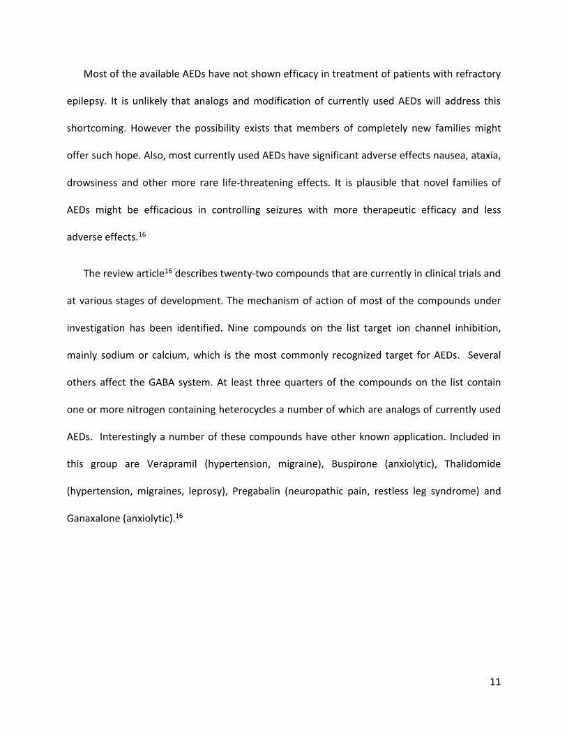
Figure 2.3.9 1H NMR spectrum of compound 40 showing stability after and equivalent of 6 months of
storage at room temperature
Since there does not seem to be a clear advantage to the compounds containing nitriles,
esters and acids at position C1, and that they also require additional steps in synthesis from the
parent ketones, these types of compounds were not further synthesized and analysed.
Furthermore, the poor stability of nitrile containing compounds could prove to be problematic
during long-term storage. Therefore, it was decided to place focus and effort on the
cyclohexenones, in particular those carrying aromatic residues at C3, as described in section 2.4
below.

41
2.4 Synthesis of 3-(4-carboxymethylphenyl)-5.5-dimethylcyclohex-2-en-1-one, 40(TD561)
The change in focus towards compounds containing aromatic residues at C3 resulted in the
synthesis of compound 40, also known as TD561, and related analogs.
The synthetic route described for the synthesis of the 3-phenyl derivative 31 is not
applicable to the preparation of 40 since the ester substituent at the para position of the phenyl
ring is not compatible with either the required lithio or Grignard intermediates. A search of the
literature28 indicated that 40 and similar substituted aromatic compounds should be accessible
by coupling reaction between the boronic acid 41 and the enol tosylate 42. This coupling is
mediated by 3% tetrakistriphenylphosphine palladium (0).
Figure 2.4.1 . General catalytic cycle for Suzuki-Miyaura couplings28

42
As seen below, the synthesis of 40 can be carried out conveniently in one pot following the
literature protocol.28 The dimedone tosylate 42 can also be prepared separately.
Figure 2.4.2. One pot, two-step, synthesis of 40 (TD561).
The major advantage to this procedure is that it is a simple two reaction – one-pot
procedure requiring less than one day to set up and complete. The isolation of pure 40 was not
trivial requiring often a minimum of two consecutive column chromatography separations.
The following procedure is representative. A mixture of dimedone (1.2 equiv) tosyl chloride
(1.5 equiv) and potassium carbonate (3 equiv) are stirred at room temperature in a 2:1 mixture
of dioxane and water for one hour; a longer time is not necessary but also not harmful. At that
point 1 equivalent of the aryl boronic acid is added followed by 0.03 equiv of Pd(PPh3)4 and the
mixture is refluxed for 90 min.
The structure assignment of 40 was consistent with the 1H and 13C NMR spectra.
Assignments of individual H and C resonances, shown on the following pages.

43
Figure 2.4.3 1H NMR spectrum of 40 (TD561) with assignments

44
Figure 2.4.4 13C NMR spectrum of 40 (TD561) with peak assignments

45
Based on the initial screening results (shown below) by voltage sensitive dye imaging (VSDI),
compound 40 was further investigated by the Poulter laboratory using the in vivo kindling
assays. The promising results in the latter tests and the observation that rats treated orally with
this compound showed no obvious side effects led to the decision by OB Pharmaceuticals, Inc.
(London, ON) under the direction of Dr. Michael Poulter (CSO), to pursue it as a potential drug
candidate. Most importantly, compound 40 when administered orally at 20 mg per kg
prevented stage 1 epileptic seizures in kindled rats whereas those treated at the same level
with AEDs such as Lamotrigine proceeded typically to stages 3 and 4.29
A considerable amount of biological data has been subsequently obtained for this
compound. It is currently being evaluated in preclinical trials by scientists at the Center for Drug
Research and Development (CDRD; Vancouver, BC). Compound 40 has also been sent to the
Epilepsy Therapy Screening Program (ETSP) at the National Institute of Neurological Disorders
and Stroke (NINDS) in Bethesda, MD, United States) for additional evaluation and verification of
its activity in vivo.
Figure 2.4.5 Structure of 40 (TD561)
It is uncertain as to why 40 was chosen by OB Pharma over a number of other potential
candidates but we can advance a hypothesis on the subject. The data for its effect on neuronal
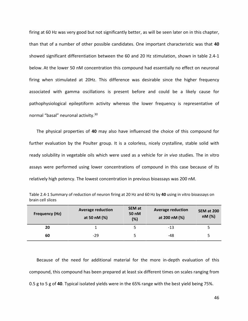
46
firing at 60 Hz was very good but not significantly better, as will be seen later on in this chapter,
than that of a number of other possible candidates. One important characteristic was that 40
showed significant differentiation between the 60 and 20 Hz stimulation, shown in table 2.4-1
below. At the lower 50 nM concentration this compound had essentially no effect on neuronal
firing when stimulated at 20Hz. This difference was desirable since the higher frequency
associated with gamma oscillations is present before and could be a likely cause for
pathophysiological epileptiform activity whereas the lower frequency is representative of
normal “basal” neuronal activity.30
The physical properties of 40 may also have influenced the choice of this compound for
further evaluation by the Poulter group. It is a colorless, nicely crystalline, stable solid with
ready solubility in vegetable oils which were used as a vehicle for in vivo studies. The in vitro
assays were performed using lower concentrations of compound in this case because of its
relatively high potency. The lowest concentration in previous bioassays was 200 nM.
Table 2.4-1 Summary of reduction of neuron firing at 20 Hz and 60 Hz by 40 using in vitro bioassays on brain cell slices
Frequency (Hz) Average reduction
at 50 nM (%)
SEM at 50 nM
(%)
Average reduction
at 200 nM (%)
SEM at 200 nM (%)
20 1 5 -13 5
60 -29 5 -48 5
Because of the need for additional material for the more in-depth evaluation of this
compound, this compound has been prepared at least six different times on scales ranging from
0.5 g to 5 g of 40. Typical isolated yields were in the 65% range with the best yield being 75%.

47
A consistent by-product accompanying the formation of 40 was identified as the
biphenyl derivative 43 based on comparison of its spectroscopic data with that reported in the
literature.31 It could not be separated from the desired product by crystallization but could be
removed by careful silica gel chromatography using pure dichloromethane as eluent where it is
much more fluorescent on analytical TLC and slightly less polar than 40. The by-product 43
elutes first. Once this product has been eluted, the desired 40 is obtained quickly using 5% ethyl
acetate: 95% DCM as eluent.
Figure 2.4.6 Structure of biphenyl by-product impurity
2.5 Potential alternate synthetic routes to 23.
Due to the purification problems associated with the preparation of multi-gram quantities
of 40, we considered alternate approaches to this compound. We investigated briefly the
sequence in which the aryl to carbon bond is made via the protected Grignard reagent 45 which
was prepared from THP protected 4-bromobenzyl alcohol 44. (shown below)

48
Figure 2.5.1 Scheme for an alternate synthetic route to 40.
Reaction of the Grignard reagent 45 with ethoxy dimedone, 29, afforded the adduct 46.
Removal of the protecting group gave the benzylic alcohol 47 which was oxidized with Jones’
reagent to form directly the acid 48. This transformation was carried out jointly with COOP
student Michael Darnowski who carried out the first and last step in the synthesis. The isolation
of 48 showed that this sequence worked in principle. All intermediates were appropriately
characterized.
Upon further consideration it was decided not to optimize this sequence since it involved
twice as many steps as the boronic acid approach. Additionally, it was realized that this would
not be easily translated into a potential large-scale approach since it involved the use of the
highly flammable ether as a solvent and the use of Cr+6 which has known toxicity as oxidizing
agent.

49
A third approach based on a Heck coupling reaction is currently being pursued by another
member of our group. A search of literature precedents led to an Organic Letters publication by
the Andrew Myers group.32 These authors pointed out the lack of reliable examples of classic
Heck reaction conditions involving cyclohexenone as the electron poor carbonyl component.
They presented a new approach involving decarboxylation of an aromatic acid and its coupling
to 4-isoprpoylcyclohexanone.
Figure 2.5.2 Decarboxylation of an aromatic acid and its coupling to 4-isoprpoylcyclohexanone
The authors pointed out that these reaction conditions worked best with ortho-substituted
aromatic acids and thus such a sequence may not be useful for a synthesis of 40. They did
however also report application of the Jeffry reaction conditions32 to the synthesis of 49.
Figure 2.5.3 Application of Jeffrey conditions of Heck reaction as a potential route to 40.
We believe that there is a reasonable possibility that this combination of reagents may lead
to a process for the preparation of 40 that could be competitive with the boronic acid route
both in terms of time and cost. (shown below)

50
Figure 2.5.4 A potential approach to synthesis of 40 (TD561).
A modification on the initial boronic acid approach was attempted, wherein the tosylate of
dimedone 42, was isolated and the synthesis was carried out in two steps. This led to an easier
separation and isolation via column chromatography and was, at the time, the best and
simplest synthetic approach to obtain 40. In order to prepare 15-20 g of 40 for preclinical
toxicity and bioavailability studies, we used this two-step boronic acid approach involving the
isolation of the intermediate. The tosylate 42 was obtained in essentially quantitative yield as a
clear viscous oil which slowly crystallized into a white solid by reaction of tosyl chloride with
dimedone in a 1,4-dioxane: water mixture (2:1 ratio) in the presence of potassium carbonate.
Reaction of 1.5 equivalents of the isolated tosylate with 1 equiv of boronic acid 41, catalyzed by
Pd(PPh3) followed by extraction with DCM yielded a reddish color solid which upon
recrystallization gave two batches of a tan solid which showed only traces of the by-product 43.
The mother liquors were chromatographed and additional 40 was obtained. The overall yield of
40 with greater than 98% purity according to 1H NMR, starting with 4.5 g of the boronic acid 41,
was 85%. The necessary preclinical studies are being carried out at the CDRD and NINDS/ESTP.

51
Figure 2.5.5 Second step of modified two pot boronic acid approach to 40 (TD561).
Recently, a COOP student in the group was asked to repeat the dimedone tosylate- boronic
acid coupling reaction via the intermediate isolate route and at the same time reduce the
tosylate to boronic acid ratio from 1.5: 1 to 1.25:1. It was found that the yield was lowered
under the latter conditions.33
2.6 Synthesis of salt and isopropyl ester analogs of compound 40.
Since 40 is a methyl ester, it seemed reasonable to prepare and evaluate both the acid 48,
its sodium and the ammonium salt 50 and 51, respectively. We considered that it was highly
plausible that 40 was a pro-drug and of the acid 48 since the methyl ester 40 should be an
excellent substrate for esterase enzymes. That this was indeed the case was shown by much
later administering 40 orally to a rat. Examination of blood serum shortly after the
administration showed only the presence the acid 48.29
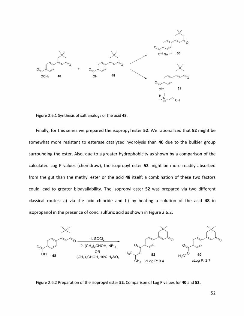
52
Figure 2.6.1 Synthesis of salt analogs of the acid 48.
Finally, for this series we prepared the isopropyl ester 52. We rationalized that 52 might be
somewhat more resistant to esterase catalyzed hydrolysis than 40 due to the bulkier group
surrounding the ester. Also, due to a greater hydrophobicity as shown by a comparison of the
calculated Log P values (chemdraw), the isopropyl ester 52 might be more readily absorbed
from the gut than the methyl ester or the acid 48 itself; a combination of these two factors
could lead to greater bioavailability. The isopropyl ester 52 was prepared via two different
classical routes: a) via the acid chloride and b) by heating a solution of the acid 48 in
isopropanol in the presence of conc. sulfuric acid as shown in Figure 2.6.2.
Figure 2.6.2 Preparation of the isopropyl ester 52. Comparison of Log P values for 40 and 52.

53
A comparison of the activity of these compounds using VSDI in rat brain tissue is given
below. As a modification of the earlier assays, the two concentrations studied were reduced
from 200 nM and 1 μM to 50 nM and 200 nM, respectively, and examined in slices that were
stimulated at both 60 and 20 Hz. The two frequencies were used to display selectivity for
compounds that preferentially reduced the higher gamma oscillation frequency believed to be
associated with pathophysiological epileptiform neuronal activity versus the lower frequency
over normal lower frequency “basal” neuronal activity.
Table 2.6-1 Summary of reduction of neuron firing at 60 Hz by 40 and its salts and isopropyl ester using in vitro bioassays on brain cell slices
Compound Average reduction
at 50 nM (%)
SEM at 50 nM
(%)
Average reduction
at 200 nM (%)
SEM at 200 nM (%)
40 -29 5 -48 5
52 -31 12 -45 6
48 -25 6 -41 6
50 -33 6 -45 5
51 -13 8 -38 5
Table 2.6-2 Summary of reduction of neuron firing at 20 Hz by 40 and its salts and isopropyl ester using in vitro bioassays on brain cell slices
Compound Average reduction
at 50 nM (%)
SEM at 50 nM
(%)
Average reduction
at 200 nM (%)
SEM at 200 nM (%)
40 1 14 -13 13
52 -7 11 -17 12
48 -20 19 -36 9
50 -6 12 -22 8
51 -10 25 -19 13

54
According to the standard error of the mean, there is no difference in the in vitro activity of
the methyl vs the isopropyl ester, 40 vs 52. The acid 48 and the sodium and ammonium salts
50 and 51 all show comparable activity at the 60 Hz frequency at the higher concentration. At
the lowest concentration of 50 nM at 60 Hz, the ammonium salt 51 shows poor inhibition.
Interestingly and somewhat surprisingly, the data at 20 Hz seems to indicate that the acid 48
shows the least amount of frequency selectivity. This would mean that it is susceptible to
producing secondary effects and could affect non-epileptiform neuronal activity because of its
strong inhibition at 20 Hz. A comparison of the in vivo activity of the methyl ester 40 and the
isopropyl ester 52 is being carried out at Centre for Drug Research and Development (CDRD).
2.7 Synthesis of amide analogs of compound 40
The synthesis of amide derivatives of 40 was undertaken next. Such compounds should be
metabolically somewhat more stable since amidases tend to cleave amide bonds more slowly
when the amine part does not represent a natural amino acid.
Six amides with varying structural features were prepared: two aliphatic with one a
secondary (54) and the other tertiary (55): one benzylic (56); and three aromatic, one tertiary
(57) and two secondary (58, 59). They were synthesized by first converting the acid 48 into the
its acid chloride 53 followed by reaction with the appropriate amine (Figure 2.7.1). Since all of
these compounds represent novel structures, they were appropriately characterized (1H and 13C
NMR, HRMS); the data is given in the Experimental Section.

55
Figure 2.7.1 Amide analogs of 40.
The bioassays for these six compounds are shown in the Table below; the data for 40 is also
included for comparison. All of the above amides show rather similar activity when taking into
account the standard error of the mean. The anilide 57 which caused the greatest reduction of
neuronal firing at the 50 nM concentration was chosen as one of five compounds for
submission to the NINDS/ESTP for further evaluation. This required an additional synthesis of
57 to produce the required quantity. Compound 58 carrying the para chloro group on the
aromatic ring was considered a backup to 57 with the idea that 58, if it had similar in vitro
biological activity compared to 57, would likely be more resistant to CYP3A4 metabolism due to
the presence of the electronegative Cl substituent on the aromatic ring. The common
metabolism of aromatic substrates by CYP enzymes including CYP3A4 results in the introduction
of a hydroxyl group. The mechanism involves initial formation of an epoxide followed by a

56
rearrangement known as the NIH shift. The aromatic ring acts as a nucleophile for the epoxide
formation. It has been observed that aromatic rings carrying strongly electron withdrawing
groups such as F, CF3, SO2R and Cl are less prone to CYP induced hydroxylation.
Figure 2.7.2 Hydroxylation by CYP3A4
A number of important drugs including Crestor were designed to be metabolised more
slowly by having fluorine containing substituents in their aromatic rings.
Although the data had poor precision due to the large standard errors of the mean (SEM)
the amides carrying an aromatic residue (56-59) appear to show considerable frequency
selectivity. All, except 58, show about a 40% reduction of VSDI activity at 200 nM when
stimulated at 60 Hz. In contrast stimulation at 20 Hz appears to cause either an increase or have
no effect on activity. Most surprisingly and unexpectedly, the positive stimulation seen with 58
and 59 appears stronger at the 50 nM as compared to the 200 nM concentration. This positive
relative change could be explained by the relatively high standard error of the mean (SEM) and
the low overall reduction compared to the control caused of the lower concentration of
compound.

57
Table 2.7-1 Summary of reduction of neuron firing at 60 Hz by 40 and its amide analogs using in vitro bioassays on brain cell slices
Compound Average reduction
at 50 nM (%)
SEM at 50 nM
(%)
Average reduction
at 200 nM (%)
SEM at 200 nM (%)
40 -29 5 -48 5
54 -19 9 -29 10
55 -32 12 -45 6
56 -30 6 -39 6
57 -24 14 -42 7
58 -22 6 -16 10
59 -20 7 -34 7
Table 2.7-2 Summary of reduction of neuron firing at 20 Hz by 40 and its amide analogs using in vitro bioassays on brain cell slices
Compound Average reduction
at 50 nM (%)
SEM at 50 nM
(%)
Average reduction
at 200 nM (%)
SEM at 200 nM (%)
40 1 14 -13 13
54 -1 31 -8 21
55 -17 15 -9 20
56 7 11 -1 21
57 25 16 47 33
58 30 25 -9 13
59 27 22 -5 13

58
2.8 Synthesis of analogs with different aromatic substituents
A number of other aromatic derivatives were prepared to map out the effect of
substituents, both electron withdrawing and electron donating on the aromatic ring.
Substituents such as F and SO2R were included since these are known to reduce the rate of
metabolism via hydroxylation in the aromatic ring via CYP enzymes.
These compounds are grouped below with the notion that they could be used to investigate
the electronic effects of para substituents. For example, in the set of three compound 60, 61,
and 62 the para nitro group makes the aromatic ring highly electron deficient.
The p-nitro derivative 60 was prepared with the help of Michael Darnowski as part of his
Honours Project via the boronic acid coupling route. Reduction to the amino derivate 61 using
H2 and Pd/C followed by methylation with methyl iodide resulted in an electron rich aromatic
ring.

59
Figure 2.8.1 Synthesis of p-nitro and amine analogs via the boronic acid approach
Table 2.8-1 Summary of reduction of neuron firing at 60 Hz by nitro and amine analogs using in vitro bioassays on brain cell slices
Compound (para substituent)
Average reduction
at 50 nM (%)
SEM at 50 nM
(%)
Average reduction
at 200 nM (%)
SEM at 200 nM (%)
60 (Nitro) -11 15 -22 17
61 (1o Amine) -33 3 -33 7
62 (2o Amine) -22 9 -35 9
Table 2.8-2 Summary of reduction of neuron firing at 20 Hz by nitro and amine analogs using in vitro bioassays on brain cell slices
Compound Average reduction
at 50 nM (%)
SEM at 50 nM
(%)
Average reduction
at 200 nM (%)
SEM at 200 nM (%)
60 (Nitro) -29 14 -11 15
61 (1o Amine) -20 6 -32 10
62 (2o Amine) -18 14 -33 14

60
The tentative conclusion is that the electron withdrawing p-nitro group (60) elicits a smaller
response at 60 Hz compared to the electron donating p-amino groups. Also, surprisingly, 60
shows higher inhibition of -29% at low concentration compared to the -11% result at higher
concentration at 20 Hz. This goes against all previous assays where a higher concentration leads
to higher reduction. Unlike in the amides above, these three derivatives show little or no
frequency selectivity. It should be pointed out that the para carboxymethyl group of 40 is, like a
nitro group, also a potent electron withdrawing group, yet the activity of the latter is
significantly superior to 60.
In the next set of five compounds three have electron donating oxygen substituents in the
para position. However, these compounds differ in several ways. The phenol 63 is both a strong
hydrogen bond donor and an H bond acceptor while 64 to 66 are only H-bond acceptors. The
methyl ether 65 is sterically small while the benzyl ether 66 adds considerable additional steric
bulk to the para position. Would that cause an increase or decrease in the in vitro activity? The
hydroxyl methyl substituent in 47 was added to this group to probe whether an OH or CH2OH at
the para position is more advantageous.
Figure 2.8.2 Structures of new series of analogs containing EWG on the benzene ring
The phenol 63 was prepared by first performing a metal-halogen exchange reaction on
TBDMS protected p-bromophenol. Subsequent reaction with 29 yielded an intermediate which

61
was treated with acetyl chloride followed by acid/base wash to give the deprotected phenol 63.
Acetylation and benzylation of 63 afforded 64 and 66, respectively.
Figure 2.8.3 Synthesis of analogs 63, 64 and 66
Additional amounts of 66 were required for evaluation of this compound by NIH. This was
accomplished via a somewhat shorter route by first benzylating 4-bromophenol to for the
bromoether 67 and carrying out the bromine-lithium exchange reaction in THF followed by
addition of ethoxydimedone. Treatment of the adduct 68 from this reaction with acid gave 66.

62
Figure 2.8.4. Alternate synthesis of 66.
Bioassay data is available for these compounds. At the time compound 65 was
submitted the concentrations used were the original of 200 nM and 1 μM and not the more
recent 50 and 200 nM which became standard in to order differentiate the more potent
compounds. Compound 47 was submitted much later at a time when the Poulter lab suspended
bioassay determination in order to focus more on obtaining data for the potential drug
candidate 40 therefore no data is available for this substance. All show significant reduction of
activity at 60 Hz and almost no effect or a positive effect at the 20 Hz stimulation. Compound
66 was selected as one of the five compounds sent to the NIH. Overall, this set of compounds
seems to be somewhat less effective at reducing firing when cells were stimulated at 60 Hz
than 40. The data obtained at 20 Hz is difficult to interpret with any certainty since the SEMs
are quite large. The `best` conclusion appears to be that these compounds have little effect on
activity when the cells were stimulated at 20 Hz.
Table 2.8-3 Summary of reduction of neuron firing at 60 Hz by benzyl ether, acid and alcohol analogs using in vitro bioassays on brain cell slices
Compound Average reduction
at 50 nM (%)
SEM at 50 nM
(%)
Average reduction
at 200 nM (%)
SEM at 200 nM (%)
63 -25 5 -38 5
64 -21 8 -34 7
65 -- -- -46 11
66 -26 9 -42 6
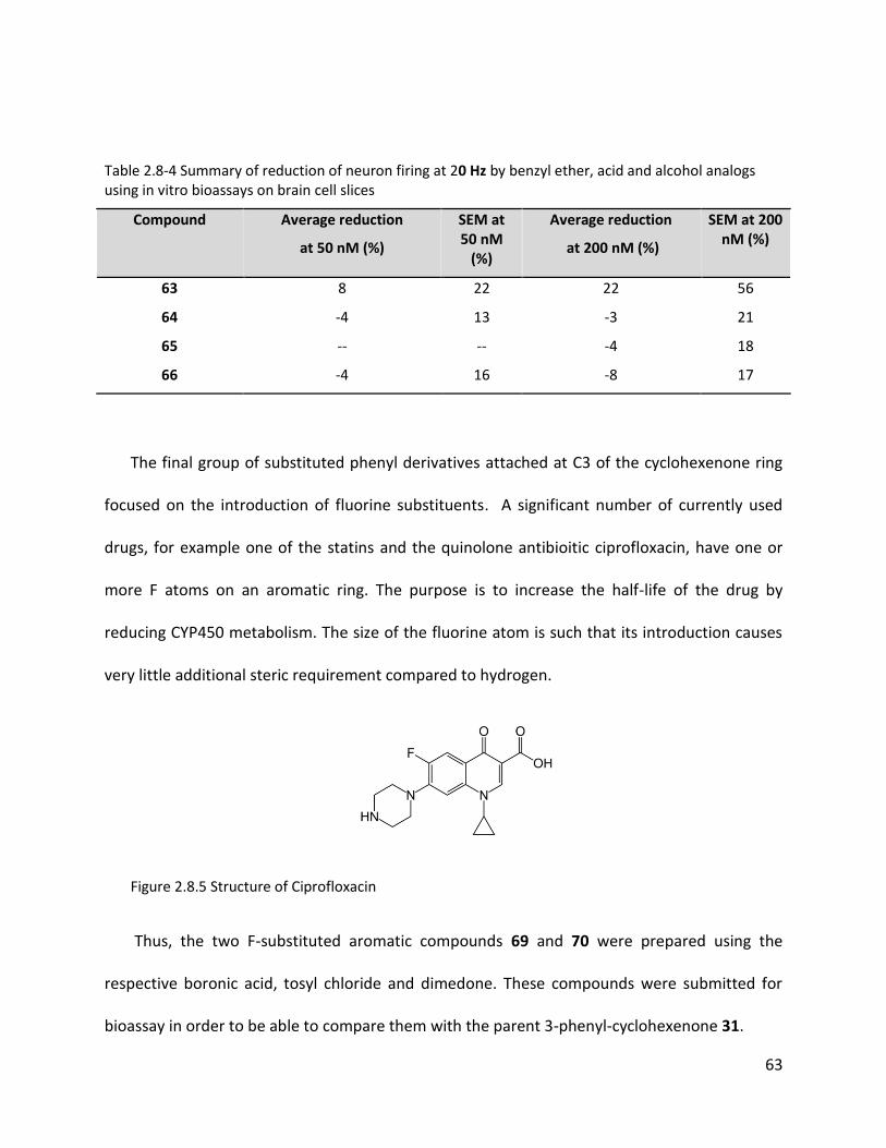
63
Table 2.8-4 Summary of reduction of neuron firing at 20 Hz by benzyl ether, acid and alcohol analogs using in vitro bioassays on brain cell slices
Compound Average reduction
at 50 nM (%)
SEM at 50 nM
(%)
Average reduction
at 200 nM (%)
SEM at 200 nM (%)
63 8 22 22 56
64 -4 13 -3 21
65 -- -- -4 18
66 -4 16 -8 17
The final group of substituted phenyl derivatives attached at C3 of the cyclohexenone ring
focused on the introduction of fluorine substituents. A significant number of currently used
drugs, for example one of the statins and the quinolone antibioitic ciprofloxacin, have one or
more F atoms on an aromatic ring. The purpose is to increase the half-life of the drug by
reducing CYP450 metabolism. The size of the fluorine atom is such that its introduction causes
very little additional steric requirement compared to hydrogen.
Figure 2.8.5 Structure of Ciprofloxacin
Thus, the two F-substituted aromatic compounds 69 and 70 were prepared using the
respective boronic acid, tosyl chloride and dimedone. These compounds were submitted for
bioassay in order to be able to compare them with the parent 3-phenyl-cyclohexenone 31.

64
Figure 2.8.6 Synthesis of fluoro-substituted aromatic compounds
The data for 31 at 20 Hz seems to be an outlier. It shows a strong increase in activity at 20
Hz, but the large uncertainty makes it hard to make a clear conclusion. When 31 was tested via
in vitro bioassay, 50 nM concentrations were not tested. Having this extra data would help
make a clearer comparison with the fluoro substituted aromatic analogs. The introduction of
the F atoms on the aromatic ring seems to have no significant effect on the in vitro activity
when compared to the parent phenyl compound 31.
Table 2.8-5 Summary of reduction of neuron firing at 60 Hz by fluoro substituted aromatic and phenyl analogs using in vitro bioassays on brain cell slices
Compound Average reduction
at 50 nM (%)
SEM at 50 nM
(%)
Average reduction
at 200 nM (%)
SEM at 200 nM (%)
69 -36 5 -33 5
70 -28 12 -38 10
31 -- -- -36 7
Table 2.8-6 Summary of reduction of neuron firing at 20 Hz by fluoro substituted aromatic and phenyl analogs using in vitro bioassays on brain cell slices
Compound Average reduction
at 50 nM (%)
SEM at 50 nM
(%)
Average reduction
at 200 nM (%)
SEM at 200 nM (%)
69 -10 9 -23 9
70 -2 12 -29 15
31 -- -- 29 34

65
Sulfone substituents on aromatic rings often serve the same purpose as F in
pharmaceuticals (CYP450 hydroxylation resistance). They are also sterically much more
demanding. A comparison of the activity of our key compound, the potential drug candidate 40
which carries the CO2CH3 at the para position of the aryl ring with the p-trifluoromethyl
derivative 71 and the p-methylsulfonyl compound 72 was also considered to be potentially
highly informative. All three substituents are strong EWG groups but only 40 has a good
hydrogen bond accepting group. Finally, these three compounds have significantly different log
Ps with the sulfone 72 being most polar and the trifluoromethyl derivative 71 least polar
amongst these three.
As above, 71 and 72 were prepared by the one-pot sequence using commercially available
4-trifluoromethyl and 4-sulfonylmethylboronic acid, respectively. They were purified via silica
gel chromatography, followed by recrystallization from ethyl acetate-hexane mixtures. Each
showed the required two sets of aromatic hydrogen doublets which in 71 were further split due
to long range F-H coupling.
Figure 2.8.7 Structures and calculated LogP of trifluoromethyl and sulfone analogs compared to 40

66
Table 2.8-7 Summary of reduction of neuron firing at 60 Hz by trifluoro and sulfone substituted aromatics compared to 40 using in vitro bioassays on brain cell slices
Compound Average reduction
at 50 nM (%)
SEM at 50 nM
(%)
Average reduction
at 200 nM (%)
SEM at 200 nM (%)
71 -19 10 -26 11
72 -12 7 -46 7
40 -29 5 -48 5
Table 2.8-8 Summary of reduction of neuron firing at 20 Hz by trifluoro and sulfone substituted aromatics compared to 40 using in vitro bioassays on brain cell slices
Compound Average reduction
at 50 nM (%)
SEM at 50 nM
(%)
Average reduction
at 200 nM (%)
SEM at 200 nM (%)
71 -22 12 -33 9
72 -14 8 -26 10
40 1 14 -13 13
The data for these three compounds indicates that reduction of neuronal firing when
stimulated at 60 Hz is highest for 40, especially at the lower, 50 nM concentration. More
importantly, compound 40 shows essentially no activity at 50 nM when the cells were
stimulated at 20 Hz and a 13% +|- 13 reduction at 200 nM. In contrast, the reduction of activity
at the lower frequency is significant for both 71 and 72 at both concentrations. Based on these
data compounds 71 and 72 will not be evaluated further.
Three heteroaromatic analogs 73-75 were prepared to investigate the possibility that a
heterocyclic substituent at position 3 in the cyclohexanone ring might yield significantly more
active compounds than the simple phenyl analog 31 due to the difference in reactivity of the
nucleophilicity of phenyl versus thienyl versus pyridyl, with thienyl being the most and pyridyl
being the least reactive towards electrophiles. The thiophene derivatives were prepared in

67
collaboration with COOP student Michael Darnowski via the one-pot procedure using the
commercially available boronic acids.
Figure 2.8.8 Structures of heteroaromatic analogs
Table 2.8-9 Summary of reduction of neuron firing at 60 Hz by heteroaromatic and phenyl analogs using in vitro bioassays on brain cell slices
Compound Average reduction
at 50 nM (%)
SEM at 50 nM
(%)
Average reduction
at 200 nM (%)
SEM at 200 nM (%)
73 8 13 -8 13
74 -29 9 -49 9
75 -31 8 -38 9
31 -- -- -36 7
Table 2.8-10 Summary of reduction of neuron firing at 20 Hz by heteroaromatic and phenyl analogs using in vitro bioassays on brain cell slices
Compound Average reduction
at 50 nM (%)
SEM at 50 nM
(%)
Average reduction
at 200 nM (%)
SEM at 200 nM (%)
73 -7 13 -6 14
74 -35 11 -27 7
75 -19 20 -32 14
31 -- -- 29 34

68
From these data, it can be inferred that reduction of neuronal firing when stimulated at
60 Hz is high for both 74 and 75. However, these compounds also reduce activity when exposed
to the lower 20 Hz frequency, which could prove to be problematic. As previously explained,
this would cause reduced firing in neurons responsible for functions crucial to life such as
controlling heartrate. These compounds do not show much potential as anti-epileptic drugs
because of their lack of selectivity between the two tested frequencies.
2.9 Sulfur and oxygen containing substituents at C3.
Two cyclohexenones carrying alkoxy substituent at C3, 28 and 29, were available as
intermediates in the synthesis of compounds such as 30 and 31. These were submitted for
bioassays. The results were sufficiently encouraging to suggest to us the preparation of the 3-
thiophenyl analog 76 and from there the corresponding sulfoxide and sulfone 77 and 78,
respectively. The bio-assay data available for these compounds are shown below.
Figure 2.9.1 Synthesis of sulfur containing analogs starting from dimedone intermediates

69
Compound 79, the analog of 76 lacking the 5,5-dimethyl group was prepared directly from
1,3-cyclohexanedione by treatment with thiophenol in the presence of TsOH as the acid
catalyst.
Figure 2.9.2 Synthesis of sulfide analogue from 1,3-cyclohexanedione
Reaction of the lithio derivative of methyl phenyl sulfone with the ethoxy ether 29 afforded
the sulfone derivative 80.
Figure 2.9.3 Synthesis of sulfone analogue 80
Several compounds, in particular the phenylthio analogs 76 and 79, show reduction of
neuronal firing at 60 Hz comparable to the 40. Unlike 40, these compounds seem to show lower
frequency selectivity. It is unfortunate that data for sulfoxide and sulfone analogs 77 and 78 are
not available at this time. The removal of gem-dimethyl group at C5 does not seem to have any
effect on the activity when comparing 76 and 79, as their VSDI bioassay data is almost identical.

70
Table 2.9-1 Summary of reduction of neuron firing at 60 Hz by sulfur derivatives 76 and 79 compared to ethyl ether 29 using in vitro bioassays on brain cell slices
Compound Average reduction
at 50 nM (%)
SEM at 50 nM
(%)
Average reduction
at 200 nM (%)
SEM at 200 nM (%)
29 -20 7 -26 8
76 -32 5 -51 5
79 -33 5 -50 5
Table 2.9-2 Summary of reduction of neuron firing at 20 Hz by sulfur derivatives 76 and 79 compared to ethyl ether 29 using in vitro bioassays on brain cell slices
Compound Average reduction
at 50 nM (%)
SEM at 50 nM
(%)
Average reduction
at 200 nM (%)
SEM at 200 nM (%)
29 -14 11 -23 13
76 -18 11 -25 12
79 -18 11 -25 12
2.10 Additional variations related to compound 40
Because of the importance of 40, despite the conclusions reached earlier when comparing
the activity of cylcohexenones 30 and 31 with the chain extended -methyl nitriles, 32 and 34,
respectively, we reacted 40 with the lithium salt derived from proprionitrile. The crude
reaction product was subsequently treated with acid to effect dehydration. This reaction
sequence yielded the expected product 81 together with 82 which is the result of the attack of
the lithio derivative on the methyl benzoate carbonyl in competition with attack at the enone
carbonyl group. These compounds were separated by silica gel column chromatography using

71
a 9:1 mixture of hexanes and ethyl acetate. Unfortunately, the desired compound 81 was
obtained in only minor quantities and never in a sufficiently pure state for a meaningful
bioassay. The fact that both stereoisomers were present further complicated the purification
process.
Figure 2.10.1 Synthesis of propionitrile analogs of 40.
Compound 82 showed two ketone carbonyl carbon absorptions at 190.0 and 199.7 ppm.
The CH(CH3)CN portion in 82 was characterized by a 1H quartet at 4.36 ppm and the methyl
doublet at 1.65 ppm. Compound 82 showed a smaller reduction of neuron firing at 60 Hz than
40.
2.11 Analogs derived from 1,3-cyclohexanedione
In order to assess the importance of the 5,5-dimethyl groups on the biological activity of the
3-substituted cyclohexenones, we prepared analogs of the most promising derivatives 40, 48
and 19. Starting from 1,3-cyclohexanedione, the same approach was used as for the isophorone
(8) starting material. Conversion to the enol tosylate 83 and the one-pot procedure with the
appropriate boronic acid led to the desired products.

72
Figure 2.11.1 Synthesis of 1,3-cyclohexanedione analogs via boronic acid approach
We also prepared the corresponding chain-extended unsaturated nitriles to mimic the
compound 19 (TD532) by reacting both 84 and 86 with lithiated propionitrile. The bioassay
results for these compounds were compared with those carrying the 5,5-dimethyl group. The
results indicate quite clearly that removal of the 5,5-dimethyl substituents results in a
significant decrease in the potency. The direct comparisons in this set of compounds are
between the 40 and 84, and the compounds 48 and 85, which both show that the compound
containing the 5,5-dimethyl have better activity at 60 Hz and better selectivity.
Figure 2.11.2 Synthesis of propionitrile analogs from 1,3-cyclohexanedione

73
Table 2.11-1 Summary of reduction of neuron firing at 60 Hz by analogs derived from 1,3-cyclohexandione using in vitro bioassays on brain cell slices
Compound Average reduction
at 50 nM (%)
SEM at 50 nM
(%)
Average reduction
at 200 nM (%)
SEM at 200 nM (%)
40 -29 5 -48 5
84 -20 5 -29 4
48 -32 6 -41 6
85 -- -- -36 7
86 -24 10 -42 7
87 -12 8 -18 8
Table 2.11-2 Summary of reduction of neuron firing at 20 Hz by analogs derived from 1,3-cyclohexandione using in vitro bioassays on brain cell slices
Compound Average reduction
at 50 nM (%)
SEM at 50 nM
(%)
Average reduction
at 200 nM (%)
SEM at 200 nM (%)
40 1 14 -13 13
84 -2 16 -7 26
48 -24 19 -36 9
85 -- -- 29 34
86 13 22 -15 20
87 -2 10 -4 11
2.12 Enol-thioether analogs
A final set of compounds comprise the simple dimedone, 26 draw in its enol form, the
ethoxy enol ether 29, and the enol thio compounds 76 and 79. For each of the latter four
compounds, most readily for the enol ethers, one can imagine a relatively simple acid catalysed
hydrolysis back to dimedone.

74
Table 2.12-1 Summary of reduction of neuron firing at 60 Hz by enol-thio analog 76 compared to dimedone 26 and ethoxy enol ether 29 using in vitro bioassays on brain cell slices
Compound Average reduction
at 50 nM (%)
SEM at 50 nM
(%)
Average reduction
at 200 nM (%)
SEM at 200 nM (%)
26 -32 5 -39 5
29 -20 7 -26 8
76 -33 5 -50 5
Table 2.12-2 Summary of reduction of neuron firing at 20 Hz by enol-thio analog 76 compared to dimedone 26 and ethoxy enol ether 29 using in vitro bioassays on brain cell slices
Compound Average reduction
at 50 nM (%)
SEM at 50 nM
(%)
Average reduction
at 200 nM (%)
SEM at 200 nM (%)
26 52 51 -12 14
29 -15 12 -26 15
76 -18 11 -25 12
The data for dimedone, 26, itself shows surprisingly strong reduction of neuronal firing at
the 60 Hz stimulation. It is difficult to interpret the results at 20 Hz due to the very high SEM.
The suggestion is that there is little or no effect at this frequency. Since dimedone exists in the
two tautomeric forms 26, it is plausible that the erratic results are due to slight changes in the
pH since the equilibrium between the enol and diketone form is highly pH dependent. The
thiophenyl derivative 76 reduces neuronal firing when stimulated at 60 Hz at a level
comparable to that of the potential clinical trial candidate 40. The major difference is that 76

75
also reduces significantly the firing at 20 Hz stimulation. Since this is a potential negative
because of the potential interference with important functions in the body such as heartrate
regulation, 76 has not been pursued further. The enol ether 29 shows no selectivity in its effect
on neuronal firing at 20 vs 60 Hz; the activity level is not remarkable.
2.13 Additional comments concerning the bio-assay data.
While two frequency VSDI has the advantage of moderately rapid screening of effect
across an entire neuronal circuit, it presented some difficulty in an accurate prediction of a
clear structure-activity relationship (SAR). Typically, the SEs were significantly larger when the
stimulation was carried out at 20 as compared to 60 Hz. Most all compounds in both the
cyclohexenone and the extended conjugated nitrile series showed some activity, but none
seemed by bioassay clearly superior to all the others. This made development of a more refined
SAR beyond the core cyclohexenone structure difficult and currently hinders the development
of more potent analogs. With regard to the present library of compounds, molecular variants
were chosen partly based on a combination of their ease of synthesis and in an attempt to
introduce a variety of functional groups. We have not yet succeeded to design a new structure
and be confident that its SAR would unequivocally predict improved biological response. Based
on the results thus far we conclude that the binding site for these molecules is capable of
accommodating a significant structure variation at positions C3 and C5 within the
cyclohexenone family.

76
The standard errors of the mean (SEM) in the measurements especially at the 20 Hz
stimulation and the 50 nM concentrations even though the number of measurements were
typically n=9-12 also make it difficult to extract meaningful data. For example, on neuron firing
when stimulated at 20 Hz for the key compound 40 was reported as +1% at 50 nM and -13% at
200 nM with SEMs as 14 and 13%, respectively. This data could be interpreted as suggesting
that 23 has no effect on neuron firing at these concentrations when the cells were stimulated at
the 20 Hz frequency. Many other examples could have been chosen to illustrate this problem.
A number of compounds appear to show an increase in neuron firing at 20 Hz. Unfortunately,
in most of these examples the SEM is sufficiently large to make the conclusion questionable.
Typically, the SEMs were significantly larger when the stimulation was carried out at 20 as
compared to 60 Hz. We recognize that bio-assay results are inherently more difficult to
reproduce than chemical reactions. Signal variance can be caused by such factors as tissue
integrity, dye loading, unintentional quenching and the choice of field of interest for signal
quantitation. Efforts are underway to better control some of these variables to make SAR
interpretation. In the present case, we are simply pointing out that the bio-assay results were
often difficult to interpret with a higher degree of confidence.
OB Pharmaceuticals chose 40 (TD561) as a potential drug candidate and develop further the
profile of this compound in vitro and in vivo. Indeed, as was pointed out earlier in this chapter,
this compound is now undergoing preclinical evaluation, including animal toxicity and
pharmacokinetics studies with the hope that the data obtained would pave the way for
clearance to carry out a Phase 1 clinical trial for safety. It is also one of five compounds
selected jointly by the NINDS/ESTP and OB Pharmaceuticals for verification of activity and

77
examination in different epilepsy and seizure animal models. The in vitro data for these five
compounds undergoing evaluation in the ESTP are shown below.
Table 2.13-1 Summary of reduction of neuron firing at 60 Hz by compounds currently being studied by NINDS using in vitro bioassays on brain cell slices
Compound Average reduction
at 50 nM (%)
SEM at 50 nM
(%)
Average reduction
at 200 nM (%)
SEM at 200 nM
(%)
40 -29 5 -48 5
51 -25 6 -41 7
65 -26 9 -42 6
57 -37 13 -44 10
Compound Average reduction
at 200 nM (%)
SEM at 200 nM
(%)
Average reduction
at 1 µM (%)
SEM at 1 µM (%)
64 -46 11 -57 6
Table 2.13-2 Summary of reduction of neuron firing at 20 Hz by compounds currently being studied by NINDS using in vitro bioassays on brain cell slices
Compound Average reduction
at 50 nM (%)
SEM at 50 nM
(%)
Average reduction
at 200 nM (%)
SEM at 200 nM
(%)
40 1 14 -13 13
51 -20 21 -36 9
65 -4 16 -8 17
57 1 1 1 12
Compound Average reduction
at 200 nM (%)
SEM at 200 nM
(%)
Average reduction
at 1 µM (%)
SEM at 1 µM (%)
64 -16 22 -4 18

78
Figure 2.13.1 Structures of compounds currently being studied by NINDS
The available in vitro data presented below for the following three compounds are not
substantially different from the above five, especially when one takes into account the standard
errors associated with these measurements.
Table 2.13-3 Summary of reduction of neuron firing at 60 Hz by other compounds with similar data compared to those currently being studied by NINDS using in vitro bioassays on brain cell slices
Compound Average reduction
at 50 nM (%)
SEM at 50 nM
(%)
Average reduction
at 200 nM (%)
SEM at 200 nM
(%)
33 -- -- -41 6
76 -33 5 -50 5
89 -28 6 -39 5
Table 2.13-4 Summary of reduction of neuron firing at 20 Hz by other compounds with similar data compared to those currently being studied by NINDS using in vitro bioassays on brain cell slices
Compound Average reduction
at 50 nM (%)
SEM at 50 nM
(%)
Average reduction
at 200 nM (%)
SEM at 200 nM
(%)
33 -- -- -9 22
76 -18 11 -25 12
89 6 21 37 9

79
Figure 2.13.2 Structure for certain analogs containing a variety of substituents
2.14 Biological evaluation of 40 (TD561): The path towards Phase 1 clinical trials and commercialization
In this section, we summarize the physicochemical and biological data that has been
obtained in this lab and by OB Pharmaceuticals. In a number of cases indicated by 29, we are
reduced to quoting results from our collaborators at OB Pharma since, for confidentiality
reasons, they did not provide details. Nevertheless, we feel that it is worthwhile to quote these
results since they illustrate the amount of effort that OB Pharma has put into this compound
and the conviction that this compound may become a plausible clinical trial candidate.
Initially it should be reiterated that 40 is structurally different than any currently approved
or pipeline anti-epileptic treatment. It is noteworthy that in subsequent in vivo studies in rats
and mice conducted by OB Pharmaceuticals, 40 did not show any deleterious side effects
(lethargy, motor impairment, feeding, etc.) at doses in excess of 25-fold its efficacious dose at
suppressing seizures indicating some relevance to the differential frequency profile in the ex
vivo VSDI bioassay.

80
Figure 2.14.1 Structural properties of 40 (TD561) and its calculated LogP value
Compound 40 is a white crystalline powder simple to produce in a one or two pot
sequence from commercial starting materials. It is thermally stable as previously demonstrated
by the accelerated stability studies.
It is chemically stable under neutral and somewhat acidic conditions but subject to basic
hydrolysis to its acid. This was demonstrated by heating it for several hours in isopropanol in
the presence of p-toluenesulfonic acid. No decomposition or reaction with isopropanol was
observed by NMR. It is orally bioavailable with predictable pharmacokinetics.29
It meets the Lipinski rule describing drug-like structures. These rules give parameters such
as MW, LogP, hydrogen bonding, which indicate predicted likely bioavailability. Compound 40 is
also effective at or lower than currently used anti-epileptic drugs (AEDs).29 It shows no
behavioral side effects in treated animals.29 Its molecular target is voltage gated sodium
channels (VGSCs) and by mechanism of action different from current AEDs.29 It has no
physiologically meaningful effect on hERG channels and endogenous cardiomyocyte activity.29 It
is negative in the Ames test,29 which is used to determine mutagenicity and carcinogenicity
caused by chemicals.

81
The purity is easily monitored by TLC and 1H NMR. A sample judged ‘pure’ by NMR was
submitted for HPLC analysis as part of the metabolism study; only one peak was visible. This
was reported by OB Pharma after analysis by the Centre for Drug Research and Development in
Vancouver. The bioavailability was proven experimentally. Oral administration of 40 results in
an ideal plasma profile with a half-life if 3.5 h. The only observable metabolite is the
corresponding acid 48.29
Compound 40 blocks 50 % of voltage gated sodium channels (VGSC) activity in single
neurons in vitro at a concentration of 25 picoM only under conditions of higher frequency 16
ms (~60 Hz) depolarizations. In contrast, three currently used anti-epileptic drugs, Phenytoin,
Lamotrigine and Carbamazepine require 40, 100 and 150 μM to reach the same effect.29 The
above observations were reported by the scientists who carried out many rat experiments
involving 40 and compared their behavior to rats administered currently used AEDs.29
40 represents a new class of anti-seizure compounds that modulate VGSC function, by
increasing the delay in the fast inactivation-reactivation cycle with a preference for high
frequency-open (60 vs 20 Hz) opening channels thus attenuating epileptic seizures. Compound
40, for example slows VGSC state recovery by approximately double the 13 milliseconds
observed for control activity.29
In separate experiments, 40 given orally once daily (20 mg/kg) inhibits the development of
kindling indicating its potential to prevent seizure onset (antiepileptogenic).29
82
Compound 40 inhibit sodium channel function. Unlike most of the current anti-epileptic
drugs, it has little effect on cardiac sodium channels. Additionally, 40 shows no effect on cardiac
mycocyte function when compared to the stimulant isoproterenol.29
Histopathological evaluation of tissues (heart liver, kidney) collected ant necropsy of kindled
rats treated with 40 showed no evidence of toxicity.29
2.15 Conclusions and future work
This project resulted from the decision to send isophorone, 8, the precursor to the parent
lead structure 1, for bio-assay and the discovery that 8 significantly reduced neuronal activity in
rat brain tissue stimulated at 60 Hz using VSDI. This suggested that other 3-substituted
cyclohex-2-en-1-ones should be evaluated. More than fifty such compounds have been
prepared and indeed compound 40 shows considerable promise as a drug candidate.
Thus far we have only investigated only a small part of the potential chemical space
surrounding the cyclohexenones. It appears that removal of the 5,5-dimethyl substituent
reduces the potency of these compounds in the in vitro bioassay. The possibility of having
substituents other than methyl at C5 or substituents at other positions in the cyclohexanone
ring has not been addressed. Two compounds can be considered to begin such an investigation.
One is the natural product 4-isopropylcyclohex-2-enone, 90 and the other is 5-phenyl-1,3-
cyclohexanedione, 91.

83
A comparison of the activity of isophorone, 8, vs 90 (see below) suggests that the latter is
more potent in inhibiting neuronal firing at 60 Hz with a 36% reduction for 90 at 200 nM
compared to 22% for isophorone 8.
Figure 2.15.1 Structures of isophorone 8 and 90
A plausible argument can be made that addition of a substituent such as the one found in
40 at C3 in 90 should result in increased potency. The preparation of several derivatives
including 92 via a Heck coupling reaction is being planned.
Figure 2.15.2 Potential synthesis of potential analogue 92
Table 2.15-1 Summary of reduction of neuron firing at 60Hz by isophorone 8 and the natural product 90 during in vitro bioassays on brain cell slices
Compound Average reduction
at 50 nM (%)
SEM at 50 nM
(%)
Average reduction
at 200 nM (%)
SEM at 200 nM
(%)
8 -- -- -22 13
90 -22 13 -36 7

84
Table 2.15-2 Summary of reduction of neuron firing at 20Hz by isophorone 8 and the natural product 90 during in vitro bioassays on brain cell slices
Compound Average reduction
at 50 nM (%)
SEM at 50 nM
(%)
Average reduction
at 200 nM (%)
SEM at 200 nM
(%)
8 -- -- -15 12
90 5 26 -24 8
5-Phenylcyclphexan-1,3-dione, 91, is commercially available. In view of the activity observed
for 5,5-dimethyl-cycohexane-1,3-dione, (dimedone), 26, it will be recommended that 91 itself
should be tested and that at least four compounds, 93-96, should be prepared in this series. All
of these compounds should be available using the dione 91 as a starting material. Together, this
group of compounds should indicate whether cyclohexenones carrying a 5-aryl substituent in
place of the 5,5-methyl substituents common to most of the compounds described in this thesis
are worth pursuing for their application to the treatment of epilepsy.
Figure 2.15.3 Structures of 5-aryl substituted analogs
Finally, we submitted the simple acyclic enone, 4-phenyl-but-3-en-2-one, 97, for the
relevant bioassays. This compound is compared structurally to 3-phenyl-5,5-dimethyl-cyclohex-
2-en-1-one 33, below. It is recognized that 97 could also be drawn as one of two rotamers,
which only one overlaps readily with 33.

85
Figure 2.15.4 Structure of open chain analogue 97 and comparison with 33
Table 2.15-3 Summary of reduction of neuron firing at 60 Hz by 97 and 33 during in vitro bioassays on brain cell slices
Compound Average reduction
at 50 nM (%)
SEM at 50 nM
(%)
Average reduction
at 200 nM (%)
SEM at 200 nM
(%)
97 -22 8 -26 5
33 -- -- -36 7
Table 2.15-4 Summary of reduction of neuron firing at 20 Hz by 97 and 33 during in vitro bioassays on brain cell slices
Compound Average reduction
at 50 nM (%)
SEM at 50 nM
(%)
Average reduction
at 200 nM (%)
SEM at 200 nM
(%)
97 -13 21 -20 12
33 -- -- 29 34
The observation of a significant reduction of neuronal firing at both 60 and 20 Hz
stimulation suggests that it might be worthwhile to consider exploring a number of open chain
enone analogs of 97 for example compounds carrying substituents in the aromatic ring (98)
which is related to 40 (TD 561) or additional carbon substituents at both C1 and at C3 (99). The

86
additional substituent at C3 would decrease the potential of interaction with glutathione via
electrophilic addition to the enone system.
Figure 2.15.5 Structures of proposed of open chained next target analogs

87
3 Experimental data
Introduction
General:
All reagents were obtained from commercial sources and used without further
purification. All solvents used in inert N2 atmosphere reactions were dried prior to use. Other
solvents were not dried before use. Most of the experimental details have been reported in the
Owen-Barry Pharmaceuticals Inc. patent published in July 2018.34
NMR:
1H NMR spectra were recorded in CDCl3 or MeOD on a Bruker Avance II 300 MHz or Bruker
Avance 400 MHz spectrometer. Chemical shifts are referenced to the residual solvent signal (δ
= 7.26) for CDCl3 or (δ = 3.31) for MeOD. 13C NMR were recorded in CDCl3 or MeOD on a Bruker
Avance 400 MHz spectrometer. Chemical shifts are referenced to the residual solvent signal (δ
= 77.16) for CDCl3 or (δ = 49.0) for MeOD. The spectroscopic data for known compounds was
compared with that reported in the literature; only the reference is supplied. Other compounds
not found in the literature do not have an associated reference.
HRMS:
To obtain the high-resolution mass of compounds, the Kratos Concept - Magnetic sector
Electron impact mass spectrometer, located in the John L. Holmes Mass Spectrometry Facility
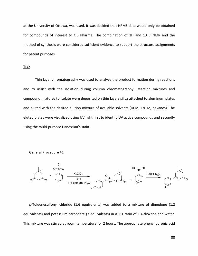
88
at the University of Ottawa, was used. It was decided that HRMS data would only be obtained
for compounds of interest to OB Pharma. The combination of 1H and 13 C NMR and the
method of synthesis were considered sufficient evidence to support the structure assignments
for patent purposes.
TLC:
Thin layer chromatography was used to analyze the product formation during reactions
and to assist with the isolation during column chromatography. Reaction mixtures and
compound mixtures to isolate were deposited on thin layers silica attached to aluminum plates
and eluted with the desired elution mixture of available solvents (DCM, EtOAc, hexanes). The
eluted plates were visualized using UV light first to identify UV active compounds and secondly
using the multi-purpose Hanessian’s stain.
General Procedure #1
p-Toluenesulfonyl chloride (1.6 equivalents) was added to a mixture of dimedone (1.2
equivalents) and potassium carbonate (3 equivalents) in a 2:1 ratio of 1,4-dioxane and water.
This mixture was stirred at room temperature for 2 hours. The appropriate phenyl boronic acid

89
(1 equivalent) and tetrakis(triphenylphosphine)palladium (0) (3% equivalents) were added and
the mixture was heated under reflux for 4 hours or until completion. The resulting mixture was
extracted with EtOAc. The extract was dried with MgSO4, filtered and evaporate via rotary
evaporator. The product was isolated via column chromatography. If further purification was
required, a subsequent crystallization was performed to obtain white crystals.
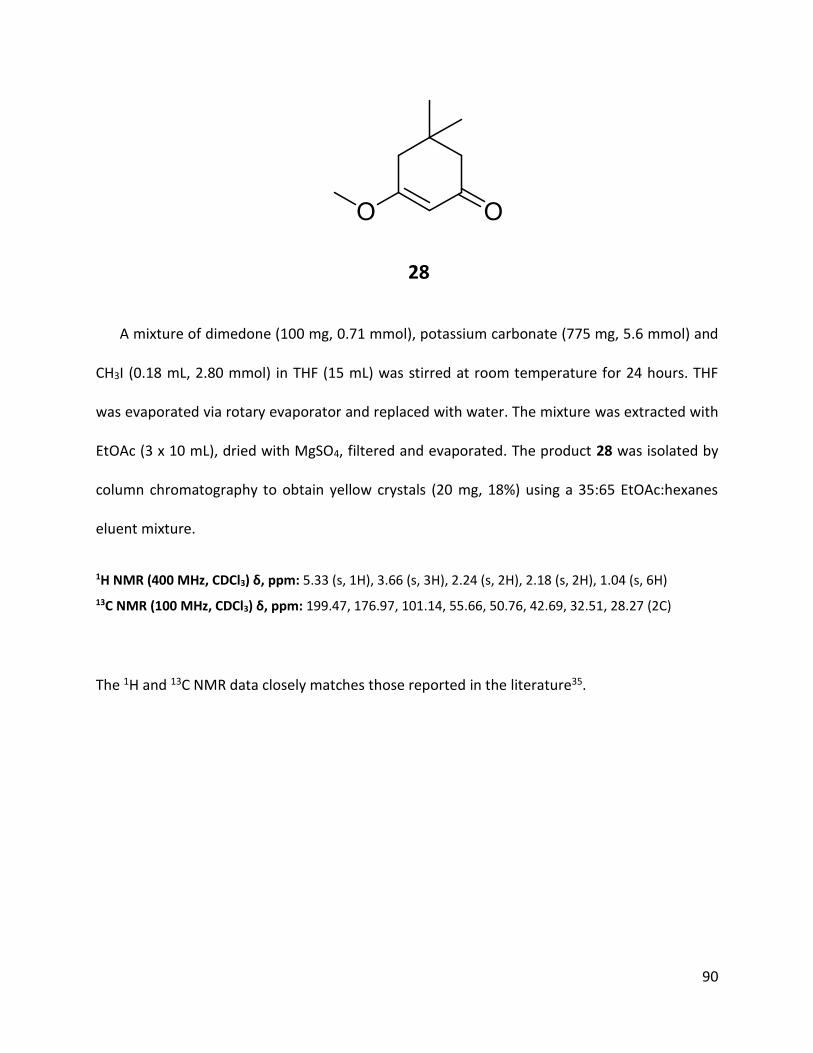
90
28
A mixture of dimedone (100 mg, 0.71 mmol), potassium carbonate (775 mg, 5.6 mmol) and
CH3I (0.18 mL, 2.80 mmol) in THF (15 mL) was stirred at room temperature for 24 hours. THF
was evaporated via rotary evaporator and replaced with water. The mixture was extracted with
EtOAc (3 x 10 mL), dried with MgSO4, filtered and evaporated. The product 28 was isolated by
column chromatography to obtain yellow crystals (20 mg, 18%) using a 35:65 EtOAc:hexanes
eluent mixture.
1H NMR (400 MHz, CDCl3) δ, ppm: 5.33 (s, 1H), 3.66 (s, 3H), 2.24 (s, 2H), 2.18 (s, 2H), 1.04 (s, 6H)
13C NMR (100 MHz, CDCl3) δ, ppm: 199.47, 176.97, 101.14, 55.66, 50.76, 42.69, 32.51, 28.27 (2C)
The 1H and 13C NMR data closely matches those reported in the literature35.

91

92

93
29
A mixture of dimedone (5.0 g, 35.67 mmol), absolute ethanol (6.7 mL, 114.93 mmol) and p-
toluenesulfonic acid (200 mg, catalytic) in benzene was refluxed for 8 hours. The mixture was
quenched using an aqueous NaHCO3 solution and extracted with EtOAc (3 x 10 mL). The
resulting extract was dried with MgSO4, filtered and evaporated. The product 29 was isolated
by column chromatography to obtain yellowish crystals (3.78g, 63%) using a 3:7 EtOAc :
hexanes eluent mixture.
1H NMR (400 MHz, CDCl3) δ, ppm: 5.32 (s, 1H), 3.88 (q, J = 7.04 Hz, 2H), 2.25 (s, 2H), 2.19 (s, 2H), 1.34 (t, J = 7.04 Hz, 3H), 1.05 (s, 6H)
13C NMR (100 MHz, CDCl3) δ, ppm: 199.58, 176.16, 101.44, 64.17, 50.68, 42.90, 32.41, 28.22 (2C), 14.07
The 1H and 13C NMR data closely matches those reported in the literature36.
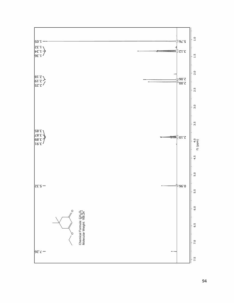
94

95

96
30
3-Methoxy-5,5-dimethylcyclohex-2-enone (28) (1.0 g, 6.48 mmol) was dissolved in dry THF
at 0oC under nitrogen atmosphere. n-Butyllithium 2.5M (6.7 mL, 114 mmol) was slowly added
and the solution was stirred for 20 minutes at 0oC. The mixture was quenched using an aqueous
NH4Cl solution and extracted with EtOAc (3 x 10 mL). The resulting extract was dried with
MgSO4, filtered and evaporated. Compound 30 was isolated by column chromatography (370
mg, 32%) using a 2.5% EtOAc in hexanes eluent mixture.
1H NMR (400 MHz, CDCl3) δ, ppm: 5.87 (s, 1H), 2.18 (m, 6H), 1.47 (m, 2H), 1.33 (qd, J = 14.36, 7.15 Hz, 2H), 1.02 (s, 6H), 0.91 (t, J = 7.28 Hz, 3H)
The 1H NMR data closely matches those reported in the literature37.

97

98
31
Magnesium turnings (0.30 g, 12.3 mmol) were added to a flask and put under nitrogen
atmosphere. A solution of bromobenzene (1.50 g, 9.55 mmol) in dry diethyl ether (10 mL) was
added and the mixture was lightly refluxed until all the magnesium turnings were consumed. A
solution of 3-methoxy-5,5-dimethylcyclohex-2-enone (28) (0.50 g, 3.24 mmol) in diethyl ether
(10 mL) was added at room temperature and the mixture was refluxed again for 30 minutes.
The reaction was quenched using water and a saturated aqueous solution of NH4Cl. The
mixture was extracted with EtOAc, dried with MgSO4, filtered and evaporated. The product 31
was isolated by column chromatography to obtain a white solid. (260 mg, 40 %) using a 5%
EtOAc in hexanes eluent mixture.
1H NMR (400 MHz, CDCl3) δ, ppm: 7.54 (ddd, J = 4.48, 2.39, 1.40 Hz, 2H), 7.43-7.40 (m, 3H), 6.42 (t, J = 1.50, 1.50 Hz, 1H), 2.66 (d, J = 1.46 Hz, 2H), 2.35 (s, 2H), 1.14 (s, 6H)
The 1H NMR data closely matches those reported in the literature38.

99

100
32
Lithium diisopropylamide 2M (2.05 mL, 4.10 mmol) was added to dry THF (15 mL) under
nitrogen atmosphere at -78oC. Propionitrile (270 mg, 4.92 mmol) was added dropwise and the
solution was stirred at -78oC for 15 minutes. 3-butyl-5,5-dimethylcyclohex-2-enone (30) (370
mg, 2.05 mmol) was added dropwise and the solution was stirred for 30 minutes. The mixture
was quenched by transferring it to a beaker containing an aqueous NH4Cl solution. The mixture
was then warmed at room temperature and diluted with water followed by extraction with
EtOAc (3 x 10 mL). The extract was dried with MgSO4, filtered and concentrated by rotary
evaporator. The hydroxyl intermediate was isolated by column chromatography.
The hydroxyl intermediate was dissolved in toluene and p-toluenesulfonic acid (100 mg,
catalytic) was added. The mixture was refluxed for 6 hours and was quenched by adding an
aqueous NaHCO3 solution. The mixture was extracted with EtOAc (3 x 10 mL) and the extract
was dried with MgSO4, filtered and evaporated by rotary evaporator. 32 was isolated by column
chromatography (170 mg, 38%) using a 5% EtOAc in hexanes eluent mixutre.
1H NMR (400 MHz, CDCl3) δ, ppm: 6.44 (s, 1/2H), 6.17 (s, 1/2H), 2.35 (d, J = 1.35 Hz, 1H), 2.14-2.07 (m, 3H), 1.94 (m, 5H), 1.47-1.37 (m, 2H), 1.36-1.24 (m, 2H), 0.93 (s, 6H), 0.90 (m, 3H)
13C NMR (100 MHz, CDCl3) δ, ppm: 151.38, 150.16, 150.02, 148.35, 121.23, 121.14, 120.32, 117.87, 99.55, 99.47, 43.69, 43.50, 43.09, 39.82, 38.24, 37.85, 31.29, 31.00, 30.90, 29.71, 29.65, 28.27 (2C), 27.94 (2C), 22.43, 15.61, 14.94, 13.99, 13.62

101
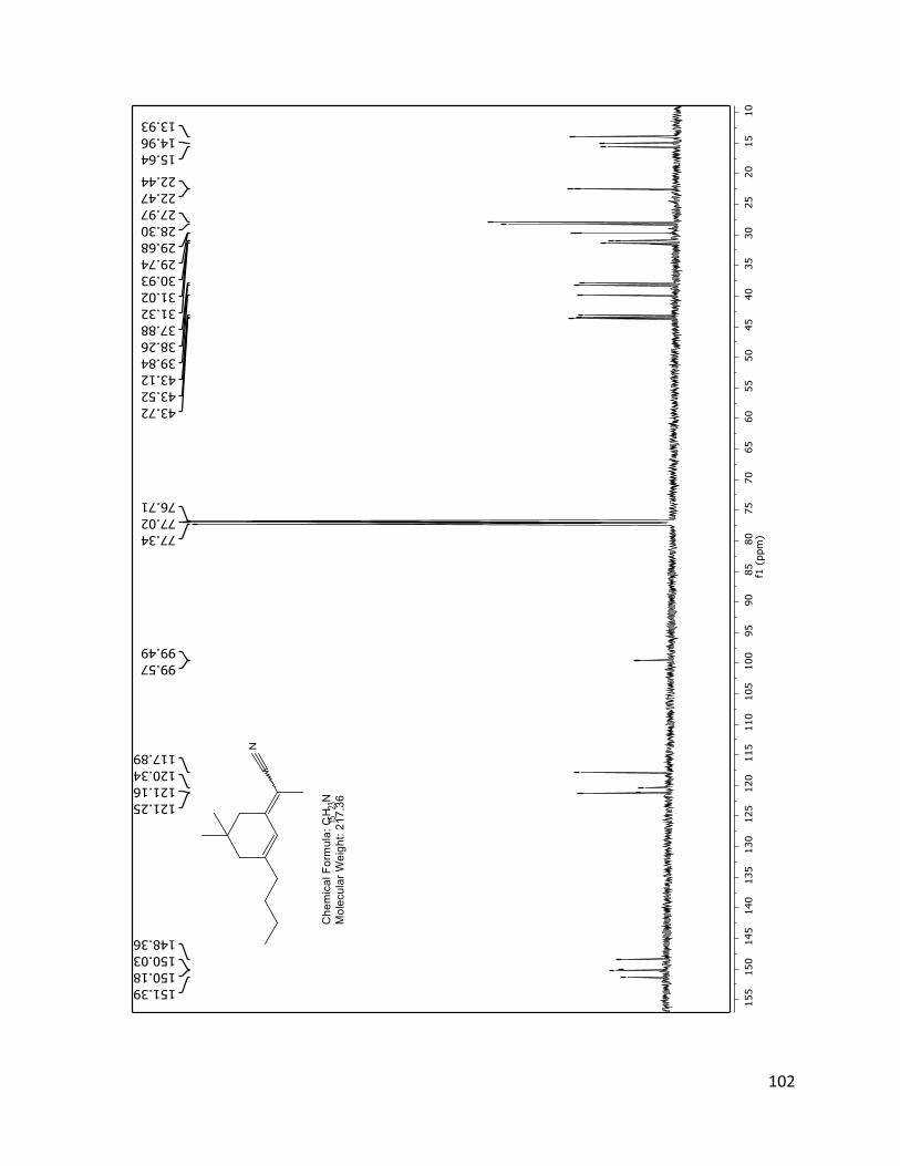
102

103
33
Lithium diisopropylamide 2M (3.22 mL, 6.44 mmol) was added to dry THF (15 mL) under
nitrogen atmosphere at -78oC. Propionitrile (426 mg, 7.73 mmol) was added dropwise and the
solution was stirred at -78oC for 15 minutes. 5,5-dimethylcyclohex-2-enone (400 mg, 3.22
mmol) was added dropwise and the solution was stirred for 30 minutes. The mixture was
quenched by transferring it to a beaker containing an aqueous NH4Cl solution. The mixture was
then warmed at room temperature and diluted with water followed by extraction with EtOAc (3
x 10 mL). The extract was dried with MgSO4, filtered and concentrated by rotary evaporator.
The hydroxyl intermediate was isolated by column chromatography.
The intermediate alcohol was dissolved in toluene and p-toluenesulfonic acid (100 mg,
catalytic) was added. The mixture was refluxed for 6 hours and was quenched by adding an
aqueous NaHCO3 solution. The mixture was extracted with EtOAc (3 x 10 mL) and the extract
was dried with MgSO4, filtered and evaporated by rotary evaporator. 33 was isolated by column
chromatography (230 mg, 44%) using a 5:25:70 eluent mixture of EtOAc : DCM : hexanes.
1H NMR (400 MHz, CDCl3) δ, ppm: 6.52 (tdd, J = 96.04, 10.06, 2.06 Hz, 1H), 6.12-6.00 (m, 1H), 2.40 (d, J = 1.26 Hz, 1H), 2.15 (s, 1H), 2.08-2.02 (m, 2H), 1.94 (d, J = 8.98 Hz, 3H), 0.95 (s, 6H),
13C NMR (100 MHz, CDCl3) δ, ppm: 150.45, 149.17, 136.00, 134.28, 125.90, 122.43, 120.52, 119.74, 102.29, 102.13, 43.62, 40.14, 39.89, 39.51, 31.03, 30.68, 28.33 (2C), 28.00 (2C), 15.57, 15.06
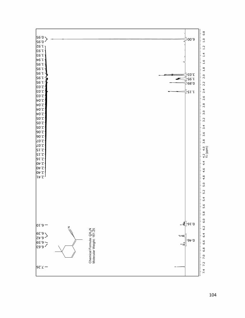
104

105

106
34
Lithium diisopropylamide 2M (0.8 mL, 1.5 mmol) was added to dry THF (15 mL) under
nitrogen atmosphere at -78oC. Propionitrile (125 mg, 1.8 mmol) was added dropwise and the
solution was stirred at -78oC for 15 minutes. 31 (150 mg, 0.75 mmol) was added dropwise and
the solution was stirred for 30 minutes. The mixture was quenched by transferring it to a
beaker containing an aqueous NH4Cl solution. The mixture was then warmed at room
temperature and diluted with water followed by extraction with EtOAc (3 x 10 mL). The extract
was dried with MgSO4, filtered and concentrated by rotary evaporator. The hydroxyl
intermediate was isolated by column chromatography.
The hydroxyl intermediate was dissolved in toluene and p-toluenesulfonic acid (100 mg,
catalytic) was added. The mixture was refluxed for 6 hours and was quenched by adding an
aqueous NaHCO3 solution. The mixture was extracted with EtOAc (3 x 10 mL) and the extract
was dried with MgSO4, filtered and evaporated by rotary evaporator. 34 was isolated by column
chromatography (80 mg, 45%) using a 10% EtOAc in hexanes eluent mixture.
1H NMR (400 MHz, CDCl3) δ, ppm: 7.51 (s, 3H), 7.37 (dd, J = 6.6, 5.1 Hz, 2H), 2.52 – 2.42 (m, 3H), 2.23 (d, J = 1.1 Hz, 1H), 2.05 (d, J = 3.2 Hz, 3H), 1.11 (s, 1H), 1.04 (d, J = 1.2 Hz, 6H)
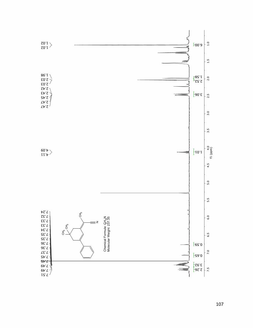
107

108
40 (TD 561)
First synthesis (1 pot reaction):
p-Toluenesulfonyl chloride (1.65 g, 8.67 mmol) was added to a mixture of dimedone (935
mg, 6.67 mmol) and potassium carbonate (2.31 g, 16.7 mmol) in a 2:1 ratio of 1,4-dioxane (16
mL) and water (8 mL). This mixture was stirred at room temperature for 2 hours. 4-
methoxycarbonylphenylboronic acid (1.35 g, 8.56 mmol) and
tetrakis(triphenylphosphine)palladium (0) (231 mg, 0.20 mmol) were added and the mixture
was heated under reflux for 4 hours or until completion. The resulting mixture was extracted
with EtOAc (3 x 10 mL). The extract was dried with MgSO4, filtered and evaporated via rotary
evaporator. The product 40 was isolated via column chromatography using a 5% EtOAc in DCM
eluent mixture. A subsequent crystallization was performed to obtain light-yellow crystals.
(0.71 g, 41 %).
1H NMR (400 MHz, CDCl3) δ, ppm: 8.09-8.00 (m, 2H), 7.63-7.48 (m, 2H), 6.44 (t, J = 1.40 Hz, 1H), 3.93 (s, 3H), 2.66 (d, J = 1.40 Hz, 2H), 2.36 (s, 2H), 1.14 (s, 6H)
13C NMR (100 MHz, CDCl3) δ, ppm: 199.78, 166.43, 156.24, 143.44, 131.16, 129.95 (2C), 126.12 (2C), 125.74, 52.30, 50.93, 42.30, 33.82, 28.38 (2C).
HRMS: Calculated for C16H18O3: 258.1256 Found: 258.1264

109
Second synthesis (2 step):
p-Toluenesulfonyl chloride (9.90 g, 52 mmol) was added to a mixture of dimedone (5.61 g,
40 mmol) and potassium carbonate (13.8 g, 100 mmol) in a 2:1 ratio of 1,4-dioxane (60 mL) and
water (30 mL). This mixture was stirred at room temperature for 15 hours. The reaction mixture
was diluted with 100 mL of water and extracted with 50 mL of DCM twice. The combined
organic layer was washed with 50 mL of saturated NH4Cl solution, dried with MgSO4 and
concentrated via rotary evaporator to isolate the tosylate intermediate (7.0 g, 23.8 mmol)
The tosylate intermediate (6.0 g, 20.4 mg), 4-methoxycarbonylphenylboronic acid (3.35 g,
18.6 mmol), potassium carbonate (5.64 g, 40.8 mmol) and
tetrakis(triphenylphosphine)palladium (0) (707 mg, 0.612 mmol) were added and the mixture
was heated under reflux for 3 hours or until completion. The resulting mixture was diluted with
50 mL of water and extracted with DCM (3 x 10 mL). The extract was dried with MgSO4, filtered
and evaporated via rotary evaporator. The product 40 was isolated via crystallization. (2.7 g,
55%).

110

111
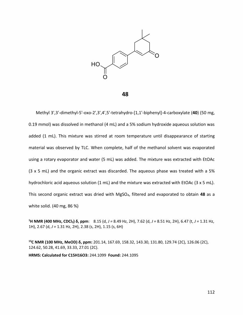
112
48
Methyl 3',3'-dimethyl-5'-oxo-2',3',4',5'-tetrahydro-[1,1'-biphenyl]-4-carboxylate (40) (50 mg,
0.19 mmol) was dissolved in methanol (4 mL) and a 5% sodium hydroxide aqueous solution was
added (1 mL). This mixture was stirred at room temperature until disappearance of starting
material was observed by TLC. When complete, half of the methanol solvent was evaporated
using a rotary evaporator and water (5 mL) was added. The mixture was extracted with EtOAc
(3 x 5 mL) and the organic extract was discarded. The aqueous phase was treated with a 5%
hydrochloric acid aqueous solution (1 mL) and the mixture was extracted with EtOAc (3 x 5 mL).
This second organic extract was dried with MgSO4, filtered and evaporated to obtain 48 as a
white solid. (40 mg, 86 %)
1H NMR (400 MHz, CDCl3) δ, ppm: 8.15 (d, J = 8.49 Hz, 2H), 7.62 (d, J = 8.51 Hz, 2H), 6.47 (t, J = 1.31 Hz, 1H), 2.67 (d, J = 1.31 Hz, 2H), 2.38 (s, 2H), 1.15 (s, 6H)
13C NMR (100 MHz, MeOD) δ, ppm: 201.14, 167.69, 158.32, 143.30, 131.80, 129.74 (2C), 126.06 (2C), 124.62, 50.28, 41.69, 33.33, 27.01 (2C).
HRMS: Calculated for C15H16O3: 244.1099 Found: 244.1095

113

114

115
50
3',3'-dimethyl-5'-oxo-2',3',4',5'-tetrahydro-[1,1'-biphenyl]-4-carboxylic acid (48) (0.1 g, 0.41
mmol) was dissolved in dichloromethane (10 mL) at room temperature. Sodium
bis(trimethylsilyl)amide 1M (0.41 mL, 0.41 mmol) was added dropwise and a white precipitate
was immediately observed. All the solvent was evaporated using a rotary evaporator and
diethyl ether (10 mL) was added to the mixture. The product 50 was obtained as a fine beige
powder by filtering the suspension. (96.5 mg, 88 %)
1H NMR (400 MHz, MeOH-D4) δ, ppm: 7.97 (d, J = 8.19 Hz, 2H), 7.60 (d, J = 8.20 Hz, 2H), 6.39 (s, 1H), 2.73 (s, 2H), 2.33 (s, 2H), 1.11 (s, 6H)
13C NMR (100 MHz, MeOH-D4) δ, ppm: 202.85, 174.01, 160.76, 142.17, 140.15, 130.77 (2C), 126.96 (2C), 125.14, 51.76, 43.20, 34.75, 28.48 (2C)

116

117

118
51
3',3'-dimethyl-5'-oxo-2',3',4',5'-tetrahydro-[1,1'-biphenyl]-4-carboxylic acid (48) (100 mg,
0.41 mmol) was dissolved in dichloromethane (10 mL). 2-Dimethylaminoethanol (36.5 mg, 0.41
mmol) in a solution of dichloromethane was added and the mixture was stirred for 10 minutes.
The solvent was completely evaporated and diethyl ether (10 mL) was added to the mixture.
The observed precipitated salt was filtered to obtain 51 as a fine white powder. (103 mg, 75 %)
1H NMR (400 MHz, CDCl3) δ, ppm: 8.08 (d, J = 8.41 Hz, 2H), 7.55 (d, J = 8.40 Hz, 2H), 6.44 (t, J = 1.23 Hz, 1H), 4.00-3.91 (m, 2H), 3.12-3.05 (m, 2H), 2.80 (s, 6H), 2.67 (d, J = 1.32 Hz, 2H), 2.35 (s, 2H), 1.14 (s, 6H)
13C NMR (100 MHz, MeOD) δ, ppm: 201.35, 159.22, 140.87, 138.30, 129.33 (2C), 125.54 (2C), 123.74, 59.19, 55.62, 50.30, 42.42 (2C), 41.74, 33.29, 27.03 (2C).

119
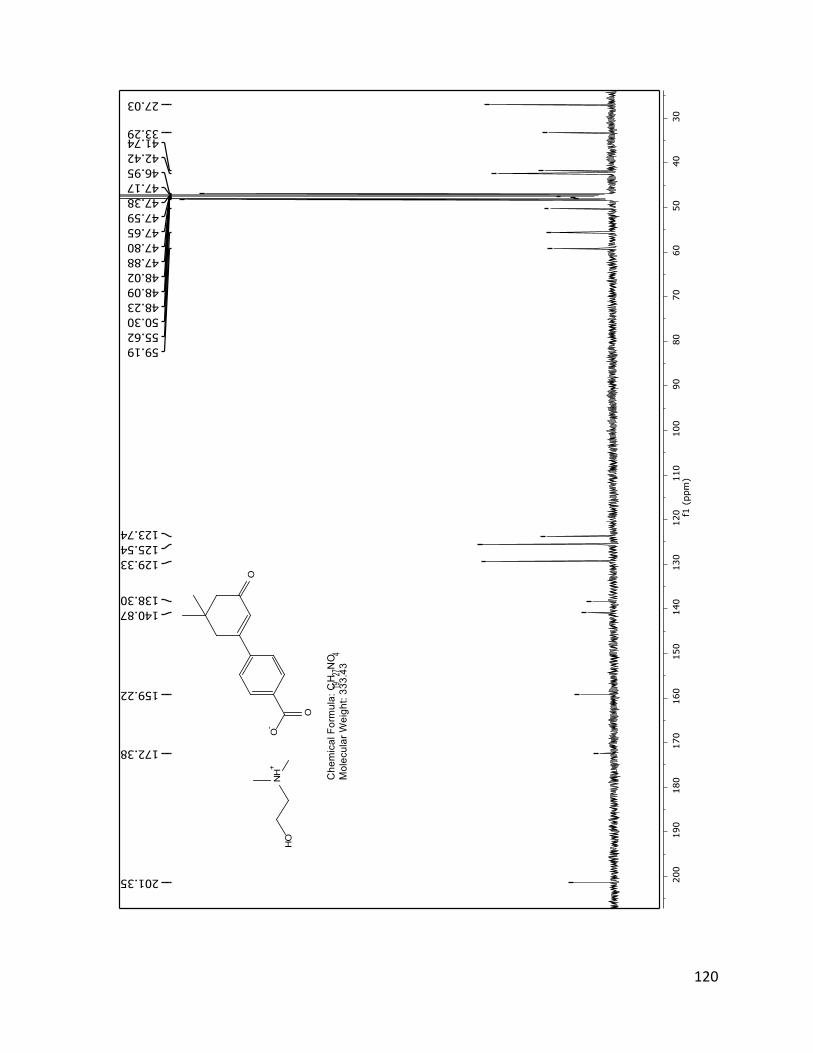
120
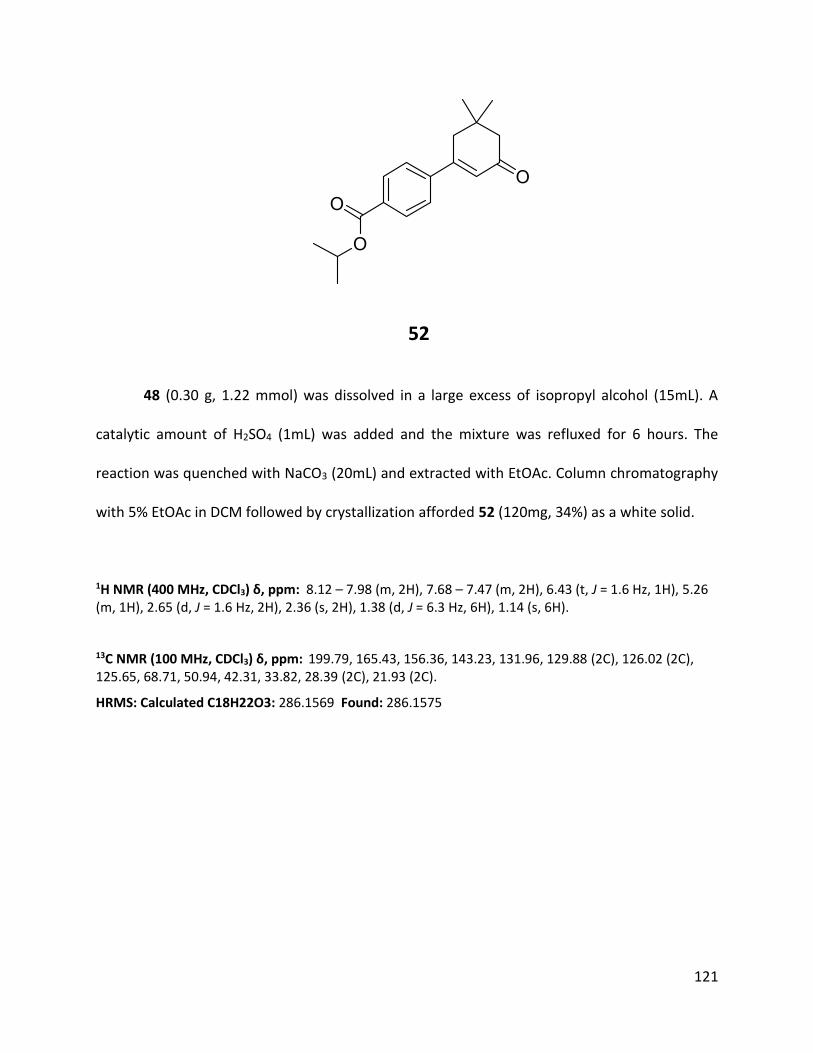
121
52
48 (0.30 g, 1.22 mmol) was dissolved in a large excess of isopropyl alcohol (15mL). A
catalytic amount of H2SO4 (1mL) was added and the mixture was refluxed for 6 hours. The
reaction was quenched with NaCO3 (20mL) and extracted with EtOAc. Column chromatography
with 5% EtOAc in DCM followed by crystallization afforded 52 (120mg, 34%) as a white solid.
1H NMR (400 MHz, CDCl3) δ, ppm: 8.12 – 7.98 (m, 2H), 7.68 – 7.47 (m, 2H), 6.43 (t, J = 1.6 Hz, 1H), 5.26 (m, 1H), 2.65 (d, J = 1.6 Hz, 2H), 2.36 (s, 2H), 1.38 (d, J = 6.3 Hz, 6H), 1.14 (s, 6H).
13C NMR (100 MHz, CDCl3) δ, ppm: 199.79, 165.43, 156.36, 143.23, 131.96, 129.88 (2C), 126.02 (2C), 125.65, 68.71, 50.94, 42.31, 33.82, 28.39 (2C), 21.93 (2C).
HRMS: Calculated C18H22O3: 286.1569 Found: 286.1575

122

123

124
54
3',3'-dimethyl-5'-oxo-2',3',4',5'-tetrahydro-[1,1'-biphenyl]-4-carboxylic acid (48) (75 mg,
0.31 mmol) was added to dichloromethane (15 mL). Thionyl chloride (62.1 mg, 0.52 mmol) was
added and the mixture was refluxed for 3 hours. The solvent and excess thionyl chloride were
evaporated using a rotary evaporator. The flask was closed using a septum and the mixture was
dissolved in benzene (15 mL) at 0oC. Isopropylamine (72.7 mg, 1.23 mmol) and triethyl amine
(124 mg, 1.23 mmol) were very slowly added simultaneously and the mixture was stirred for 5
minutes at 0oC. The reaction was quenched with water and an aqueous solution of NH4Cl. The
mixture was extracted with EtOAc (3 x 10 mL), dried with MgSO4 and filtered. The extract was
treated with an aqueous solution of 5% sodium hydroxide. The product 54 was isolated by
column chromatography with 30% EtOAc in hexanes to obtain a white fine powder. (50 mg, 57
%)
1H NMR (400 MHz, CDCl3) δ, ppm: 7.79 (d, J = 8.37 Hz, 2H), 7.57 (d, J = 8.37 Hz, 2H), 6.42 (s, 1H), 5.95 (d, J = 6.25 Hz, 1H), 4.30 (qd, J = 13.30, 6.56 Hz, 1H), 2.65 (d, J = 1.15 Hz, 2H), 2.35 (s, 2H), 1.28 (d, J = 6.55 Hz, 6H), 1.14 (s, 6H)
13C NMR (100 MHz, CDCl3) δ, ppm: 199.84, 165.78, 156.34, 141.81, 136.00, 127.28 (2C), 126.24 (2C), 125.30, 50.89, 42.23, 42.03, 33.77, 28.36 (2C), 22.80 (2C)

125

126

127
55
3',3'-Dimethyl-5'-oxo-2',3',4',5'-tetrahydro-[1,1'-biphenyl]-4-carboxylic acid (48) (0.1 g, 0.41
mmol) was added to dichloromethane (15 mL). Thionyl chloride (81.9 mg, 0.69 mmol) was
added and the mixture was refluxed for 3 hours. The solvent and excess thionyl chloride were
evaporated using a rotary evaporator. The flask was closed using a septum and the mixture was
dissolved in benzene (15 mL) at 0oC. Piperidine (70.0 mg, 0.82 mmol) and triethyl amine (82.8
mg, 0.82 mmol) were very slowly added simultaneously and the mixture was stirred for 5
minutes at 0oC. The reaction was quenched with water and an aqueous solution of NH4Cl. The
mixture was extracted with EtOAc (3 x 10 mL), dried with MgSO4 and filtered. The extract was
treated with an aqueous solution of 5% sodium hydroxide. The product 55 was isolated by
column chromatography with 35% EtOAc in hexanes to obtain a white fine powder. (90 mg, 70
%)
1H NMR (400 MHz, CDCl3) δ, ppm: 7.59-7.52 (m, 2H), 7.46-7.41 (m, 2H), 6.42 (t, J = 1.45 Hz, 1H), 3.78-3.66 (br, 2H), 3.43-3.28 (br, 2H), 2.64 (d, J = 1.42 Hz, 2H), 2.35 (s, 2H), 1.72-1.61 (m, 4H), 1.57-1.49 (m, 2H), 1.13 (s, 6H)
13C NMR (100 MHz, MeOD) δ, ppm: 199.90, 169.44, 156.62, 140.06, 137.89, 127.29 (2C), 126.25 (2C), 124.98, 50.93, 42.31, 33.79, 28.40 (2C), 24.56.
HRMS: Calculated for C20H25NO2: 311.1885 Found: 311.1889

128
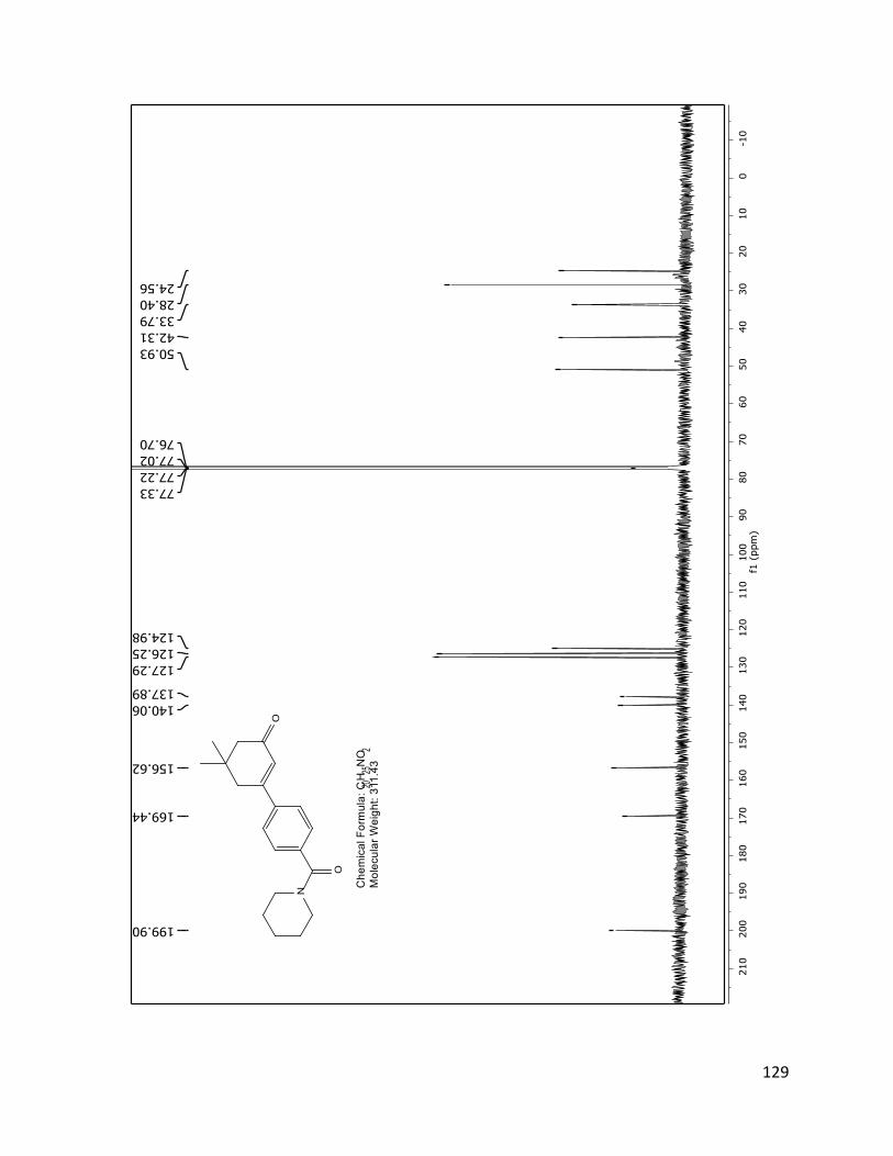
129

130
56
3',3'-Dimethyl-5'-oxo-2',3',4',5'-tetrahydro-[1,1'-biphenyl]-4-carboxylic acid (48) (100 mg,
0.41 mmol) was added to dichloromethane (15 mL). Thionyl chloride (82.7 mg, 0.70 mmol) was
added and the mixture was refluxed for 3 hours. The solvent and excess thionyl chloride were
evaporated using a rotary evaporator. The flask was closed using a septum and the mixture was
dissolved in benzene (15 mL) at 0oC. Benzyl amine (175 mg, 1.64 mmol) and triethyl amine (166
mg, 1.64 mmol) were very slowly added simultaneously and the mixture was stirred for 5
minutes at 0oC. The reaction was quenched with water and an aqueous solution of NH4Cl. The
mixture was extracted with EtOAc (3 x 10 mL), dried with MgSO4 and filtered. The extract was
treated with an aqueous solution of 5% sodium hydroxide. The product 56 was isolated by
column chromatography with 30% EtOAc in hexanes to obtain a beige fine powder. (70 mg, 51
%)
1H NMR (400 MHz, CDCl3) δ, ppm: 7.85-7.81 (m, 2H), 7.60-7.55 (m, 2H), 7.38-7.28 (m, 5H), 6.49-6.43 (m, 1H), 6.42 (t, J = 1.44 Hz, 1H), 4.66 (d, J = 5.64 Hz, 2H), 2.64 (d, J = 1.43 Hz, 2H), 2.35 (s, 2H), 1.14 (s, 6H)
13C NMR (101 MHz, CDCl3) δ, ppm: 199.82, 166.43, 156.21, 142.19, 137.94, 135.36, 128.86 (2C), 127.98 (2C), 127.77, 127.41 (2C), 126.38 (2C), 125.47, 50.92, 44.28, 42.28, 33.81, 28.39 (2C).

131

132

133
57
3',3'-dimethyl-5'-oxo-2',3',4',5'-tetrahydro-[1,1'-biphenyl]-4-carboxylic acid (48) (100 mg,
0.41 mmol) was added to dichloromethane (15 mL). Thionyl chloride (82.7 mg, 0.70 mmol) was
added and the mixture was refluxed for 3 hours. The solvent and excess thionyl chloride were
evaporated using a rotary evaporator. The flask was closed using a septum and the mixture was
dissolved in benzene (15 mL) at 0oC. Aniline (152 mg, 1.64 mmol) and triethyl amine (166 mg,
1.64 mmol) were very slowly added simultaneously and the mixture was stirred for 5 minutes at
0oC. The reaction was quenched with water and an aqueous solution of NH4Cl. The mixture was
extracted with EtOAc (3 x 10 mL), dried with MgSO4 and filtered. The extract was treated with
an aqueous solution of 5% sodium hydroxide. The product 57 was isolated by column
chromatography with 3% EtOAc in hexanes to obtain a white fine powder. (50 mg, 38 %)
1H NMR (400 MHz, CDCl3) δ, ppm: 7.95-7.90 (m, 2H), 7.83 (br, 1H), 7.68-7.61 (m, 4H), 7.42-7.36 (m, 2H), 7.21-7.15 (m, 1H), 6.45 (t, J = 1.44 Hz, 1H), 2.67 (d, J = 1.45 Hz, 2H), 2.37 (s, 2H), 1.16 (s, 6H)
13C NMR (100 MHz, CDCl3) δ, ppm: 199.85, 164.81, 156.14, 142.47, 137.75, 135.99, 129.18 (2C), 127.52 (2C), 126.56 (2C), 125.61, 124.81, 120.24 (2C), 50.94, 42.29, 33.84, 28.41 (2C)
HRMS: Calculated for C21H21NO2: 319.1572 Found: 319.1554

134

135

136
58
48 (0.10 g, 0.41 mmol) was dissolved in DCM (10 mL). Oxalyl chloride (0.10 mL, 1.13
mmol) was added with a few drops of DMF. The reaction was then left to bubble at room
temperature for 15 minutes. The mixture is then concentrated by rotary evaporator. The
oily/solid mixture is redissolved in in DCM and 4-chloroaniline (0.07 mL, 0.82 mmol) was added,
followed by refluxing for 1 hour. The mixture is washed with a 5% HCl (10mL) solution, then
with a 5% NaOH solution and extracted with DCM (10 mL). The organic phase is dried with
MgSO4, filtered and concentrated by rotary evaporator. The product 58 is obtained by column
chromatography purification with 30% EtOAc in hexanes (0.04 g, 28%).
1H NMR (400 MHz, CDCl3) δ, ppm: 8.13 (d, J = 8.5 Hz, 1H), 7.91 (d, J = 8.4 Hz, 1H), 7.63 (dd, J = 9.4, 7.8 Hz, 3H), 7.35 (d, J = 8.8 Hz, 1H), 6.45 (d, J = 3.9 Hz, 1H), 2.67 (d, J = 1.6 Hz, 2H), 2.37 (d, J = 1.0 Hz, 2H), 1.15 (d, J = 1.4 Hz, 6H).

137

138
59
3',3'-dimethyl-5'-oxo-2',3',4',5'-tetrahydro-[1,1'-biphenyl]-4-carboxylic acid (48) (100 mg,
0.41 mmol) was added to dichloromethane (15 mL). Thionyl chloride (82.7 mg, 0.70 mmol) was
added and the mixture was refluxed for 3 hours. The solvent and excess thionyl chloride were
evaporated using a rotary evaporator. The flask was closed using a septum and the mixture was
dissolved in benzene (15 mL) at 0oC. N-methyl aniline (175 mg, 1.64 mmol) and triethyl amine
(166 mg, 1.64 mmol) were very slowly added simultaneously and the mixture was stirred for 5
minutes at 0oC. The reaction was quenched with water and an aqueous solution of NH4Cl. The
mixture was extracted with EtOAc (3 x 10 mL), dried with MgSO4 and filtered. The extract was
treated with an aqueous solution of 5% sodium hydroxide. The product 59 was isolated by
column chromatography with 35% EtOAc in hexanes to obtain a beige fine powder. (60 mg, 44
%)
1H NMR (400 MHz, CDCl3) δ, ppm: 7.33 (s, 4H), 7.28-7.22 (m, 2H), 7.19-7.14 (m, 1H), 7.07-7.03 (m, 2H), 6.32 (t, J = 1.48 Hz, 1H), 3.51 (s, 3H), 2.55 (d, J = 1.47 Hz, 2H), 2.30 (s, 2H), 1.09 (s, 6H)
13C NMR (100 MHz, CDCl3) δ, ppm: 199.83, 169.67, 156.34, 144.64, 140.02, 137.23, 129.32 (2C), 129.17 (2C), 126.89 (2C), 126.76, 125.49 (2C), 124.99, 50.88, 42.07, 38.46, 33.66, 28.36 (2C)
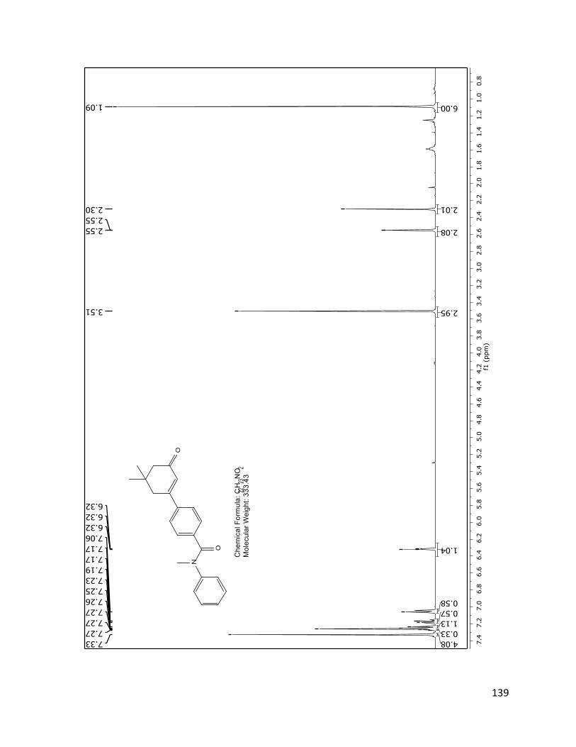
139
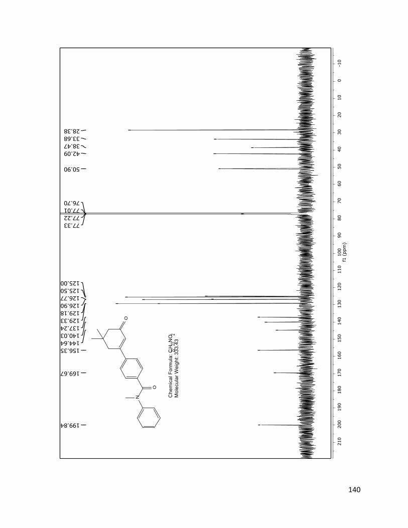
140

141
60
p-toluenesulfonyl chloride (2.30 g, 12.0 mmol) was added to a mixture of dimedone (2.02 g,
14.4 mmol) and potassium carbonate (2.35 g, 16.5 mmol) in a 2:1 ratio of 1,4-dioxane (26 mL)
and water (13 mL). This mixture was stirred at room temperature for 2 hours. 4-
nitrophenylboronic acid (2.0 g, 12.0 mmol) and tetrakis(triphenylphosphine)palladium (0) (416
mg, 0.36 mmol) were added and the mixture was heated under reflux for 4 hours or until
completion. The resulting mixture was extracted with EtOAc (3 x 10 mL). The extract was dried
with MgSO4, filtered and evaporate via rotary evaporator. The product 60 was isolated via
column chromatography with 15% EtOAc in hexanes. (1.33 g, 45 %).
1H NMR (400 MHz, CDCl3) δ, ppm: 8.27 (d, J = 9.0 Hz, 2H), 7.67 (d, J = 9.0 Hz, 2H), 6.45 (s, 1H), 2.66 (d, J = 1.6 Hz, 2H), 2.38 (s, 2H), 1.16 (s, 6H).
13C NMR (100 MHz, CDCl3) δ, ppm: δ 199.39, 154.76, 148.40, 145.46, 127.04 (2C), 126.85, 123.98 (2C), 50.85, 42.34, 33.90, 29.70, 28.35 (2C).

142

143

144
61
60 (210 mg, 0.86 mmol) was dissolved in methanol (10 mL). Palladium on carbon
(50 mg) was added and hydrogen gas pressure was added to the system and stirred for 2 hours.
The mixture was then run through a celite column and then washed with 5% HCl solution
followed by DCM extraction and a 10% NaOH wash. The organic phase was concentrated by
rotary evaporator. The product 61 was obtained by column chromatography with 20% EtOAc in
hexanes (125 mg, 67%)
1H NMR (400 MHz, CDCl3) δ, ppm: 7.43 – 7.34 (m, 2H), 6.70 – 6.61 (m, 2H), 6.35 (d, J = 1.4 Hz, 1H), 2.57 (d, J = 1.4 Hz, 2H), 2.28 (s, 2H), 1.08 (s, 6H).

145

146
62
61 (100 mg, 0.46 mmol) was dissolved in DCM (10 mL). K2CO3 (80 mg, 1.3 mmol) was
added followed by iodomethane (98 mg, 0.7 mmol) and the reaction was stirred for 1 hour. The
mixture was quenched with a solution of NH4Cl and the organic phase was dried over MgSO4
and concentrated by rotary evaporator. The product 62 was isolated by column
chromatography with 15% EtOAc in hexanes. (30 mg, 28%)
1H NMR (400 MHz, CDCl3) δ, ppm: 7.47 (d, J = 8.8 Hz, 2H), 6.61 (dd, J = 8.9, 1.2 Hz, 2H), 6.39 (t, J = 1.4 Hz, 1H), 2.89 (s, 3H), 2.61 (d, J = 1.4 Hz, 2H), 2.31 (s, 2H), 1.11 (s, 6H).

147

148
63
4-Bromophenol (4.33 g, 25 mmol) and imidazole (2.21 g, 32.5 mmol) were dissolved in a 1:1
ratio mixture of tetrahydrofuran (15 mL) and dimethylformamide (15 mL). tert-
Butyldimethylsilyl chloride (4.15 g, 27.5 mmol) and 4-dimethylaminopyridine (trace amount)
were added and the reaction was stirred for 12 hours at room temperature. The mixture was
diluted with water (30 mL) and ether (40 mL) and then extracted with EtOAc (3 x 20 mL). The
organic extracts were dried with MgSO4, filtered and evaporated using a rotary evaporator. The
silyl protected intermediate was isolated by column chromatography. (5.63 g, 68%)
The silyl protected intermediate (320 mg, 0.97 mmol) was dissolved in EtOAc (25 mL) and an
aqueous 5% sodium hydroxide solution (25 mL) was added. The aqueous phase was kept and
treated with an aqueous 10% hydrochloric acid solution (15 mL). This mixture was extracted
with EtOAc (3 x 10 mL) and this organic extract was dried with MgSO4, filtered and evaporated
to obtain 63 as a beige solid. (150 mg, 71%)
Total synthesis yield = 48%
1H NMR (300 MHz, MeOH-D4) δ, ppm: 7.57-7.48 (m, 2H), 6.94-6.73 (m, 2H), 6.34 (t, J = 1.39 Hz, 1H), 2.70 (d, J = 1.36 Hz, 2H), 2.32 (s, 2H), 1.12 (s, 6H)
13C NMR (101 MHz, MeOD) δ, ppm: 201.60, 159.81, 159.78, 129.36, 127.77 (2C), 120.57, 115.24 (2C), 50.18, 41.44, 33.13, 27.10 (2C).HRMS: Calculated for C14H16O2: 216.1150 Found: 216.1174
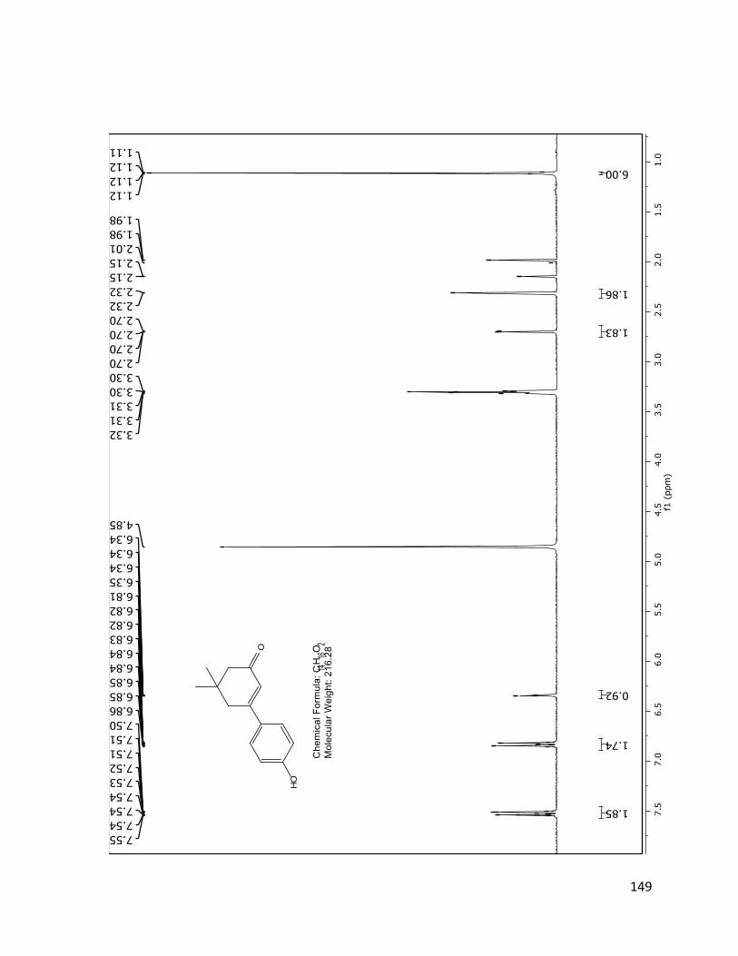
149

150

151
64
4'-hydroxy-5,5-dimethyl-5,6-dihydro-[1,1'-biphenyl]-3(4H)-one (63) (50 mg, 0.23 mmol) was
dissolved in dichloromethane (10 mL) and 4-dimethylaminopyridine (trace amounts) was
added. Acetic anhydride (27.7 mg, 0.46 mmol) and triethyl amine (70.1 mg, 0.69 mmol) were
added and the solution was stirred for 30 minutes at room temperature. The reaction mixture
was then poured into a flask containing a saturated aqueous solution of NaHCO3 and extracted
with dichloromethane (3 x 10 mL). The organic extract was dried with MgSO4, filtered and
evaporated. The product 64 was isolated by column chromatography with 10% EtOAc in
hexanes to obtain a light-beige solid. (25 mg, 42%)
1H NMR (400 MHz, CDCl3) δ, ppm: 7.59-7.50 (m, 2H), 7.20-7.06 (m, 2H), 6.38 (s, 1H), 2.62 (d, J = 1.25 Hz, 2H), 2.33 (s, 2H), 2.31 (s, 3H), 1.12 (s, 6H)
13C NMR (101 MHz, CDCl3) δ, ppm: 199.94, 169.21, 156.47, 151.90, 136.64, 127.38 (2C), 124.45, 121.97 (2C), 50.89, 42.34, 33.78, 28.40 (2C), 21.13.

152

153

154
65
p-toluenesulfonyl chloride (247.8 mg, 1.3 mmol) was added to a mixture of dimedone
(140.2 mg, 1.0 mmol) and potassium carbonate (345 mg, 2.5 mmol) in a 2:1 ratio of 1,4-dioxane
(4 mL) and water (2 mL). This mixture was stirred at room temperature for 1 hour. 4-
methylphenyl boronic acid (182.4 mg, 1.2 mmol) and tetrakis(triphenylphosphine)palladium (0)
(34.7 mg, 0.03 mmol) were added and the mixture was heated under reflux for 2 hours or until
completion. The resulting mixture was extracted with EtOAc (3 x 10 mL). The extract was dried
with MgSO4, filtered and evaporate via rotary evaporator. The product 65 was isolated via
column chromatography with 5% EtOAc in hexanes followed by a wash with an aqueous 10%
NaOH solution to obtain white crystals (130 mg, 56%).
1H NMR (400 MHz, CDCl3) δ, ppm: 7.51-7.40 (m, 2H), 6.89-6.84 (m, 2H), 6.32 (s, 1H), 3.77 (s, 3H), 2.55 (d, J = 1.25 Hz, 2H), 2.25 (s, 2H), 1.06 (s, 6H)
13C NMR (100 MHz, CDCl3) δ, ppm: 199.94, 161.03, 156.92, 130.72, 127.48 (2C), 122.24, 113.93 (2C), 55.14, 50.59, 41.79, 33.38, 28.21 (2C)
HRMS: Calculated for C15H18O2: 230.1307 Found: 230.1323

155

156

157
66
First synthesis (via compound 63):
4'-hydroxy-5,5-dimethyl-5,6-dihydro-[1,1'-biphenyl]-3(4H)-one (63) (50 mg, 0.23 mmol) was
dissolved in dry acetone (15 mL). Potassium carbonate (73.5 mg, 0.53 mmol) and benzyl
bromide (43.4 mg, 0.25 mmol) were added and the reaction mixture was refluxed for 3 hours.
The mixture was filtered to get rid of the excess potassium carbonate and water was added to
the remaining solution. This mixture was extracted with EtOAc (3 x 10 mL), dried with MgSO4,
filtered and evaporated. The product 66 was isolated by column chromatography with 7.5%
EtOAc in hexanes to obtain a light-yellow solid. (48 mg, 71%)
1H NMR (400 MHz, CDCl3) δ, ppm: 7.55-7.30 (m, 7H), 7.00 (d, J = 8.86 Hz, 2H), 6.39 (s, 1H), 5.10 (s, 2H), 2.61 (d, J = 0.80 Hz, 2H), 2.32 (s, 2H), 1.12 (s, 6H)
13C NMR (100 MHz, CDCl3) δ, ppm: 200.09, 160.37, 156.86, 136.50, 131.34, 128.69 (2C), 128.17, 127.73 (2C), 127.44 (2C), 122.75, 115.06 (2C), 70.11, 50.90, 42.11, 33.67, 28.48 (2C)
HRMS: Calculated for C21H22O2: 306.1620 Found: 306.1587

158
Second synthesis (via compound 29):
A round bottom flask was heated dry and an inert N2 was created within and placed in a
-78oC acetone/dry ice bath. Compound 29 (1.02 g, 6.1 mmol) was dissolved in dry THF (10 mL)
and added to the flask. N-BuLi 2.5M (3.04 mL, 7.6 mmol) was added to the mixture and stirred
for 5 minutes. 1,4-Bromophenyl benzyl ether was dissolved in dry THF (10 mL) and added to the
reaction mixture dropwise. After 15 minutes, the reaction mixture was quenched with water
(15 mL) and extracted with DCM (10 mL x 2). The organic layer was dried with MgSO4, filtered
and concentrated via rotary evaporator. The product 66 was isolated by column
chromatography with 7.5% EtOAc in hexanes. (1.3 g, 72%)

159

160

161
69
p-toluenesulfonyl chloride (1.42 g, 7.42 mmol) was added to a mixture of dimedone (800
mg, 5.71 mmol) and potassium carbonate (1.97 g, 14.3 mmol) in a 2:1 ratio of 1,4-dioxane (12
mL) and water (6 mL). This mixture was stirred at room temperature for 2 hours. 4-
fluorophenylboronic acid (1.08 g, 6.85 mmol) and tetrakis(triphenylphosphine)palladium(0)
(197 mg, 0.17 mmol) were added and the mixture was heated under reflux for 3 hours or until
completion. The resulting mixture was extracted with EtOAc (3 x 10 mL). The extract was dried
with MgSO4, filtered and evaporate via rotary evaporator. The product 69 was isolated via
column chromatography with 15% EtOAc in hexanes to obtain a light-brown solid. (0.71 g, 57
%).
1H NMR (400 MHz, CDCl3) δ, ppm: 7.56-7.43 (m, 2H), 7.13-6.97 (m, 2H), 6.33 (t, J = 1.39 Hz, 1H), 2.59 (d, J = 1.45 Hz, 2H), 2.30 (s, 2H), 1.10 (s, 6H)
HRMS: Calculated for C14H15FO: 218.1107 Found: 218.1091

162

163
70
p-toluenesulfonyl chloride (1.77 g, 9.27 mmol) was added to a mixture of dimedone (1.00 g,
7.13 mmol) and potassium carbonate (2.46 g, 17.8 mmol) in a 2:1 ratio of 1,4-dioxane (14 mL)
and water (7 mL). This mixture was stirred at room temperature for 2 hours. 2,4-
difluorophenylboronic acid (1.35 g, 8.56 mmol) and tetrakis(triphenylphosphine)palladium(0)
(247 mg, 0.21 mmol) were added and the mixture was heated under reflux for 3 hours or until
completion. The resulting mixture was extracted with EtOAc (3 x 10 mL). The extract was dried
with MgSO4, filtered and evaporate via rotary evaporator. The product 70 was isolated via
column chromatography with 15% EtOAc in hexanes to obtain a beige solid. (0.73 g, 43%).
1H NMR (400 MHz, CDCl3) δ, ppm: 7.31 (dt, J = 8.62, 6.36 Hz, 1H), 6.96-6.82 (m, 2H), 6.24 (s, 1H), 2.60 (t, J = 1.61 Hz, 2H), 2.34 (s, 2H), 1.12 (s, 6H)
13C NMR (100 MHz, CDCl3) δ, ppm: 199.58, 163.40, 160.15, 153.86, 129.92, 127.89, 124.29, 111.81, 104.79, 51.00, 43.67, 34.15, 28.17 (2C)

164

165

166
71
p-toluenesulfonyl chloride (0.78 g, 4.10 mmol) was added to a mixture of dimedone (0.37 g,
3.16 mmol) and potassium carbonate (1.09 g, 7.89 mmol) in a 2:1 ratio of 1,4-dioxane (14 mL)
and water (7 mL). This mixture was stirred at room temperature for 2 hours.
4(trifluoromethyl)phenylboronic acid (0.5 g, 2.63 mmol) and
tetrakis(triphenylphosphine)palladium(0) (0.09 mg, 0.08 mmol) were added and the mixture
was heated under reflux for 3 hours or until completion. The resulting mixture was extracted
with EtOAc (3 x 10 mL). The extract was dried with MgSO4, filtered and evaporate via rotary
evaporator. The product 71 was isolated via column chromatography with 30% EtOAc in
hexanes followed by recrystallization to obtain a white solid. (0.35 g, 41%).
1H NMR (400 MHz, CDCl3) δ, ppm: 7.72 – 7.56 (m, 3H), 6.42 (s, 1H), 2.65 (d, J = 1.6 Hz, 2H), 2.37 (s, 2H), 1.15 (s, 6H).
HRMS: Calculated for C15H15F3O: 268.1075 Found: 268.1082
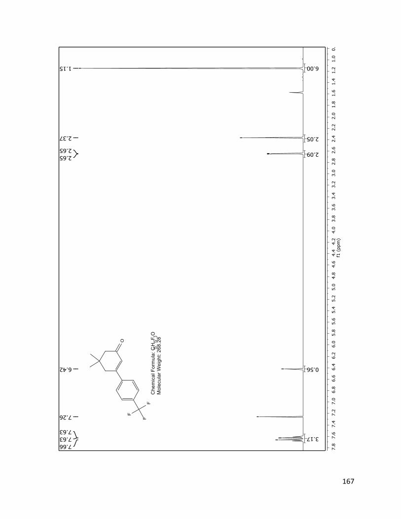
167

168
73
p-toluenesulfonyl chloride (0.97 g, 5.1 mmol) was added to a mixture of dimedone (0.55 g,
3.9 mmol) and potassium carbonate (1.35 g, 9.8 mmol) in a 2:1 ratio of 1,4-dioxane (8 mL) and
water (4 mL). This mixture was stirred at room temperature for 1 hour. 4-pyridinyl boronic acid
(0.4 g, 3.3 mmol) and tetrakis(triphenylphosphine)palladium (0) (0.11 g, 0.1 mmol) were added
and the mixture was heated under reflux for 2 hours or until completion. The resulting mixture
was extracted with EtOAc (3 x 10 mL). The extract was dried with MgSO4, filtered and evaporate
via rotary evaporator. The product 73 was isolated via column chromatography with 60% EtOAc
in hexanes followed by a wash with a 5% HCl solution then with an aqueous 10% NaOH solution
to obtain white crystals (0.32 g, 31%).
1H NMR (400 MHz, CDCl3) δ, ppm: 8.62 (t, J = 4.4 Hz, 2H), 7.33 (dq, J = 4.5, 1.6 Hz, 2H), 6.41 (dq, J = 3.3, 1.6 Hz, 1H), 2.57 (q, J = 1.7 Hz, 2H), 2.35 – 2.28 (m, 2H), 1.12 – 1.06 (m, 6H).

169

170
74
p-toluenesulfonyl chloride (1.16 g, 6.1 mmol) was added to a mixture of dimedone (0.65 g,
4.7 mmol) and potassium carbonate (1.62 g, 11.7 mmol) in a 2:1 ratio of 1,4-dioxane (12 mL)
and water (6 mL). This mixture was stirred at room temperature for 1 hour. 2-thienylboronic
acid (0.5 g, 3.9 mmol) and tetrakis(triphenylphosphine)palladium (0) (0.135 g, 0.12 mmol) were
added and the mixture was heated under reflux for 2 hours or until completion. The resulting
mixture was extracted with EtOAc (3 x 10 mL). The extract was dried with MgSO4, filtered and
evaporate via rotary evaporator. The product 74 was isolated via column chromatography with
10% EtOAc in hexanes. (0.427 g, 53%).
1H NMR (400 MHz, CDCl3) δ, ppm: 7.35 – 7.24 (m, 2H), 6.98 (dd, J = 5.1, 3.8 Hz, 1H), 6.30 (t, J = 1.5 Hz, 1H), 2.53 (d, J = 1.5 Hz, 2H), 2.19 (s, 2H), 1.01 (s, 6H).
13C NMR (100 MHz, CDCl3) δ, ppm: 199.41, 150.21, 142.97, 128.70, 128.26, 127.29, 121.59, 50.94, 41.97, 33.51, 28.39 (2C).

171

172
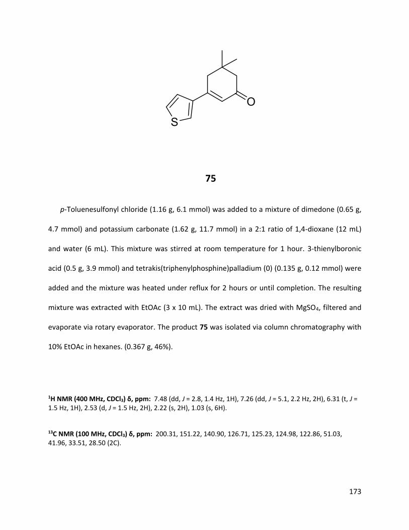
173
75
p-Toluenesulfonyl chloride (1.16 g, 6.1 mmol) was added to a mixture of dimedone (0.65 g,
4.7 mmol) and potassium carbonate (1.62 g, 11.7 mmol) in a 2:1 ratio of 1,4-dioxane (12 mL)
and water (6 mL). This mixture was stirred at room temperature for 1 hour. 3-thienylboronic
acid (0.5 g, 3.9 mmol) and tetrakis(triphenylphosphine)palladium (0) (0.135 g, 0.12 mmol) were
added and the mixture was heated under reflux for 2 hours or until completion. The resulting
mixture was extracted with EtOAc (3 x 10 mL). The extract was dried with MgSO4, filtered and
evaporate via rotary evaporator. The product 75 was isolated via column chromatography with
10% EtOAc in hexanes. (0.367 g, 46%).
1H NMR (400 MHz, CDCl3) δ, ppm: 7.48 (dd, J = 2.8, 1.4 Hz, 1H), 7.26 (dd, J = 5.1, 2.2 Hz, 2H), 6.31 (t, J = 1.5 Hz, 1H), 2.53 (d, J = 1.5 Hz, 2H), 2.22 (s, 2H), 1.03 (s, 6H).
13C NMR (100 MHz, CDCl3) δ, ppm: 200.31, 151.22, 140.90, 126.71, 125.23, 124.98, 122.86, 51.03, 41.96, 33.51, 28.50 (2C).

174

175

176
76
Thiophenol (3.77 g, 34.2 mmol), 5,5-dimethylcyclohexane-1,3-dione (4.0 g, 28.5 mmol) and
anhydrous ferrous chloride (0.93 g, 5.71 mmol) were added to a flask and stirred at room
temperature for 5 hours. The mixture was dissolved in dichloromethane (30 mL) and washed
with a 10% sodium hydroxide solution in an extraction funnel. The aqueous phase was treated
with bleach to get rid of the strong thiophenol smell. The organic phase was dried with MgSO4,
filtered and concentrated via rotary evaporator. The product 76 was isolated via column
chromatography with 20% EtOAc in hexanes (1.47 g, 22%).
1H NMR (400 MHz, CDCl3) δ, ppm: 7.45 (tdt, J = 3.93, 2.51, 1.89 Hz, 5H), 5.48 (s, 1H), 2.40 (s, 2H), 2.23 (s, 2H), 1.08 (s, 6H)\
HRMS: Calculated for C14H16OS: 232.0922 Found: 232.0932
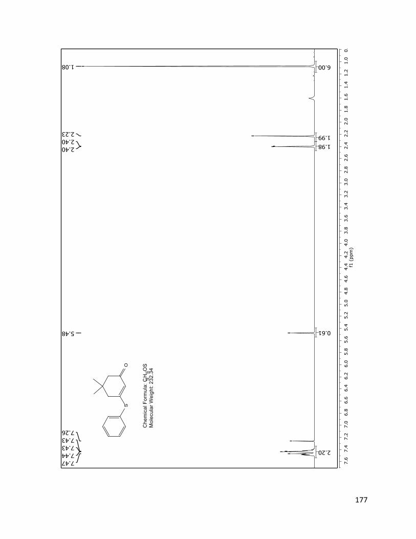
177

178
77
5,5-Dimethyl-3-(phenylthio)cyclohex-2-enone (76) (1.09g, 4.69 mmol) was dissolved in
dichloromethane (15 mL) and the solution was cooled to 0oC. mCPBA was dissolved in a small
amount of dichloromethane and added dropwise. The mixture was stirred over 30 mins at 0oC,
then warmed to room temperature and stirred for an additional 1 hour. The reaction was
quenched with a 10% aqueous solution of sodium carbonate. The mixture was extracted with
dichloromethane (2 x 10 mL) and washed a last time with a solution of brine (20 mL). The
organic extract was dried with MgSO4, filtered and concentrated via rotary evaporator. The
sulfoxide 77 was isolated using column chromatography with 20% EtOAc in hexanes to obtain a
clear yellow oil (550 mg, 47%)
1H NMR (400 MHz, CDCl3) δ, ppm: 7.66 – 7.54 (m, 2H), 7.55 – 7.43 (m, 3H), 6.72 (t, J = 1.6 Hz, 1H), 2.24 (d, J = 1.4 Hz, 2H), 2.14 – 2.01 (m, 2H), 0.86 (d, J = 24.9 Hz, 6H).

179

180
78
5,5-DImethyl-3-(phenylthio)cyclohex-2-enone (76) (1.09g, 4.69 mmol) was dissolved in
dichloromethane (15 mL) and the solution was cooled to 0oC. mCPBA was dissolved in a small
amount of dichloromethane and added dropwise. The mixture was stirred over 30 minutes at
0oC, then warmed to room temperature and stirred for an additional 1 hour. The reaction was
quenched with a 10% aqueous solution of sodium carbonate. The mixture was extracted with
dichloromethane (2 x 10 mL) and washed a last time with a solution of brine (20 mL). The
organic extract was dried with MgSO4, filtered and concentrated via rotary evaporator. The
sulfone 78 was isolated using column chromatography with 20% EtOAc in hexanes to obtain a
clear yellow oil (300 mg, 24%)
1H NMR (400 MHz, CDCl3) δ, ppm: 7.90 (ddd, J = 7.22, 2.93, 1.67 Hz, 2H), 7.70 ( m, 1H), 7.62-7.57 (m, 2H), 6.72 (t, J = 1.73 Hz, 1H), 2.39 (d, J = 1.75 Hz, 2H), 2.27 (s, 2H), 0.96 (s, 6H)

181

182
79
Thiophenol (4.7 g, 42.8 mmol), cyclohexane-1,3-dione (4.0 g, 35.7 mmol) and anhydrous
ferrous chloride (0.93 g, 5.71 mmol) were added to a flask and stirred at room temperature for
5 hours. The mixture was dissolved in dichloromethane (30 mL) and washed with a 10% sodium
hydroxide solution in an extraction funnel. The aqueous phase was treated with bleach to get
rid of the strong thiophenol smell. The organic phase was dried with MgSO4, filtered and
concentrated via rotary evaporator. The product 79 was isolated via column chromatography
with 20% EtOAc in hexanes (1.54 g, 22%).
1H NMR (400 MHz, CDCl3) δ, ppm: 7.55 – 7.35 (m, 5H), 5.55 – 5.42 (m, 1H), 2.52 (ddd, J = 6.4, 5.6, 1.2 Hz, 2H), 2.36 (dd, J = 7.3, 5.9 Hz, 2H), 2.14 – 1.95 (m, 2H).

183

184
80
nBuLi 2.5M (0.48 mL, 1.19 mmol) was added to dry THF (10 mL) under N2 atmospthere
to a flask cooled to -78oC. Phenyl methyl sulfone (0.19 g, 1.19 mmol) was dissolved in dry THF (5
mL) and added dropwise. Ethoxydimedone 29 (0.2 g, 1.19 mmol) was dissolved in dry THF (5
mL) and added dropwise. The reaction was quenched with a solution of NH4Cl (10 mL) and
extracted with H2O and EtOAc (10 mL x 2). The organic phase was dried over MgSO4 and
concentrated by rotary evaporator. The product 80 was isolated by column chromatography
with 20% EtOAc in hexanes to obtain a clear yellow oil (0.03 g, 10%)
1H NMR (400 MHz, CDCl3) δ, ppm: 7.92 – 7.75 (m, 2H), 7.65 (d, J = 7.5 Hz, 1H), 7.55 (dd, J = 8.3, 7.0 Hz, 2H), 5.56 (t, J = 0.9 Hz, 1H), 3.90 (d, J = 0.8 Hz, 2H), 2.41 (d, J = 1.8 Hz, 2H), 2.17 (s, 2H), 1.02 (s, 6H).
HRMS: Calculated for C15H18O3S: 278.0977 Found: 278.0962

185

186
82
Lithium Diisopropylamide 2M (3.87 mL, 7.74 mmol) was added to dry THF (25 mL) under
nitrogen atmosphere at -78oC. Propionitrile (0.06 mL, 7.74 mmol) was added dropwise and the
solution was stirred at -78oC for 15 minutes. 3',3'-dimethyl-5'-oxo-2',3',4',5'-tetrahydro-[1,1'-
biphenyl]-4-carboxylic acid (40) (100 mg, 0.387 mmol) was added dropwise and the solution
was stirred for 30 minutes. The mixture was quenched by transferring it to a beaker containing
an aqueous NH4Cl solution. The mixture was then warmed at room temperature and diluted
with water followed by extraction with EtOAc (3 x 10 mL). The extract was dried with MgSO4,
filtered and concentrated by rotary evaporator. The hydroxyl intermediate was isolated by
column chromatography.
The hydroxyl intermediate was dissolved in toluene and p-toluenesulfonic acid (50 mg,
catalytic) was added. The mixture was refluxed for 3 hours and was quenched by adding an
aqueous NaHCO3 solution. The mixture was extracted with EtOAc (3 x 10 mL) and the extract
was dried with MgSO4, filtered and evaporated by rotary evaporator. The product 82 was
isolated by column chromatography with 40% EtOAc in hexanes (30 mg, 28%).
1H NMR (400 MHz, CDCl3) δ, ppm: 8.07-7.98 (m, 2H), 7.71-7.61 (m, 2H), 6.45 (s, 1H), 4.36 (q, J = 7.18 Hz, 1H), 2.65 (d, J = 1.53 Hz, 2H), 2.36 (d, J = 5.96 Hz, 2H), 1.65 (d, J = 7.14 Hz, 3H), 1.14 (s, 6H)

187

188
84
p-Toluenesulfonyl chloride (371 mg, 1.95 mmol) was added to a mixture of 1,3-
cyclohexanedione (168 mg, 1.5 mmol) and potassium carbonate (432 mg, 3.0 mmol) in a 2:1
ratio of 1,4-dioxane (10 mL) and water (5 mL). This mixture was stirred at room temperature for
1 hour. 4-Methoxycarbonylphenylboronic acid (225 mg, 1.25 mmol) and
tetrakis(triphenylphosphine)palladium (0) (7.22 mg, 0.006 mmol) were added and the mixture
was heated under reflux for 1.5 hours or until completion. The resulting mixture was extracted
with EtOAc (2 x 25 mL). The extract was dried with MgSO4, filtered and evaporated via rotary
evaporator. The product 84 was isolated via column chromatography with DCM. (0.09 g, 26%).
1H NMR (400 MHz, CDCl3) δ, ppm: 8.09 – 8.01 (m, 2H), 7.61 – 7.53 (m, 2H), 6.43 (t, J = 1.5 Hz, 1H), 3.92 (s, 3H), 2.77 (td, J = 6.0, 1.5 Hz, 2H), 2.49 (dd, J = 7.5, 5.9 Hz, 2H), 2.22 – 2.10 (m, 2H)
13C NMR (100 MHz, CDCl3) δ, ppm: 199.55, 166.44, 158.39, 143.21, 131.20, 129.96 (2C), 126.79, 126.05 (2C), 52.30, 37.25, 28.10, 22.75.
HRMS: Calculated for C14H14O3: 230.0943 Found: 230.0975

189

190

191
85
84 (20 mg, 0.09 mmol) was added to a 5% solution of NaOH in water (2 mL) and
methanol (8 mL) and stirred at room temperature for 12 hours. The reaction mixture was
washed with EtOAc (20 mL). The aqueous phase was treated with a 5% HCl solution (10 mL)
followed by an extraction with EtOAc (20 mL). The resulting organic phase was dried over
MgSO4 and concentrated by rotary evaporator to obtain 85. (10 mg, 51%)
1H NMR (400 MHz, MeOD) δ, ppm: 8.07 (d, J = 8.5 Hz, 2H), 7.72 (d, J = 8.5 Hz, 2H), 6.44 (s, 1H), 2.96 – 2.75 (m, 2H), 2.50 (dd, J = 7.5, 6.0 Hz, 2H), 2.27 – 2.06 (m, 2H).
13C NMR (100 MHz, MeOD) δ, ppm: 201.05, 167.71, 160.61, 143.11, 131.76, 129.73 (2C), 125.99 (2C), 125.51, 36.63, 27.69, 22.43.

192

193

194
86
p-Toluenesulfonyl chloride (440 mg, 2.31 mmol) was added to a mixture of 1,3-
cyclohexanedione (200 mg, 1.78 mmol) and potassium carbonate (511 mg, 3.7 mmol) in a 2:1
ratio of 1,4-dioxane (10 mL) and water (5 mL). This mixture was stirred at room temperature for
1 hour. 4-Methoxyphenylboronic acid (225 mg, 1.48 mmol) and
tetrakis(triphenylphosphine)palladium (0) (0.135 g, 0.12 mmol) were added and the mixture
was heated under reflux for 1.5 hours or until completion. The resulting mixture was extracted
with EtOAc (2 x 25 mL). The extract was dried with MgSO4, filtered and evaporated via rotary
evaporator. The product 86 was isolated via column chromatography with 5% EtOAc in
hexanes. (0.2 g, 38%).
1H NMR (400 MHz, CDCl3) δ, ppm: 7.54 – 7.46 (m, 2H), 6.95 – 6.87 (m, 2H), 6.38 (t, J = 1.4 Hz, 1H), 3.83 (s, 3H), 2.73 (td, J = 6.1, 1.4 Hz, 2H), 2.45 (dd, J = 7.4, 6.0 Hz, 2H), 2.18 – 2.06 (m, 2H).
13C NMR (100 MHz, CDCl3) δ, ppm: 199.90, 161.23, 159.09, 130.83, 127.63(2C), 123.72, 114.15 (2C), 55.40, 37.21, 27.88, 22.79.
HRMS: Calculated for C13H14O2: 202.0994 Found: 202.0997

195

196

197
87
To dry THF (12 mL) under N2 atmosphere at -78oC is added LDA 2M (0.434 mL, 0.868
mmol) dropwise. Propionitrile (0.07 mL, 1.04 mmol) was added dropwise, followed by a
solution of 85 (0.1 g, 0.434 mmol) in dry THF (5 mL). The reaction was stirred during 15 min at -
78oC. The reaction was quenched with NH4Cl at room temperature and extracted with EtOAc
(20 mL). The organic phase was washed with water (20 mL), dried over MgSO4 and
concentrated by rotary evaporator. The hydroxyl intermediate was isolated by column
chromatography.
The hydroxyl intermediate (10 mg) was dissolved in toluene (10 mL) and PTSA (30 mg,
0.174 mmol) was added and the reaction was refluxed for 2 hours. The reaction was quenched
by a saturated NaCO3 solution. The mixture was extracted with EtOAc (10 mL) and the organic
phase was washed with water (10 mL). The resulting organic phase was dried over MgSO4,
filtered and concentrated by rotary evaporator. The isomeric mixture 87 was obtained by
column chromatography with 10% EtOAc in hexanes. (0.01 g, 9%)
1H NMR (400 MHz, CDCl3) δ, ppm: 8.11 – 7.95 (m, 2H), 7.61 – 7.46 (m, 2H), 7.15 (d, J = 1.6 Hz, 1/2H), 6.85 (t, J = 1.6 Hz, 1/2H), 3.91 (d, J = 1.0 Hz, 3H), 2.74 – 2.54 (m, 3H), 2.50 – 2.38 (m, 1H), 2.07 – 1.90 (m, 4H), 1.95 – 1.82 (m, 2H)
13C NMR (100 MHz, CDCl3) δ, ppm: 166.74, 166.65, 150.64, 149.33, 145.59, 144.85, 144.52, 144.12, 129.92, 129.86, 129.81, 129.74 (2C), 129.48 (2C), 125.56 (2C), 125.47 (2C), 125.04, 121.67, 120.51, 119.76, 103.03, 102.45, 52.21, 52.16, 29.63, 28.19, 27.49, 26.02, 22.97, 22.38, 21.87, 16.13, 15.16.

198
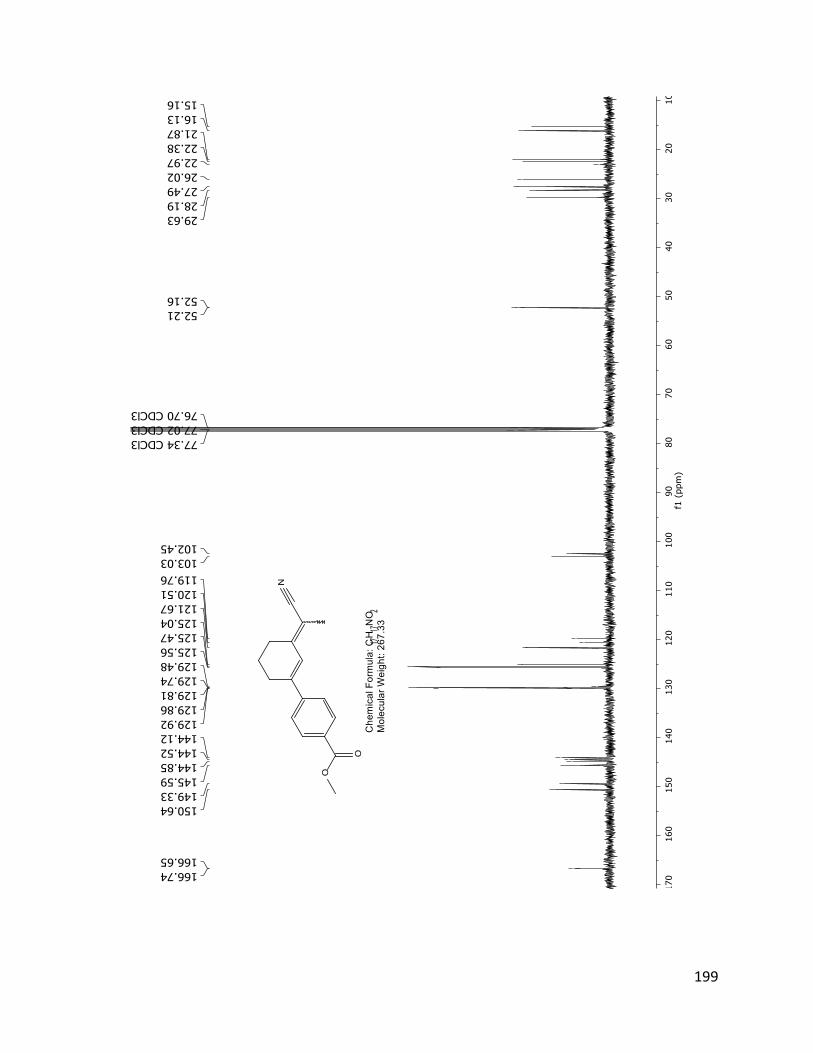
199

200
88
To dry THF (12 mL) under N2 atmosphere at -78oC is added LDA 2M (0.434 mL, 0.868
mmol) dropwise. Propionitrile (0.07 mL, 1.04 mmol) was added dropwise, followed by a
solution of 86 (0.1 g, 0.434 mmol) in dry THF (5 mL). The reaction was stirred during 15 min at -
78oC. The reaction was quenched with NH4Cl at room temperature and extracted with EtOAc
(20 mL). The organic phase was washed with water (20 mL), dried over MgSO4 and
concentrated by rotary evaporator. The hydroxyl intermediate was isolated by column
chromatography.
The hydroxyl intermediate (10 mg) was dissolved in toluene (10 mL) and PTSA (30 mg,
0.174 mmol) was added and the reaction was refluxed for 2 hours. The reaction was quenched
by a saturated NaCO3 solution. The mixture was extracted with EtOAc (10 mL) and the organic
phase was washed with water (10 mL). The resulting organic phase was dried over MgSO4,
filtered and concentrated by rotary evaporator. The product 88 was obtained by column
chromatography with 10% EtOAc in hexanes. (0.01 g, 9%)
1H NMR (400 MHz, CDCl3) δ, ppm: 7.49 (dd, J = 10.6, 8.8 Hz, 2H), 7.14 – 6.67 (m, 3H), 3.85 (d, J = 1.3 Hz, 3H), 2.81 – 2.38 (m, 4H), 2.09 – 1.84 (m, 5H)
13C NMR (100 MHz, CDCl3) δ, ppm: 160.10, 159.96, 151.44, 150.15, 146.19, 144.70, 132.86, 132.54, 126.92, 126.85, 121.81, 121.04, 120.26, 118.53, 113.98, 113.92, 100.44, 100.01, 55.37, 55.35, 29.74, 28.19, 27.50, 26.12, 22.51, 22.01, 15.95, 15.00.

201

202
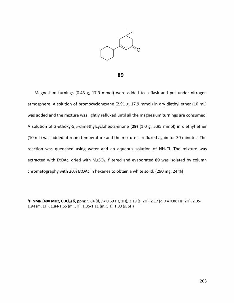
203
89
Magnesium turnings (0.43 g, 17.9 mmol) were added to a flask and put under nitrogen
atmosphere. A solution of bromocyclohexane (2.91 g, 17.9 mmol) in dry diethyl ether (10 mL)
was added and the mixture was lightly refluxed until all the magnesium turnings are consumed.
A solution of 3-ethoxy-5,5-dimethylcyclohex-2-enone (29) (1.0 g, 5.95 mmol) in diethyl ether
(10 mL) was added at room temperature and the mixture is refluxed again for 30 minutes. The
reaction was quenched using water and an aqueous solution of NH4Cl. The mixture was
extracted with EtOAc, dried with MgSO4, filtered and evaporated 89 was isolated by column
chromatography with 20% EtOAc in hexanes to obtain a white solid. (290 mg, 24 %)
1H NMR (400 MHz, CDCl3) δ, ppm: 5.84 (d, J = 0.69 Hz, 1H), 2.19 (s, 2H), 2.17 (d, J = 0.86 Hz, 2H), 2.05-1.94 (m, 1H), 1.84-1.65 (m, 5H), 1.35-1.11 (m, 5H), 1.00 (s, 6H)

204

205
References:
1 World Health Organization (WHO) Epilepsy Fact Sheet (2005) http://www.who.int/mediacentre/factsheets/fs999/en/
2 Bromfield, EB, Cavazos, JE, Sirven JI 2006; An Introduction to Epilepsy, American Epilepsy Society
3 Arzimanoglou, A, Ben-Menachem, E, Cramer, J, Glauser, T, Seeruthun, R, Harrison, M, The evolution of antiepileptic drug development and regulation, Epileptic Disord, (2010) vol. 12, no. 1, pp. 3-15
4 Hauptmann, A, Luminal bei Epilepsie, Münch Med Wochenschr, (1912), vol. 59, pp. 1907-9.
5 Merritt H, Putnam TJ. Sodium diphenyl hydantoinate in the treatment of convulsive disorders. J Am Med Soc 1938; 111: 1068-73.
6 Schindler W, Häfliger F. Über Derivate des Iminodibenzyls. Helv Chim Acta 1954; 37: 472-83.
7Abernathy D., Bergin P.; Prescribing issues associated with anticonvulsant medications for epilepsy, Best Practice Journal (2009), vol. 24, pp. 4-13
8 Arroyo S, de la Morena A.; Life-threatening adverse events of antiepileptic drugs, Epilepsy Res (2001), 47, pp. 155-74
9 Hesdorffer DC, Kanner AM; The FDA alert on suicidality and antiepileptic drugs: Fire or false alarm? Epilepsia, (2009), 50(5), pp.978-86.
10 Schmidt D.,Drug treatment of epilepsy: Options and limitations,Epilepsy & Behavior, (2009), 14, pp. 56–65
11 Kwan P, Brodie MJ. Effectiveness of first antiepileptic drug. Epilepsia 2001; 42: 1255-60
12 Simeone, T.A.; Rho, J.M.; Antiepileptic Drug Mechanisms Encyclopedia of Basic Epilepsy Research, (2009), pp. 59–66
13 Richerson, G.B.; Ransom, C.B.; ANTIEPILEPTIC DRUGS: GABA Transporters as Targets of Antiepileptic Drugs, Encyclopedia of Basic Epilepsy Research, (2009), pp. 66–74
14 Jeffrys, J.; Advances in understanding basic mechanisms of epilepsy and seizures, Seizure (2010) 19 (10), pp. 638-646
15 Schousboe, A.; Madsen, H.; White S.; Karsten K.; Neuronal and non-neuronal GABA transporters as targets for antiepileptic drugs, Pharmacology & Therapeutics, (2010), 125, pp.394–401
16 Kaur H., Kumar B., Medhi B., Antiepileptic drugs in development pipeline: A recent update, eNeurologicalSci, (2016), 4, pp. 42-51
17 McCoy, B., Wang, L., Zak, M., Al‐Mehmadi, S., Kabir, N., Alhadid, K., McDonald, K., Zhang, G., Sharma, R., Whitney, R., Sinopoli, K.; A prospective open‐label trial of a CBD/THC cannabis oil in dravet syndrome, Annals of Clinical and Translational Neurology, (2018), 5, 9, pp. 1077-1088
18 Rahman, A., Choudhary, M.I., Shaheen, F., Ganesan, A., Simjee, S.U., Raza, A.M, (2008) United States Patent No. US 7,399,888 B2
19 Rahman, A.U., Choudhary, M.I., Shaheen, F., Simjee, S.U., Kahn, N., Malhi, S.M., Shah, S.U.A., Ashraf, M.N., (2014) Synthesis and Biological studies of an isomeric mixture of isoxylitones and its analogs. US. Patent Application 0221682A1

206
20 Morimoto, K., Fahnestock, M., Racine, R.J.; Kindling and status epilepticus models of epilepsy: rewiring the brain, Progress in Neurobiology, (2004), 73 (1), pp 1-60
21 Saikely, A. (2015). Synthesis of Isoxylitone analogs for the treatment of epilepsy (Master’s thesis). Retrieved from https://ruor.uottawa.ca/handle/10393/33424
22 Gavrilovic IT, Hormigo A, Yahalom J, DeAngelis LM, Abrey LE, Long-term follow-up of high-dose methotrexate-based therapy with and without whole brain irradiation for newly diagnosed primary CNS lymphoma, J Clin Oncol. (2006) 1;24(28): pp.4570-4.
23 McIntyre et al., Modeling the excitability of mammalian nerve fibers: influence of afterpotentials on the recovery cycle, J Neurophysiol. (2002) 87(2): pp.995-1006.
24 Gavrilovici et al., The loss of interneuron functional diversity in the piriform cortex after induction of experimental epilepsy, Neurobiology of Disease (48) 3, pp.317-328
25 Wińska K, Grudniewska A, Chojnacka A, Białońska A, Wawrzeńczyk C., Enzymatic resolution of racemic secondary cyclic allylic alcohols. Tetrahedron Asymmetry, 2010; 21, 6 pp. 670 – 678
26 Zegarski, J.; Howard, B. Syntheses of marine ochtodane antifeedants via hydrolytic dehalogenation. Tetrahedron Letters, (1985) 26(11), 1363-6
27 Stability testing of active pharmaceutical ingredients and finished pharmaceutical products, WHO technical report series no. 953, 2009, Annex 2
28 Buchwald S. L., Martin R., Palladium-Catalyzed Suzuki−Miyaura Cross-Coupling Reactions Employing Dialkylbiaryl Phosphine Ligands, Acc. Chem. Res., 2008, 41 (11), pp 1461–1473
29 Michael Poulter, private communication
30 Hirashima SI, Nobuta T, Tada N, Miura T, Itoh A., Direct Aerobic Photo-Oxidative Synthesis of Aromatic Methyl Esters from Methyl Aromatics via Dimethyl Acetals, Organic Letters, (2010) 12 (16), pp.3645-3647
31 Guchhait, K.; Priyadarshani G.; Synthesis of 2-Arylpyridopyrimidinones, 6-Aryluracils, and Tri- and Tetrasubstituted Conjugated Alkenes via Pd-Catalyzed Enolic C–O Bond Activation–Arylation. J. Org. Chem., (2015), 80 (12), pp 6342–6349
32 Hermes D, Trenité DG, Winawer J., Gamma oscillations and photosensitive epilepsy, Curr Biol. 2017 May 8; 27(9): R336–R338.
33 Myers, A.; Tanaka, D.; Heck-Type Arylation of 2-Cycloalken-1-ones with Arylpalladium Intermediates Formed by Decarboxylative Palladation and by Aryl Iodide Insertion, Organic Letters, (2004) 6(3), pp. 433-436
34 K. Lakevold. Private communication.
35 Poulter M., Durst.; (2018) Anticonvulsant Compounds, United States Patent Pub No. US 2018/0194715 A1
36 Yoshino, T., Ng, F., Danishefsky, S.J., A total synthesis of xestodecalactone A and proof of its absolute stereochemistry: interesting observations on dienophilic control with 1, 3-disubstituted nonequivalent allenes. Journal of the American Chemical Society, (2006) vol. 128, no. 43, pp.14185-14191
37 Cui, Z.S., Zhang, Z.H., Liu, S.F., An efficient conversion of β-diketones into β-keto enol ethers with P2O5/SiO2 under solvent-free conditions. Journal of Chemical Research, (2006) no. 6, pp.390-392.
38 Castelani, Priscila; Tetrahedron 2005, V61(9)

207
39 Tsukamoto, H., Ueno, T., Kondo, Y., Palladium (0)-catalyzed alkylative cyclization of alkynals and alkynones: Remarkable trans-addition of organoboronic reagents. Journal of the American Chemical Society, (2006) vol. 128, no. 5, pp.1406-1407.
40 Brodie, M.; Antiepileptic drug therapy the story so far, Seizure (2010) vol. 19, pp. 650–655
41 Cheval, N.P., Dikova, A., Blanc, A., Weibel, J.M., Pale, P., Vinyl Nosylates: An Ideal Partner for Palladium‐Catalyzed Cross‐Coupling Reactions. Chemistry–A European Journal, (2013) vol. 19, no.27, pp.8765-8768.
42 Dikova, A., Cheval, N.P., Blanc, A., Weibel, J.M., Pale, P., Handy Protocols using Vinyl Nosylates in Suzuki–Miyaura Cross‐Coupling Reactions. Advanced Synthesis & Catalysis, (2015) vol. 357, no. 18, pp. 4093-4100.