synthesis and mechanical properties of conductive
TRANSCRIPT

SYNTHESIS AND MECHANICAL PROPERTIES OF CONDUCTIVE
COMPOSITE POLYLACTIC ACID/POLYANILINE SCAFFOLD FOR
POTENTIAL TISSUE ENGINEERING
FARAH NURULJANNAH BINTI DAHLI
A thesis submitted in fulfilment of the
requirements for the award of the degree of
Master of Philosophy
Faculty of Chemical and Energy Engineering
Universiti Teknologi Malaysia
DECEMBER 2016

iii
ACKNOWLEDGEMENTS
In the name of Allah S.W.T the most gracious and the most merciful.
Alhamdulillah, with the utmost blessing from Allah and in the remembrance to our
prophet Muhammad P.B.U.H His most beloved messenger of all time, the path of my
master degree has come to a completion.
I would like to take this opportunity to show my deepest gratitude to Dr.
Saiful Izwan Bin Dato’ Abd Razak who had stand in front of me to lead my way in
achieving my master degree. This thesis has become a reality due to his wholly
support in knowledge, guidance and moral support along with the will from Allah
S.W.T. The appreciation also goes to my co-supervisor, Assoc. Prof. Dr. Abdul
Razak Rahmat and Prof. Dato' Ir. Dr. Mohammed Rafiq bin Dato' Abdul Kadir for
their provision on supervising my research.
Not to be forgotten my beloved mother, Zakiah Binti Hashim for placing her
highest belief in me to complete this thesis. Same thanks to my family members and
friends who are willing to bear with me through thick and torn together in making
my dream come true. Finally, tremendous assistance from all of my colleagues in
polymeric biomaterials lab would not be forgotten and always be in my thought and
prayer.

iv
ABSTRACT
This thesis reports a new composite scaffold material that is conductive and
porous made from degradable polylactic acid (PLA) and conducting polyaniline
(PANI) which has the potential for use in promoting tissue regeneration. The
conductive scaffold was successfully prepared using a simple yet effective method
known as freeze extraction method. The doped PANI was synthesised using
conventional method of oxidative chemical polymerization. The electrical
percolation state was successfully obtained at 3 wt% of PANI inclusion and reached
at useable conductivity level for tissue engineering application at 4 wt% PANI, 2.91
x 10-3 Scm-1. 4 wt% inclusion of PANI was justified as the best PLA/PANI
composite scaffold because it met the criterion as an electro-responsive material
where the conductivity achieved was higher than 10-3 Scm-1. It is also much suitable
material in the regeneration of skin tissue (fibroblast) because the mean pore size
achieved was at 35.82 μm and optimum tensile strength at 3.08 MPa. The UV-
spectrum of the conductive scaffold displayed transition peaks of PANI indicating
the PANI was still in its conducting doped state inside the scaffold. Incubation for 24
weeks for in-vitro degradation revealed that the PANI component delayed the
degradation of PLA. Preliminary bioactivity test results indicated that the doping
agent able to form chelate at the scaffold surface and this could assist in the
formation of in-vitro apatite during the biomimetic immersion.
v
ABSTRAK
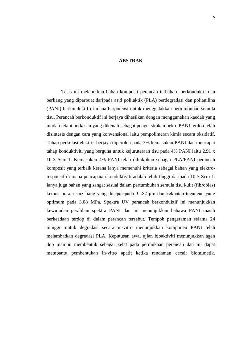
Tesis ini melaporkan bahan komposit perancah terbaharu berkonduktif dan
berliang yang diperbuat daripada asid polilaktik (PLA) berdegradasi dan polianilina
(PANI) berkonduktif di mana berpotensi untuk menggalakkan pertumbuhan semula
tisu. Perancah berkonduktif ini berjaya dihasilkan dengan menggunakan kaedah yang
mudah tetapi berkesan yang dikenali sebagai pengekstrakan beku. PANI terdop telah
disintesis dengan cara yang konvensional iaitu pempolimeran kimia secara oksidatif.
Tahap perkolasi elektrik berjaya diperoleh pada 3% kemasukan PANI dan mencapai
tahap konduktiviti yang berguna untuk kejuruteraan tisu pada 4% PANI iaitu 2.91 x
10-3 Scm-1. Kemasukan 4% PANI telah dibuktikan sebagai PLA/PANI perancah
komposit yang terbaik kerana ianya memenuhi kriteria sebagai bahan yang elektro-
responsif di mana pencapaian konduktiviti adalah lebih tinggi daripada 10-3 Scm-1.
Ianya juga bahan yang sangat sesuai dalam pertumbuhan semula tisu kulit (fibroblas)
kerana purata saiz liang yang dicapai pada 35.82 μm dan kekuatan tegangan yang
optimum pada 3.08 MPa. Spektra UV perancah berkonduktif ini menunjukkan
kewujudan peralihan spektra PANI dan ini menunjukkan bahawa PANI masih
berkeadaan terdop di dalam perancah tersebut. Tempoh pengeraman selama 24
minggu untuk degradasi secara in-vitro menunjukkan komponen PANI telah
melambatkan degradasi PLA. Keputusan awal ujian bioaktiviti menunjukkan agen
dop mampu membentuk sebagai kelat pada permukaan perancah dan ini dapat
membantu pembentukan in-vitro apatit ketika rendaman cecair biomimetik.

vi
TABLE OF CONTENTS
CHAPTER TITLE PAGE
DECLARATION ii
ACKNOWLEDGEMENTS iii
ABSTRACT iv
ABSTRAK v
TABLE OF CONTENTS vi
LIST OF TABLES x
LIST OF FIGURES xi
LIST OF ABBREVIATIONS xiii
LIST OF SYMBOLS xiv
lxi
1 INTRODUCTION 1
1.1 Overview 1
1.2 Problem Statement 3
1.3 Objectives of Study 4
1.4 Scopes of Study 4
2 LITERATURE REVIEW 6
2.1 Tissue Engineering 6
2.2 Scaffolds 7
2.3 Synthetic Biodegradable Polymers 9
2.3.1 Polylactic Acid (PLA) 10
2.4 Scaffolds Preparation 11

vii
2.4.1 Freeze Extraction 13
2.5 Composite Scaffolds 14
2.6 Electrically Conductive Polymers 16
2.6.1 Polyaniline (PANI) 16
2.6.2 Acid Doping and Bioactivity 19
2.6.3 PANI in Tissue Engineering 21
2.7 PLA/PANI Composite 22
2.8 Summary of Literature Review 23
3 MATERIALS AND METHODS 24
3.1 Materials And Reagents 24
3.2 Experiment Methods 27
3.2.1 Synthesis of PANI 27
3.2.2 Preparation Composite Scaffold by Freeze
Extraction 28
3.3 Characteristic and Testing 29
3.3.1 DC Conductivity Testing 29
3.3.2 Porosity and Pore Size 30
3.3.3 Degree of Swelling 30
3.3.4 Mechanical Testing 31
3.3.5 UV-vis Spectroscopy 32
3.3.6 Scanning Electron Microscopy 32
3.3.7 In-Vitro degradation 33
3.3.8 In-Vitro Bioactivity 33
3.4 Flow Diagram of the Research Methodology 34
4 RESULTS AND RESULTS 36
4.1 Morphology And Appearance of Synthesized
PANI 36
4.2 Electrical Conductivity of Composite Scaffold 37
4.3 Morphology of Composite Scaffold 39
4.4 Porosity, Pore Size And Degree of Swelling 41
4.5 Tensile Properties of Composite Scaffold 42
viii
4.6 UV-vis Spectra of Composite Scaffold 43
4.7 In-vitro degradation of Composite Scaffold 44
4.8 In-vitro Bioactivity of Composite Scaffold 45
5 CONCLUSIONS 48
5.1 Conclusion 48
5.2 Recommendations 49
REFERENCES 50
LIST OF PUBLICATIONS 61
APPENDICES 62-63

x
LIST OF TABLES
TABLE NO. TITLE PAGE
3.1 Chemicals and reagents 26
3.2 Ion concentration of 1.5 SBF 34
4.1 Porosity, pore size and degree of swelling 41
4.2 Tensile properties 42

xi
LIST OF FIGURES
FIGURE NO. TITLE PAGE
2.1 Repeating unit of PLA (Gruber et al, 2003) 10
2.2 Different oxidation states in PANI:
(a) leucoemeraldine,
(b) emeraldine, (c)nigraniline, d) pernigraniline 17
2.3 Reversible transformations from ES to EB of
PANI 18
2.4 Reaction mechanism of polyaniline via radical
cation polymerization 19
3.1 Chemical structure of aniline 24
3.2 Schematic diagram of the synthesis of PANI 27
3.3 Freeze extraction of scaffold 28
3.4 Flow diagram of the research methodology 35
4.1 (a)SEM image of synthesized PANI at 10,000
magnification (b)PANI suspended in 1,4-dioxane 37
4.2 DC conductivity of PLA scaffold as function of
PANI loading 38
4.3 SEM images of neat PLA at (a) 350 (b) 20,000
magnifications 39
4.4 SEM images of PLA/4PANI at (a) 350
(b) 20,000 (c)100,000 and (d) 200,000
magnifications, and (e) 200,000 magnification of
PLA/5PANI 40
4.5 UV-vis spectra of (a) neat doped PANI, and
(b)PLA/4PANI 43
4.6 SEM images of (a) neat PLA and PLA/4PANI
after immersion in PBS for 24 weeks 45

xi
4.7 SEM images of (a) neat PLA and PLA/4PANI
after 5 days of soaking in SBF (HA marked in
circles) 46
4.8 EDX spectra of PLA/4PANI after 5 days of
soaking in SBF 47
4.9 Schematic illustration for the formation of
chelation complex of citric acid and calcium ion
47

xii
LIST OF ABBREVIATIONS
1D - One dimensional
3D - Three dimensional
CA-cit - Calcium-citric acid
CHCl3 - trichloromethane
DC - Direct Current
FC - Fast cooling rate
FDA - Food and Drug Administration
HA - hydroxyl apatite
m-ABA - m-Aminobenzoic acid
mmol - Milimole
MWCNT - Multiwall Carbon Nanotubes
PANI - Polyaniline
PANI-CSA PANI-camphor sulfonic acid
PEDOT - Poly(3,4-ethylene dioxythiophene)
PLA - Polylactic Acid
PLLA - Poly-L-Lactic Acid
PGA - Polyglycolic Acid
PPY - Polypyrrole
PPV - Poly(p-phenylene-vinylene)
PYG - Polypyrrole/graphene
SBF - Simulated body fluid
SC - Slow cooling rate
SEM - Scanning Electron Microscopy
UV-vis - Ultraviolet-Visible Spectroscopy
xiii
LIST OF SYMBOLS
α - Alpha
A - Area
E - Electric field intensity
I - Current
J - Current density
σ - Conductivity of the material
Ms - Mass of scaffold after immersion in water
Md - Mass of dry scaffold
ρ - Resistivity
π - Pi bonding orbital
π* - Antibonding Pi orbital
R - Resistance
t - thickness
V - Voltage
Vd - Apparent volume
Vp - Pore volume
Wd - Surplus weight of scaffold after degradation
Wi - Initial weight of scaffold before degradation
CHAPTER 1
INTRODUCTION
1.1 Overview
Scaffolds in tissue engineering refer to biodegradable materials which are
highly porous that can act as template for tissue regeneration (Yang et al., 2001).
Synthetic biodegradable scaffold such as polylactic acid (PLA) has found wide range
of pharmaceutical applications in the tissue regeneration of skin (Mohiti‐Asli et al.,
2015), cartilage (Muhonen et al., 2015), blood vessel (Li et al., 2015) and cardiac
valve (Iop and Gerosa 2015). The advantages of PLA are its synthetically
controllable degradation rate (Cui et al., 2015), good mechanical properties (Shi et
al., 2015) and biocompatibility (Abdal-hay et al., 2015) plus it can be produced from
renewable resource (Yang et al., 2015).
The methods of preparing a porous PLA scaffold are diverse which includes,
thermally induced phase separation (Mannella et al., 2015), 3D printing (Rosenzweig
et al., 2015), porogen leaching (Choudhury et al., 2015), the highly popular freeze
drying (Salerno et al., 2015) and electrospinning (Morelli et al., 2015). Another
method to produce polymeric porous scaffold is the simple freeze extraction (Adeli
et al., 2011).

2
Though there are few reports on PLA scaffold prepared by freeze extraction
method with the inclusion of other fillers or reinforcements such as carbon nanotubes
(Adeli et al., 2011), chitosan and alginate (Yuan et al., 2009), bioactive glass (El-
Kady et al., 2010), to date there are no reported studies on the preparation of freeze
extracted porous conductive scaffold of PLA with the inclusion of conducting
polymers such as of polyaniline (PANI).
PANI is one of the most promising conducting polymers for wide range of
applications (Li et al., 2008) mainly due to its ease of synthesis and preparation
(Bhadra et al., 2009), excellent electrical properties (Wang et al., 2015) and being
biocompatible (Bidez et al., 2006). Inclusion of conductive PANI filler in the PLA
scaffold might open up opportunities in many biomedical applications such as tissue
engineering. It is only quite recently that the tuneable electroactivity of PANI has
been explored in the area of diverse biomedical applications, such as for scaffolds in
tissue engineering (Qazi et al., 2014).
Earlier in vivo test revealed that various forms of PANI caused minimal
inflammation after 50 weeks of implantation beneath the dorsal skin of rats (Wang et
al., 1999). It was also shown that PANI can be a good reducing agents and effective
scavengers of free radicals when present in biological media (Gizdavic-Nikolaidis et
al., 2004). Investigation on adhesion and proliferation of cardiac myocytes on PANI
concluded that PANI potential usefulness as an electroactive conductive polymer in
cell-culture experiments (Bidez et al., 2006), able to stimulate cell differentiation to
cardiomyocites (Borriello et al., 2011) and biocompatible for both healthy and cancer
cells after some modifications (Yslas et al., 2015). However, due to its brittleness
and nonprocessability (Saini et al., 2012), it should be incorporated into other
polymers that able to be fabricated into a tissue engineered scaffold.
Therefore, the main aim of this study is to prepare and investigate the effects
of PANI addition on the properties of PLA scaffold prepared using freeze extraction.
This new type of conductive composite scaffold is expected to exhibit new and

3
enhanced properties including the ease of processing and low cost. Such conductive
scaffold may be usable in many applications in tissue engineering and biomedical
implants such as controllable electrically responsive cell growth scaffold,
controllable drug delivery sites and skin graft for wounds.
1.2 Problem Statements
Most of the research works on PLA composite scaffold are focused on the
mechanical and morphology improvement. Nonetheless it was shown that certain
material can enhance Schwann cell growths for neural tissue engineering upon
applied voltage (Baniasadi et al., 2015). This could decrease the time taken for the
cells to fully mature and it could lessen the time for patients to wait for their new
regenerate tissue. Thus it seems feasible to induce a certain degree of electrical
conductivity to a scaffold material in order to obtain cell responsive properties for
tissue engineering. Though being reported, the study on conductive scaffold is still
limited to some extent.
Freeze drying is a widely used method to prepare porous scaffold but it is
time and energy consuming (Baldino et al., 2015). Plus the resulting freeze dried
scaffold usually produced unwanted surface skin which requires additional process
thus becomes economically uncompetitive (Sachlos and Czernuszka, 2003). In
regards to conductive scaffolds, they have been fabricated using the electrospinning
method mainly due to their nanofiber formation which led to high porosity (McKeon
2010, Shokry et al., 2015). Though the electrospinning process seemed feasible,
various cumbersome factors should be taken into consideration to obtain its
nanofiber form such as applied voltage, solvent mixtures, distance between the tip
and the collector, viscosity of the polymer solution, flow rate and even
humidity/temperature of the spinning chamber (Subbiah et al., 2005).

4
Being relatively new in the tissue engineering field, conductive scaffold
prepared using freeze extraction has many unexplored features and characteristics.
Many aspects that should be studied which includes the electrical conductivity
enhancement, morphology, pore size and porosity, electronic transitions,
biodegradability and bioactivity.
1.3 Objectives
This study was conducted in order to fulfil the following objectives:
1. To prepare conductive composite scaffold of PLA/PANI via freeze
extraction
2. To characterize the electrical, physical and morphological properties of
the PLA/PANI scaffold
3. To evaluate the preliminary in-vitro degradation and preliminary
bioactivity test
1.4 Scope of Study
In order to satisfy all the outlined objectives, the scopes of this research are
undertaken according to the following:
Initially, PANI was synthesized according to conventional method as
reported in literatures. The synthesized PANI was characterized for its morphology,
color appearance, DC electrical conductivity and UV-vis spectroscopy. Following

5
that, the as synthesized PANI will be used as fillers in the preparation of conductive
scaffold.
Next step was to prepare the scaffold by the inclusions of PANI within the
PLA using freeze extraction. Amount of PANI used were (0.5, 1, 2, 3, 4, 5 wt%).
The resulting conductive composite scaffolds were evaluated in terms of its DC
conductivity, tensile properties, porosity, pore size and degree of swelling. Scaffold
of PLA/PANI with a suitable electrical conductivity value and good physical
characteristics were identified and further tested using UV-vis spectroscopy and
scanning electron microscope.
Consequently the conductive composite scaffold was tested for in-vitro
degradation; evaluating the weight loss and the resulting morphology. Bioactivity
test of the conductive scaffold was done by immersion in simulated body fluid
solution (SBF), followed by the evaluation of hydroxyl apatite growth on the sample.

50
REFERENCES
Abdal-hay, A., Hussein, K. H., Casettari, L., Khalil, K. A., and Hamdy, A. S. (2016).
Fabrication of novel high performance ductile poly (lactic acid) nanofiber
scaffold coated with poly (vinyl alcohol) for tissue engineering applications.
Materials Science and Engineering: C. 60, 143–150.
Adeli, H., Zein, S. H. S., Tan, H. S., Akil, H. M., and Ahmad, A. L. (2011).
Synthesis, characterization and biodegradation of novel poly (L-
lactide)/multiwalled carbon nanotube porous scaffolds for tissue engineering
applications. Current Nanoscience. 7(3), 323–332.
Azmi, S., Razak, S. I. A., Kadir, M. R. A., Iqbal, N., Hassan, R., Nayan, N. H. M.,
Wahab, A. H. A., and Shaharuddin, S. (2016). Reinforcement of Poly (vinyl
alcohol) Hydrogel with Halloysite Nanotubes as Potential Biomedical
Materials. Soft Materials. In Press. DOI: 10.1080/1539445X.2016.1242500
Badami, A. S., Kreke, M. R., Thompson, M. S., Riffle, J. S., and Goldstein, A. S.
(2006). Effect of fiber diameter on spreading, proliferation, and differentiation
of osteoblastic cells on electrospun poly (lactic acid) substrates. Biomaterials.
27(4), 596-606.
Baldino, L., Concilio, S., Cardea, S., De Marco, I., and Reverchon, E. (2015).
Complete glutaraldehyde elimination during chitosan hydrogel drying by SC-
CO2 processing. The Journal of Supercritical Fluids. 103, 70–76.
Baniasadi, H., Ramazani S. A. A., and Mashayekhan, S. (2015). Fabrication and
characterization of conductive chitosan/gelatin-based scaffolds for nerve tissue
engineering. International Journal of Biological Macromolecules. 74, 360-366.
Baniasadi, H., Ramazani S. A. A., Mashayekhan, S., Farani, M. R., Ghaderinezhad,
F., and Dabaghi, M. (2015). Design, fabrication, and characterization of novel
porous conductive scaffolds for nerve tissue engineering. International
Journal of Polymeric Materials and Polymeric Biomaterials. 64(18), 969-977.

50
Bean, A. C., and Tuan, R. S. (2015). Fiber diameter and seeding density influence
chondrogenic differentiation of mesenchymal stem cells seeded on electrospun
poly (ε-caprolactone) scaffolds. Biomedical Materials. 10(1), 015018.
Bhadra, S., Khastgir, D., Singha, N. K., and Lee, J. H. (2009). Progress in
preparation, processing and applications of polyaniline. Progress in Polymer
Science. 34(8), 783–810.
Bidez, P. R., Li, S., MacDiarmid, A. G., Venancio, E. C., Wei, Y., and Lelkes, P. I.
(2006). Polyaniline, an electroactive polymer, supports adhesion and
proliferation of cardiac myoblasts. Journal of Biomaterials Science, Polymer
Edition. 17(1-2), 199-212.
Borriello, A., Guarino, V., Schiavo, L., Alvarez-Perez, M. A., and Ambrosio, L.
(2011). Optimizing PANi doped electroactive substrates as patches for the
regeneration of cardiac muscle. Journal of Materials Science:Materials in
Medicine. 22(4), 1053–1062.
Budyanto, L., Goh, Y. Q., and Ooi, C. P. (2009). Fabrication of porous poly(L-
lactide)(PLLA) scaffolds for tissue engineering using liquid-liquid phase
separation and freeze extraction. Journal of Material Science: Material
Medicine. 20, 105-111.
Choudhury, M., Mohanty, S., and Nayak, S. (2015). Effect of different solvents in
solvent casting of porous scaffolds – in biomedical and tissue engineering
applications. Journal of Tissue Science & Engineering. 5, 1–9.
Chutipakdeevong, J., Ruktanonchai, U., and Supaphol, P. (2015). Hybrid biomimetic
electrospun fibrous mats derived from poly (ε‐caprolactone) and silk fibroin
protein for wound dressing application. Journal of Applied Polymer Science,
132(11).
Coleman, J. N., Khan, U., Blau, W. J., and Gun’ko, Y. K. (2006). Small but strong: a
review of the mechanical properties of carbon nanotube–polymer composites.
Carbon. 44(9), 1624-1652.
Cui, M., Liu, L., Guo, N., Su, R., and Ma, F. (2015). Preparation, cell compatibility
and degradability of collagen-modified poly (lactic acid). Molecules. 20(1),
595–607.
Cui, W., Zhou, Y., and Chang, J. (2016). Electrospun nanofibrous materials for tissue
engineering and drug delivery. Science and Technology of Advanced
Materials.11(1), 014108.
51
Densakulprasert, N., Wannatong, L., Chotpattananont, D., Hiamtup, P., Sirivat, A.,
and Schwank, J. (2005). Electrical conductivity of polyaniline/zeolite
composites and synergetic interaction with CO. Materials Science and
Engineering: B. 117(3), 276-282.
Dhandayuthapani, B., Yoshida, Y., Maekawa, T., and Kumar, D. S. (2011).
Polymeric scaffolds in tissue engineering application: a review. International
Journal of Polymer Science. 2011.
Ding, H. J., Shen, J. Y., Wan, M. X., and Chen, Z. J. (2008). Formation mechanism
of polyaniline nanotubes by a simplified template-free method.
Macromolecular Chemistry and Physics. 209(8), 864-871.
El-Kady, A. M., Saad, E. A., El-Hady, B. M. A., and Farag, M. M. (2010). Synthesis
of silicate glass/poly (L-lactide) composite scaffolds by freeze-extraction
technique: characterization and in vitro bioactivity evaluation. Ceramics
International. 36(3), 995-1009.
Gao, J., Sansinena, J. M., and Wang, H. L. (2003). Chemical vapor driven
polyaniline sensor/actuators. Synthetic Metals. 135, 809-810.
Garlotta, D. (2001). A literature review of poly(lactic acid). Journal of Polymers and
the Environment. 9(2), 63-84.
Ghanbari, K., Mousavi, M. F., Shamsipur, M., and Karami, H. (2007). Synthesis of
polyaniline/graphite composite as a cathode of Zn-polyaniline rechargeable
battery. Journal of Power Sources. 170(2), 513-519.
Gizdavic-Nikolaidis, M., Travas-Sejdic, J., Bowmaker, G. A., Cooney, R. P.,
Thompson, C., and Kilmartin, P. A. (2004). The antioxidant activity of
conducting polymers in biomedical applications. Current Applied Physics.
4(2), 347–350.
Green, R. A., Baek, S., Poole-Warren, L. A., and Martens, P. J. (2016). Conducting
polymer-hydrogels for medical electrode applications. Science and Technology
of Advanced Materials. 11(1), 014107.
Gruber, P., and O’Brien, M. (2003). Polylactides “NatureWorks™ PLA.
Biopolymers. 4, 235-252.
Guo, B., Glavas, L., and Albertsson, A. C. (2013). Biodegradable and electrically
conducting polymers for biomedical applications. Progress in Polymer
Science. 38(9), 1263-1286.
52
Guo, Y., and Zhou, Y. (2007). Polyaniline nanofibers fabricated by electrochemical
polymerization: a mechanistic study. European Polymer Journal. 43(6), 2292-
2297.
Heeger, A. J. (2010). Semiconducting polymers: the third generation. Chemical
Society Reviews. 39(7), 2354-2371.
Ho, M., Kuo, P., Hsieh, H., Hsien, T., Hou, L., Lai, J., and Wang, D. (2004).
Preparation of porous scaffolds by using freeze-extraction and freeze-gelation
methods. Journal of Biomaterials. 25, 129-138.
Huang, J. (2006). Synthesis and applications of conducting polymer polyaniline
nanofibers. Pure and Applied Chemistry. 78(1), 15-27.
Huang, J., and Kaner, R. B. (2004). A general chemical route to polyaniline
nanofibers. Journal of the American Chemical Society. 126(3), 851-855.
Humpolicek, P., Kasparkova, V., Saha, P., and Stejskal, J. (2012). Biocompatibility
of polyaniline. Journal of Synthetic Materials. 162, 722-727.
Iop, L., and Gerosa, G. (2015). Guided tissue regeneration in heart valve
replacement: from preclinical research to first-in-human trials. BioMed
Research International, 2015. ID:432901.
Jang, K. S., Lee, H., and Moon, B. (2004). Synthesis and characterization of water
soluble polypyrrole doped with functional dopants. Synthetic Metals. 143(3),
289-294.
Jiang, Y., Wang, Z. H., and Cromack, K. R. (2002). Effect of sulfonic acid group on
polyaniline backbone. J Am Chem Soc. 113(7), 2665-2671.
Joziasse, C. A. P., Grijpma, D. W., Bergsma, J. E., Cordewener, F. W., Bos, R. R.
M., and Pennings, A. J. (1998). The influence of morphology on the
(hydrolytic degradation of as-polymerized and hot-drawn poly (L-lactide)).
Colloid and Polymer Science. 276(11), 968-975.
Kai, D., Prabhakaran, M. P., Jin, G., and Ramakrishna, S. (2013). Biocompatibility
evaluation of electrically conductive nanofibrous scaffolds for cardiac tissue
engineering. Journal of Materials Chemistry B. 1(17), 2305-2314.
Kim, B. J., Oh, S. G., Han, M. G., and Im, S. S. (2000). Preparation of polyaniline
nanoparticles in micellar solutions as polymerization medium. Langmuir.
16(14), 5841-5845.
Kokubo, T., and Takadama, H. (2006). How Useful is SBF in predicting in vivo bone
bioactivity?. Biomaterials. 27(15), 2907-2915.
53
Kokubo, T., Ito, S., Shigematsu, M., Sanka, S., and Yamamuro, T. (1987). Fatigue
and life-time of bioactive glass-ceramic AW containing apatite and
wollastonite. Journal of Materials Science. 22(11), 4067-4070.
Kulkarni, M. V., Viswanath, A. K., Marimuthu, R., and Seth, T. (2004). Synthesis
and characterization of polyaniline doped with organic acids. Journal of
Polymer Science Part A: Polymer Chemistry. 42(8), 2043-2049.
Laska, J., Zak, K., and Proń, A. (1997). Conducting Blends of Polyaniline with
Conventional Polymers. Synthetic Metals. 84(1), 117-118.
Lasprilla A. J. R, Martinez, G. A. R., and Lunelli, B. H. (2012). Poly-lactic acid
synthesis for application in biomedical devices – A review. Journal of
Biotechnology Advances. 30, 321-328.
Leong, W. S., Wu, S. C., Ng, K., and Tan, L. P. (2016). Electrospun 3D multi-scale
fibrous scaffold for enhanced human dermal fibroblast infiltration.
International Journal of Bioprinting. 2(1).
Li, C., Bai, H., and Shi, G. Q. (2009). Conducting polymer nanomaterials:
electrosynthesis and applications. Chemical Society Reviews. 38(8), 2397-
2409.
Li, D., Huang, J., and Kaner, R. B. (2008). Polyaniline nanofibers: a unique polymer
nanostructure for versatile applications. Accounts of Chemical Research. 42(1),
135-145.
Li, Z., Zhao, X., Ye, L., Coates, P., Caton-Rose, F., and Martyn, M. (2015).
Fibrillation of chain branched poly (lactic acid) with improved blood
compatibility and bionic structure. Chemical Engineering Journal. 279, 767–
776.
Lu, X. F., Zhang, W. J., Wang, C., Wen, T. C., and Wei, Y. (2010). One-dimensional
conducting polymer nanocomposites: synthesis properties and applications.
Progress in Polymer Science. 36(5), 671-712.
Luo, Y., Engelmayr, G., Auguste, D. T., Ferreira, L. D. S., Karp, J. M., Saigal, R.,
and Langer, R. (2007). Three-dimensional scaffolds. Lanza, Langer and
Vacanti (Eds.) Principles of Tissue Engineering, 3rd Edition (pp. 3-6). London:
Academic Press.
MacDiarmid, A. G. (2001). “Synthetic metals”: a novel role for organic polymers.
Current Applied Physics. 1(4), 269-279.

54
Mannella, G. A., Conoscenti, G., Pavia, F. C., La Carrubba, V., and Brucato, V.
(2015). Preparation of polymeric foams with a pore size gradient via thermally
induced phase separation (TIPS). Materials Letters. 160, 31–33.
Martínez-Pérez, C. A., Olivas-Armendariz, I., Castro-Carmona, J. S., and García-
Casillas, P. E. (2011). Scaffolds for tissue engineering via thermally induced
phase separation. Advances in Regenerative Medicine: InTech. 275-294.
Mattioli-Belmonte, M., Giavaresi, G., Biagini, G., Virgili, L., Giacomini, M., Fini,
M., Giantomassi, D. Natali, P. Torricelli, and Giardino, R. (2003). Tailoring
biomaterial compatibility: in vivo tissue response versus in vitro cell behavior.
The International Journal of Artificial Organs. 26(12), 1077-1085.
McKeon, K. D., Lewis, A., and Freeman, J. W. (2010). Electrospun poly (D, L-
lactide) and polyaniline scaffold characterization. Journal of Applied Polymer
Science. 115(3), 566–1572.
Meszynska, A., Pollet, E., Odelius, K., Hakkarainen, M., and Avérous, L. (2015).
Effect of Oligo‐Hydroxyalkanoates on Poly (3‐Hydroxybutyrate‐co‐4‐
Hydroxybutyrate)‐Based Systems. Macromolecular Materials and
Engineering. 300(6), 661-666.
Meyer, J. L., and Thomas Jr, W. C. (1982). Trace metal-citric acid complexes as
inhibitors of calcification and crystal growth. I. Effects of Fe (III), Cr (III) and
Al (III) complexes on calcium phosphate crystal growth. The Journal of
Urology. 128(6), 1372-1375.
Mohiti-Asli, M., Saha, S., Murphy, S. V., Gracz, H., Pourdeyhimi, B., Atala, A., and
Loboa, E.G. (2015). Ibuprofen loaded PLA nanofibrous scaffolds increase
proliferation of human skin cells in vitro and promote healing of full thickness
incision wounds in vivo. Journal of Biomedical Materials Research Part B:
Applied Biomaterials. 2015(00B), 000–000.
Morelli, S., Salerno, S., Holopainen, J., Ritala, M., and De Bartolo, L. (2015).
Osteogenic and osteoclastogenic differentiation of co-cultured cells in
polylactic acid– nanohydroxyapatite fiber scaffolds. Journal of Biotechnology.
204, 53–62.
Muhonen, V., Salonius, E., Haaparanta, A. M., Järvinen, E., Paatela, T., Meller, A.,
and Kiviranta, I. (2015). Articular cartilage repair with recombinant human
type II collagen/polylactide scaffold in a preliminary porcine study. Journal of
Orthopaedic Research. 34, 745–753
55
Najim, T. S., and Salim, A. J. (2014). Polyaniline nanofibers and nanocomposites:
preparation, characterization and application for Cr(IV) and phosphate ions
removal from aqueous solution. Arabian Journal of Chemistry.
Nakanishi, K., Okuma, M., and Katayama, S. (1993). U.S. Patent No. 5,259,985.
Washington, DC: U.S. Patent and Trademark Office.
Nam, Y. S., and Park, T. G. (1999). Porous biodegradable polymeric scaffolds
prepared by thermally induced phase separation. Journal of Biomedical
Materials Research. 47(1), 8-17.
O’Brien, F. J. (2011). Biomaterials & scaffolds for tissue engineering. Materials
Today. 14(3), 88-95.
O’Brien, F. J., Harley, B. A., Yannas, I. V., and Gibson, L. J. (2005). The effect of
pore size on cell adhesion in collagen-GAG scaffolds. Biomaterials. 26(4),
433-441.
Odedra, D., Chiu, L., Reis, L., Rask, F., Chiang, K., and Radisic, M. (2011). Cardiac
tissue engineering. Burdick, J. A. and Mauck, R.L. (Eds.). Biomaterials for
Tissue Engineering Applications. (pp. 421-456). London: Springer-Verlag.
Pandey, S. S., Annapoorni, S., and Malhotra, B. D. (1993). Synthesis and
characterization of poly (aniline-co-o-anisidine). A processable conducting
copolymer. Macromolecules. 26(12), 3190-3193.
Pham, Q. P., Sharma, U., and Mikos, A. G. (2006). Electrospinning of polymeric
nanofibers for tissue engineering applications: a review. Tissue Engineering.
12(5), 1197-1211.
Picciani, P. H., Medeiros, E. S., Pan, Z., Orts, W. J., Mattoso, L. H., and Soares B. G.
(2009). Development of conducting polyaniline/poly(lactic acid) nanofibers by
electrospinning. Journal of Applied Polymer Science. 112(2), 744-753.
Pollet, E., and Avérous, L. (2012). Biodegradable polymers. Pollet, E. and Avérous,
L. (Eds.) Environmental Silicate Nano-biocomposites. (pp. 13-39). London:
Springer.
Qazi, T. H., Rai, R., and Boccaccini, A. R. (2014). Tissue engineering of electrically
responsive tissues using polyaniline based polymers: a review. Biomaterials.
35(33), 9068-9086.
Rahman, N. A., Gizdavic-Nikolaidis, M., Ray, S., Easteal, A. J., and Travas-Sejdic,
J. (2010). Functional electrospun nanofibers of poly(lactic acid) blends with

56
polyaniline or poly(aniline-co-benzoic acid). Journal of Synthetic Metals. 160,
2015-2022.
Rakhmatia, Y. D., Ayukawa, Y., Atsuta, I., Furuhashi, A., and Koyano, K. (2015).
Fibroblast attachment onto novel titanium mesh membranes for guided bone
regeneration. Odontology. 103(2), 218-226.
Razak, S. I. A., Abdul Rahman, W. A. W., Hashim, S., and Yahya, M. Y. (2013).
Enhanced interfacial interaction and electronic properties of novel conducting
kenaf/polyaniline biofibers. Polymer-Plastics Technology and Engineering.
52(1), 51-57.
Razak, S. I. A., Ahmad, A. L., Zein, S. H. S., and Boccaccini, A. R. (2009). MnO 2-
filled multiwalled carbon nanotube/polyaniline nanocomposites with enhanced
interfacial interaction and electronic properties. Scripta Materialia. 61(6), 592-
595.
Razak, S. I. A., Dahli, F. N., Wahab, I. F., Abdul Kadir, M. R., Muhamad, I. I.,
Yusof, A. H. M., and Adeli, H. (2016). A Conductive polylactic
acid/polyaniline porous scaffold via freeze extraction for potential biomedical
applications. Soft Materials. 14(2), 78-86.
Razak, S. I. A., Rahman, W. A. W. A., and Yahya, M. Y. (2012). Electrically
conductive nanocomposites of epoxy/polyaniline nanowires doped with formic
acid: effect of loading on the conduction and mechanical properties. NANO. 7,
1250039.
Razak, S. I. A., Sharif, N. F. A., and Muhamad, I. I. (2014). Polyaniline coated
halloysite nanotubes: effect of para-hydroxybenzene sulfonic acid doping.
Composite Interfaces. 21(8), 715-722.
Razak, S. I. A., Wahab, I. F., Kadir, M. R. A., Khudzari, A. Z. M., Yusof, A. H. M.,
Dahli, F. N., Nayan, H. N. M., and Anand, T. J. S. (2016). Biomimetic growth
of hydroxyapatite on kenaf fibers. BioResources. 11(1), 1971-1981.
Rengier, F., Mehndiratta, A., von Tengg-Kobligk, H., Zechmann, C. M.,
Unterhinninghofen, R., Kauczor, H. U., and Giesel, F. L. (2010). 3D printing
based on imaging data: review of medical applications. International Journal
of Computer Assisted Radiology and Surgery. 5(4), 335-341.
Rhee, S. H., and Tanaka, J. (1999). Effect of citric acid on the nucleation of
hydroxyapatite in a simulated body fluid. Biomaterials. 20(22), 2155-2160.

57
Rosenzweig, D. H., Carelli, E., Steffen, T., Jarzem, P., and Haglund, L. (2015). 3D-
printed ABS and PLA scaffolds for cartilage and nucleus pulposus tissue
regeneration. International Journal of Molecular Sciences. 16(7), 15118-
15135.
Sachlos, E., and Czernuszka, J. T. (2003). Making tissue engineering scaffolds work.
Review: the application of solid freeform fabrication technology to the
production of tissue engineering scaffolds. European Cells & Materials. 5(29),
39–40.
Saini, P., Choudhary, V., Vijayan, N., and Kotnala, R. K. (2012). Improved
electromagnetic interference shielding response of poly (aniline)-coated fabrics
containing dielectric and magnetic nanoparticles. The Journal of Physical
Chemistry C. 116(24), 13403–13412.
Salerno, A., Fernández-Gutiérrez, M., del Barrio, J. S. R., and Domingo, C. (2015).
Bio-safe fabrication of PLA scaffolds for bone tissue engineering by
combining phase separation, porogen leaching and scCO2 drying. The Journal
of Supercritical Fluids. 97, 238–246.
Serrano, W., Melendez, A., Ramos, I., and Pinto, N. J. (2014). Electropsun
composite poly(lactic acid)/polyaniline nanofibers from low concentrations in
CHCl3: Making a biocompatible polyester electro-active. Journals of Polymer.
55, 5727-5733.
Sharifian, I. (2011). Conductive and biodegradable polyaniline/starch blends and
their composites with polystyrene. Iran Polymers Journals. 20(4), 319-328.
Shi, H., Gan, Q., Liu, X., Ma, Y., Hu, J., Yuan, Y., and Liu, C. (2015). Poly (glycerol
sebacate)-modified polylactic acid scaffolds with improved hydrophilicity,
mechanical strength and bioactivity for bone tissue regeneration. RSC
Advances. 5(97), 79703–79714.
Shokry, H., Vanamo, U., Wiltschka, O., Niinimäki, J., Lerche, M., Levon, K., and
Sahlgren, C. (2015). Mesoporous silica particle-PLA–PANI hybrid scaffolds
for cell-directed intracellular drug delivery and tissue vascularization.
Nanoscale. 7(34), 14434–14443.
Stejskal, J., and Gilbert, R. G. (2002). Polyaniline. Preparation of a conducting
polymer (IUPAC technical report). Pure Applied Chemistry. 74, 857-867

58
Stejskal, J., Sapurina, I., and Trchoya, M. (2010). Polyaniline nanostructures and the
role of aniline oligomers in their formation. Progress Polymer Science. 35,
1420–1481.
Subbiah, T., Bhat, G. S., Tock, R. W., Parameswaran, S., and Ramkumar, S. S.
(2005). Electrospinning of nanofibers. Journal of Applied Polymer Science.
96(2), 557–569.
Vacanti, J., and Vacanti, C. A. (2007). The history and scope of tissue engineering.
Lanza, Langer and Vacanti (Ed.) Principles of Tissue Engineering, 3rd Edition
(pp. 3-6). London: Academic Press.
Van Lieshout, M. I., Vaz, C. M., Rutten, M. C. M., Peters, G. W. M., and Baaijens,
F. P. T. (2006). Electrospinning versus knitting: two scaffolds for tissue
engineering of the aortic valve. Journal of Biomaterials Science, Polymer
Edition. 17(1-2), 77-89.
Wan, Y., Huang, W., Wang, Z., and Zhu, X.X. (2004). Preparation and
characterization of high loading porous crosslinked poly (vinyl alcohol) resins.
Polymer. 45(1), 71–77.
Wang, N., Li, H., Chen, T., Wang, J., and Shen, Q. (2014). Formation and
comparison of poly(L-lactide)-guided polyaniline morphology via normal and
phase alternated interfacial polymerization. Journal of Materials Letters. 137,
203-205.
Wang, W., Li, C., Shi, X., Liu, H., and Sun, L. (2016). Synthesis and characterization
of polyaniline coating modification micro copper powder. In Advanced
Graphic Communications, Packaging Technology and Materials (pp. 1031-
1037). Springer Singapore.
Wang, Y., Ji, H., Shi, H., Zhang, T., and Xia, T. (2015). Fabrication and
characterization of stearic acid/polyaniline composite with electrical
conductivity as phase change materials for thermal energy storage. Energy
Conversion and Management. 98, 322-330.
Weiller, B. H., Virji, S., Baker, C., Huang, J., Li, D., and Kaner, R. B. (2016).
Polyaniline nanofibers and composite materials for chemical detection. Une.
13, 15.
Whang, K., Thomas, C. H., Healy, K. E., and Nuber, G. (1995). A novel method to
fabricate bioabsorbable scaffolds. Polymers. 36(4), 837-842.

59
Williams, D. F. (Ed.) (1987). Definitions in biomaterials. Proceedings of a consensus
conference of the European Society for Biomaterials. 3-5 March 1986. Chester,
England. Volume 4.
Woodruff, M. A., and Hutmacher, D. W. (2010). The return of a forgotten polymer—
polycaprolactone in the 21st century. Progress in Polymer Science. 35(10),
1217-1256.
Yang, J., Shi, G., Bei, J., Wang, S., Cao, Y., Shang, Q., and Wang, W. (2002).
Fabrication and surface modification of macroporous poly (l‐lactic acid) and
poly (l‐lactic‐co‐glycolic acid) (70/30) cell scaffolds for human skin fibroblast
cell culture. Journal of Biomedical Materials Research. 62(3), 438-446.
Yang, J., Webb, A. R., and Ameer, G. A. (2004). Novel citric acid‐based
biodegradable elastomers for tissue engineering. Advanced Materials. 16(6),
511-516.
Yang, S., Leong, K. F., Du, Z., and Chua, C. K. (2001). The design of scaffolds for
use in tissue engineering. Part I. Traditional factors. Tissue Engineering. 7(6),
679–689.
Yang, S., Madbouly, S. A., Schrader, J. A., Srinivasan, G., Grewell, D., McCabe, K.
G., and Graves, W. R. (2015). Characterization and biodegradation behavior of
biobased poly (lactic acid) and soy protein blends for sustainable horticultural
applications. Green Chemistry. 17(1), 380–393.
Yslas, E.I., Cavallo, P., Acevedo, D. F., Barbero, C. A., and Rivarola, V. A. (2015).
Cysteine modified polyaniline films improve biocompatibility for two cell
lines. Materials Science and Engineering: C. 51, 51–56.
Yuan, N. Y., Lin, Y. A., Ho, M. H., Wang, D. M., Lai, J. Y., and Hsieh, H. J. (2009).
Effects of the cooling mode on the structure and strength of porous scaffolds
made of chitosan, alginate, and carboxymethyl cellulose by the freeze-gelation
method. Carbohydrate Polymers. 78(2), 349-356.
Yuan, X., Mak, A. F., and Yao, K. (2002). Comparative observation of accelerated
degradation of poly (L-lactic acid) fibres in phosphate buffered saline and a
dilute alkaline solution. Polymer degradation and stability. 75(1), 45-53.
Zhang, H. Z., Liu, X., Yang, M., and Zhu, L. (2015). Silk fibroin/sodium algenate
composite nano-fibrous scaffold prepared through thermally induced phase-
separation (TIPS) method for biomedical applications. Materials Science and
Engineering C. 55, 8-13.
60
Zhang, R., and Ma, P. X. (2004). Biomimetic polymer/apatite composite scaffolds
for mineralized tissue engineering. Macromolecular Bioscience. 4(2), 100-111.
Zhang, X., Qi, H., Wang, S., Feng, L., Ji, Y., Tao, L., and Wei, Y. (2012). Cellular
responses of aniline oligomers: a preliminary study. Toxicology Research. 1(3),
201-205.