synthesis and characterization of gold and gold...
TRANSCRIPT

SYNTHESIS AND CHARACTERIZATION OF GOLD AND
GOLD-CUPROUS OXIDE NANOSTRUCTURES
GUL NAZ
UNIVERSITI TEKNOLOGI MALAYSIA

SYNTHESIS AND CHARACTERIZATION OF GOLD AND
GOLD-CUPROUS OXIDE NANOSTRUCTURES
GUL NAZ
A thesis submitted in fulfilment of the
requirements for the award of the degree of
Doctor of Philosophy (Physics)
Faculty of Science
Universiti Teknologi Malaysia
FEBRUARY 2017

iii
DEDICATION
To my beloved parents, who without their enthusiasm and
encouragement, I would never step in this way
and
to my kind, mindful understanding husband, who supported me
and did way more than his share on each step of the way
and
to someone special….. Abiha, Haitham & Isbah
Thank you for waiting all this while.

iv
ACKNOWLEDGEMENT
In the name of ALLAH, the most gracious and the most merciful,
Alhamdulillah, all praises to ALLAH for giving me health, strength, patience,
perseverance and blessing in completing this thesis. I would like to express my deep
and sincere gratitude to my research supervisor, Prof. Dr. Zulkafli Bin Othaman, for
giving me the opportunity to join his research group. His supervision and constant
support, guidance and advice throughout the research project, from the start to the
end had enabled me to develop an understanding of this project. I am extremely
grateful for what he has offered me. Special thanks to my co-supervisor, Prof. Dr.
Mustaffa Bin Shamsuddin for providing laboratory facilities and his generous
guidance, encouragement, advice, motivation throughout the course of the research.
I am also indebted to The Islamia University of Bahawalpur, Pakistan for the
financial support and study leave. I also extend my sincerest gratitude to the
technical staff of the Department of Physics and Department of Chemistry, Universiti
Teknologi Malaysia, in particular, to Madam Hamitul Asma Gazali, and Laboratory
colleagues for their full friendliness and assistance in handling the instruments.
I also would like to thank my family, especially my mother and father for
always believing in me, for their continuous love, prayers and their support in my
decisions. I am very much thankful to my lovely husband and my children for their
love, understanding and continuing support that enabled me to complete this research
work. Thank you for being a wonderful family. And my special thanks go to my
sisters and brothers for their never ending support either emotionally or physically
throughout my study.

v
ABSTRACT
In the past few years, substantial efforts have been invested into the synthesis
and characterization of plasmonic gold nanostructures owing to their unique size and
shape-dependent physical and chemical properties. Gold (Au) nanostructures (NSs)
are of great interest for scientific research because of their attractive applications in
numerous fields, built upon their interesting surface plasmon resonance (SPR)
features and biocompatibility. Corresponding to these fascinating features, multi-
faceted Au NSs have been synthesized using a quaternary ammonium cationic
surfactant, methyltrioctylammonium chloride (Aliquat 336), as a shaping and
stabilizing agent. Transmission electron microscopy (TEM) and ultraviolet-visible
(UV-Vis) spectroscopy analyses confirm the existence of Aliquat 336 stabilized NSs
that are demonstrated to achieve minimal ligand density in the form of mono-
molecular layer onto the Au surface. Thermogravimetric analysis (TGA) and
dynamic light scattering (DLS) experiments have been performed to quantify the
ligand density on the surface of Au. Fourier transform infrared (FTIR) and X-ray
photoelectron spectroscopy (XPS) measurements are accomplished to determine the
structure and binding of ligand molecules to the Au surface. Zeta potential (+24.3
mV) of the nanoparticles (NPs) shows that the particles are positively charged and
sufficiently stable in nature. The quats surfactant also manipulates the growth of
extremely elongated Au nanorods (aspect ratio within 10-57) and nanowires
following one-step hydrothermal syntheses. A pronounced change in the shapes of
Au NSs strongly depends on the growth parameters including ligand contents,
reaction temperature and reaction duration. As-synthesized Au NSs i.e. multi-faceted
and cubic nanoparticles are coated with cuprous oxide to form Au-Cu2O core-shell
nano-morphologies in which efficient shape evolution of the Cu2O shell is achieved
through fine adjustment of the ratio H2O:NH2OH∙HCl. The effect of particle
morphology and shell thickness on the optical properties of truncated-octahedra,
cuboctahedra and nanoflowers Au-Cu2O having sizes within 90-230 nm shows that
the SPR band of the Au-core shifts progressively to red with increasing shell
thickness. A comparative study to correlate the photoluminescence (PL) analyses of
core-shell nanostructures with their photocatalytic activities towards the
decomposition of methyl orange shows that truncated-octahedra and nanoflowers,
bounded by (111) facets, are photocatalytically more active. The results are in good
agreement with the PL analysis in that cuboctahedra with more (100) catalytically
inactive sites reveal a comparatively sharp emission peak.

vi
ABSTRAK
Beberapa tahun kebelakangan ini, usaha yang besar telah dilaburkan dalam
sintesis dan pencirian nanostruktur plasmonik aurum kerana sifat fizik dan kimia
yang bergantung kepada saiz dan bentuknya yang unik. Nanostruktur (NS) aurum
(Au) mendapat perhatian yang tinggi untuk penyelidikan saintifik kerana aplikasinya
yang menarik dalam pelbagai bidang yang terbina di atas ciri resonans plasmon
permukaan (SPR) yang menarik dan keserasian-bio. Sepadan dengan cirinya yang
menarik, pelbagai bentuk NS Au telah disintesis menggunakan surfaktan kation
ammonium kuaterner, metiltrioktilammonium klorida (Aliquat 336), sebagai agen
pembentukan dan penstabilan. Analisis mikroskop elektron (TEM) dan spektroskopi
ultra lembayung-nampak (UV-Vis) mengesahkan kehadiran nanostruktur yang
distabilkan oleh Aliquat 336 yang menunjukkan pencapaian ketumpatan ligan
minimum dalam bentuk lapisan molekul mono di atas permukaan Au. Analisis
termogravimetri (TGA) dan serakan cahaya dinamik (DLS) telah dijalankan untuk
mengukur ketumpatan ligan di atas permukaan Au. Pengukuran analisis spektroskopi
inframerah (FTIR) dan specktroskopi fotoelektron sinar-X (XPS) disempurnakan
untuk menentukan struktur dan ikatan molekul ligan pada permukaan Au. Keupayaan
zeta nanozarah (+24.3 mV) menunjukkan bahawa zarah tersebut bercas positif dan
berkeadaan cukup stabil. Surfaktan quats ini juga memanipulasi pertumbuhan
nanorod Au memanjang (nisbah aspek antara 10-57) dan nanowayar mengikut
sintesis hidroterma selangkah. Perubahan ketara pada bentuk NS Au amat
bergantung kepada parameter pertumbuhan termasuk kandungan ligan, suhu tindak
balas dan masa tindak balas. NS Au tersedia sintesis iaitu pelbagai permukaan dan
nanozarah kubus adalah diselaputi oleh kuprus oksida untuk membentuk petala teras
Au-Cu2O nanomorfologi yang mana evolusi bentuk petala Cu2O yang efisyen
diperolehi melalui pelarasan kecil nisbah H2O:NH2OH∙HCl. Kesan morfologi zarah
dan ketebalan petala terhadap sifat optik Au-Cu2O oktahedron terpenggal,
kuboktahedra dan nanobunga dengan saiz sekitar 90-230 nm menunjukkan bahawa
jalur SPR dari teras Au berganjak ke arah merah dengan pertambahan ketebalan
petala. Satu kajian perbandingan untuk mengaitkan analisis kefotopendarcahayaan
(PL) dari nanostruktur petala-teras dengan aktiviti pemangkinan berfoto mereka
terhadap penguraian metil oren menunjukkan bahawa oktahedron terpenggal dan
nanobunga disempadani oleh permukaan (111), adalah lebih aktif secara
pemangkinan berfoto. Keputusan adalah sepadan dengan analisis PL yang mana
kuboktahedra yang mempunyai banyak permukaan (100) yang tidak aktif katalitik
mempamirkan puncak pemancaran lebih tajam.

vii
TABLE OF CONTENTS
CHAPTER TITLE PAGE
DECLARATION ii
DEDICATION iii
ACKNOWLEDGEMENT iv
ABSTRACT v
ABSTRAK vi
TABLE OF CONTENTS vii
LIST OF TABLES xi
LIST OF FIGURES xii
LIST OF ABREVIATIONS xvii
LIST OF SYMBOLS xix
LIST OF APPENDICES xxi
1 INTRODUCTION 1
1.1 Research Background 1
1.2 Problem statement 3
1.3 Research Questions 5
1.4 Research Objectives 6
1.5 Scope of Research 6
1.6 Significance of the Research 7
2 LITERATURE REVIEW 8
2.1 Introduction to Surface Plasmon Resonance 8
2.2 Why Gold is Distinctive? 10
2.3 Quaternary Ammonium Cations/Quats 11
2.4 Quats Stabilized Gold Nanostructures 11

viii
2.4.1 Cetyltrimethylammonium Bromide (CTAB) 12
2.4.2 Tetraoctylammonium Bromide (TOAB) 13
2.4.3 Cetyltrimethylammonium Chloride (CTAC) 13
2.4.4 Drawback of Long Chain Cationic Ligands 14
2.5 Ionic Liquids (ILs) for Synthesis of Metal Nanoparticles 14
2.6 Aliquat 336 As an Ionic Liquid (IL) Based on Quaternary
Ammonium Cations 15
2.7 Applications of Gold Nanostructures 16
2.7.1 Detection of Metal Ions 17
2.7.2 Enhancement of the Surface-enhanced Raman
Scattering (SERS) Signals 18
2.7.3 Biochemical Sensing 19
2.7.4 Biomedical Applications 19
2.7.5 Catalysis 20
2.8 Metal-semiconductor Core-shell Nanostructures 21
2.9 Applications of Metal-semiconductor Core-shell
Nanostructures 22
2.9.1 Semiconductor Photoluminescence Enhancement
via Surface Plasmon Absorption in Metal-core 22
2.9.2 LSPR-mediated Charge Separation at the Metal-
semiconductor Interface 23
2.9.3 Charge Transfer Activities 26
2.10 Gold-Cuprous Oxide (Au-Cu2O) Core-shell
Nanostructures 27
3 EXPERIMENTAL 30
3.1 Materials 30
3.2 Preparation of Gold Nanostructures 31
3.2.1 Preparation of Aliquat 336 Stabilized Multi-
faceted Gold Nanoparticles 31
3.2.2 Preparation of Gold Nanostructures Including
Nanorods, Nanowires, Nanotriangles and
Nanocubes 32

ix
3.2.2.1 Effect of Reaction Duration 33
3.2.2.2 Effect of Reaction Temperature 33
3.2.2.3 Effect of Quantity of Aliquat 336 33
3.2.2.4 Effect of the Ratio HAuCl4:Na3C6H5O7 34
3.3 Preparation of Au-Cu2O Core-shell Nanostructures 34
3.4 Characterization Techniques 36
3.4.1 Ultraviolet and Visible (UV-Vis) Spectroscopy 36
3.4.2 Transmission Electron Microscopy (TEM) 37
3.4.3 Energy Dispersive X-ray Spectroscopy
(EDX or DES) 38
3.4.4 X-ray Diffraction Spectroscopy (XRD) 38
3.4.5 Thermal Gravimetric Analysis (TGA) 39
3.4.6 Dynamic Light Scattering (DLS) 40
3.4.7 X-ray Photoelectron Spectroscopy (XPS) 41
3.4.8 Fourier Transform Infrared (FTIR) Spectrosopy 42
3.4.9 Photoluminescence Spectroscopy (PL) 43
3.5 Photocatalytic Activity Measurements 44
4 RESULTS AND DISCUSSION 45
4.1 Multi-faceted Gold Nanoparticles with Minimal Ligand
Density 45
4.1.1 Existence of Translational Gold Nanoparticles 45
4.1.2 Multi-faceted Gold Nanoparticles 47
4.1.3 Monolayer Illustration of the Ligand Molecules
on Nanoparticle Surface 50
4.1.4 Support to Mono-layer Assembly on the Surface
of Gold Nanoparticles by Thermogravimetric
Analysis (TGA) 51
4.1.5 Measurement of the Size and Charge on the
Nanoparticle 52
4.1.6 Measurement of the Ligand Density 53
4.1.7 EDX Spectra of Translational and Multi-faceted
Au NPs 54

x
4.1.8 X-ray Photoelectron Spectroscopy (XPS) for
Elemental Analysis 55
4.1.9 The FTIR Studies 57
4.1.10 Photoluminescence Spectra 58
4.2 Variously Shaped Gold Nanostructures i.e. Nanorods,
Nanowires, Nanotriangles, Nanocubes 59
4.2.1 TEM Imaging and EDX Spectrum of Gold
Nanorods 59
4.2.2 XRD, Shape and Size Measurements 60
4.2.3 Absorption Spectra of Gold Nanostructures 62
4.2.4 Synthetic Parameters Dependent Shape Variation 64
4.2.5 Mechanism of Growth of Gold Nanostructures 66
4.2.6 Photoluminescence Study of Gold Nanostructures 68
4.3 Au-Cu2O Metal-semiconductor Core-shell
Nanostructures 69
4.3.1 TEM Imaging of Au-Cu2O Core-shell
Nanostructures 69
4.3.2 XRD and EDX Analyses 73
4.3.3 Influence of Synthetic Parameters on the Optical
Properties of Au-Cu2O Nanostructures 74
4.3.4 Photoluminescence Studies of Au-Cu2O
Heterostructures 78
4.3.5 Photocatalytic Analysis for Degradation of
Organic Dye 79
5 CONCLUSION AND FURTHER STUDIES 83
5.1 Conclusion 83
5.2 Further Studies 85
REFERENCES 87
Appendices A1-E 104-117
xi
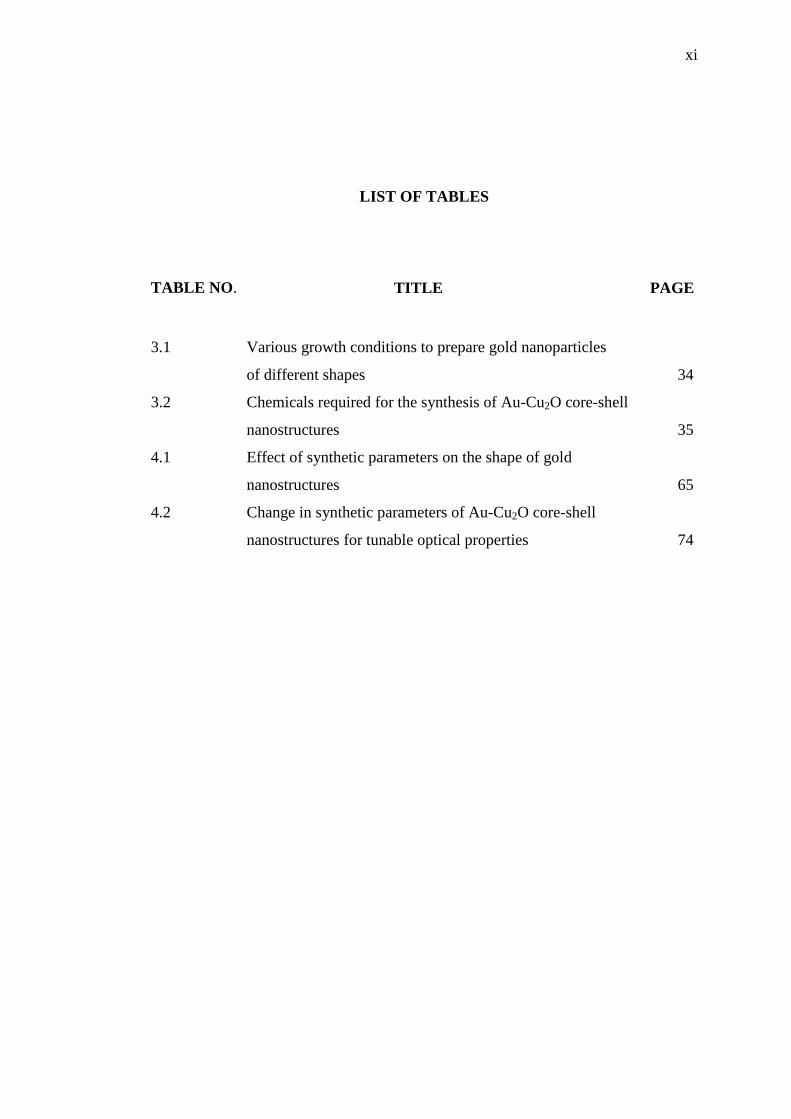
LIST OF TABLES
TABLE NO. TITLE PAGE
3.1 Various growth conditions to prepare gold nanoparticles
of different shapes 34
3.2 Chemicals required for the synthesis of Au-Cu2O core-shell
nanostructures 35
4.1 Effect of synthetic parameters on the shape of gold
nanostructures 65
4.2 Change in synthetic parameters of Au-Cu2O core-shell
nanostructures for tunable optical properties 74
xii
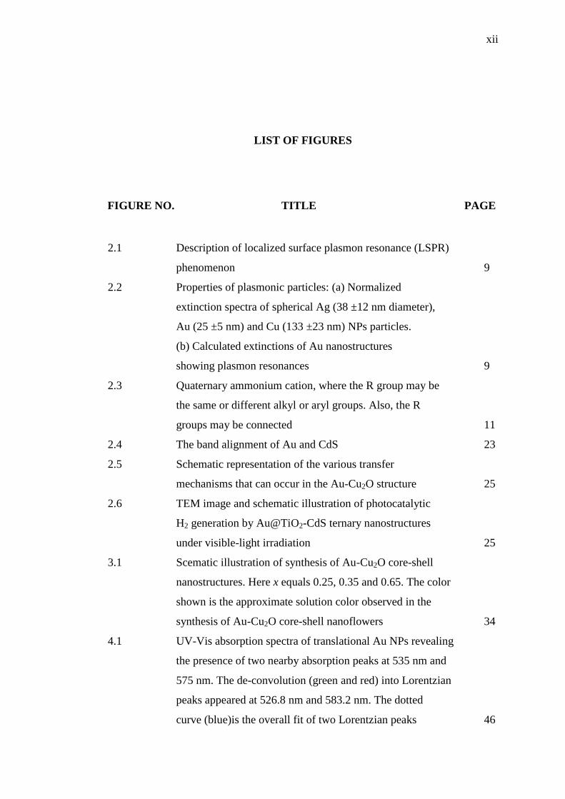
LIST OF FIGURES
FIGURE NO. TITLE PAGE
2.1 Description of localized surface plasmon resonance (LSPR)
phenomenon 9
2.2 Properties of plasmonic particles: (a) Normalized
extinction spectra of spherical Ag (38 ±12 nm diameter),
Au (25 ±5 nm) and Cu (133 ±23 nm) NPs particles.
(b) Calculated extinctions of Au nanostructures
showing plasmon resonances 9
2.3 Quaternary ammonium cation, where the R group may be
the same or different alkyl or aryl groups. Also, the R
groups may be connected 11
2.4 The band alignment of Au and CdS 23
2.5 Schematic representation of the various transfer
mechanisms that can occur in the Au-Cu2O structure 25
2.6 TEM image and schematic illustration of photocatalytic
H2 generation by Au@TiO2-CdS ternary nanostructures
under visible-light irradiation 25
3.1 Scematic illustration of synthesis of Au-Cu2O core-shell
nanostructures. Here x equals 0.25, 0.35 and 0.65. The color
shown is the approximate solution color observed in the
synthesis of Au-Cu2O core-shell nanoflowers 34
4.1 UV-Vis absorption spectra of translational Au NPs revealing
the presence of two nearby absorption peaks at 535 nm and
575 nm. The de-convolution (green and red) into Lorentzian
peaks appeared at 526.8 nm and 583.2 nm. The dotted
curve (blue)is the overall fit of two Lorentzian peaks 46

xiii
4.2 TEM images of translational Au NPs in the intermediate
reaction stage (step 3): (a) bunch of NPs appearing to adopt
some morphological transformation due to non-spherical
shape and (b) two close-by NPs with an inset showing
the size uniformity 46
4.3 (a) De-convoluted UV-Vis spectra showing the LSPR
absorption peaks of multi-faceted Au NPs at 534.5 and
650 nm and (b) a comparison showing relevance
absorption of seed nanoparticles with translational (step 3)
and multi-faceted (step 4) NPs during the reaction.
The absorption peak of the multi-faceted NPs is rather
sharp as compared to conventional spherical NPs and
translational NPs 47
4.4 A comparison of absorption spectra of freshly prepared multi-
faceted Au NPs after six month storage at 4 ºC 48
4.5 (a) and (b) TEM images of multi-faceted Au NPs, (c) magnified
view of a multi-faceted Au NP with seven-fold of symmetry
as marked, and (d) the HRTEM image displaying the lattice
constant of 2.33 Å corresponding to the Au(111) lattice planes 49
4.6 Aliquat 336 molecule (a) symbolic representation, (b) schematic
view, (c) stabilized multi-faceted Au NP, (d) a monolayer
of the ligand molecules illustrating a hexagonal pattern formed
due to overlapping of long hydrophobic chains on three sides
under van der Waals stabilization 50
4.7 TGA spectrum of the Aliquat 336-stabilized multi-faceted
Au NPs 52
4.8 (a) DLS data showing the corresponding size distribution
and (b) graph showing apparent ζ-potential of multi-faceted
Au NPs at +24.3 mV 53
4.9 EDX patterns of (a) intermediate translational and
(b) multi-faceted Au NPs 55
4.10 (a) XPS spectrum of Aliquat 336 stabilized multi-faceted
Au NPs and (b) high-resolution XPS spectrum of the
Au-4f7/2 at 84 eV and Au-4f5/2 at 87.75 eV 56

xiv
4.11 A comparison of FTIR spectra between (a) pure Aliquat 336
and (b) washed and dried samples of Aliquat 336-coated
gold nanoparticles supports both presence and nature of
attachment of Aliquat 336 molecules with the gold
nanostructures 57
4.12 Room temperature PL spectra of Aliquat 336 stabilized gold
NPs of multi-faceted (upper curve) and translational
(lower curve) 59
4.13 TEM images of as synthesized Au NRs. Most of them are
mono-dispersed ((a), (b), (c), (d), (g) and (h)) and few have
appeared in the bunch ((f) and (i)) 60
4.14 EDX spectrum showing the high purity formation of
Aunanorods 61
4.15 (a) XRD pattern of Au NRs, (b) shape distribution of Au
nanostructures in Figure 4.13 and (c) length distribution
of NRs in Figure 4.13 62
4.16 The UV-Vis absorption spectra of gold NRs (orange),
NWs (red), NCs (blue) and NTs (green) 63
4.17 TEM images of variously shaped gold nanostructures formed
due to variation in the synthetic parameters, including
(a) nanowires, (b) nanocubes, (c) nanotriangles and
(d) nanohexagons to nanotriangles. The actual solution
colours are also shown by an inset in each image 65
4.18 Schematic illustration of the reaction pathways that leads to
fcc Au nanocrystals of different shapes. The yellow, red
and purple colours represent the (100), (110) and (111)
facets, respectively 67
4.19 TEM images representing schematic of the formation of gold
nanostructures including gradual change in shape 68
4.20 Room temperature PL spectra of Aliquat 336-stabilized gold
NRs (blue curve), NTs (magenta curve) and nanocubes
(pink curve) 69

xv
4.21 TEM images of Au-Cu2O core-shell nanostructures; ((a) and
(b)) truncated-octahedral nanostructures formed from cubic Au
nanoparticles, ((c) and (d)) cuboctahedral nanostructures
formed from the same cubic Au nanoparticles, ((e) and
(f)) nanoflowers formed using multi-faceted Au nanoparticles
as templetes 71
4.22 HRTEM images performed to measure the fringe spacing of
Cu2O shell. The inset shows the orientation of Cu2O-shell
facets with respect to the Au nanocrystal facets 72
4.23 XRD patterns of nanoflower and truncated-octahedra
Au-Cu2O core-shell heterostructures 73
4.24 UV-Vis spectra of cubic Au nanoparticles and truncated-
octahedral Au-Cu2O core-shell nanostructures. The solution
colour for truncated-octahedra is gray with green hue having
the SPR band of the composite structure at 716 nm 75
4.25 ((a) and (b)) TEM images of truncated-octahedra with more
shell thickness, evidenced by more red-shift of SPR band, and
(c) UV-Vis absorption spectra of Samples A, B and C,
showing the effect of ratio NH2OH.HCl:NaOH on their
SPR bands. The solution colour of Sample B is green with
yellow hue 76
4.26 UV-Vis absorption spectra of cubic Au NPs and unique
cuboctahedral Au-Cu2O nanostructures with their SPR band
extending up to IR region of light. The solution colour
shows an orange hue 77
4.27 UV-Vis absorption spectra of multi-faceted Au nanoparticles
and flower-like Au-Cu2O nanostructures (including Sample D
and E) with the SPR band falling in the visible region of
light. The solution colour is green 77
4.28 Room temperature PL spectra of Au-Cu2O core-shell
heterostructures including truncated-octahedra, cuboctahedra
and nanoflowers 79
xvi
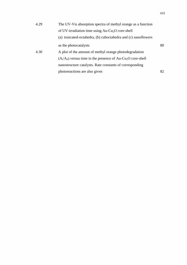
4.29 The UV-Vis absorption spectra of methyl orange as a function
of UV-irradiation time using Au-Cu2O core-shell
(a) truncated-octahedra, (b) cuboctahedra and (c) nanoflowers
as the photocatalysts 80
4.30 A plot of the amount of methyl orange photodegradation
(At/A0) versus time in the presence of Au-Cu2O core-shell
nanostructure catalysts. Rate constants of corresponding
photoreactions are also given 82

xvii
LIST OF ABBREVIATIONS
A336 - Aliquat 336
AgNO3 - Silver Nitrate
CH3 - Methyl
CH2 - Methylene
CTAB - Cetyltrimethyl Ammonium Bromide
CTAC - Cetyltrimethyl Ammonium Bromide
CTEAB - Cetyltriethyl Ammonium Bromide
C25H54ClN - Methyltrioctyl Ammonium Chloride/Aliquat 336
CuCl2 - Copper Chloride
Cu2O - Cuprous Oxide
DI - Deionized Water
DLS - Dynamic Light Scattering
EDX - Energy Dispersive X-rays
FRET - Fluorescence Resonance Energy Transfer
FTIR - Fourier Transformation Infrared
HAuCl4 - Hydrochloroauric Acid
HR-TEM - High Resolution Transmission Electron Microscopy
H2S - Hydrogen Sulfide
JCPD - Joint Committee for Powder Diffraction
KBr - Potassium Bromide
LSPR - Localised Surface Plasmon Resonance
MO - Methyl Orange
NaBH4 - Sodium Borohydride
NaBr - Sodium Bromide
NaOH - Sodium Hydroxide
NCs - Nanocubes
NPs - Nanoparticles

xviii
NRs - Nanorods
NSs - Nanostructures
NTs - Nanotriangles
NWs - Nanowires
oop - Out-of-plane
PL - Photoluminescence
Quats - Quaternary Ammonium Cations
rpm - Revolution per Minute
RTILs - Room Temperature Ionic Liquids
SDS - Sodium Dodecyl Sulphate
SHE - Standard Hydrogen Electrode
TEM - Transmission Electron Microscopy
TGA - Thermogravimetric Analysis
TOAB - Tetraoctyl Ammonium Bromide
UV-Vis - Ultraviolet-Visible
XPS - X-ray Photoelectron Spectroscopy
XRD - X-ray Diffraction

xix
LIST OF SYMBOLS
Å - Angstrom
Al - Aluminium
Au - Gold
a.u. - Arbitrary unit
Ag - Silver
Br - Bromine
C - Carbon
ca. - Circa
Cl - Chlorine
cm-1
- Frequency
Cu - Copper
o - Degree angle
oC - Degree Celsius
eV - Electron volt
h - Hours
H - Hydrogen
kV - Kilo volt
λ - Lambda
L - Length
µL - Micro litre
mg - Milli gram
mL - Milli litre
mV - Milli volt
min - Minute
M - Molarity
nm - Nanometre
N - Nitrogen

xx
N - Number
n - Number of carbon atoms
O - Oxygen
Pd - Palladium
% - Percentage
R - Radius
ρ - Density
s - Second
θ - Theta
W - Watt
x - Volume of NH2OH∙HCl in mL
Xe - Xenon
ζ - Zeta
xxi
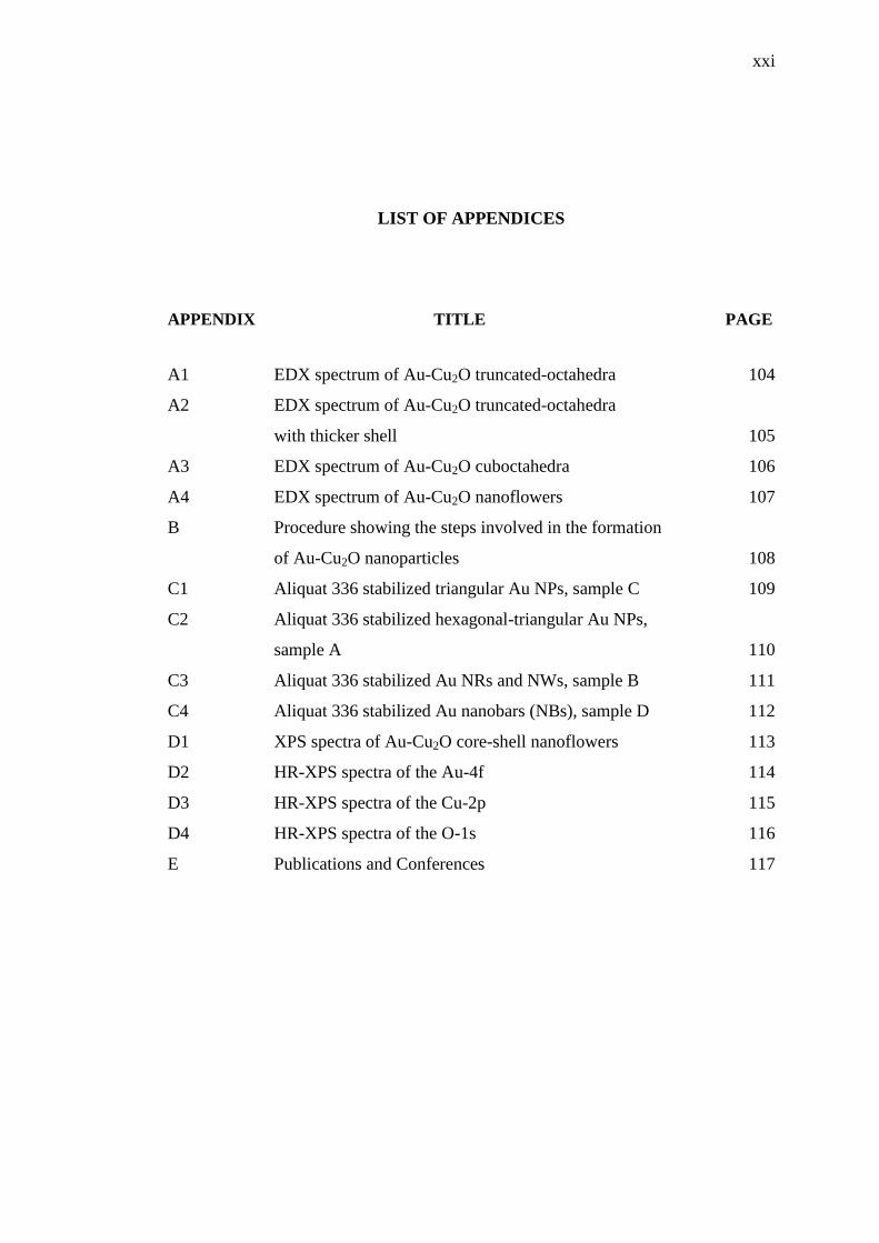
LIST OF APPENDICES
APPENDIX TITLE PAGE
A1 EDX spectrum of Au-Cu2O truncated-octahedra 104
A2 EDX spectrum of Au-Cu2O truncated-octahedra
with thicker shell 105
A3 EDX spectrum of Au-Cu2O cuboctahedra 106
A4 EDX spectrum of Au-Cu2O nanoflowers 107
B Procedure showing the steps involved in the formation
of Au-Cu2O nanoparticles 108
C1 Aliquat 336 stabilized triangular Au NPs, sample C 109
C2 Aliquat 336 stabilized hexagonal-triangular Au NPs,
sample A 110
C3 Aliquat 336 stabilized Au NRs and NWs, sample B 111
C4 Aliquat 336 stabilized Au nanobars (NBs), sample D 112
D1 XPS spectra of Au-Cu2O core-shell nanoflowers 113
D2 HR-XPS spectra of the Au-4f 114
D3 HR-XPS spectra of the Cu-2p 115
D4 HR-XPS spectra of the O-1s 116
E Publications and Conferences 117
CHAPTER 1
INTRODUCTION
1.1 Research Background
Undeniably, plasmonic gold (Au) nanostructures (NSs) are promising
material for their novel applications in various emerging fields of science,
technology and engineering [1, 2]. They have been a material of choice owing to a
combination of unique properties including the flexibility for surface alteration, the
tunable localized surface plasmon resonance (LSPR), the fascinating catalytic
activities at the nanoscale, and biocompatibility. Indeed, these Au NSs exhibiting
strong resonances in the visible/NIR region are model candidates for the
enhancement of Raman signals [3, 4] and diverse biomedical applications [5].
The growth of plasmonic Au NSs through wet chemical processes typically
requires a surfactant as capping and shape-directing agent. In fact, cationic
quaternary ammonium salts (quats) are used as the most essential surfactants [6]. It
is acknowledged that cetyltrimethylammonium bromide (CTAB) being very efficient
as directing agent allows the formation of Au nanoparticles (NPs) with varying
shapes including nanorods, hexagons and triangles [7, 8]. Especially, Au nanorods
with high aspect ratio are prepared using a seed-mediated growth method in an
aqueous micellar template by properly adjusting the CTAB concentration during the
reaction [9, 10]. The CTAB analogue, cetyltrimethylammonium chloride (CTAC) is
also used for the synthesis of anisotropic Au NPs with different shapes such as cubic,
trisoctahedra, and rhombic dodecahedra [11]. Nevertheless, these long chain cationic

2
ligands with higher ligand density in terms of bilayer have appeared to be more toxic
and thus limit their potential biomedical applications [12].
Methyltrioctylammonium chloride (Aliquat 336) is another quats reagent. It
is a less stable cationic ligand than the usual CTAB/CTAC ligands due to its three
dimensional short hydrocarbon chains and low affinity. However, it is more stable
against air and moisture attack than other cationic ligands and easier to handle [13].
Unlike a bilayer in CTAB/CTAC ligands, a mono-hydrophobic layer of Aliquat 336
molecules can stabilize Au NPs, where three hydrocarbon chains of the ligand
molecule overlap on three sides with those of another ligand molecules on the NPs
surface. Accordingly, the formation of a hexagonally patterned monolayer of the
ligand molecules on the Au NPs surface may overcome the ligand density problem
related to in vivo applications.
Another limitation with the CTAB assisted, seed-mediated gold nanorods
(NRs) synthesis is that the growth conditions control using these synthetic strategies
usually offers complexes like its aspect ratio reduction with the growth progression
[14, 15]. In this research, Au NRs of very high aspect ratio (ranging from 10 to 57)
have been prepared in an aqueous solution at 85 oC using Aliquat 336 as a phase
transfer reagent. Such anisotropic Au nanoparticles (NPs) have been used for various
biological and sensing applications due to their unique size, composition and
structure dependent optical properties [5]. However, the stability and surface
functionalization of Au NPs still remain problematic in many situations [16] due to
physicochemical limitations associated with them. An ideal solution is to encapsulate
these Au NPs with a semiconductor protective shell.
The interest in cuprous oxide, Cu2O, as a semiconductor began with the
invention of the Cu2O rectifier by Grondahl in the 1920s [17]. Cuprous oxide is a
semiconductor material with p-type conductivity due to copper vacancies. The
energy band gap of Cu2O is 2.17 eV and it has a high optical absorption coefficient
in the visible region [18]. The crystal structure of Cu2O is cuprite with a lattice
constant of 4.27 Å [19]. Considerable work was done on Cu2O characterization from
1930 to 1940. Photosensitive devices based on Cu2O were investigated in the 1930s

3
and B. Lange reviewed this work in 1939 [20]. The successful preparation of Cu2O
nanocrystals with systematic shape evolution from cubic to hexapod and octahedral
structures by a facile aqueous solution approach have shown enhanced photocatalytic
activity [21].
Formation of localized surface plasmon resonant (LSPR) cuprous oxide
coated gold (Au-Cu2O) core-shell nanostructures, during the last few years, with
precise geometrical and shape control of the components and their characterization
has presented remarkable attention. The characterization of these metal-
semiconductor core-shell nanostructures plays an important role either in
fundamental research or in technological uses, covering from fabrication and
characterization to device processing. It has been investigated that several
geometrical parameters (shell thickness, size of the core, spacing between core and
shell, etc.) of Au-Cu2O core-shell nanoparticles systematically fine-tune the light
absorption and scattering properties of these particles across the visible and near-
infrared regions [22]. Despite significant lattice mismatch of 4.3% between the
different gold surfaces and the lattice planes of Cu2O, excellent interfacial epitaxial
growth and systematic morphological evolution of these structures can still be
achieved [23] to have enhanced optical and catalytic properties.
1.2 Problem Statement
Quaternary ammonium cations/quats surfactants such as CTAC, CTAB and
cetyltriethylammonium bromide (CTEAB)-stabilized Au NSs have drawn an
interesting attention for applications based on their size and shape dependent optical
properties [24-26]. The drawback of these bilayer-surfactants protection of
nanoparticles has been their toxicity due to higher ligand density for in vivo [27] and
deficient long-term stability in terms of aggregation as the long alkyl chains of
CTAB/CTAC tend to trigger more van der Waals interactions among themselves
[28]. This research involves the use of another cationic ligand,
metyhyltrioctylammonium chloride (Aliquat 336), with rather short alkyl chains as a

4
phase transfer reagent to produce multi-faceted Au nanoparticles stabilized by
monolayer of the ligand molecules.
The preparation of Au NRs traditionally involves a seed-mediated growth
mechanism in the presence of cetyltrimethylammonium bromide/CTAB as a shape
directing and capping agent [3] and has been reported many times. This seed-growth
approach can produce Au NRs with aspect ratios (length/diameter) as much as 27:1.
However, the growth conditions control usually offers complexes [15] with another
limitation related to its aspect ratio reduction with the growth progression [14]. Thus,
an alternative synthesis method is required to achieve dispersed and elongated Au
NRs with localized surface plasmon resonance (LSPR) effects in the IR region.
The surface ligand and aspect ratio of Au NRs are prerequisite for near-field
optical response [29]. Usually, NRs aspect ratio is directing surfactant‟s nature
dependent (in an aqueous solution) [30] and a surfactant‟s (CTAB) concentrated
solution is necessary. CTAB binds to the surface of Au as bilayer structure and has
limitations in terms of its toxicity and stability [27, 31]. Often, AgNO3 is used as
additive for selective binding and packing of CTAB but it reduces the repulsion
between the surfactant head groups [32]. Despite this additive, preparation of NRs
with aspect ratio > 7 becomes difficult [9]. Many experiments exhibited the effect of
alkyltrimethylammonium (surfactant) tail length [30] and surfactant‟s head group
[26] on Au NRs growth. The change of Au NRs morphology (aspect ratio) via
different synthesis temperature programs has been reported [33], but the impact of
reaction time duration on morphological change is not yet documented. Here, an
alternative single-step synthesis method is adopted to achieve dispersed and
elongated Au NRs and nanowires (NWs) with localized surface plasmon resonance
(LSPR) effects in the near IR region to avoid a complex seed-mediated growth
mechanism in the presence of CTAB as a shape directing and capping agent.
Au NRs of very high aspect ratio (ranging from 10 to 57) have been prepared
in an aqueous solution at 85 oC using Aliquat 336 as a phase transfer reagent. The
effects of the ligand concentration, reaction temperature and time on the structure,
optical behavior, and the product yield are determined. Aliquat 336 has also shown a
5
capability to produce a variety of Au NSs, like Au nanocubes (NCs) and
nanotriangles (NTs), by controlling the growth parameters during the reaction.
As one distinctive combination of metal nanoparticles with localized surface
plasmon resonance and metal-oxide semiconductors, Au-Cu2O metal-semiconductor
core-shell nanostructures have attracted a great deal of attention because of their
novel structure and potential application in solar energy conversion [34]. Despite the
recent achievements in the systematic growth of these heterostructures at different
levels and their catalytic activities, further investigation on various shapes-dependent
optical properties of Au-Cu2O nanocrystals are lacking. For example, a lot of
attention has been given to measurements of photocatalytic performance of Au-Cu2O
core-shell nanostructures [35], but the effect of various shaped Au-Cu2O
nanoparticles (e.g. cuboctahedron, octahedron) on other optical properties like
photoluminescence has not been systematically investigated. Furthermore, most
studies are lacking the rich structural variety of semiconductor-shell that may be
produced by employing core particles of different shapes, and their characterization.
In this research, Au-Cu2O core-shell nano-morphologies are synthesized by
facile wet chemical approach and exposed to light with different shapes in order to
investigate for correlation between photoluminescence and photocatalytic
performance. Furthermore, the cooperative morphology between plasmonic metal
and semiconductor nanostructures is explored along with their special plasmon
resonant optical properties that show interesting tunability during the structural
evolution.
1.3 Research Questions
The study involves following research questions:
i. How the shape evolution of gold (Au) and Au-Cu2O core-shell
nanostructures can be obtained by using Aliquat 336 surfactant and
gold-cores of different shapes, respectively?

6
ii. Does the surface functionalization of these nanostructures require the
ability to tune the nanoparticles morphology?
iii. How does the shape and surface orientation of gold and Au-Cu2O
nanostructures affect the LSPR based optical properties and how these
can be harvested towards applications, like Photoluminescence and
photocatalysis?
1.4 Research Objectives
The research objectives of the study include:
i. To synthesize quats-functionalized gold (Au) nanostructures (NSs)
for the measurement of the ligand density on the surface of Au NSs.
ii. To prepare Au-Cu2O core-shell nanostructures using gold-cores of
different shapes, for improved LSPR based optical properties.
iii. To determine the influence of growth parameters on the
structural/optical properties of Au and Au-Cu2O core-shell
nanostructures.
iv. To determine the relationship between photoluminescence and
photocatalytic properties of Au-Cu2O core-shell nanostructures.
1.5 Scope of the Research
This research involves the syntheses of functionalized gold (Au) and cuprous
oxide coated gold (Au-Cu2O) core-shell nanostructures with various shape evolution
by varying the growth parameters. In the first, preparation of multi-faceted Au
nanoparticles (NPs) with minimal ligand density using a quaternary ammonium
cationic ligand as a shaping and stabilizing agent was encountered. The stability and
nature of binding of the ligand to the Au NPs surface was accomplished by Fourier
transform infrared spectroscopy (FTIR), X-ray photoelectron spectroscopy (XPS),

7
dynamic light scattering (DLS), Zeta potential and thermogravimetric analysis
(TGA). Then, the effect of different synthetic parameters like reaction temperature,
reaction duration, gold precursor and the ligand concentration on the syntheses of
other Au NSs (e.g. cubic, triangular, rod and wire-like) were studied. The
modification of multi-faceted and cubic Au NSs with Au-Cu2O core-shell
nanostructures for enhanced LSPR based optical properties was successfully
accomplished. The unique surface plasmon absorption of various gold nanostructures
and Au-Cu2O core-shell nanostructures was taken by UV-Vis spectroscopy. The size,
morphology and chemical composition of these nanostructures were studied by
transmission electron microscope (TEM), X-ray diffractometer (XRD) and energy
dispersive X-ray (EDX). Influence of growth parameters on structural and optical
properties of Au-Cu2O core-shell nanostructures was investigated. Gold and Au-
Cu2O core-shell nanostructures were also supposed to explore the influence of the
LSPR on the photoluminescence emission peaks of these nanostructures. The Au-
Cu2O core-shell nanostructures were also examined comparatively as photocatalysts
towards the decomposition of organic dye. The results showed that the core-shell
nanostructures with more exposed (111) surfaces were catalytically more active, in
good agreement with PL analysis where catalytically inactive (100) surfaces revealed
a comparatively sharp emission peak.
1.6 Significance of the Research
In this research, the motivation for the syntheses of plasmonic Au NSs with
shape-dependent optical properties comes from the choice of another cationic ligand,
methyltrioctylammonium chloride/Aliquat 336, having superior properties while
altering the growth parameters. Especially, Aliquat 336 surfactant is utilized to
prepare high aspect ratio Au NRs and NWs without any usual seed-mediated growth
mechanism. The modification of Au NSs with Au-Cu2O metal-semiconductor core-
shell NSs resulted in enhanced LSPR based optical and photocatalytic properties.
REFERENCES
1. Daniel, M.-C. and Astruc, D. Gold Nanoparticles: Assembly, Supramolecular
Chemistry, Quantum-size-related Properties, and Applications toward
Biology, Catalysis, and Nanotechnology. Chemical Reviews. 2003. 104(1):
293-346.
2. Kale, M. J., Avanesian, T. and Christopher, P. Direct Photocatalysis by
Plasmonic Nanostructures. ACS Catalysis. 2013. 4(1): 116-128.
3. Smitha, S. L., Gopchandran, K. G., Ravindran, T. R. and Prasad, V. S. Gold
Nanorods with Finely Tunable Longitudinal Surface Plasmon Resonance as
SERS Substrates. Nanotechnology. 2011. 22(26): 265705.
4. Zhu, J., Gao, J., Li, J.-J. and Zhao, J.-W. Improve the Surface-enhanced
Raman Scattering from Rhodamine 6G Adsorbed Gold Nanostars with
Vimineous Branches. Applied Surface Science. 2014. 322(0): 136-142.
5. Cobley, C. M., Chen, J., Cho, E. C., Wang, L. V. and Xia, Y. Gold
Nanostructures: A Class of Multifunctional Materials for Biomedical
Applications. Chemical Society Reviews. 2011. 40(1): 44-56.
6. Nikoobakht, B. and El-Sayed, M. A. Evidence for Bilayer Assembly of
Cationic Surfactants on the Surface of Gold Nanorods. Langmuir. 2001.
17(20): 6368-6374.
7. Gole, A. and Murphy, C. J. Seed-mediated Synthesis of Gold Nanorods:
Role of the Size and Nature of the Seed. Chemistry of Materials. 2004.
16(19): 3633-3640.
8. Wang, Z., Yuan, J., Zhou, M., Niu, L. and Ivaska, A. Synthesis,
Characterization and Mechanism of Cetyltrimethylammonium Bromide
Bilayer-encapsulated Gold Nanosheets and Nanocrystals. Applied Surface
Science. 2008. 254(20): 6289-6293.

88
9. Jana, N. R., Gearheart, L. and Murphy, C. J. Wet Chemical Synthesis of High
Aspect Ratio Cylindrical Gold Nanorods. The Journal of Physical Chemistry
B. 2001. 105(19): 4065-4067.
10. Smith, D. K. and Korgel, B. A. The Importance of the CTAB Surfactant on
the Colloidal Seed-mediated Synthesis of Gold Nanorods. Langmuir. 2008.
24(3): 644-649.
11. Wu, H.-L., Kuo, C.-H. and Huang, M. H. Seed-mediated Synthesis of Gold
Nanocrystals with Systematic Shape Evolution from Cubic to Trisoctahedral
and Rhombic Dodecahedral Structures. Langmuir. 2010. 26(14): 12307-
12313.
12. Bozich, J. S., Lohse, S. E., Torelli, M. D., Murphy, C. J., Hamers, R. J. and
Klaper, R. D. Surface Chemistry, Charge and Ligand Type Impact the
Toxicity of Gold Nanoparticles to Daphnia Magna. Environmental Science:
Nano. 2014. 1(3): 260-270.
13. Mikkola, J.-P., Virtanen, P. and Sjoholm, R. Aliquat 336®-a Versatile and
Affordable Cation Source for an Entirely New Family of Hydrophobic Ionic
Liquids. Green Chemistry. 2006. 8(3): 250-255.
14. Park, K., Drummy, L. F., Wadams, R. C., Koerner, H., Nepal, D., Fabris, L.
and Vaia, R. A. Growth Mechanism of Gold Nanorods. Chemistry of
Materials. 2013. 25(4): 555-563.
15. Li, W. and Xia, Y. Facile Synthesis of Gold Octahedra by Direct Reduction
of HAuCl4 in an Aqueous Solution. Chemistry – An Asian Journal. 2010.
5(6): 1312-1316.
16. Wangoo, N., Shekhawat, G., Wu, J.-S., Bhasin, A. K., Suri, C. R., Bhasin, K.
K. and Dravid, V. Green Synthesis and Characterization of Size Tunable
Silica-capped Gold Core–shell Nanoparticles. Journal of Nanoparticle
Research. 2012. 14(8): 1-9.
17. Grondahl, L. O. The Copper-Cuprous-Oxide Rectifier and Photoelectric Cell.
Reviews of Modern Physics. 1933. 5(2): 141-168.
18. Parretta, A., Jayaraj, M. K., Di Nocera, A., Loreti, S., Quercia, L. and Agati,
A. Electrical and Optical Properties of Copper Oxide Films Prepared by
Reactive RF Magnetron Sputtering. Physica Status Solidi (a). 1996. 155(2):
399-404.
19. Wyckoff, R. W. G. Crystal Structures: Wiley. 1963.

89
20. Lange, B. Photoelements and Their Applications. Reinhold, New York. 1939.
21. Ho, J.-Y. and Huang, M. H. Synthesis of Submicrometer-sized Cu2O Crystals
with Morphological Evolution from Cubic to Hexapod Structures and their
Comparative Photocatalytic Activity. The Journal of Physical Chemistry C.
2009. 113(32): 14159-14164.
22. Zhang, L., Blom, D. A. and Wang, H. Au–Cu2O Core–shell Nanoparticles: A
Hybrid Metal-semiconductor Heteronanostructure with Geometrically
Tunable Optical Properties. Chemistry of Materials. 2011. 23(20): 4587-
4598.
23. Wang, Y. Q., Nikitin, K. and McComb, D. W. Fabrication of Au–Cu2O
Core–shell Nanocube Heterostructures. Chemical Physics Letters. 2008.
456(4–6): 202-205.
24. Angelomé, P. C., Heidari Mezerji, H., Goris, B., Pastoriza-Santos, I., Pérez-
Juste, J., Bals, S. and Liz-Marzán, L. M. Seedless Synthesis of Single
Crystalline Au Nanoparticles with Unusual Shapes and Tunable LSPR in the
near-IR. Chemistry of Materials. 2012. 24(7): 1393-1399.
25. Xiao, J. and Qi, L. Surfactant-assisted, shape-controlled Synthesis of Gold
Nanocrystals. Nanoscale. 2011. 3(4): 1383-1396.
26. Kou, X., Zhang, S., Tsung, C.-K., Yeung, M. H., Shi, Q., Stucky, G. D., Sun,
L., Wang, J. and Yan, C. Growth of Gold Nanorods and Bipyramids using
CTEAB Surfactant. The Journal of Physical Chemistry B. 2006. 110(33):
16377-16383.
27. Vigderman, L., Manna, P. and Zubarev, E. R. Quantitative Replacement of
Cetyl Trimethylammonium Bromide by Cationic Thiol Ligands on the
Surface of Gold Nanorods and their Extremely Large Uptake by Cancer
Cells. Angewandte Chemie International Edition. 2012. 51(3): 636-641.
28. Hsu, C.-L., Wang, K.-H., Chang, C.-H., Hsu, W.-P. and Lee, Y.-L. Surface
Modification of Gold Nanoparticles and their Monolayer Behavior at the
Air/Water Interface. Applied Surface Science. 2011. 257(7): 2756-2763.
29. Habteyes, T. G. Direct Near-field Observation of Orientation-dependent
Optical Response of Gold Nanorods. The Journal of Physical Chemistry C.
2014. 118(17): 9119-9127.

90
30. Gao, J., Bender, C. M. and Murphy, C. J. Dependence of the Gold Nanorod
Aspect Ratio on the Nature of the Directing Surfactant in Aqueous Solution.
Langmuir. 2003. 19(21): 9065-9070.
31. Kinnear, C., Dietsch, H., Clift, M. J. D., Endes, C., Rothen-Rutishauser, B.
and Petri-Fink, A. Gold Nanorods: Controlling their Surface Chemistry and
Complete Detoxification by a Two-step Place Exchange. Angewandte Chemie
International Edition. 2013. 52(7): 1934-1938.
32. Ye, X., Jin, L., Caglayan, H., Chen, J., Xing, G., Zheng, C., Doan-Nguyen,
V., Kang, Y., Engheta, N., Kagan, C. R. and Murray, C. B. Improved Size-
tunable Synthesis of Monodisperse Gold Nanorods through the Use of
Aromatic Additives. ACS Nano. 2012. 6(3): 2804-2817.
33. Liu, F.-K., Chang, Y.-C., Ko, F.-H. and Chu, T.-C. Microwave Rapid Heating
for the Synthesis of Gold Nanorods. Materials Letters. 2004. 58(3–4): 373-
377.
34. Kuo, C.-H., Yang, Y.-C., Gwo, S. and Huang, M. H. Facet-dependent and Au
Nanocrystal-enhanced Electrical and Photocatalytic Properties of Au−Cu2O
Core−shell Heterostructures. Journal of the American Chemical Society.
2010. 133(4): 1052-1057.
35. Wang, Z., Zhao, S., Zhu, S., Sun, Y. and Fang, M. Photocatalytic Synthesis
of M/Cu2O (M = Ag, Au) Heterogeneous Nanocrystals and their
Photocatalytic Properties. CrystEngComm. 2011. 13(7): 2262-2267.
36. Faraday, M. Experimental Relations of Gold (and other Metals) to Light P.
Trans. R. Soc. London 1857. 147: 145-181.
37. Linic, S., Christopher, P. and Ingram, D. B. Plasmonic-metal nanostructures
for efficient conversion of solar to chemical energy. Nat Mater. 2011. 10(12):
911-921.
38. Rycenga, M., Cobley, C. M., Zeng, J., Li, W., Moran, C. H., Zhang, Q., Qin,
D. and Xia, Y. Controlling the Synthesis and Assembly of Silver
Nanostructures for Plasmonic Applications. Chemical Reviews. 2011. 111(6):
3669-3712.
39. Zhang, H. and Govorov, A. O. Optical Generation of Hot Plasmonic Carriers
in Metal Nanocrystals: The Effects of Shape and Field Enhancement. The
Journal of Physical Chemistry C. 2014. 118(14): 7606-7614.
91
40. Lewinski, N., Colvin, V. and Drezek, R. Cytotoxicity of Nanoparticles.
Small. 2008. 4(1): 26-49.
41. Shukla, R., Bansal, V., Chaudhary, M., Basu, A., Bhonde, R. R. and Sastry,
M. Biocompatibility of Gold Nanoparticles and their Endocytotic Fate Inside
the Cellular Compartment: A Microscopic Overview. Langmuir. 2005.
21(23): 10644-10654.
42. Xia, Y., Xiong, Y., Lim, B. and Skrabalak, S. E. Shape-controlled Synthesis
of Metal Nanocrystals: Simple Chemistry Meets Complex Physics?
Angewandte Chemie International Edition. 2009. 48(1): 60-103.
43. Schuller, J. A., Barnard, E. S., Cai, W., Jun, Y. C., White, J. S. and
Brongersma, M. L. Plasmonics for Extreme Light Concentration and
Manipulation. Nat Mater. 2010. 9(3): 193-204.
44. Atwater, H. A. and Polman, A. Plasmonics for Improved Photovoltaic
Devices. Nat Mater. 2010. 9(3): 205-213.
45. Reineck, P., Gómez, D., Ng, S. H., Karg, M., Bell, T., Mulvaney, P. and
Bach, U. Distance and Wavelength Dependent Quenching of Molecular
Fluorescence by Au@SiO2 Core–shell Nanoparticles. ACS Nano. 2013. 7(8):
6636-6648.
46. Juergensen, L., Busnarda, J., Caux, P.-Y. and Kent, R. A. Fate, Behavior, and
Aquatic Toxicity of the Fungicide DDAC in the Canadian Environment.
Environmental Toxicology. 2000. 15(3): 174-200.
47. Yu, P., Wen, X., Toh, Y.-R., Ma, X. and Tang, J. Fluorescent Metallic
Nanoclusters: Electron Dynamics, Structure, and Applications. Particle &
Particle Systems Characterization. 2015. 32(2): 142-163.
48. Sardar, R., Funston, A. M., Mulvaney, P. and Murray, R. W. Gold
Nanoparticles: Past, Present, and Future. Langmuir. 2009. 25(24): 13840-
13851.
49. Shukla, S., Priscilla, A., Banerjee, M., Bhonde, R. R., Ghatak, J., Satyam, P.
V. and Sastry, M. Porous Gold Nanospheres by Controlled Transmetalation
Reaction: A Novel Material for Application in Cell Imaging. Chemistry of
Materials. 2005. 17(20): 5000-5005.
50. Chang, C.-C., Wu, H.-L., Kuo, C.-H. and Huang, M. H. Hydrothermal
Synthesis of Monodispersed Octahedral Gold Nanocrystals with Five
92
Different Size Ranges and Their Self-assembled Structures. Chemistry of
Materials. 2008. 20(24): 7570-7574.
51. Sau, T. K. and Murphy, C. J. Self-assembly Patterns Formed upon Solvent
Evaporation of Aqueous Cetyltrimethylammonium Bromide-coated Gold
Nanoparticles of Various Shapes. Langmuir. 2005. 21(7): 2923-2929.
52. Wiley, B. J., Chen, Y., McLellan, J. M., Xiong, Y., Li, Z.-Y., Ginger, D. and
Xia, Y. Synthesis and Optical Properties of Silver Nanobars and Nanorice.
Nano Letters. 2007. 7(4): 1032-1036.
53. Sau, T. K. and Murphy, C. J. Room Temperature, High-yield Synthesis of
Multiple Shapes of Gold Nanoparticles in Aqueous Solution. Journal of the
American Chemical Society. 2004. 126(28): 8648-8649.
54. Saunders, A. E., Sigman, M. B. and Korgel, B. A. Growth Kinetics and
Metastability of Monodisperse Tetraoctylammonium Bromide Capped Gold
Nanocrystals. The Journal of Physical Chemistry B. 2004. 108(1): 193-199.
55. Thomas, K. G., Zajicek, J. and Kamat, P. V. Surface Binding Properties of
Tetraoctylammonium Bromide-Capped Gold Nanoparticles. Langmuir. 2002.
18(9): 3722-3727.
56. Thomas, K. G. and Kamat, P. V. Making Gold Nanoparticles Glow:
Enhanced Emission from a Surface-bound Fluoroprobe. Journal of the
American Chemical Society. 2000. 122(11): 2655-2656.
57. Isaacs, S. R., Cutler, E. C., Park, J.-S., Lee, T. R. and Shon, Y.-S. Synthesis
of Tetraoctylammonium-protected Gold Nanoparticles with Improved
Stability. Langmuir. 2005. 21(13): 5689-5692.
58. Briñas, R. P., Maetani, M. and Barchi, J. J. A Survey of Place-exchange
Reaction for the Preparation of Water-soluble Gold Nanoparticles. Journal of
colloid and interface science. 2013. 392: 415-421.
59. Wu, H.-L., Chen, C.-H. and Huang, M. H. Seed-Mediated Synthesis of
Branched Gold Nanocrystals Derived from the Side Growth of Pentagonal
Bipyramids and the Formation of Gold Nanostars. Chemistry of Materials.
2008. 21(1): 110-114.
60. Lee, Y. W., Kim, M., Kim, Z. H. and Han, S. W. One-Step Synthesis of
Au@Pd Core−shell Nanooctahedron. Journal of the American Chemical
Society. 2009. 131(47): 17036-17037.
93
61. Lee, N.-S., Kim, D., Kang, H., Park, D. K., Han, S. W. and Noh, J. Structural
Transitions of Octanethiol Self-assembled Monolayers on Gold Nanoplates
after Mild Thermal Annealing. The Journal of Physical Chemistry C. 2011.
115(13): 5868-5874.
62. Zeng, J., Zheng, Y., Rycenga, M., Tao, J., Li, Z.-Y., Zhang, Q., Zhu, Y. and
Xia, Y. Controlling the Shapes of Silver Nanocrystals with Different Capping
Agents. Journal of the American Chemical Society. 2010. 132(25): 8552-
8553.
63. Ma, Y., Li, W., Cho, E. C., Li, Z., Yu, T., Zeng, J., Xie, Z. and Xia, Y.
Au@Ag Core−shell Nanocubes with Finely Tuned and Well-controlled Sizes,
Shell Thicknesses, and Optical Properties. ACS Nano. 2010. 4(11): 6725-
6734.
64. Garabagiu, S. and Bratu, I. Thiol Containing Carboxylic Acids Remove the
CTAB Surfactant onto the Surface of Gold Nanorods: An FTIR
Spectroscopic Study. Applied Surface Science. 2013. 284(0): 780-783.
65. Alkilany, A. M., Lohse, S. E. and Murphy, C. J. The Gold Standard: Gold
Nanoparticle Libraries To Understand the Nano–bio Interface. Accounts of
Chemical Research. 2012. 46(3): 650-661.
66. Neouze, M.-A. About the Interactions Between Nanoparticles and
Imidazolium Moieties: Emergence of Original Hybrid Materials. Journal of
Materials Chemistry. 2010. 20(43): 9593-9607.
67. Itoh, H., Naka, K. and Chujo, Y. Synthesis of Gold Nanoparticles Modified
with Ionic Liquid Based on the Imidazolium Cation. Journal of the American
Chemical Society. 2004. 126(10): 3026-3027.
68. Dinda, E., Si, S., Kotal, A. and Mandal, T. K. Novel Ascorbic Acid Based
Ionic Liquids for the In Situ Synthesis of Quasi-spherical and Anisotropic
Gold Nanostructures in Aqueous Medium. Chemistry. 2008. 14(18): 5528-
5537.
69. Weng, J., Wang, C., Li, H. and Wang, Y. Novel Quaternary Ammonium
Ionic Liquids and their Use as Dual Solvent-catalysts in the Hydrolytic
Reaction. Green Chemistry. 2006. 8(1): 96-99.
70. Chowdhury, S., Mohan, R. S. and Scott, J. L. Reactivity of Ionic Liquids.
Tetrahedron. 2007. 63(11): 2363-2389.

94
71. Hoh, Y.-C., Chuang, W.-S. and Yueh, P.-S. The Extraction of Platinum from
Pt(II) Chloride Solution by a Quaternary Ammonium Compound. Journal of
Chemical Technology and Biotechnology. Chemical Technology. 1985. 35(1):
41-47.
72. Nayl, A. A. Extraction and Separation of Co(II) and Ni(II) from Acidic
Sulfate Solutions using Aliquat 336. J Hazard Mater. 2010. 173(1-3): 223-
230.
73. Xu, J., Paimin, R., Shen, W. and Wang, X. An Investigation of Solubility of
Aliquat 336 in Different Extracted Solutions. Fibers and Polymers. 2003.
4(1): 27-31.
74. Oliveira, E., Núñez, C., Santos, H. M., Fernández-Lodeiro, J., Fernández-
Lodeiro, A., Capelo, J. L. and Lodeiro, C. Revisiting the Use of Gold and
Silver Functionalised Nanoparticles as Colorimetric and Fluorometric
Chemosensors for Metal Ions. Sensors and Actuators B: Chemical. 2015.
212: 297-328.
75. Zhang, J., Wang, Y., Xu, X. and Yang, X. Specifically Colorimetric
Recognition of Calcium, Strontium, and Barium Ions using 2-
mercaptosuccinic Acid-functionalized Gold Nanoparticles and its Use in
Reliable Detection of Calcium Ion in Water. Analyst. 2011. 136(19): 3865-
3868.
76. Lai, Y.-J. and Tseng, W.-L. Role of 5-thio-(2-nitrobenzoic acid)-capped Gold
Nanoparticles in the Sensing of Chromium(vi): Remover and Sensor. Analyst.
2011. 136(13): 2712-2717.
77. Zhao, L., Jin, Y., Yan, Z., Liu, Y. and Zhu, H. Novel, Highly Selective
Detection of Cr(III) in Aqueous Solution Based on a Gold Nanoparticles
Colorimetric Assay and its Application for Determining Cr(VI). Analytica
Chimica Acta. 2012. 731: 75-81.
78. Weng, Z., Wang, H., Vongsvivut, J., Li, R., Glushenkov, A. M., He, J., Chen,
Y., Barrow, C. J. and Yang, W. Self-assembly of Core-satellite Gold
Nanoparticles for Colorimetric Detection of Copper Ions. Analytica Chimica
Acta. 2013. 803: 128-134.
79. Zhang, F., Zeng, L., Zhang, Y., Wang, H. and Wu, A. A Colorimetric Assay
Method for Co2+
Based on Thioglycolic acid Functionalized Hexadecyl
95
Trimethyl ammonium Bromide Modified Au Nanoparticles (NPs).
Nanoscale. 2011. 3(5): 2150-2154.
80. Krpetić, Ž., Guerrini, L., Larmour, I. A., Reglinski, J., Faulds, K. and
Graham, D. Importance of Nanoparticle Size in Colorimetric and SERS-
based Multimodal Trace Detection of Ni(II) Ions with Functional Gold
Nanoparticles. Small. 2012. 8(5): 707-714.
81. Sung, Y.-M. and Wu, S.-P. Highly Selective and Sensitive Colorimetric
Detection of Ag(I) using N-1-(2-mercaptoethyl)adenine Functionalized Gold
Nanoparticles. Sensors and Actuators B: Chemical. 2014. 197: 172-176.
82. Liu, J.-M., Wang, H.-F. and Yan, X.-P. A Gold Nanorod Based Colorimetric
Probe for the Rapid and Selective Detection of Cu2+
ions. Analyst. 2011.
136(19): 3904-3910.
83. Velu, R., Ramakrishnan, V. T. and Ramamurthy, P. Colorimetric and
Fluorometric Chemosensors for Selective Signaling toward Ca2+
and Mg2+
by
Aza-crown Ether Acridinedione-functionalized Gold Nanoparticles.
Tetrahedron Letters. 2010. 51(33): 4331-4335.
84. Habeeb Muhammed, M. A., Verma, P. K., Pal, S. K., Retnakumari, A.,
Koyakutty, M., Nair, S. and Pradeep, T. Luminescent Quantum Clusters of
Gold in Bulk by Albumin-induced Core Etching of Nanoparticles: Metal Ion
Sensing, Metal-enhanced Luminescence, and Biolabeling. Chemistry – A
European Journal. 2010. 16(33): 10103-10112.
85. Zhang, G., Li, Y., Xu, J., Zhang, C., Shuang, S., Dong, C. and Choi, M. M. F.
Glutathione-protected Fluorescent Gold Nanoclusters for Sensitive and
Selective Detection of Cu2+
. Sensors and Actuators B: Chemical. 2013. 183:
583-588.
86. Pyne, S., Sahoo, G. P., Bhui, D. K., Bar, H., Sarkar, P., Maity, A. and Misra,
A. FRET Based Ultra Sensor for Detection of Hg (II) in Water: A
Comparative Study using Citrate and Marcapto Propanoic Acid as Stabilizer
of AuNPs. Sensors and Actuators B: Chemical. 2011. 160(1): 1141-1148.
87. Yuan, Z., Peng, M., He, Y. and Yeung, E. S. Functionalized Fluorescent Gold
Nanodots: Synthesis and Application for Pb2+
Sensing. Chemical
Communications. 2011. 47(43): 11981-11983.
88. Wu, Y.-S., Huang, F.-F. and Lin, Y.-W. Fluorescent Detection of Lead in
Environmental Water and Urine Samples using Enzyme Mimics of Catechin-

96
Synthesized Au Nanoparticles. ACS Applied Materials & Interfaces. 2013.
5(4): 1503-1509.
89. Sun, J., Zhang, J. and Jin, Y. 11-Mercaptoundecanoic Acid Directed One-pot
Synthesis of Water-soluble Fluorescent Gold Nanoclusters and Their Use as
Probes for Sensitive and Selective Detection of Cr3+
and Cr6+
. Journal of
Materials Chemistry C. 2013. 1(1): 138-143.
90. Seo, S.-H., Kim, B.-M., Joe, A., Han, H.-W., Chen, X., Cheng, Z. and Jang,
E.-S. NIR-light-induced Surface-enhanced Raman Scattering for Detection
and Photothermal/Photodynamic Therapy of Cancer Cells using Methylene
Blue-embedded Gold Nanorod@SiO2 Nanocomposites. Biomaterials. 2014.
35(10): 3309-3318.
91. Guo, Z., Hwang, J., Zhao, B., Chung, J. H., Cho, S. G., Baek, S.-J. and Choo,
J. Ultrasensitive Trace Analysis for 2,4,6-trinitrotoluene Using Nano-
dumbbell Surface-enhanced Raman Scattering Hot Spots. Analyst. 2014.
139(4): 807-812.
92. Pazos-Pérez, N., Barbosa, S., Rodríguez-Lorenzo, L., Aldeanueva-Potel, P.,
Pérez-Juste, J., Pastoriza-Santos, I., Alvarez-Puebla, R. A. and Liz-Marzán,
L. M. Growth of Sharp Tips on Gold Nanowires Leads to Increased Surface-
enhanced Raman Scattering Activity. The Journal of Physical Chemistry
Letters. 2010. 1(1): 24-27.
93. Zhang, Z., Yang, P., Xu, H. and Zheng, H. Surface Enhanced Fluorescence
and Raman Scattering by Gold Nanoparticle Dimers and Trimers. Journal of
Applied Physics. 2013. 113(3): 033102.
94. Pilo-Pais, M., Watson, A., Demers, S., LaBean, T. H. and Finkelstein, G.
Surface-enhanced Raman Scattering Plasmonic Enhancement Using DNA
Origami-based Complex Metallic Nanostructures. Nano Letters. 2014. 14(4):
2099-2104.
95. Dan, X., Jiangjiang, G., Weina, W., Xuehai, Y., Kai, X. and Xudong, J.
Development of Chitosan-coated Gold Nanoflowers as SERS-active Probes.
Nanotechnology. 2010. 21(37): 375101.
96. Dawson, K. and Riordan, A. O. Towards Nanowire (Bio) Sensors. Journal of
Physics: Conference Series. 2011. 307(1): 012004.
97. Wang, Q., Min, F. and Zhu, J. Preparation of Gold Nanowires and its
Application in Glucose Biosensing. Materials Letters. 2013. 91(0): 9-11.
97
98. Hu, J., Wang, Z. and Li, J. Gold Nanoparticles With Special Shapes:
Controlled Synthesis, Surface-enhanced Raman Scattering, and The
Application in Biodetection. Sensors (Basel, Switzerland). 2007. 7(12): 3299-
3311.
99. Huang, X., El-Sayed, I. H., Qian, W. and El-Sayed, M. A. Cancer Cell
Imaging and Photothermal Therapy in the Near-Infrared Region by Using
Gold Nanorods. Journal of the American Chemical Society. 2006. 128(6):
2115-2120.
100. Wijaya, A., Schaffer, S. B., Pallares, I. G. and Hamad-Schifferli, K. Selective
Release of Multiple DNA Oligonucleotides from Gold Nanorods. ACS Nano.
2009. 3(1): 80-86.
101. Kuo, W.-S., Chang, C.-N., Chang, Y.-T., Yang, M.-H., Chien, Y.-H., Chen,
S.-J. and Yeh, C.-S. Gold Nanorods in Photodynamic Therapy, as
Hyperthermia Agents, and in Near-infrared Optical Imaging. Angewandte
Chemie. 2010. 122(15): 2771-2775.
102. Haruta, M., Kobayashi, T., Sano, H. and Yamada, N. Novel Gold Catalysts
for the Oxidation of Carbon Monoxide at a Temperature far Below 0 °C.
Chemistry Letters. 1987. 16(2): 405-408.
103. Huang, D., Bai, X. and Zheng, L. Ultrafast Preparation of Three-dimensional
Dendritic Gold Nanostructures in Aqueous Solution and their Applications in
Catalysis and SERS. The Journal of Physical Chemistry C. 2011. 115(30):
14641-14647.
104. Sai Krishna, K., Navin, C. V., Biswas, S., Singh, V., Ham, K., Bovenkamp,
G. L., Theegala, C. S., Miller, J. T., Spivey, J. J. and Kumar, C. S. S. R.
Millifluidics for Time-resolved Mapping of the Growth of Gold
Nanostructures. Journal of the American Chemical Society. 2013. 135(14):
5450-5456.
105. Parkera, S. C. and Campbell, C. T. Reactivity and Sintering Kinetics of
Au/TiO2(110) Model Catalysts: Particle Size Effects. Topics in Catalysis
2007. 44: 3-13.
106. Wang, L., Zhu, Y., Wang, J.-Q., Liu, F., Huang, J., Meng, X., Basset, J.-M.,
Han, Y. and Xiao, F.-S. Two-dimensional Gold Nanostructures with High
Activity for Selective Oxidation of Carbon-Hydrogen Bonds. Nat Commun.
2015. 6: 1-8.

98
107. Lei, Y. and Chim, W.-K. Highly Ordered Arrays of Metal/Semiconductor
Core−shell Nanoparticles with Tunable Nanostructures and
Photoluminescence. Journal of the American Chemical Society. 2005. 127(5):
1487-1492.
108. Gao, P. X., Lao, C. S., Ding, Y. and Wang, Z. L. Metal/Semiconductor
Core/Shell Nanodisks and Nanotubes. Advanced Functional Materials. 2006.
16(1): 53-62.
109. AiLing, Y., ShunPin, L., YuJin, W., LeLe, W., XiChang, B. and RenQiang,
Y. Synthesis of Ag@Cu2O Core-shell Metal-semiconductor Nanoparticles
and Conversion to Ag@Cu Core-shell Bimetallic Nanoparticles. Science
China Technological Sciences 2015. 58(5): 881-888.
110. Sun, Z., Yang, Z., Zhou, J., Yeung, M. H., Ni, W., Wu, H. and Wang, J. A
General Approach to the Synthesis of Gold–Metal Sulfide Core–Shell and
Heterostructures. Angewandte Chemie International Edition. 2009. 48(16):
2881-2885.
111. Chen, Y.-S., Frey, W., Kim, S., Homan, K., Kruizinga, P., Sokolov, K. and
Emelianov, S. Enhanced Thermal Stability of Silica-coated Gold Nanorods
for Photoacoustic Imaging and Image-guided Therapy. Optics Express. 2010.
18(9): 8867-8878.
112. Herring, N. P., AbouZeid, K., Mohamed, M. B., Pinsk, J. and El-Shall, M. S.
Formation Mechanisms of Gold–Zinc Oxide Hexagonal Nanopyramids by
Heterogeneous Nucleation using Microwave Synthesis. Langmuir. 2011.
27(24): 15146-15154.
113. Christopher, P., Ingram, D. B. and Linic, S. Enhancing Photochemical
Activity of Semiconductor Nanoparticles with Optically Active Ag
Nanostructures: Photochemistry Mediated by Ag Surface Plasmons. The
Journal of Physical Chemistry C. 2010. 114(19): 9173-9177.
114. Zhang, N., Liu, S. and Xu, Y.-J. Recent Progress on Metal
Core@Semiconductor Shell Nanocomposites as a Promising Type of
Photocatalyst. Nanoscale. 2012. 4(7): 2227-2238.
115. Bai, X., Zong, R., Li, C., Liu, D., Liu, Y. and Zhu, Y. Enhancement of
Visible Photocatalytic Activity via Ag@C3N4 Core–shell Plasmonic
Composite. Applied Catalysis B: Environmental. 2014. 147: 82-91.

99
116. Dick, K., Dhanasekaran, T., Zhang, Z. and Meisel, D. Size-dependent
Melting of Silica-encapsulated Gold Nanoparticles. Journal of the American
Chemical Society. 2002. 124(10): 2312-2317.
117. Li, J. F., Huang, Y. F., Ding, Y., Yang, Z. L., Li, S. B., Zhou, X. S., Fan, F.
R., Zhang, W., Zhou, Z. Y., WuDe, Y., Ren, B., Wang, Z. L. and Tian, Z. Q.
Shell-isolated Nanoparticle-enhanced Raman Spectroscopy. Nature. 2010.
464(7287): 392-395.
118. Kamat, P. V. and Shanghavi, B. Interparticle Electron Transfer in
Metal/Semiconductor Composites. Picosecond Dynamics of CdS-capped
Gold Nanoclusters. The Journal of Physical Chemistry B. 1997. 101(39):
7675-7679.
119. Lin, H. Y., Chen, Y. F., Wu, J. G., Wang, D. I. and Chen, C. C. Carrier
Transfer Induced Photoluminescence Change in Metal-semiconductor Core-
shell Nanostructures. Applied Physics Letters. 2006. 88(16): 161911.
120. Haldar, K. K., Sen, T. and Patra, A. Au@ZnO Core−shell Nanoparticles are
Efficient Energy Acceptors with Organic Dye Donors. The Journal of
Physical Chemistry C. 2008. 112(31): 11650-11656.
121. Hirakawa, T. and Kamat, P. V. Photoinduced Electron Storage and Surface
Plasmon Modulation in Ag@TiO2 Clusters. Langmuir. 2004. 20(14): 5645-
5647.
122. Kamat, P. V. Quantum Dot Solar Cells. Semiconductor Nanocrystals as Light
Harvesters. The Journal of Physical Chemistry C. 2008. 112(48): 18737-
18753.
123. Li, J., Cushing, S. K., Bright, J., Meng, F., Senty, T. R., Zheng, P., Bristow,
A. D. and Wu, N. Ag@Cu2O Core-shell Nanoparticles as Visible-light
Plasmonic Photocatalysts. ACS Catalysis. 2013. 3(1): 47-51.
124. Cushing, S. K., Li, J., Meng, F., Senty, T. R., Suri, S., Zhi, M., Li, M.,
Bristow, A. D. and Wu, N. Photocatalytic Activity Enhanced by Plasmonic
Resonant Energy Transfer from Metal to Semiconductor. Journal of the
American Chemical Society. 2012. 134(36): 15033-15041.
125. Fang, J., Xu, L., Zhang, Z., Yuan, Y., Cao, S., Wang, Z., Yin, L., Liao, Y.
and Xue, C. Au@TiO2–CdS Ternary Nanostructures for Efficient Visible-
light-driven Hydrogen Generation. ACS Applied Materials & Interfaces.
2013. 5(16): 8088-8092.

100
126. Hirakawa, T. and Kamat, P. V. Charge Separation and Catalytic Activity of
Ag@TiO2 Core−shell Composite Clusters under UV−irradiation. Journal of
the American Chemical Society. 2005. 127(11): 3928-3934.
127. Sudeep, P. K., Takechi, K. and Kamat, P. V. Harvesting Photons in the
Infrared. Electron Injection from Excited Tricarbocyanine Dye (IR-125) into
TiO2 and Ag@TiO2 Core−shell Nanoparticles. The Journal of Physical
Chemistry C. 2007. 111(1): 488-494.
128. Kim, Y., Park, K. Y., Jang, D. M., Song, Y. M., Kim, H. S., Cho, Y. J.,
Myung, Y. and Park, J. Synthesis of Au−Cu2S Core−shell Nanocrystals and
their Photocatalytic and Electrocatalytic Activity. The Journal of Physical
Chemistry C. 2010. 114(50): 22141-22146.
129. Lee, J.-S., Shevchenko, E. V. and Talapin, D. V. Au−PbS Core−shell
Nanocrystals: Plasmonic Absorption Enhancement and Electrical Doping via
Intra-particle Charge Transfer. Journal of the American Chemical Society.
2008. 130(30): 9673-9675.
130. Yang, T.-T., Chen, W.-T., Hsu, Y.-J., Wei, K.-H., Lin, T.-Y. and Lin, T.-W.
Interfacial Charge Carrier Dynamics in Core−shell Au-CdS Nanocrystals.
The Journal of Physical Chemistry C. 2010. 114(26): 11414-11420.
131. Li, P., Wei, Z., Wu, T., Peng, Q. and Li, Y. Au−ZnO Hybrid Nanopyramids
and Their Photocatalytic Properties. Journal of the American Chemical
Society. 2011. 133(15): 5660-5663.
132. Zhang, L., Jing, H., Boisvert, G., He, J. Z. and Wang, H. Geometry Control
and Optical Tunability of Metal–Cuprous Oxide Core–shell Nanoparticles.
ACS Nano. 2012. 6(4): 3514-3527.
133. Liu, D.-Y., Ding, S.-Y., Lin, H.-X., Liu, B.-J., Ye, Z.-Z., Fan, F.-R., Ren, B.
and Tian, Z.-Q. Distinctive Enhanced and Tunable Plasmon Resonant
Absorption from Controllable Au@Cu2O Nanoparticles: Experimental and
Theoretical Modeling. The Journal of Physical Chemistry C. 2012. 116(7):
4477-4483.
134. Kuo, C.-H., Hua, T.-E. and Huang, M. H. Au Nanocrystal-directed Growth of
Au−Cu2O Core−shell Heterostructures with Precise Morphological Control.
Journal of the American Chemical Society. 2009. 131(49): 17871-17878.

101
135. Zhu, J., Lu, N., Chen, W., Kong, L., Yang, Y., Ma, D. and Huang, S.
Influence of Au Nanoparticle Shape on Au@Cu2O Heterostructures. Journal
of Nanomaterials. 2015. 2015: 9.
136. Wang, W.-C., Lyu, L.-M. and Huang, M. H. Investigation of the Effects of
Polyhedral Gold Nanocrystal Morphology and Facets on the Formation of
Au–Cu2O Core–shell Heterostructures. Chemistry of Materials. 2011. 23(10):
2677-2684.
137. Kong, L., Chen, W., Ma, D., Yang, Y., Liu, S. and Huang, S. Size Control of
Au@Cu2O Octahedra for Excellent Photocatalytic Performance. Journal of
Materials Chemistry. 2012. 22(2): 719-724.
138. Sun, S. Recent Advances in Hybrid Cu2O-based Heterogeneous
Nanostructures. Nanoscale. 2015. 7(25): 10850-10882.
139. Rai, P., Khan, R., Raj, S., Majhi, S. M., Park, K.-K., Yu, Y.-T., Lee, I.-H. and
Sekhar, P. K. Au@Cu2O Core-shell Nanoparticles as Chemiresistors for Gas
Sensor Applications: Effect of Potential Barrier Modulation on the Sensing
Performance. Nanoscale. 2014. 6(1): 581-588.
140. Majhi, S. M., Rai, P., Raj, S., Chon, B.-S., Park, K.-K. and Yu, Y.-T. Effect
of Au Nanorods on Potential Barrier Modulation in Morphologically
Controlled Au@Cu2O Core–shell Nanoreactors for Gas Sensor Applications.
ACS Applied Materials & Interfaces. 2014. 6(10): 7491-7497.
141. Energy Dispersive X-ray Spectroscopy. Handbook of Analytical Methods for
Materials 2014. Materials Evaluation and Engineering, Inc.
142. http://las.perkinelmer.com/Trainings/Courses.htm
143. X-Ray Photoelectron Spectroscopy. Handbook of Analytical Methods for
Materials. 2014. Materials Evaluation and Engineering, Inc.
144. Albanese, A., Tang, P. S. and Chan, W. C. W. The Effect of Nanoparticle
Size, Shape, and Surface Chemistry on Biological Systems. Annual Review of
Biomedical Engineering. 2012. 14(1): 1-16.
145. Jackson, A. M., Myerson, J. W. and Stellacci, F. Spontaneous Assembly of
Subnanometre-ordered Domains in the Ligand Shell of Monolayer-protected
Nanoparticles. Nat Mater. 2004. 3(5): 330-336.
146. Shen, M., Du, Y., Hua, N. and Yang, P. Microwave Irradiation Synthesis and
Self-assembly of Alkylamine-stabilized Gold Nanoparticles. Powder
Technology. 2006. 162(1): 64-72.

102
147. Jana, N. R., Gearheart, L. A., Obare, S. O., Johnson, C. J., Edler, K. J., Mann,
S. and Murphy, C. J. Liquid Crystalline Assemblies of Ordered Gold
Nanorods. Journal of Materials Chemistry. 2002. 12(10): 2909-2912.
148. Liu, H., Doane, T. L., Cheng, Y., Lu, F., Srinivasan, S., Zhu, J.-J. and Burda,
C. Control of Surface Ligand Density on PEGylated Gold Nanoparticles for
Optimized Cancer Cell Uptake. Particle & Particle Systems
Characterization. 2015. 32(2): 197-204.
149. Huang, C.-J., Chiu, P.-H., Wang, Y.-H., Meen, T.-H. and Yang, C.-F.
Synthesis and Characterization of Gold Nanodogbones by the Seeded
Mediated Growth Method. Nanotechnology. 2007. 18(39): 395603.
150. Pavia D. L., L. G. M., Kriz G. S. and Vyvyan J. A. Introduction to
Spectroscopy 2009. 4th Edition
151. Imura, K., Nagahara, T. and Okamoto, H. Photoluminescence from Gold
Nanoplates Induced by Near-field Two-photon Absorption. Applied Physics
Letters. 2006. 88(2): -.
152. Khalil, M. M. H., Ismail, E. H. and El-Magdoub, F. Biosynthesis of Au
Nanoparticles using Olive Leaf Extract. Arabian Journal of Chemistry. 2012.
5(4): 431-437.
153. Zheng, J., Zhou, C., Yu, M. and Liu, J. Different Sized Luminescent Gold
Nanoparticles. Nanoscale. 2012. 4(14): 4073-4083.
154. Zhang, T., Lu, G., Shen, H., Shi, K., Jiang, Y., Xu, D. and Gong, Q.
Photoluminescence of a Single Complex Plasmonic Nanoparticle. Sci. Rep.
2014. 4.
155. Goldys, E. M. and Sobhan, M. A. Fluorescence of Colloidal Gold
Nanoparticles is Controlled by the Surface Adsorbate. Advanced Functional
Materials. 2012. 22(9): 1906-1913.
156. Pissuwan, D., Valenzuela, S. M. and Cortie, M. B. Prospects for Gold
Nanorod Particles in Diagnostic and Therapeutic Applications. Biotechnology
and Genetic Engineering Reviews. 2008. 25(1): 93-112.
157. Barbosa, S., Agrawal, A., Rodríguez-Lorenzo, L., Pastoriza-Santos, I.,
Alvarez-Puebla, R. A., Kornowski, A., Weller, H. and Liz-Marzán, L. M.
Tuning Size and Sensing Properties in Colloidal Gold Nanostars. Langmuir.
2010. 26(18): 14943-14950.

103
158. Li, C., Qiu, C., Zhang, Y. and Li, D. Self-assembly of PEGylated gold
nanorods and its optical limiting property. Materials Letters. 2015. 140(0):
184-186.
159. Khoury, C. G. and Vo-Dinh, T. Gold Nanostars For Surface-enhanced Raman
Scattering: Synthesis, Characterization and Optimization. The Journal of
Physical Chemistry C. 2008. 112(48): 18849-18859.
160. Wiley, B., Herricks, T., Sun, Y. and Xia, Y. Polyol Synthesis of Silver
Nanoparticles: Use of Chloride and Oxygen to Promote the Formation of
Single-crystal, Truncated Cubes and Tetrahedrons. Nano Letters. 2004. 4(9):
1733-1739.
161. Lofton, C. and Sigmund, W. Mechanisms Controlling Crystal Habits of Gold
and Silver Colloids. Advanced Functional Materials. 2005. 15(7): 1197-1208.
162. Kim, H., Xiang, C., Güell, A. G., Penner, R. M. and Potma, E. O. Tunable
Two-photon Excited Luminescence in Single Gold Nanowires Fabricated by
Lithographically Patterned Nanowire Electrodeposition. The Journal of
Physical Chemistry C. 2008. 112(33): 12721-12727.
163. Khatua, S. and Orrit, M. Probing, Sensing, and Fluorescence Enhancement
with Single Gold Nanorods. The Journal of Physical Chemistry Letters. 2014.
5(17): 3000-3006.
164. Chen, T., Tian, L., Chen, Y., Liu, B. and Zhang, J. A Facile One-pot
Synthesis of Au/Cu2O Nanocomposites for Nonenzymatic Detection of
Hydrogen Peroxide. Nanoscale Research Letters. 2015. 10(1): 1-7.
165. Yang, Y.-C., Wang, H.-J., Whang, J., Huang, J.-S., Lyu, L.-M., Lin, P.-H.,
Gwo, S. and Huang, M. H. Facet-dependent Optical Properties of Polyhedral
Au-Cu2O Core-shell Nanocrystals. Nanoscale. 2014. 6(8): 4316-4324.
166. R.K. Swarnkar, S. C. S. a. R. G. Optical Characterizations of Copper Oxide
Nanomaterial ICOP 2009-International Conference on Optics and Photonics,
CSIO, Chandigarh, India 2009.