synergistic efficacy of inhibiting mycn and mtor signaling
TRANSCRIPT

RESEARCH ARTICLE Open Access
Synergistic efficacy of inhibiting MYCN andmTOR signaling against neuroblastomaMatthew J. Kling1, Connor N. Griggs1, Erin M. McIntyre1, Gracey Alexander1, Sutapa Ray1, Kishore B. Challagundla2,Shantaram S. Joshi3, Don W. Coulter1 and Nagendra K. Chaturvedi1*
Abstract
Background: Neuroblastoma (NB) patients with MYCN amplification or overexpression respond poorly to currenttherapies and exhibit extremely poor clinical outcomes. PI3K-mTOR signaling-driven deregulation of proteinsynthesis is very common in NB and various other cancers that promote MYCN stabilization. In addition, both theMYCN and mTOR signaling axes can directly regulate a common translation pathway that leads to increasedprotein synthesis and cell proliferation. However, a strategy of concurrently targeting MYCN and mTOR signaling inNB remains unexplored. This study aimed to investigate the therapeutic potential of targeting dysregulated proteinsynthesis pathways by inhibiting the MYCN and mTOR pathways together in NB.
Methods: Using small molecule/pharmacologic approaches, we evaluated the effects of combined inhibition ofMYCN transcription and mTOR signaling on NB cell growth/survival and associated molecular mechanism(s) in NBcell lines. We used two well-established BET (bromodomain extra-terminal) protein inhibitors (JQ1, OTX-015), and aclinically relevant mTOR inhibitor, temsirolimus, to target MYCN transcription and mTOR signaling, respectively. Thesingle agent and combined efficacies of these inhibitors on NB cell growth, apoptosis, cell cycle and neurosphereswere assessed using MTT, Annexin-V, propidium-iodide staining and sphere assays, respectively. Effects of inhibitorson global protein synthesis were quantified using a fluorescence-based (FamAzide)-based protein synthesis assay.Further, we investigated the specificities of these inhibitors in targeting the associated pathways/molecules usingwestern blot analyses.
Results: Co-treatment of JQ1 or OTX-015 with temsirolimus synergistically suppressed NB cell growth/survival byinducing G1 cell cycle arrest and apoptosis with greatest efficacy in MYCN-amplified NB cells. Mechanistically, theco-treatment of JQ1 or OTX-015 with temsirolimus significantly downregulated the expression levels ofphosphorylated 4EBP1/p70-S6K/eIF4E (mTOR components) and BRD4 (BET protein)/MYCN proteins. Further, thiscombination significantly inhibited global protein synthesis, compared to single agents. Our findings alsodemonstrated that both JQ1 and temsirolimus chemosensitized NB cells when tested in combination with cisplatinchemotherapy.
Conclusions: Together, our findings demonstrate synergistic efficacy of JQ1 or OTX-015 and temsirolimus againstMYCN-driven NB, by dual-inhibition of MYCN (targeting transcription) and mTOR (targeting translation). Additionalpreclinical evaluation is warranted to determine the clinical utility of targeted therapy for high-risk NB patients.
Keywords: Neuroblastoma, MYCN protein, mTOR signaling, Small molecule inhibitors
© The Author(s). 2021 Open Access This article is licensed under a Creative Commons Attribution 4.0 International License,which permits use, sharing, adaptation, distribution and reproduction in any medium or format, as long as you giveappropriate credit to the original author(s) and the source, provide a link to the Creative Commons licence, and indicate ifchanges were made. The images or other third party material in this article are included in the article's Creative Commonslicence, unless indicated otherwise in a credit line to the material. If material is not included in the article's Creative Commonslicence and your intended use is not permitted by statutory regulation or exceeds the permitted use, you will need to obtainpermission directly from the copyright holder. To view a copy of this licence, visit http://creativecommons.org/licenses/by/4.0/.The Creative Commons Public Domain Dedication waiver (http://creativecommons.org/publicdomain/zero/1.0/) applies to thedata made available in this article, unless otherwise stated in a credit line to the data.
* Correspondence: [email protected] of Pediatrics, Hematology/Oncology Division, University ofNebraska Medical Center, 986395, Nebraska Medical Center, Omaha, NE, USAFull list of author information is available at the end of the article
Kling et al. BMC Cancer (2021) 21:1061 https://doi.org/10.1186/s12885-021-08782-9

BackgroundNeuroblastoma (NB) is the most common extracranialpediatric solid tumor of neural crest origin and accountsfor approximately 10% of childhood cancers and 15% ofcancer-related deaths in children. Approximately 50% ofNB patients are diagnosed with high-risk disease, anddespite intensive multimodal therapy options (includingradiation, surgery, and chemotherapy), effective treat-ment for these patients remains elusive [1, 2]. Particu-larly, amplification of the neural MYC (MYCN)oncogene, which occurs in 20–30% of all NB tumorsand nearly 50% of the high- risk cases, remains a keypredictor of poor outcomes. MYCN-amplified NB tu-mors typically exhibit high malignancy, metastatic prop-erties, and treatment resistance [3, 4]. Therefore,upstream and downstream regulatory components of theMYCN-driven tumorigenic programs contain promisingtargets for the identification of novel therapeutics forthese high-risk patients.One of the most frequently deregulated oncogenic
pathways in cancers, is the protein synthesis (translation)pathway that drives increased cell proliferation and can-cer progression/resistance [5, 6]. Similar to MYC pro-tein, MYCN plays an important role in protein synthesisby controlling the transcription of several componentsof protein synthesis machinery including components in-volved in mRNA translation and ribosome biogenesis[7–10]. Similar to MYC protein, MYCN itself is consid-ered to be an undruggable target because of its shorthalf-life and complex protein structure; however, target-ing epigenetic regulators of MYCN provides a promisingalternative strategy [11, 12]. Bromodomain and extra-terminal (BET) family proteins have been shown to pro-mote MYCN transcription. In preclinical studies, inhibit-ing BET protein function has shown promise as atherapeutic strategy to target MYCN in NB and othercancers [13–17].mTOR signaling is another key regulator of protein
synthesis, which is frequently deregulated in cancers in-cluding NB [18–20]. MTOR kinase regulates proteinsynthesis by phosphorylating key translation factors(4EBP1/eIF4E) upstream of the translation initiationcomplex [18]. Notably, it has been shown that mTORsignaling can stabilize MYCN protein levels by inducingMYCN translation [21]. Together, these observationssuggest the potential to block deregulated MYCN-drivenproliferation by co-delivering drugs that target globaltranscription and translation.We hypothesize that combined inhibition of transcrip-
tion (by BET-protein inhibition) and translation (bymTOR inhibition) will synergistically blockade globalprotein synthesis and proliferation in MYC-driven NBtumor cells. Using small molecule/pharmacologic ap-proaches, we tested this hypothesis by targeting BET
with JQ1 or OTX-015 and mTOR with temsirolimus, inNB cell lines.
MethodsCell lines and inhibitorsNon-MYCN-amplified NB cell lines (SK-K-AS, SK-N-SH) and MYCN-amplified NB cell lines (SK-N-BE2,IMR-32, and SK-N-DZ) were purchased from AmericanType Culture Collection (USA). Non-MYCN-amplifiedNB cell line CHLA-255 was provided by Dr. KishoreChallagundla (UNMC). The identity of cell lines wasconfirmed by their respective cell bank using STR ana-lyses. Cell lines were also verified for mycoplasma-freecondition using the MycoSensor-PCR assay kit (Agilent-Technologies, USA). Cell lines were cultured in Eagle’sMinimal Essential Medium (EMEM) or Roswell ParkMemorial Institute (RPMI)-1640 media containing 10%fetal bovine serum and 1% penicillin-streptomycin (Invi-trogen Life Technologies, USA). Experiments were per-formed under 8–10 passages for each cell line. Smallmolecule inhibitors (JQ1, OTX-015 and temsirolimus)and cisplatin (a chemotherapeutic drug) were purchasedfrom Sellekchem LLC (USA).
Cell viability assayEffects of inhibitors on NB Cell growth/viability wereassessed using the MTT assay as previously described[22, 23].
Neurosphere assayEffects of inhibitors on NB spheres were performedusing the neurosphere/sphere assay as previously de-scribed [22].
Cell cycle distribution and apoptosis analysesAnalysis of cell cycle distribution in NB cells was per-formed using a propidium iodide (PI staining) flow cy-tometry kit (Abcam, UK) according to manufacturer’sinstructions. Apoptosis in NB cells was assessed usingan Annexin-V flow-cytometry assay kit (BD-Biosciences,USA) following the manufacturer’s instructions. Theflow cytometry analysis of Annexin-V/PI stained cellswas determined using the FACSCalibur cell sorter sys-tem (BD Biosciences, USA).
Global protein synthesis assayNB cells were treated with inhibitors alone or in com-bination in 96-well plates (2 × 10⁴ cells/well) for 24 h.After treatment, culture media was replaced with freshmedia containing O-propargyl-puromycin (OPP) and in-cubated for 30 min at 37 °C to be incorporated in trans-lating polypeptide chains. Cells were then fluorescentlystained with 5-FAM-Azide. The detection offluorescent-labelled OPP was performed using the
Kling et al. BMC Cancer (2021) 21:1061 Page 2 of 13

Protein Synthesis Assay Kit (#601100, Cayman Chem-ical, USA), according to the manufacturer’s instructions.
ImmunoblottingWestern blot analysis of the inhibitor-treated NB cellswas performed as described previously [23]. Primary andsecondary antibodies used in this analysis includedMYCN (Cell Signaling Technology #9405), BRD4 (CellSignaling Technology #13440), p-4EBP1 (Ser65, Cell Sig-naling Technology #9456) 4EBP1 (Cell Signaling Tech-nology #9452), p-eIF4E (Ser209, Cell SignalingTechnology #9741), eIF4E (Cell Signaling Technology#9742), p-S6K (Thr421/Ser424, Cell Signaling Technol-ogy #9204), S6K (Cell Signaling Technology #9202),Nestin (Santacruz Biotechnology #sc-23,927), SOX2(Cell Signaling Technology #3579), GAPDH (Cell Signal-ing Technology #2118), Cyclophillin B (Cell SignalingTechnology #43603), CD133 (BD Biosciences) and HRP-conjugated secondary antibodies (anti-Rabbit/Mouse,Jackson ImmunoResearch Laboratory).
Statistical analysisEach experiment was repeated at last an additional threetimes and the mean ± standard error values calculated.Statistical significance (p-value) was analyzed using two-tailed Student’s t-test or analysis of variance (ANOVA)and p-values > 0.05 considered significant. GraphPadPrism-V6 software was used to determine IC50 valuesand dose-response curves of inhibitors in NB cell lines.The Chou-Talalay combination index (CI) method wasused to analyze synergy/interaction between inhibitorsby using CalcuSyn software (Biosoft, UK). CI < 0.9 indi-cates synergism, 0.9–1.1 additivity and > 1.1 antagonism.
ResultsSynergistic effects of JQ1 and temsirolimus on NB cellgrowthWe used small molecule inhibitors JQ1 and temsiroli-mus (TEM, hereafter) to target MYCN transcription(BET proteins) and mTOR signaling, respectively, [24,25] in NB. Using the MTT assay, we first determinedthe IC50 of each inhibitor on cell viability of three non-MYCN- and three MYCN-amplified NB cell lines. Ourresults showed that as single agents, JQ1 and TEMinhibited NB cell growth with relatively lower IC50
values in MYCN-amplified NB cell lines (Table 1), sug-gesting superior efficacy of each inhibitor againstMYCN-driven NB cell lines, compared to non-MYCN-amplified NB cell lines.We next tested the efficacy of combined JQ1/TEM to
explore potential for synergistic growth inhibition on NBcells. NB cell lines, including three MYCN-amplified andtwo non-MYCN-amplified NB cell lines, were treatedwith increasing concentrations of JQ1 and TEM alone or
in combination for 72 h. As shown in Fig. 1, co-treatment of JQ1/TEM significantly suppressed growthof all NB cell in a dose-dependent manner, comparedwith single agent treatment. Again, this co-treatmenthad greater on growth inhibition effects on MYCN-amplified cell lines, compared to non-MYCN-amplifiedcell line. The combination index (CI) analyses by Chou-Talalay method [26] confirmed that combination ofJQ1/TEM had strong synergistic inhibitory effects onNB cell growth, with CI values ranging 0.3–0.8. Theseresults suggested synergistic anti-NB potential ofMYCN/mTOR inhibition.
Co-treatment with JQ1 and TEM induces cell cycle arrestand apoptosisTo determine combined effects of JQ1 and TEM on cellcycle and apoptosis, two highly MYCN-amplified NB(SK-N-BE2, SK-N-DZ) cell lines were treated with a sub-optimal concentration of each inhibitor alone or in com-bination and subjected to cell cycle and apoptosisanalyses using propidium-iodide and Annexin-V stain-ing, respectively, followed by flow cytometry. The cellcycle analyses in both MYCN-amplified cell lines re-vealed that JQ1 and TEM alone slightly caused cell cyclearrest in G1 phase, while co-treatment with JQ1 andTEM drastically arrested the cells in G1 phase (Fig. 2a).The Annexin-V assay demonstrated that treatment withJQ1 or TEM alone increased the percentage of apoptoticcells. However, the co-treatment with JQ1 and TEM re-sulted in significant further induction of apoptosis inboth NB lines (Fig. 2b) and showed consistency with theresults of the MTT growth study. These results suggestthat the combination of these two inhibitors suppresses
Table 1 I3C50: MTT assay 72 h
Kling et al. BMC Cancer (2021) 21:1061 Page 3 of 13

growth and/or survival of MYCN-amplified NB cellsin vitro.
Co-treatment with JQ1 and TEM downregulates theexpression of MYCN and mTOR signaling componentsTo establish the molecular mechanism(s) associated withJQ1/TEM anti-NB activity, we examined the expression/activation of key components of MYCN and mTOR sig-naling pathways by western blotting in SK-N-BE2 and SK-N-DZ cell lines. Single agent treatments of MYCN-amplified cells with JQ1 or TEM significantly suppressedexpression of MYCN and BET protein BRD4 and down-regulated the levels of phosphorylated/activated signalingproteins (p-S6K, p-4EBP1, and p-eIF4E) of the mTOR(translation) pathway (Fig. 3a and b). Co-treatment withJQ1 and TEM further downregulated the expression ofthe above mentioned mTOR signaling components, aswell as MYCN expression, compared with single agenttreatments. These data suggest that concurrent inhibitionof MYCN transcription and mTOR signaling coopera-tively suppresses the protein synthesis pathway, justifyingwhy this combined inhibition exerts the greatest antitu-mor effects in MYCN-amplified NB.
Co-treatment with JQ1 and TEM inhibits global proteinsynthesisBecause of the key role of MYCN/mTOR signaling incontrolling global protein synthesis, we further testedwhether the inhibition of MYCN/mTOR represented aglobal blockage of protein synthesis. To this end, we per-formed protein synthesis assay using a robust chemicalmethod based on a cell permeable analogue of puro-mycin, O-Propargyl-puromycin (OPP), in SK-N-BE2 andSK-N-DZ cell lines treated with JQ1 and TEM alone orin combination. Incorporation of OPP to nascent poly-peptide chains can be labeled via copper catalyzed click-chemistry using 5-FAMAzide in order to quantify totalprotein synthesis. In this assay, we used cycloheximideas the positive control for protein synthesis inhibition.This fluorescence-based assay displayed high proteinsynthesis in control solvent (DMSO)-treated cells andstrong inhibition of protein synthesis when blocked withcycloheximide (Fig. 3c). Although treatments of JQ1 andTEM alone showed strong inhibitory effects on proteinsynthesis, the combination of these two caused signifi-cant further reduction in total protein synthesis, result-ing in the lowest fluorescent signal in both NB cell lines
Fig. 1 Synergistic effects of JQ1 and temsirolimus (TEM) on NB cell growth. Cell viability (MTT) assay showing dose-dependent growth effects ofJQ1/TEM in NB cell lines at 72 h. Viable cells (%) is relative to DMSO-treated cells. Values represent mean ± SEM. Bar graphs show combinationindex (CI) analyses for the synergism of JQ1 and TEM in NB cell lines
Kling et al. BMC Cancer (2021) 21:1061 Page 4 of 13

and suggesting a synergistic effect of the MYCN andmTOR targeting on global protein synthesis.
Combined effect of JQ1 and TEM on neurosphereformationWe next investigated the effect of JQ1 and TEM, alone orcombined, in a neurosphere model of MYCN-amplifiedNB cells. Figure 4a shows micrographs of sphere forma-tion in SK-N-BE2 cells. Treatment with JQ1 or TEM
alone significantly inhibited sphere formation, with furtherreduction of sphere formation when both were combined(Fig. 4b). We further tested the effects of JQ1 and TEMon the expression of neural stem cell markers (Nestin,CD133, SOX2) in SK-N-BE2 spheres by western blot ana-lysis. We observed that JQ1 and TEM, either alone orcombined, strongly inhibited the expression of these stemcell markers and inhibited MYCN expression (Fig. 4c).These data suggest that combined inhibition of MYC
Fig. 2 Combined effects of JQ1 and TEM on cell cycle arrest and apoptosis. (a) Representative flow cytometry plots show cell cycle distribution inSK-N-BE2 cells treated with JQ1 (0.5 μM) and TEM (2 μM) alone or combined for 24 h. On the right, graphs show the quantification of cell cycledistribution in two MYCN-amplified (SK-N-BE2, SK-N-DZ) NB cell lines treated with JQ1 (0.5 μM) and TEM (2 μM) alone or combined for 24 h. (b)Representative flow scatter diagrams show apoptosis induction in SK-N-BE2 cells treated with JQ1 (0.5 μM) and TEM (2 μM) alone or combined for72 h. On the right, bar graphs show the quantification of Annexin-V/PI double positive apoptotic cells in two MYCN-amplified (SK-N-BE2, SK-N-DZ)and one non-MYCN-amplified (SK-N-AS) NB cell lines treated with JQ1 (0.5 μM) and TEM (2 μM) alone or combined for 72 h. Values, mean ± SEM.*p < 0.05; **p < 0.01; ***p < 0.001 (Student-t-test)
Kling et al. BMC Cancer (2021) 21:1061 Page 5 of 13

transcription and mTOR signaling has anti-tumor effectson neurospheres and associated stem cell markers.
JQ1 and TEM chemosensitizes NB cellsGiven the limited success of current therapies, we nextsought to determine whether JQ1 or TEM could en-hance the anti-NB efficacy of chemotherapy by
sensitizing NB cells. Cisplatin is one of the most com-mon chemotherapeutic drugs in the treatment of NB pa-tients. To evaluate the enhanced efficacy of inhibitors oncisplatin-mediated NB cytotoxicity, we treated NB (SK-N-AS, SK-N-BE2, SK-N-DZ) cell lines with cisplatin andeither JQ1, TEM, or the combination cisplatin alone orin combination, in stepwise doses. At 72 h we
Fig. 3 Combined effects of JQ1 and TEM on target pathways/molecules and global protein synthesis. (a) Western blot images for the expressionof key components of MYCN/mTOR signaling in two MYCN-amplified NB cell lines following treatment with JQ1 (0.5 μM) and TEM (2 μM) aloneand combined for 24 h. The original uncropped images of these blots are provided in the Additional File 1 (Fig. S1 and S2). (b) Bar graphs showthe quantification of expression of key target proteins (shown in western blot images) relative to the control (DMSO) in the combined blots ofSK-N-BE2 and SK-N-DZ cells after GAPDH (loading control) normalization using Image J software. The values represent the mean ± SEM of threereplicates of blot. *p < 0.05; **p < 0.01; ***p < 0.001 (Student t test, vehicle/ or single agents vs. combination). (c) Overall protein synthesismeasurement by OPP-incorporation following treatment with JQ1 (0.5 μM) and TEM (2 μM) alone or in combination for 24 h. CHX (50 μg/ml, 1 h)was used as a positive control for protein synthesis inhibition. Values represent mean ± SEM. **p < 0.01 (Student-t-test)
Kling et al. BMC Cancer (2021) 21:1061 Page 6 of 13

determined cell growth using the MTT assay. Resultsshown in Fig. 5 clearly show that co-treatment of NBcell lines with inhibitors (JQ1 or TEM) and cisplatin sig-nificantly inhibited cell growth in a dose-dependentmanner, compared to cisplatin and inhibitors alone.Combination index analyses further show that JQ1 orTEM synergistically increased the cytotoxicity of cis-platin in all NB cell lines tested. Of these combinations,JQ1 demonstrated a significantly greater efficacy in en-hancing cisplatin-mediated NB cytotoxicity. Results alsoindicated a higher sensitivity of MYCN-amplified NBcells to these combined treatments, compared to non-MYCN-amplified SK-N-AS cells. In summary, these datashow that JQ1 or TEM either combined together or in-dividually combined with cisplatin chemotherapy, syner-gistically inhibits NB cell growth.
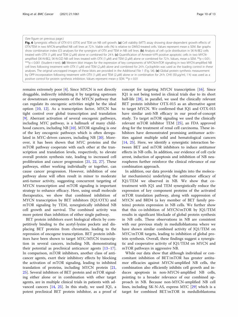
Synergistic anti-cancer efficacies of OTX-015 and TEM inNB cellsIn previous experiments, the rationale for using JQ1 wasits advantages over other BET inhibitors in preclinicalcancer studies. Preclinical studies with JQ1 offered a
great opportunity to better understand the biology ofBET proteins and validate these proteins as the anti-cancer targets [14, 24]. In addition, JQ1 has been shownto efficiently target MYC/MYCN transcription inpediatric NB and medulloblastoma [15–17]. However,JQ1 is not being considered in clinical trials because ofits short half-life [24]. As the proof of concept that if thecombined inhibition of BET protein and mTOR signal-ing can be translated into the clinic, we also utilized aclinically relevant BET protein inhibitor OTX-015(OTX, hereafter) which is currently in clinical trials forseveral advanced cancers [14]. Using previously reporteddoses of OTX in NB cell lines [16], we tested its com-bination efficacy with TEM in two MYCN-amplified NBcell lines. As shown in Fig. 6a, co-treatment of OTX andTEM significantly suppressed growth of NB cell lines ina dose-dependent manner, compared to single agentsalone. CI analyses further confirmed that combination ofOTX and TEM had a highly synergistic inhibitory effecton NB cell growth with a CI value below 0.7. The resultsfrom cell cycle (PI staining) and apoptosis (Annexin-V/PI staining) analyses in NB cells showed that the
Fig. 4 Combined effects of JQ1 and TEM on neurosphere formation. (a) Representative micrograph of spheres of SK-N-BE2 cells with thetreatment of control-(DMSO) or JQ1 (0.5 μM) and TEM (2 μM) alone or combined for 7 days. Scale bar, 1000 μm. (b) Quantification of the numberof neurospheres following treatments. The values represent mean ± SEM. **p < 0.01; ***p < 0.001 (Student-t-test). (c) Western blot analysis for theexpression of neural stem cell markers following treatment of neurospheres with JQ1 (0.5 μM) and TEM (2 μM) alone or combined for three days.Cyclophilin was used as the loading control in this analysis. The original uncropped images of these blots are provided in the Additional File 1(Fig. S3)
Kling et al. BMC Cancer (2021) 21:1061 Page 7 of 13

combination significantly induced G1 cell cycle arrest andapoptosis (Fig. 6b and c), compared to single agents. Ourwestern blot results showed that compared to single agenttreatments, combination of OTX and TEM significantly sup-pressed expression of MYCN and downregulated the levelsof key downstream targets (phosphorylated-4EBP1/eIF4E) ofthe mTOR pathway (Fig. 6d). The results from global proteinsynthesis investigation using OPP-based assay, further
demonstrated synergistic inhibition of general protein syn-thesis rate by OTX and TEM, compared to single agents(Fig. 6e). These results consistently suggested the importanceof co-targeting BET protein and mTOR signaling in NB.
DiscussionDespite the availability of intensive multimodal therapy,the prognosis for patients with MYCN-amplified NB
Fig. 5 JQ1 and TEM chemosensitize NB cells. MTT results showing the dose-dependent effects of JQ1 and TEM alone or combined with cisplatin(Cis) chemotherapy in NB cell lines at 72 h, as indicated. The values represent the means ± SEM. Bar graphs show combination index (CI) analysesfor the synergism between JQ1/Cis or TEM/Cis in NB cell lines
Kling et al. BMC Cancer (2021) 21:1061 Page 8 of 13

Fig. 6 (See legend on next page.)
Kling et al. BMC Cancer (2021) 21:1061 Page 9 of 13
remains extremely poor [4]. Since MYCN is not directlydruggable, indirectly inhibiting it by targeting upstreamor downstream components of the MYCN pathway thatcan regulate its oncogenic activities might be the idealoption [10, 12]. As a transcription factor, MYCN hastight control over global transcription and translation[9]. Aberrant activation of several oncogenic pathways,including MYC pathway, is a hallmark of many child-hood cancers, including NB [10]. MTOR signaling is oneof the key oncogenic pathways which is often deregu-lated in MYC-driven cancers, including NB [20]. More-over, it has been shown that MYC proteins and themTOR pathway cooperate with each other at the tran-scription and translation levels, respectively, to elevateoverall protein synthesis rate, leading to increased cellproliferation and cancer progression [21, 22, 27]. Thesepathways, either working individually or together, cancause cancer progression. However, inhibition of onepathway alone will often result in minor to moderateanti-tumor activity, suggesting concurrent targeting ofMYCN transcription and mTOR signaling is importantstrategy to enhance efficacy. Here, using small moleculetherapeutics, we show that combined inhibition ofMYCN transcription by BET inhibitors (JQ1/OTX) andmTOR signaling by TEM, synergistically inhibited NBcell growth and survival. The combined activity wasmore potent than inhibition of either single pathway.BET protein inhibitors exert biological effects by com-
petitively binding to the acetyl-lysine pockets and dis-placing BET proteins from chromatin, leading to therepression of oncogene transcription. BET protein inhib-itors have been shown to target MYC/MYCN transcrip-tion in several cancers, including NB, demonstratingtheir potential as preclinical anticancer agents [13–17].In comparison, mTOR inhibitors, another class of anti-cancer agents, exert their inhibitory effects by blockingthe activation of mTOR signaling, leading to inhibitedtranslation of proteins, including MYCN protein [21,25]. Several inhibitors of BET protein and mTOR signal-ing either alone or in combination with other targetagents, are in multiple clinical trials in patients with ad-vanced cancers [14, 25]. In this study, we used JQ1, apotent preclinical BET protein inhibitor, as a proof of
concept for targeting MYCN transcription [16]. SinceJQ1 is not being tested in clinical trials due to its shorthalf-life [28], in parallel, we used the clinically relevantBET protein inhibitor OTX-015 as an alternative agentto target MYCN. We confirmed that JQ1 and OTX-015have similar anti-NB efficacy in our proof-of-conceptstudy. To target mTOR signaling we used the clinicallyrelevant mTOR inhibitor TEM [25], an FDA approveddrug for the treatment of renal cell carcinoma. These in-hibitors have demonstrated promising antitumor activ-ities against multiple solid and hematological cancers[14, 25]. Here, we identify a synergistic interaction be-tween BET and mTOR inhibitors to induce antitumoreffects in NB cells. In addition, our evidence of cell cyclearrest, induction of apoptosis and inhibition of NB neu-rospheres further reinforce the clinical relevance of ourcombination approach.In addition, our data provide insights into the molecu-
lar mechanism(s) underlying the antitumor efficacy ofJQ1/TEM we observed in NB. We show that co-treatment with JQ1 and TEM synergistically reduce theexpression of key component proteins of the activatedmTOR translation pathway, along with suppression ofMYCN and BRD4 (a key member of BET family pro-teins) protein expression in NB cells. We further showthat this co-inhibition of MYCN/mTOR by JQ1/TEMresults in significant blockade of global protein synthesisin NB cells. These observations in NB are consistentwith our previous study in medulloblastoma where wehave shown similar combined activity of JQ1/TEM onMYC/mTOR targets, leading to inhibition of global pro-tein synthesis. Overall, these findings suggest a synergis-tic and cooperative activity of JQ1/TEM on MYCN andmTOR pathways in aggressive NB.While our data show that although individual or con-
comitant inhibition of BET/mTOR has greater antitu-mor efficacies against MYCN-amplified NB cells, thecombination also efficiently inhibits cell growth and in-duces apoptosis in non-MYCN-amplified NB cells,pointing to a broader relevance of our combined ap-proach in NB. Because non-MYCN-amplified NB celllines, including SK-N-AS, express MYC [29] which is atarget for combined BET/mTOR in medulloblastoma
(See figure on previous page.)Fig. 6 Synergistic effects of OTX-015 (OTX) and TEM on NB cell growth. (a) Cell viability (MTT) assay showing dose-dependent growth effects ofOTX/TEM in two MYCN-amplified NB cell lines at 72 h. Viable cells (%) is relative to DMSO-treated cells. Values represent mean ± SEM. Bar graphsshow combination index (CI) analyses for the synergism of OTX and TEM in NB cell lines. (b) Analysis of cell cycle distribution in SK-N-BE2 cellstreated with OTX (1 μM) and TEM (2 μM) alone or combined for 24 h. (c) Quantification of Annexin-V/PI positive apoptotic cells in two MYCN-amplified (SK-N-BE2, SK-N-DZ) NB cell lines treated with OTX (1 μM) and TEM (2 μM) alone or combined for 72 h. Values, mean ± SEM. **p < 0.01;***p < 0.001 (Student-t-test). (d) Western blot images for the expression of key components of MYCN/mTOR signaling in two MYCN-amplified NBcell lines following treatment with OTX (1 μM) and TEM (2 μM) alone and combined for 24 h. Cyclophilin was used as the loading control in theseanalyses. The original uncropped images of these blots are provided in the Additional File 1 (Fig. S4). (e) Global protein synthesis measurementby OPP-incorporation following treatment with OTX (1 μM) and TEM (2 μM) alone or in combination for 24 h. CHX (50 μg/ml, 1 h) was used as apositive control for protein synthesis inhibition. Values represent mean ± SEM. **p < 0.01
Kling et al. BMC Cancer (2021) 21:1061 Page 10 of 13

cells [22]. MYC may be a secondary target for these in-hibitors in the context of NB.In NB and other cancers, MYCN and mTOR signaling
have been shown to play key roles in cancer stem cellsand contribute to relapse and drug-resistance. NB cells ex-press neural stem cell markers such as CD133 and Nestinand have the ability to form neurospheres [30–33]. Ourinitial findings with the anti-NB efficacy of MYCN/mTORinhibition on neurospheres and stem cell markers indicatethat individual or combined inhibition of MYCN/mTORmight target cancer stem cells - potentially minimizing re-currence of NB. Mounting evidence suggest that increasedprotein synthesis in cells not only correlates with in-creased cell proliferation/survival, but also often involvesin stem cell fate, including neural stem cell markers [34,35]. In line with these, our data is consistent that coopera-tive inhibition of MYCN/mTOR by proposed drugs down-regulates the expression/activation of MYCN/mTOR keytargets and inhibits global protein synthesis, thereby en-ables cell growth/cell cycle arrest, apoptosis and inhibitsneurosphere formation/stem cell markers.Since overexpression or amplification of MYC/MYCN
and activation of mTOR signaling are often associatedwith chemoresistance in many cancers including NB[36–38], we also wanted to see whether inhibition ofthese signaling pathways helps to chemosensitize NBcells. Our results revealed that both JQ1 and TEM sig-nificantly enhanced cisplatin-induced growth inhibitionin MB cells, suggesting that inhibition of MYCN/mTORsignaling not only inhibits cell proliferation and survivalof NB cells, but also sensitizes NB cells to chemotherapy.The exact mechanism of synergy between these inhibi-tors and cisplatin requires further investigation. How-ever, activation of both MYC and PI3K-mTOR signalingpathways have been shown to be associated withplatinum-based therapy resistance [39–41]. The cytotox-icity of cisplatin involves the damaging of DNA replica-tion/repair mechanisms. Similarly, targeting MYC andmTOR signaling have been shown to induce DNA dam-age response and contribute susceptibility to cisplatin-induced cell death. Therefore, co-induction of DNAdamage by cisplatin and JQ1 or TEM could be the po-tential mechanism of observed synergy.In clinical trials studies, TEM alone or in combination
with other clinical agents, such as chemotherapeuticdrugs temzilomide and irrinotican, has shown no signifi-cant benefit for relapsed NB patients [42, 43]. This sug-gests further exploration of combining TEM with othertargeted therapies. Our data indicate that one of poten-tial combinatorial target could be the inhibition of BETproteins. Since inhibitors of BET proteins, includingOTX and structurally similar inhibitors to JQ1, are cur-rently in clinical trials for several cancers, it is morelikely that combination of TEM with BET inhibitors can
be translated in clinic for NB and other MYC/MYCN-driven cancers therapies.
ConclusionsOur study demonstrates that targeting dysregulated pro-tein synthesis pathway by pharmacologic dual-inhibitionof MYCN transcription (by BET protein inhibition) andmTOR signaling has significant preclinical anti-NB effi-cacies in inducing cell growth inhibition, cell cycle arrestand apoptosis in vitro. Combination of JQ1 or OTX withTEM synergistically inhibited global protein synthesis bydownregulating the key components and downstreamtargets of MYCN/mTOR signaling. Thus, this study isthe first to demonstrate synergy in the combined inhib-ition of BET protein and mTOR signaling in NB. Ourstudy also revealed that inhibition of MYCN/mTOR notonly inhibits cell growth and survival in NB cells, butalso chemosensitizes NB cells. Beyond NB, this thera-peutic approach can be of broader relevance for therapyof MYC/MYCN-addicted cancers, as we have previouslyshown a synergistic antitumor efficacy of BET/mTORinhibitors in MYC-driven medulloblastoma [22]. Whilefurther studies using appropriate in vivo models areneeded to evaluate this combination, our study high-lights a basis for considering this combination approachas a new therapy for NB.
AbbreviationsBET: Bromodomain extra-terminal; CI: Combination index; FBS: Fetal bovineserum; h: Hour; IC50: Inhibitory concentration of inhibitor with 50% inhibition;NB: Neuroblastoma; mTOR: Mammalian Target of Rapamycin; MTT: 3-(4,5-dimethylthiazol-2-yl)-2,5-diphenyltetrazolium bromide;; MYC: v-myc avianmylocytomatosis viral oncogene homolog; MYCN: v-myc avianmylocytomatosis viral oncogene neuroblastoma homolog; OPP: O-Propargyl-puromycin; OTX: OTX-015; TEM: Temsirolimus; μM: Micromolar
Supplementary InformationThe online version contains supplementary material available at https://doi.org/10.1186/s12885-021-08782-9.
Additional file 1.
AcknowledgementsThe authors thank the Flow Cytometry and Tissue Science Core Facilities atUNMC for their assistance with these studies. The authors thank MattSandbulte for his help in editing this manuscript. We thank all members ofthe CHRI-PCRG labs for their useful discussion and suggestion on this work.
Authors’ contributionsNKC and DC designed the study. NKC, MJK, CNG, EMM, GA and SRperformed the experiments and analyzed the data. KBC, SSJ and DCcontributed significantly to the interpretation of the data. NKC and MJKwrote the manuscript. All authors read and approved the final manuscript.
FundingThis work was fully supported by the State of Nebraska through the PediatricCancer Research Grant Funds (LB905) awarded to D. W. Coulter, MD. Thisfunding had no role in the study design, data collection and analysis,interpretation of the data, decision to publish, or writing the manuscript.
Kling et al. BMC Cancer (2021) 21:1061 Page 11 of 13

Availability of data and materialsAll data generated or analyzed during this study are included in this article.
Declarations
Ethics approval and consent to participateNot applicable.
Consent for publicationNot applicable.
Competing interestsThe authors declare that they have no competing interest.
Author details1Department of Pediatrics, Hematology/Oncology Division, University ofNebraska Medical Center, 986395, Nebraska Medical Center, Omaha, NE, USA.2Department of Biochemistry and Molecular Biology, University of NebraskaMedical Center, Omaha, NE, USA. 3Department of Genetics, Cell Biology andAnatomy, University of Nebraska Medical Center, Omaha, NE, USA.
Received: 2 July 2021 Accepted: 14 September 2021
References1. Maris JM. Recent advances in neuroblastoma. N Engl J Med. 2010;362(23):
2202–11. https://doi.org/10.1056/NEJMra0804577.2. Tolbert VP, Matthay KK. Neuroblastoma: clinical and biological approach to
risk stratification and treatment. Cell Tissue Res. 2018;372(2):195–209. https://doi.org/10.1007/s00441-018-2821-2.
3. Matthay KK, Maris JM, Schleiermacher G, Nakagawara A, Mackall CL, Diller L,et al. Neuroblastoma. Nat Rev Dis Primers. 2016;2(1):16078. https://doi.org/10.1038/nrdp.2016.78.
4. Huang M, Weiss WA. Neuroblastoma and MYCN. Cold Spring Harb PerspectMed. 2013;3(10):a014415. https://doi.org/10.1101/cshperspect.a014415.
5. Truitt ML, Ruggero D. New frontiers in translational control of the cancergenome. Nat Rev Cancer. 2017;17(5):332. https://doi.org/10.1038/nrc.2017.30.
6. Robichaud N, Sonenberg N, Ruggero D, Schneider RJ. Translational Controlin Cancer. Cold Spring Harb Perspect Biol. 2019;11(7):11(7). https://doi.org/10.1101/cshperspect.a032896.
7. Ruggero D. The role of Myc-induced protein synthesis in cancer. CancerRes. 2009;69(23):8839–43. https://doi.org/10.1158/0008-5472.CAN-09-1970.
8. van Riggelen J, Yetil A, Felsher DW. MYC as a regulator of ribosomebiogenesis and protein synthesis. Nat Rev Cancer. 2010;10(4):301–9. https://doi.org/10.1038/nrc2819.
9. Boon K, Caron HN, van Asperen R, Valentijn L, Hermus MC, van Sluis P, et al.N-myc enhances the expression of a large set of genes functioning inribosome biogenesis and protein synthesis. EMBO J. 2001;20(6):1383–93.https://doi.org/10.1093/emboj/20.6.1383.
10. Liu R, Shi P, Wang Z, Yuan C, Cui H. Molecular mechanisms of MYCNDysregulation in cancers. Front Oncol. 2021;10:625332. https://doi.org/10.3389/fonc.2020.625332.
11. Wolpaw AJ, Bayliss R, Büchel G, Dang CV, Eilers M, Gustafson WC, et al.Drugging the “Undruggable” MYCN oncogenic transcription factor:overcoming previous obstacles to impact childhood cancers. Cancer Res.2021;81(7):1627–32. https://doi.org/10.1158/0008-5472.CAN-20-3108.
12. Chen H, Liu H, Qing G. Targeting oncogenic Myc as a strategy for cancertreatment. Signal Transduct Target Ther. 2018;3(1):5. https://doi.org/10.1038/s41392-018-0008-7.
13. Delmore JE, Issa GC, Lemieux ME, Rahl PB, Shi J, Jacobs HM, et al. BETbromodomain inhibition as a therapeutic strategy to target c-Myc. Cell.2011;146(6):904–17. https://doi.org/10.1016/j.cell.2011.08.017.
14. Stathis A, Bertoni F. BET proteins as targets for anticancer treatment. CancerDiscov. 2018;8(1):24–36. https://doi.org/10.1158/2159-8290.CD-17-0605.
15. Bandopadhayay P, Bergthold G, Nguyen B, Schubert S, Gholamin S,Tang Y, et al. BET bromodomain inhibition of MYC-amplifiedmedulloblastoma. Clin Cancer Res. 2014;20(4):912–25. https://doi.org/10.1158/1078-0432.CCR-13-2281.
16. Henssen A, Althoff K, Odersky A, Beckers A, Koche R, Speleman F, et al.Targeting MYCN-driven transcription by BET-Bromodomain inhibition. Clin
Cancer Res. 2016;22(10):2470–81. https://doi.org/10.1158/1078-0432.CCR-15-1449.
17. Puissant A, Frumm SM, Alexe G, Bassil CF, Qi J, Chanthery YH, et al.Targeting MYCN in neuroblastoma by BET bromodomain inhibition. CancerDiscov. 2013;3(3):308–23. https://doi.org/10.1158/2159-8290.CD-12-0418.
18. Wang X, Proud CG. The mTOR pathway in the control of protein synthesis.Physiology (Bethesda). 2006;21:362–9. https://doi.org/10.1152/physiol.00024.2006.
19. Hsieh AC, Liu Y, Edlind MP, Ingolia NT, Janes MR, Sher A, et al. The translationallandscape of mTOR signalling steers cancer initiation and metastasis. Nature.2012;485(7396):55–61. https://doi.org/10.1038/nature10912.
20. Mei H, Wang Y, Lin Z, Tong Q. The mTOR signaling pathway in pediatricneuroblastoma. Pediatr Hematol Oncol. 2013;30(7):605–15. https://doi.org/10.3109/08880018.2013.798058.
21. Vaughan L, Clarke PA, Barker K, Chanthery Y, Gustafson CW, Tucker E, et al.Inhibition of mTOR-kinase destabilizes MYCN and is a potential therapy forMYCN-dependent tumors. Oncotarget. 2016;7(36):57525–44. https://doi.org/10.18632/oncotarget.10544.
22. Chaturvedi NK, Kling MJ, Griggs CN, Kesherwani V, Shukla M, McIntyre EM,et al. A novel combination approach targeting an enhanced proteinsynthesis pathway in MYC-driven (group 3) Medulloblastoma. Mol CancerTher. 2020;19(6):1351–62. https://doi.org/10.1158/1535-7163.MCT-19-0996.
23. Chaturvedi NK, Rajule RN, Shukla A, Radhakrishnan P, Todd GL, Natarajan A,et al. Novel treatment for mantle cell lymphoma including therapy-resistanttumor by NF-kB and mTOR dual-targeting approach. Mol Cancer Ther. 2013;12(10):2006–17. https://doi.org/10.1158/1535-7163.MCT-13-0239.
24. Shorstova T, Foulkes WD, Witcher M. Achieving clinical success with BETinhibitors as anti-cancer agents. Br J Cancer. 2021;124(9):1478–90. https://doi.org/10.1038/s41416-021-01321-0.
25. Tian T, Li X, Zhang J. mTOR signaling in Cancer and mTOR inhibitors in solidtumor targeting therapy. Int J Mol Sci. 2019;20(3):755. https://doi.org/10.3390/ijms20030755.
26. Chou TC. Drug combination studies and their synergy quantification usingthe Chou-Talalay method. Cancer Res. 2010;70(2):440–6. https://doi.org/10.1158/0008-5472.CAN-09-1947.
27. Pourdehnad M, Truitt ML, Siddiqi IN, Ducker GS, Shokat KM, Ruggero D. Mycand mTOR converge on a common node in protein synthesis control thatconfers synthetic lethality in Myc-driven cancers. Proc Natl Acad Sci U S A.2013;110(29):11988–93. https://doi.org/10.1073/pnas.1310230110.
28. Li F, MacKenzie KR, Jain P, Santini C, Young DW, Matzuk MM. Metabolism ofJQ1, an inhibitor of bromodomain and extra terminal bromodomainproteins, in human and mouse liver microsomes. Biol Reprod. 2020;103(2):427–36. https://doi.org/10.1093/biolre/ioaa043.
29. Zimmerman MW, Liu Y, He S, Durbin AD, Abraham BJ, Easton J, et al. MYCdrives a subset of high-risk pediatric Neuroblastomas and is activatedthrough mechanisms including enhancer hijacking and focal enhanceramplification. Cancer Discov. 2018;8(3):320–35. https://doi.org/10.1158/2159-8290.CD-17-0993.
30. Yang L, Shi P, Zhao G, Xu J, Peng W, Zhang J, et al. Targeting cancer stemcell pathways for cancer therapy. Signal Transduct Target Ther. 2020;5(1):8.https://doi.org/10.1038/s41392-020-0110-5.
31. Yoshida GJ. Emerging roles of Myc in stem cell biology and novel tumortherapies. J Exp Clin Cancer Res. 2018;37(1):173. https://doi.org/10.1186/s13046-018-0835-y.
32. Rickman DS, Schulte JH, Eilers M. The expanding world of N-MYC-driventumors. Cancer Discov. 2018 Feb;8(2):150–63. https://doi.org/10.1158/2159-8290.CD-17-0273.
33. Bahmad HF, Mouhieddine TH, Chalhoub RM, Assi S, Araji T, Chamaa F, et al.The Akt/mTOR pathway in cancer stem/progenitor cells is a potentialtherapeutic target for glioblastoma and neuroblastoma. Oncotarget. 2018;9(71):33549–61. https://doi.org/10.18632/oncotarget.26088.
34. Tahmasebi S, Amiri M, Sonenberg N. Translational control in stem cells.Front Genet. 2019;9:709. https://doi.org/10.3389/fgene.2018.00709.
35. Baser A, Skabkin M, Martin-Villalba A. Neural stem cell activation and therole of protein synthesis. Brain Plast. 2017;3(1):27–41. https://doi.org/10.3233/BPL-160038.
36. Kumari A, Folk WP, Sakamuro D. The dual roles of MYC in genomicinstability and Cancer Chemoresistance. Genes (Basel). 2017;8(6):158. https://doi.org/10.3390/genes8060158.
37. Yogev O, Almeida GS, Barker KT, George SL, Kwok C, Campbell J, et al. Invivo modeling of Chemoresistant neuroblastoma provides new insights into
Kling et al. BMC Cancer (2021) 21:1061 Page 12 of 13

Chemorefractory disease and metastasis. Cancer Res. 2019;79(20):5382–93.https://doi.org/10.1158/0008-5472.CAN-18-2759.
38. Jiang BH, Liu LZ. Role of mTOR in anticancer drug resistance: perspectivesfor improved drug treatment. Drug Resist Updat. 2008;11(3):63–76. https://doi.org/10.1016/j.drup.2008.03.001.
39. David-West G, Ernlund A, Gadi A, Schneider RJ. mTORC1/2 inhibition re-sensitizes platinum-resistant ovarian cancer by disrupting selectivetranslation of DNA damage and survival mRNAs. Oncotarget. 2018;9(69):33064–76.
40. Reyes-González JM, Armaiz-Peña GN, Mangala LS, Valiyeva F, Ivan C, Pradeep S,et al. Targeting c-MYC in platinum-resistant ovarian Cancer. Mol Cancer Ther.2015;14(10):2260–9. https://doi.org/10.1158/1535-7163.MCT-14-0801.
41. Miller AL, Fehling SC, Garcia PL, Gamblin TL, Council LN, van WaardenburgRCAM, et al. The BET inhibitor JQ1 attenuates double-strand break repairand sensitizes models of pancreatic ductal adenocarcinoma to PARPinhibitors. EBioMedicine. 2019;44:419–30. https://doi.org/10.1016/j.ebiom.2019.05.035.
42. Geoerger B, Kieran MW, Grupp S, Perek D, Clancy J, Krygowski M, et al.Phase II trial of temsirolimus in children with high-grade glioma,neuroblastoma and rhabdomyosarcoma. Eur J Cancer. 2012;48(2):253–62.https://doi.org/10.1016/j.ejca.2011.09.021.
43. Mody R, Naranjo A, Van Ryn C, Yu AL, London WB, Shulkin BL, et al.Irinotecan-temozolomide with temsirolimus or dinutuximab in children withrefractory or relapsed neuroblastoma (COG ANBL1221): an open-label,randomised, phase 2 trial. Lancet Oncol. 2017;18(7):946–57. https://doi.org/10.1016/S1470-2045(17)30355-8.
Publisher’s NoteSpringer Nature remains neutral with regard to jurisdictional claims inpublished maps and institutional affiliations.
Kling et al. BMC Cancer (2021) 21:1061 Page 13 of 13