synchronized activation of sympathetic vasomotor, cardiac ...€¦ · synchronized activation of...
TRANSCRIPT

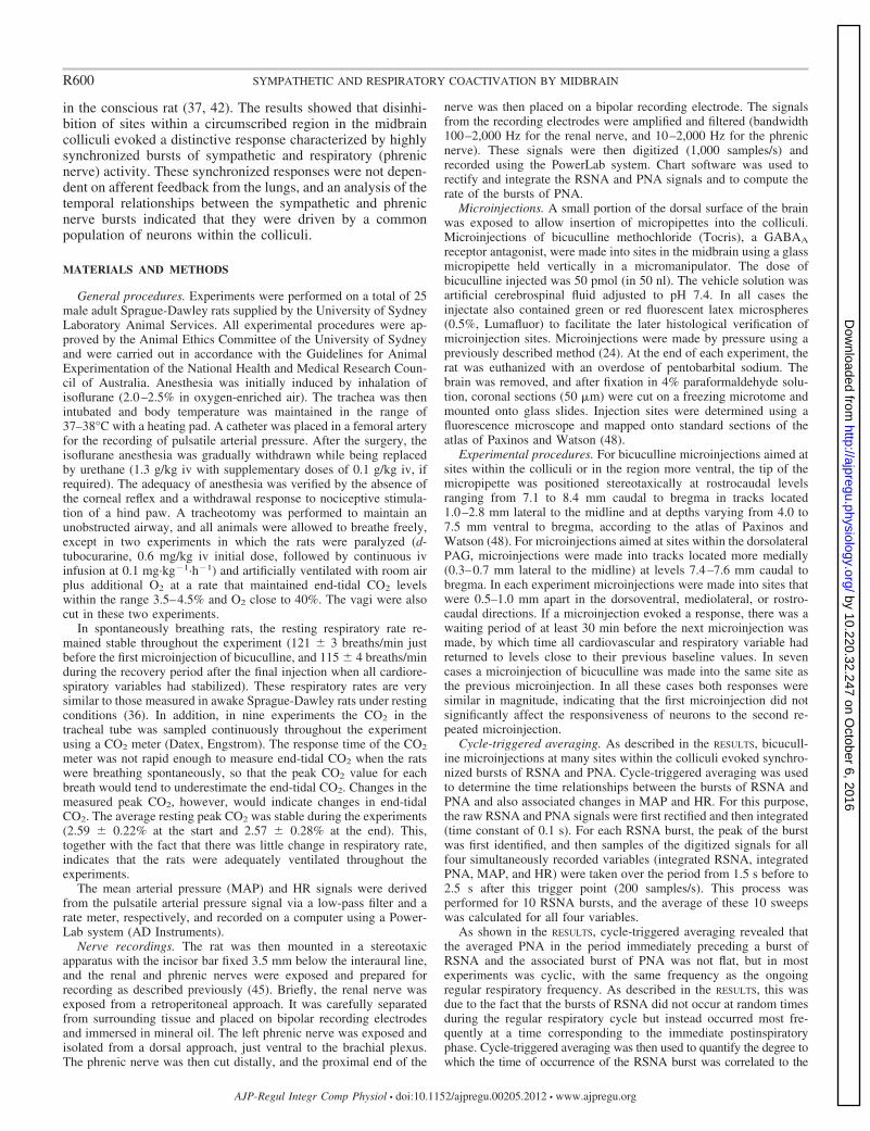
Synchronized activation of sympathetic vasomotor, cardiac, and respiratoryoutputs by neurons in the midbrain colliculi
Kamon Iigaya,1* Flávia Camargos de Figueirêdo Müller-Ribeiro,1,2* Jouji Horiuchi,1,3
Lachlan M. McDowall,1 Eugene Nalivaiko,4 Marco A. P. Fontes,2 and Roger A. L. Dampney1
1School of Medical Sciences (Physiology) and Bosch Institute for Biomedical Research, University of Sydney, Australia;2Laboratório de Hipertensão, Departamento de Fisiologia e Biofísica, Instituto de Ciências Biológicas, Universidade Federalde Minas Gerais, Minas Gerais, Brazil; 3Department of Biomedical Engineering, Toyo University, Saitama, Japan;and 4School of Biomedical Sciences and Pharmacy, University of Newcastle, Callaghan, Australia
Submitted 8 May 2012; accepted in final form 12 July 2012
Iigaya K, Müller-Ribeiro FC, Horiuchi J, McDowall LM,Nalivaiko E, Fontes MA, Dampney RA. Synchronized activationof sympathetic vasomotor, cardiac, and respiratory outputs byneurons in the midbrain colliculi. Am J Physiol Regul Integr CompPhysiol 303: R599 –R610, 2012. First published July 18, 2012;doi:10.1152/ajpregu.00205.2012.—The superior and inferior col-liculi are believed to generate immediate and highly coordinateddefensive behavioral responses to threatening visual and auditorystimuli. Activation of neurons in the superior and inferior colliculihave been shown to evoke increases in cardiovascular and respi-ratory activity, which may be components of more generalizedstereotyped behavioral responses. In this study, we examined thepossibility that there are “command neurons” within the colliculi thatcan simultaneously drive sympathetic and respiratory outputs. Inanesthetized rats, microinjections of bicuculline (a GABAA receptorantagonist) into sites within a circumscribed region in the deep layersof the superior colliculus and in the central and external nuclei of theinferior colliculus evoked a response characterized by intense andhighly synchronized bursts of renal sympathetic nerve activity(RSNA) and phrenic nerve activity (PNA). Each burst of RSNA hada duration of �300–400 ms and occurred slightly later (peak to peaklatency of 41 � 8 ms) than the corresponding burst of PNA. Thebursts of RSNA and PNA were also accompanied by transient in-creases in arterial pressure and, in most cases, heart rate. Synchro-nized bursts of RSNA and PNA were also evoked after neuromuscularblockade, artificial ventilation, and vagotomy and so were not depen-dent on afferent feedback from the lungs. We propose that thesynchronized sympathetic-respiratory responses are driven by a com-mon population of neurons, which may normally be activated by anacute threatening stimulus.
command neuron; defensive behavior; heart rate; arterial pressure
THE MAMMALIAN SUPERIOR COLLICULUS is a key brain region thatreceives visual and other signals arising from a stimulus in theexternal environment and processes these signals to produce anappropriate somatomotor response (e.g., an orienting response,in which the head and eyes are moved to allow focusing on anovel stimulus) (19, 26). In addition, however, there is consid-erable evidence that the superior colliculus also generates defen-sive responses, such as avoidance or escape, evoked by a threat-ening stimulus such as the presence of a predator (5, 7, 9, 16, 19).Furthermore, stimulation of neurons in the superior colliculus canalso evoke increases in blood pressure, heart rate (HR), and
respiratory activity (39, 40), and it has been proposed that sucheffects are part of a coordinated response to stimuli that triggerorienting or avoidance/defensive behaviors (19).
The inferior colliculus receives auditory signals from severalnuclei in the lower brain stem and has an essential role in theprocessing of auditory signals (15, 50). Similar to the superiorcolliculus, activation of neurons in the inferior colliculus canevoke a behavioral response similar to defensive behavior (8,12), as well as increases in blood pressure and HR (8). It hasbeen proposed that the inferior colliculus is part of a moregeneralized system in the dorsal midbrain, which also includesthe superior colliculus and periaqueductal gray (PAG), whichintegrates the somatomotor and autonomic responses to threat-ening stimuli signaled by visual, auditory, and somatosensoryinputs (7, 57).
Distinct populations of neurons in the superior colliculus cangenerate different patterns of coordinated responses that arerapid in onset and that are appropriate for particular stimuli(19, 26). Similarly, evidence from anatomical, neurophysio-logical, and behavioral studies of the inferior colliculus haveled to the proposal that there are neurons in the inferiorcolliculus that respond to particular auditory stimuli, such asparticular sounds made by prey or predators, and which thentrigger immediate responses termed fixed action patterns,which are appropriate for each stimulus (14).
These observations thus raise the possibility that there areneurons within the superior and inferior colliculi that arecapable of generating, in addition to somatomotor responses, acoordinated cardiovascular and respiratory response that isappropriate for a defensive behavioral response (e.g., escape),which is triggered by a threatening stimulus. Disinhibition (bymicroinjection of bicuculline, a GABAA receptor antagonist)of sites within the superior and inferior colliculi is a highlyeffective means of evoking defensive behavioral responses,indicating that the neurons generating such responses receive atonic GABAergic inhibition (5, 6, 8, 19). The aim of this study,therefore, was to determine the effects of disinhibition ofneurons (by microinjection of bicuculline) within the superiorand inferior colliculi on cardiovascular function [arterial pres-sure, HR, and renal sympathetic nerve activity (RSNA)] andrespiratory function [phrenic nerve activity (PNA)]. TheRSNA was measured because although renal sympatheticnerves regulate tubules and the juxtaglomerular granular cellsas well as blood vessels in the kidney (21), RSNA is closelycorrelated with arterial pressure and is believed to be a reliableindicator of overall sympathetic vasomotor activity (10). Fur-thermore, RSNA is increased in response to threatening stimuli
* Kamon Iigaya and Flávia Müller-Ribeiro contributed equally to this work.Address for reprint requests and other correspondence: R. A. L. Dampney,
School of Medical Sciences (Physiology) and Bosch Institute for BiomedicalResearch, F13, The Univ. of Sydney, NSW 2006, Australia (e-mail:[email protected]).
Am J Physiol Regul Integr Comp Physiol 303: R599–R610, 2012.First published July 18, 2012; doi:10.1152/ajpregu.00205.2012.
0363-6119/12 Copyright © 2012 the American Physiological Societyhttp://www.ajpregu.org R599
by 10.220.32.247 on October 6, 2016
http://ajpregu.physiology.org/D
ownloaded from
in the conscious rat (37, 42). The results showed that disinhi-bition of sites within a circumscribed region in the midbraincolliculi evoked a distinctive response characterized by highlysynchronized bursts of sympathetic and respiratory (phrenicnerve) activity. These synchronized responses were not depen-dent on afferent feedback from the lungs, and an analysis of thetemporal relationships between the sympathetic and phrenicnerve bursts indicated that they were driven by a commonpopulation of neurons within the colliculi.
MATERIALS AND METHODS
General procedures. Experiments were performed on a total of 25male adult Sprague-Dawley rats supplied by the University of SydneyLaboratory Animal Services. All experimental procedures were ap-proved by the Animal Ethics Committee of the University of Sydneyand were carried out in accordance with the Guidelines for AnimalExperimentation of the National Health and Medical Research Coun-cil of Australia. Anesthesia was initially induced by inhalation ofisoflurane (2.0–2.5% in oxygen-enriched air). The trachea was thenintubated and body temperature was maintained in the range of37–38°C with a heating pad. A catheter was placed in a femoral arteryfor the recording of pulsatile arterial pressure. After the surgery, theisoflurane anesthesia was gradually withdrawn while being replacedby urethane (1.3 g/kg iv with supplementary doses of 0.1 g/kg iv, ifrequired). The adequacy of anesthesia was verified by the absence ofthe corneal reflex and a withdrawal response to nociceptive stimula-tion of a hind paw. A tracheotomy was performed to maintain anunobstructed airway, and all animals were allowed to breathe freely,except in two experiments in which the rats were paralyzed (d-tubocurarine, 0.6 mg/kg iv initial dose, followed by continuous ivinfusion at 0.1 mg·kg�1·h�1) and artificially ventilated with room airplus additional O2 at a rate that maintained end-tidal CO2 levelswithin the range 3.5–4.5% and O2 close to 40%. The vagi were alsocut in these two experiments.
In spontaneously breathing rats, the resting respiratory rate re-mained stable throughout the experiment (121 � 3 breaths/min justbefore the first microinjection of bicuculline, and 115 � 4 breaths/minduring the recovery period after the final injection when all cardiore-spiratory variables had stabilized). These respiratory rates are verysimilar to those measured in awake Sprague-Dawley rats under restingconditions (36). In addition, in nine experiments the CO2 in thetracheal tube was sampled continuously throughout the experimentusing a CO2 meter (Datex, Engstrom). The response time of the CO2
meter was not rapid enough to measure end-tidal CO2 when the ratswere breathing spontaneously, so that the peak CO2 value for eachbreath would tend to underestimate the end-tidal CO2. Changes in themeasured peak CO2, however, would indicate changes in end-tidalCO2. The average resting peak CO2 was stable during the experiments(2.59 � 0.22% at the start and 2.57 � 0.28% at the end). This,together with the fact that there was little change in respiratory rate,indicates that the rats were adequately ventilated throughout theexperiments.
The mean arterial pressure (MAP) and HR signals were derivedfrom the pulsatile arterial pressure signal via a low-pass filter and arate meter, respectively, and recorded on a computer using a Power-Lab system (AD Instruments).
Nerve recordings. The rat was then mounted in a stereotaxicapparatus with the incisor bar fixed 3.5 mm below the interaural line,and the renal and phrenic nerves were exposed and prepared forrecording as described previously (45). Briefly, the renal nerve wasexposed from a retroperitoneal approach. It was carefully separatedfrom surrounding tissue and placed on bipolar recording electrodesand immersed in mineral oil. The left phrenic nerve was exposed andisolated from a dorsal approach, just ventral to the brachial plexus.The phrenic nerve was then cut distally, and the proximal end of the
nerve was then placed on a bipolar recording electrode. The signalsfrom the recording electrodes were amplified and filtered (bandwidth100–2,000 Hz for the renal nerve, and 10–2,000 Hz for the phrenicnerve). These signals were then digitized (1,000 samples/s) andrecorded using the PowerLab system. Chart software was used torectify and integrate the RSNA and PNA signals and to compute therate of the bursts of PNA.
Microinjections. A small portion of the dorsal surface of the brainwas exposed to allow insertion of micropipettes into the colliculi.Microinjections of bicuculline methochloride (Tocris), a GABAA
receptor antagonist, were made into sites in the midbrain using a glassmicropipette held vertically in a micromanipulator. The dose ofbicuculline injected was 50 pmol (in 50 nl). The vehicle solution wasartificial cerebrospinal fluid adjusted to pH 7.4. In all cases theinjectate also contained green or red fluorescent latex microspheres(0.5%, Lumafluor) to facilitate the later histological verification ofmicroinjection sites. Microinjections were made by pressure using apreviously described method (24). At the end of each experiment, therat was euthanized with an overdose of pentobarbital sodium. Thebrain was removed, and after fixation in 4% paraformaldehyde solu-tion, coronal sections (50 �m) were cut on a freezing microtome andmounted onto glass slides. Injection sites were determined using afluorescence microscope and mapped onto standard sections of theatlas of Paxinos and Watson (48).
Experimental procedures. For bicuculline microinjections aimed atsites within the colliculi or in the region more ventral, the tip of themicropipette was positioned stereotaxically at rostrocaudal levelsranging from 7.1 to 8.4 mm caudal to bregma in tracks located1.0–2.8 mm lateral to the midline and at depths varying from 4.0 to7.5 mm ventral to bregma, according to the atlas of Paxinos andWatson (48). For microinjections aimed at sites within the dorsolateralPAG, microinjections were made into tracks located more medially(0.3–0.7 mm lateral to the midline) at levels 7.4–7.6 mm caudal tobregma. In each experiment microinjections were made into sites thatwere 0.5–1.0 mm apart in the dorsoventral, mediolateral, or rostro-caudal directions. If a microinjection evoked a response, there was awaiting period of at least 30 min before the next microinjection wasmade, by which time all cardiovascular and respiratory variable hadreturned to levels close to their previous baseline values. In sevencases a microinjection of bicuculline was made into the same site asthe previous microinjection. In all these cases both responses weresimilar in magnitude, indicating that the first microinjection did notsignificantly affect the responsiveness of neurons to the second re-peated microinjection.
Cycle-triggered averaging. As described in the RESULTS, bicucull-ine microinjections at many sites within the colliculi evoked synchro-nized bursts of RSNA and PNA. Cycle-triggered averaging was usedto determine the time relationships between the bursts of RSNA andPNA and also associated changes in MAP and HR. For this purpose,the raw RSNA and PNA signals were first rectified and then integrated(time constant of 0.1 s). For each RSNA burst, the peak of the burstwas first identified, and then samples of the digitized signals for allfour simultaneously recorded variables (integrated RSNA, integratedPNA, MAP, and HR) were taken over the period from 1.5 s before to2.5 s after this trigger point (200 samples/s). This process wasperformed for 10 RSNA bursts, and the average of these 10 sweepswas calculated for all four variables.
As shown in the RESULTS, cycle-triggered averaging revealed thatthe averaged PNA in the period immediately preceding a burst ofRSNA and the associated burst of PNA was not flat, but in mostexperiments was cyclic, with the same frequency as the ongoingregular respiratory frequency. As described in the RESULTS, this wasdue to the fact that the bursts of RSNA did not occur at random timesduring the regular respiratory cycle but instead occurred most fre-quently at a time corresponding to the immediate postinspiratoryphase. Cycle-triggered averaging was then used to quantify the degree towhich the time of occurrence of the RSNA burst was correlated to the
R600 SYMPATHETIC AND RESPIRATORY COACTIVATION BY MIDBRAIN
AJP-Regul Integr Comp Physiol • doi:10.1152/ajpregu.00205.2012 • www.ajpregu.org
by 10.220.32.247 on October 6, 2016
http://ajpregu.physiology.org/D
ownloaded from

ongoing normal respiratory rhythm. This was measured by calculatingthe ratio of the amplitude of the oscillation (i.e., maximum � minimum)of the averaged PNA calculated using cycle-triggered averaging over theperiod from 1.5 s before to 0.75 s before the trigger point (peak of RSNAburst), to the average value of the amplitudes of the oscillations of PNAover the same time period for each of the 10 individual recordings thatwere used in determining the cycle-triggered averaging. A ratio of100% would imply that the RSNA burst had occurred at exactly thesame time point in the normal respiratory cycle for each of the 10individual recordings, whereas a value close to zero would imply thatthe timing of the RSNA burst had no consistent relation to the normalrespiratory cycle. This ratio will be referred to in this paper as the“respiratory cycle correlation index.”
Statistical analysis. As described in the RESULTS, responses wereclassified into three groups according to the degree of synchronizationbetween bursts of RSNA and of PNA (i.e., strong, moderate, and littleor no RSNA/PNA burst synchrony). The amplitudes of RSNA andPNA bursts in the groups with strong and moderate RSNA/PNA burstsynchrony were compared using the t-test. The peak changes in MAP,HR, integrated RSNA and integrated PNA were compared for all threegroups using one-way ANOVA. If this revealed a statistically signif-icant difference (P � 0.05), a subsequent pair-wise comparison wasperformed using the t-test with application of the Helm step-downprocedure for multiple comparisons (55). All values are presented asmeans � SE. A chi-squared analysis was also used to test whether thedifferences in regional distribution of sites evoking different types ofresponses were statistically significant.
RESULTS
General description of cardiovascular and respiratory re-sponses evoked by bicuculline microinjection into the colliculiand surrounding regions. Microinjections of bicuculline (50pmol) were made into 119 sites in 23 experiments in sponta-neously breathing rats. The centers of the injection sites ex-tended rostrocaudally from 7.1 to 8.6 mm caudal to bregma[atlas of Paxinos and Watson, (48)] and were mapped on tothree levels at 7.20, 7.80, and 8.3 mm caudal to bregma. Thesites were located within the intermediate and deep layers ofthe superior colliculus, the external and central nuclei of theinferior colliculus, PAG, and in the region ventral to thesuperior and inferior colliculi, including the precuneiform andcuneiform nuclei (Fig. 1).
Our subsequent analysis allowed us to classify the responsesevoked from these sites into different types, depending on thepattern of the evoked changes in PNA and their relationshipwith evoked changes in RSNA. In the first type (n � 8), therewere large increases in both integrated RSNA and integratedPNA of at least 50% above baseline that were accompanied byan increase in MAP and (in 7 of 8 cases) by an increase in HR(see Fig. 2 for an example of such a response). In most cases(5 of 8) bicuculline microinjections also evoked a change in the
1 mm
Level -7.2
Level -7.8
Level -8.3
DpGInG
ECIC
DpWh
PrCnF
dmPAGdl
lvl
Aq
ECIC
CIC
DpG
InG
PrCnF
dllvl
Aq
PAGdm
ECIC
PAGCIC
dm
l
vl
Aq
CnF
Strong RSNA/PNA burst synchrony (n=26)
Moderate RSNA/PNA burst synchrony (n=29)
Significant response, no RNSA/PNA bursting (n=3)
Other responses, no RNSA/PNA bursting (n=56)
RSNA bursting, no PNA bursting (n=5)
Fig. 1. Coronal sections of the rat brain fromthe atlas of Paxinos and Watson (with permis-sion, 48), showing the centers of injectionsites of bicuculline (50 pmol) in the superiorand inferior colliculi and surrounding regionsin 23 experiments in spontaneously breathingrats. The heading above each section indicatesthe distance of that section in millimeters frombregma. The different colored filled circles indi-cate the pattern of response evoked from eachsite, as shown in the key. Aq, aqueduct; CIC,central nucleus of the inferior colliculus; CnF,cuneiform nucleus; dm, dorsomedial; dl, dorso-lateral; DpG, deep gray layer of the superiorcolliculus; DpWh, deep white layer of the supe-rior colliculus; ECIC, external nucleus of theinferior colliculus; InG, intermediate graylayer of the superior colliculus; l, lateral;PAG, periaqueductal gray; PrCnF, precunei-form nucleus; vl, ventrolateral.
R601SYMPATHETIC AND RESPIRATORY COACTIVATION BY MIDBRAIN
AJP-Regul Integr Comp Physiol • doi:10.1152/ajpregu.00205.2012 • www.ajpregu.org
by 10.220.32.247 on October 6, 2016
http://ajpregu.physiology.org/D
ownloaded from

pattern of RSNA, such that intense bursts occurred at intervalsof 2–5 s (e.g., Fig. 2C). These bursts of RSNA had a durationof �300–400 ms. In all these five cases, however, the PNAmaintained a consistent regular rhythm during the periodswhen bursts of RSNA were evoked by bicuculline microinjec-tions (e.g., Fig. 2C).
In contrast, bicuculline microinjections at other sitesevoked a different type of response that was characterizedby synchronous bursts of both RSNA and PNA, as shown
for example in Fig. 3. The onset of these synchronous burstsof RSNA and PNA mostly occurred just after the precedingpeak of a regular PNA burst, as shown for example in Fig.3C. Each synchronous burst of RSNA and PNA was accom-panied by an increase in arterial pressure, the onset of whichalso coincided with the RSNA and PNA bursts. As illus-trated in Fig. 3D, the intense PNA bursts evoked from suchsites were consistently associated with synchronous RSNAbursts.
ECIC
CIC
CnF
PAGAq
80
240
160
80
240
160Integrated
RSNA(%baseline)
460
320
390
180
60
120
5 min
240
120180
AP(mmHg)
HR(beats/min)
Respiratoryactivity
(%baseline)
PNAburst rate
(bursts/min)
Bic
-1
0
+1
0
0.5
1.0
-1
0
+1
180
60120
0
0.5
1.0
2 s
AP(mmHg)
RSNA(units)
IntegratedRSNA(units)
PNA(units)
IntegratedPNA
(units)
Before bicuculline After bicuculline
A
B C
D E
*
Fig. 2. Example of response with renal sym-pathetic nerve activity (RSNA) bursting with-out synchronous phrenic nerve activity (PNA)bursting. Chart recordings show changes in ar-terial pressure (AP), heart rate (HR), integratedRSNA (time constant 5 s), respiratory activity(integrated PNA, time constant 5 s), and PNAburst rate (A) evoked by bicuculline (Bic) mi-croinjection into a site within the central nu-cleus of the inferior colliculus. Examples ofchart recordings at a faster chart speed beforeand after the bicuculline microinjection, at thetime points indicated by the gray vertical bars inA above, are shown in B and C, respectively.For the recordings in B and C, the time constantfor the integrated RSNA and PNA signals wasset at 0.1 s. The injection site as marked bythe fluorescent tracer (indicated by *) isshown in the photomicrograph in D. Thelocation of the injection site as mapped on toa standard section (at the level 8.04 mmcaudal to bregma) is indicated by the gray-filled circle in E, whereas the rectangle in Eindicates the region corresponding to the pho-tomicrograph in D.
R602 SYMPATHETIC AND RESPIRATORY COACTIVATION BY MIDBRAIN
AJP-Regul Integr Comp Physiol • doi:10.1152/ajpregu.00205.2012 • www.ajpregu.org
by 10.220.32.247 on October 6, 2016
http://ajpregu.physiology.org/D
ownloaded from

Cycle-triggered averaging was used to quantify the magni-tudes of the synchronous RSNA and PNA bursts and theassociated changes in MAP and HR (Fig. 4). Responses withstrong RSNA/PNA burst synchrony were defined as responsesin which the peak amplitude of the averaged synchronous PNAburst was at least 100% greater than the baseline averaged PNA(e.g., Fig. 4A). Such responses were evoked from 26 sites. Re-sponses with moderate RSNA/PNA burst synchrony were defined
as responses in which the peak amplitude of the averaged syn-chronous PNA burst was 30–99% greater than the baselineaveraged PNA (e.g., Fig. 4B). Such responses were evoked from29 other sites. Cycle-triggered averaging was also performed forthe five responses of the first type, in which the PNA maintaineda regular rhythm during the periods when bursts of RSNA wereevoked by bicuculline microinjections. This analysis confirmedthat in all five cases the averaged RSNA burst was not
IntegratedRSNA
(%baseline)
5 min
80
200
140
80
200
140
450
350
400
200
60
130
180
80130
AP(mmHg)
HR(beats/min)
Respiratoryactivity
(%baseline)
PNAburst rate
(bursts/min)
Bic
A
Before bicuculline After bicuculline
AP(mmHg)
RSNA(units)
IntegratedRSNA(units)
PNA(units)
IntegratedPNA
(units)
190
70
130
-1
+10
0
1.0
0.5
-1
+10
0
1.0
0.5
2 s
B C
0
1.0
0.5
0
1.0
0.5
200
80
140
10 s
AP(mmHg)
IntegratedRSNA(units)
IntegratedPNA
(units)
D
Fig. 3. Example of response with strongRSNA/PNA burst synchrony. A: chart record-ings of a bicuculline-evoked response inwhich bursts of RSNA occurred, each ofwhich was accompanied by a burst of PNA. Band C: chart recordings before and after thebicuculline microinjection, at the time pointsindicated by the gray vertical bars in A above,whereas D is a longer section of the chartrecording that includes the section (indicatedby the gray shading) shown in C. For theslower chart speed recordings in A the timeconstant for the integrated RSNA and PNAsignals was set at 5 s, and for the faster speedrecordings in B, C, and D it was set at 0.1 s.
R603SYMPATHETIC AND RESPIRATORY COACTIVATION BY MIDBRAIN
AJP-Regul Integr Comp Physiol • doi:10.1152/ajpregu.00205.2012 • www.ajpregu.org
by 10.220.32.247 on October 6, 2016
http://ajpregu.physiology.org/D
ownloaded from

associated with a significant increase in the averaged PNA(e.g., Fig. 4C).
Immediately afer each burst of RSNA, there was a period of1–2 s during which the level of RSNA was greatly reducedcompared with the baseline level that immediately precededthe onset of the RSNA burst. This postburst depression ofRSNA was consistently observed whether or not the RSNAburst was associated with a synchronous PNA burst (Figs. 2C,3C, and 4).
At the remaining 56 sites (Fig. 1), bicuculline microinjec-tions evoked responses that did not fit into any of the categoriesdescribed above. In most cases there was no response or else a
very small response, although in a few cases there was asignificant change in just one variable (RSNA, PNA, or HR)that was accompanied by little or no change in other variables.
Because responses with strong and moderate RSNA/PNAsynchrony were defined according to the magnitudes of thePNA bursts as described above, there were clear differences inthe amplitudes of the PNA bursts when the two groups werecompared (Fig. 5).
At the same time, there were also clear and significantdifferences between the two groups with respect to the mag-nitudes of the associated RSNA bursts and increases in MAPand HR (Fig. 5). The changes in HR associated with responses
0 1.0 2.0-1.0
400
100
200
0
300
200
150
250
100
50
125
120
130
115
110
MAP(mmHg)
IntegratedRSNA
(%baseline)
IntegratedPNA
(%baseline)
0 1.0 2.0-1.0Time relative to RSNA peak (s)
0 1.0 2.0-1.0
B CA
470
460
480
450
470
460
480
450
440
430
450
420
400
100
200
0
300
200
150
250
100
50
125
120
130
115
110
400
100
200
0
300
200
150
250
100
50
125
120
130
115
110
HR(beats/min)
Fig. 4. Examples of cycle-triggered averag-ing, showing averages of 10 sweeps of inte-grated RSNA, integrated PNA, MAP, andHR for a response with strong RSNA/PNAsynchrony (A), moderate RSNA/PNA syn-chrony (B), and no significant RSNA/PNAsynchrony (C). In each case the peak of theintegrated RSNA signal was taken as the trig-ger point (indicated by the vertical dashed line),and the signals were averaged over the periodfrom 1.5 s before to 2.5 s after the trigger point.The time constant for the integrated RSNA andPNA signals was set at 0.1 s.
Strong RSNA/PNA burst synchrony (n=26)
Moderate RSNA/PNA burst synchrony (n=29)
∆RS
NA
bur
st a
mpl
itude
(% b
asel
ine)
300
200
100
400
0
500 **
0
∆MA
P(m
mH
g)
15
10
5
20
**
∆PN
A b
urst
am
plitu
de(%
bas
elin
e)
120
80
40
160
0
200**
0
∆HR
(bea
ts/m
in)
25
20
5
30
**
15
10
Fig. 5. Grouped data (means � SE) showingthe relative magnitudes of the averagedRSNA and PNA bursts and associatedchanges in MAP and HR for bicuculline-evoked responses with strong and moderateRSNA/PNA burst synchrony, as determinedusing cycle-triggered averaging. **P �0.001, vs. moderate RSNA/PNA burst syn-chrony.
R604 SYMPATHETIC AND RESPIRATORY COACTIVATION BY MIDBRAIN
AJP-Regul Integr Comp Physiol • doi:10.1152/ajpregu.00205.2012 • www.ajpregu.org
by 10.220.32.247 on October 6, 2016
http://ajpregu.physiology.org/D
ownloaded from
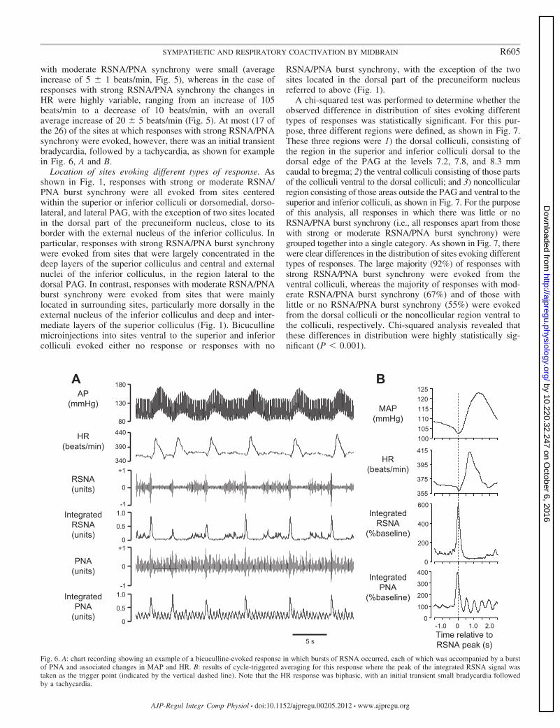
with moderate RSNA/PNA synchrony were small (averageincrease of 5 � 1 beats/min, Fig. 5), whereas in the case ofresponses with strong RSNA/PNA synchrony the changes inHR were highly variable, ranging from an increase of 105beats/min to a decrease of 10 beats/min, with an overallaverage increase of 20 � 5 beats/min (Fig. 5). At most (17 ofthe 26) of the sites at which responses with strong RSNA/PNAsynchrony were evoked, however, there was an initial transientbradycardia, followed by a tachycardia, as shown for examplein Fig. 6, A and B.
Location of sites evoking different types of response. Asshown in Fig. 1, responses with strong or moderate RSNA/PNA burst synchrony were all evoked from sites centeredwithin the superior or inferior colliculi or dorsomedial, dorso-lateral, and lateral PAG, with the exception of two sites locatedin the dorsal part of the precuneiform nucleus, close to itsborder with the external nucleus of the inferior colliculus. Inparticular, responses with strong RSNA/PNA burst synchronywere evoked from sites that were largely concentrated in thedeep layers of the superior colliculus and central and externalnuclei of the inferior colliculus, in the region lateral to thedorsal PAG. In contrast, responses with moderate RSNA/PNAburst synchrony were evoked from sites that were mainlylocated in surrounding sites, particularly more dorsally in theexternal nucleus of the inferior colliculus and deep and inter-mediate layers of the superior colliculus (Fig. 1). Bicucullinemicroinjections into sites ventral to the superior and inferiorcolliculi evoked either no response or responses with no
RSNA/PNA burst synchrony, with the exception of the twosites located in the dorsal part of the precuneiform nucleusreferred to above (Fig. 1).
A chi-squared test was performed to determine whether theobserved difference in distribution of sites evoking differenttypes of responses was statistically significant. For this pur-pose, three different regions were defined, as shown in Fig. 7.These three regions were 1) the dorsal colliculi, consisting ofthe region in the superior and inferior colliculi dorsal to thedorsal edge of the PAG at the levels 7.2, 7.8, and 8.3 mmcaudal to bregma; 2) the ventral colliculi consisting of those partsof the colliculi ventral to the dorsal colliculi; and 3) noncollicularregion consisting of those areas outside the PAG and ventral to thesuperior and inferior colliculi, as shown in Fig. 7. For the purposeof this analysis, all responses in which there was little or noRSNA/PNA burst synchrony (i.e., all responses apart from thosewith strong or moderate RSNA/PNA burst synchrony) weregrouped together into a single category. As shown in Fig. 7, therewere clear differences in the distribution of sites evoking differenttypes of responses. The large majority (92%) of responses withstrong RSNA/PNA burst synchrony were evoked from theventral colliculi, whereas the majority of responses with mod-erate RSNA/PNA burst synchrony (67%) and of those withlittle or no RSNA/PNA burst synchrony (55%) were evokedfrom the dorsal colliculi or the noncollicular region ventral tothe colliculi, respectively. Chi-squared analysis revealed thatthese differences in distribution were highly statistically sig-nificant (P � 0.001).
5 s
AP(mmHg)
RSNA(units)
IntegratedRSNA(units)
PNA(units)
IntegratedPNA
(units)
180
80
130
-1
+1
0
0
1.0
0.5
-1
+1
0
0
1.0
0.5
440
340
390HR
(beats/min)
A
0 1.0 2.0-1.0
395
375
415
355
600
200
0
400
300
200
400
100
0
125120115110105100
IntegratedPNA
(%baseline)
IntegratedRSNA
(%baseline)
MAP(mmHg)
HR(beats/min)
Time relative toRSNA peak (s)
B
Fig. 6. A: chart recording showing an example of a bicuculline-evoked response in which bursts of RSNA occurred, each of which was accompanied by a burstof PNA and associated changes in MAP and HR. B: results of cycle-triggered averaging for this response where the peak of the integrated RSNA signal wastaken as the trigger point (indicated by the vertical dashed line). Note that the HR response was biphasic, with an initial transient small bradycardia followedby a tachycardia.
R605SYMPATHETIC AND RESPIRATORY COACTIVATION BY MIDBRAIN
AJP-Regul Integr Comp Physiol • doi:10.1152/ajpregu.00205.2012 • www.ajpregu.org
by 10.220.32.247 on October 6, 2016
http://ajpregu.physiology.org/D
ownloaded from

Time relationships between PNA and RSNA bursts. Cycle-triggered averaging was used to determine the latency betweenthe peaks of the PNA and RSNA bursts, where the trigger pointwas the peak of the RSNA burst. As shown in Fig. 4, thisshowed that the peak of the PNA burst occurred just before thepeak of the associated RSNA burst. In the case of responseswith strong RSNA/PNA burst synchrony, the time differencebetween the peaks was 41 � 8 ms.
As noted above, the onset of PNA bursts mostly occurredjust after the preceding peak of a regular PNA burst, as shownfor example in Fig. 3C. The fact that these nonregular PNAbursts did not occur randomly throughout the normal respira-tory cycle is also reflected in the results of the cycle-triggeredaveraging analysis, which showed that the averaged PNApreceding the RSNA/PNA bursts was not flat but rathershowed a clear cyclic pattern at the same frequency as theregular respiratory rhythm (e.g., Figs. 4 and 6B). The degree towhich, for each response, the time of occurrence of the RSNAburst was correlated to the ongoing normal respiratory rhythmwas measured by calculating the “respiratory cycle correlationindex.” This index is fully explained and defined in the MATE-RIALS AND METHODS, but in brief a value of 100% would implythat the RSNA burst had occurred at exactly the same timepoint in the normal respiratory cycle for each RSNA burstevoked from a particular site, whereas a value close to zerowould imply that the timing of the RSNA burst had noconsistent relation to the normal respiratory cycle. The resultsshowed that the respiratory cycle correlation index was 30.6 �2.6% for the 26 responses in which there was strong RSNA/PNA burst correlation, implying that the synchronous RSNAand PNA bursts did not occur at random points in the normal
respiratory cycle. As already noted, the onset of the PNA burstoccurred most frequently just after the preceding peak of aregular PNA burst (e.g., Fig. 3C).
For the 29 responses in which there was moderate RSNA/PNA burst correlation and the 5 responses in which RSNAbursts occurred but without any significant change in the PNApattern, the respiratory cycle correlation index was calculatedto be 28.0 � 2.0% and 35.8 � 3.6%, respectively. Thecalculated values of this index were not significantly differentfor all three categories of response (P � 0.329). Thus thetiming of the onset of the RSNA burst in all cases was relatedto the normal respiratory cycle, regardless of whether or not theRSNA burst was also associated with a PNA burst.
PNA and RSNA bursts in rats with neuromuscular blockade,vagotomy, and artificial ventilation. In two experiments, neu-romuscular blockade was induced, the vagi were cut, and therats were artificially ventilated. Under these conditions, thePNA burst rate was much less than in intact spontaneouslybreathing rats (e.g., Fig. 8), but bicuculline microinjections stillevoked strong RSNA/PNA burst synchrony (e.g., Fig. 8B). Thesites from which such responses were evoked were locatedwithin the deep layer of the superior colliculus and in theexternal nucleus of the inferior colliculus, as in the spontane-ously breathing rats.
It is also clear from the results of the experiment shown inFig. 8B that the synchronized RSNA/PNA bursts occurred justafter the normal respiratory burst, i.e., within the postinspira-tory period, consistent with the results obtained in spontane-ously breathing rats. This experiment also showed that theamplitude of the PNA burst synchronized with the RSNA burstwas very similar to that of the preceding regular PNA burst.
DISCUSSION
Previous studies have found that chemical stimulation ordisinhibition of neurons within the superior colliculus canincrease blood pressure, HR, and respiration (5, 39, 40). Ourstudy confirmed this observation, but the main new finding wasthat disinhibition of neurons within both the superior andinferior colliculi can result in a highly synchronized activationof sympathetic and respiratory outputs. The sites at which themost intense synchronized sympathetic/respiratory bursts ofactivity were evoked were located primarily in the central andexternal nuclei of the inferior colliculus, particularly the moreventral parts of those nuclei, and the deep layers of the superiorcolliculus. It has previously been shown that microinjection ofbicuculline into these regions in conscious rats evokes a be-havioral defense response characterized by freezing or escape(5, 6, 8). The central and external nuclei of the inferiorcolliculus receive mainly auditory inputs (15, 50) and the deeplayers of the superior colliculus receive afferent inputs from awide variety of brain nuclei, conveying auditory and somato-sensory inputs in addition to visual inputs (19, 26).
It is well established that a threatening stimulus evokes, inaddition to a behavioral defense response, a cardiorespiratoryresponse that includes increases in arterial pressure, cardiacoutput, RSNA, and respiratory activity (18, 36, 37, 42). Theincrease in arterial pressure increases the perfusion of activeskeletal muscles, while the increase in RSNA together withincreases in the activity of sympathetic vasomotor nervesinnervating other viscera redistributes the increased cardiac
Strong RSNA/PNA burst synchrony
Moderate RSNA/PNA burst synchrony
0
Pro
porti
on(%
tota
l site
s in
regi
on)
75
50
25
100
Other responses
Dorsal colliculi(DC)
Ventral colliculi(VC)
Outside colliculi(OC)
Level -8.3ECIC
CIC
vl
DC
VC
OC
Level -7.8
ECICCIC
DpGInG
DCVC
OC
Level -7.2DpG
InG
ECIC
DpWhDC
VC
OC
Fig. 7. Relative proportions of responses with strong, moderate, or little or noRSNA/PNA burst synchrony evoked from sites centered in the dorsal colliculi(DC), ventral colliculi (VC), or in the region outside and ventral to the colliculi(OC). The locations of these three regions are shown at the top.
R606 SYMPATHETIC AND RESPIRATORY COACTIVATION BY MIDBRAIN
AJP-Regul Integr Comp Physiol • doi:10.1152/ajpregu.00205.2012 • www.ajpregu.org
by 10.220.32.247 on October 6, 2016
http://ajpregu.physiology.org/D
ownloaded from

output to the active skeletal muscles (18). At the same time, theincrease in respiratory activity increases oxygen uptake in thelungs. Thus our findings are consistent with the view that thereare neurons within the superior and inferior colliculi that cangenerate highly coordinated behavioral responses, accompa-nied by appropriate cardiovascular and respiratory changes,that allow an animal to escape from a sudden threat such as theappearance of a predator, signaled by visual or auditory inputs(5, 19).
Pattern of the evoked response. It has been previouslyobserved that low-frequency bursts of sympathetic activity,similar to those observed in the present study, can be evokedby microinjection of bicuculline into the paraventricular nu-cleus of the hypothalamus (41). The mechanisms responsiblefor this bursting pattern are not clear but may be a consequenceof the complete blockade of GABA-mediated inhibition andresultant hyperexcitability of neurons, which can lead to aparoxysmal effect (31). Regardless of the precise mechanismsthat are responsible for the bicuculline-evoked bursting activ-ity, however, it had the effect of revealing a mechanism withinthe colliculi that can generate highly synchronized activation ofsympathetic and respiratory outputs.
As pointed out in the RESULTS, irregular bursts of PNA couldbe distinguished by the fact that they were superimposed uponthe normal rhythmic bursts of PNA and were always associatedwith a synchronized burst of RSNA. In addition, cycle-trig-gered averaging revealed that the bursts of RSNA were alsoaccompanied by transient but substantial increases in MAP of10–15 mmHg, suggesting that sympathetic activity in nonrenalvascular beds was also increased.
On the other hand, changes in HR were more variable, but inmost cases were biphasic, with an initial transient bradycardiafollowed by a tachycardia. Although we did not test the relativecontributions of cardiac vagal and sympathetic nerves to theHR response, a possible explanation for the variability in this
response was that there were simultaneous increases in bothcardiac vagal and sympathetic activity. Such coactivation iscommonly evoked by stimulation of various reflexes, includingchemoreceptor, nociceptor, and ocular reflexes, and it has beensuggested that such coactivation is the norm rather than theexception (46, 47). In addition, it is interesting to note thatcardiac vagal and sympathetic coactivation is also often evokedwhen an alerting stimulus evokes an orienting reflex (1, 4),which, at least when evoked by a visual stimuli, is generated byneurons within the superior colliculus (19, 57).
Possible mechanisms producing synchronized sympatheticand respiratory activation. There are two general possiblemechanisms that could produce the observed synchrony be-tween bursts of RSNA and PNA. First, an increase in respira-tory activity could activate receptors in the lungs or chest wallthat then reflexly increases RSNA. This explanation can beruled out, however, because synchronized bursts of RSNA andPNA still occurred in paralyzed, artificially ventilated, andvagotomized rats, in which afferent feedback related to lunginflation was eliminated. Thus a much more likely explanationis that the synchronized bursts of RSNA and PNA were drivenby a common central mechanism generated by activation ofneurons within the colliculi.
Consistent with this hypothesis, the bursts of RSNA did notoccur precisely simultaneously with the PNA bursts, butlagged slightly, such that the latency between the peaks of thePNA and RSNA bursts was 41 � 8 ms. Such a lag would beexpected if both the PNA and RSNA bursts were generated bydisinhibition of a common population of neurons within thecolliculi, because postganglionic sympathetic nerve fibers areunmyelinated (35) and have a slower conduction velocity thanmyelinated axons of phrenic motoneurons (49). There do notappear to be any direct projections to the renal sympatheticpreganglionic or phrenic motoneurons in the spinal cord fromthe superior or inferior colliculi (43, 56), and so effects on
Before bicuculline
AP(mmHg)
RSNA(units)
IntegratedRSNA(units)
PNA(units)
IntegratedPNA
(units)
200
60
130
-1
+10
0
1.0
0.5
-1
+1
0
0
1.0
0.5
A BAfter bicuculline
2 s
Fig. 8. Chart recording in one experiment ofAP, RSNA, and PNA before (A) and after(B) bicuculline microinjection into the infe-rior colliculus of a rat with neuromuscularblockade, vagotomy, and artificial ventila-tion. The time constant for the integratedRSNA and PNA signals was set at 0.1 s.
R607SYMPATHETIC AND RESPIRATORY COACTIVATION BY MIDBRAIN
AJP-Regul Integr Comp Physiol • doi:10.1152/ajpregu.00205.2012 • www.ajpregu.org
by 10.220.32.247 on October 6, 2016
http://ajpregu.physiology.org/D
ownloaded from

these outputs must be mediated via other central neurons,labeled S and R respectively, in Fig. 9.
Bursts of RSNA sometimes occurred without bursts of PNA,although the reverse was never observed. A possible explana-tion for this is that the central sympathetic neurons that receiveinputs from the command neurons in the colliculi have a lowerthreshold for firing than the respiratory neurons that also receiveinputs from the command neurons, as depicted in Fig. 9.
It is well known that there are links between the respiratorypattern generator that is responsible for the regular ongoingrespiratory rhythm and central neurons driving sympatheticactivity (2, 29, 30, 44). It could therefore be suggested that theproposed “command neurons” may simply provide an input tothe respiratory pattern generator, and thus indirectly generatean increase in sympathetic activity via the normal couplingbetween the respiratory pattern generator and sympatheticoutflow. There are three observations that suggest, however,that this is unlikely to be the underlying mechanism. First, theirregular bursts of PNA that were associated with bursts ofRSNA occurred at different times in the normal respiratory
cycle, even though they occurred most frequently just after thepreceding peak of a regular PNA burst. Thus they appear tobe generated by a different mechanism to that which drives thenormal respiratory cycle. Second, in many cases the amplitudeof the irregular bursts of PNA was moderate but was never-theless associated with a marked increase in RSNA (e.g., Fig.4B). Third, and most importantly, in the experiments in whichthe rats were paralyzed and artificially ventilated (e.g., Fig.8B), the irregular bursts of PNA were clearly distinct from thepreceding normal regular PNA burst. In these cases, the am-plitude of the irregular bursts was similar to those of the regularbursts, but it was only the irregular bursts that were associatedwith synchronized bursts of RSNA. We therefore conclude thatthe synchrony between PNA and RSNA bursts is due to amechanism that is separate to the respiratory-sympathetic cou-pling that occurs during normal breathing. In our model, therefore,we propose that the respiratory neurons that drive PNA in re-sponse to inputs from the putative command neurons do soindependently of the respiratory pattern generator (Fig. 9).
An interesting feature of the bursts of RSNA, whether or notthey were accompanied by synchronous bursts of PNA, is thateach burst was immediately followed by a period of 1–2 sduring which the level of RSNA was greatly reduced comparedwith the baseline level that immediately preceded the onset ofthe burst. It has been shown that strong activation of sympa-thetic preganglionic neurons has a depressant effect on theirexcitability, such that the neurons may become inactive for aperiod of some seconds (51). This phenomenon would there-fore explain our observation that RSNA was markedly de-pressed following each intense burst of activity. The functionalsignificance of this phenomenon of postexcitatory depression isunclear, although it has been suggested that sympatheticpreganglionic neurons may act as a low-pass filter, such thatthe output of these neurons matches the properties of theeffectors (e.g., vascular smooth muscle) that are unable torespond to high-frequency stimulation (51).
As is also described above, the synchronized bursts ofRSNA and PNA tended to occur most frequently immediatelyafter a normal regular PNA burst, during the normal postin-spiratory period. This occurred both in spontaneously breath-ing rats and in artificially ventilated vagotomized rats, in whichafferent feedback from the lungs was eliminated. This suggeststhat the command neurons driving the synchronized bursts ofRSNA and PNA receive an input from the respiratory patterngenerator, such that the probability of their firing is increasedduring the normal postinspiratory period. Therefore, as de-picted in Fig. 9, we propose that these command neuronsreceive at least three inputs: 1) a tonic GABAergic inhibitoryinput, 2) a tonic excitatory input that evokes powerful activa-tion of these neurons when the tonic inhibitory input isblocked, and 3) a modulatory input arising from the respiratorypattern generator, as described above. The sources of theproposed tonic inhibitory and excitatory inputs to the colliculiare unknown. There is also little information available inregard to putative modulatory inputs to the colliculi from therespiratory pattern generator, although there is an afferent inputto the deep and intermediate layers of the superior colliculusfrom the rostral ventral respiratory cell group in the medulla(28), which could conceivably serve this function.
Commandneuron
SNA PNA
S R
RPG
-
? ?
+
++
Fig. 9. Schematic diagram showing proposed connections generating synchro-nized RSNA/PNA responses. The lines with arrows indicate proposed connec-tions that could be either direct (monosynaptic) or indirect (polysynaptic). Theneuron labeled S regulates the sympathetic outflow, whereas the neuronlabeled R regulates phrenic nerve activity. It is proposed that these neuronsreceive inputs from a common population of neurons, labeled commandneurons, located within the superior and inferior colliculi. It is also proposedthat the command neurons in turn receive a tonic inhibitory input (indicated bythe � sign), as well as excitatory inputs (indicated by � signs) from neuronsthat generate the normal respiratory rhythm (i.e., the respiratory patterngenerator, RPG) as well as other excitatory inputs from an unknown source(s).The input from the RPG could increase the probability of firing of commandneurons during the inspiratory or postinspiratory phase and thus account for thegreater frequency of RSNA and PNA bursts during that phase of the normalrespiratory cycle. Finally, it is proposed that the S neurons have a lowerthreshold of firing than the R neurons, which would account for the fact thatRSNA bursts can occur without synchronous bursts of PNA bursts, whereasthe reverse was not observed.
R608 SYMPATHETIC AND RESPIRATORY COACTIVATION BY MIDBRAIN
AJP-Regul Integr Comp Physiol • doi:10.1152/ajpregu.00205.2012 • www.ajpregu.org
by 10.220.32.247 on October 6, 2016
http://ajpregu.physiology.org/D
ownloaded from

Central pathways subserving sympathetic/respiratory re-sponses from the midbrain colliculi. The projections of thesuperior colliculus have been studied primarily with respect totheir control of motor functions related to orienting and escapebehavior (19, 26). These projections appear to be organizedinto separate output channels, each with their specific targetsand afferent inputs (19). There are descending projections tothe pontomedullary reticular nuclei, but these are largely me-dial and do not appear to include nuclei known to controlsympathetic or respiratory function, such as the rostral ventro-lateral medulla or the dorsal and ventral respiratory groups (53).On the other hand, it is interesting to note that there is a projectionto the midbrain PAG (25, 53), including a significant projection toits dorsolateral portion that regulates sympathetic and respiratoryactivity (34, 59). In addition, there is a projection to the lateralhypothalamus (23) that may also regulate sympathetic and respi-ratory outputs. Previous studies have indicated that cardiovascularand respiratory responses evoked from the dorsolateral PAG aremediated by ascending projections to the dorsomedial hypothal-amus (20, 33). In the present study, as well as previous studies (8,39, 40) in which cardiovascular and/or respiratory responses tostimulation of the colliculi have been measured, decerebrationwas not performed, and so the possibility remains that cardiovas-cular and respiratory responses evoked from the superior andinferior colliculi are mediated by ascending projections to thehypothalamus, either directly or via neurons in the dorsolateralPAG.
Similarly, most studies of the outputs from the inferiorcolliculus have focused on its role in auditory processing (e.g.,Refs. 3, 11), and there is little information available concerningpossible pathways by which neurons in the inferior colliculuscould regulate sympathetic or respiratory function. There is astrong projection from the inferior to the superior colliculus,however (27), and it is conceivable that this projection is linkedto the neurons within the superior colliculus that regulate thesympathetic and respiratory outputs. Clearly, however, furtherdetailed studies are required to identify the output pathwaysfrom both the superior and inferior colliculi that are responsiblefor synchronized activation of sympathetic and respiratoryactivity.
Perspectives and Significance
Studies in conscious rats have shown that disinhibition ofsites within the central and external nuclei of the inferiorcolliculus and the deep layers of the superior colliculus evokesa defensive behavioral response (5, 6, 8, 19). Our finding thatneurons within this region can evoke a highly synchronizedsympathetic and respiratory response therefore raises the pos-sibility that there may be a common population of commandneurons within these regions that are capable of simultaneouslydriving somatomotor, cardiovascular, and respiratory re-sponses as part of a generalized and highly coordinated defensereaction. Such a view is entirely compatible with the proposalby Redgrave and colleagues (19, 39, 40, 52–54) that neurons inthe superior colliculus can generate both immediate orientingresponses as well as defensive responses (including changes incardiovascular and respiratory function) that are appropriatefor an emergency, as well as the more recent proposal byCasseday and Covey (14) that neurons in the inferior colliculustrigger “fixed action patterns” appropriate for escape or pursuit.
The anatomical organization and functional properties of themammalian superior colliculus are very similar to its homo-logue, the optic tectum, in reptiles (58), indicating that thissystem developed at an early stage in evolution. In a study intoads, Cordeiro de Sousa and Hoffmann (17) found that elec-trical stimulation of the optic tectum evoked an immediateincrease in arterial pressure, HR, and respiratory activity, aresponse that was similar to that which occurs during naturallyevoked avoidance behavior. Thus the mechanisms within thesuperior and inferior colliculi of the rat that can evoke syn-chronized sympathetic and respiratory activity may representthe mammalian homologue of a phylogenetically ancient de-fense system. Further studies will be required to determine thefunctional relationship between this system and the well-known defense systems in the hypothalamus and midbrainPAG (13, 22, 32, 38).
ACKNOWLEDGMENTS
We thank Assoc. Prof. Kevin Keay for very helpful discussions andcomments on the manuscript.
GRANTS
This work was supported by grants from the National Health and MedicalResearch Council of Australia (to R. A. L. Dampney and J. Horiuchi), CAPESof Brazil (to F. C. F. M. -Ribeiro) and Fapemig and CNPq of Brazil (toM. A. P. Fontes).
DISCLOSURES
No conflicts of interest, financial or otherwise, are declared by the author(s).
AUTHOR CONTRIBUTIONS
Author contributions: K.I., F.C.d.F.M.-R., and J.H. performed experiments;K.I., F.C.d.F.M.-R., J.H., L.M.M., and R.A.L.D. analyzed data; K.I., F.C.d.F.M.-R., J.H., L.M.M., and R.A.L.D. interpreted results of experiments; K.I.,F.C.d.F.M.-R., J.H., L.M.M., E.N., M.A.P.F., and R.A.L.D. edited and revisedmanuscript; K.I., F.C.d.F.M.-R., J.H., L.M.M., E.N., M.A.P.F., and R.A.L.D.approved final version of manuscript; J.H., E.N., M.A.P.F., and R.A.L.D. concep-tion and design of research; R.A.L.D. prepared figures; R.A.L.D. drafted manu-script.
REFERENCES
1. Abdeen OA, Taylor BK, Youngblood KL, Printz MP. Peripheral betaadrenergic blockade modifies airpuff startle-induced heart rate responses.J Pharmacol Exp Ther 272: 282–289, 1995.
2. Adrian ED, Bronk DW, Phillips G. Discharges in mammalian sympa-thetic nerves. J Physiol 74: 115–133, 1932.
3. Aparicio MA, Vinuela A, Saldana E. Projections from the inferiorcolliculus to the tectal longitudinal column in the rat. Neuroscience 166:653–664, 2010.
4. Baudrie V, Tulen JH, Blanc J, Elghozi JL. Autonomic components ofthe cardiovascular responses to an acoustic startle stimulus in rats. J AutonPharmacol 17: 303–309, 1997.
5. Brandão ML, Anseloni VZ, Pandossio JE, De Araujo JE, CastilhoVM. Neurochemical mechanisms of the defensive behavior in the dorsalmidbrain. Neurosci Biobehav Rev 23: 863–875, 1999.
6. Brandão ML, Borelli KG, Nobre MJ, Santos JM, Albrechet-Souza L,Oliveira AR, Martinez RC. GABAaergic regulation of the neural orga-nization of fear in the midbrain tectum. Neurosci Biobehav Rev 29:1299–1311, 2005.
7. Brandão ML, Cardoso SH, Melo LL, Motta V, Coimbra NC. Neuralsubstrate of defensive behavior in the midbrain tectum. Neurosci BiobehavRev 18: 339–346, 1994.
8. Brandão ML, Tomaz C, Borges PC, Coimbra NC, Bagri A. Defensereaction induced by microinjections of bicuculline into the inferior col-liculus. Physiol Behav 44: 361–365, 1988.
9. Brandão ML, Troncoso AC, de Souza Silva MA, Huston JP. Therelevance of neuronal substrates of defense in the midbrain tectum to
R609SYMPATHETIC AND RESPIRATORY COACTIVATION BY MIDBRAIN
AJP-Regul Integr Comp Physiol • doi:10.1152/ajpregu.00205.2012 • www.ajpregu.org
by 10.220.32.247 on October 6, 2016
http://ajpregu.physiology.org/D
ownloaded from

anxiety and stress: empirical and conceptual considerations. Eur J Phar-macol 463: 225–233, 2003.
10. Burgess DE, Hundley JC, Li SG, Randall DC, Brown DR. Multifiberrenal SNA recordings predict mean arterial blood pressure in unanesthe-tized rat. Am J Physiol Regul Integr Comp Physiol 273: R851–R857, 1997.
11. Caicedo A, Herbert H. Topography of descending projections from theinferior colliculus to auditory brainstem nuclei in the rat. J Comp Neurol328: 377–392, 1993.
12. Cardoso SH, Coimbra NC, Brandão ML. Defensive reactions evokedby activation of NMDA receptors in distinct sites of the inferior colliculus.Behav Brain Res 63: 17–24, 1994.
13. Carrive P. The periaqueductal gray and defensive behavior: functionalrepresentation and neuronal organization. Behav Brain Res 58: 27–47,1993.
14. Casseday JH, Covey E. A neuroethological theory of the operation of theinferior colliculus. Brain Behav Evol 47: 311–336, 1996.
15. Casseday JH, Fremouw T, Covey E. The inferior colliculus: a hub forthe central auditory system. In: Integrative Functions in the MammalianAuditory Pathway, edited by Oertel D, Popper AN, Fay RR. New York:Springer-Verlag, 2002, vol. 15, p. 238–318.
16. Cohen JD, Castro-Alamancos MA. Neural correlates of active avoidancebehavior in superior colliculus. J Neurosci 30: 8502–8511, 2010.
17. Cordeiro de Sousa MB, Hoffmann A. Autonomic adjustments duringavoidance and orienting responses induced by electrical stimulation of thecentral nervous system in toads (Bufo paracnemis). J Comp Physiol B 155:381–386, 1985.
18. Dampney RAL, Horiuchi J, McDowall LM. Hypothalamic mechanismscoordinating cardiorespiratory function during exercise and defensivebehaviour. Auton Neurosci 142: 3–10, 2008.
19. Dean P, Redgrave P, Westby GW. Event or emergency? Two responsesystems in the mammalian superior colliculus. Trends Neurosci 12:137–147, 1989.
20. de Menezes RC, Zaretsky DV, Fontes MAP, DiMicco JA. Cardiovas-cular and thermal responses evoked from the periaqueductal grey requireneuronal activity in the hypothalamus. J Physiol 587:1201–1215, 2009.
21. DiBona GF. Neural control of the kidney: functionally specific renalsympathetic nerve fibers. Am J Physiol Regul Integr Comp Physiol 279:R1517–R1524, 2000.
22. DiMicco JA, Samuels BC, Zaretskaia MV, Zaretsky DV. The dorso-medial hypothalamus and the response to stress: part renaissance, partrevolution. Pharmacol Biochem Behav 71: 469–480, 2002.
23. Fallon JH, Moore RY. Superior colliculus efferents to the hypothalamus.Neurosci Lett 14: 265–270, 1979.
24. Fontes MA, Tagawa T, Polson JW, Cavanagh SJ, Dampney RAL.Descending pathways mediating cardiovascular response from dorsome-dial hypothalamic nucleus. Am J Physiol Heart Circ Physiol 280: H2891–H2901, 2001.
25. Furigo IC, de Oliveira WF, de Oliveira AR, Comoli E, Baldo MV,Mota-Ortiz SR, Canteras NS. The role of the superior colliculus inpredatory hunting. Neuroscience 165: 1–15, 2010.
26. Gandhi NJ, Katnani HA. Motor functions of the superior colliculus. AnnRev Neurosci 34: 205–231, 2011.
27. Garcia Del Cano G., Gerrikagoitia I., Alonso-Cabria A., Martinez-MillanL. Organization and origin of the connection from the inferior to thesuperior colliculi in the rat. J Comp Neurol 499: 716–731, 2006.
28. Gaytan SP, Pasaro R. Connections of the rostral ventral respiratoryneuronal cell group: an anterograde and retrograde tracing study in the rat.Brain Res Bull 47: 625–642, 1998.
29. Guyenet P. Cardiorespiratory integration. In: Central Regulation of Au-tonomic Functions (2nd ed), edited by Llewellyn-Smith I, Verberne A.New York: Oxford, 2011, p. 180–201.
30. Häbler H-J, Jänig W, Michaelis M. Respiratory modulation in theactivity of sympathetic neurones. Prog Neurobiol 43: 567–606, 1994.
31. Heyer EJ, Nowak LM, Macdonald RL. Bicuculline: a convulsant withsynaptic and nonsynaptic actions. Neurology 31: 1381–1390, 1981.
32. Hilton SM. The defence-arousal system and its relevance for circulatoryand respiratory control. J Exp Biol 100: 159–174, 1982.
33. Horiuchi J, McDowall LM, Dampney RAL. Vasomotor and respiratoryresponses evoked from the dorsolateral periaqueductal grey are mediatedby the dorsomedial hypothalamus. J Physiol 587: 5149–5162, 2009.
34. Iigaya K, Horiuchi J, McDowall LM, Dampney RAL. Topographicalspecificity of regulation of respiratory and renal sympathetic activity by
the midbrain dorsolateral periaqueductal gray. Am J Physiol Regul IntegrComp Physiol 299: R853–R861, 2010.
35. Jänig W. Pre- and postganglionic vasoconstrictor neurons: differentiation,types, and discharge properties. Ann Rev Physiol 50: 525–539, 1988.
36. Kabir MM, Beig MI, Baumert M, Trombini M, Mastorci F, Sgoifo A,Walker FR, Day TA, Nalivaiko E. Respiratory pattern in awake rats:effects of motor activity and of alerting stimuli. Physiol Behav 101: 22–31,2010.
37. Kanbar R, Orea V, Barres C, Julien C. Baroreflex control of renalsympathetic nerve activity during air-jet stress in rats. Am J Physiol RegulIntegr Comp Physiol 292: R362–R367, 2007.
38. Keay KA, Bandler R. Parallel circuits mediating distinct emotionalcoping reactions to different types of stress. Neurosci Biobehav Rev 25:669–678, 2001.
39. Keay KA, Dean P, Redgrave P. N-methyl D-aspartate (NMDA) evokedchanges in blood pressure and heart rate from the rat superior colliculus.Exp Brain Res 80: 148–156, 1990.
40. Keay KA, Redgrave P, Dean P. Cardiovascular and respiratory changeselicited by stimulation of rat superior colliculus. Brain Res Bull 20: 13–26,1988.
41. Kenney MJ, Weiss ML, Patel KP, Wang Y, Fels RJ. Paraventricularnucleus bicuculline alters frequency components of sympathetic nervedischarge bursts. Am J Physiol Heart Circ Physiol 281: H1233–H1241,2001.
42. Li SG, Lawler JE, Randall DC, Brown DR. Sympathetic nervousactivity and arterial pressure responses during rest and acute behavioralstress in SHR versus WKY rats. J Auton Nerv Syst 62: 147–154, 1997.
43. Lois JH, Rice CD, Yates BJ. Neural circuits controlling diaphragmfunction in the cat revealed by transneuronal tracing. J Appl Physiol 106:138–152, 2009.
44. Malpas SC. The rhythmicity of sympathetic nerve activity. Prog Neuro-biol 56: 65–96, 1998.
45. McDowall LM, Horiuchi J, Dampney RAL. Effects of disinhibition ofneurons in the dorsomedial hypothalamus on central respiratory drive. AmJ Physiol Regul Integr Comp Physiol 293: R1728–R1735, 2007.
46. Paton JF, Boscan P, Pickering AE, Nalivaiko E. The yin and yang ofcardiac autonomic control: vago-sympathetic interactions revisited. BrainRes Rev 49: 555–565, 2005.
47. Paton JF, Nalivaiko E, Boscan P, Pickering AE. Reflexly evokedcoactivation of cardiac vagal and sympathetic motor outflows: observa-tions and functional implications. Clin Exp Pharmacol Physiol 33: 1245–1250, 2006.
48. Paxinos G, Watson C. The Rat Brain in Stereotaxic Coordinates. Oxford,UK: Elsevier, 2004.
49. Pickering M, Jones JF. Comparison of the motor discharge to the cruraland costal diaphragm in the rat. Respir Physiol Neurobiol 159: 21–27,2007.
50. Pollack GD, Xie R, Gittelman JX, Andoni S, Li N. The dominance ofinhibition in the inferior colliculus. Hearing Res 274: 27–39, 2011.
51. Polosa C. The silent period of sympathetic preganglionic neurons. Can JPhysiol Pharmacol 45: 1033–1045, 1967.
52. Redgrave P, Dean P, Mitchell IJ, Odekunle A, Clark A. The projectionfrom superior colliculus to cuneiform area in the rat. I. Anatomical studies.Exp Brain Res 72: 611–625, 1988.
53. Redgrave P, Mitchell IJ, Dean P. Descending projections from thesuperior colliculus in rat: a study using orthograde transport of wheatgerm-agglutinin conjugated horseradish peroxidase. Exp Brain Res 68: 47–167,1987.
54. Redgrave P, Mitchell IJ, Dean P. Further evidence for segregated outputchannels from superior colliculus in rat: ipsilateral tecto-pontine andtecto-cuneiform projections have different cells of origin. Brain Res 413:170–174, 1987.
55. Shaffer JP. Modified sequentially rejective multiple test procedures. J AmStat Assoc 81: 826–831, 1986.
56. Sly JD, Colvill L, McKinley MJ, Oldfield JB. Identification of neuralprojections from the forebrain to the kidney, using the virus pseudorabies.J Auton Nerv Syst 77: 73–82, 1999.
57. Stein BE. Neural mechanisms for synthesizing sensory information andproducing adaptive behaviors. Exp Brain Res 123: 124–135, 1998.
58. Stein BE, Gaither NS. Sensory representation in reptilian optic tectum:some comparisons with mammals. J Comp Neurol 202: 69–87, 1981.
59. Subramanian HH, Balnave RJ, Holstege G. The midbrain periaqueduc-tal gray control of respiration. J Neurosci 28: 12274–12283, 2008.
R610 SYMPATHETIC AND RESPIRATORY COACTIVATION BY MIDBRAIN
AJP-Regul Integr Comp Physiol • doi:10.1152/ajpregu.00205.2012 • www.ajpregu.org
by 10.220.32.247 on October 6, 2016
http://ajpregu.physiology.org/D
ownloaded from