sweet spots in functional glycomics
TRANSCRIPT
Sweet spots in functional glycomicsJames C Paulson, Ola Blixt & Brian E Collins
Information contained in the mammalian glycome is decoded by glycan-binding proteins (GBPs) that mediate diverse functions including host-pathogen interactions, cell trafficking and transmembrane signaling. Although information on the biological roles of GBPs is rapidly expanding, challenges remain in identifying the glycan ligands and their impact on GBP function. Protein-glycan interactions are typically low affinity, requiring multivalent interactions to achieve a biological effect. Though many glycoproteins can carry the glycan structure recognized by the GBP, other factors, such as recognition of protein epitopes and microdomain localization, may restrict which glycoproteins are functional ligands in situ. Recent advances in development of glycan arrays, synthesis of multivalent glycan ligands, bioengineering of cell-surface glycans and glycomics databases are providing new tools to identify the ligands of GBPs and to elucidate the mechanisms by which they participate in GBP function.
The mammalian glycome includes glycans of glycoproteins, glycolipids, proteoglycans and glycosylphosphatidylinositol (GPI) anchors, com-prising a highly diverse set of structures that rivals the proteome in complexity1,2. The glycome is generated post-translationally through nontemplate synthesis, which is directed by over 200 glycosyltransferase genes whose differential expression and combined specificities dictate the unique spectrum of structures produced by a given cell. Many pathogens have GBPs that recognize host glycan structures as recep-tors for attachment3–5; eukaryotic organisms have in turn developed GBPs that recognize pathogen glycans in the innate immune response. It is believed that this evolutionary ‘war’ has played a role in the gen-eration of the diversity of glycan structures6. GBPs have also evolved to recognize ‘self ’ glycans at the cell surface, forming receptor-ligand pairs that mediate a variety of functions including cell-cell adhesion and trafficking and cell signaling1,3.
Although the functions of selected mammalian GBPs are well docu-mented, those of many others are not known; detailed information about the effect of receptor-ligand interactions on GBP functions is missing for most. Progress has been hampered in part by the complexityof the mammalian glycome itself. Identification of the ‘true’ ligands of a GBP is confounded by the fact that although many glycoproteins may carry the glycan structure recognized by the GBP, other proper-ties of the glycoprotein may be required for interaction with the GBP in situ (for example, compatible microdomain localization of the GBP and ligand). Moreover, the affinity of GBPs for their glycan ligands is typically low (Kd of micromolar to millimolar), and multivalent inter-actions are required to achieve a biological effect. This review provides a brief overview of the major functions ascribed to GBPs and of recent advances in glycan array technology, design of multivalent synthetic ligands and bioengineering of cell-surface glycans—developments that promise to accelerate progress in this field by addressing the unique challenges in studying GBP-ligand interactions.
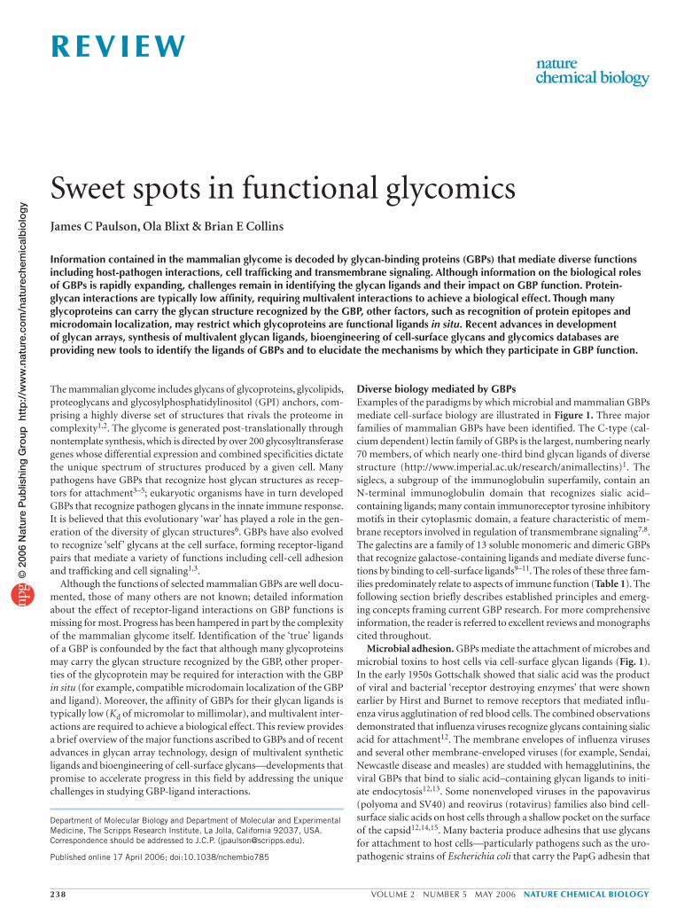
Diverse biology mediated by GBPsExamples of the paradigms by which microbial and mammalian GBPs mediate cell-surface biology are illustrated in Figure 1. Three major families of mammalian GBPs have been identified. The C-type (cal-cium dependent) lectin family of GBPs is the largest, numbering nearly 70 members, of which nearly one-third bind glycan ligands of diverse structure (http://www.imperial.ac.uk/research/animallectins)1. The siglecs, a subgroup of the immunoglobulin superfamily, contain anN-terminal immunoglobulin domain that recognizes sialic acid–containing ligands; many contain immunoreceptor tyrosine inhibitory motifs in their cytoplasmic domain, a feature characteristic of mem-brane receptors involved in regulation of transmembrane signaling7,8. The galectins are a family of 13 soluble monomeric and dimeric GBPs that recognize galactose-containing ligands and mediate diverse func-tions by binding to cell-surface ligands9–11. The roles of these three fam-ilies predominately relate to aspects of immune function (Table 1). The following section briefly describes established principles and emerg-ing concepts framing current GBP research. For more comprehensive information, the reader is referred to excellent reviews and monographs cited throughout.
Microbial adhesion. GBPs mediate the attachment of microbes and microbial toxins to host cells via cell-surface glycan ligands (Fig. 1). In the early 1950s Gottschalk showed that sialic acid was the product of viral and bacterial ‘receptor destroying enzymes’ that were shown earlier by Hirst and Burnet to remove receptors that mediated influ-enza virus agglutination of red blood cells. The combined observations demonstrated that influenza viruses recognize glycans containing sialic acid for attachment12. The membrane envelopes of influenza viruses and several other membrane-enveloped viruses (for example, Sendai, Newcastle disease and measles) are studded with hemagglutinins, the viral GBPs that bind to sialic acid–containing glycan ligands to initi-ate endocytosis12,13. Some nonenveloped viruses in the papovavirus (polyoma and SV40) and reovirus (rotavirus) families also bind cell-surface sialic acids on host cells through a shallow pocket on the surface of the capsid12,14,15. Many bacteria produce adhesins that use glycans for attachment to host cells—particularly pathogens such as the uro-pathogenic strains of Escherichia coli that carry the PapG adhesin that
Department of Molecular Biology and Department of Molecular and Experimental Medicine, The Scripps Research Institute, La Jolla, California 92037, USA. Correspondence should be addressed to J.C.P. ([email protected]).
Published online 17 April 2006; doi:10.1038/nchembio785
238 VOLUME 2 NUMBER 5 MAY 2006 NATURE CHEMICAL BIOLOGY
R E V I E W©
200
6 N
atur
e P
ublis
hing
Gro
up h
ttp
://w
ww
.nat
ure.
com
/nat
urec
hem
ical
bio
log
y

binds Galα1-4Gal on epithelial cells and the ulcer-causing Helicobacter pylori that contains multiple adhesins recognizing Lewis A– and sialic acid–containing glycolipids on gut epithelial cells4,5,16,17. Bacterial tox-ins that bind to host cell glycans typically have a pentameric structure, with the toxic subunit carried to the cell-surface by five identical bind-ing subunits, as exemplified by shiga toxin (Galα1-4Gal) and cholera toxin (ganglioside GM1)18.
Glycoprotein targeting. The initial evidence for the existence of mam-malian GBPs was the observation of Ashwell and co-workers in 1968 that asialoglycoproteins were rapidly cleared from blood. This process was shown to be due to a galactose-binding ‘C-type’ lectin on hepatocytes that bound and efficiently endocytosed glycoproteins with glycans containing terminal galactose. Several endocytic C-type lectin family receptors with different specificities were soon found on macrophages and endothelial cells1,19. Because circulating glycoproteins are normally ‘capped’ with sialic acids and have long circulatory half-lives, glycoprotein clearance may not be the primary function of these GBPs (see below). However, a role in the regulation of serum levels of glycopeptide hormones such as leutropin has been clearly established for the mannose/SO4
2–-GalNAc receptor on hepatic sinusoidal endothelial cells20.
Innate and acquired immunity. Recognition of pathogen-associated molecular patterns (PAMPs), specifically recognition of pathogen glycans, is a major function of GBPs in the immune system (Table 1)7,8,11,21–27. Members of the C-type lectin family are the largest group of non-Toll-like receptor pattern recognition molecules and are increasingly recog-nized to work in concert with Toll-like receptors to effect an immune response21,23,25,28. All major GBP families are believed to be involved in PAMP recognition. Siglecs7,8 and galectins11,29 also have key roles in regu-lating aspects of cell signaling, differentiation and apoptosis of leukocytes that mediate the innate and acquired immune responses.
The soluble collectins, a subfamily of the C-type lectin family, and the structurally related ficolins are multimeric collagenous lectins found in extracellular fluids and blood that opsonize microbial pathogens by binding to glycan-based PAMPs. Once bound, they can initiate phagocytosis by macrophages, and in the case of mannose binding protein (MBL) and ficolins they can initiate an alternative comple-ment pathway for direct killing of the organism by substituting for the complement molecule C1q and antibody recognition in the classical pathway22,24,26,30.
A large group of membrane-bound macrophage and dendritic cell C-type lectins have diverse glycan specificities and mediate innate immune responses to a wide variety of pathogens including viruses (for example, HIV and Ebola), bacteria and mycobacteria (for exam-ple, H. pylori, Streptococcus pneumoniae and Mycobacterium tuberculo-sis), and yeast (Aspergillus fumigatus and Candida albicans)21,23,25,28,31. As these lectins are endocytic receptors, the organisms are typically endocytosed and degraded once bound. As a consequence (particu-larly for dendritic cells), the GBPs provide a bridge to the acquired immune response by causing antigenic peptides that are released from the ingested organism to be presented via major histocompatibility molecules to T cells. The lectin dendritic cell–specific intercellular adhesion molecule–grabbing nonintegrin (DC-SIGN) is proposed to have a dual role: it acts as an endocytic receptor for uptake of some pathogens, and it stabilizes cell-cell interactions during presentation of the antigen to T cells by binding glycans on its cognate T cell ligand, intercellular adhesion molecule-3 (ref. 23). Some organisms, such as the HIV and hepatitis C viruses, have evolved to subvert these pathways, avoiding an immune response and even promoting their dissemination by blocking the degradation pathway and/or cytokine responses after endocytosis by the dendritic cells (DCs). The viruses then resurface to infect other cells at a remote site21,23.
Microbial GBPs
Soluble GBPs
Cell surface GBPs: extracellular glycans
Cell surface GBPs: cell surface glycans
Bacterialcolonization
Virus attachmentand endocytosis
Opsinization
Glycanantibody
Glycoproteinlattice
Glycoproteintargeting
Microberecognition
Clathrin-coated pit
Clathrin-coated pit
Trans ligand:cell adhesion
Cis ligand:receptorsignalling
Toxinattachment
Figure 1 Cell-surface biology mediated by GBPs. Microbial GBPs mediate attachment of microbes and microbial toxins to host cells. Soluble mammalian GBPs mediate a host of biological interactions by binding to microbes (opsonization) and cell-surface glycans. Cell-surface GBPs mediate attachment of microbes (innate immunity) and glycoproteins before endocytosis and bind cell-surface glycans to mediate cell adhesion (trans) or modulate the activity of cell signaling receptors (cis or trans). GBPs are represented by the ‘Pac-Man’ symbol, and glycans are represented by a single sugar ring. In reality, the glycan-binding site of a typical GBP is a shallow pocket on the surface of the protein1. A, A subunit of AB5 toxin.
NATURE CHEMICAL BIOLOGY VOLUME 2 NUMBER 5 MAY 2006 239
R E V I E W©
200
6 N
atur
e P
ublis
hing
Gro
up h
ttp
://w
ww
.nat
ure.
com
/nat
urec
hem
ical
bio
log
y

Most leukocytes involved in innate and acquired immune responses (for example, macrophages, DCs, natural killer cells, eosinophils, neu-trophils and B cells) also express one or more members of the siglec family, each of which has unique specificities for sialoside ligands7,8. Analogous to the C-type lectins and consistent with their recognition of sialic acid–containing ligands, siglecs are capable of binding and mediating the uptake of sialylated pathogens (for example, Neisseria meningitidis)7. However, accumulating evidence suggests that the most important role for most siglecs is regulation of cell signaling via immu-noreceptor tyrosine inhibitory motifs in their cytoplasmic domain7,8. Best understood is CD22 (Siglec-2), a B cell siglec, whose regulation of B cell receptor signaling is modulated by interactions, both by cis ligands on the B cells and by trans ligands on opposing cells, that cause its redistribution to sites of cell contact32,33.
Members of the galectin family are differentially expressed as soluble proteins by many leukocytes, endothelial cells and other cells that are present at the sites of leukocyte development and function. Bivalent galectins can cross-link glycoproteins with multiple glycan ligands into lattice formations (Fig. 1) and modulate their activity and underlying biology. Galectins have been proposed to induce apoptosis of imma-ture thymocytes and activated T cells, modulate neutrophil degranula-tion and phagocytosis by macrophages, and participate in the innate immune response to various microorganisms (either directly by bind-ing to the microbe or indirectly by modulating the response of the neutrophils and macrophages that respond to them)11,27,29,34,35.
Cell adhesion and cell trafficking. Trafficking of leukocytes by the selectin group of the C-type lectin family defines the classic paradigm for GBPs mediating the initial tethering of cells to the endothelial cells of blood vessels. Tethering results in the arrest and migration of
cells into the target tissue (for example, inflammatory tissue or lymph nodes). The three different selectins (E-, P- and L-selectin), the dif-ferential expression of their respective sialyl–Lewis X–bearing36 and proteoglycan37 ligands, and other factors (for example, the cytokine milieu) account for the differential trafficking of lymphocytes to lymph nodes, neutrophils to sites of inflammation, CD4+ T cells to sites of chronic inflammation, and DCs, natural killer cells and various other cells to their sites of action36,38,39. Another C-type lectin, DC-SIGN, has been shown to mediate DC adhesion to neutrophils and CD4+ T cells through recognition of the integrin Mac-1 and intercellular adhesion molecule-3 as ligands, respectively23,40. Several siglecs also mediate cell-cell interactions, particularly in the context of cell signal-ing, as discussed above7,8. In a somewhat different paradigm, the glial cell–associated siglec myelin-associated glycoprotein (MAG or Siglec-4) recognizes sialoside ligands on neuronal cells, resulting in inhibition of neurite outgrowth7,8,41.
Challenges in analysis of GBP-ligand interactionsAs mentioned above, the intrinsic affinity of GBPs for their glycan ligands is weak (Kd values ~ 1–1,000 µM), and as a consequence mul-tivalent GBP-ligand interactions are required to effect biologically relevant outcomes. For analysis of GBP-ligand interactions in vitro, this fact dictates that assay methods incorporate multivalent display of both ligands and the GBP to achieve sufficient avidity for binding to be detected. Similarly, synthetic probes based on the glycan ligand must be either multivalent, comprised of higher affinity analogs, or both. Identification of the in situ ligand of the GBP is also complicated by the fact that all glycoproteins produced by a cell are exposed to the same glycosylation machinery. Thus, although the glycan structure
Table 1 Roles of GBPs in immune functionProcess and GBP family Functions
Innate immunity
Soluble C-type lectins: collectins, ficolins Opsonization of pathogens
Initiation of alternative complement pathway
C-type lectins: e.g., DC-SIGN, mannose receptor, Dectin-1 Binding and endocytosis of pathogen
Bridge to acquired immunity
Harboring and dissemination of infectious agents (e.g., HIV, Ebola)
Siglecs Recognition and uptake of sialylated pathogens
Regulation of cell signaling, activation, adhesion
Galectins Regulation of DC maturation and cytokine secretion
Pathogen sensing and killing
Acquired immunity
C-type lectins: e.g., DC-SIGN, mannose receptor, Dectin-1 Binding and endocytosis leading to processing of pathogen and presentation of pathogen peptides to T cells by MHC
C-type lectin: DC-SIGN Maintenance of contact between DCs and T cells during antigen presentation
Siglecs Regulation of cell signaling
Recognition of self
Galectins Apoptosis
Regulation of cytokine secretion
Regulation of TCR signaling threshold
Leukocyte trafficking
C-type lectins: E- and P-selectin Trafficking of various leukocytes to sites of inflammation
Platelet leukocyte thrombus formation
C-type lectins: L-selectin Trafficking of lymphocytes to lymph nodes
C-type lectins: DC-SIGN Trafficking of DCs from periphery to lymph nodes
Galectins Regulation of T cell trafficking into peritoneum
Abbreviations: MHC, major histocompatibility; TCR, T-cell receptor.
240 VOLUME 2 NUMBER 5 MAY 2006 NATURE CHEMICAL BIOLOGY
R E V I E W©
200
6 N
atur
e P
ublis
hing
Gro
up h
ttp
://w
ww
.nat
ure.
com
/nat
urec
hem
ical
bio
log
y

recognized by a GBP may be carried by many glycoproteins, only selected proteins are the effective ligands in situ. As a case in point, E-selectin binds multiple neutrophil glycoprotein ligands contain-ing sialyl–Lewis X glycans, whereas P-selectin selectively recognizes P-selectin glycoprotein ligand-1 (PSGL-1) by virtue of its extended binding pocket, which recognizes sulfated tyrosines in addition to the glycan structure42–44. As a further consideration, the in situ interac-tions of a GBP with its ligand may be constrained by the restricted localization of the GBP and its ligand to microdomains (for example, ‘sweet spots’), physically excluding other glycoproteins with the glycan sequence recognized by the GBP from being physiologically relevant in situ ligands45. In this case, glycoproteins that bind to the GBP in vitro may not be ligands in situ. Notwithstanding such limitations, advances in the synthesis of monovalent and multivalent glycans and in the bioengineering of cell-surface glycans are providing new approaches to assess the specificity of GBPs for their glycan ligands and the biology mediated by GBP-ligand interactions.
Glycan microarrays for functional glycomicsLack of tools for interrogating the ability of a protein to bind carbo-hydrate ligands or for assessing the detailed specificity of a protein already known to bind carbohydrates has severely limited the pace of biological discovery for GBPs. Recent advances in the construction of glycan microarrays that display a diverse glycan library directly address these limitations by allowing the specificity of a GBP or candidate GBP to be assessed for a large number of glycans simultaneously. Advances have occurred on two fronts: the method of attachment of the glycan library to the ‘chip’ or solid surface and the generation of the glycan library that is arrayed on the chip46–52.
Glycan array formats. The history of glycan arrays arguably dates back to the use of thin-layer chromatography to separate complex mix-tures of glycolipids and to probe the chromatogram with a GBP (for example, an antibody or a toxin) to identify which glycolipids were recognized53. Feizi and co-workers extended this idea to include non-glycolipid carbohydrates by attaching lipids to the reducing end of isolated glycans to create neoglycolipids that could be ‘arrayed’ on chro-matograms or 96-well plates54,55. In the past 5 years there has been an explosion of methods for creating modern versions of glycan micro-arrays, most of which use (or can be adapted to use) state-of-the-art array technologydeveloped for printing cDNA microarrays on glass slides. The methods differ primarily in the method of attachment of the glycan to the solid support: attachment is either noncova-lent or covalent (Fig. 2).
Several glycan array formats are based on noncovalent association of glycans or modi-fied glycans with appropriately prepared sur-faces. Unmodified bacterial polysaccharides and proteoglycans bind well to nitrocellulose56 or oxidized polystyrene57. Glycans linked to lipids or alkyl chains allow adsorption to poly-styrene and to nitrocellulose or polyvinylidene fluoride membranes or coated glass slides47,48. Glycans linked to fluorous tags (C8F17) were recently shown to adsorb avidly to glass slides coated with fluoroalkylsilane and even to resist washing with detergents58. An alternative and
useful approach uses glycans linked to biotin for affinity adsorption to surfaces coated with streptavidin59,60.
Increasing attention is being placed on array formats using gly-cans with functionalized spacers that react with a complementary-activated surface to form a covalent bond, as illustrated for several examples in Figure 2 (refs. 61–65). In one format, unmodified glycans
OO
OO
OOHO
OO
OHO
O
OO O
OHO
OHO
OOHO
OOHO
OO XHO
OOPO
OO X
OOPO
X
OOHO
OO X
OORnO
OO X
OORnO
OOH
OO
OHO
OOHO
OO
OO
Y
Y
Y
Y
Y
Y
Y
Y
Y
Y
Y
Y
Y
Y
Y
Y
Y
Y
Y
Y
Chemicalsynthesis
Enzymaticsynthesis
Glycoproteinglycolipid glycans
Deprotection
Deprotection
Multistepglycosylation
One-stepglycosylation
Glycolipid
Glycoprotein
Elongationdeprotection
ElongationEndoglycosidasecleavage
Reducing andderivatization
Glycan library
Diverse glycan array
Figure 3 Strategies for assembling a diverse functionalized glycan library. Shown are chemical derivatization of monosaccharide precursors containing a functional group X followed by diversification using chemical and/or enzymatic approaches, and isolation of glycoprotein and glycolipid glycans by glycosidase cleavage and subsequent derivatization to contain the same functional group X. Note that the functional group X can be either an adsorptive group for noncovalent attachment or a reactive group for covalent attachment.
OO XRO
Y Y Y
OO
XR
O
Y Y
OO
XR
O
OO
XR
OO
ORO
OO
RO
OO
RO
OO
RO
Surface Adsorptive group
Nitrocellulose Alkyl, lipid, none
Oxidizedpolystyrene
Alkyl, lipid, none
Fluoroalkylsilane C8F17
Streptavidin Biotin
Surface Y Reactive group X
Thiol Maleimide (thioether)
Alkyne Azide (cycloaddition)
Amine (amide)NHS
Benzoquinone Cyclopentadiene(cycloaddition)
Aryl-CF3 diazarine None (carbeneinsertion)
a bNoncovalent CovalentAdsorptivegroup
Reactivegroup
Adsorptive surface Functionalized surface
Figure 2 Methods for attachment of glycans to the surface of a microarray. (a) Noncovalent immobilization via an adsorptive surface. (b) Covalent coupling via a reaction of a reactive group X with a functionalized surface Y.
NATURE CHEMICAL BIOLOGY VOLUME 2 NUMBER 5 MAY 2006 241
R E V I E W©
200
6 N
atur
e P
ublis
hing
Gro
up h
ttp
://w
ww
.nat
ure.
com
/nat
urec
hem
ical
bio
log
y

are covalently coupled to the surface of aryl-CF3 diazarine–coated slides by carbene insertion66. Although this could be very useful for polysac-charides, it remains to be seen whether such random coupling will be useful for small glycans.
With so many alternatives for construction of glycan arrays, it is not possible to conclude that one format is superior to all others. However, many practical issues are ultimately considered during the development of large-scale arrays. From the chemistry perspective, the ease of gen-erating a diverse functionalized-glycan library and the efficiency of the coupling reaction are major factors, particularly for small (microgram) quantities of glycans obtained from biological sources (Fig. 3). The sur-face to which the glycans are attached is also critical for the subsequent interrogation with labeled GBP, as low background binding is essential for specific binding to be detected. Such factors can ultimately take precedence over otherwise elegant array chemistries.
Glycan library. Ultimately, the utility of a glycan array depends on an appropriate match between the types of glycan structures it contains and the specificity of the GBP being analyzed. The ideal array would contain the entire glycome of an organism on a single chip, so that any GBP could be assessed. In practice, however, current arrays are limited to displaying libraries of natural and synthetic glycans that can be practically assembled.
An achievable goal for creating a diverse glycan array is to envision all sources of glycans and develop compatible chemistries that will allow them to carry an adsorptive or reactive group required for printing on the desired array surface (Fig. 3). All glycans synthesized de novo by chemical or chemoenzymatic synthesis rely on the chemical introduc-tion of the selected functional group, typically as a glycoside linker at the reducing terminal sugar residue. Chemical strategies for elongation to larger glycans include traditional solution-phase chemical synthe-sis67,68, automated solid-phase synthesis and one-pot reactivity-based glycosylations52,69,70. The automated methods offer great potential to develop large and diverse glycan libraries. At present, however, the diversity that can be achieved is still limited by difficulties in the syn-thesis of some linkages.
A highly complimentary approach is the use of regio- and stereospe-cific glycosylating enzymes—glycosyltransferases that generate glyco-sidic linkages in one-step reactions between an unprotected acceptor and a sugar donor nucleotide. With new advances in molecular biology,recombinant enzymes and accessory enzymes for sugar-nucleotide regeneration are now being readily expressed in large quantities for synthesis of complex oligosaccharides50,71. Although enzymes have relatively strict substrate specificities, many of them offer substantial flexibility for synthesis of unnatural analogs (Fig. 5). Enzymes can also be used to elaborate glycans synthesized chemically. For example, sialic acid, a common monosaccharide that frequently terminates oligosac-charide sequences on various glycoproteins and glycolipids, is noto-riously difficult to apply in chemical glycosylation but can be easily introduced enzymatically as a final step by a sialyltransferase72,73.
Despite advances, high-throughput synthesis of branched complex-type glycans of glycoproteins and glycolipids to cover the diversity of the mammalian glycome is not yet practical. New methods for isolating and characterizing natural glycans from glycoproteins and glycolipids74,75 are providing an alternative to obtaining these molecules. Enzymatic modification of glycans isolated in abundance using glycosyltransfer-ases or glycosidases can further increase the diversity of the glycans obtained from biological sources76. Although the amounts that can be obtained are typically small (for example, <1 mg), 100 µg of a glycan is sufficient for printing 5,000–10,000 glycan arrays.
Because glycans from natural sources are often released by endogly-cosidases, they contain a reducing sugar that can in principle be used
GBP staining
Fluorescence detection and processed data
Bioinformatics
GBP
Secondaryantibody
Anti-glycanantibody
Fluorophore Virus
Glycan microarray slide
Glycans
Cartoon representation
IUPAC 2D representation
IUPAC code
Linear code
Sub structure search interface
General information
α6 β4 β SP2
NeuAc α2 6 Gal β1 4GlcNAc
NeuAc α2-6 Gal β1-4 GlcNAc β-sp
NNα6Aβ4GNβ-sp
Load this structure for sub structure search
Glycan family:Sub family:Scientific name:
SyntheticSyntheticNeu5Acα6Galβ4GlcNAcβ-sp
Rel
ativ
e flu
ores
cenc
e
Figure 4 Glycan microarrays reveal novel glycan specificities of GBPs. Fluorescently labeled GBPs are readily detected after staining the glycan microarray surface using a confocal slide scanner or plate reader. Top, nonlabeled GBPs, antibodies or pathogens are detected with an overlay of labeled secondary antibodies. A higher degree of GBP multivalencyis achieved by overlaying precomplex GBP with labeled secondary antibodies for stable multivalent binding. Middle, the data obtained from the image analysis (two of six replicates shown in the inset) generate atwo-dimensional bar chart to reveal relative signals for each glycan on the array. Bottom, data may be integrated into a relational bioinformatics database for user-friendly navigation to biological information.
242 VOLUME 2 NUMBER 5 MAY 2006 NATURE CHEMICAL BIOLOGY
R E V I E W©
200
6 N
atur
e P
ublis
hing
Gro
up h
ttp
://w
ww
.nat
ure.
com
/nat
urec
hem
ical
bio
log
y

for introducing the desired linker containing a functional group needed for incorporation into covalent arrays. In practice, however, this seemingly simple transformation has been a major barrier to the use of glycans obtained from natural sources, particularly for those obtained in small quantities. Several groups have exploited conversion of the reducing end to the glycosylamine to introduce an amine functionality while retaining the ring struc-ture76,77. A new approach promoted by Xia and co-workers78 exploits reductive amina-tion with diaminopyridine, resulting in ring opening and a free amino group that can be used directly for coupling to N-hydroxysuccin-imide–activated glass slides. These and related methods will undoubtedly be optimized for efficiency for the derivation of glycans isolated from biological sources and their transfer to glycan arrays.
In summary, the technologies required for producing functionalized synthetic and natural glycans are in place and are expected to benefit from further efficiencies that will increase the pace of assembling highly diverse glycan librar-ies for populating glycan arrays. In principle, these methods can be adapted for the introduc-tion of any adsorptive or reactive group, making glycan libraries accessible to most of the array formats that have been developed (for example, Fig. 2). However, with finite resources being a practical consideration, diverse glycan libraries will ideally be generated for populating array formats that are proven.
Sweet spots reveal GBP specificitySince the introduction of the first glycan arrays in 2002, their application for analysis of the specificities of diverse GBPs has rapidly grown46,47,56,63. For analysis on the array, GBPs must be directly labeled with a fluorescent tag or indirectly labeled with antibodies or some other agent carrying the fluorescent label that binds to a tagged GBP (Fig. 4). Because GBPs typically bind to their glycan ligands with low affinity, the indirect method of cross-link-ing with antibodies is sometimes beneficial or even required to obtain sufficient valency to acquire a ‘signal.’ Several array platforms that display diverse glycan libraries have been developed and are sought out by investiga-tors for collaborative analysis of GBP function. The Consortium for Functional Glycomics (CFG) has produced a glycan library compris-ing over 300 glycans with amino linkers and representing terminal structures of glycolipids and N- and O-linked glycans of glycoproteins (http://www.functionalglycomics.org). This library, coupled to bio-tin linkers, was initially arrayed in 384-well polystyrene plates coated with streptavidin59,79,80 and is now available in microarrays printed directly on glass slides activated with N-hydroxysuccinimide (NHS)63. Investigators wishing to use the array have analysis done by the CFG. The results obtained are posted on the CFG website and linked to
databases for visualizing the structures recognized; links to biological information about natural structures that bear the sequence detected are also included (Fig. 4)81. A similar-sized glycan library of glycans linked to lipids (‘neoglycolipids’) has been generated for printing on nitrocellulose surfaces47,82 by Feizi and co-workers, now affiliated with the newly formed UK Glycochip Consortium (http://www.glycochips.org.uk). The array of polysaccharides reported by Wang is currently the largest array of bacterial and pathogen glycans56, and a commercial array with a library of 45 glycans in a slide-based microwells array has been used in several applications83,84.
OOHRO
OORO
OOOR
OOHRO
OORO
ORO O
OOR
R OO
Golgi
Cytosolic protein
O-linkedGlcNAc
O-linked
N-linked
N-linked
O-linked
OO
AzHN
OHOH
O
OO
HO
OHOHOH
O
CO2H
HOR1
R2
OH
OOH
HO
OH
HONHO
N3
OOH
HO OH
HONHO
N3
OHN
OH
O
R1
R2OH
OH CO2H
OOH
HOR2
HO
HN
O
R1
OO
HO
OH
HOAzHN
OHO
O
HO
OHO
AcHN
OH
O OHO
AcHN
OH
OO
HO
O
HO
HO OO
OH
HO
O
OHO
CO2H
HOR1
R2
OH
OHO
CO2H
HOR1
R2
OH
OHO
AcHN
OH
OO
HOHO
OOH
OHO
AcHN
OH
OO
HOHO
OOH
HO
R1
N-linked
R1
R2
Mannosamineanalogs Sialic acid
analogs
GlcNAz
GalNAz
Figure 5 Biosynthetic engineering of cell glycans. Monosaccharide derivatives carrying unnatural substituents (R) are taken up by cells, converted to the corresponding activated nucleotide sugar (N) and then transferred onto nascent oligosaccharide chains. Representative analogs of sialic acid, mannosamine, galactosamine, and glucosamine shown to be taken up by cells and incorporated into representative glycoproteins are illustrated. After uptake, mannosamine derivatives are rapidly converted to the corresponding sialic acid derivative.
NATURE CHEMICAL BIOLOGY VOLUME 2 NUMBER 5 MAY 2006 243
R E V I E W©
200
6 N
atur
e P
ublis
hing
Gro
up h
ttp
://w
ww
.nat
ure.
com
/nat
urec
hem
ical
bio
log
y

Glycan arrays are best suited for screening the specificity of GBPs, after which more classical methods of examining protein-ligand inter-actions can be used to study details of affinity and relative specificity. Numerous plant lectins, glycan-specific antibodies and microbial toxins used as routine research tools have been analyzed for their specificity on glycan arrays. This raises the possibility of having all such reagents sys-tematically analyzed and using the data as a resource for interpretation of results acquired using these reagents (see also CFG website)46,47,63,85. Representative members of the C-type lectin63,79–82,86,87, siglec59,63,88 and galectin63 families have also been successfully assessed for their glycan specificity on glycan microarray formats. Notably, recombinant DC-SIGN, a C-type lectin, has been independently analyzed on two different platforms; it showed dual specificity for high-mannose and fucose-containing glycans, indicating that the array format itself did not greatly influence the specificity observed63,79. Human eosinophil Siglec-8 and its murine paralog Siglec-F were found to recognize a new ligand, 6’-sulfo-sialyl–Lewis X59,88, and the C-type–like lectin Dectin-2 was, surprisingly, shown to bind high-mannose glycans81.
Glycan array analysis of (i) the specificity of the hemagglutinin from the 1918 pandemic human influenza virus (H1N1) and (ii) receptor-site mutants containing selected amino acids corresponding to the H1 avian virus consensus sequence showed that only two amino acids were required to change the specificity from recognition of human receptors (NeuAcα2-6Gal) to recognition of avian type receptors (NeuAcα2-3Gal)89. As evaluation of the specificity using whole influenza virus is also possible63, glycan microarrays may be useful in surveying human isolates of avian and other animal influenza viruses for mutations that would facilitate binding to human type receptors.
Glycan microarrays are also capable of detecting carbohydrate-specificantibodies in human sera56,63,90–93. Normal sera contain a variety of glycan-specific antibodies that are related to blood groups or to expo-sure to microbial flora. Several reports describe altered glycan-antibody profiles from sera of cancer patients90,92, suggesting a potential use of glycan arrays in cancer diagnosis90–93.
Initial attempts to evaluate the binding of bacteria and even eukary-otic cells to glycan arrays also appear promising46,84. Such activities will encourage further use of glycan microarrays to study protein-glycan interactions in these more biologically complex systems.
In related developments, several groups have evaluated the potential utility of lectins or microarrays to profile the glycan structures of gly-coproteins66,94, cells95 or pathogen glycans96. The idea is to array vari-ous plant lectins and/or mammalian GBPs, collectively called lectins, onto the microarray slides and assess which lectins recognize and bind to the sample containing an unknown glycan structure. By knowing the specificity of the lectins used in the array, elements of the glycan structure can be inferred. In this regard, efforts to elucidate detailed glycan specificities of the lectins used in the microarrays (see above) will aid the interpretation of the information obtained using lectin microarrays.
Bioengineering of cell-surface glycansUsing synthetic derivatives of the sialic acid precursor N-acetylman-nosamine, Reutter and co-workers showed that unnatural sugar analogs can be taken up by cells, converted to their corresponding nucleotide sugar and efficiently transferred onto the nascent oligosaccharide chain to be presented on the cell surface (Fig. 5)97–102. This pioneering work was expanded by Bertozzi and colleagues to include sugar derivatives with substituents that carry functional groups, allowing selective chem-istry to be done on cell surfaces displaying these derivatives100,103–105. An expansive library of sugar analogs are now known to be incorpo-rated into cell-surface glycoproteins when ‘fed’ to cells (Fig. 5); this
demonstrates the impressive promiscuity of many of the enzymes in the glycan biosynthesis pathways100. Increasingly this approach is being exploited to advance the understanding of the roles of glycans in cell-surface biology, as described below.
Owing to the prominent role of sialic acid–containing glycans in cell adhesion and communication106, sialic acid has been the major target of glycan biosynthetic engineering. Incorporation of unnatural sialic acids into cell-surface glycans can be accomplished using either the desired sialic acid analog directly or the corresponding analog of the biosynthetic precursor N-acetylmannosamine. Although many unnatu-ral substituents can be accommodated at the C-9 and N-acyl posi-tions, there are exceptions. Some mannosamine derivatives with long N-acyl chains, for example, are inefficiently converted to sialic acid100. In these cases, using the corresponding sialic acid derivative will largely overcome this blockade, suggesting that the defect is in the sialic acid synthetase. Poor uptake of some derivatives by cells can be overcome by peracetylating them, which makes them more membrane perme-able101,107. Once internalized, the peracetylated derivatives are rapidly deacetylated to the unprotected sugar by nonspecific esterases.
Several reports document altered functions of cells displaying sialic acid analogs. For example, substitution of the N-acetyl group of sialic acid with N-propanoyl blocks papovavirus infection99. N-propanoyl analogs also increase calcium signaling and differentiation108 and neuronal outgrowth100, although it is unknown whether GBPs play a role in these effects. Recognition of neuronal glycoproteins by myelin-associated glycoprotein (or Siglec-4), an inhibitor of neurite regenera-tion, is blocked by converting neuronal sialic acids from N-acetyl to N-glycolyl109. In contrast, a sialic acid derivative with an aromatic ring at position nine (9-AAz) is preferred by CD22 over the natural sialic acid45. As these derivatives can be used in a variety of cell types, the approach represents a rapid alternative to gene knockouts to investigate the function of a particular GBP.
Recent data suggest that unnatural N-acyl analogs of sialic acid can be used to regulate the activity of sialyltransferases. N-butanoyl mannosamine derivatives, for example, specifically inhibit the sialyl-transferase that forms the polysialic acid (PSA) polymers NeuAc(α2-8NeuAc)n-(PSA)100. This derivative may prove useful to regulate neuritogenesis as cells fed the derivatives have decreased PSA on the protein neural cell adhesion molecule; thus they have increased inhi-bition of dorsal root ganglion neuronal outgrowth110,111. Long-chain N-acyl derivatives have similar effects on bacterial systems that salvage sialic acids. In Haemophilus ducreyi, N-octanoyl sialic acid inhibits the sialyltransferase that is necessary for synthesis of the PSA chains of sialic acid found on the outer wall of the organism. This leads to marked changes in lipopolysaccharide expression112. In contrast, promyelo-cytic HL60 cells fed N-propanoyl mannosamine have greatly increased expression of the selectin ligand sialyl–Lewis X epitope (NeuAcα2,3Gal(Fucα2,3)β1,4GalNAc) and increased selectin-mediated rolling as compared to the nonderivatized mannosamine113. In this case the derivative may enhance sialyl–Lewis X expression by acting as a better donor or acceptor of a glycosyltransferase responsible for sialyl–Lewis X synthesis.
In another application, Han and co-workers developed a method to identify the cis glycoprotein ligands of the B cell siglec CD22 using B cells grown in the presence of a sialic acid analog with a photoreactive aryl azide moiety at the C-9 position45,114. When exposed to UV light, sialic acids bearing the aryl azide moiety react with any protein contactingthem, resulting in cross-linking of the lectin domain of CD22 to its cis glycan ligand45. ‘Fishing out’ and characterizing the cross-linked CD22 showed that it selectively recognized glycans of other CD22 molecules as cis ligands, resulting in formation of homomultimers. In contrast,
244 VOLUME 2 NUMBER 5 MAY 2006 NATURE CHEMICAL BIOLOGY
R E V I E W©
200
6 N
atur
e P
ublis
hing
Gro
up h
ttp
://w
ww
.nat
ure.
com
/nat
urec
hem
ical
bio
log
y

CD22 was found to recognize many B cell glycoproteins in cell lysates; thus the interactions of GBPs with their true in situ ligands cannot be deduced from in vitro experiments alone. Extension of this protein-glycan cross-linking approach may be useful for identification of the in situ ligands of GBPs.
Analogs of N-acetylglucosamine (GlcNAc) and N-acetylgalactos-amine (GalNAc) are similarly taken up and incorporated into glycans (Fig. 5)100,115. The N-azidogalactosamine (GalNAz) derivatives are readily taken up by cells and incorporated as the first sugar added to the polypeptide chain in O-linked glycans. This is particularly important for proteomics applications because the azide in the N-acyl substituent serves as a chemical tag for proteins that contain O-linked glycans116. The GlcNAz derivative also has interest from a proteomics stand-point, primarily as a method to detect the multitude of cytoplasmic and nuclear proteins modified by O-GlcNAc residues115. Interestingly, GlcNAz is only weakly incorporated into cell-surface glycans115,117; this probably reflects an inability of the derivative to be recognized by the GlcNAc transferases involved in N- and O-glycan synthesis.
A different approach to modifying cell-surface glycans is to expose cells to saccharide ‘primers’ that can act as decoy substrates for the gly-cosyltransferases that add terminal sugars to glycoprotein glycans. In this case the analogs are meant not to be incorporated but to competitively inhibit terminal glycosylation by competing with natural acceptors118. The peracetylated disaccharide Galβ1,3GlcNAcβ-O-R, for example, is taken up by tumors and effectively competes as an acceptor for the trans-ferases that make the sialyl–Lewis X epitope; the result is a decrease in the expression of cell-surface sialyl–Lewis X. The reduced expression of sialyl–Lewis X on tumor cells attenuates metastasis and tumor burden in vivo119. Additional decoys have been used to decrease heparin sulfate and chondroitin sulfate synthesis or O-linked glycan biosynthesis118.
As illustrated by the examples above, the cell machinery can be made to perform what would be difficult chemistry in vitro and to synthesize the complex glycan structure and present it in the normal environ-ment. Recent results suggest that these methods can be extended to in vivo applications103. Although they are well described as methods to introduce chemical reactivity, their future holds great promise as modulators of GBP biology.
Synthetic multivalent glycan probesInteractions of GBPs with their glycan ligands are generally low affinity, requiring multivalent interactions to achieve a biological effect (Fig. 1). Consequently, designers of synthetic ligands or glycan-based inhibitors have focused on multimeric structures29,120–122. Highly valent glyco-dendrimers and glycopolymers have been demonstrated as tools to study GBP biology as they inhibit the binding of toxins123, viruses124,125, selectin-mediated inflammation126 and leukocyte trafficking127,128. However, despite major efforts of synthetic chemists and biologists, cluster glycosides of intermediate valency have in large part resulted in only modest gains in avidity. The rational design of multivalent ligands is confounded by (i) the low affinity of GBP-glycan interactions and (ii) the fact that the distances between the binding sites of multivalent GBPs are large or undefined, and in the case of monovalent membrane GBPs they not only are undefined but also are in constant motion. A com-prehensive review of multivalent glycan inhibitors is beyond the scope of this discussion, but the reader is referred to excellent reviews on the topic120–122. Instead, this review will focus on recent new approaches to ‘biology driven’ inhibitor design that have the potential to achieve gains in affinity with modest levels of valency.
Several groups have exploited rational design–based tethering ligands to approximate the spacing of multimeric GBPs. A natural example of
this ‘cluster effect’ can be observed with the multimeric asialo-glycopro-tein and its N-linked ligand. Here, in these pioneering studies, increasing from mono- to triantennary galactose-terminated N-linked glycans to match the multimeric receptor increased binding 200,000-fold129. In an elegant extension of rational design, Bundle and co-workers considered the three-dimensional spacing of the shiga-like toxin lectin domains in designing a multivalent backbone130,131. The shiga-like toxin exists as a pentameric structure with each of the five arms containing a glycan-binding domain. An oligovalent inhibitor containing the glycan ligand attached to a core by linkers tailored to fit the recent crystal structure resulted in a subnanomolar inhibitor130,131 with 90% efficacy in vivo132. Interestingly, the pentavalent-tailored inhibitor gave similar levels of pro-tection as a dendrimeric structure in vivo132. Nonetheless the directed, tailored approach for glycan modeling holds great promise for designing specific inhibitors with high potency and better bioavailability in vivo.
A more difficult problem is the creation of multivalent inhibitors for monovalent membrane GBPs that achieve multivalency by clus-tering with other GBPs. To this end efforts have focused on a more biology-driven inhibitor design. An example of this is the design of a self-assembled probe that strings glycan-containing cyclo-dextran ‘beads’ onto a pseudo polyrotaxane ‘string’ (Fig. 6)133. These glycan beads have complete mobility to spin around and ‘slide’ along the string to match GBP spacing (‘ligand adaptation’). Such mobility even allows the inhibitor to match the rigid spacing of the dimeric galectin Gal I, in which the GBP domains face in opposite directions. This type of approach is likely to have general application to cell-surface GBPs, as the beads could slide to mirror the spacing and lateral mobility observed in GBPs in the fluidic membrane environment.
An alternative approach allows the biology to drive assembly of the multimer. To this end, homomultimerization into polymeric arrays of
Heterobifunctional assembly
Ligand adaptation
SAPBifunctionalligand
AB5toxin
Figure 6 Biological assembly of multivalent ligands. Top, inhibitors of the appropriate spacing are formed using biological ‘templates.’ In this example, a heterobifunctional ligand bridges the pentameric ligand-binding domains of the SAP and AB5 toxins, which are similarly spaced135,136. As shown here, SAP binding of the heterobifunctional ligand stabilizes the ligand presentation for the AB5 toxins. Such heterobifunctional ligands can be monovalent or tethered pentavalent structures. Bottom, inhibitors may have free motion to adapt to GBP density and spacing133. cyclo-dextran ‘beads’ have free rotational and lateral mobility around the pseudo polyrotaxane ‘string,’ allowing it to match the distribution of membrane-bound GBP.
NATURE CHEMICAL BIOLOGY VOLUME 2 NUMBER 5 MAY 2006 245
R E V I E W©
200
6 N
atur
e P
ublis
hing
Gro
up h
ttp
://w
ww
.nat
ure.
com
/nat
urec
hem
ical
bio
log
y
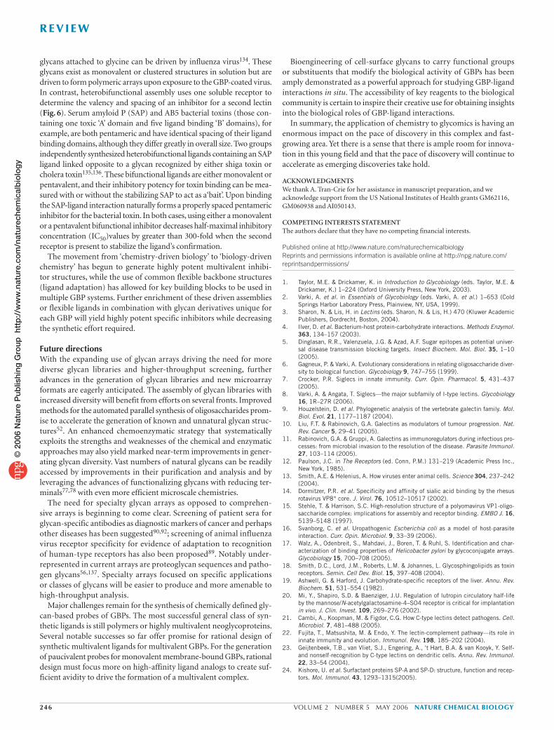
glycans attached to glycine can be driven by influenza virus134. These glycans exist as monovalent or clustered structures in solution but are driven to form polymeric arrays upon exposure to the GBP-coated virus. In contrast, heterobifunctional assembly uses one soluble receptor to determine the valency and spacing of an inhibitor for a second lectin (Fig. 6). Serum amyloid P (SAP) and AB5 bacterial toxins (those con-taining one toxic ‘A’ domain and five ligand binding ‘B’ domains), for example, are both pentameric and have identical spacing of their ligand binding domains, although they differ greatly in overall size. Two groups independently synthesized heterobifunctional ligands containing an SAP ligand linked opposite to a glycan recognized by either shiga toxin or cholera toxin135,136. These bifunctional ligands are either monovalent or pentavalent, and their inhibitory potency for toxin binding can be mea-sured with or without the stabilizing SAP to act as a‘bait’. Upon binding the SAP-ligand interaction naturally forms a properly spaced pentameric inhibitor for the bacterial toxin. In both cases, using either a monovalent or a pentavalent bifunctional inhibitor decreases half-maximal inhibitory concentration (IC50)values by greater than 300-fold when the second receptor is present to stabilize the ligand’s confirmation.
The movement from ‘chemistry-driven biology’ to ‘biology-driven chemistry’ has begun to generate highly potent multivalent inhibi-tor structures, while the use of common flexible backbone structures (ligand adaptation) has allowed for key building blocks to be used in multiple GBP systems. Further enrichment of these driven assemblies or flexible ligands in combination with glycan derivatives unique for each GBP will yield highly potent specific inhibitors while decreasing the synthetic effort required.
Future directionsWith the expanding use of glycan arrays driving the need for more diverse glycan libraries and higher-throughput screening, further advances in the generation of glycan libraries and new microarray formats are eagerly anticipated. The assembly of glycan libraries with increased diversity will benefit from efforts on several fronts. Improved methods for the automated parallel synthesis of oligosaccharides prom-ise to accelerate the generation of known and unnatural glycan struc-tures52. An enhanced chemoenzymatic strategy that systematically exploits the strengths and weaknesses of the chemical and enzymatic approaches may also yield marked near-term improvements in gener-ating glycan diversity. Vast numbers of natural glycans can be readily accessed by improvements in their purification and analysis and by leveraging the advances of functionalizing glycans with reducing ter-minals77,78 with even more efficient microscale chemistries.
The need for specialty glycan arrays as opposed to comprehen-sive arrays is beginning to come clear. Screening of patient sera forglycan-specific antibodies as diagnostic markers of cancer and perhaps other diseases has been suggested90,92; screening of animal influenza virus receptor specificity for evidence of adaptation to recognition of human-type receptors has also been proposed89. Notably under-represented in current arrays are proteoglycan sequences and patho-gen glycans56,137. Specialty arrays focused on specific applications or classes of glycans will be easier to produce and more amenable to high-throughput analysis.
Major challenges remain for the synthesis of chemically defined gly-can-based probes of GBPs. The most successful general class of syn-thetic ligands is still polymers or highly multivalent neoglycoproteins. Several notable successes so far offer promise for rational design of synthetic multivalent ligands for multivalent GBPs. For the generation of paucivalent probes for monovalent membrane-bound GBPs, rational design must focus more on high-affinity ligand analogs to create suf-ficient avidity to drive the formation of a multivalent complex.
Bioengineering of cell-surface glycans to carry functional groups or substituents that modify the biological activity of GBPs has been amply demonstrated as a powerful approach for studying GBP-ligand interactions in situ. The accessibility of key reagents to the biological community is certain to inspire their creative use for obtaining insights into the biological roles of GBP-ligand interactions.
In summary, the application of chemistry to glycomics is having an enormous impact on the pace of discovery in this complex and fast-growing area. Yet there is a sense that there is ample room for innova-tion in this young field and that the pace of discovery will continue to accelerate as emerging discoveries take hold.
ACKNOWLEDGMENTSWe thank A. Tran-Crie for her assistance in manuscript preparation, and we acknowledge support from the US National Institutes of Health grants GM62116, GM060938 and AI050143.
COMPETING INTERESTS STATEMENTThe authors declare that they have no competing financial interests.
Published online at http://www.nature.com/naturechemicalbiologyReprints and permissions information is available online at http://npg.nature.com/reprintsandpermissions/
1. Taylor, M.E. & Drickamer, K. in Introduction to Glycobiology (eds. Taylor, M.E. & Drickamer, K.) 1–224 (Oxford University Press, New York, 2003).
2. Varki, A. et al. in Essentials of Glycobiology (eds. Varki, A. et al.) 1–653 (Cold Springs Harbor Laboratory Press, Plainview, NY, USA, 1999).
3. Sharon, N. & Lis, H. in Lectins (eds. Sharon, N. & Lis, H.) 470 (Kluwer Academic Publishers, Dordrecht, Boston, 2004).
4. Ilver, D. et al. Bacterium-host protein-carbohydrate interactions. Methods Enzymol. 363, 134–157 (2003).
5. Dinglasan, R.R., Valenzuela, J.G. & Azad, A.F. Sugar epitopes as potential univer-sal disease transmission blocking targets. Insect Biochem. Mol. Biol. 35, 1–10 (2005).
6. Gagneux, P. & Varki, A. Evolutionary considerations in relating oligosaccharide diver-sity to biological function. Glycobiology 9, 747–755 (1999).
7. Crocker, P.R. Siglecs in innate immunity. Curr. Opin. Pharmacol. 5, 431–437 (2005).
8. Varki, A. & Angata, T. Siglecs—the major subfamily of I-type lectins. Glycobiology 16, 1R–27R (2006).
9. Houzelstein, D. et al. Phylogenetic analysis of the vertebrate galectin family. Mol. Biol. Evol. 21, 1177–1187 (2004).
10. Liu, F.T. & Rabinovich, G.A. Galectins as modulators of tumour progression. Nat. Rev. Cancer 5, 29–41 (2005).
11. Rabinovich, G.A. & Gruppi, A. Galectins as immunoregulators during infectious pro-cesses: from microbial invasion to the resolution of the disease. Parasite Immunol. 27, 103–114 (2005).
12. Paulson, J.C. in The Receptors (ed. Conn, P.M.) 131–219 (Academic Press Inc., New York, 1985).
13. Smith, A.E. & Helenius, A. How viruses enter animal cells. Science 304, 237–242 (2004).
14. Dormitzer, P.R. et al. Specificity and affinity of sialic acid binding by the rhesus rotavirus VP8* core. J. Virol. 76, 10512–10517 (2002).
15. Stehle, T. & Harrison, S.C. High-resolution structure of a polyomavirus VP1-oligo-saccharide complex: implications for assembly and receptor binding. EMBO J. 16, 5139–5148 (1997).
16. Svanborg, C. et al. Uropathogenic Escherichia coli as a model of host-parasite interaction. Curr. Opin. Microbiol. 9, 33–39 (2006).
17. Walz, A., Odenbreit, S., Mahdavi, J., Boren, T. & Ruhl, S. Identification and char-acterization of binding properties of Helicobacter pylori by glycoconjugate arrays. Glycobiology 15, 700–708 (2005).
18. Smith, D.C., Lord, J.M., Roberts, L.M. & Johannes, L. Glycosphingolipids as toxin receptors. Semin. Cell Dev. Biol. 15, 397–408 (2004).
19. Ashwell, G. & Harford, J. Carbohydrate-specific receptors of the liver. Annu. Rev. Biochem. 51, 531–554 (1982).
20. Mi, Y., Shapiro, S.D. & Baenziger, J.U. Regulation of lutropin circulatory half-life by the mannose/N-acetylgalactosamine-4–SO4 receptor is critical for implantation in vivo. J. Clin. Invest. 109, 269–276 (2002).
21. Cambi, A., Koopman, M. & Figdor, C.G. How C-type lectins detect pathogens. Cell. Microbiol. 7, 481–488 (2005).
22. Fujita, T., Matsushita, M. & Endo, Y. The lectin-complement pathway—its role in innate immunity and evolution. Immunol. Rev. 198, 185–202 (2004).
23. Geijtenbeek, T.B., van Vliet, S.J., Engering, A., ‘t Hart, B.A. & van Kooyk, Y. Self- and nonself-recognition by C-type lectins on dendritic cells. Annu. Rev. Immunol. 22, 33–54 (2004).
24. Kishore, U. et al. Surfactant proteins SP-A and SP-D: structure, function and recep-tors. Mol. Immunol. 43, 1293 –1315(2005).
246 VOLUME 2 NUMBER 5 MAY 2006 NATURE CHEMICAL BIOLOGY
R E V I E W©
200
6 N
atur
e P
ublis
hing
Gro
up h
ttp
://w
ww
.nat
ure.
com
/nat
urec
hem
ical
bio
log
y

25. McGreal, E.P., Martinez-Pomares, L. & Gordon, S. Divergent roles for C-type lectins expressed by cells of the innate immune system. Mol. Immunol. 41, 1109–1121 (2004).
26. Takahashi, K. & Ezekowitz, R.A. The role of the mannose-binding lectin in innate immunity. Clin. Infect. Dis. 41(Suppl.), S440–S444 (2005).
27. Die, I. & Cummings, R.D. Glycans modulate immune responses in helminth infec-tions and allergy. Chem. Immunol. Allergy 90, 91–112 (2006).
28. Brown, G.D. Dectin-1: a signalling non-TLR pattern-recognition receptor. Nat. Rev. Immunol. 6, 33–43 (2006).
29. Brewer, C.F., Miceli, M.C. & Baum, L.G. Clusters, bundles, arrays and lattices: novel mechanisms for lectin-saccharide-mediated cellular interactions. Curr. Opin. Struct. Biol. 12, 616–623 (2002).
30. Takahashi, K., Ip, W.E., Michelow, I.C. & Ezekowitz, R.A. The mannose-binding lectin: a prototypic pattern recognition molecule. Curr. Opin. Immunol. 18, 16–23 (2006).
31. Taylor, P.R., Gordon, S. & Martinez-Pomares, L. The mannose receptor: link-ing homeostasis and immunity through sugar recognition. Trends Immunol. 26, 104–110 (2005).
32. Collins, B.E., Smith, B.A., Bengtson, P. & Paulson, J.C. Ablation of CD22 in ligand-deficient mice restores B cell receptor signaling. Nat. Immunol. 7, 199–206 (2006).
33. Tedder, T.F., Poe, J.C. & Haas, K.M. CD22: a multifunctional receptor that regu-lates B lymphocyte survival and signal transduction. Adv. Immunol. 88, 1–50 (2005).
34. Dai, S.Y. et al. Galectin-9 induces maturation of human monocyte-derived den-dritic cells. J. Immunol. 175, 2974–2981 (2005).
35. Delacour, D. et al. Galectin-4 and sulfatides in apical membrane trafficking in enterocyte-like cells. J. Cell Biol. 169, 491–501 (2005).
36. Lowe, J.B. Glycosyltransferases and glycan structures contributing to the adhesive activities of L-, E- and P-selectin counter-receptors. Biochem. Soc. Symp. 69, 33–45 (2002).
37. Wang, L., Fuster, M., Sriramarao, P. & Esko, J.D. Endothelial heparan sulfate deficiency impairs L-selectin- and chemokine-mediated neutrophil trafficking during inflammatory responses. Nat. Immunol. 6, 902–910 (2005).
38. Morris, M.A. & Ley, K. Trafficking of natural killer cells. Curr. Mol. Med. 4, 431–438 (2004).
39. Yoneyama, H., Matsuno, K. & Matsushimaa, K. Migration of dendritic cells. Int. J. Hematol. 81, 204–207 (2005).
40. van Gisbergen, K.P., Geijtenbeek, T.B. & van Kooyk, Y. Close encounters of neu-trophils and DCs. Trends Immunol. 26, 626–631 (2005).
41. Venkatesh, K. et al. The Nogo-66 receptor homolog NgR2 is a sialic acid-depen-dent receptor selective for myelin-associated glycoprotein. J. Neurosci. 25, 808–822 (2005).
42. Katayama, Y., Hidalgo, A., Chang, J., Peired, A. & Frenette, P.S. CD44 is a physio-logical E-selectin ligand on neutrophils. J. Exp. Med. 201, 1183–1189 (2005).
43. Satoh, T., Kaneko, M., Wu, M.H., Yokozeki, H. & Nishioka, K. Contribution of selectin ligands to eosinophil recruitment into the skin of patients with atopic dermatitis. Eur. J. Immunol. 32, 1274–1281 (2002).
44. Somers, W.S., Tang, J., Shaw, G.D. & Camphausen, R.T. Insights into the molecular basis of leukocyte tethering and rolling revealed by structures of P- and E-selectin bound to SLe(X) and PSGL-1. Cell 103, 467–479 (2000).
45. Han, S., Collins, B.E., Bengston, P. & Paulson, J.C. Homo-multimeric complexes of CD22 in B cells revealed by protein-glycan crosslinking. Nat. Chem. Biol. 1, 93–97 (2005).
46. Disney, M.D. & Seeberger, P.H. The use of carbohydrate microarrays to study car-bohydrate-cell interactions and to detect pathogens. Chem. Biol. 11, 1701–1707 (2004).
47. Feizi, T. & Chai, W. Oligosaccharide microarrays to decipher the glyco code. Nat. Rev. Mol. Cell Biol. 5, 582–588 (2004).
48. Feizi, T., Fazio, F., Chai, W. & Wong, C.H. Carbohydrate microarrays—a new set of technologies at the frontiers of glycomics. Curr. Opin. Struct. Biol. 13, 637–645 (2003).
49. Shin, I., Park, S. & Lee, M.R. Carbohydrate microarrays: an advanced technology for functional studies of glycans. Chemistry (Easton) 11, 2894–2901 (2005).
50. Blixt, O. & Razi, N. in Synthesis of Carbohydrates through Biotechnology (eds. Wang, P.G. & Ichikawa, Y.) 93–112 (American Chemical Society, Washington, DC, 2004).
51. Hanson, S., Best, M., Bryan, M.C. & Wong, C.H. Chemoenzymatic synthesis of oli-gosaccharides and glycoproteins. Trends Biochem. Sci. 29, 656–663 (2004).
52. Seeberger, P.H. & Werz, D.B. Automated synthesis of oligosaccharides as a basis for drug discovery. Nat. Rev. Drug Discov. 4, 751–763 (2005).
53. Magnani, J.L., Smith, D.F. & Ginsburg, V. Detection of gangliosides that bind cholera toxin: direct binding of 125I-labeled toxin to thin-layer chromatograms. Anal. Biochem. 109, 399–402 (1980).
54. Tang, P.W. & Feizi, T. Neoglycolipid micro-immunoassays applied to the oligosac-charides of human milk galactosyltransferase detect blood-group related antigens on both O- and N-linked chains. Carbohydr. Res. 161, 133–143 (1987).
55. Tang, P.W., Gool, H.C., Hardy, M., Lee, Y.C. & Feizi, T. Novel approach to the study of the antigenicities and receptor functions of carbohydrate chains of glycopro-teins. Biochem. Biophys. Res. Commun. 132, 474–480 (1985).
56. Wang, D., Liu, S., Trummer, B.J., Deng, C. & Wang, A. Carbohydrate microarrays for the recognition of cross-reactive molecular markers of microbes and host cells. Nat. Biotechnol. 20, 275–281 (2002).
57. Willats, W.G., Rasmussen, S.E., Kristensen, T., Mikkelsen, J.D. & Knox, J.P. Sugar-coated microarrays: a novel slide surface for the high-throughput analysis of glycans. Proteomics 2, 1666–1671 (2002).
58. Ko, K.S., Jaipuri, F.A. & Pohl, N.L. Fluorous-based carbohydrate microarrays. J. Am. Chem. Soc. 127, 13162–13163 (2005).
59. Bochner, B.S. et al. Glycan array screening reveals a candidate ligand for Siglec-8.J. Biol. Chem. 280, 4307–4312 (2005).
60. Galanina, O.E., Mecklenburg, M., Nifantiev, N.E., Pazynina, G.V. & Bovin, N.V. GlycoChip: multiarray for the study of carbohydrate-binding proteins. Lab Chip 3, 260–265 (2003).
61. Park, S. & Shin, I. Fabrication of carbohydrate chips for studying protein-carbohydrate interactions. Angew. Chem. Int. Edn. Engl. 41, 3180–3182 (2002).
62. Dyukova, V.I. et al. Hydrogel glycan microarrays. Anal. Biochem. 347, 94–105 (2005).
63. Blixt, O. et al. Printed covalent glycan array for ligand profiling of diverse glycan binding proteins. Proc. Natl. Acad. Sci. USA 101, 17033–17038 (2004).
64. Bryan, M.C., Lee, L.V. & Wong, C.H. High-throughput identification of fucosyltrans-ferase inhibitors using carbohydrate microarrays. Bioorg. Med. Chem. Lett. 14, 3185–3188 (2004).
65. Mrksich, M. An early taste of functional glycomics. Chem. Biol. 11, 739–740 (2004).
66. Angeloni, S. et al. Glycoprofiling with micro-arrays of glycoconjugates and lectins. Glycobiology 15, 31–41 (2005).
67. Garegg, P.J. Synthesis and reactions of glycosides. Adv. Carbohydr. Chem. Biochem. 59, 69–134 (2004).
68. Nicolaou, K.C. & Mitchell, H.J. Adventures in carbohydrate chemistry: new synthetic technologies, chemical synthesis, molecular design, and chemical biology. Angew. Chem. Int. Edn Engl. 40, 1576–1624 (2001).
69. Tanaka, H., Adachi, M. & Takahashi, T. One-pot synthesis of sialo-containing glycosyl amino acids by use of an N-trichloroethoxycarbonyl-β-thiophenyl sialoside. Chemistry (Easton) 11, 849–862 (2005).
70. Ye, X.S. & Wong, C.H. Anomeric reactivity-based one-pot oligosaccharide synthesis: a rapid route to oligosaccharide libraries. J. Org. Chem. 65, 2410–2431 (2000).
71. Endo, T., Koizumi, S., Tabata, K., Kakita, S. & Ozaki, A. Large-scale production of N-acetyllactosamine through bacterial coupling. Carbohydr. Res. 316, 179–183 (1999).
72. Blixt, O., Collins, B.E., van den Nieuwenhof, I.M., Crocker, P.R. & Paulson, J.C. Sialoside specificity of the siglec family assessed using novel multivalent probes: identification of potent inhibitors of myelin-associated glycoprotein. J. Biol. Chem. 278, 31007–31019 (2003).
73. Blixt, O. et al. Chemoenzymatic synthesis of 2-azidoethyl-ganglio-oligosaccharides GD3, GT3, GM2, GD2, GT2, GM1, and GD1a. Carbohydr. Res. 340, 1963–1972 (2005).
74. Campbell, C.T. & Yarema, K.J. Large-scale approaches for glycobiology. Genome Biol. 6, 236 (2005).
75. Hirabayashi, J. Lectin-based structural glycomics: glycoproteomics and glycan profil-ing. Glycoconj. J. 21, 35–40 (2004).
76. Kajihara, Y., Yamamoto, N., Miyazaki, T. & Sato, H. Synthesis of diverse asparagine linked oligosaccharides and synthesis of sialylglycopeptide on solid phase. Curr. Med. Chem. 12, 527–550 (2005).
77. Hackenberger, C.P., O’Reilly, M.K. & Imperiali, B. Improving glycopeptide synthesis: a convenient protocol for the preparation of β-glycosylamines and the synthesis of glycopeptides. J. Org. Chem. 70, 3574–3578 (2005).
78. Xia, B. et al. Versatile fluorescent derivatization of glycans for glycomic analysis. Nat. Methods 2, 845–850 (2005).
79. Guo, Y. et al. Structural basis for distinct ligand-binding and targeting properties of the receptors DC-SIGN and DC-SIGNR. Nat. Struct. Mol. Biol. 11, 591–598 (2004).
80. van Vliet, S.J. et al. Carbohydrate profiling reveals a distinctive role for the C-type lectin MGL in the recognition of helminth parasites and tumor antigens by dendritic cells. Int. Immunol. 17, 661–669 (2005).
81. McGreal, E.P. et al. The carbohydrate recognition domain of dectin-2 is a C-type lectin with specificity for high-mannose. Glycobiology, published online 19 January 2006 (doi:10.1093/glycob/cwj077).
82. Galustian, C. et al. High and low affinity carbohydrate ligands revealed for murine SIGN-R1 by carbohydrate array and cell binding approaches, and differing specificities for SIGN-R3 and langerin. Int. Immunol. 16, 853–866 (2004).
83. Schwarz, M. et al. A new kind of carbohydrate array, its use for profiling antiglycan anti-bodies, and the discovery of a novel human cellulose-binding antibody. Glycobiology 13, 749–754 (2003).
84. Nimrichter, L. et al. Intact cell adhesion to glycan microarrays. Glycobiology 14, 197–203 (2004).
85. Wang, R., Liu, S., Shah, D. & Wang, D. A practical protocol for carbohydrate microar-rays. Methods Mol. Biol. 310, 241–252 (2005).
86. Coombs, P.J., Graham, S.A., Drickamer, K. & Taylor, M.E. Selective binding of the scavenger receptor C-type lectin to Lewis(x)trisaccharide and related glycan ligands. J. Biol. Chem. 280, 22993–22999 (2005).
87. Palma, A.S. et al. Ligands for the β-glucan receptor, dectin-1, assigned using ‘designer’ microarrays of oligosaccharide probes (neoglycolipids) generated from glucan polysac-charides. J. Biol. Chem. 281, 5771–5779 (2005).
88. Tateno, H., Crocker, P.R. & Paulson, J.C. Mouse Siglec-F and human Siglec-8 are functionally convergent paralogs that are selectively expressed on eosinophils and recognize 6’-sulfo-sialyl Lewis X as a preferred glycan ligand. Glycobiology 15, 1125–1135 (2005).
NATURE CHEMICAL BIOLOGY VOLUME 2 NUMBER 5 MAY 2006 247
R E V I E W©
200
6 N
atur
e P
ublis
hing
Gro
up h
ttp
://w
ww
.nat
ure.
com
/nat
urec
hem
ical
bio
log
y
89. Stevens, J. et al. Glycan microarray analysis of the hemagglutinins from modern and pandemic influenza viruses reveals different receptor specificities. J. Mol. Biol. 355, 1143–1155 (2006).
90. Huflejt, M.E. et al. Glycan array identifies specific signatures of anti-glycan auto-antibodies in sera of breast cancer patients: diagnostic, prognostic and therapeutic opportunities. Breast Cancer Res. Treat. 94, S85 (2005).
91. Huang, C.Y. et al. Carbohydrate microarray for profiling the antibodies interacting with Globo H tumor antigen. Proc. Natl. Acad. Sci. USA 103, 15–20 (2006).
92. Lawrie, C.H. et al. Cancer-associated carbohydrate identification in Hodgkin’s lym-phoma by carbohydrate array profiling. Int. J. Cancer, published online 4 January 2006 (doi:10.1002/iij.21762).
93. Manimala, J.C., Li, Z., Jain, A., Vedbrat, S. & Gildersleeve, J.C. Carbohydrate array analysis of anti-Tn antibodies and lectins reveals unexpected specificities: implications for diagnostic and vaccine development. Chembiochem 6, 2229–2241 (2005).
94. Pilobello, K.T., Krishnamoorthy, L., Slawek, D. & Mahal, L.K. Development of a lectin microarray for the rapid analysis of protein glycopatterns. ChemBioChem 6, 985–989 (2005).
95. Zheng, T., Peelen, D. & Smith, L.M. Lectin arrays for profiling cell surface carbohydrate expression. J. Am. Chem. Soc. 127, 9982–9983 (2005).
96. Hsu, K.L., Pilobello, K.T. & Mahal, L.K. Analyzing the dynamic bacterial glycome with a lectin microarray approach. Nat. Chem. Biol. 2, 153–157 (2006).
97. Kayser, H. et al. Biosynthesis of a nonphysiological sialic acid in different rat organs, using N-propanoyl-D-hexosamines as precursors. J. Biol. Chem. 267, 16934–16938 (1992).
98. Kayser, H., Geilen, C.C., Paul, C., Zeitler, R. & Reutter, W. Incorporation of N-acyl-2-amino-2-deoxy-hexoses into glycosphingolipids of the pheochromocytoma cell line PC 12. FEBS Lett. 301, 137–140 (1992).
99. Keppler, O.T. et al. Biosynthetic modulation of sialic acid-dependent virus-recep-tor interactions of two primate polyoma viruses. J. Biol. Chem. 270, 1308–1314 (1995).
100. Dube, D.H. & Bertozzi, C.R. Metabolic oligosaccharide engineering as a tool for glycobiology. Curr. Opin. Chem. Biol. 7, 616–625 (2003).
101. Bertozzi, C.R. & Kiessling, L.L. Chemical glycobiology. Science 291, 2357–2364 (2001).
102. Keppler, O.T., Horstkorte, R., Pawlita, M., Schmidt, C. & Reutter, W. Biochemical engi-neering of the N-acyl side chain of sialic acid: biological implications. Glycobiology 11, 11R–18R (2001).
103. Prescher, J.A., Dube, D.H. & Bertozzi, C.R. Chemical remodelling of cell surfaces in living animals. Nature 430, 873–877 (2004).
104. Luchansky, S.J., Goon, S. & Bertozzi, C.R. Expanding the diversity of unnatural cell-surface sialic acids. ChemBioChem 5, 371–374 (2004).
105. Prescher, J.A. & Bertozzi, C.R. Chemistry in living systems. Nat. Chem. Biol. 1, 13–21 (2005).
106. Angata, T. & Varki, A. Chemical diversity in the sialic acids and related alpha-keto acids: an evolutionary perspective. Chem. Rev. 102, 439–469 (2002).
107. Jones, M.B. et al. Characterization of the cellular uptake and metabolic conversion of acetylated N-acetylmannosamine (ManNAc) analogues to sialic acids. Biotechnol. Bioeng. 85, 394–405 (2004).
108. Horstkorte, R., Rau, K., Laabs, S., Danker, K. & Reutter, W. Biochemical engineering of the N-acyl side chain of sialic acid leads to increased calcium influx from intracellular compartments and promotes differentiation of HL60 cells. FEBS Lett. 571, 99–102 (2004).
109. Collins, B.E., Fralich, T.J., Itonori, S., Ichikawa, Y. & Schnaar, R.L. Conversion of cellular sialic acid expression from N-acetyl- to N-glycolylneuraminic acid using a synthetic precursor, N-glycolylmannosamine pentaacetate: inhibition of myelin-associ-ated glycoprotein binding to neural cells. Glycobiology 10, 11–20 (2000).
110. Charter, N.W., Mahal, L.K., Koshland, D.E., Jr & Bertozzi, C.R. Biosynthetic incorpo-ration of unnatural sialic acids into polysialic acid on neural cells. Glycobiology 10, 1049–1056 (2000).
111. Charter, N.W., Mahal, L.K., Koshland, D.E., Jr & Bertozzi, C.R. Differential effects of unnatural sialic acids on the polysialylation of the neural cell adhesion molecule and neuronal behavior. J. Biol. Chem. 277, 9255–9261 (2002).
112. Goon, S., Schilling, B., Tullius, M.V., Gibson, B.W. & Bertozzi, C.R. Metabolic incorpo-ration of unnatural sialic acids into Haemophilus ducreyi lipooligosaccharides. Proc. Natl. Acad. Sci. USA 100, 3089–3094 (2003).
113. Horstkorte, R., Rau, K., Reutter, W., Nohring, S. & Lucka, L. Increased expression of the selectin ligand sialyl-Lewis(x) by biochemical engineering of sialic acids. Exp. Cell Res. 295, 549–554 (2004).
114. Luchansky, S.J. & Bertozzi, C.R. Azido sialic acids can modulate cell-surface interac-tions. ChemBioChem 5, 1706–1709 (2004).
115. Vocadlo, D.J., Hang, H.C., Kim, E.J., Hanover, J.A. & Bertozzi, C.R. A chemical approach for identifying O-GlcNAc-modified proteins in cells. Proc. Natl. Acad. Sci. USA 100, 9116–9121 (2003).
116. Hang, H.C., Yu, C., Kato, D.L. & Bertozzi, C.R. A metabolic labeling approach toward proteomic analysis of mucin-type O-linked glycosylation. Proc. Natl. Acad. Sci. USA 100, 14846–14851 (2003).
117. Saxon, E. et al. Investigating cellular metabolism of synthetic azidosugars with the Staudinger ligation. J. Am. Chem. Soc. 124, 14893–14902 (2002).
118. Fuster, M.M. & Esko, J.D. The sweet and sour of cancer: glycans as novel therapeutic targets. Nat. Rev. Cancer 5, 526–542 (2005).
119. Fuster, M.M., Brown, J.R., Wang, L. & Esko, J.D. A disaccharide precursor of sialyl Lewis X inhibits metastatic potential of tumor cells. Cancer Res. 63, 2775–2781 (2003).
120. Collins, B.E. & Paulson, J.C. Cell surface biology mediated by low affinity multivalent protein-glycan interactions. Curr. Opin. Chem. Biol. 8, 617–625 (2004).
121. Gestwicki, J.E., Cairo, C.W., Strong, L.E., Oetjen, K.A. & Kiessling, L.L. Influencing receptor-ligand binding mechanisms with multivalent ligand architecture. J. Am. Chem. Soc. 124, 14922–14933 (2002).
122. Lundquist, J.J. & Toone, E.J. The cluster glycoside effect. Chem. Rev. 102, 555–578 (2002).
123. Nishikawa, K. et al. A therapeutic agent with oriented carbohydrates for treatment of infections by Shiga toxin-producing Escherichia coli O157:H7. Proc. Natl. Acad. Sci. USA 99, 7669–7674 (2002).
124. Kensinger, R.D., Catalone, B.J., Krebs, F.C., Wigdahl, B. & Schengrund, C.L. Novel polysulfated galactose-derivatized dendrimers as binding antagonists of human immunodeficiency virus type 1 infection. Antimicrob. Agents Chemother. 48, 1614–1623 (2004).
125. Gambaryan, A.S. et al. Polymer-bound 6’ sialyl-N-acetyllactosamine protects mice infected by influenza virus. Antiviral Res. 68, 116–123 (2005).
126. Rele, S.M. et al. Dendrimer-like PEO glycopolymers exhibit anti-inflammatory prop-erties. J. Am. Chem. Soc. 127, 10132–10133 (2005).
127. Ali, M., Hicks, A.E., Hellewell, P.G., Thoma, G. & Norman, K.E. Polymers bearing sLex-mimetics are superior inhibitors of E-selectin-dependent leukocyte rolling in vivo. FASEB J. 18, 152–154 (2004).
128. Mowery, P. et al. Synthetic glycoprotein mimics inhibit L-selectin-mediated rolling and promote L-selectin shedding. Chem. Biol. 11, 725–732 (2004).
129. Lee, R.T., Lin, P. & Lee, Y.C. New synthetic cluster ligands for galactose/N-acetyl-galactosamine-specific lectin of mammalian liver. Biochemistry 23, 4255–4261 (1984).
130. Kitov, P.I. et al. Shiga-like toxins are neutralized by tailored multivalent carbohydrate ligands. Nature 403, 669–672 (2000).
131. Kitov, P.I., Shimizu, H., Homans, S.W. & Bundle, D.R. Optimization of tether length in nonglycosidically linked bivalent ligands that target sites 2 and 1 of a Shiga-like toxin. J. Am. Chem. Soc. 125, 3284–3294 (2003).
132. Mulvey, G.L. et al. Assessment in mice of the therapeutic potential of tailored,multivalent Shiga toxin carbohydrate ligands. J. Infect. Dis. 187, 640–649 (2003).
133. Nelson, A. et al. A self-assembled multivalent pseudopolyrotaxane for bindinggalectin-1. J. Am. Chem. Soc. 126, 11914–11922 (2004).
134. Bovin, N.V., Tuzikov, A.B., Chinarev, A.A. & Gambaryan, A.S. Multimeric glycothera-peutics: new paradigm. Glycoconj. J. 21, 471–478 (2004).
135. Liu, J. et al. Protein heterodimerization through ligand-bridged multivalent pre-organization: enhancing ligand binding toward both protein targets. J. Am. Chem. Soc. 127, 2044–2045 (2005).
136. Solomon, D. et al. Heterobifunctional multivalent inhibitor-adaptor mediatesspecific aggregation between Shiga toxin and a pentraxin. Org. Lett. 7, 4369–4372 (2005).
137. Fukui, S., Feizi, T., Galustian, C., Lawson, A.M. & Chai, W. Oligosaccharide micro-arrays for high-throughput detection and specificity assignments of carbohydrate-protein interactions. Nat. Biotechnol. 20, 1011–1017 (2002).
248 VOLUME 2 NUMBER 5 MAY 2006 NATURE CHEMICAL BIOLOGY
R E V I E W©
200
6 N
atur
e P
ublis
hing
Gro
up h
ttp
://w
ww
.nat
ure.
com
/nat
urec
hem
ical
bio
log
y