surviving sepsis in veterinary medicine federico
TRANSCRIPT

Surviving Sepsis in Veterinary Medicine
Federico Montealegre-Golcher, DVM, MS, PhD, CPBE
Medical & Scientific Director
Nova BioMedical, 200 Prospect Avenue Waltham, MA 02543
(787) 4673487

Introduction
Sepsis and septic shock are major causes for morbidity and mortality in patients in both human and
veterinary medicine. In people, the true incidence of sepsis is unknown since there is variability in the
reported cases. A recent report shows that clinical data from 409 hospitals, sepsis was present in 6%
of adult hospitalizations, and in contrast to claims-based analyses, neither the incidence of sepsis nor
the combined outcome of death or discharge to hospice changed significantly between 2009-2014.1
Regardless of the precision of the data, is clear that the burden in health and health care expenses
due to direct and indirect costs in treating patients are significant.
Published data indicate that sepsis leads the top 5 most expensive conditions treated in U.S.
hospitals.2 In Veterinary Medicine, the incidence and the burden of disease is poorly described. As in
people, sepsis in Veterinary Medicine is likely to be a major cause of mortality in hospitalized dogs
and cats. In fact, available data shows that mortality can reach 50%.3 Sepsis is a complex syndrome
that its definitions change on a regular basis, and a literature search for the last 5 years yielded near
9,000 citations covering a wide of areas including pathogenesis, biomarkers, clinical trials, etc.
Our purpose is to provide a summary to primary care Veterinarians of the most important aspects in:
a) Mechanisms and Pathogenesis, b) Laboratory diagnosis focusing on Biomarkers, and c) Diagnosis
and Treatment Challenges in Small Animals. These sections will assist Veterinarians in the
appropriate categorization of a patient as having systemic inflammatory response, sepsis or septic
shock and should bring the attention of all personnel involved to rapidly identify and manage the
syndrome and organ dysfunctions and hence, an unfavorable outcome. Also, it will help for
preparedness on resource allocation for the best possible patient’s care.
Chapter 1. Mechanisms and Pathogenesis.
Definitions.4,5
What is Systemic Inflammatory Response?
o Clinical manifestation of systemic inflammation, which results from either:
Infectious insult (septic SIRS).
Noninfectious insult (non-septic or sterile SIRS).
What is Sepsis?
o Dysregulated (pro & anti) inflammatory response to infection/trauma.
o Progressive, organic slow and causing multiple vital organ failure coupled with critical
reduction in tissue perfusion.
What is septic shock?
o A subset of sepsis in which underlying circulatory and cellular/metabolic abnormalities are
profound enough to substantially increase mortality.
o Despite adequate intravascular fluid resuscitation, sepsis-associated:
Acute circulatory failure.
Persistent arterial hypotension.

What is multi-organ dysfunction syndrome (MODS)?
o Physiologic derangements of at least 2 major organ systems associated with SIRS.
o In people there is the Sequential Organ Failure Assessment (SOFA) score (0 to 4):
Respiratory, Coagulation, Liver, Cardiovascular, Central Nervous System, and Renal.6
What is CARS?
o The counter-inflammatory response syndrome.
o Anti-inflammatory cytokines IL-4 and IL-10 that are responsible for decreasing the pro-
inflammatory effect of TNF-α, IL-1, IL-6, and IL-8.
What is MARS?
o Is a mixed antagonist response syndrome.
o Triggered between SIRS (Hyper-inflammatory) and CARS (hypo-inflammatory).

Pathophysiology of sepsis (See Table below)
Table 1. Outline of events leading to SIRS, ARS, MARS, Sepsis, Septic Shock and MODS.7
1. Insult Uncontrolled infection, major trauma, circulatory shock, tissue necrosis, apoptosis, anaphylaxis
2. Triggers Receptors: Pathogen-associated molecular patterns (PAMPs) Damage -associated molecular patterns (DAMPs)
3. Sensors and effector cells
Complex protein systems (coagulation), vascular and tissue cells and blood and lymphatic cells (neutrophils, platelets)
4. Mediators and biomarkers
>300 cytokines/The Cytokine Storm, lactic acid, coagulation proteins, me
5. Impact on organ function
Brain: confusion, Lung: respiratory distress, Cardiovascular: shock, liver: failure Kidney: failure, Gut – loos of barrier, microcirculation: edema, capillary leak, DIC.
6. Outcome Effective source control: Normalization and resolution Ineffective source controls: MOS, death
Immune response: local vs systemic:
Concept: The immune response to pathogens triggers local reactions that with time, become systemic. Locally
beneficial host defense mechanisms can become detrimental when activated systemically. The septic reaction
travels via the vascular system to spread inflammation throughout the body leading to organ failure. Early
infections represent a race between the ability of pathogens to multiply and spread and the hosts' ability to
sequester and kill pathogens before they disseminate. This race starts after resident innate immune cells
expressing TLRs recognize pathogens, leading to local vasodilation, increased vascular permeability,
recruitment of neutrophils and monocytes, and local coagulopathy. if the inoculum is high, the pathogen
evades host defenses, or if the host response is slow to gain control over multiplying pathogens, then both the
pathogen and the inflammatory response aimed at pathogen containment extend beyond the local

environment, and both systemic infection and systemic inflammation. Figure 1 summarizes how sepsis is a
numeric and geographic race between bacterial growth and host defense.
Figure 1. Local to systemic inflammatory response.
In the Figure 1, the following steps can be recognized : A) after the initial inoculum, bacteria or other
pathogens begin to propagate within local compartments, B) if the immune response is sufficiently
fast, then the spread of pathogens is limited by defense mechanisms and C) if the infection spread
outside a single compartment and the infection point where specific host defense mechanisms shift
from benefit to detriment is crossed, then both infection and the inflammatory response to the
infection become systemic, resulting in diffuse organ injury and shock. This is an ineffective immune
response with systemic pathogen dissemination, systemically elevated cytokines (The Cytokine
Storm), and multiple organ failure.
What are the TOLL receptors and why they are important?
Immune response is triggered by receptor sensing either bacterial products or damaged cell-derived
products. Both receptors will initiate the pro inflammatory response as a mechanism of defense. The
cells of immune system express various pattern recognition receptors (PRRs) that detect danger via
Systemic
Local
Systemic
Local
Systemic
Local
Blo
od
ves
sel
Pathogen
Host Immunity
Time Pa
tho
gen
Lo
ad
an
d In
fla
mm
ati
on
A
B
C

recognizing specific pathogen-associated molecular patterns (PAMPs) and mount a specific immune
response. There are 13 TLRs described.8
Table 1. A list of the TLR receptors and their pathogen ligands.9
PAMPs TOLL (TLR) Pathogen
Zymozan, 1,2,6 Gram positives
Endotoxins 4 (most important) Gran negatives
Flagelin 5 Bacteria and e flagella
dsRNA 3 Virus
dsDNA 7, 8 Virus
CpG 9 Bacteria, DNA
Cells are activated and the inflammasomes are triggered. An inflammasome is a macromolecular complex that
is required for caspase-1 activation and cleavage of inactive pro-IL-1to its biologically active form IL-1 and also
IL18. IL-1 is an endogenous fever trigger.10 Activation of inflammasomes during sepsis and trauma serve to
amplify inflammatory signaling influencing the Cytokine Storm. Nine types have been identified.11
Sepsis: bifasic response: hiperinflammatory and hipoinflammatory.12
Immune response during SIRS, Sepsis and Septic Shock has 2 phases: hyper-inflammatory and a hypo-
inflammatory which lead to three possible outcomes: a) controlled anti-inflammatory response reaching
homeostasis, b) uncontrolled anti-inflammatory response reaching homeostasis and c) uncontrolled anti-
inflammatory and patient dies. When diagnosing and treating patients, SIRS, Sepsis, Septic Shock and MODS
we need to consider that there is a temporal relationship during the course of the disease. Therefore,
monitoring strategies and biomarkers will change over time. During the hyper inflammatory phase, there is a
low risk for a second bacterial infection, In contrast, during the hypo inflammatory phase, there is a high risk for
a secondary pathogen infection, immunosuppression and immune dysfunction. Biomarker evaluations can be
performed over time, as well as monitoring therapy. The next figure depicts the disease evolution over time.

Figure 2. The immune response in sepsis.13,14
Neutrophils in sepsis and organ failure
Review.
o First line of defense.
o Main cell type involved in acute inflammation
o Main functions: secretion of molecules, migration and phagocytosis.
o Dysfunctional neutrophil biology in sepsis will lead to significant changes that will contribute to
the development of secondary complications and MOD.
o When infection occurs: mobilization, marginalization and rolling in the endothelium, and
adhesion and transmigration through the wall of the blood vessels with significant changes in
their morphology and biology.
Changes in the neutrophil biology during sepsis:
o Changes in the neutrophils elasticity.15
Pro-inflammatory mediators and released bacterial products result increased leukocyte
stiffness.
Neutrophils become sequestered in the capillary beds (lungs, liver).
Controlled anti-inflammatory response reaching homeostasis
Uncontrolled anti-inflammatory response reaching homeostasis
Uncontrolled anti-inflammatory and patients succumbs H
yp
o-i
nfl
am
ma
tory
H
yp
er-
infl
am
ma
tory
Time (days)
Immune dysfunction
Low risk for 2nd
infection
A
B
C
High risk for 2nd
infection
Immune-suppression
Immune Homeostasis
Biomarker assessment over time
Diagnosis Monitoring Outcome

Reduction of leukocyte movements during sepsis may contribute even more to
neutrophil sequestration and vascular occlusion, thus promoting tissue ischemia and
dysfunction of various organs.
o Alterations in neutrophil-endothelial cell and chemotaxis.15
Profiles of adhesion molecule expression in neutrophils, endothelial cells further
promote firm and neutrophil adhesion in the vasculature.
Movement to the sites of infection controlled by cytokines and microparticles
o Deterioration of neutrophil migration 16
Compromised due to excessive release of pro-inflammatory mediators
What are microparticles?
o Microparticles are proinflammatory vesicles and procoagulants released from neutrophils,
platelets and other cells.17
o Activation of resting platelets, increased increase P-selectin expression, and perpetuate
thrombus formation.18
o Triger proinflammatory mediators by activating endothelial cells (IL-6).18
o Activation of classic pathway of complement and fix C4 and C3 fragments.18
Expansion of inflammation by neutroohils.19
o Acute phase, neutrophils entering the circulation can spread inflammation in other organs, and
leading to damage.
o Late phases, there is a state of immune refractoriness with reasonably high amounts of anti-
inflammatory cytokines and specific inhibitors of cytokines.
o Lack of maintenance of the balance between excessive inflammation vs anti-inflammatory
mechanisms.
o Yystemic neutrophil activation induce the release of NETs into the blood vessels causing
endothelial damage, culminating with worsening sepsis and possible death.
What are NETs?
Are neutrophil’s weapons against bacteria. To kill these pathogens neutrophils use strategies such as
phagocytosis, degranulation and NETs formation. The latter, are webs of DNA coated in anti-
microbial proteins that are released into the vasculature during sepsis and contribute to organ
damage.
Once the neutrophils are activated: There is release of nucleosomes in response to stimuli by
infection or inflammation. The neutrophil extracellular traps (NETs) are composed of nucleosomes
with granular components . Networks link and kill microbes. The nets also immobilize platelets and
erythrocytes triggering DIC.20 Figure 3 summarizes the NETs formation.

Figure 3. Neutrophil activation and NET formation.21
Platelets in sepsis and organ failure.
Review.22
o Small (2–4 μm).
o Anucleate.
o Discoid-shaped cytoplasmic fragments released in the bloodstream during the fragmentation of
polyploid megakaryocytes in bone marrow sinusoidal blood vessels.
o Short lifespan, of up to 10 days.
o Rapidly accumulate at the site of infection.
o Express Toll-like receptors.
Platelet tasks23
o Hemostasis
Detection of vascular breach
Response to endothelial alarms
Clot formation
Regulation of vessel permeability
Sealing of vascular breaches during uleykocyte transmigration
Blood-lymph separation
o Immunity
Direct pathogen detection
Pathogen binding and degranulation
Leukocyte recruitment
Response to leukocyte alarms (NETs)
Physical interaction with leukocytes signal exchange
Microbes LPS Granules
Endothelium
Chromatin decondensation
Neutrophil
Adhesion to endohelium
Histones
Enzymes

Antimicrobial mechanisms in platelets.24
o Activated metabolic status.
o Expression of receptors mediating increased adhesion to injured or infected tissues.
o Motility toward and intensification at sites of tissue injury or infection mediated by C3a and
C5a.
o Generation of reactive oxygen species including superoxide, peroxide, and hydroxyl radicals.
o Extension of pseudopodia that interact with microbial pathogens and host cells.
o Cytoskeletal remodeling to facilitate granule mobilization and organization.
o Degranulation and processing of preformed granule molecules, including host defense
peptides.
o Platelets bridge innate and adaptive immunity.
Platelet micropartiles.25
o Is a distributed storage pool of bioactive effectors, exerting pro-inflammatory and pro-
thrombotic properties in the immediate microenvironment of their formation.
o Contribute to myocardial dysfunction in sepsis by decreasing myocardial.
How platelets cause multiple organ failure?
o Immune cell recruitment and hyper inflammation.24
o Development of vaso-occlusive thrombi in capillary vascular beds.24
o Direct cell toxic effects of platelets and platelet derived micro particles on endothelial cells.24
o Formation of DIC.24
o Platelet-endothelial adhesion, platelet-leukocyte aggregates, and NETs all contribute to the
formation of micro-thrombi in small vessels.26
o The cells involved release cytokines and chemokines resulting cellular recruitment, which
can become pathological self-sustaining dysregulated process resulting in septic shock.25
o During sepsis, even when organ perfusion is preserved, patchy areas of reduced oxygen
delivery with consequent mitochondrial dysfunction influencing mortality.25
o Platelets will induce.25
Acute respiratory distress (ARD).
Disseminated Intravascular Coagulation (DIC).
Acute Kidney Injury (AKI).

Figure 4. Summary the consequences of neutrophil-platelet-endothelium activation and interplay in
sepsis/septic in shock and organ failure.21
The Cytokine Storm
What is the cytokine storm?
o Is the systemic expression of a healthy and vigorous immune system resulting in the release
of more than 150 known inflammatory mediators.
o Consequences:
Local and systemic inflammation.
What are the cytokines most commonly associated with the storm?27
o See Table 2. Cytokines associated with the storm.
Sepsis Septic Shock
Activation
Neutrophils Platelets
NETs
Fibrin formation + Plug development
Organ Failure
Histones
Inhibition of fibrinolysis
Activation of endothelium
Activation of coagulation
Capillary leak and edema formation

Table 2. Cytokines associated with the storm.
Type Actions
Interferons Regulation innate immunity
Interleukins ( pro-inflammatory IL-1α and IL-1β; antflammatory IL-10)
Growth and differentiation (pro-inflammatory)
Chemokines Leukocyte recruitment (pro-inflammatory)
Colony-stimulating factors Stimulation of progenitor cells
Tumor necrosis factor Prinflammatory
o Cytokine storm begins at a local site and spreads throughout the body via the systemic
circulation expanding to other organs.
o Acute lung injury progresses into respiratory distress.
o Hypotension, hyper- or hypothermia, leukocytosis or leukopenia, and often thrombocytopenia.
Surviving sepsis in Veterinary Medicine: Chapter 2- Laboratory diagnosis
Introduction:
The septic response is an extremely complex chain of events involving inflammatory and anti-
inflammatory processes, humoral and cellular reactions and circulatory abnormalities.
An early diagnosis of sepsis helps to initiate rapid treatment, improve outcomes and reduce
unnecessary antibiotic therapy. Diagnostic biomolecular markers can aid veterinarians to simplify,
accelerate and objectify outcomes. Process from diagnosis and process monitoring to verification and
timely correction of therapy.
Currently, there is no ideal and clinical gold standard for the diagnosis of sepsis, as microbiology may
not be sensitive enough and laboratory tests are unspecific for use as a reference standard.
What are biomarkers?28
o Characteristic that is objectively measured and evaluated as an indicator of normal biological
process, pathogenic process, or pharmacologic response to a therapeutic intervention.
o Usefulness is evaluated by:
Capacity to provide timely information beyond what is readily available from routine
physiologic and clinical data (Speed + Accuracy).

Sensitivity and specificity must be an acceptable value.
Classification of sepsis markers
o Cytokine/chemokine biomarkers - IL-1 receptor antagonist & TNF.
o Cell markers – CD40.
o Receptor - Fc-gamma RIII.
o Coagulation - Activated partial thromboplastin time (aPTT).
o Vascular endothelial damage - Platelet-derived growth factor (PDGF), ELAM1, ICAM1,
o Vasodilation - Angiotensin converting enzyme (ACE).
o Organ dysfunction – Troponin.
o Acute phase proteins – Calcitonin, Hepcidin.
o Metabolic – Lactate.
o Vasodilation - Neuropeptide, NO, Substance P.
Roles of the Biomarkers29.
o Identify patient with ↑ probability of disease, adverse outcome, or benefit from intervention
o Identify presence or absence of pathologic state or process
o Aid in risk stratification/prognosis
o Monitor response to an intervention or treatment
o Serve as surrogate endpoint
Lactate
What is lactate?
o Lactate is a by-product of glycolysis, energy pack.
o Glycolysis has 2 enzymatic pathways:
Cytoplasm – first set of enzymatic reactions - mostly anaerobic – poorly perfused tissues
- 1 glucose will produce 2 pyruvates and 2 ATP, pyruvate either is converted into lactate
or moves to the mitochondria into Krebs cycle for the second set of reactions.
Mitochondria – second set of reactions - mostly aerobic- lactate is oxidized and produces
18 ATPs. If low oxygen exists, pyruvate is converted into lactate to maintain energy.
Lactate/pyruvate ration increases (normal pyruvate/lactate ratio is 10 pyruvate:1 lcatate).
Once molecular oxygen is again available, and if the mitochondrial function is
preserved, the excess lactate is rapidly metabolized back through pyruvate into CO2 and
H2O via the Krebs cycle.
Where the lactate is metabolized?
o Liver.
o Kidney – lactate clearance – pH and blood flow critical for clearance .

Lactate as a marker for cellular stress.
o Under stress, lactate is a source of energy in the same cell where it is produced and also in
other cells where it can be used as an important fuel for oxidation and glucose generation.
o Lactate Clearance as independent predictor of mortality.
o Targeting resuscitation in sepsis to achieve a lactate ‘clearance’ of at least 10%.
Sampling techniques
o Lactate production by red blood cells continues following blood sampling, blood samples must
be analyzed rapidly (<5 minutes) following sampling.30
o Blood samples submitted for laboratory analysis should be collected in tubes containing
sodium fluoride (NaF).28
o Solutions containing 5% dextrose, in normal saline or Ringer’s lactate, have been shown to
result in significant increases in blood lactate concentration over time.31
o Stress and struggling (induced by bathing) can increase a tenfold in plasma lactate
concentrations in cats.29
o Regional differences: minimal, venous samples – locally concentrations, arterial – systemic
levels.32
Laboratory methods for serial lactate measurements.
o Central laboratory - ~ 30 min depending on patient load and sample management.
Enzymatic.
o Blood gas analyzer - ~30 min depending on patient load and sample management.
Biosensor.
o Handheld devices (PoC) – 15 seconds.
Enzymatic biosensors.
Early lactate measurements
o To allow a correct triage and address the patient to the proper hospital structure.
o To allow the proper therapy to be administered with no delay (Time-to-antibiotics, dedicated
therapy).
o To allow an efficient patients’ monitoring @ ward.
o Delays – will impact patient outcome and cost.
o Reduced TAT (Turn-Around-Time).
o Reduced Sample volume.
o Improved key performance indicators.
TTT (Time-to-Triage) or TTA (Time-to-Antibiotics).

Lactate concentrations.33
Table 2. Lactate Concentrations in Mature Dogs and Cats.
Lactate concentration (mmol/L)
Significance
<2.5 Normal
2.5-4.9 Mild elevation
5-7 Moderate elevation
>7 Severe elevation
Interpretation of lactate concentrations.34
o Increased blood lactate can only be caused by increased anaerobic or aerobic lactate
production, and eventually combined with decreased lactate clearance.
o Aerobic lactate production, either global or focal (especially in the lungs), is the result of
activation of the inflammation cascade.
Figure 5. Interpretation of hyperlactemia.
Figure 5 illustrates the blood lactate concentrations reflecting the balance between lactate
production, either anaerobic (mainly in tissue hypoxia) or aerobic, and lactate clearance. The
clearance is the sum of the endogenous oxidative-phosphorylation lactate production and the
Hyperlactemia
Anaerobic Aerobic
Tissue hypoxia
Increased WBC metabolism
Inflammation mediated
Accelerated glycolysis
Inhibition of pyruvate dehydrogenase
↑ Lactate production ↓ Lactate clearance
Decreased blood flow
Decreased liver and renal function

additional lactate production under the influence of inflammation, and lactate clearance, mainly by
the liver.
Hyperlactemia.35
Table 3 details the types of hyper-lactemia.
Type A Hyper-lactemia (due to tissue hypoxia)
Increased Oxygen demand Decreased oxygen delivery
Exercise Seizures Shivering
Septic shock Blood loss Severe anemia
Type B Hyper-lactemia (not due to hypoxia)
B1 Associated
with underlying disease
B2 Associated with
Drugs or Toxins B3 Inborn errors in
Metabolism
Sepsis Diabetes Liver Disease Neoplasia
Acetaminophen Activated Charcoal Cyanide Ethylene Glycol Methanol
Mitochondrial myopathies Enzymatic diseases
Clinical use of lactate in critical care patients
o In people.
Lactate monitored pts had significantly less length of stay, and decreased mortality and
early lactate detection and serial measurements provide clinical benefit.36
Lactate clearance was higher in survivors than in non-survivors, and low clearance is
predictor of death at day 28.37
o In Veterinary Medicine.
Lactate concentrations and lactate clearance were good prognostic indicators in dogs with
septic peritonitis.38
Point-of-care rapid devices are useful in the clinic.39
Procalcitonin
What is procalcitonin (PCT)?40
o Procalcitonin is a 116 amino-acid peptide - Precursor of the hormone Calcitonin.
o Synthesis in healthy persons in the C-Cells of the thyroid.
o Endocrine regulator.
o PCT is enzymatically converted to calcitonin and then stored in endocrine granules.

o Released only under certain stress (e.g. magnesium, gastrin).
What is the role of PCT in the absence of infection?41
o Alternative (cytokine-like) pathway during sepsis: ‘Hormokine’.
o Bacterial toxins (gran +/gram-) and cytokines stimulate production of Procalcitonin in all
parenchymal tissue.
o PCT is immediately released into the bloodstream is short lived.
Why Procalcitonin can be used as a biomarker in sepsis?42
o PCT mRNA is expressed in many tissues including ling, liver, kidney, adrenal, colon,
leukocytes.
Methods to detect Procalcitonin.43
o Enzymatic Immunoassays.
o Chemiluminscent.
o Immunofluorescent: time-resolved amplified cryptate emission (TRACE).
Based on non‐radiating energy transfer from donor molecule (europium cryptate) to
acceptor molecule (XL665) as a result of the completed immune reaction.
Procalcitonin improves diagnosis in people.44
o PCT levels accurately differentiate sepsis from noninfectious inflammation*
o PCT has been demonstrated to be the best marker for differentiating patients with sepsis from
those with systemic inflammatory reaction not related to infectious cause.
Clinically relevant levels in antibiotic therapy in people45 Importance is to manage antibiotic therapy.
Table 4. Procalcitonin levels and antibiotic therapy in sepsis.
Procalcitonin level Interpretation Antibiotic therapy
<0.1 μl/L Bacterial infection very unlikely
Withhold
0.1-0.25 μl/L Bacterial infection unlikely
Withhold
0.25-0.5 μl/L Bacterial infection likely Start or continue
>0.5 μl/L Bacterial infection very likely
Start or continue
Procalcitonin in Veterinary Medicine.
o Detection of dog Procalcitonin
No very consistent.46

o SIRS detection
PCT quantified by ELISA is helpful the detection SIRS in horses47.
o Detection – lung diseases.
PCT is useful as a biomarker in equine lung diseases.48
o Organ dysfunction
Serial procalcitonin monitoring may offer valuable prognostic information in
canine sepsis, wherein early decreases in PCT concentrations are associated
with survival.49
Conclusions.
Lactate
o Useful marker for organ dysfunction.
o Endpoint for resuscitation in patients with sepsis and septic shock as part of the sepsis
bundles.
o Sepsis-3 definitions lactate levels were included in defining patients with septic shock.
o Serial lactate measurements are useful in monitoring treatment effectiveness to various
therapeutic interventions, and therefore, is recommended in the sepsis bundle for septic shock,
especially when the initial level is high.
o Monitoring lactate clearance through serial measurements has been demonstrated to be a
useful predictor of morbidity and mortality. Patients with a decrease in an initially elevated
lactate level within 24 hours have significantly better outcomes than patients whose lactate
remains elevated.
o Elevated levels of lactate are not considered specific for either the diagnosis of sepsis, or
predicting mortality, unless thoughtfully coupled with the overall clinical picture.
o Point-of-Care (POC) determinations: results in 15 seconds, easy to perform, small volume,
good correlation with central lab.
Procalcitonin
o PCT is the only analyte approved by the FDA for the assessment of risk for developing
severe sepsis in critically ill patients upon their first day of admission to intensive care units.
Severe sepsis is no longer part of the sepsis 3 definition.
o Lactate and C-Reactive Protein are FDA-approved but not specifically for sepsis.2
o PCT is closely associated with inflammation, but not completely specific for infection.
Evidence has shown that it may be elevated in a number of disorders in the absence of
infection, especially following trauma.2 Using a single concentration value for the diagnosis
or prognosis of sepsis is not practical.
o PCT should always be interpreted carefully in the context of medical history and other clinical
information as recommended in the Surviving Sepsis Campaign.2
o Immunoassays ~ 50 min to produce results.

Surviving sepsis in Veterinary Medicine: Chapter 3- Diagnosis and Treatment Challenges in
Small Animals
Introduction.
Keypoints
o Clinical syndrome of local expanding to systemic inflammation in response to infection or
injury.
o Alterations in the regulation of vasomotor tone, increased vascular permeability, dysfunctional
microcirculation and coagulation abnormalities.
o Treatment should be directed towards resuscitation, early administration of liquids and
antibiotics if bacterial sepsis is suspected, within 1 hour or less.
o Untreated sepsis leads septic shock:
o Hypotension, vascular leak and microvascular dysfunction
o Global oxygen debt, organ failure and death.
o What dogs and cats present?
Vague signs of illness.
Possibly conflicting information from owners.
o What to do?
o Immediately perform diagnostics and treatment bundle. Time matters.
o Decision?
Treat dogs or cats symptomatically as outpatients or,
Recommend hospitalization or referral.
o What do we have to keep in mind if we suspect sepsis?50
o Measure lactate level.
o Blood sample for cultures.
o Administer broad-spectrum antibiotics.
o Begin rapid administration of crystalloids.
o Apply vasopressors if patient is hypotensive or after fluid resuscitation.
Diagnosis.
Initial steps
o Patient history.
o Physical examination.
o Laboratory work (Stat, BGA, blood cultures).
o Preliminary diagnostic findings = pt history + physical + lab.
o Watch for fluid overload.
o Options:
Suspect and confirm systemic inflammatory response.
Suspect and confirm sepsis, and septic shock.
Patient history & physical
o History:
Diarrhea, lethargy, loss of appetite, mental depression, vomiting (both cats and dogs).
o Physical exam:
Focus on vitals and BAR (bright and alert behavior)!!
Temperature.
Heart rate.
Respiratory rate.
Weight (fluid therapy.
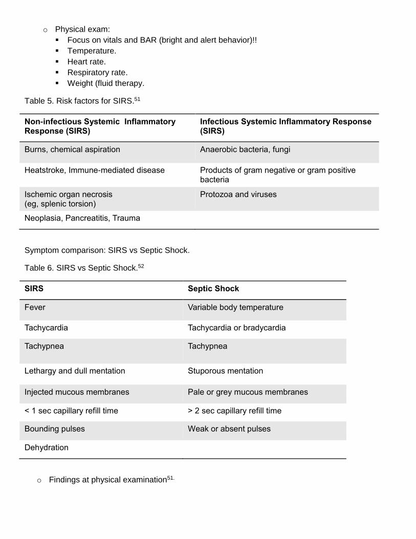
Table 5. Risk factors for SIRS.51
Non-infectious Systemic Inflammatory Response (SIRS)
Infectious Systemic Inflammatory Response (SIRS)
Burns, chemical aspiration Anaerobic bacteria, fungi
Heatstroke, Immune-mediated disease Products of gram negative or gram positive bacteria
Ischemic organ necrosis (eg, splenic torsion)
Protozoa and viruses
Neoplasia, Pancreatitis, Trauma
Symptom comparison: SIRS vs Septic Shock.
Table 6. SIRS vs Septic Shock.52
SIRS Septic Shock
Fever Variable body temperature
Tachycardia Tachycardia or bradycardia
Tachypnea Tachypnea
Lethargy and dull mentation Stuporous mentation
Injected mucous membranes Pale or grey mucous membranes
< 1 sec capillary refill time > 2 sec capillary refill time
Bounding pulses Weak or absent pulses
Dehydration
o Findings at physical examination51.

Table 7. Findings at physical examination: comparison Dogs vc Cats
Findings Dogs Cats
Early or hyper-dynamic phase Bounding pulses Brick red mucous membranes Fever Tachycardia Tachypnea
Yes Yes Yes Yes Yes
No No Yes No Yes
Late phase or advanced disease progression Hypotension Hypothermia Pale mucous membranes Weak pulses
Yes Yes Yes Yes
Yes Yes Yes Yes
Laboratory work53
o Venous blood gas analysis (BGA) and/or lactate.
Send out for bacterial and antibiotic sensitivity.
o Complete blood count (CBC).
o Biochemistry profile.
Stat if you are equipped.
o Urinalysis.
o Clotting profile.
PT, aPTT.
+/- fibrinogen .
+/- D-dimers, fibrin degradation products (FDPs.)
o Which samples can we use for lab work?
Blood.
Bronchoalveolar lavage fluid or endotracheal/transtracheal wash fluid
Joint fluid.
Peritoneal effusion (increased fluid entering the cavity or decreased removal).
Pleural effusion.
Urine.
Treatment.
The Bundle Concept
o Survival rates have improved through the use of treatment bundles.
o “Bundle of care is a group of therapies that, when instituted together, result in better outcomes
than if each individual component were to be implemented alone.”54
o Bundles have proven efficacious in reducing sepsis mortality.55
o Brief bundle components.56
Lactate-
Samples for cultures-
Early antibiotic administration-
Hypotension treatment with fluids and vasopressors-
Target central venous pressure and ScvO2 (oxygen debt)-
Overview
o Initial hemodynamic stabilization.
o Alleviating the underlying cause.
o Intensive care support.
Organization:
o Initial resuscitation (1-3 hours) and fluids.
o Antibiotic therapy, debridement and blood Cultures.
o Long term management (>3 hours)
Key points on treatment and resuscitation and of SIRS, sepsis and septic shock
o Time: 0 or immediate as patient checks inn
Identify
Confirm suspicion
o Time: 20 min?
Resuscitate & Reassess
O2, IV fluids, antibiotics
o Time: 30 min?
Investigate and confirm suspicion
o Time: 1 hr?
Disposition
Set limitations
Surgery
Pt evolution
Hospitalize, ICU
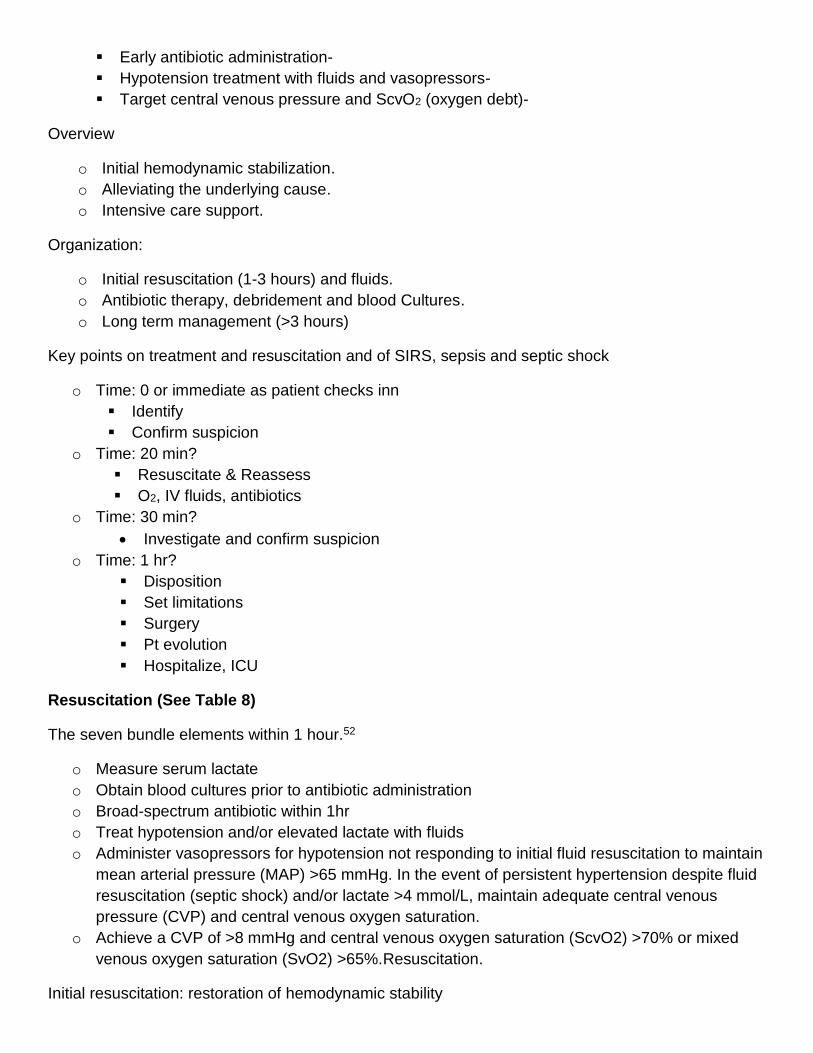
Resuscitation (See Table 8)
The seven bundle elements within 1 hour.52
o Measure serum lactate
o Obtain blood cultures prior to antibiotic administration
o Broad-spectrum antibiotic within 1hr
o Treat hypotension and/or elevated lactate with fluids
o Administer vasopressors for hypotension not responding to initial fluid resuscitation to maintain
mean arterial pressure (MAP) >65 mmHg. In the event of persistent hypertension despite fluid
resuscitation (septic shock) and/or lactate >4 mmol/L, maintain adequate central venous
pressure (CVP) and central venous oxygen saturation.
o Achieve a CVP of >8 mmHg and central venous oxygen saturation (ScvO2) >70% or mixed
venous oxygen saturation (SvO2) >65%.Resuscitation.
Initial resuscitation: restoration of hemodynamic stability

o Aims of the goal-directed therapy.
Central venous pressure.
Mean arterial pressure.
o Place an IV catheter.
Cephalic or saphenous initially.
o Administer isotonic crystalloid boluses.
LRS, Plasmalyte-A, Plasmalyte-148, Normosol-R.
20-25 mL/kg IV over 15 minutes.
o Re-assess perfusion parameters.
o Continue until perfusion restored up to 80-100 mL/ kg.
o Best to use balanced electrolyte solutions (NaCl reduces renal flow).
Table 8. Response to Fluid Resuscitation.
Parameter Target
Heart rate 80-140 bpm
Respiratory rate 18-24/min
Pulses Palpable femoral & dorsal pedal
Systolic BP 100-120 mmHg
Mean BP 70-80 mmHg
Lactate < 2 mmol/L
Urine output > 1 mL/kg/hour
Mentation Responsive
Stabilization.57
Patients with SIRS/sepsis may be resuscitated and supported with one or more of the following fluids:
o Crystalloids and blood component therpy: Isotonic, hypertonic or synthetic
o Isotonic crystalloids (very important in treatment).
Pts with severe cardiovascular issues: administer and repeat small 10 to 20 mL/kg boluses
and monitor pt response to each bolus.
o Important: do not overload leading to pulmonary edema.
o Monitor improvement: pulse quality, decreased lactate level decreased heart rate, improved
mentation.
o Synthetic colloids.
Useful in pts with SIRS/sepsis, especially if they are hypo-proteinemic.
Large molecules that not leave the vascular space, help pull fluid to, and keep it within, the
vascular space.

o Hydroxyethyl starch solutions –
20 mL/kg Q 24 H in dogs and cats.
dogs should be delivered in 5-mL/kg increments up to 20 mL/kg.
cats, 3- to 5-mL/kg increments up to 10 mL/kg.
Infusion rate should be constant to 1 to 2 mL/kg/H can be administered to increase
oncotic. pressure in stable hypo-proteinemic patients.
o Blood products
Red blood cell (RBC) transfusion.
Recent blood loss (or RBC lysis).
Requirement for general anesthesia and surgery, packed cell volume (PCV) below 25% to
30%.
o Frozen products.
Evidence of bleeding, prolonged clotting times.
o Lyophilized Products (albumin).
Severely hypoalbuminemic with concurrent hypovolemia and hypotension.
Doses.
Packed RBCs: 10 to 15 mL/kg.
Fresh whole blood: 20 to 25 mL/kg.
Fresh frozen plasma: 15 mL/kg.
Lyophilized Albumin (canine): 800-884 mg/kg 6 hrs.
Antibiotics.
o Key points:
Early empiric therapy will improve survival rates.
Initiate broad spectrum while waiting blood culture results.
Selected antibiotics should be effective against gram-positive, gram-negative, and
anaerobic bacteria.
o Typical first line:
Ampicillin/Unasyn + Amikacin.
Ampicillin/Unasyn + Enrofloxacin.
Cefazolin + Cefotaxime.
Cefoxitin.
Clindamycin + Enrofloxacin.
o How long to treat: 1-2 weeks

Table 9. Antibiotics, dosages and spectrum.
Drug Dose Spectrum
Amikacin 15-30 mg/kg IB q24 h Gram Pos: + Gram Neg: ++
Ampicillin Unasyn
20-30 mg/kg q 8 h Gram Pos: + Gram Neg: + Anaerobes: +
Cefazolin 20-30 mg/kg q 8 h Gram Pos: + Gram Neg: ± Anaerobes: ±
Cefoxitin 20-30 mg/kg q 6-8 h Gram Pos: + Gram Neg: + Anaerobes: +
Clindamycin 11-22 mg/kg q 12 h Gram Pos: + Gram Neg: + Anaerobes: +
Enrofloxacin 10-20 mg/kg q 24 h Gram Pos: ± Gram Neg: ++
Metronidazole 10-15 mg/kg IV q 12 h Anaerobes ++
Monitoring the patient.
o Sepsis management bundle.52
o Vital signs.
o Blood pressure.
o ECG.
o Pulse oximetry.
o Lab work.
PCV, TP, BGA, Lactate, Electrolytes q 6-12 h.
o Pain score.
o Nursing
Change positioning rotate recumbency.
Passive range of motion.
Head above bed 30o.
sternal, or semi-sternal positioning.
Nebulization & coupage if pneumoni.
Wound/incision management.

Prognosis.58,59
o Overall mortality = 47%
o Dogs without MODS = 25%
o Dogs with MODS = 70%
o ScvO2 and base deficit are useful in predicting the prognosis of dogs with septic shock
o Animals with a higher ScvO2 and lower base deficit at admission to the ICU
o have a lower probability of death.
Conclusions.
o SIRS and Sepsis can be very serious conditions with a cautious to poor prognosis.
o Rapid diagnosis and and treatment with fluid resuscitation and antibiotics are crucial.
o If required and ASAP, surgical intervention or other source to control underlying conditions.
o Post-operative care and monitoring are intensive and expensive.
o The development of septic shock and requirement for vasopressors are poor prognostic
indicators.
o Early Goal-Oriented-Therapy (EGOT) according to bundles may improve survival rates.
What do we have to keep in mind if we suspect sepsis?
o Measure lactate level and blood sample for cultures.
o Administer broad-spectrum antibiotics, and begin rapid administration of crystalloids.
o Apply vasopressors if patient is hypotensive or after fluid resuscitation.
References
1 Rhee C, Dantes R, Epstein L, et al. Incidence and Trends of Sepsis in US Hospitals Using Clinical vs Claims Data, 2009-2014. JAMA. 2017;318(13):1241–1249. doi:10.1001/jama.2017.13836 2 Torio et al. https://www.hcup-us.ahrq.gov/reports/statbriefs/sb160.jsp. Accessed on 10/09/2018 3 de Laforcade A et al. J Hemostatic changes in dogs with naturally occurring sepsis. Vet Intern Med. 2003;17(5):674-9 4 https://www.cdc.gov/sepsis/datareports/index.html. Accessed on 10/12/2018 5 American College of Chest Physicians/Society of Critical Care Medicine Consensus Conference: Definitions for sepsis and organ failure and guidelines for the use of innovative therapies in sepsis". Critical Care Medicine 20 (6): 864–74. 1992. 6 Singer M, Deutschman CS, Seymour CW, et al. The Third International Consensus Definitions for Sepsis and Septic Shock (Sepsis-3). JAMA. 2016;315(8):801-810. doi:10.1001/jama.2016.0287. 7 Reinhart et al. New Approaches to Sepsis: Molecular Diagnostics and Biomarkers Clin. Microbiol. Rev. 2012; 25; 609-634 8 Vijay K. Toll-like receptors in immunity and inflammatory diseases: Past, present, and future. Int Immunopharmacol. 2018 Jun;59:391-412. doi: 10.1016/j.intimp.2018.03.002. Epub 2018 May 4. 9 Mitchell J. et al.. Critical role of toll-like receptors and nucleotide oligomerisation domain in the regulation of health and disease. Journal of Endocrinology 2007;193:323-330. 10 Kremer et al. Complicated Pericarditis Understanding Risk Factors and Pathophysiology to Inform Imaging and Treatment. J. AM. Col. Cardiology 2016; 68: 21

11Sharma D, Kanneganti TD The cell biology of inflammasomes: Mechanisms of inflammasome activation and regulation. J Cell Biol. 2016 Jun 20;213(6):617-29. doi: 10.1083/jcb.201602089. 12 Boomer et al. The changing immune system in sepsis. Virulence 2014, 5:1, 45–56 13 Kojic et al. Are there new approaches for diagnosis, therapy guidance and outcome prediction of sepsis. World Exp Med.2015; 5: 50-63 14 Hotchkiss RS, et al. Immunosuppression in sepsis: a novel understanding of the disorder and a new therapeutic approach. Lancet Infect Dis 2013; 13: 260–268 15 Lerman Y, Kim M. Neutrophil Migration under normal and sepsis conditions. Cardiovasc Hematol Disord Drug Targets. 2015 ; 15: 19–28 16 Trevelin SC, Alves‑Filho JC, Sônego F, Turato W, Nascimento DC, Souto FO, et al. Toll‑ like receptor 9 activation in neutrophils impairs chemotaxis and reduces sepsis outcome. Crit Care Med
2012;40:2631‑7. doi: 10.1097/CCM.0b013e318258fb70 17 Souza et al. Microparticles: markers and mediators of sepsis-induced microvascular dysfunction, immunosuppression, and AKI. Kidney 2015, 87;6: pp1100–1108 18 Angelillo-Scherrer A. Leukocyte-Derived Microparticles in Vascular Homeostasis. Circulation Research. 2012;110:356-369 19 Lerman Y, Kim M. Neutrophil Migration under normal and sepsis conditions. Cardiovasc Hematol Disord Drug Targets. 2015 ; 15(1): 19–28. 20 Miyata T, Fan X. A second hit for thrombotic microangiopathies (TMAs). Blood 2012 Vol. 120;6 21 Lipinska-Gediga M . Neutrophils, NETs, NETosis — old or new factors in sepsis and septic shock. Anaesthesiol Intensive Therapy 2017;49:235–240. 22 Dewitte et al. Blood platelets and sepsis pathophysiology: A new therapeutic prospect? Ann. Intensive Care 2017: 7:115 23 Liang Yao Li J, Zarbock A, Hidalgo A. Platelets as autonomous drones for hemostatic and immune surveillance. J. Exp. Med. 2017;214:2193–2204 24 Yeaman MR. Platelets in defense against bacterial pathogens. Life Sci. 2010;67:525–544 25 de Stoppelaar. SF, van t Veer C, van der Poll. The role of platelets in sepsis. Thromb Haemost 2014; 112: 666–677 26 Greco. E et al. Platelets and Multi-Organ Failure in Sepsis . Int. J. Mol. Sci. 2017, 18, 2200 27 Adapted from Tisoncik JR et al. Into the Eye of the Cytokine Storm. Microbiol Mol Biol Rev. 2012;76: 16–32. 28 Atkinson AJ, Colburn WA, DeGruttola VG, et al. Biomarkers and surrogate endpoints: preferred definitions and conceptual framework. Clin Pharmacol Ther. 2001;69(3):89–95. 29 Marshall JC et al. Biomarkers of Sepsis. Crit Care Med. 2009;37:2290-8. 30 Christopher MM, O’Neill S. Effect of specimen collection and storage on blood glucose and lactate concentrations in healthy, hyperthyroid and diabetic cats. Vet Clin Pathol 2000;29:22-28. 31 Rand JS, Kinnaird E, Baglioni A, et al. Acute stress hyperglycemia in cats is associated with struggling and increased concentrations of lactate and norepinephrine. J Vet Intern Med 2002;16:123-132. 32 Hughes, D., et al. Effect of sampling site, repeated sampling, pH, and PCO2 on plasma lactate concentration in healthy dogs. Am J Vet Res 1999; 60:521-524. 33 Boag A, Hughes D: Assessment and treatment of perfusion abnormalities in the emergency patient. Vet Clin North Am Small Anim Pract 2005;35:319-342. 34 De Backer D. Lactic Acidosis. Intensive Care Med 2003; 9:699-702 35 Rosenstein PG and Huyghes D. Hyperlactermia. Chapter 56. Hyperlactemia. In: Small Animal Critical Care Medicine, Deborah Silverstein & Kate Hooper (Eds), Elsiever, Missouri, 2015.1st Ed. 36 Jansen TC et al. Early lactate-guided therapy in intensive care unit patients: a multicenter, open-label, randomized controlled trial." Am J Respir Crit Care Med 2010;182: 752-761. 37 Marty P et al. Lactate clearance for death prediction in severe sepsis or septic shock patients during the first 24 hours in Intensive Care Unit: an observational study. Annals of Intensive Care. 2013;3:3

38 Cortinelli S, Seth M, Kellet-Gregory LM . Plasma lactate concentrations in septicperitonitis: A retrospective study of 83 dogs (2007–2012). Journal of Veterinary Emergency and Critical Care 25(3) 2015, pp 388–395 39 Acierno MJ & Mitchell MA. . Evaluation of four point-of-care meters for rapid determination of blood lactate concentrations in dogs. J Am Vet Med Assoc 2007;230:1315–1318 40 Morgenthaler NG et al. Detection of procalcitonin (PCT) in healthy controls and patients with local infection by a sensitive ILMA. Clin Lab. 2002;48(5-6):263-70. 41 Linscheid et al. In Vitro and in Vivo Calcitonin I Gene Expression in Parenchymal Cells: A Novel Product of Human Adipose Tissue. Endocrinology 2003;144:5578–5584 42 Müller B. et al., Ubiquitous Expression of the Calcitonin-I Gene in Multiple Tissues in Response to Sepsis J Clin Endocrinol Metab.2001 86;396-404. 43 Chan T, Gu F. Early diagnosis of sepsis using serum biomarkers Expert Rev Mol Diagn 2011;11:487–96 44 Simon L. et al. Serum procalcitonin and C-reactive protein levels as markers of bacterial infection: a systematic review and meta-analysis.Clin Infect Dis. 2004; 39:206-217. 45 Schuetz P, Christ-Crain M, Wolbers M, et al. “Procalcitonin guided antibiotic therapy and hospitalization inpatients with lower respiratory tract infections: a prospective, multicenter, randomized controlled trial.” BMC Health Svcs Res 2007;7:102 46 Floras ANK et al. Investigation of a Commercial ELISA for the Detection of Canine Procalcitonin. J Vet Intern Med 2014;28:599–602 47 Bonelli F et al. Plasma Procalcitonin Concentration in Healthy Horses and Horses Affected by Systemic Inflammatory Response Syndrome. J Vet Intern Med 2015;29:1689–1691 48 Barton et al. Procalcitonin as a biomarker in equine chronic pneumopathies. BMC Veterinary Research 2016;12:281 49 Troia R et al. Plasma procalcitonin concentrations predict organ dysfunction and outcome in dogs with sepsis. BMC Vet Res. 2018; 14: 111. 50 Levy M, Evans LE, Rhodes A, et al. The Surviving Sepsis Campaign Bundle: 2018 update. Int. Med 2018;44:6 51 Silverstein D. Systemic Inflammatory Response Syndrome & Sepsis Part 1: Recognition & Diagnosis. Today’s Veterinary Practice 2015: January/February. 52 DeClue A. Sepsis and the Systemic Inflammatory Response. In Veterinary Internal Medicine. Diseases of the Dogs and the Cat. Stephen J. Ettinger, Edward C. Feldman, Ettiene Cote (Eds), 8th Editio. Elsieve, St Lois, MO, 2017. Chapter 132, pp554-560 53 Modified from Mittleman E, Otto CM. Sepsis and septic Shock. Chapter 91, pp 472-480. In Small Animal Critical Care Medicine (2ed), Deborah Silverstein & Kate Hoper (eds). Elsiver Sanders, St. Louis, MI. 2015 54 Cinel I, Dellinger RP. Guidelines for severe infections: are they useful? Curr Opin Crit Care. 2006;12:483-8. 55 Nguyen HB, Corbett SW, Steele R et al. Implementation of a bundle of quality indicators for the early management of severe sepsis and septic shock is associated with decreased mortality. Crit Care Med. 2007;35:1105-12. 56 Mittleman E, Otto CM. Sepsis and septic Shock. Chapter 91, pp 472-480. In Small Animal Critical Care treatment-systemic-inflammatory-response-syndrome-sepsis/Accessed on 09/20/2018 57 Silverstein D. https://todaysveterinarypractice.com/systemic-inflammatory-response-syndrome-sepsis-part-2-stabilization-treatment-systemic-inflammatory-response-syndrome-sepsis/Accessed on 09/20/2018 58 Kenney E, et al. Association between outcome and organ system dysfunction in dogs with sepsis: 114 cases (2003-2007). JAVMA 2010; 236:1 59 Conti-Patara A, et al. Changes in tissue perfusion parameters in dogs with severe sepsis/septic
shock in response to goal‐directed hemodynamic optimization at admission to ICU and the relation to outcomeVet Emerg Crit Care 2012; 22: 409–418.