surgical treatment for epstein-barr virus otomastoiditis ... · in this case, surgical...
TRANSCRIPT

J Int Adv Otol 2017; 13(1): 143-6 • DOI: 10.5152/iao.2017.2788
Case Report
INTRODUCTIONThe cause of acquired facial nerve palsy (FNP) in the pediatric population can be classified as infectious, traumatic, malignancy associated, hypertension associated, and idiopathic (Bell’s palsy) (Table 1). Toddlers and preteenagers may be at higher risk for FNP because of infectious and traumatic causes [1].
The main pathogens causing FNP in children are Borrelia burgdorferi (50%), idiopatic or Bell’s palsy (26%), otitis media (OM) (12%), varicella-zoster virus (6%), Herpes simplex virus (4%), and coxsackie (2%) [2]. A thorough research of the literature showed that al-though rare, Epstein–Barr virus (EBV) can also be linked to FNP [3, 4]. Here we present the case of two brothers with an EBV-derived unilateral FNP.
CASE PRESENTATIONA 7-month-old boy (patient A) without previous otologic history was referred to our Ear Nose Throat tertiary referral center for the evaluation of a left-sided unilateral FNP that developed after 2 days of fever. A ventilation tube (VT) was placed in the referring hospital 2 days earlier because of signs of OM with effusion. On examination in our hospital 4 days after onset, House–Brackmann (HB) [5] score grade IV (Table 2), hepatosplenomegaly, and bilateral enlarged cervical glands were observed. Otoscopy showed VT with clear otorrhea, without clinical signs of mastoiditis. A polymerase chain reaction (PCR) on EBV was conducted and confirmed an acute EBV infection.
Contrast-enhanced computed tomographic (cCT) scan (Aquilion ONE ViSION Edition; Toshiba Medical Systems, Otawara, Japan) demonstrated opacification of the mastoid and middle ear as seen in otomastoiditis. Contrast-enhanced magnetic resonance im-aging (cMRI) scan (Siemens Avanto 1,5T, Erlangen, Germany) demonstrated otitis media and mastoiditis, particularly around the apex pars petrosae, with signs of abscess formation. From the day of onset, the patient was pharmacologically treated for 2 days with azithromycin (Zithromax by Pfizer BV, New York City, United States of America), followed by 2 days of ceftriaxone and simul-taneously for 7 days with eardrops consisting hydrocortisone, colistin, and bacitracin (Bacicoline-B by Daleco Pharma BV, Enspijk, The Netherlands).
Based on the diagnosis of EBV-induced mastoiditis complicated by FNP that did not improve with antibiotic treatment, mastoid-ectomy and atticoantrotomy were performed 6 days after onset of FNP. Granulation tissue from the mastoid was positive for EBV.
143
Surgical Treatment for Epstein-Barr Virus Otomastoiditis Complicated by Facial Nerve Paralysis: A Case Report of Two Young Brothers and Review of Literature
We report the case of two young brothers with Epstein–Barr virus (EBV) otomastoiditis complicated by a facial nerve paralysis. The boys, aged 7 months (patient A) and 2 years and 8 months (patient B), were diagnosed with a facial nerve paralysis House–Brackmann (HB) grade IV (A) and V (B). After unsuccessful pharmacological treatment, patient A underwent mastoidectomy and atticoantrotomy and patient B underwent a trans-mastoidal surgical decompression of the facial nerve. They recovered to HB grades I and II facial nerve palsy (FNP), respectively. Although rare and relatively unknown, EBV should be considered in the differential diagnosis of children with FNP of unknown cause. Surgical intervention may be a viable therapy with good recovery.
KEYWORDS: Facial paralysis, otitis media, Epstein–Barr virus infections, surgical decompression
Evelien van Eeten, Hubert Faber, Dirk KunstDepartment of Otolaryngology, Radboud University Medical Center, Nijmegen, Netherlands
Corresponding Address: Hubert Faber E-mail: [email protected]
Submitted: 26.06.2016 Revision received: 11.11.2016 Accepted: 08.02.2017©Copyright 2017 by The European Academy of Otology and Neurotology and The Politzer Society - Available online at www.advancedotology.org
After the surgical treatment, patient A clinically recovered to a HB II, with minimal asymmetry and eye closure after 1 month, and HB I 6 months later.
Two months later, his brother (patient B), aged 2 years and 8 months, showed a left-sided unilateral FNP grade V HB. FNP was preceded by 2 days of otalgia and fever. A secondary referral center placed a VT that showed thickened mucosa without signs of acute OM. He only had a medical history of adenotonsillectomy because of obstructive sleep apnea syndrome.
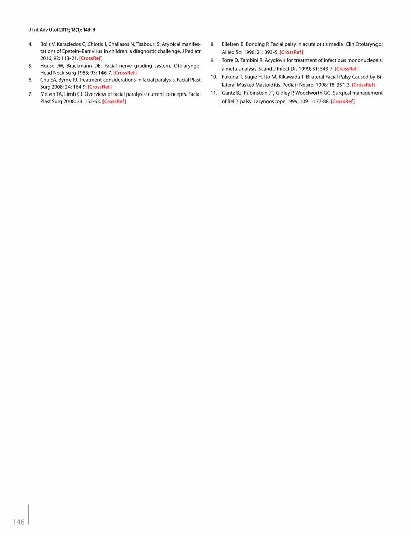
Polymerase chain reaction on EBV that was conducted consider-ing the disease course of his brother indicated an acute EBV in-fection. cCT showed otomastoiditis, with complete opacification of the mastoid and middle ear. cMRI showed slight coloration of the geniculate ganglion on the tympanal and mastoidal part of the facial nerve (Figure 1). Otoacoustic emissiontest reported bilateral normal inducible emissions. Two EMGs 14 and 37 days after onset found a severe neuropathy with a non-inducible musculus orbicu-laris oculi and musculus nasalis. Over 90% difference in compound
muscle action potential (CMAP) amplitude was found in relation to the right side.
The patient was initially pharmacologically treated for 5 days with amoxicillin/clavulanic acid. After PCR on EBV was positive, treatment was switched to 5 days ceftriaxone. During the same period, he was treated with glucocorticoids and eardrops consisting hydrocorti-sone, colistin, and bacitracin (Bacicoline-B by Daleco Pharma BV, En-spijk, The Netherlands).
After 6 weeks, no improvement of FNP was seen, and transmastoidal surgical decompression of the facial nerve from geniculate ganglion to the stylomastoid foramen was performed. During surgery, a pale white facial nerve was observed that regained vascularization after decom-pression (Video 1. See corresponding video/movie images at http://www.advanceotology.org). The postoperative course was complicated by a bacterial mastoiditis that needed surgery. After the second sur-gery, Patient B recovered to a HB III in 3 weeks and HB II after 6 months, with movement and only slight asymmetry of the mouth. We obtained written informed consent from the parents of the brothers.
144
J Int Adv Otol 2017; 13(1): 143-6
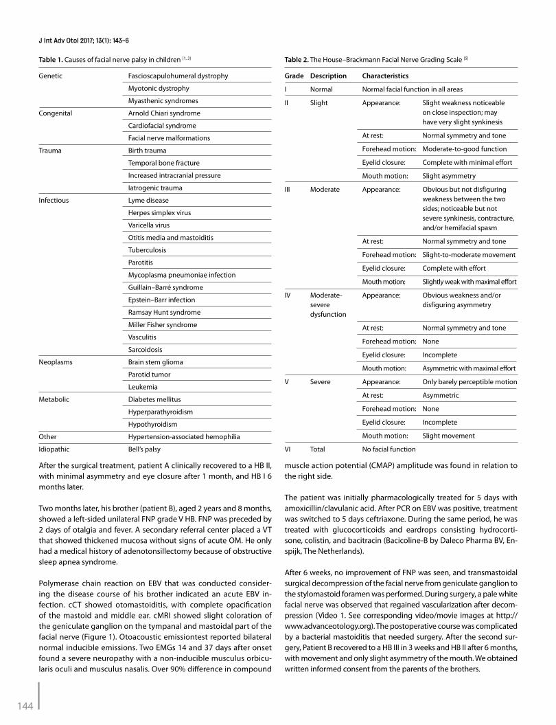
Table 1. Causes of facial nerve palsy in children [1, 3]
Genetic Fascioscapulohumeral dystrophy
Myotonic dystrophy
Myasthenic syndromes
Congenital Arnold Chiari syndrome
Cardiofacial syndrome
Facial nerve malformations
Trauma Birth trauma
Temporal bone fracture
Increased intracranial pressure
Iatrogenic trauma
Infectious Lyme disease
Herpes simplex virus
Varicella virus
Otitis media and mastoiditis
Tuberculosis
Parotitis
Mycoplasma pneumoniae infection
Guillain–Barré syndrome
Epstein–Barr infection
Ramsay Hunt syndrome
Miller Fisher syndrome
Vasculitis
Sarcoidosis
Neoplasms Brain stem glioma
Parotid tumor
Leukemia
Metabolic Diabetes mellitus
Hyperparathyroidism
Hypothyroidism
Other Hypertension-associated hemophilia
Idiopathic Bell’s palsy
Table 2. The House–Brackmann Facial Nerve Grading Scale [5]
Grade Description Characteristics
I Normal Normal facial function in all areas
II Slight Appearance: Slight weakness noticeable on close inspection; may have very slight synkinesis
At rest: Normal symmetry and tone
Forehead motion: Moderate-to-good function
Eyelid closure: Complete with minimal effort
Mouth motion: Slight asymmetry
III Moderate Appearance: Obvious but not disfiguring weakness between the two sides; noticeable but not severe synkinesis, contracture, and/or hemifacial spasm
At rest: Normal symmetry and tone
Forehead motion: Slight-to-moderate movement
Eyelid closure: Complete with effort
Mouth motion: Slightly weak with maximal effort
IV Moderate- Appearance: Obvious weakness and/or severe disfiguring asymmetry dysfunction
At rest: Normal symmetry and tone
Forehead motion: None
Eyelid closure: Incomplete
Mouth motion: Asymmetric with maximal effort
V Severe Appearance: Only barely perceptible motion
At rest: Asymmetric
Forehead motion: None
Eyelid closure: Incomplete
Mouth motion: Slight movement
VI Total No facial function

DISCUSSIONEpstein-Barr virus is a relatively unknown cause of FNP; it is inciden-tally reported, and case reports focus on diagnosis rather than treat-ment. Consequently, literature on treatment of EBV-induced FNP is inconclusive and mainly based on expert opinions. Choosing the most appropriate treatment should be guided by etiology and severity of FNP. Each patient needs an individual approach based on the patient’s condition, duration and cause of paralysis, physical finding, EMG, au-diogram, anatomical status of the facial nerve, and expectations [6].
A conservative approach, potentially supplemented with EMG fol-low-up, could be indicated if the facial nerve is anatomically intact and EMG shows less than a 90% difference in CMAP. Recovery of an infection-derived FNP usually takes less than 1 month. Nearly always substantial (HB II) to complete (HB I) recovery is attained [1]. In other EBV-associated neurological complications, the prognosis is good; therefore, intervention is not ushered. This could also be the case for EBV-associated FNP [6].
Pharmacological treatment can be performed using corticosteroids, antibiotics, or antiviral medication. Inflammation and associated ede-ma are considered to be one of causes of FNP. Reducing this edema with corticosteroids accelerates spontaneous clinical improvement of FNP. Controversy remains on the benefit of steroid treatment use in children as clear studies are not available [7]. If a bacterial infection, such as OM, is the cause of FNP, antibiotics should be administered. OM-associated FNP is less prevalent since the antibiotic era. In the pre-antibiotic era 0.5%–0.7% of OM were complicated by FNP be-cause of persistent inflammation of the fallopian canal. Currently, this complication has become exceptional with an incidence of 0.005% [8]. When FNP is caused by EBV, treatment with antiviral therapy is ques-tionable. A meta-analysis showed no clinical benefit comparing the treatment of EBV infections with acyclovir and placebo treatment [9].
Surgical intervention could be indicated when a conservative or pharmacological approach is not sufficient. The rapid improved vas-cularization of the facial nerve after decompression suggests that a decreased vascular perfusion might be an important factor in the pathogenesis of FNP; therefore, surgical decompression is necessary
[10]. As a guide to determine when decompression is warranted, EMG measurements can be used. Patients with less than 90% reduction are likely to return to a HB grade I or II. If patients have more than 90% reduction of CMAP within 14 days, they are likely to have a HB grade III or IV. In this case, surgical decompression provided significant im-
provement to normal or near-normal recovery. The best results in pa-tients with Bell’s palsy with more than 90% reduction in CMAP within 14 days of onset were achieved if surgery was performed within 2 or 3 weeks after onset of FNP [11].
There are different surgical approaches to establish facial nerve de-compression. The different approaches are based on multiple surgical techniques, such as the transmastoid approach reaching the tympanic and mastoid segments and the geniculate ganglion. The middle fossa approach can be added to reach the labyrinthine segment [7].
The most suitable treatment for the pediatric population with an EBV-de-rived FNP is still unknown. In our cases, the question remains whether surgical treatment or natural course caused the clinical improvement of FNP. Larger studies comparing the different treatments of FNP with the natural course are needed in order to provide a clear answer.
Informed Consent: Written informed consent was obtained from the parents of the patients who participated in this study.
Peer-review: Externally peer-reviewed.
Author Contributions: Concept - E.V.E., H.F.; Design - E.V.E., H.F.; Supervision - H.F., D.K.; Resources - E.V.E.; Materials - E.V.E.; Data Collection and/or Pro-cessing - E.V.E., H.F.; Analysis and/or Interpretation - E.V.E., H.F., D.K.; Literature Search - E.V.E., H.F., D.K.; Writing Manuscript - E.V.E., H.F., D.K.; Critical Review - H.F., D.K.
Acknowledgements: We thank the two brothers and their parents for their permission to use this data.
Conflict of Interest: No conflict of interest was declared by the authors.
Financial Disclosure: The authors declared that this study has received no financial support.
Video 1. Facial nerve decompression.
REFERENCES1. Lorch M, Teach SJ. Facial nerve palsy: etiology and approach to diagnosis
and treatment. Pediatr Emerg Care 2010; 26: 763-9. [CrossRef]2. Cook SP, Macartney KK, Rose CD, Hunt PG, Eppes SC, Reilly JS. Lyme dis-
ease and seventh nerve paralysis in children. Am J Otolaryngol 1997; 18: 320-3. [CrossRef]
3. Pavlou E, Gkampeta A, Arampatzi M. Facial nerve palsy in childhood. Brain Dev 2011; 33: 644-50. [CrossRef]
145
Eeten et al. Surgical Treatment for Facial Paralysis in Epstein-Barr Virus Otomastoiditis
Figure 1. a, b. MRI of the skullbase shows slight enhanced signal at the geniculate ganglion on the left side. A subtle finding found on the axial and coronal slides
a b
4. Bolis V, Karadedos C, Chiotis I, Chaliasos N, Tsabouri S. Atypical manifes-tations of Epstein–Barr virus in children: a diagnostic challenge. J Pediatr 2016; 92: 113-21. [CrossRef]
5. House JW, Brackmann DE. Facial nerve grading system. Otolaryngol Head Neck Surg 1985; 93: 146-7. [CrossRef]
6. Chu EA, Byrne PJ. Treatment considerations in facial paralysis. Facial Plast Surg 2008; 24: 164-9. [CrossRef]
7. Melvin TA, Limb CJ. Overview of facial paralysis: current concepts. Facial Plast Surg 2008; 24: 155-63. [CrossRef]
8. Ellefsen B, Bonding P. Facial palsy in acute otitis media. Clin Otolaryngol
Allied Sci 1996; 21: 393-5. [CrossRef]9. Torre D, Tambini R. Acyclovir for treatment of infectious mononucleosis:
a meta-analysis. Scand J Infect Dis 1999; 31: 543-7. [CrossRef]10. Fukuda T, Sugie H, Ito M, Kikawada T. Bilateral Facial Palsy Caused by Bi-
lateral Masked Mastoiditis. Pediatr Neurol 1998; 18: 351-3. [CrossRef]11. Gantz BJ, Rubinstein JT, Gidley P, Woodworth GG. Surgical management
of Bell’s palsy. Laryngoscope 1999; 109: 1177-88. [CrossRef]
146
J Int Adv Otol 2017; 13(1): 143-6