surgery of the cardiovascular system
TRANSCRIPT
Surgery of the
Cardiovascular SystemDr. Pınar CAN

Anatomy
The right atrium and left atrium are divided by
an atrial septum and receive blood from the
systemic and pulmonary venous circulations,
respectively.
Blood is carried from the systemic circulation to
the right atrium by the cranial and caudal vena
cavae.

Blood is carried to the left atrium by multiple
pulmonary veins located on the dorsal aspect of
the heart.
Blood flows from the right and left atrium into
their respective ventricles through the right
atrioventricular (tricuspid) and left
atrioventricular (mitral) valves

The right ventricle pumps blood to the
pulmonary arterial circulation via the main
pulmonary artery or trunk.
The pulmonic (pulmonary) valve is situated
between the right ventricle and main pulmonary
artery.
The left ventricle pumps blood to the systemic
arterial circulation via the aorta.
The aortic valve is located between the left
ventricle and aorta.


The pericardium is a thick,
two-layer sac composed of
outer fibrous and inner serous
layers.
The pericardial cavity is
located between two layers
(visceral and parietal) of
serous pericardium and
normally contains a small
amount of fluid.

PREOPERATIVE CONSIDERATIONS
Cardiac surgery includes procedures performed
on the ventricles, atria, cardiac valves, or great
vessels.
Some cardiac surgeries are performed on a
closed beating heart, but others are open
procedures in which a major cardiac structure
must be opened to accomplish the repair.
The latter require a strategy to arrest or support
the circulation during the repair.

Short procedures (<4 minutes) can be performed with
venous inflow occlusion and brief circulatory arrest.
Longer open cardiac repairs require cardiopulmonary
bypass to support cardiopulmonary function during
surgery.

Animals requiring cardiac surgery often have prior
cardiovascular compromise that should be corrected or
controlled medically when possible before anesthetic
induction.
Congestive heart failure (CHF), particularly pulmonary
edema, should be managed with diuretics (e.g.,
furosemide) and angiotensin-converting enzyme (ACE)
inhibitors (e.g., enalapril, benazepril, lisinopril) and an
inodilator (pimobendan) before surgery.
Ventricular tachycardia should be suppressed before
surgery with class I antiarrhythmic drugs (i.e., lidocaine
and procainamide).

All animals should undergo a
complete echocardiographic
evaluation before cardiac surgery;
an incomplete or inaccurate
diagnosis can have devastating
consequences.
With the advent of Doppler
echocardiography, cardiac
catheterization is no longer
routinely necessary before cardiac
surgery.

ANESTHESIA
Anesthesia of the patient with cardiac compromise has risks that vary depending on the cause of the underlying disease.
Preanesthetic medication is appropriate for most animals undergoing cardiac surgery.
Parenteral opioids (i.e., hydromorphone, butorphanol, buprenorphine, and fentanyl) induce sedation with minimal cardiovasculareffects; however, all opioids can producerespirator depression and/ or bradycardia.

α2-Agonists (e.g., demedetomidine) and
acepromazine should be avoided in cardiac
patients owing to significant alterations in
hemodynamic parameters associated with their
administration.
Anticholinergics (i.e., atropine and glycopyrrolate)
should be administered only as needed.
Benzodiazepines (e.g., diazepam 0.2 mg/kg,
midazolam 0.2 mg/kg) have minimal
cardiopulmonary effects and enhance sedation
when given alone or combined with opioids.

Induction of anesthesia should be undertaken
with caution in animals with cardiopulmonary
compromise.
Propofol produces rapid induction but causes
essentially the same cardiovascular compromise
as thiobarbiturates.
The addition of fentanyl decreases propofol
requirements in healthy dogs with minimal
alteration in cardiovascular parameters.

A balanced anesthetic approach using benzodiazepine,
opioids, and modest amounts of inhalant is generally
much safer.
Thoracic surgery always requires controlled ventilation.
Mechanical ventilation should achieve a tidal volume of 6
to 10 ml/kg of body weight at an inspiratory pressure of
less than 20 cm of water.
PATENT DUCTUS ARTERIOSUS
The ductus arteriosus is a fetal vessel that connects the main pulmonary artery and the descending aorta.
During development, it shunts blood away from collapsed fetal lungs.
Normally, it closes shortly after birth during the transition rom fetal to extrauterine life.
Continued patency of the ductus arteriosus for longer than a few days after birth is called patent ductus arteriosus (PDA).

PDA is one of the most common congenital heart
defects of dogs; it occurs infrequently in cats. PDA
typically causes a left-to-right shunt that results in
volume overload of the left ventricle and produces
left ventricular dilation.
Progressive left ventricular dilation distends the
mitral valve annulus, causing secondary
regurgitation and additional ventricular overload.
This severe volume overload leads to left-sided CHF
and pulmonary edema, usually within the first year
of life.

SURGICAL TREATMENT
Intravascular coils, vascular plugs, and duct occluders are now used routinely for closure of patent ductus arteriosus.
Standard surgical correction of left-to-right patent ductus arteriosus is accomplished by ligation of the ductus arteriosus
Intravascular coil
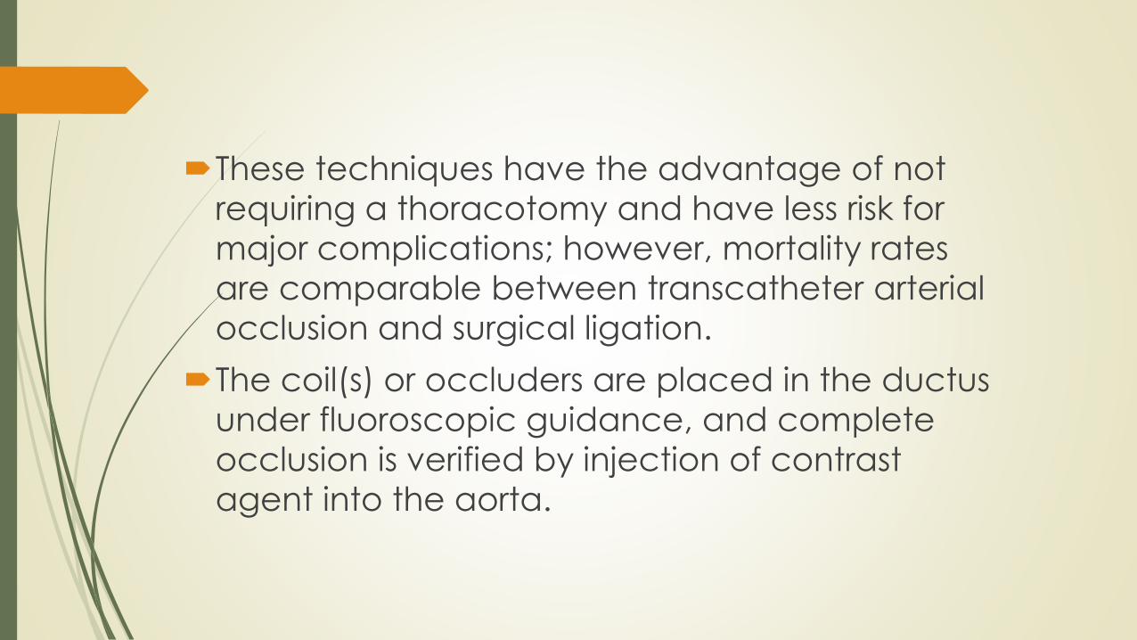
These techniques have the advantage of not
requiring a thoracotomy and have less risk for
major complications; however, mortality rates
are comparable between transcatheter arterial
occlusion and surgical ligation.
The coil(s) or occluders are placed in the ductus
under fluoroscopic guidance, and complete
occlusion is verified by injection of contrast
agent into the aorta.
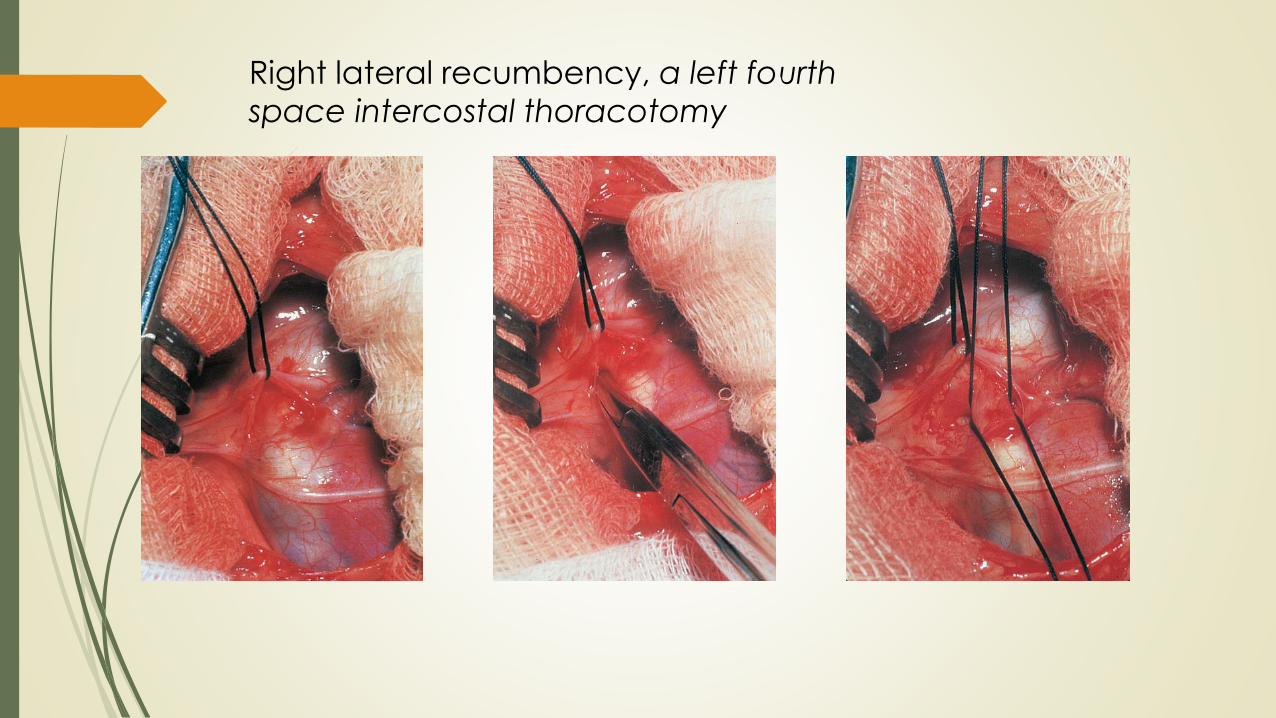
Right lateral recumbency, a left fourth
space intercostal thoracotomy

PULMONIC STENOSIS
Pulmonic stenosis (PS) is a congenital narrowing
of the pulmonic valve, pulmonary artery, or right
ventricular outflow tract.
Valvular stenosis may be simple, consisting of
incomplete separation of valve leaflets, or it be
due to valve dysplasia characterized by a
hypoplastic valve annulus and thickened
immobile valve leaflets.

English Bulldogs, Scottish Terriers, Wirehaired Fox
Terriers, Beagles, Miniature Schnauzers, Cocker
Spaniels, Samoyeds, and Mastiffs are at increased
risk for developing PS.
Young animals with PS are often asymptomatic.
Advanced cases may present with exercise
intolerance, syncope, or abdominal distention from
ascites.
SURGICAL TREATMENT
Therapy for PS is based on its degree of severity and the type of lesion present.
Severity is judged by the presence of signs, the extent of right ventricular hypertrophy, and the magnitude of the systolic pressure gradient.
Animals with PS that have no signs, mild hypertrophy, and a pressure gradient less than 50 mmHg generally do not require surgical intervention.
Surgical options for correction of PS include valve dilation and patch-graft valvuloplasty. With the advent of balloon valvuloplasty, operative valve dilation is seldom indicated.

TETRALOGY OF FALLOT
Tetralogy of Fallot is a complex congenital heart defect that consists of PS, VSD, a dextropositioned overriding aorta, and right ventricular hypertrophy.
most common congenital heart defect that causes cyanosis in small animals.
It occurs in cats and a variety of canine breeds.
A shortened life span is expected in these animalsbecause of complications of hyperviscosity-inducethromboembolism or sudden death (caused by polycythemia).


Breeds most commonly reported to have tetralogy of
Fallot include Keeshonden, English Bulldogs, Poodles,
Schnauzers, Terriers, Collies, and Shelties.
Clinical findings at presentation include moderate to
severe exercise intolerance, exertional tachypnea,
collapse, and syncope.
SURGICAL TREATMENT
Surgery should be considered for severely cyanotic
animals to lessen clinical signs and prolong life.
Animals with resting arterial oxygen saturation less than
70% should be considered candidates for surgery.
Palliative surgeries for tetralogy include isolated
correction of the PS or creation of a systemicto-
pulmonary shunt (e.g., Blalock-Taussig shunt)
Several types of systemic-to-pulmonary shunt
have been used to palliate tetralogy of Fallot.
A modified Blalock-Taussig shunt is accomplished
by harvesting the left subclavian artery as a free
autogenous graft and placing it between the
aorta and the main pulmonary artery.


PERICARDIAL EFFUSION ANDPERICARDIAL CONSTRICTION
The pericardium is a fibroserous envelope that
encompasses the heart and great vessels.
Pericardial effusion is an abnormal accumulation of
fluid within the pericardial sac.
Cardiac tamponade refers to the decompensated
phase of cardiac compression that results from an
unchecked rise in intrapericardiac fluid pressure.

Pericardial constriction results from restrictive fibrosis of
the parietal and/or visceral pericardium that interferes
with diastolic function of the heart.
Diseases that affect primarily the pericardium account
for approximately 1% of cardiovascular disease.
Pericardial diseases are uncommon in cats; pericardial
effusion is most often related to CHF

The most common of which are those resulting
in the accumulation of pericardial effusion.

Pericardial effusion can be transudative,
exudative (inflammatory), or
sanguineous.
Causes of pericardial transudation
include right-sided CHF,
hypoproteinemia, and incarceration of
a liver lobe within the pericardial cavity.
Transudative pericardial effusions are
usually subclinical.

Infectious pericarditis is an uncommon cause of
pericardial effusion in dogs and cats, usually
producing a purulent or fibrinous exudate.
Bacterial pericarditis can arise from bite wounds
to the thorax, migrating foreign bodies, or
hematogenous seeding.
Feline infectious peritonitis and toxoplasmosis
are potential causes of feline inflammatory
effusions.
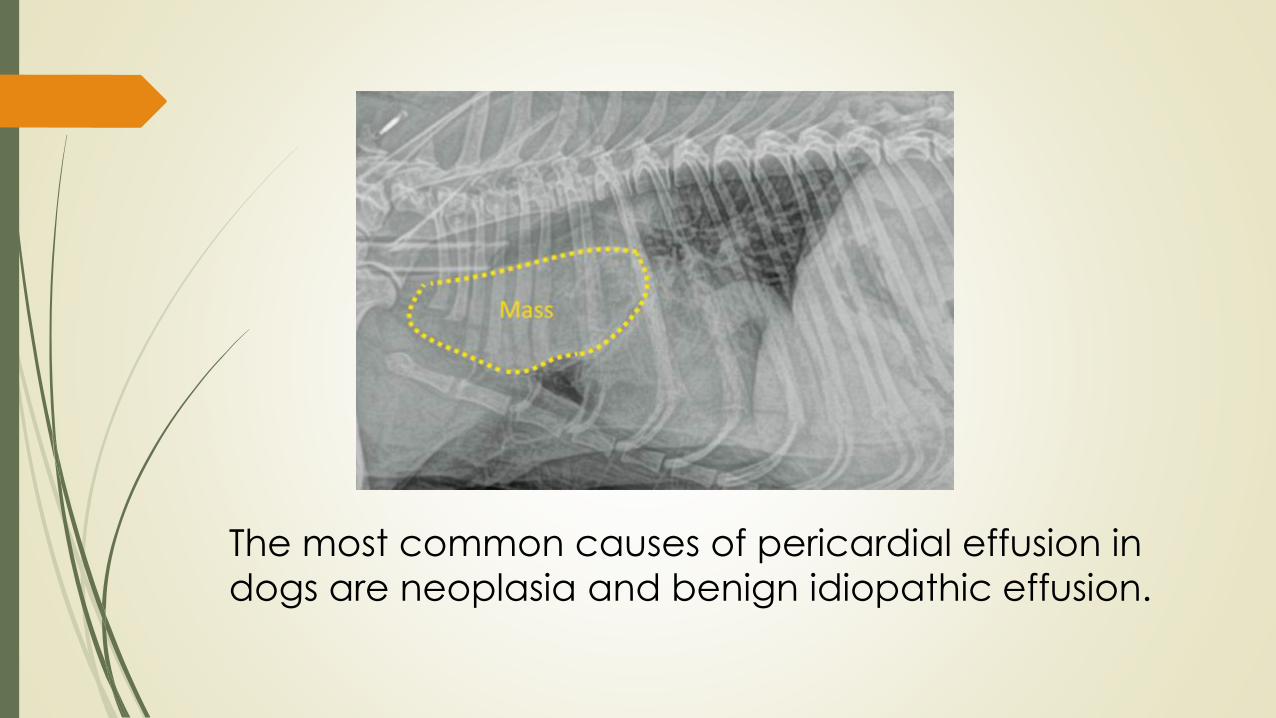
The most common causes of pericardial effusion in
dogs are neoplasia and benign idiopathic effusion.

Idiopathic (benign) pericardial effusions are common in
dogs; this condition has not been reported in cats.
Idiopathic benign and neoplastic pericardial effusions are
more commonly observed in large and giant breed dogs.
Clinical signs; weakness, lethargy, exercise intolerance,
and/or collapse. Patients often have right-sided
congestion, ascites, and/or pleural effusion.
The most common owner complaint with constrictive
pericarditis is abdominal enlargement.
Dyspnea, tachypnea, weakness, syncope, and/or weight
loss are noted less frequently

Pericardiocentesis is the treatment of choice for initial stabilization of dogs and cats with pericardial effusion and cardiac tamponade.
When performed properly, pericardiocentesis is associated with minimal complications.
This is most commonly between the fourth and fifth intercostal spaces at
the costochondral junction. Attach a 14 to 18 g needle or over-the-needle
catheter to a three way stopcock, extension tubing, and syringe to allow
constant negative pressure to be applied during insertion and drainage.

SURGICAL TREATMENT
Although temporary relief of cardiac tamponade
is provided by pericardiocentesis, long-term
palliation of pericardial effusion often requires
pericardiectomy.
Pericardiectomy can be performed through an
intercostal thoracotomy or a median sternotomy
or it can be done with thoracoscopy.


Partial, subtotal, or total pericardiectomy can be
performed depending on the underlying cause and
the surgical approach (thoracotomy, sternotomy, or
thoracoscopy) chosen.

Subphrenic (Subtotal) Pericardiectomy
via Right Thoracotomy
A, For subtotal pericardiectomy via a right fifth
intercostal thoracotomy, incise the epicardium
vertically and horizontally ventral to the right
phrenic nerve.
B, Carefully extend the incision around the vena
cava, taking care to identify the vessel Wallwhile making the incision.
C, Gently retract the heart and extend the
incision across the left side, ventral to the left
phrenic nerve. D, Divide the pericardiophrenic ligament with
cautery or between ligatures. Removal of
smaller portions of the pericardium may be
equally as effective.

Total Pericardiectomy
Thoracoscopic Pericardiectomy!!
