surface modification of pvdf using non-mammalian sources ... · tissue engineering constructs and...
TRANSCRIPT
TISSUE ENGINEERING CONSTRUCTS AND CELL SUBSTRATES Original Research
Surface modification of PVDF using non-mammalian sourcesof collagen for enhancement of endothelial cell functionality
Jun Kit Wang1,2,3 • Gordon Minru Xiong3 • Baiwen Luo3 • Chee Chong Choo4 •
Shaojun Yuan5 • Nguan Soon Tan4,6,7 • Cleo Choong3,7
Received: 18 May 2015 / Accepted: 12 December 2015 / Published online: 12 January 2016
� The Author(s) 2015. This article is published with open access at Springerlink.com
Abstract Although polyvinylidene fluoride (PVDF) is
non-toxic and stable in vivo, its hydrophobic surface has
limited its bio-applications due to poor cell-material inter-
action and thrombus formation when used in blood con-
tacting devices. In this study, surface modification of PVDF
using naturally derived non-mammalian collagen was
accomplished via direct surface-initiated atom transfer
radical polymerisation (SI-ATRP) to enhance its cytocom-
patibility and hemocompatibility. Results showed that Type
I collagen was successfully extracted from fish scales and
bullfrog skin. The covalent immobilisation of fish scale-
derived collagen (FSCOL) and bullfrog skin-derived col-
lagen (BFCOL) onto the PVDF surface improves the
attachment and proliferation of human umbilical vein
endothelial cells (HUVECs). Furthermore, both FSCOL and
BFCOL had comparable anti-thrombogenic profiles to that
of commercially available bovine collagen (BVCOL). Also,
cell surface expression of the leukocyte adhesion molecule
was lower on HUVECs cultured on non-mammalian col-
lagen surfaces than on BVCOL, which is an indication of
lower pro-inflammatory response. Overall, results from this
study demonstrated that non-mammalian sources of colla-
gen could be used to confer bioactivity to PVDF, with
comparable cell-material interactions and hemocompati-
bility to BVCOL. Additionally, higher expression levels of
Type IV collagen in HUVECs cultured on FSCOL and
BFCOL were observed as compared to BVCOL, which is an
indication that the non-mammalian sources of collagen led
to a better pro-angiogenic properties, thus making them
suitable for blood contacting applications.
1 Introduction
Due to its excellent thermal stability, high chemical and
degradation resistance and favorable mechanical proper-
ties, polyvinylidene fluoride (PVDF), is both biostable and
non-toxic in vivo [1]. However, the lack of surface func-
tionalisable groups on PVDF for protein-ligand interaction
leads to poor cell adhesion and proliferation [2]. Thus,
surface modification is required in order to enhance cell-
material interactions on PVDF. Improved biocompatibility
of PVDF can be achieved by introducing bioactive mole-
cules via physisorption or chemical bonding [3], however,
Electronic supplementary material The online version of thisarticle (doi:10.1007/s10856-015-5651-8) contains supplementarymaterial, which is available to authorized users.
& Cleo Choong
1 Residues and Resource Reclamation Centre (R3C), Nanyang
Environment and Water Research Institute (NEWRI),
Nanyang Technological University, 1 Cleantech Loop,
Singapore 637141, Singapore
2 Interdisciplinary Graduate School, Nanyang Technological
University, 50 Nanyang Avenue, Singapore 639798,
Singapore
3 School of Materials Science and Engineering, Nanyang
Technological University, 50 Nanyang Avenue,
Singapore 639798, Singapore
4 School of Biological Sciences, Nanyang Technological
University, 60 Nanyang Avenue, Singapore 637551,
Singapore
5 College of Chemical Engineering, Sichuan University, 19
Wangjiang Road, Wuhou, Chengdu, Sichuan, China
6 Institute of Molecular and Cell Biology, 61 Biopolis Drive,
Proteos, A*STAR, Singapore 138673, Singapore
7 KK Research Centre, KK Women’s and Children Hospital,
100 Bukit Timah Road, Singapore 229899, Singapore
123
J Mater Sci: Mater Med (2016) 27:45
DOI 10.1007/s10856-015-5651-8
physically-adsorbed molecules tend to detach upon expo-
sure to heat or solvent, since they are held in place by weak
Van der Waals forces [4]. As such, the alternative approach
of using chemical bonds is preferred.
Surface-initiated atom transfer radical polymerisation
(SI-ATRP) is an example of a versatile and robust chemical
surface modification process that can be used for a large
variety of monomers and functional groups. This method
relies on the reversible redox activation of dormant alkyl
halide terminated polymer chain end by halogen transfer to
a transition metal complex. SI-ATRP has previously been
used to effectively improve surface hydrophilicity and anti-
fouling properties of PVDF by grafting of hydrophilic
polymers from the secondary fluorine atoms of the PVDF
backbone [5]. The pendant reactive groups on the side
chain of polymer brushes, including hydroxyl, amino,
carboxyl and epoxy group, served as the anchoring sites for
the binding of biomolecules and proteins, such as collagen,
to promote cell-material interaction [1].
Collagen, as a natural material, has excellent biocom-
patibility, low antigenicity and is able to biodegrade into
physiologically non-toxic products [6]. Thus, collagen has
wide applications in tissue engineering either on its own, or
in combination with other biomaterials as hybrid materials
for the fabrication of porous scaffolds for bone graft, skin
substitute, drug delivery, wound dressings and artificial
blood vessel [7]. Currently, the main commercial source of
collagen is acid-solubilised collagen (ASC), which is often
derived from non-human sources, such as bovine or por-
cine dermis and bone [6]. However, the clinical applica-
tions of these materials have been limited due to the
outbreaks of bovine spongiform encephalopathy (BSE),
transmissible spongiform encephalopathy (TSE) and foot
and mouth disease (FMD) in cattle and pigs. Furthermore,
collagen derived from porcine material may have religious
restrictions in certain countries [6, 8]. In addition, although
recombinant collagen has been developed to minimise the
diseases transmission associated with animals, this process
requires complex processing steps involving a large num-
ber of enzymes [9]. Therefore, an alternative source of
collagen is highly desirable.
Although both fish and bullfrog serve as sources of meat
for human consumption here in Asia, the scales from the
fish and the skin from the bullfrog are commonly treated as
waste material from farm processing as they are inedible
[10]. However, these waste products can potentially serve
as low cost, alternative sources of protein to replace
mammalian collagen, since no risk of animal-related dis-
ease transmission has been associated with both fish scale-
derived collagen (FSCOL) and bullfrog skin-derived col-
lagen (BFCOL) [6, 10]. In fact, the in vivo biocompati-
bility of FSCOL has recently been demonstrated in its
clinical application as a scaffold to cultivate corneal cells
for corneal regeneration [11]. Collagen derived from
bullfrog skin has also been used as a collagen film for drug
delivery using a model protein, bovine serum albumin
(BSA) [12]. However, at present, limited studies have been
carried out to compare between different sources of non-
mammalian collagen and a detailed study involving the
conjugating of these extracted collagens to PVDF surfaces
via SI-ATRP method to compare their efficacy for
improving biocompatibility and cell proliferation has not
yet been carried out. Such investigations will allow for
more wide-scale application of these non-mammalian
sources of collagens in biomedical research applications.
In the current study, the efficiency of the acid solubili-
sation method for extracting collagen from fish scales and
bullfrog skin was investigated and the isolated collagen
material was further characterised by attenuated total
reflection-Fourier transform infrared spectroscopy (ATR-
FTIR) and sodium dodecyl sulfate polyacrylamide gel
electrophoresis (SDS-PAGE) analyses. The acid solubilisa-
tion method was used instead of pepsin solubilised treatment
to prevent the conversion of collagen into monomeric sub-
units by pepsin. This conversion would otherwise have
caused a loss of telepeptides and crosslinking sites, which
would have affected the effectiveness of the collagen
crosslinking mechanism [13]. Subsequently, the SI-ATRP
method was used to modify the hydrophobic PVDF surface
with the different sources of extracted collagen. Commer-
cially available Type I collagen from bovine Achilles tendon
(BVCOL) was used as a control. The collagen-enriched
PVDF films were characterised by ATR-FTIR. In parallel,
in vitro cellular studies were carried out to assess the bio-
compatibility and cell-material interaction of the different
sources of collagen conjugated onto the functionalised
PVDF by observing the proliferation profile and the extent
of endothelialisation of human umbilical vein endothelial
cells (HUVECs). In addition, the hemocompatibility of the
endothelialised PVDF films was evaluated by observing the
platelet activation, and the inflammatory response of dif-
ferent sources of collagen at the material-tissue interface
was investigated by observing the expression levels of
leukocyte adhesion markers ICAM-1 and VCAM-1.
2 Experimental section
2.1 Materials
Polyvinylidenefluoride (PVDF) sheets were purchased from
Goodfellow Cambridge Ltd. (Huntingdon, Cambridgeshire,
UK). Scales from giant snakehead (Channa Micropeltes)
and skin tissue from American bullfrog (Rana Castebeiana)
were kindly provided by KhaiSeng Trading & Fish Farm Pte
Ltd, Singapore. Acetic acid (99.8–100.5 %), lyophilised
45 Page 2 of 15 J Mater Sci: Mater Med (2016) 27:45
123

collagen from bovine Achilles tendon, sodium hydroxide
(NaOH, C 98 %), sodium chloride (NaCl, C 99 %), 2-hy-
droxyethyl methacrylate (HEMA, 97 %), copper (I) bromide
(CuBr, 98 %), copper (II) bromide (CuBr2, 99 %), 1,1,4,
7,10,10-hexamethyltriethylenetetramine (HMTETA, 97 %),
anhydrous dimethyl sulfoxide (DMSO, C 99.5 %), 1,10-carbonyldiimidazole (CDI, reagent grade), tetrahydrofuran
(THF, C 99.9 %), fluorescein diacetate, primer sets, bovine
serum albumin (BSA, C 98 %), adenosine-50-diphosphate
(ADP, C 95 %), 3,30,5,50-Tetramethylbenzidine (TMB, C
99 %) and sulfuric acid (99.999 %) were obtained from
Sigma-Aldrich Chemical CO. (St. Louis, MO, USA) and
were used without further purification. SnakeSkin� dialysis
tubing (10 K MWCO) and P-selectin (CD62P, Clone AK-6)
were purchased from Thermo Scientific-Pierce (Rockford,
IL, USA). Disodium ethylenediaminetetraacetatedehydrate
(EDTA, C 99 %), Bio-SafeTM Coomassie Brilliant Blue
R-250, 10 % Tween 20 and SsoAdvancedTM SYBR� Green
Supermix were obtained from Bio-Rad (Hercules, CA,
USA). Human umbilical vein endothelial cells (HUVECs,
EndoGROTM, SCCE001) and EndoGRO-LS Complete
Media Kit (SCME001) were purchased from Merck Milli-
pore (Darmstadt, Hesse, Germany). PrestoBlue� Cell Via-
bility Reagent (Molecular Probes�), SuperScript� III First-
Strand Synthesis SuperMix (InvitrogenTM) and Alexa Flu-
or� 488 goat anti-mouse IgG (H ? L) antibody were
obtained from Life Technologies (Singapore). RNeasy�
Mini Kit was obtained from Qiagen (Hilden, North Rhine-
Westphalia, Germany). Purified mouse IgG1 j isotype
control antibody, purified anti-human CD54 (ICAM-1) and
purified anti-human CD106 (VCAM-1) were obtained from
Biolegend� (San Diego, CA, USA). Anti-mouse IgG
(H ? L) antibody (Human Serum Adsorbed and Peroxidase
Labeled) was obtained from Kirkegaard & Perry Laborato-
ries, Inc. KPL (Gaithersburg, MD, USA). Recombinant
human tumour necrosis factor alpha (TNFa) was obtained
from R&D SystemsTM (Minneapolis, MN, USA).
2.2 Isolation and purification of collagen from fish
scales and bullfrog skin
ASC was extracted from fish scales and bullfrog skin fol-
lowing a previously established method of acid solubilisation
and followed neutral salt precipitation [14, 15]. All the
extraction steps were performed at 4 �C unless otherwise
specified. Briefly, for the isolation of collagen from fish
scales, the fish scales were washed thoroughly with distilled
water for several times to remove impurities. Subsequently,
the scales were treated with 0.5 M NaOH of solid/solvent
ratio of 1:10 (w/v) to remove non-collagenous protein and
pigments for 48 h while changing the solution every 24 h.
After 48 h of alkaline treatment, calcium phosphate com-
pounds were removed from the scales prior to acid
extraction. Decalcification was performed by stirring the
scales in 0.5 M EDTA at pH 7.7 for 48 h with changes in the
solution every 24 h to allow for more effective decalcifica-
tion of the scales [15]. The scales were rinsed with distilled
water and extraction was allowed to proceed in 0.5 M acetic
acid for 48 h. Collagen was salted out by transferring the
viscous extract into a clean flask where the extracted filtrate
was mixed with NaCl to a final concentration of 0.9 M and
left undisturbed to induce salting out of collagen. The fibrillar
suspension was then centrifuged at 4 �C, 10,0009g for 1 h
and the supernatant was removed. The protein pellet was
subsequently reconstituted in a small amount of 0.5 M acetic
acid and dialysed using SnakeSkin� dialysis tubing against
0.1 M acetic acid, followed by distilled water for 24 h each.
The purified collagen solution was lyophilised and kept at
4 �C for further use [14]. The collagen extraction process for
bullfrog skin was similar to that for fish scales, except that the
demineralisation step was not required.
2.3 Characterisation of collagen
The collagen fibres in the fish scale and bullfrog skin were
imaged prior to the extraction process using a scanning
electron microscope (SEM; JEOL Co., Tokyo, Japan). The
samples were sputtered with a thin layer of gold using SPI
Module sputter coater (SPI Supplies Inc., West Chester,
PA, USA) and imaged under an accelerating potential of
5 kV. The chemical composition of the extracted collagen
was characterised using Spectrum GX Fourier transform
infrared spectrometer (FTIR, Perkin Elmer Inc. Waltham,
MA, USA) equipped with a smart performer accessory (i.e.
germanium (Ge) crystal with an incident angle of 45�), and
a sampling area of 2 mm with attenuated total reflection
(ATR) mode to identify the different functional groups of
the collagen. BVCOL was used as control. The extracted
collagen was pressed onto the Ge crystal and the resulting
spectra were collected at a resolution of 4 cm-1 in the
range of 4000–650 cm-1 over 32 scans. The amino acid
composition of the FSCOL and BFCOL was characterised
qualitatively and quantitatively using the Hitachi amino
acid analyser L-8900 (Hitachi, Tokyo, Japan) with BVCOL
as a control. Sodium dodecyl sulfate polyacrylamide gel
electrophoresis (SDS-PAGE) of the isolated collagen was
performed following the method established by Laemmli
[16]. 10 lg of lyophilised collagen was dissolved in 19
SDS Loading Dye, heated at 95 �C for 5 min and resolved
in 10 % polyacrylamide gel; migration at 0.2 A for 1 h
45 min. After electrophoresis, the gel was stained with
Bio-SafeTM Coomassie Brilliant Blue R-250 for visualisa-
tion of the protein bands. The protein bands were visualised
using enhanced chemiluminescent substrate (ECL) on
photographic film. BVCOL was used as markers of alpha-
(a), beta- (b) and gamma- (c) chains mobilities [17, 18].
J Mater Sci: Mater Med (2016) 27:45 Page 3 of 15 45
123

2.4 Surface functionalisation of PVDF films
via direct surface-initiated ATRP of HEMA
The hydrophilic pHEMA brushes were first grafted from the
PVDF surface via direct SI-ATRP using the secondary flu-
orine atoms as initiating sites. The overall reaction pathway
for the surface functionalisation of PVDF with collagen
using the SI-ATRP method is illustrated in Scheme 1.
Briefly, the PVDF films were cut into circular-shaped
specimens of 1.5 cm in diameter each. The films were
soaked in acetone, ethanol and deionised water for 5 min
each time to remove grease or organic contaminants
before being dried in a vacuum oven at 40 �C. A typical
solution polymerisation process with a molar ratio of
[HEMA (monomer)]:[CuBr (catalyst)]:[CuBr2 (deactivator)]:
[HMTETA (ligand)] at 100.0:1.0:0.2:2.0 in deionised water
was carried out as follows. The clean PVDF films were
added into a 50 mL round bottom flask containing 5 mL of
deionised water and 1.5 mL of HEMA (12.37 mmol). After
the reaction mixture was degassed with pure argon for
30 min, 17.7 mg of CuBr (0.12 mmol), 5.5 mg of CuBr2
(0.025 mmol) and 68 lL of HMTETA (0.25 mmol) were
added into the reaction mixture. The reaction tube was sealed
and placed in an 80 �C oil bath for 8 h. At the end of reaction
period, PVDF films were removed from the flask and washed
with copious amount of ethanol and deionised water to
remove the physically-adsorbed reactants, if any. The resul-
tant pHEMA-grafted PVDF films (defined as PVDF-g-
pHEMA) were dried in vacuum oven at 40 �C and stored at
room temperature in a drying cabinet AD-080 (Digi-Cabi,
Singapore) prior to being used.
2.5 Collagen immobilisation onto the functionalised
PVDF films
To immobilise the extracted collagen onto the PVDF films,
the PVDF-g-pHEMA films were immersed in 5 mL of
DMSO containing 0.5 g of CDI to activate the terminal
hydroxyl groups (–OH) on the side chains of the pHEMA
brushes. The reaction was allowed to proceed at room
temperature for 48 h to give rise to the PVDF-g-pHEMA-
CDI surface. After their removal from the reaction mixture,
the resulting substrates were rinsed with copious amounts
of THF and deionised water, and dried in a 37 �C oven for
24 h. The dried films were subsequently immersed in
3 mg/mL of collagen solution dissolved in 0.5 M acetic
acid, and mildly stirred at room temperature for 72 h. The
film samples were taken out from the flask after reaction
and washed with ethanol, followed by deionised water,
before drying in vacuum oven preset at 40 �C for further
usage. The successful grafting of the polymer brushes via
SI-ATRP and the conjugation of collagen were confirmed
using ATR-FTIR analyses using similar procedures
described in Sect. 2.3, since different functional groups on
the films’ surface at the end of each reaction step could be
identified using this method.
2.6 In vitro endothelial cell culture
Cellular studies involving HUVECs were performed using
previously established protocols [19]. Briefly, HUVECs
were seeded and expanded in tissue culture flasks using
defined, low-serum EndoGRO-LS endothelial cell culture
medium supplemented with 100 U/mL penicillin and
100 g/mL streptomycin. The cells were maintained in a
5 % CO2 environment at 37 �C with saturated humidity,
and the culture medium was replaced every alternate day.
The cells were subcultured upon reaching 80 % confluency
first by washing in 19 phosphate buffered saline (PBS),Scheme 1 Reaction path used for surface grafting of collagen from
PVDF by direct SI-ATRP
45 Page 4 of 15 J Mater Sci: Mater Med (2016) 27:45
123

and followed by detaching them from culture flask surfaces
using 0.25 % Trypsin–EDTA. HUVECs between passages
4–6 were used for subsequent experiments.
2.7 Cell proliferation and viability studies
The PVDF films were sterilised with ethylene oxide (EtO)
for 12 h prior to cell seeding with an initial density of
5 9 104 cells per film in a 12-well plate. The HUVECs
were cultured at 37 �C, 5 % CO2 in a saturated humidity.
Cell proliferation was determined on days 1, 3, 5 and 7
using the PrestoBlue� cell viability reagent according to
the manufacturer’s instructions [19]. Briefly, at the end of
each incubation period, culture medium was removed and
0.5 mL of 10 % PrestoBlue� solution (diluted in culture
medium) was added to the wells. The plates were then
incubated in a 5 % CO2 atmosphere at 37 �C for 1 h. The
fluorescence analysis using an excitation wavelength of
560 nm and an emission wavelength of 590 nm was carried
out with a SpectraMax M2 microplate reader (Molecular
Devices, Sunnyvale, CA, USA). Cell numbers were cal-
culated using a standard curve correlating known cell
numbers with fluorescence.
After incubation for 7 days, the confluency and viability
of the HUVECs covering the collagen-enriched PVDF
films was observed using fluorescence-based chemicals, as
a measure of the extent of endothelialisation on the colla-
gen-enriched PVDF films. Fluorescein diacetate (FDA)
was used to assess the viable live cells (green fluores-
cence). Briefly, culture medium was removed from each
well and each sample was washed with 19 PBS for three
times. The working solution containing 20 lM FDA was
then added directly to each sample. After incubation at
room temperature for 5 min, the surfaces were observed
using a Zeiss Axio Observer.Z1 inverted fluorescence
microscope fitted with a camera (Carl Zeiss MicroImaging
GmbH, Gottingen, Germany) at an excitation of 490 nm
and an emission of 515 nm on each of the surfaces.
2.8 RNA isolation and gene expression using real-
time polymerase chain reaction (qPCR)
The phenotypes of the cells seeded on the collagen-en-
riched PVDF films were examined by comparing mRNA
expression for pro-thrombotic factors [von Willebrand
factor (vWF) and plasminogen activator inhibitor-1 (PAI-
1)], anti-thrombotic factors [tissue plasminogen activator
(tPA) and endothelial nitric oxide synthase (eNOS)] and
Type IV collagen. RNA was isolated from HUVECs on the
collagen-enriched PVDF films after 7 days of growth using
RNeasy� Mini Kit according to manufacturer’s directions.
Yield of the extracted RNA was quantified by spec-
trophotometric measurement using NanoDropTM 2000
(Thermo Scientific, Wilmington, DE, USA) and 100 ng of
total RNA was taken for reverse transcription into cDNA
using the SuperScript� III First-Strand Synthesis Super-
Mix. The primer sets used (Table 1) were obtained from
previously published reports and synthesised by OLIGO
Sigma [20, 21].
qPCR was performed on a Bio-Rad C1000TM thermal
cycler (Bio-Rad, Hercules, CA, USA). Reactions were
carried out in a total volume of 20 lL containing 10 lL
SsoAdvancedTM SYBR� Green supermix, 2 lL template
cDNA, 200 nM forward primer, 200 nM reverse primer,
where the remaining amount is filled with RNase/DNase-
free water, at an annealing temperature of 58 �C using
previously established conditions [19]. GAPDH was used
as the internal housekeeping control for the normalisation
of initial cDNA loading amounts across different samples.
The threshold cycle (Ct) values obtained from qPCR were
used in the calculation of relative mRNA expression by
the DDCt method while all values obtained were com-
pared and normalised against the tissue culture plastic
(TCPs) control.
2.9 Platelet activation study
Citrated whole blood was collected from healthy human
volunteers using procedures approved by the Nanyang
Technological University Institutional Review Board (IRB-
2014-08-002). Platelet activation on the different sources
of collagen with an endothelialised surface was investi-
gated through the detection of P-selectin (CD62P)
expressed on freshly-derived platelets using previously
established protocols [22]. Briefly, 500 lL of fresh human
platelet rich plasma (PRP) was incubated with the
endothelialised collagen surfaces at 37 �C for 2 h. At the
end of the incubation, the surfaces were washed thoroughly
three times with copious amounts of 19 PBS solution,
Table 1 Sequences of primers for quantitative qPCR
Transcripts Primer sequence (50 to 30)
vWF S: CACCATTCAGCTAAGAGGAGG
A: GCCCTGGCAGTAGTGGATA
PAI-1 S: TTGGTGAAGGGTCTGCTGTG
A: GGCTCCTTTCCCAAGCAAGT
tPA S: ATGGGAAGACATGAATGCAC
A: GAAAGGGGAAGGAGACTTGA
eNOS S: AGCTGTGCTGGCATACAGGA
A: ATGGTAACATCGCCGCAGAC
Collagen IV S: CCTGGCTTGAAAGGTGATAAG
A: CCCGCTATCCCTTGATCTC
GAPDH S: CCCCTTCATTGACCTCAACTACA
A: TTGCTGATGATCTTGAGGCTGT
J Mater Sci: Mater Med (2016) 27:45 Page 5 of 15 45
123

followed by the addition of 1 mL of staining buffer (5 w/
v% BSA in 19 PBS) for blocking of non-specific antibody
binding sites for 15 min. Following that, 5 lL of anti-
CD62P IgG1 (1:200 v/v%) was added to each sample,
followed by incubation at 37 �C for 1 h. Non-activated
platelets incubated with non-staining antibody (IgG1 iso-
type control, 1:200 v/v%) were used as the negative control
for the staining, while adenosine diphosphate (ADP)-acti-
vated platelets (treatment with 30 lM ADP, 2 h) incubated
with anti-CD62P antibody was used as the positive control
for the assay. The samples were being washed three times
with PBS-T (5 w/v% BSA in 19 PBS with 0.5 v/v%
Tween-20) before incubation with 5 lL of horseradish
peroxidase-conjugated sheep anti-mouse polycolonal anti-
body at 1:200 v/v% and at 37 �C for 1 h. Subsequently, the
samples were washed three times with PBS-T, followed by
the addition of 200 lL of 3,30,5,50-tetramethylbenzidine
(TMB) chromogenic solution for 10 min. The colour
reaction was stopped by addition of 400 lL of 1 M H2SO4,
and the optical densities (OD) were measured at 450 nm
using a SpectraMax M2 microplate reader (Molecular
Devices, Sunnyvale, CA, USA) [19, 22].
2.10 Quantitation of cell adhesion molecules ICAM-
1 and VCAM-1 by flow cytometry
The material-induced inflammatory response of different
sources of collagen was further investigated by looking at
the interactions at the material-tissue interface through the
expression of leukocyte adhesion molecules by HUVECs
seeded on the collagen. On the HUVECs, intracellular
adhesion molecule (ICAM-1) and vascular cell adhesion
molecule (VCAM-1) are the cytokine-regulated cell sur-
face molecules that mediate leukocyte migration and
adhesion to the endothelium as part of the responses eli-
cited during vascular injury or inflammation. Thus, the
expression levels of ICAM-1 and VCAM-1 are indicators
of HUVECs activation during pro-inflammatory states [23,
24]. HUVECs cultured on different sources of collagen
were trypsinised and washed with 19 PBS before incu-
bation with staining buffer (5 w/v% BSA in 19 PBS) for
blocking of non-specific antibody binding sites for 15 min
on ice. ICAM-1 or VCAM-1 mouse IgG anti-human anti-
bodies (1:200 v/v%) were then added to the HUVECs in
staining buffer on ice and incubated for 45 min. HUVECs
stained with mouse IgG isotype control antibody were used
as the negative control, while TNFa-stimulated HUVECs
(treatment with 100 ng/mL TNFa, 16 h) was used as the
positive control for the assay. The HUVECs were then
washed twice with PBS-T by centrifugation, following
which the HUVECs were stained with goat Alexa Fluor-
488-conjugated anti-mouse IgG (H ? L) antibody
(1:200 v/v%) for 20 min on ice. The HUVECs were
washed twice with PBS-T before the surface expression of
ICAM-1 and VCAM-1 was determined using the
LSRFortessaTM X-20 flow cytometer (BD Biosciences, San
Jose, CA, USA) [23, 25].
2.11 Statistical analysis
The quantitative results in this study were expressed as
mean ± standard deviation (SD). All experiments were
carried out in triplicate (n = 3) unless otherwise specified
and statistical significance was determined using Kruskal–
Wallis nonparametric one-way analysis of variance and
Mann–Whitney U test. The differences were considered
statistically significant when P\ 0.05.
3 Results
3.1 Characterisation and confirmation of extraction
of Type I collagen
SEM analysis showed the presence of collagen fibres in
both fish scale and bullfrog skin prior to the extraction
process (Fig. 1). The fish scale of giant snakehead con-
sisted of tightly packed co-aligned collagen fibres with
diameters of 0.20–0.41 lm. As the result of the perpen-
dicular alignment of the collagen fibres between the adja-
cent lamellae planes, highly ordered plywood structures
were observed, similar to those reported by others showing
the unique arrangement in teleost fish [26, 27]. Meanwhile,
a random distribution of collagen fibres with thicker
diameters of 1.21–3.63 lm was observed for the bullfrog
Fig. 1 Extraction of collagen from food processing waste material in
the form of a Snakehead scales and b Bullfrog skin. The SEM images
show the presence of collagen fibres of different thicknesses and
orientation. (Inset: Scale bar = 50 lm)
45 Page 6 of 15 J Mater Sci: Mater Med (2016) 27:45
123
skin collagen. The thick bundles of collagen fibres had a
flat, tape-like shape that divided into fine bundles that ran
in various directions, intersecting one another to form a
loose meshwork structure. After the extraction process,
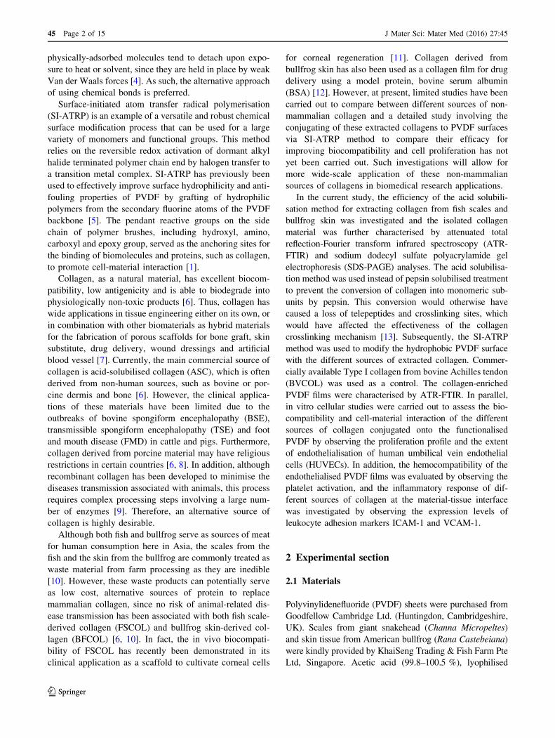
ATR-FTIR analysis showed the presence of characteristic
amide peaks representing amide A, B, I, II and III for the
FSCOL and BFCOL samples, which were also found in
BVCOL (Fig. 2a). The ATR-FTIR band wavenumber for
amide A is located at about 3300 cm-1 while amide B is at
2920 cm-1. The presence of amide I is indicated by the
appearance of the C=O bond at about 1630 cm-1, amide II
is contributed by the N–H bond (1530 cm-1) and amide III
is typically detected through complex bands from a mixture
of several coordinate displacements (around 1200 cm-1)
[28]. The presence of the peak at 1450 cm-1 for all the
FSCOL, BFCOL and BVCOL indicated the presence of the
collagen triple helical structure where the ratio of the
absorbance peak intensity of amide III to 1450 cm-1 is a
measurement of the relative amount of the helical structure
preserved in the collagen [29]. The ratio of the absorbance
peak intensities of amide III and 1450 cm-1 for FSCOL,
BFCOL and BVCOL were all around 1.0, thus indicating
that the helical structures of all the samples were similar to
each other and to ASC from other sources [28].
The amino acid compositions of the different sources of
collagen are shown in Table 2, where glycine was found to
be the most abundant amino acid, which is typical for
collagen [6]. Amongst the different sources of collagen,
FSCOL contained higher amounts of threonine, alanine and
histidine and lower amounts of arginine, aspartic acid,
leucine and proline, while BFCOL contained higher
amounts of arginine, glycine and aspartic acid as well as
serine, lysine and proline as compared to BVCOL. In
general, arginine, serine and lysine are amino acid residues
involved in post-translational modifications of proteins
such as acetylation, methylation and phosphorylation [30],
while hydroxylation of proline has been proposed to be
important for folding and stabilisation of the collagen triple
helix [31]. Hence, the differences in the amino acid com-
position could potentially lead to different collagen-mate-
rial interactions.
When the subunit compositions of FSCOL and BFCOL
were further resolved by SDS-PAGE, Type I collagen, with
several distinct band sizes corresponding to molecular
weights of approximately 400 kDa and 250 kDa for c- and
b-chains respectively, and approximately 139 kDa and
129 kDa for a1- and a2-chains respectively, were visible
after Coomassie Brilliant Blue staining for both FSCOL
and BFCOL (Fig. 2b). In addition, the width ratio of the
a1- and a2-chains for both FSCOL and BFCOL was found
to be approximately 2:1, which is the typical ratio for Type
I collagen [17, 18]. Also, the overall electrophoretic
mobility of FSCOL and BFCOL were similar to the
BVCOL, which was used as a positive control [32, 33].
Taken together, the results showed that Type I collagen
could be successfully extracted from fish scales and bull-
frog skin using the acid solubilisation method.
3.2 Direct surface-initiated ATRP of PVDF surface
for grafting of pHEMA brushes
As shown schematically in Scheme 1, the reaction steps
involved in the biofunctionalisation of PVDF include direct
Fig. 2 Characterisation of
collagen showing a ATR-FTIR
spectra of the characteristic
amide peaks of collagen (Dotted
lines) and b SDS-PAGE
analysis showing the protein
expression levels of Type I
collagen. (1) FSCOL, (2)
BFCOL, (3) BVCOL and (4)
protein ladder as marker
J Mater Sci: Mater Med (2016) 27:45 Page 7 of 15 45
123

SI-ATRP from the pristine PVDF to graft pHEMA brushes,
activation of hydroxyl groups on side chains of pHEMA
brush with a CDI biolinker, and covalent conjugation of
different sources of collagen. At each reaction step,
chemical analysis by way of ATR-FTIR was carried out to
identify peak changes. From the ATR-FTIR spectrum of
pristine PVDF (Fig. 3a), the characteristic bands with
wavenumber near 1177.91 cm-1 is attributed to –CF2
stretching vibration, other characteristic bands include the
–C–H and –CH2 stretching with a wavenumber near
2986.22 and 1401.50 cm-1, respectively, the asymmetric
stretching vibrations of the –CH2 groups at 3023 cm-1.
Upon grafting of pHEMA, a broad absorption band near
3396.68 cm-1, attributable to terminal hydroxyl (-OH)
group, was observed in the ATR-FTIR spectrum of PVDF-
g-pHEMA. The appearance of an additional absorption
peak at 1718.35 cm-1, attributable to the C=O stretching
vibrations, indicates the presence of the ester carbonyl
group of pHEMA [1]. Both characteristic peaks of pHEMA
of –OH and C=O were absent in the spectrum of pristine
PVDF, but present after the graft polymerisation reaction.
Taken together, the ATR-FTIR spectra showed the suc-
cessful grafting of the pHEMA onto the PVDF using the
SI-ATRP method.
3.3 Conjugation of collagen to the functionalised
PVDF surface
In order to immobilise the extracted collagen to the func-
tionalised PVDF films, a biofunctional linker, CDI, was
introduced to activate the terminal –OH groups in side
chains of pHEMA brushes. The resultant imidazole car-
bamate groups terminated PVDF-g-pHEMA films can be
used to covalently conjugate the extracted collagen. The
CDI modification of PVDF-g-pHEMA films resulted in the
split of the absorption bands at 1762.53 cm-1 for the car-
bonyl groups of acrylate and 1724.85 cm-1 for the imi-
dazole functionalities from the CDI compound (Fig. 3a)
[34, 35]. The newly formed imide bond indicated the
successful tethering of CDI onto the pHEMA-modified
films. Also the decrease in the intensities of –OH stretching
(near 3396.68 cm-1) was an indication of the conversion of
hydroxyl group of pHEMA into the imidazole carbamate
groups. Figure 3b shows the covalent immobilised colla-
gen on the functionalised PVDF films. Additional charac-
teristic absorption peaks of Type I collagen were observed
on the ATR-FTIR spectrum showing the amide B (near
2958.86 cm-1, C–H stretching), amide I (near
1661.26 cm-1, C=O stretching) and amide II (near
1555.42 cm-1, C–N stretching and N–H bending) indi-
cated successful immobilisation of collagen onto the
functionalised PVDF films via SI-ATRP.
3.4 Cellular studies
The cytocompatibility of the collagen-enriched PVDF
films was evaluated by quantitatively measuring the cell
adhesion and proliferation activity of HUVECs using the
PrestoBlue� assay over a 7 day period. It was observed
that the hydrophobic pristine PVDF exhibited poor initial
affinities for cell adhesion and hence could not support
subsequent cell proliferation (Fig. 4a). For the pHEMA-
modified PVDF films, even though PVDF-g-pHEMA and
PVDF-g-pHEMA-CDI surfaces improved the hydrophilic-
ity of the PVDF films due to the presence of hydrophilic
polymer brushes on the hydrophobic PVDF surfaces and
Table 2 Amino acid composition of FSCOL and BFCOL with BVCOL as reference
Types of amino acid FSCOL (residues/1000) BFCOL (residues/1000) BVCOL (residues/1000)
Aspartic acid 41.40 49.29 48.45
Threonine 26.01 20.88 18.41
Serine 36.77 50.10 33.93
Glutamic acid 70.48 74.94 74.00
Glycine 383.70 393.94 367.72
Alanine 134.42 122.21 122.17
Valine 18.04 11.85 19.30
Methionine 5.11 0.00 0.00
Isoleucine 10.35 8.45 12.02
Leucine 20.41 20.90 30.68
Phenylalanine 11.36 10.70 12.12
Lysine 24.69 28.24 23.43
Histidine 7.10 6.43 5.20
Arginine 49.10 55.86 54.28
Proline 130.65 146.19 136.59
45 Page 8 of 15 J Mater Sci: Mater Med (2016) 27:45
123

imidazole carbamate groups (Fig. S1), no significant
improvements for cell attachment and cell proliferation
profiles were observed due to the absence of biological
motifs available to the cells. On the other hand, signifi-
cantly higher (P\ 0.05) cell attachment and growth was
observed for the pHEMA-grafted PVDF films conjugated
with the different sources of Type I collagen. As compared
to the pristine PVDF films, the collagen-tethered films had
at least 600 % more number of cells attached at day 7.
However, no significant differences between the prolifer-
ation data of different species of collagen-enriched PVDF
films could be observed, thus suggesting that both non-
mammalian sources of collagen could be promising alter-
natives to the currently commercially available source.
In addition, observations from fluorescence staining of
viable cells (Fig. 4b) were in agreement with the cell
proliferation results, as the pristine PVDF film had the least
number of cells attached at day 7. The PVDF-g-pHEMA
and PVDF-g-pHEMA-CDI were observed to have low
numbers of viable cells attached despite an improvement in
surface hydrophilicity (Fig. S1). On the other hand, a
confluent layer of cells was observed on the collagen-en-
riched PVDF surfaces conjugated with different sources of
Type I collagen (PVDF-g-pHEMA-FSCOL, PVDF-g-
pHEMA-BFCOL and PVDF-g-pHEMA-BVCOL). Overall,
this confirmed that collagen-enrichment was important for
promoting cell-material interactions for the endotheliali-
sation of functionalised PVDF films.
3.5 Gene expression studies
Figure 5 shows the gene expression levels for pro-throm-
botic (vWF, PAI-1) and anti-thrombotic (tPA, eNOS)
markers, which were measured from the cells growing
collagen-enriched PVDF films. A lower PAI-1 but higher
tPA mRNA expression level was observed for the cells
growing on PVDF-g-pHEMA-BFCOL as compared to
those on the PVDF-g-pHEMA-BVCOL substrates. Studies
of PAI-1 have demonstrated that inhibiting PAI-1 could
enhance thrombolysis in experimental animal models [36],
while conversely, elevated levels of PAI-1 contributed to
thrombus formation and venous occlusion [37]. PAI-1 is an
important inhibitor of plasminogen activators, a class of
key enzymes that potentiate fibrinolytic activity in the
blood plasma, and of which tPA belongs to [38]. The
secretion of tPA from HUVECs, together with its regula-
tion in blood plasma levels by PAI-1, forms the important
tPA/PAI-1 axis for maintaining basal anti-thrombotic
activity in the blood plasma [39]. Overall, the tPA/PAI-1
ratios of FSCOL- and BFCOL-enriched PVDF films were
Fig. 3 ATR-FTIR results after a Conjugation of pHEMA chains onto
PVDF films followed by tethering of CDI biolinker onto the surface
functionalised PVDF films and b Immobilisation of different sources
of collagen onto the functionalised PVDF films. (Dotted lines
Characteristic peaks for PVDF substrate; Solid lines Characteristic
peaks resulted after each functionalisation ATRP step)
J Mater Sci: Mater Med (2016) 27:45 Page 9 of 15 45
123
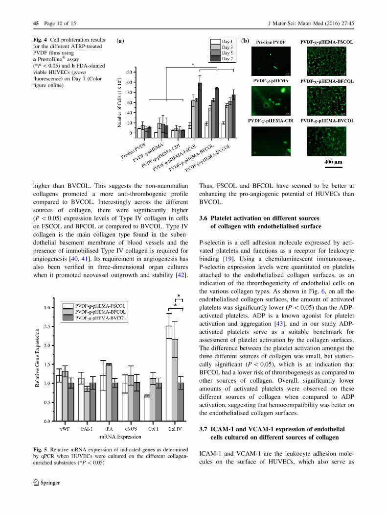
higher than BVCOL. This suggests the non-mammalian
collagens promoted a more anti-thrombogenic profile
compared to BVCOL. Interestingly across the different
sources of collagen, there were significantly higher
(P\ 0.05) expression levels of Type IV collagen in cells
on FSCOL and BFCOL as compared to BVCOL. Type IV
collagen is the main collagen type found in the suben-
dothelial basement membrane of blood vessels and the
presence of immobilised Type IV collagen is required for
angiogenesis [40, 41]. Its requirement in angiogenesis has
also been verified in three-dimensional organ cultures
when it promoted neovessel outgrowth and stability [42].
Thus, FSCOL and BFCOL have seemed to be better at
enhancing the pro-angiogenic potential of HUVECs than
BVCOL.
3.6 Platelet activation on different sources
of collagen with endothelialised surface
P-selectin is a cell adhesion molecule expressed by acti-
vated platelets and functions as a receptor for leukocyte
binding [19]. Using a chemiluminescent immunoassay,
P-selectin expression levels were quantitated on platelets
attached to the endothelialised collagen surfaces, as an
indication of the thrombogenicity of endothelial cells on
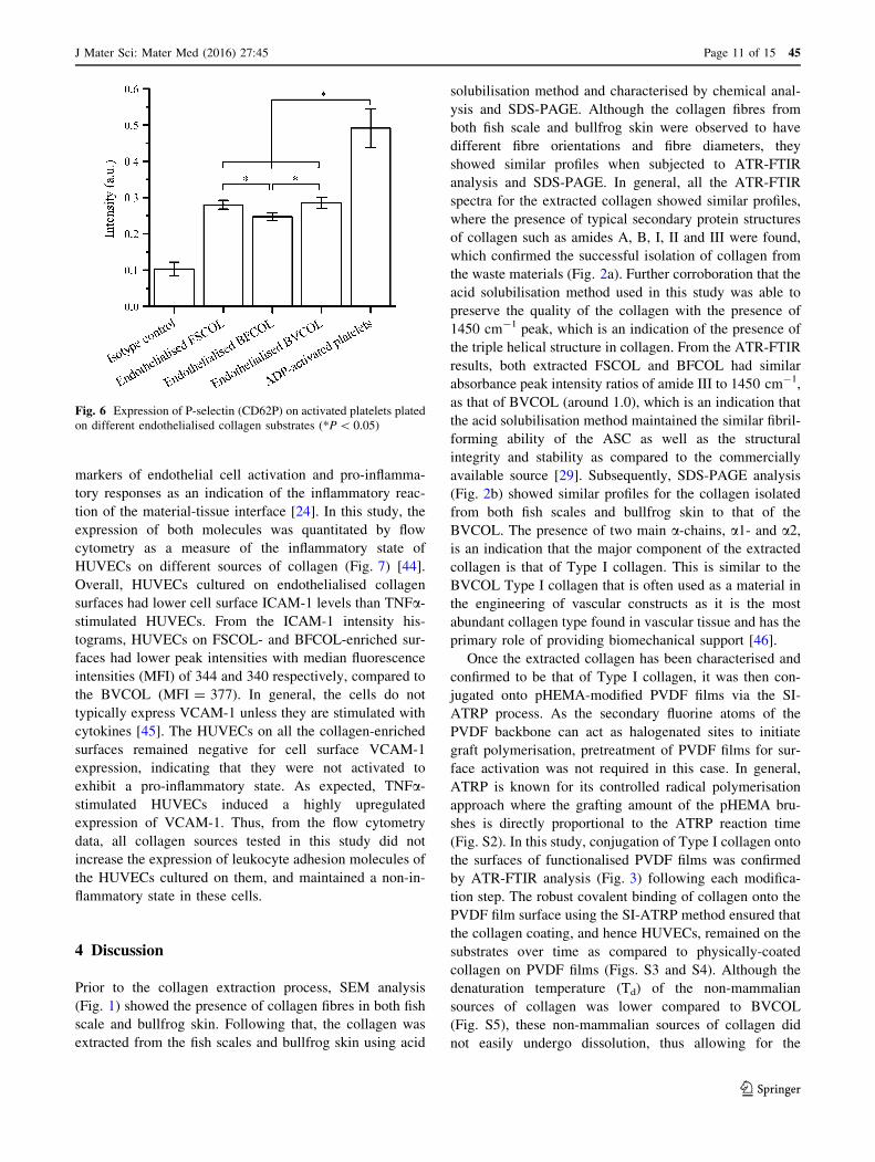
the various collagen types. As shown in Fig. 6, on all the
endothelialised collagen surfaces, the amount of activated
platelets was significantly lower (P\ 0.05) than the ADP-
activated platelets. ADP is a known agonist for platelet
activation and aggregation [43], and in our study ADP-
activated platelets serve as a suitable benchmark for
assessment of platelet activation by the collagen surfaces.
The difference between the platelet activation amongst the
three different sources of collagen was small, but statisti-
cally significant (P\ 0.05), which is an indication that
BFCOL had a lower risk of thrombogenesis as compared to
other sources of collagen. Overall, significantly lower
amounts of activated platelets were observed on these
different sources of collagen when compared to ADP
activation, suggesting that hemocompatibility was better on
the endothelialised collagen surfaces.
3.7 ICAM-1 and VCAM-1 expression of endothelial
cells cultured on different sources of collagen
ICAM-1 and VCAM-1 are the leukocyte adhesion mole-
cules on the surface of HUVECs, which also serve as
Fig. 4 Cell proliferation results
for the different ATRP-treated
PVDF films using
a PrestoBlue� assay
(*P\ 0.05) and b FDA-stained
viable HUVECs (green
fluorescence) on Day 7 (Color
figure online)
Fig. 5 Relative mRNA expression of indicated genes as determined
by qPCR when HUVECs were cultured on the different collagen-
enriched substrates (*P\ 0.05)
45 Page 10 of 15 J Mater Sci: Mater Med (2016) 27:45
123
markers of endothelial cell activation and pro-inflamma-
tory responses as an indication of the inflammatory reac-
tion of the material-tissue interface [24]. In this study, the
expression of both molecules was quantitated by flow
cytometry as a measure of the inflammatory state of
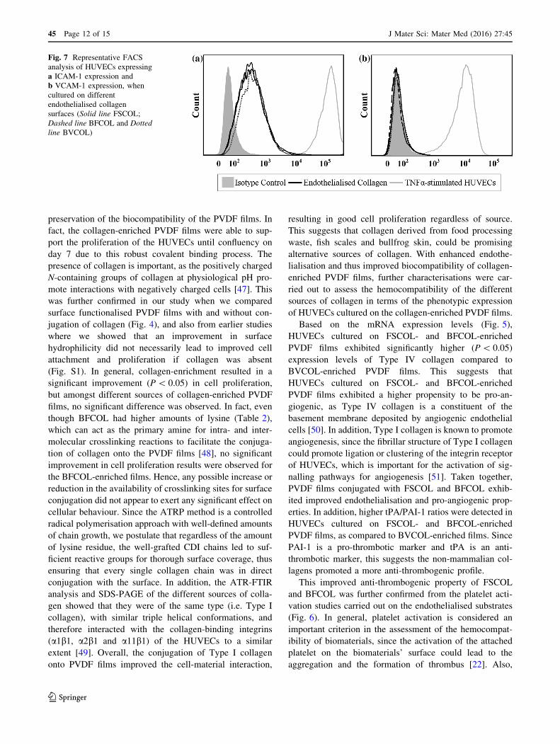
HUVECs on different sources of collagen (Fig. 7) [44].
Overall, HUVECs cultured on endothelialised collagen
surfaces had lower cell surface ICAM-1 levels than TNFa-
stimulated HUVECs. From the ICAM-1 intensity his-
tograms, HUVECs on FSCOL- and BFCOL-enriched sur-
faces had lower peak intensities with median fluorescence
intensities (MFI) of 344 and 340 respectively, compared to
the BVCOL (MFI = 377). In general, the cells do not
typically express VCAM-1 unless they are stimulated with
cytokines [45]. The HUVECs on all the collagen-enriched
surfaces remained negative for cell surface VCAM-1
expression, indicating that they were not activated to
exhibit a pro-inflammatory state. As expected, TNFa-
stimulated HUVECs induced a highly upregulated
expression of VCAM-1. Thus, from the flow cytometry
data, all collagen sources tested in this study did not
increase the expression of leukocyte adhesion molecules of
the HUVECs cultured on them, and maintained a non-in-
flammatory state in these cells.
4 Discussion
Prior to the collagen extraction process, SEM analysis
(Fig. 1) showed the presence of collagen fibres in both fish
scale and bullfrog skin. Following that, the collagen was
extracted from the fish scales and bullfrog skin using acid
solubilisation method and characterised by chemical anal-
ysis and SDS-PAGE. Although the collagen fibres from
both fish scale and bullfrog skin were observed to have
different fibre orientations and fibre diameters, they
showed similar profiles when subjected to ATR-FTIR
analysis and SDS-PAGE. In general, all the ATR-FTIR
spectra for the extracted collagen showed similar profiles,
where the presence of typical secondary protein structures
of collagen such as amides A, B, I, II and III were found,
which confirmed the successful isolation of collagen from
the waste materials (Fig. 2a). Further corroboration that the
acid solubilisation method used in this study was able to
preserve the quality of the collagen with the presence of
1450 cm-1 peak, which is an indication of the presence of
the triple helical structure in collagen. From the ATR-FTIR
results, both extracted FSCOL and BFCOL had similar
absorbance peak intensity ratios of amide III to 1450 cm-1,
as that of BVCOL (around 1.0), which is an indication that
the acid solubilisation method maintained the similar fibril-
forming ability of the ASC as well as the structural
integrity and stability as compared to the commercially
available source [29]. Subsequently, SDS-PAGE analysis
(Fig. 2b) showed similar profiles for the collagen isolated
from both fish scales and bullfrog skin to that of the
BVCOL. The presence of two main a-chains, a1- and a2,
is an indication that the major component of the extracted
collagen is that of Type I collagen. This is similar to the
BVCOL Type I collagen that is often used as a material in
the engineering of vascular constructs as it is the most
abundant collagen type found in vascular tissue and has the
primary role of providing biomechanical support [46].
Once the extracted collagen has been characterised and
confirmed to be that of Type I collagen, it was then con-
jugated onto pHEMA-modified PVDF films via the SI-
ATRP process. As the secondary fluorine atoms of the
PVDF backbone can act as halogenated sites to initiate
graft polymerisation, pretreatment of PVDF films for sur-
face activation was not required in this case. In general,
ATRP is known for its controlled radical polymerisation
approach where the grafting amount of the pHEMA bru-
shes is directly proportional to the ATRP reaction time
(Fig. S2). In this study, conjugation of Type I collagen onto
the surfaces of functionalised PVDF films was confirmed
by ATR-FTIR analysis (Fig. 3) following each modifica-
tion step. The robust covalent binding of collagen onto the
PVDF film surface using the SI-ATRP method ensured that
the collagen coating, and hence HUVECs, remained on the
substrates over time as compared to physically-coated
collagen on PVDF films (Figs. S3 and S4). Although the
denaturation temperature (Td) of the non-mammalian
sources of collagen was lower compared to BVCOL
(Fig. S5), these non-mammalian sources of collagen did
not easily undergo dissolution, thus allowing for the
Fig. 6 Expression of P-selectin (CD62P) on activated platelets plated
on different endothelialised collagen substrates (*P\ 0.05)
J Mater Sci: Mater Med (2016) 27:45 Page 11 of 15 45
123
preservation of the biocompatibility of the PVDF films. In
fact, the collagen-enriched PVDF films were able to sup-
port the proliferation of the HUVECs until confluency on
day 7 due to this robust covalent binding process. The
presence of collagen is important, as the positively charged
N-containing groups of collagen at physiological pH pro-
mote interactions with negatively charged cells [47]. This
was further confirmed in our study when we compared
surface functionalised PVDF films with and without con-
jugation of collagen (Fig. 4), and also from earlier studies
where we showed that an improvement in surface
hydrophilicity did not necessarily lead to improved cell
attachment and proliferation if collagen was absent
(Fig. S1). In general, collagen-enrichment resulted in a
significant improvement (P\ 0.05) in cell proliferation,
but amongst different sources of collagen-enriched PVDF
films, no significant difference was observed. In fact, even
though BFCOL had higher amounts of lysine (Table 2),
which can act as the primary amine for intra- and inter-
molecular crosslinking reactions to facilitate the conjuga-
tion of collagen onto the PVDF films [48], no significant
improvement in cell proliferation results were observed for
the BFCOL-enriched films. Hence, any possible increase or
reduction in the availability of crosslinking sites for surface
conjugation did not appear to exert any significant effect on
cellular behaviour. Since the ATRP method is a controlled
radical polymerisation approach with well-defined amounts
of chain growth, we postulate that regardless of the amount
of lysine residue, the well-grafted CDI chains led to suf-
ficient reactive groups for thorough surface coverage, thus
ensuring that every single collagen chain was in direct
conjugation with the surface. In addition, the ATR-FTIR
analysis and SDS-PAGE of the different sources of colla-
gen showed that they were of the same type (i.e. Type I
collagen), with similar triple helical conformations, and
therefore interacted with the collagen-binding integrins
(a1b1, a2b1 and a11b1) of the HUVECs to a similar
extent [49]. Overall, the conjugation of Type I collagen
onto PVDF films improved the cell-material interaction,
resulting in good cell proliferation regardless of source.
This suggests that collagen derived from food processing
waste, fish scales and bullfrog skin, could be promising
alternative sources of collagen. With enhanced endothe-
lialisation and thus improved biocompatibility of collagen-
enriched PVDF films, further characterisations were car-
ried out to assess the hemocompatibility of the different
sources of collagen in terms of the phenotypic expression
of HUVECs cultured on the collagen-enriched PVDF films.
Based on the mRNA expression levels (Fig. 5),
HUVECs cultured on FSCOL- and BFCOL-enriched
PVDF films exhibited significantly higher (P\ 0.05)
expression levels of Type IV collagen compared to
BVCOL-enriched PVDF films. This suggests that
HUVECs cultured on FSCOL- and BFCOL-enriched
PVDF films exhibited a higher propensity to be pro-an-
giogenic, as Type IV collagen is a constituent of the
basement membrane deposited by angiogenic endothelial
cells [50]. In addition, Type I collagen is known to promote
angiogenesis, since the fibrillar structure of Type I collagen
could promote ligation or clustering of the integrin receptor
of HUVECs, which is important for the activation of sig-
nalling pathways for angiogenesis [51]. Taken together,
PVDF films conjugated with FSCOL and BFCOL exhib-
ited improved endothelialisation and pro-angiogenic prop-
erties. In addition, higher tPA/PAI-1 ratios were detected in
HUVECs cultured on FSCOL- and BFCOL-enriched
PVDF films, as compared to BVCOL-enriched films. Since
PAI-1 is a pro-thrombotic marker and tPA is an anti-
thrombotic marker, this suggests the non-mammalian col-
lagens promoted a more anti-thrombogenic profile.
This improved anti-thrombogenic property of FSCOL
and BFCOL was further confirmed from the platelet acti-
vation studies carried out on the endothelialised substrates
(Fig. 6). In general, platelet activation is considered an
important criterion in the assessment of the hemocompat-
ibility of biomaterials, since the activation of the attached
platelet on the biomaterials’ surface could lead to the
aggregation and the formation of thrombus [22]. Also,
Fig. 7 Representative FACS
analysis of HUVECs expressing
a ICAM-1 expression and
b VCAM-1 expression, when
cultured on different
endothelialised collagen
surfaces (Solid line FSCOL;
Dashed line BFCOL and Dotted
line BVCOL)
45 Page 12 of 15 J Mater Sci: Mater Med (2016) 27:45
123

collagen receptors have been identified on platelets, most
notably a2b1 integrin and the immunoglobulin superfamily
GPVI, which is why the direct interaction of collagen with
platelets has been known to trigger the activation of
thrombogenic initiation [19]. Therefore, the endotheliali-
sation of the collagen surfaces is an important step in the
reduction of collagen-induced thrombogenicity. In the
current study, the amount of P-selectin expression of the
platelets was investigated, since it is one of the biomarkers
found to be increased on the platelet surface following
activation [52]. The extent of platelet activation could be
determined by comparing the P-selectin expression on
platelets attached to the endothelialised collagen surfaces
with the ADP-activated platelets [19, 22]. From the results
(Fig. 6), all the platelets in contact with the endothelialised
collagen surfaces did indeed have significantly lower
(P\ 0.05) platelet activation as compared to ADP-acti-
vated platelets. Furthermore, the lowest amount of acti-
vated platelets was observed on the endothelialised
BFCOL surfaces. In addition, the plasma recalcification
assay was performed on different sources of endothe-
lialised collagen surfaces by incubating the surfaces with
platelet poor plasma, followed by measuring fibrin clot
initiation times when the plasma was recalcified with cal-
cium chloride [53]. The clot initiation times for FSCOL,
BFCOL and BVCOL were 17.5 ± 3.6, 16.9 ± 1.1 and
16.1 ± 3.5 min respectively, which is significantly longer
(P\ 0.05) than the timings of TCPs control and pristine
PVDF with timings of 9.4 ± 0.7 and 6.0 ± 0.5 min,
respectively (Fig. S6). Similar to the results from the pla-
telet activation study, the endothelialised collagen surfaces
had a significantly longer (P\ 0.05) clot formation time as
compared to TCPs control and pristine PVDF. In addition,
the non-mammalian sources of collagen had slightly pro-
longed clot formation times as compared to BVCOL.
Taken together, the endothelialised collagen surfaces
demonstrated better anti-coagulation responses, which is a
phenomenon that is also reported by others [54, 55].
Lastly, the material-induced inflammatory response of
HUVECs in contact with different sources of collagen was
studied by investigating level of activation of HUVECs
cultured on the collagen. The activation of HUVECs is
typically associated to the initial step of inflammation,
caused by the increased infiltration of leukocytes into the
inflamed tissue, which is mediated by leukocyte adhesion
molecules such as ICAM-1 and VCAM-1. Thus, the acti-
vation of HUVECs is commonly used to investigate
inflammatory reactions [25]. From the results (Fig. 7), the
cells on collagen-enriched surfaces all have low constitu-
tive expression levels of ICAM-1 with little or no expres-
sion levels of VCAM-1. These values were compared to
TNFa-stimulated HUVEC controls, since TNFa is a pro-
inflammatory cytokine known to increase the expression of
cell adhesion molecules [44]. Taken together, the expres-
sion profiles of the HUVECs on all the collagen-enriched
surfaces used in this study is in line with that of inactivated
HUVECs in culture (i.e. low constitutive expression of
ICAM-1, no expression of VCAM-1) [23]. Overall, it can
be deduced that the non-mammalian sources of collagen,
similar to the BVCOL, did not trigger a pro-inflammatory
phenotype in the HUVECs.
5 Conclusion
Type I collagen was successfully extracted from fish scales
and bullfrog skin and chemically conjugated onto PVDF
films using the direct SI-ATRP approach. The acid solu-
bilisation method used in this study led to the preservation
of the quality of the extracted collagen, since FSCOL and
BFCOL both had similar triple helical confirmation to that
of commercially available Type I BVCOL. The collagen-
enriched PVDF films significantly enhanced (P\ 0.05)
cell-biomaterial interactions by improving the cell attach-
ment and cell proliferation, which then led to a better
coverage of HUVECs on the collagen-enriched PVDF
films. As compared to BVCOL, the non-mammalian
sources of collagen led to endothelialised surfaces with
higher expression levels of Type IV collagen by HUVECs
and comparable anti-thrombogenic properties, thus making
them more suitable candidates for blood contacting appli-
cations. In addition, the low expression levels of ICAM-1
and VCAM-1 indicated that these non-mammalian sources
of collagen did not elicit pro-inflammatory phenotypes in
the cells. Taken together, our study not only has high-
lighted the feasibility of using FSCOL and BFCOL as cost
effective substitutes for BVCOL, but has also demonstrated
an effective chemical functionalisation scheme for PVDF
substrates via SI-ATRP process. The conversion of the
abundantly available food processing wastes, fish scales
and bullfrog skin, into collagen is a demonstration of an
effective waste-to-resource management to utilise the
underexploited biological resources. These sources of non-
mammalian collagen can potentially be used as alternative
resource resilient biomaterials for tissue engineering
applications.
Acknowledgments The authors are grateful for the research
scholarship from Nanyang Environment and Water Research Insti-
tute/ Interdisciplinary Graduate School, Nanyang Technological
University, Singapore (NEWRI/IGS) and the research grant supported
by the Singapore National Research Foundation under its Environ-
ment & Water Technologies Strategic Research Programme and
administered by the Environment & Water Industry Programme
Office (EWI) of the Public Utility Board (PUB). They would also like
to acknowledge the funding support from the Singapore Ministry of
Education Academic Research Fund (AcRF) Tier 1 (RG52/13
2013-T1-002-227) funding.
J Mater Sci: Mater Med (2016) 27:45 Page 13 of 15 45
123

Open Access This article is distributed under the terms of the
Creative Commons Attribution 4.0 International License (http://crea
tivecommons.org/licenses/by/4.0/), which permits unrestricted use,
distribution, and reproduction in any medium, provided you give
appropriate credit to the original author(s) and the source, provide a
link to the Creative Commons license, and indicate if changes were
made.
References
1. Meng J-Q, Chen C-L, Huang L-P, Du Q-Y, Zhang Y-F. Surface
modification of PVDF membrane via AGET ATRP directly from
the membrane surface. Appl Surf Sci. 2011;257:6282–90.
2. Zhu LP, Yu JZ, Xu YY, Xi ZY, Zhu BK. Surface modification of
PVDF porous membranes via poly(DOPA) coating and heparin
immobilization. Colloids Surf B. 2009;69:152–5.
3. Ameringer T, Ercole F, Tsang K, Coad B, Hou X, Rodda A, et al.
Surface grafting of electrospun fibers using ATRP and RAFT for
the control of biointerfacial interactions. Biointerphases.
2013;8:1–11.
4. Balamurugan SS, Subramanian B, Bolivar JG, McCarley RL.
Aqueous-based initiator attachment and ATRP grafting of poly-
mer brushes from poly(methyl methacrylate) substrates. Lang-
muir. 2012;28:14254–60.
5. He F, Luo B, Yuan S, Liang B, Choong C, Pehkonen SO. PVDF film
tethered with RGD-click-poly(glycidyl methacrylate) brushes by
combination of direct surface-initiated ATRP and click chemistry
for improved cytocompatibility. RSC Adv. 2014;4:105–17.
6. Song E, Yeon Kim S, Chun T, Byun H-J, Lee YM. Collagen
scaffolds derived from a marine source and their biocompatibil-
ity. Biomaterials. 2006;27:2951–61.
7. Lee CH, Singla A, Lee Y. Biomedical applications of collagen.
Int J Pharm. 2001;221:1–22.
8. Sadowska M, Kołodziejska I, Niecikowska C. Isolation of col-
lagen from the skins of Baltic cod (Gadus morhua). Food Chem.
2003;81:257–62.
9. Shoseyov O, Posen Y, Grynspan F. Human recombinant type I
collagen produced in plants. Tissue Eng Part A. 2013;19:1527–33.
10. Li H, Liu BL, Gao LZ, Chen HL. Studies on bullfrog skin col-
lagen. Food Chem. 2004;84:65–9.
11. Lin CC, Ritch R, Lin SM, Ni MH, Chang YC, Lu YL, et al. A
new fish scale-derived scaffold for corneal regeneration. Eur
Cells Mater. 2010;19:50–7.
12. Li H, Wang D, Li S, Liu B, Gao L. Sustained release of BSA
from a novel drug delivery matrix—bullfrog skin collagen film.
Macromol Biosci. 2004;4:454–7.
13. Bannister DW, Burns AB. Pepsin treatment of avian skin colla-
gen. Effects on solubility, subunit composition and aggregation
properties. Biochem J. 1972;129:677–81.
14. Zhang F, Wang A, Li Z, He S, Shao L. Preparation and charac-
terisation of collagen from freshwater fish scales. Food Nutr Sci.
2011;2:818–23.
15. Takeshi Suzuki N. Isolation of collagen from fish waste mate-
rial—skin, bone and fins. Food Chem. 2000;68:277–81.
16. Laemmli UK. Cleavage of structural proteins during the assembly
of the head of bacteriophage T4. Nature. 1970;227:680–5.
17. Gomez-Guillen MC, Turnay J, Fernandez-Dıaz MD, Ulmo N,
Lizarbe MA, Montero P. Structural and physical properties of
gelatin extracted from different marine species: a comparative
study. Food Hydrocolloids. 2002;16:25–34.
18. Singh P, Benjakul S, Maqsood S, Kishimura H. Isolation and
characterisation of collagen extracted from the skin of striped
catfish (Pangasianodon hypophthalmus). Food Chem. 2011;124:
97–105.
19. Xiong GM, Yuan S, Tan CK, Wang JK, Liu Y, Yang Tan TT,
et al. Endothelial cell thrombogenicity is reduced by ATRP-
mediated grafting of gelatin onto PCL surfaces. J Mater Chem B.
2014;2:485–93.
20. Pankajakshan D, Krishnan VK, Krishnan LK. Functional stability
of endothelial cells on a novel hybrid scaffold for vascular tissue
engineering. Biofabrication. 2010;2:041001-1–-10.
21. Prasad CK, Krishnan LK. Regulation of endothelial cell pheno-
type by biomimetic matrix coated on biomaterials for cardio-
vascular tissue engineering. Acta Biomater. 2008;4:182–91.
22. Yang Z, Tu Q, Zhu Y, Luo R, Li X, Xie Y, et al. Mussel-inspired
coating of polydopamine directs endothelial and smooth muscle
cell fate for re-endothelialization of vascular devices. Adv
Healthcare Mater. 2012;1:548–59.
23. van der Zijpp YJ, Poot AA, Feijen J. ICAM-1 and VCAM-1
expression by endothelial cells grown on fibronectin-coated
TCPS and PS. J Biomed Mater Res Part A. 2003;65:51–9.
24. Autieri MV. Pro- and anti-inflammatory cytokine networks in
atherosclerosis. ISRN Vasc Med. 2012;2012:1–17.
25. Kim I, Moon SO, Kim SH, Kim HJ, Koh YS, Koh GY. Vascular
endothelial growth factor expression of intercellular adhesion
molecule 1 (ICAM-1), vascular cell adhesion molecule 1
(VCAM-1), and E-selectin through nuclear factor-kappa B acti-
vation in endothelial cells. J Biol Chem. 2001;276:7614–20.
26. Ikoma T, Kobayashi H, Tanaka J, Walsh D, Mann S.
Microstructure, mechanical, and biomimetic properties of fish
scales from Pagrus major. J Struct Biol. 2003;142:327–33.
27. Bigi A, Burghammer M, Falconi R, Koch MHJ, Panzavolta S,
Riekel C. Twisted plywood pattern of collagen fibrils in teleost
scales: an X-ray diffraction investigation. J Struct Biol.
2001;136:137–43.
28. Ahmad M, Benjakul S, Nalinanon S. Compositional and
physicochemical characteristics of acid solubilized collagen
extracted from the skin of unicorn leatherjacket (Aluterus
monoceros). Food Hydrocolloids. 2010;24:588–94.
29. Guzzi Plepis AMD, Goissis G, Das-Gupta DK. Dielectric and
pyroelectric characterization of anionic and native collagen.
Polym Eng Sci. 1996;36:2932–8.
30. Blom N, Sicheritz-Ponten T, Gupta R, Gammeltoft S, Brunak S.
Prediction of post-translational glycosylation and phosphoryla-
tion of proteins from the amino acid sequence. Proteomics.
2004;4:1633–49.
31. Vitagliano L, Berisio R, Mazzarella L, Zagari A. Structural bases
of collagen stabilization induced by proline hydroxylation.
Biopolymers. 2001;58:459–64.
32. Wang L, Liang Q, Wang Z, Xu J, Liu Y, Ma H. Preparation and
characterisation of type I and V collagens from the skin of Amur
sturgeon (Acipenser schrenckii). Food Chem. 2014;148:410–4.
33. Wang L, An X, Xin Z, Zhao L, Hu Q. Isolation and characteri-
zation of collagen from the skin of deep-sea redfish (Sebastes
mentella). J Food Sci. 2007;72:E450–5.
34. Evans MD, Steele JG. Polymer surface chemistry and a novel
attachment mechanism in corneal epithelial cells. J Biomed
Mater Res. 1998;40:621–30.
35. Fukano Y, Knowles NG, Usui ML, Underwood RA, Hauch KD,
Marshall AJ, et al. Characterization of an in vitro model for
evaluating the interface between skin and percutaneous bioma-
terials. Wound Repair Regen. 2006;14:484–91.
36. Levi M, Biemond BJ, van Zonneveld AJ, ten Cate JW, Pan-
nekoek H. Inhibition of plasminogen activator inhibitor-1 activity
results in promotion of endogenous thrombolysis and inhibition
of thrombus extension in models of experimental thrombosis.
Circulation. 1992;85:305–12.
45 Page 14 of 15 J Mater Sci: Mater Med (2016) 27:45
123

37. Erickson LA, Fici GJ, Lund JE, Boyle TP, Polites HG, Marotti
KR. Development of venous occlusions in mice transgenic for the
plasminogen activator inhibitor-1 gene. Nature. 1990;346:74–6.
38. Chandler WL, Trimble SL, Loo SC, Mornin D. Effect of PAI-1
levels on the molar concentrations of active tissue plasminogen
activator (t-PA) and t-PA/PAI-1 complex in plasma. Blood.
1990;76:930–7.
39. Chandler WL, Alessi MC, Aillaud MF, Henderson P, Vague P,
Juhan-Vague I. Clearance of tissue plasminogen activator (TPA)
and TPA/plasminogen activator inhibitor type 1 (PAI-1) com-
plex: relationship to elevated TPA antigen in patients with high
PAI-1 activity levels. Circulation. 1997;96:761–8.
40. Xu J, Rodriguez D, Petitclerc E, Kim JJ, Hangai M, Yuen SM,
et al. Proteolytic exposure of a cryptic site within collagen type
IV is required for angiogenesis and tumor growth in vivo. J Cell
Biol. 2001;154:1069–80.
41. Kramer RH, Fuh G-M, Karasek MA. Type IV collagen synthesis
by cultured human microvascular endothelial cells and its depo-
sition into the subendothelial basement membrane. Biochemistry.
1985;24:7423–30.
42. Bonanno E, Iurlaro M, Madri JA, Nicosia RF. Type IV collagen
modulates angiogenesis and neovessel survival in the rat aorta
model. In Vitro Cell Dev Biol Anim. 2000;36:336–40.
43. Puri RN, Colman RW. ADP-induced platelet activation. Crit Rev
Biochem Mol Biol. 1997;32:437–502.
44. Min JK, Kim YM, Kim SW, Kwon MC, Kong YY, Hwang IK,
et al. TNF-related activation-induced cytokine enhances leuko-
cyte adhesiveness: induction of ICAM-1 and VCAM-1 via TNF
receptor-associated factor and protein kinase C-dependent NF-
kappaB activation in endothelial cells. J Immunol. 2005;175:
531–40.
45. Carlos TM, Schwartz BR, Kovach NL, Yee E, Rosa M, Osborn L,
et al. Vascular cell adhesion molecule-1 mediates lymphocyte
adherence to cytokine-activated cultured human endothelial cells.
Blood. 1990;76:965–70.
46. Pang Y, Greisler HP. Using a type 1 collagen-based system to
understand cell-scaffold interactions and to deliver chimeric
collagen-binding growth factors for vascular tissue engineering.
J Invest Med. 2010;58:845–8.
47. Cheng Z, Teoh SH. Surface modification of ultra thin poly (ep-
silon-caprolactone) films using acrylic acid and collagen. Bio-
materials. 2004;25:1991–2001.
48. Sinz A. Chemical cross-linking and mass spectrometry to map
three-dimensional protein structures and protein–protein interac-
tions. Mass Spectrom Rev. 2006;25:663–82.
49. Sweeney SM, Orgel JP, Fertala A, McAuliffe JD, Turner KR, Di
Lullo GA, et al. Candidate cell and matrix interaction domains on
the collagen fibril, the predominant protein of vertebrates. J Biol
Chem. 2008;283:21187–97.
50. Bahramsoltani M, Slosarek I, De Spiegelaere W, Plendl J. Angio-
genesis and collagen type IV expression in different endothelial cell
culture systems. Anat Histol Embryol. 2014;43:103–15.
51. Twardowski T, Fertala A, Orgel JP. San Antonio JD. Type I
collagen and collagen mimetics as angiogenesis promoting
superpolymers. Curr Pharm Des. 2007;13:3608–21.
52. Reed GL, Houng AK, Bianchi C. Comparative biochemical and
ultrastructural studies of P-selectin in rabbit platelets. Comp
Biochem Physiol Part B. 1998;119:729–38.
53. Xiong GM, Yuan S, Wang JK, Do AT, Tan NS, Yeo KS, et al.
Imparting electroactivity to polycaprolactone fibers with heparin-
doped polypyrrole: modulation of hemocompatibility and
inflammatory responses. Acta Biomater. 2015;23:240–9.
54. Chang Y, Chang W-J, Shih Y-J, Wei T-C, Hsiue G-H. Zwitte-
rionic sulfobetaine-grafted poly(vinylidene fluoride) membrane
with highly effective blood compatibility via atmospheric
plasma-induced surface copolymerization. ACS Appl Mater
Interfaces. 2011;3:1228–37.
55. McGuigan AP, Sefton MV. The thrombogenicity of human
umbilical vein endothelial cell seeded collagen modules. Bioma-
terials. 2008;29:2453–63. doi:10.1016/j.biomaterials.2008.02.010.
J Mater Sci: Mater Med (2016) 27:45 Page 15 of 15 45
123