surface-modified polystyrene beads as photografting...
TRANSCRIPT

Sf
La
b
a
ARR2AA
KMPPIC
1
tnmp<sWlc[
cces
3
(
0d
Journal of Chromatography A, 1216 (2009) 807–814
Contents lists available at ScienceDirect
Journal of Chromatography A
journa l homepage: www.e lsev ier .com/ locate /chroma
urface-modified polystyrene beads as photografting imprinted polymer matrixor chromatographic separation of proteins
ei Qina, Xi-Wen Hea, Wei Zhanga, Wen-You Lia,∗, Yu-Kui Zhanga,b,∗∗
Department of Chemistry, Nankai University, Tianjin 300071, ChinaNational Chromatographic Research and Analysis Center, Dalian Institute of Chemical Physics, Chinese Academy of Sciences, Dalian 116011, China
r t i c l e i n f o
rticle history:eceived 8 June 2008eceived in revised form8 November 2008ccepted 1 December 2008vailable online 7 December 2008
eywords:
a b s t r a c t
A new and facile fabricating method for lysozyme molecularly imprinted polymer beads (lysozyme-MIPbeads) in aqueous media was presented. Mesoporous chloromethylated polystyrene beads (MCP beads)containing dithiocarbamate iniferter (initiator transfer agent terminator) were used as supports for thegrafting of lysozyme imprinted copolymers with acrylamide and N,N′-methylenebisacrylamide throughsurface initiated living-radical polymerization (SIP). After the polymerization, a layer of lysozyme-MIPwas formed on the MCP beads. The SIP allowed an efficient control of the grafting process and sup-pressed solution propagation. Therefore, the obtained lysozyme-MIP beads had a large quantity ofwell-distributed pores on the surface without any visible gel formation in solution and were more
olecular imprintingrotein separationolystyrene beadsniferterhromatography
advantageous comparing with traditional MIPs which were prepared by traditionally initiated radicalpolymerization. The obtained composites were characterized by Fourier transform infrared spectroscopy,elemental analysis, nitrogen sorption analysis and scanning electron microscopy. Chromatographicbehaviors of the column packed with lysozyme-MIP beads exhibited ability in separating lysozyme fromcompetitive protein (bovine hemoglobin, bovine serum albumin, ovalbumin or cytochrome c) in aqueous
mobile phase.. Introduction
Molecular imprinting has been proved to be an importantool for the rapid fabrication of organic polymeric and inorganicetwork-structured materials that selectively bind a templateolecule [1,2]. Commonly this technique has demonstrated great
otential when targeting small molecules of molecular weight1500, while extending the technique to biological macromoleculesuch as proteins, whole cells and viruses has been proven difficult.
hen imprinting proteins, there are a number of inherent prob-ems to address that are related to the molecular size, complexity,onformational flexibility, solubility and sensitivity to environment3].
Though protein imprinting is proved to be one of the most
hallenging tasks, the field is progressing rapidly. The first suc-ess in protein imprinting was achieved by the group of Galdt al. [4]. A number of molecular imprinting polymers (MIPs)elective for proteins have now been reported using methodolo-∗ Corresponding author at: Department of Chemistry, Nankai University, Tianjin00071, China. Tel.: +86 22 2349 4962; fax: +86 22 2350 2458.∗∗ Corresponding author. Tel.: +86 411 84379560; fax: +86 411 84379560.
E-mail addresses: [email protected] (W.-Y. Li), [email protected]. Zhang).
021-9673/$ – see front matter © 2008 Elsevier B.V. All rights reserved.oi:10.1016/j.chroma.2008.12.007
© 2008 Elsevier B.V. All rights reserved.
gies including sol–gel [5,6], metal chelating [7,8], hybrid-DNA [3],epitope approach [9,10] and acrylate chemistry [11–14] throughthree-dimensional [5,11] or two-dimensional [14–18] imprinting.Recently, several new methods have been adopted to prepare pro-tein MIPs [19] and accelerated the process of protein molecularimprinting technology. Among the above methods, polyacrylamideimprinting of proteins has been proven useful for the separationof proteins in chromatographic mode [11,20]. However, so far, theprotein imprinting materials used as stationary phase of HPLC toseparate proteins are rare [15].
Surface grafting on supports has been recently proposed as anew surface imprinting technology for the synthesis of molecularlyimprinted polymer with a particular morphology and better acces-sibility to the specific binding sites [21]. Unfortunately, solutionpolymerization and resulting gelation are difficult to be avoided intraditional surface initiated radical polymerization. The extra solu-tion polymer would lead to the heterogeneous recognition sitesand peak tailing when used as stationary phase [22]. One solutionto this shortcoming is to use the iniferter (initiator transfer agentterminator) which is one kind of surface initiated living-radical
polymerization (SIP) initiators. The iniferter traditionally has beenused in a dissolved form, but here polymerization in solution couldbe avoided by attaching the iniferter on support surface before itis activated [23–25]. Under the ultraviolet irradiation, the inifer-ter decomposes into active radical and non-active radical, where
8 togr. A
tigtbrItti
otpwtae
FoAb
08 L. Qin et al. / J. Chroma
he active radical bound to the support surface initiates polymer-zation while the non-active radical in solution mainly reacts withrowing radical to form a “dormant species”. Compared with theraditional radical polymerization, this polymerization process cane well controlled by iniferter due to the avoidance of the adverseeactions such as radical coupling or disproportionation action [23].niferter was also found important applications in the manufac-uring of micropatterned or biocompatible surfaces [23]. However,he use of this concept for the preparation of protein molecularlymprinted polymer has no precedent.
Lysozyme is considered as an important index in the diagnosisf various diseases including tuberculosis and fungal meningitis. Inhis paper, lysozyme was chosen as template protein and meso-
orous chloromethylated polystyrene (MCP) containing iniferteras used as a rigid support for grafting the polyacrylamide gelo produce lysozyme-MIP beads. The lysozyme-MIP beads werepplied to separate lysozyme from competitive proteins only usingnvironment-friendly aqueous mobile phase.
ig. 1. Schematic representation of generation of mesoporous chloromethylated polystf the resulting supports (modified MCP beads) for the grafting of lysozyme imprintedfter the polymerization, a layer of lysozyme-MIP was formed on the MCP beads, whicox.
1216 (2009) 807–814
2. Experimental
2.1. Materials
Bovine hemoglobin (Hb, pI (isoelectric point) 6.9, MW 66.0 kDa)and cytochrome c (from bovine heart, pI 9.8, MW 12.3 kDa) wereobtained from Sigma (St Louis, MO, USA). Lysozyme (pI 11.2, MW13.4 kDa), bovine serum albumin (BSA, pI 4.9, MW 68.0 kDa) andovalbumin (pI 4.7, MW 43.0 kDa) were purchased from DingGuoBiotech (Beijing, China). MCP beads (40–50 �m) with chlorine sub-stitution of 2.8 mmol/g were purchased from Nankai Hecheng S&T(Tianjin, China). Acrylamide (AAm), N,N′-methylenebisacrylamide(MBAA), potassium persulfate (KPS) and hydrogen sulfite sodium
(NaHSO3) were purchased from KeMiOu Chemical (Tianjin, China).Diethyldithiocarbamate sodium (DEDTC) and sodium dodecyl sul-fate (SDS) were purchased from Institute of GuangFu Fine Chemicals(Tianjin, China). Phosphate buffer solution (PBS, 10 mM Na2HPO4and 10 mM NaH2PO4, pH 6.2) was used as working medium. Theyrene beads (MCP beads) modified with a dithiocarbamate iniferter and the usecopolymers with acrylamide (AAm) and N,N′-methylenebisacrylamide (MBAA).
h was defined as MIP beads. The immobilized iniferter was labeled in the dashed
togr. A
ot
2
2
csdia(to
2
tpwl(guitlr(tHpltnct
2
mao
as
aPauao
spc
D
beads were packed into stainless steel columns (100 mm × 4.6 mmi.d.), respectively. 100 �l of 1 mg/ml solution of single protein ora protein mixture was injected. The protein mixture containedlysozyme and one of the competitive proteins (Hb, BSA, ovalbuminor cytochrome c).
L. Qin et al. / J. Chroma
ther chemicals and solvents were of analytical grade or bet-er.
.2. Preparation of MIP beads
.2.1. Modification of MCP with iniferterThe iniferter was introduced by the reaction of the MCP-bound
hloromethyl group with DEDTC. Briefly, 1.36 g of DEDTC was dis-olved in 12 ml of dry ethanol, and then it was dropped intory ethanol (20 ml) containing MCP beads (4.5 g). The result-
ng suspension was heated at 60 ◦C under stirring for 12, 24, 36nd 48 h, respectively. The MCP beads modified with inifertermodified-MCP beads) were successively washed with double dis-illed water and methanol, and then dried at 40 ◦C under vacuumvernight.
.2.2. Preparation of lysozyme-MIP beadsThe modified-MCP beads were subsequently used for the pho-
ografting of imprinting polymer films. The general preparingrocedure was depicted as Fig. 1. 2.0 g of modified-MCP beadsas suspended to the prepolymerization mixture consisting of
ysozyme (90 mg), AAm (2.8 g) and MBAA (0.30 g) in 40 ml of PBS10 mM, pH 6.2). Thereafter, the mixture was purged with nitro-en and then sealed. Polymerization reaction was initiated byltraviolet irradiation in an ice water bath. The subsequent graft-
ng polymerization was performed for certain hours to controlhe thickness of the polymer films. After the polymerization, theysozyme-MIP beads were washed by repeated centrifugation ande-suspension with double distilled water, solution of acetic acid10%, v/v) containing SDS (10%, w/v) and double distilled watero elute the template, and then dried prior to be packed into thePLC column. The traditional lysozyme MIP (lysozyme-MIPt) wasrepared using the same prepolymerization recipe as that of the
ysozyme-MIP beads, except initiated by traditional redox initia-or (KPS and NaHSO3) at room temperature for 24 h. Molecularon-imprinting beads (NIP beads and NIPt beads) were preparedorresponding to the MIP beads but without the addition of theemplate.
.3. Characterization techniques
Fourier transform infrared (FT-IR) spectra of MCP beads,odified-MCP beads and lysozyme-MIP beads were recorded with
n Avatar 360 instrument (Nicolet, Waltham, MA, USA) in the rangef 4000–500 cm−1 using KBr pellets.
Elemental analyses were performed with Vario EL elementalnalyzer (Elementar, Hanau, Germany) after carefully drying of theamples.
Nitrogen adsorption–desorption analysis was done at 77 K onMicromeritics TriStar 3000 porosimeter (Norcross, GA, USA).
rior to the measurements, 100–150 mg of the samples was heatedt 60 ◦C for at least 10 h. The surface areas (S) were evaluatedsing the BET method. The total pore volumes (Vp) and theverage pore diameter (dp) were evaluated using the BJH the-ry.
Scanning electron microscopy (SEM) of the imprinted beads wastudied with a Quanta 200 (FEI, Eindhoven, The Netherlands). Thearticle size distribution was reflected by SEM. The averages werealculated by the following formula [26]:
k∑n D
n = i=1
i i
k∑i=1
ni
1216 (2009) 807–814 809
Dw =
k∑i=1
niD4i
k∑i=1
niD3i
U = Dw
Dn
where U is the polydispersity index, Dn is the number-average diam-eter, Dw is the weight-average diameter, N is the total number of themeasured particles, and Di is the particle diameters of the deter-mined microspheres. When the value of U ranges between 1.0 and1.1, it means the polymer microspheres are mono-dispersed. Thevalue of U is much closer to 1.0, the mono-dispersity of the polymermicrospheres much better.
2.3.1. Film thickness calculationsThe calculation of the film thickness d (nm) was performed as
follows [25]
d = mN MW
MN�S× 103
m = %N
100−
(%N MW
MN
)
where mN is the weight of nitrogen of the grafted polymer pergram of MCP beads, MW is the average molecular weight of thegrafted polymer assuming stoichiometric incorporation of reac-tive monomer, MN is the average molecular weight of the nitrogenfraction of the grafted polymer, � (g/ml) is the average density ofmonomers, and S (m2/g) is the specific surface area of the MCPbeads.
2.3.2. HPLC measurements and evaluationThe HPLC measurements were carried out on a Shimadzu LC-
20AD (Kyoto, Japan). The lysozyme imprinting and non-imprinting
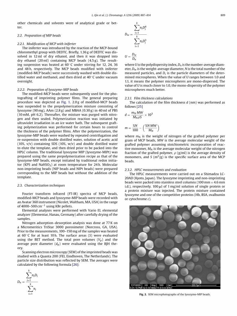
Fig. 2. SEM microphotographs of the lysozyme-MIP beads.

810 L. Qin et al. / J. Chromatogr. A 1216 (2009) 807–814
Fs
(koa
cN
p
3
3
tuTiuvpisw
iuaw
TPb
B
MMLLLN
Table 2Elemental analysis of the support beads and the molecular imprinting beads.
Bead type %C %N
MCP beads 79.9 0.00Modified MCP beads12a 78.5 1.39Modified MCP beads24a 76.8 1.65Modified MCP beads36a 76.0 1.68Modified MCP beads48a 74.3 1.70
cific surface area, total pore volume and average pore diameter oflysozyme-MIP beads and NIP beads were different (Table 1). The
ig. 3. Pore size distribution of the lysozyme-MIP beads calculated from the nitrogenorption measurements.
The capacity factors (kt and kc) and the separation factor˛) were calculated using the following formulas: kt = (tt − t0)/t0,c = (tc − t0)/t0, and ˛ = kt/kc, where tt and tc are the retention timesf the template protein and the competitive protein, respectively,nd t0 is the retention time of the void marker, thiourea.
The chromatographic imprinting factor (CIF) for the template isalculated from the capacity factors obtained on MIP column andIP column (CIF = kMIP/kNIP).
The resolution (Rs) is calculated by the following equation:R = 2(tt−tc)
(Wt+Wc) where Wt and Wc are the peak widths of the tem-late and the competitive protein, respectively.
. Results and discussion
.1. The advantage of using iniferter
An advantage of using immobilized iniferter is the stability ofhe dithiocarbamate radical. Since the dithiocarbamate radical isnlikely to initiate new chains, propagation in solution is minimal.he effect of the SIP on the imprinting beads was well-illustratedn SEM of the lysozyme-MIP beads (Fig. 2). The grafting polymersing SIP appeared homogeneous with no agglomeration and noisible non-grafted polymer in the polymerization solution. Theolydispersity index (U) of 1.036 was calculated from Fig. 2, which
ndicated that the lysozyme-MIP beads were of mono-dispersionize. Besides, the pore size distribution of the lysozyme-MIP beadsas homogeneous (Fig. 3) through the SIP.
Moreover, the iniferter allowed an efficient control of the graft-ng process, so the film thickness was tunable by controlling theltraviolet irradiation time (Table 1). The thickness of the filmffected the chromatographic selectivity of the MIP column, whichill be discussed below. Of particular interest was the use of a
able 1ore properties of the support beads and film thickness of the molecular imprintingeads.
ead type S (m2/g) Vp (ml/g) dp (nm) da (nm)
CP beads 59.2 0.377 28.5odified MCP beads24b 39.3 0.343 32.2
ysozyme-MIP beads2c 41.3 0.332 32.0 4.70ysozyme-MIP beads4c 39.7 0.255 30.2 5.62ysozyme-MIP beads6c 33.8 0.217 29.0 7.10IP beads 28.4 0.198 20.6
a The film thickness (d) was calculated from formula.b It is the reaction time of the chloromethyl groups with DEDTC.c It is the reaction time of the polymerization under ultraviolet irradiation.
Lysozyme-MIP beads4b 60.5 5.91
a It is the reaction time of the chloromethyl groups with DEDTC.b It is the reaction time of the polymerization under ultraviolet irradiation.
hydrophobic MCP bead surface to graft a hydrophilic polyacry-lamide imprinted polymer. Some reports have indicated that suchinterfaces can stabilize imprinted site, even when using polar,water-containing monomer/template systems [27].
3.2. Modification time of MCP beads with iniferter
In the grafting of polymers from the modified-MCP beads, thedensity of initiator groups is an important factor controlling thedensity and homogeneity of the grafted layer. The reaction time ofthe chloromethyl groups with DEDTC could influence the densityof initiator groups (Table 2). It can be seen from Table 2 that the %Nof modified-MCP beads increased with the increase of the reactiontime. When the reaction time was over 24 h, the %N of modified-MCP beads increased slowly. So the time chosen as the reactiontime of modification was 24 h.
3.3. Characteristic studies
The IR spectra of MCP beads, modified-MCP beads, andlysozyme-MIP beads were shown in Fig. 4. It was notable that themodified-MCP beads showed new peaks at 1414.44, 1351.30 and1205.11 cm−1 corresponding to thiocarbonyl group of the iniferter,which suggested that the iniferter had been modified to the MCPbeads. The presence of the iniferter was also checked by elementalanalysis (Table 2, reduction of %C and appearance of %N). The strongpeak at 1654.56 cm−1 of lysozyme-MIP beads was attributed to thecarbonyl groups of the polyacrylamide. The IR results indicated thatimprinting polymers have been grafted from the modified-MCPbeads. It was further confirmed by elemental analysis of the beadsbefore (C% = 76.8%, N% = 1.65%) and after (C% = 60.5%, N% = 5.91%)polymerization.
The MCP beads were mesoporous as judged from the nitrogensorption data (S = 59.2 m2/g; dp = 28.5 nm; Vp = 0.377 ml/g). Spe-
pore diameter of lysozyme-MIP beads (30.2 nm) was possibly inthe range of diffusion of lysozyme molecule whose molecular size is
Fig. 4. FT-IR spectra of (a) MCP beads, (b) modified-MCP beads and (c) lysozyme-MIPbeads.

L. Qin et al. / J. Chromatogr. A 1216 (2009) 807–814 811
F zyme-m
3cd
(MSoTiplweic
3
3
larhuta
ig. 5. SEM microphotographs of the surface of (a) modified-MCP beads, (b) lysoicrophotographs were the correspondence of the entire beads.
.0 nm × 3.0 nm × 4.5 nm [28]. On the basis of the data, it was con-luded that lysozyme-MIP beads have effective pore volume andimensions for lysozyme molecules.
The surface morphology of the beads was observed by SEMFig. 5). From Fig. 5a, it can be seen that the surface of modified-
CP beads was rugged after being modified with the iniferter. In theEM photograph of lysozyme-MIP beads (Fig. 5b), a large quantityf well-distributed pores could be observed and they were netlike.his porous structure on the surface of lysozyme-MIP beads facil-tated the mass transfer rate of releasing and rebinding templaterotein. Due to the imprinting effect, the surface morphology of
ysozyme-MIP beads and NIP beads were different (Fig. 5b and c),hich was consistent with nitrogen sorption data (Table 1). How-
ver, in the case of lysozyme-MIPt beads (Fig. 5d), the graftingmprinted polymer was not homogeneous, which was the short-oming of the traditional polymerization.
.4. Chromatographic analysis
.4.1. Imprinting effect of lysozyme-MIP beadsThe retention behavior of lysozyme on lysozyme-MIP column,
ysozyme-MIPt column and the corresponding NIP column werenalyzed in the same condition (Fig. 6). It can be found that the
etention capacity of lysozyme-MIP column to the template wasigher than that of NIP column (Table 3). The lysozyme-MIP col-mn had a good imprinting effect (CIF = 12.8). These data indicatedhat molecular imprinting process had resulted in the formationnd preservation of a microenvironment based on shape memo-MIP beads, (c) NIP beads and (d) lysozyme-MIPt beads. The top left corner SEM
rial size and positions of functional groups of the template on thesurface of the lysozyme-MIP beads [29]. For NIP beads, however,the non-specific adsorption had dominant effect due to the lackof imprinting process and there were no suitable cavities for therecognition of lysozyme.
Although the lysozyme-MIPt column had imprinting effect insome degree with the CIF of 2.64 (Fig. 6c), it was not better thanlysozyme-MIP column (Fig. 6a and b). And both the lysozyme-MIPt column and NIPt column showed peaks tailing in binding thetemplate. Lysozyme-MIP beads provided better chromatographicproperty mainly due to the narrow size distributions and homoge-neous pore size.
3.4.2. Recognition specificity of the imprinting columnThe selectivity of the imprinting column was tested by using Hb,
BSA, ovalbumin or cytochrome c as competitive protein which isdifferent in mass and charge from lysozyme. In the SIP, the graftedlayer thickness increased with the increase of the reaction time.After 4 h polymerization, the lysozyme-MIP column had a good res-olution (Fig. 7). The lysozyme-MIP beads prepared using shorterpolymerization time (2 h) exhibited only partial resolution andweak retention of the template. Longer grafting time (6 h) increasedthe number of recognition site and resulted in strong retention of
the template, but the Rs did not follow this trend. Over thicknessof the imprinting layer did not benefit for the mass transfer effi-ciency of the protein. The capacity factor of lysozyme-MIP columnfor lysozyme was much higher than that for Hb, BSA, ovalbuminor cytochrome c (Table 3). The lysozyme-MIP column exhibited
812 L. Qin et al. / J. Chromatogr. A 1216 (2009) 807–814
Table 3Chromatographic properties of the lysozyme-MIP column and the NIP column to the single protein.
Proteins Lysozyme-MIP column NIP column
Retention time (min) Capacity factor (k) Retention time (min) Capacity factor (k)
Lysozyme 9.21 1.53 4.10 0.12Hb 3.98 0.09 3.89 0.07BSA 3.88 0.06 3.86 0.05Ovalbumin 3.97 0.09 3.84 0.05Cytochrome c 4.02 0.10 3.91 0.07
Column: 100 mm × 4.6 mm (i.d.); sample amount: 100 �l (1 mg/ml); detection wavelength: 280 nm; mobile phase: PBS (10 mM, pH 6.2); flow rate: 0.2 ml/min.
Fig. 6. Chromatograms of lysozyme on (a) the lysozyme-MIP column, (b) the NIPcolumn and (c) the lysozyme-MIPt column (. . .) and NIPt column (—). Column:100 mm × 4.6 mm (i.d.); sample amount: 100 �l (1 mg/ml); detection wavelength:280 nm; mobile phase: PBS (10 mM, pH 6.2); flow rate: 0.2 ml/min. Peak identifica-tion: (1) lysozyme.
Fig. 7. Chromatograms of the mixture of lysozyme and cytochrome c on the
lysozyme-MIP column prepared in different hours. Column: 100 mm × 4.6 mm (i.d.);sample amount: 100 �l (1 mg/ml); detection wavelength: 280 nm; mobile phase:PBS (10 mM, pH 6.2); flow rate: 0.2 ml/min. Peak identification: (1) lysozyme and(2) cytochrome c.good recognition for template from a protein mixture (Table 4 andFig. 8). Concerning lysozyme-MIPt column, no selectivity could bedemonstrated for the template.
As known, the peptide bonds of the protein could form hydrogenbonds with AAm even in polar solution [30]. Multiple-point hydro-gen bonds interaction between the template protein and AAm canform strong interactions [14]. The imprinting column discriminatedproteins by the synergistic effects of shape complementarity and
multiple weak interactions provided by the functional monomers.In the cases of Hb, BSA or ovalbumin, the molecular volume waslarger than that of lysozyme so that they had less chance to entirelyslip into the imprinting cavities created by lysozyme and to interactTable 4Chromatographic properties of the lysozyme-MIP column to the protein mixture.
Protein mixture Lysozyme-MIP column
Capacity factor (k) Separation factor (˛) Resolution (Rs)
1Hb 0.09 16.66 0.82Lysozyme 1.50
2BSA 0.06 19.33 0.75Lysozyme 1.16
3Ovalbumin 0.13 11.46 0.83Lysozyme 1.49
4Cytochrome c 0.04 25.25 0.82Lysozyme 1.01
Column: 100 mm × 4.6 mm (i.d.); sample amount: 100 �l (1 mg/ml); detection wave-length: 280 nm; mobile phase: PBS (10 mM, pH 6.2); flow rate: 0.2 ml/min.

L. Qin et al. / J. Chromatogr. A 1216 (2009) 807–814 813
Fig. 8. Chromatograms of the mixture of lysozyme with one of Hb, BSA, ovalbumin or cytochrome c on the lysozyme-MIP column. Column: 100 mm × 4.6 mm (i.d.); sampleamount: 100 �l (1 mg/ml); detection wavelength: 280 nm; mobile phase: PBS (10 mM, pH 6.2); flow rate: 0.2 ml/min. Peak identification: (1) lysozyme; (2) Hb; (3) BSA; (4)ovalbumin and (5) cytochrome c.
Table 5Reproducibility of the lysozyme-MIP column for the mixture of lysozyme and cytochrome c.
Protein mixture Resolution (Rs)
First time Second time Third time Fourth time Fifth time
Cytochrome c0.82 0.82 0.83 0.79 0.80L
C lengt
wsmqiwcwcic
palut(
3
fit
ysozyme
olumn: 100 mm × 4.6 mm (i.d.); sample amount: 100 �l (1 mg/ml); detection wave
ith the functional groups. Therefore, lysozyme-MIP column couldeparate the mixture of lysozyme with one of Hb, BSA or ovalbu-in. The molecular volume and pI of cytochrome c and lysozyme are
uite similar. Though cytochrome c was small enough to get into themprinting cavities, the recognition sites of the imprinting cavities
ere not complementary to the cytochrome c. Consequently, theytochrome c could not form entirely multiple weak interactionsith the functional groups, so the retention time of cytochromewas shorter than that of the template protein. Concerning the
mprinted composites, the high selectivity to lysozyme was suc-essfully demonstrated.
In contrast, for NIP column, it was difficult to form multi-le hydrogen bonds and strong interaction between the templatend NIP-beads, which led to the weak retention capacity ofysozyme, and so as for the competitive proteins. The NIP col-mn did not display any protein recognition ability and retainedhe five kinds of proteins nearly close to the void fractionTable 3).
.5. Reusability and stability of lysozyme-MIP column
Using the mixture of lysozyme and cytochrome c as example,ve times of selective separation experiments were performed onhe same lysozyme-MIP column (Table 5). The results showed that
h: 280 nm; mobile phase: PBS (10 mM, pH 6.2); flow rate: 0.2 ml/min.
the lysozyme-MIP column was very stable and maintained the sep-aration capacity at almost constant value.
4. Conclusions
In the present study, this new approach significantly broadensthe scope of protein imprinting through surface initiated living-radical polymerization and demonstrates that iniferter-modifiedsupport materials can be used to prepare protein molecularlyimprinted polymer. It is a new stationary phase for the separa-tion of bioactive compounds only using aqueous mobile phase.MIP beads prepared by SIP exhibited a better imprinting effectthan traditional MIP beads prepared by traditionally initiated rad-ical polymerization. Through using SIP, the thickness of imprintingpolymer layer could be rationally controlled to obtain good selectiv-ity. The lysozyme-MIP column exhibited a pronounced imprintingeffect and was capable of separating the template from competitiveproteins, whereas the NIP column had no selective properties.
Acknowledgements
We are grateful to the National Basic Research Program ofChina (973 Program, nos. 2007CB914100 and 2006CB705703), theNational Nature Science Foundation of China (no. 20875049) and

8 togr. A
Tc
R
[[
[
[[
[
[[[[
14 L. Qin et al. / J. Chroma
ianjin Natural Science Foundation (no. 06YFJMJC02800) for finan-ial support.
eferences
[1] K. Haupt, K. Mosbach, Chem. Rev. 100 (2000) 2495.[2] Y. Lei, K. Masbach, Chem. Mater. 20 (2008) 859.[3] N.W. Turner, C.W. Jeans, K.R. Brain, C.J. Allender, V. Hlady, D.W. Britt, Biotechnol.
Progr. 22 (2006) 1474.[4] M. Gald, O. Norrlöw, B. Sellergren, N. Siegbahn, K. Mosbach, J. Chromatogr. 347
(1985) 11.[5] D.L. Venton, E. Gudipati, Biochim. Biophys. Acta 1250 (1995) 117.[6] T. Shiomi, M. Matsui, F. Mizukami, K. Sakaguchi, Biomaterials 26 (2005)
5564.[7] S. Mallik, S.D. Plunkett, P.K. Dhal, R.D. Johnson, D. Pack, D. Shnek, F.H. Arnold,
New J. Chem. 18 (1994) 299.
[8] M. Kempe, M. Glad, K. Mosbach, J. Mol. Recog. 8 (1995) 35.[9] A. Rachkov, M.J. Hu, E. Bulgarevich, T. Matsumoto, N. Minoura, Anal. Chim. Acta504 (2004) 191.10] H. Nishino, C.S. Huang, K.J. Shea, Angew. Chem. Int. Ed. 45 (2006) 2392.11] S. Hjertén, J.L. Liao, K. Nakazato, Y. Wang, G. Zamaratskaia, H.X. Zhang, Chro-
matographia 44 (1997) 227.
[
[[
[
1216 (2009) 807–814
12] S.H. Ou, M.C. Wu, T.C. Chou, C.C. Liu, Anal. Chim. Acta 504 (2004) 163.[13] J.T. Huang, J. Zhang, J.Q. Zhang, S.H. Zheng, J. Appl. Polym. Sci. 95 (2005) 358.[14] X.S. Pang, G.X. Cheng, S.L. Lu, E.J. Tang, Anal. Bioanal. Chem. 384 (2006) 225.[15] T.Y. Guo, Y.Q. Xia, J. Wang, M.D. Song, B.H. Zhang, Biomaterials 26 (2005) 5737.[16] H.Q. Shi, W.B. Tsai, M.D. Garrison, S. Ferrari, B.D. Ratner, Nature 398 (1999) 593.[17] C.J. Tan, S. Wangrangsimakul, R. Bai, Y.W. Tong, Chem. Mater. 20 (2008) 118.[18] Y. Li, H.H. Yang, Q.H. You, Z.X. Zhuang, X.R. Wang, Anal. Chem. 78 (2006) 317.[19] D.E. Hansen, Biomaterials 28 (2007) 4178.20] J.L. Liao, Y. Wang, S. Hjertén, Chromatographia 42 (1996) 259.21] C. Sulizky, B. Rueckert, A.J. Hall, F. Lanza, K. Unger, B. Sellergren, Macromolecules
35 (2002) 79.22] R.E. Fairhurst, C. Chassaing, R.F. Venn, A.G. Mayes, Biosens. Bioelectron. 20
(2004) 1098.23] Y. Nakayama, T. Matsuda, Langmuir 15 (1999) 5560.24] B. Rueckert, A.J. Hall, B. Sellergren, J. Mater. Chem. 12 (2002) 2275.25] M.M. Titirici, B. Sellergren, Chem. Mater. 18 (2006) 1773.26] F. Bai, X.L. Yang, W.Q. Huang, Macromolecules 37 (2004) 9746.
27] S.A. Piletsky, H. Matuschewski, U. Schedler, A. Wilpert, E.V. Piletska, T.A. Thiele,M. Ulbricht, Macromolecules 33 (2000) 3092.28] G. Wulff, Angew. Chem. Int. Ed. 34 (1995) 1812.29] A. Bossi, S.A. Piletsky, E.V. Piletska, P.G. Righetti, A.P.F. Turner, Anal. Chem. 73
(2001) 5281.30] C. Yu, K. Mosbach, J. Org. Chem. 62 (1997) 4057.