suppression of notch signaling in the neonatal mouse ovary
TRANSCRIPT
Suppression of Notch Signaling in the NeonatalMouse Ovary Decreases Primordial Follicle Formation
Daniel J. Trombly, Teresa K. Woodruff, and Kelly E. Mayo
Department of Biochemistry, Molecular Biology, and Cell Biology and Center for Reproductive Science (D.J.T., T.K.W.,K.E.M.), Northwestern University, Evanston, Illinois 60208; and Department of Obstetrics and Gynecology (T.K.W.),Northwestern University, Chicago, Illinois 60611
Notch signaling directs cell fate during embryogenesis by influencing cell proliferation, differen-tiation, and apoptosis. Notch genes are expressed in the adult mouse ovary, and roles for Notch inregulating folliculogenesis are beginning to emerge from mouse genetic models. We investigatedhow Notch signaling might influence the formation of primordial follicles. Follicle assembly takesplace when germ cell syncytia within the ovary break down and germ cells are encapsulated bypregranulosa cells. In the mouse, this occurs during the first 4–5 d of postnatal life. The expressionof Notch family genes in the neonatal mouse ovary was determined through RT-PCR measure-ments. Jagged1, Notch2, and Hes1 transcripts were the most abundantly expressed ligand, recep-tor, and target gene, respectively. Jagged1 and Hey2 mRNAs were up-regulated over the periodof follicle formation. Localization studies demonstrated that JAGGED1 is expressed in germ cellsprior to follicle assembly and in the oocytes of primordial follicles. Pregranulosa cells that surroundgerm cell nests express HES1. In addition, pregranulosa cells of primordial follicles expressedNOTCH2 and Hey2 mRNA. We used an ex vivo ovary culture system to assess the requirement forNotch signaling during early follicle development. Newborn ovaries cultured in the presence of�-secretase inhibitors, compounds that attenuate Notch signaling, had a marked reduction in primor-dial follicles compared with vehicle-treated ovaries, and there was a corresponding increase in germcells that remained within nests. These data support a functional role for Notch signaling in regulatingprimordial follicle formation. (Endocrinology 150: 1014–1024, 2009)
Ovarian follicles are the functional units within the femalegonad that nurture maturation of the oocyte and enable
production of steroid hormones. Follicles are comprised of threecells types: oocytes, surrounding granulosa cells, and an externalthecal cell layer. Select numbers of follicles mature in response tocirculating gonadotropins and to the local actions of growthfactors during the female reproductive cycle (1). Follicle matu-ration continues until ovulation, when an egg or eggs competentfor fertilization are extruded from the ovary and the remainingsomatic cells of the follicle luteinize. Although much is knownabout how secondary follicles progressively develop into pre-ovulatory follicles, the molecular events mediating primordialfollicle formation and initial follicle growth are less clear.
In mice, primordial germ cells migrate to the urogenital ridgearound embryonic d 11 (2). By embryonic d 13.5, synchronous
rounds of mitotic division in the female gonad yield clusters ofoocytes arranged in syncytia commonly referred to as cysts ornests. (3). Syncytia persist until germ cells undergo a wave ofapoptosis near the time of birth (4). During programmed nestbreakdown, germ cells are encapsulated by squamous somaticcells (pregranulosa cells) to generate primordial follicles. Thenewborn mouse ovary contains few primordial follicles, whereasat postnatal d 2 approximately 40% of germ cells are withinprimordial follicles (4). This number increases to greater than80% by postnatal d 6 (4, 5). Perturbations during the criticalperiod of primordial follicle formation can significantly affectthe size of the primordial follicle pool and follicular phenotypes.For example, administration of activin to neonatal mice in-creases the primordial follicle pool by 30% (5), whereas theovaries of neonatal mice injected with estradiol, the synthetic
ISSN Print 0013-7227 ISSN Online 1945-7170Printed in U.S.A.Copyright © 2009 by The Endocrine Societydoi: 10.1210/en.2008-0213 Received February 13, 2008. Accepted September 16, 2008.First Published Online September 25, 2008
Abbreviations: Ct, Cycle threshold; DAPT, N-(N-(3,5-difluorophenacetyl)-L-alanyl)-S-phe-nylglycine t-butyl ester; DMSO, dimethylsulfoxide; DSL, Drosophila homolog-� and -Ser-rate, and the C. elegans homolog Lag-2; GAPDH, glyceraldehyde-3-phosphate dehydro-genase; Hes, hairy and enhancer of split; L-685,458, (5S)-(t-butoxycarbonylamino)-6-phenyl-(4R)hydroxy-(2R)benzylhexanoyl)-L-leu-L-phe-amide; MOF, multioocytic follicle;TUNEL, terminal deoxynucleotidyl transferase-mediated deoxyuridine triphosphate nickend labeling.
R E P R O D U C T I O N - D E V E L O P M E N T
1014 endo.endojournals.org Endocrinology, February 2009, 150(2):1014–1024
at Northwestern Univ Sci-Engineering Library on February 17, 2009 endo.endojournals.orgDownloaded from

estrogen diethylstilbestrol, or the phytoestrogen genistein de-velop multioocytic follicles (MOFs) (6–10). MOFs, which havetwo or more germ cells trapped within a follicle boundary (6, 7),are also observed in mouse ovary cultures treated with estradiol(11). These structures likely arise from incomplete breakdown ofgerm cell nests.
Contacts between germ cells and somatic cells are establishedas early as embryonic d 13.5 in the mouse ovary (4). Thus, com-munication between germ cells and pregranulosa cells is likelyimportant for orchestrating follicle assembly. Given the manyroles of the Notch signaling pathway in cell communication andmorphogenesis, this pathway is a likely candidate for regulatingearly follicle development. Notch signaling affects cell fate dur-ing embryogenesis and in turn influences cell proliferation, dif-ferentiation, and apoptosis (12).
Originally characterized in Drosophila and Caenorhabditiselegans, components of the Notch pathway have several mam-malian orthologues. These include four Notch receptors(Notch1–4), and five Notch ligands: Jagged1, Jagged2, Delta-like 1, Delta-like 3, and Delta-like 4 (13). Notch ligands belongto the Drosophila homolog Delta and Serrate, and the C. eleganshomolog Lag-2 (DSL) family of proteins (14). Notch genes en-code conserved transmembrane receptors, and the DSL ligandsare also membrane bound. Signaling occurs between apposingcells that express Notch receptors and DSL ligands. After ligandbinding, a cascade of proteolytic cleavages of the Notch receptorensues (15). The active form of Notch, the Notch intracellulardomain, is generated by cleavage at the receptor juxtamembraneregion by the �-secretase complex (16). Liberated Notch intra-cellular domain translocates into the nucleus in which it associ-ates with the transcriptional regulator C-promoter binding fac-tor 1/suppressor of hairless/Lag-1 (CSL) to promote Notch targetgene transcription (13). Well-characterized Notch target genesinclude two families of basic-helix-loop-helix transcription fac-tors: hairy and enhancer of split (Hes) and a related family (Hesrelated with YRPW motif, hairy related transcription factor)(17–20). Depending on the cellular context, Notch signaling isreduced or potentiated by Fringe proteins, a class of glycosyl-transferases that modify the receptor (21). In mammals, the threeFringe proteins that modulate Notch signaling are Lunatic,Manic, and Radical Fringe (22). Interestingly, the Lunatic Fringeknockout mouse ovary exhibits meiotic defects and developsMOFs (23). An analogous phenotype occurs in the Drosophilagonad, in which the absence of Notch signaling during egg cham-ber formation results in fused egg chambers (24). Taken to-gether, these data suggest that Notch signaling may have con-served roles in follicle development within the female gonad.
We tested the hypothesis that Notch signaling is required forfollicle assembly in the mouse ovary. Our studies centered on theinterval between birth and postnatal d 4, the time period whenmost primordial follicles form in the mouse. We found thatNotch receptors and ligands were expressed in a complementarypattern in the neonatal mouse ovary, with Notch2 in granulosacells and Jagged1 in germ cells. Germ cell nests were retained andprimordial follicles were reduced in ovaries cultured with�-secretase inhibitors that suppress Notch signaling. These data
support a functional role for Notch signaling in regulating pri-mordial follicle formation.
Materials and Methods
Animal treatment and tissue collectionCD-1 mice (Harlan, Indianapolis, IN) were maintained on a 12-h
light, 12-h dark cycle (lights off at 1700 h) with food and water availablead libitum. Breeders (90–180 d old) were fed with a soy-free mouse chow(Harlan 7926) to limit exogenous phytoestrogen intake through food.Matings were timed, and vaginal plug detection was considered d 0.5 ofpregnancy. Ovaries were collected from newborn mice (within 2 h ofbirth) or mice at different postnatal times. Day 0 marks the first 24 h afterbirth. Ovaries were either stored at �80 C for subsequent RNA isolation,immediately fixed for follicle counting and immunohistochemical stud-ies, or prepared for ex vivo culture. Animals were cared for in accordancewith all federal and institutional guidelines.
Antibodies and inhibitorsJagged1 (SC-6011) and Notch2 (SC-5545) polyclonal antibodies
were purchased from Santa Cruz Biotechnology, Inc. (Santa Cruz, CA),and used at a 1:50 and 1:100 dilution, respectively. A monoclonalNotch2 antibody (C651.6DbHN) was provided by the Iowa Develop-mental Studies Hybridoma Bank (Iowa City, IA) and used at 8 �g/ml. AHes1 polyclonal antibody (25) was kindly provided by Dr. Tetsuo Sudo(Toray Industries Inc., Tokyo, Japan) and used at a 1:500 dilution.�-Secretase inhibitors (5S)-(t-butoxycarbonylamino)-6-phenyl-(4R)hy-droxy-(2R)benzylhexanoyl)-L-leu-L-phe-amide (L-685,458; L1790) andN-(N-(3,5-difluorophenacetyl)-L-alanyl)-S-phenylglycine t-butyl ester(DAPT; D5942) were purchased from Sigma-Aldrich (St. Louis, MO).
Semiquantitative RT-PCR and real-time PCRFor semiquantitative PCR studies, dissected ovaries from postnatal d
3 CD-1 mice were stripped of the bursal sac and immediately frozen. AnRNeasy kit (QIAGEN, Valencia, CA) was used to isolate RNA frompooled ovaries (n � 12), and 500 ng RNA was reverse transcribed usingavian myeloma virus reverse transcriptase. One quarter of the resultingcDNA was used in PCRs. Primers for ribosomal protein RPL19 wereused as a control, and PCRs were run for 26 cycles. 32P-dCTP was in-corporated during PCR, and products were visualized after exposingdried gels to Hyperfilm (GE Healthcare, Piscataway, NJ). (See supple-mental Table 1, published as supplemental data on The Endocrine So-ciety’s Journals Online web site at http://endo.endojournals.org, forprimer sequences and amplicon sizes). For real-time PCR experiments,ovaries were dissected from d 0, 2, 6, and 18 CD-1 mice. One microgramof RNA from each set of pooled ovaries (n � 12–14 for d 0–6; n � 2 forday 18) was used for each reverse transcription reaction, and 100 ng ofcDNA served as the template for PCR. Real-time PCR assays were per-formed using SYBR Green PCR master mix (Applied Biosystems, FosterCity, CA), and mouse RPL19 served as the internal control (26). Thecomparative cycle threshold (Ct) method (��Ct) was used for relativequantification (27). To examine mRNA expression in cultured ovaries,two ovaries were pooled, and eluates from RNeasy columns were pre-cipitated. RNA samples were resuspended in a small volume of dieth-ylpyrocarbonate water, and real-time PCR experiments were performedusing 50 ng of cDNA. Relative mRNA values were normalized to RPL19.
In situ hybridizationOvarian sections from CD-1 mice were processed for in situ hybrid-
ization as previously described (28), with modifications. A 630 bp Hey2fragment was amplified from d 18 mouse cDNA and cloned into thepcDNA3 vector (Invitrogen Corp., Carlsbad, CA). Primers used to gen-erate the Hey2 amplicon were: forward 5�-TGAAGATGCTCCAGGC-
Endocrinology, February 2009, 150(2):1014–1024 endo.endojournals.org 1015
at Northwestern Univ Sci-Engineering Library on February 17, 2009 endo.endojournals.orgDownloaded from

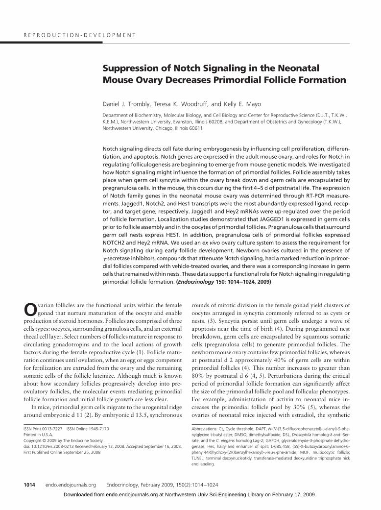
TACAGG-3� and reverse 5�-ATACCGACAAGGGTGGGCTGAT-3�. AHey2 riboprobe was prepared using digoxigenin-11-UTP (Roche Ap-plied Science, Indianapolis, IN) and the Riboprobe combination system(Promega Corp., Madison, WI). Detection of mRNA was achieved usinga digoxigenin-AP antibody (Roche Applied Science, Indianapolis, IN)and the nitro blue tetrazolium chloride/5-bromo-4-chloro-3-indolyl-phosphate, 4-toluidine salt substrate (Sigma-Aldrich).
ImmunohistochemistryOvaries harvested from CD-1 mice were fixed in 4% paraformalde-
hyde overnight at 4 C. Before tissue processing, samples were dehydratedwith 50 and then 70% ethanol. Ovaries were further dehydrated througha graded series of ethanols and infiltrated with paraffin. Embedded sam-ples were sectioned at 5 �m. Tissue sections were dewaxed and thenrehydrated. Antigen retrieval was performed by microwaving samples onhigh power for 2 min and on low power for 7 min in 0.01 M sodium citrate(pH 6). After incubating in 3% H2O2/PBS for 15 min to block endoge-nous peroxidase activity, tissue sections were blocked with avidin/biotinreagents (Vector Laboratories, Inc., Burlingame, CA). Blocking wasachieved by immersing slides in a solution containing 10% serum for 1 hat room temperature. Samples were incubated with primary antibodiesdiluted in blocking serum overnight at 4 C and were then exposed tobiotin-conjugated secondary antibodies (1:200 dilution; Vector Labo-ratories) for 30 min at room temperature. The ABC reagent (VectastainElite ABC kits; Vector Laboratories) was used according the manufac-turer’s instructions. All washes were performed in PBS-Tween 20 afterincubations with antibody and ABC reagent. The 3,3�-diaminobenzidinesubstrate (Vector Laboratories) was used for colorometric detection, andsamples were counterstained with hematoxylin. A proliferating cell nu-clear antigen staining kit (Zymed Laboratories, South San Francisco,CA) was used to identify proliferating cells in tissue sections. Terminaldeoxynucleotidyl transferase-mediated deoxyuridine triphosphate nickend labeling (TUNEL) staining was performed using the DeadEnd flu-orometric TUNEL system kit (Promega) to assess cellular apoptosis. Allsections processed for fluorescence detection were mounted in mediumcontaining 4�,6-diamidino-2-phenylindole (Vector Laboratories). Im-munohistochemical images were acquired on a Nikon E600 microscopeusing a Spot Insight Mosaic 11.2 color digital camera (Diagnostic In-struments, Sterling Heights, MI) and Advanced Spot Imaging software(version 4.6; Universal Imaging, Downington, PA). Immunofluorescentimages were generated using a Leica DM5000B fluorescence microscopeand OpenLab 4.0 software (Improvision, Lexington, MA).
Organ culture and morphometric analysisOvaries from newborn CD-1 mice were dissected and the bursal sac
removed in PBS. Ovaries were placed in 5-�l drops of media and culturedfor 4 d on 0.4-�m floating filters (Millicell-CM; Millipore Corp., Bil-lerica, MA) at 37 C in a chamber containing 5% CO2. Filters were placedin 14-mm culture wells and rested on top of 0.4 ml DMEM-F12 mediasupplemented with 0.1% Albumax (Invitrogen), penicillin-streptomycin(Invitrogen), 0.1% BSA (Sigma), 27.5 �g/ml transferrin (Sigma), and0.05 mg/ml L-ascorbic acid (Sigma). Culture medium contained 1, 10, or50 �g/ml insulin (Sigma) and was changed daily. The media formulationwas adapted from Kezele and Skinner (29). In studies using �-secretaseinhibitors, ovaries were cultured for 1, 2, or 4 d in media containing either10 �M L-685,458 or 20 �M DAPT. Vehicle-treated ovaries were culturedin media containing 0.2% dimethylsulfoxide (DMSO). Cultured ovarieswere fixed in 4% paraformaldehyde and stained with hematoxylin andeosin. To compare follicle formation between in vivo and cultured ova-ries, ovaries were isolated from littermate animals the same day culturedovaries were fixed. Follicle populations among isolated and culturedovaries were quantified. Ovaries were sectioned at 5 �m, and follicleswere counted in every fifth section as described elsewhere (5) to avoidduplicate counts. Images of ovarian sections were analyzed using ImageJsoftware (National Institutes of Health, Bethesda, MD), and follicleswere manually counted by two individuals. Germ cells not surroundedby pregranulosa cells were scored as unassembled (remaining in nests).
Oocytes surrounded by pregranulosa cells or a mixture of squamous andcuboidal somatic cells were scored as primordial follicles. Primary andsecondary follicles were scored when oocytes were surrounded by a sin-gle or double layer of cuboidal granulosa cells, respectively. Primary andsecondary follicle counts were added together (developing follicles). Fol-licle populations were expressed as a percentage of the total number ofgerm cells counted.
StatisticsData are presented as means � SEM. One-way ANOVA followed by
a Tukey-Kramer post hoc analysis was used for statistical comparisonsamong multiple groups. ANOVA was performed using GraphPadPRISM 4.0 (GraphPad Software, Inc., San Diego, CA). For statisticalcomparisons between two groups, the Student’s two-tailed t test wasused. P � 0.05 was considered significant.
Results
Notch family genes are expressed in the neonatalmouse ovary
Johnson et al. (30) described the expression patterns for sev-eral Notch pathway genes in the adult mouse ovary. We inves-tigated Notch gene expression in the neonatal mouse ovary dur-ing the period when primordial follicles form. The d 0 mouseovary chiefly contains germ cells that are arranged in nests (sup-plemental Fig. 1A). The ovarian architecture changes dramati-cally 3 d into postnatal development as germ cell nests break-down. Ovaries at this stage contain a heterogeneous populationof germ cell nests, primordial and primary follicles (supplementalFig. 1B). Because Notch signaling may affect processes mediatinggerm cell nest breakdown and/or initial follicle growth, we usedneonatal d 3 ovaries for semiquantitative RT-PCR assays to de-termine the expression profiles for Notch family genes. Tran-scripts for all Notch receptors were expressed in the d 3 mouseovary, with Notch2 showing the highest expression (Fig. 1A).Jagged1 and Jagged2 mRNAs were more abundant than theDelta-like ligands in the neonatal ovary. Hes1 and Hey2 tran-scripts showed the highest abundance of the eight Notch targetgenes tested. We also examined the expression of the three Fringemolecules, and Radical Fringe showed the strongest expression(data not shown). We performed real-time PCR experiments todetermine whether Notch family genes, those strongly expressedby d 3, were regulated during postnatal development (Fig. 1B).There was no change in Notch2, Notch3, Jagged2, and Hes1mRNA expression between d 0 and 6, but a significant decreasewas observed in d 18 ovaries compared with earlier ovaries (Fig.1B). In contrast, Jagged1 mRNA increased 2.5-fold during thepeak period of follicle formation, between d 0 and 6, and thendeclined such that by d 18 (prepubertal), the level of Jagged1transcript was similar to that found in d 0 ovaries (Fig. 1B). Hey2mRNA increased 5-fold between d 0 and 6 and then increasedfurther, 10-fold, in d 18 ovaries compared with d 0 ovaries (Fig.1B). These data show that multiple Notch genes are expressedand dynamically regulated during the time of follicle formation.
Immunohistochemical experiments were used to determinethe cellular localization of NOTCH2, JAGGED1, and HES1proteins (Fig. 2). NOTCH2 was expressed in pregranulosa cellsof the newborn mouse ovary (Fig. 2A). Light NOTCH2 staining
1016 Trombly et al. Notch Signaling in Neonatal Mouse Ovary Endocrinology, February 2009, 150(2):1014–1024
at Northwestern Univ Sci-Engineering Library on February 17, 2009 endo.endojournals.orgDownloaded from

was also observed in germ cells of the newborn mouse ovary.NOTCH2 was localized predominantly in the pregranulosacells of primordial follicles and granulosa cells of primaryfollicles in the d 3 ovary (Fig. 2B and supplemental Fig. 2A).In contrast, JAGGED1 was expressed in germ cells of germ cellnests in the newborn ovary (Fig. 2C). At d 3, JAGGED1 con-tinued to be expressed in oocytes of primordial and primaryfollicles (Fig. 2D and supplemental Fig. 2B). HES1 was ex-pressed in pregranulosa cells that surround germ cell nests(Fig. 2E) in the newborn ovary. HES1 expression was main-tained in pregranulosa cells surrounding primordial folliclesin the d 3 ovary, whereas oocytes of early primary follicles alsostained positively for HES1 (Fig. 2F). Due to the lack of areliable Hey2 antibody, we examined Hey2 mRNA localiza-tion via in situ hybridization. Hey2 transcripts were expressedin the pregranulosa cells and oocytes of follicles in the d 3ovary (Fig. 3A and supplemental Fig. 2C). RT-PCR experi-ments verified that Hey2 transcripts are expressed in purifiedpreparations of primary granulosa cells (data not shown).Incubation of d 3 ovary sections with a sense Hey2 proberevealed negligible staining (Fig. 3B). Thus, at a time whenfollicle formation is occurring, JAGGED1 and NOTCH2 areexpressed in germ cells and pregranulosa cells respectively,consistent with reports in Drosophila (24) in which Notchligand-receptor pairs mediate germ cell-somatic cell interac-tions. That NOTCH2 and HES1 are expressed in early gran-ulosa cells suggests that the Notch pathway is active at thetime of follicle assembly.
Ex vivo ovary cultureCollectively, our gene expression and immunohistochemistry
data support the hypothesis that Notch signaling occurs duringearly folliculogenesis, but how Notch functions during this pe-riod of ovary development is unknown. Although Notch3 andNotch4 knockout animals are viable and fertile (31, 32), targeteddisruption of other Notch receptor or ligand genes results ineither embryonic or perinatal lethality (33). An alternativemethod for disrupting the function of proteins is through the useof small molecule inhibitors, a strategy that has been successfullyperformed in ex vivo organ culture experiments (34). Thus, weused an ex vivo ovary culture system to address functional rolesfor Notch signaling during follicle assembly. In this system, new-born mouse ovaries are maintained in culture for 4 d, a time spanthat allows for follicle formation. To validate the organ culturesystem, we compared follicle formation and initial growth be-tween cultured and in vivo ovaries. Cultured ovaries and ovariesisolated from littermate animals were fixed and stained for his-tological analysis at the end of the 4-d culture period. Isolatedand cultured ovaries contained primary follicles in the medulla ofthe ovary and primordial follicles in the ovarian cortex (supple-mental Fig. 3, A and B). Although primordial follicles formed inthis ex vivo culture system, isolated ovaries contained 10% moreprimordial follicles than cultured ovaries (supplemental Fig. 3C).In addition, isolated ovaries had significantly more developingfollicles (primary and secondary) compared with cultured ova-ries (supplemental Fig. 3C). The ovary culture duration andmedia formulation may account for the lower percentage of
B
Rel
ativ
e m
RN
A
A
RPL
19
Not
ch1
Not
ch2
Not
ch3
Not
ch4
Jagg
ed1
Jagg
ed2
DII
1
DII
3
DII
4
Hey
1
Hey
2
Hes
1
Hes
2
Hes
3
Hes
5
Hes
6
Hes
7
-RT
BP194
118
0
0.5
1.0
1.5
2.0
2.5
3.0
Notch2 Notch3 Jagged1 Jagged2 Hes1 Hey20
2
4
6
8
10
12Day 0Day 2Day 6Day 18
Day 0
.05
.10.15.20.25
0N2 N3 J1 J2 Hs1Hy2
*** *
*
**
*** **
*
***
***
Rel
ativ
e H
ey2
mR
NA
FIG. 1. Notch family gene expression in the neonatal mouse ovary. A, RT-PCR was performed to detect Notch receptor, ligand, and target gene transcripts in ovariesfrom d 3 CD-1 mice. Primers for ribosomal protein RPL19 were used as a control. A complete list of primer sequences can be found in supplemental Table 1. Notch2primers were used for the negative control reverse transcription reaction. B, Real-time PCR was used to examine the regulation of selected Notch family transcriptsduring postnatal ovarian development. The second y-axis represents relative mRNA values for Hey2. The comparative Ct method (��Ct) was used for relativequantification. Genes were normalized to RPL19, and the graph shows changes for each gene relative to d 0. The inset shows mRNA values for Notch2 (N2), Notch3(N3), Jagged1 (J1), Jagged2 (J2), Hes1 (Hs1), and Hey2 (Hy2) at d 0, normalized to RPL19. The graph represents the average of samples from three to five experiments.*, P � 0.05; **, P � 0.01; ***, P � 0.001.
Endocrinology, February 2009, 150(2):1014–1024 endo.endojournals.org 1017
at Northwestern Univ Sci-Engineering Library on February 17, 2009 endo.endojournals.orgDownloaded from

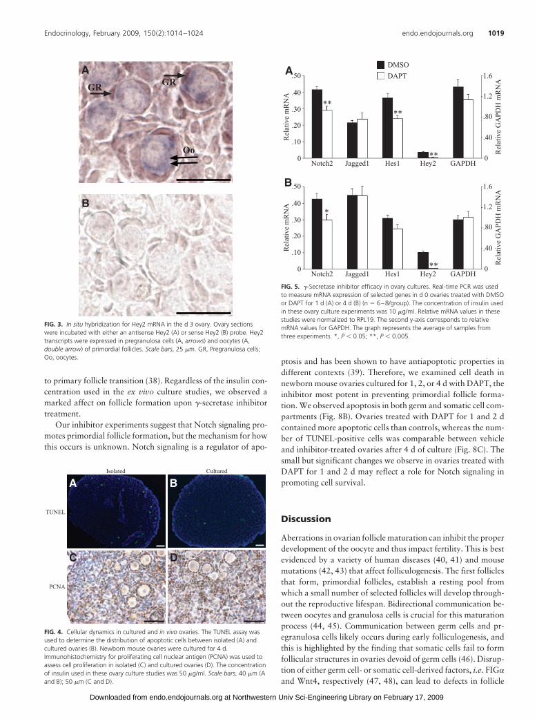
developing follicles in cultured ovaries. Immunolocalizationexperiments revealed that JAGGED1 and HES1 proteins wereexpressed in the same cell types as observed in isolated ovaries(supplemental Fig. 3, D and E). Isolated and cultured ovariesdisplayed a similar distribution and number of apoptotic (Fig. 4,A and B) and proliferating cells (Fig. 4, C and D). Thus, theculture conditions do not appear to cause cell death, and somaticcells continue to proliferate normally. These findings suggest thatfollicle dynamics and Notch family protein expression in ex vivocultured ovaries are similar to those of in vivo ovaries.
Attenuating Notch signaling decreases primordial follicleformation
Because primordial follicles assemble in this culture system,weused this exvivoapproach in subsequent studies to investigatefollicle formation. We tested the ability of two chemically dis-tinct �-secretase inhibitors, DAPT and L-685,458 (35, 36), toblock Notch signaling in ovary cultures. We first validated theefficacy of these compounds in cell lines known to express Notch
signaling components and demonstratedthat both inhibitors strongly suppressNotch signaling without affecting cell via-bility (supplemental Fig. 4). Hes1 and Hey2are Notch target genes and thus provide ameasure of Notch signaling activity (37).Therefore, we examined the mRNA expres-sion of these genes in cultured ovariestreated with vehicle or DAPT. Day 0 ovariescultured for 1 d with 20 �M DAPT displayeda 35% decrease in Hes1 mRNA levels and a90% decrease in Hey2 mRNA comparedwith controls (Fig. 5A). Hey2 mRNA down-regulation persisted when ovaries were cul-tured for 4 d with DAPT (Fig. 5B). Similarly,Notch2 mRNA decreased 30% upon DAPTtreatment for 1 d, and this level of receptordown-regulationwasmaintainedwhenova-ries were treated for 4 d with the inhibitor(Fig. 5, A and B). In contrast, Jagged1 andglyceraldehyde-3-phosphate dehydroge-nase (GAPDH) mRNA levels were notchanged by DAPT (Fig. 5). These studies re-veal that Notch signaling can be suppressedby culturing ovaries with a �-secretaseinhibitor.
To examine the actions of Notch duringearly follicle development, ovary cultureswere treated with DAPT or L-685,458 for4 d, and the ovarian histology was analyzed.Ovaries treated with DMSO for 4 d werechiefly composed of primordial follicles.Some small germ cells nests persisted inthese ovaries and were found near the ovar-ian cortex (Fig. 6A). Conversely, DAPT-treated ovaries showed expanded tracts ofgerm cells not assembled into follicles (Fig.6B). Follicle counting data revealed that
DAPT-treated ovaries had a significantly reduced percentage ofprimordial follicles, 35 vs. 58% for controls, and a correspond-ingly significant increase in the percentage of germ cells remain-ing in nests, 64 vs. 42% for controls (Fig. 6E). There was nosignificant difference in the percentage of more advanced, de-veloping follicles between vehicle and inhibitor-treated ovaries(Fig. 6E), although the number of these follicles is low. Similar toDAPT, L-685,458-treated ovaries displayed reduced germ cellnest breakdown compared with controls (Fig. 7, A and B).L-685,458-treated ovaries had a lower percentage of primordialfollicles, 48 vs. 64% for controls, and this was accompanied bya rise in the percentage of germ cells not encapsulated by somaticcells, 50 vs. 34% for controls (Fig. 7E). Vehicle and L-685,458-treated ovaries had comparable percentages of developing folli-cles (Fig. 7E). Ovaries exposed to L-685,458 and cultured inmedia containing 50 �g/ml insulin had a higher percentage ofdeveloping follicles than that found in DAPT-treated ovaries,which were cultured in media containing 1 �g/ml insulin. This isexpectedbecause insulinhasbeen shownpromote theprimordial
Newborn Day 3
Notch2
A B
E F
Hes1
Jagged1
C D
GR
Oo
GR
GRGR
Oo
GRGR
Oo
GR
GR
GE
GE
FIG. 2. NOTCH2, JAGGED1, and HES1 localization in the neonatal mouse ovary. In the newborn ovary,NOTCH2 and HES1 were expressed in pregranulosa cells of germ cell nests (A and E, arrows), whereas theNotch ligand JAGGED1 was expressed in germ cells of germ cell nests (C, arrows). Three days intopostnatal development, the pregranulosa cells of primordial follicles stained positively for NOTCH2 andHES1 (B and F, arrows), and oocytes surrounded by a mixture of squamous and cuboidal somatic cells (F,double arrow) also expressed HES1. By d 3, JAGGED1 expression was maintained in oocytes of primordialfollicles (D, arrows). Experiments were repeated three times and representative images are shown. Scalebars, 25 �m. GR, Pregranulosa cells; GE, germ cells; Oo, oocytes.
1018 Trombly et al. Notch Signaling in Neonatal Mouse Ovary Endocrinology, February 2009, 150(2):1014–1024
at Northwestern Univ Sci-Engineering Library on February 17, 2009 endo.endojournals.orgDownloaded from
to primary follicle transition (38). Regardless of the insulin con-centration used in the ex vivo culture studies, we observed amarked affect on follicle formation upon �-secretase inhibitortreatment.
Our inhibitor experiments suggest that Notch signaling pro-motes primordial follicle formation, but the mechanism for howthis occurs is unknown. Notch signaling is a regulator of apo-
ptosis and has been shown to have antiapoptotic properties indifferent contexts (39). Therefore, we examined cell death innewborn mouse ovaries cultured for 1, 2, or 4 d with DAPT, theinhibitor most potent in preventing primordial follicle forma-tion. We observed apoptosis in both germ and somatic cell com-partments (Fig. 8B). Ovaries treated with DAPT for 1 and 2 dcontained more apoptotic cells than controls, whereas the num-ber of TUNEL-positive cells was comparable between vehicleand inhibitor-treated ovaries after 4 d of culture (Fig. 8C). Thesmall but significant changes we observe in ovaries treated withDAPT for 1 and 2 d may reflect a role for Notch signaling inpromoting cell survival.
Discussion
Aberrations in ovarian follicle maturation can inhibit the properdevelopment of the oocyte and thus impact fertility. This is bestevidenced by a variety of human diseases (40, 41) and mousemutations (42, 43) that affect folliculogenesis. The first folliclesthat form, primordial follicles, establish a resting pool fromwhich a small number of selected follicles will develop through-out the reproductive lifespan. Bidirectional communication be-tween oocytes and granulosa cells is crucial for this maturationprocess (44, 45). Communication between germ cells and pr-egranulosa cells likely occurs during early folliculogenesis, andthis is highlighted by the finding that somatic cells fail to formfollicular structures in ovaries devoid of germ cells (46). Disrup-tion of either germ cell- or somatic cell-derived factors, i.e. FIG�
and Wnt4, respectively (47, 48), can lead to defects in follicle
B
AGRGR
Oo
FIG. 3. In situ hybridization for Hey2 mRNA in the d 3 ovary. Ovary sectionswere incubated with either an antisense Hey2 (A) or sense Hey2 (B) probe. Hey2transcripts were expressed in pregranulosa cells (A, arrows) and oocytes (A,double arrow) of primordial follicles. Scale bars, 25 �m. GR, Pregranulosa cells;Oo, oocytes.
DC
BACulturedIsolated
PCNA
TUNEL
FIG. 4. Cellular dynamics in cultured and in vivo ovaries. The TUNEL assay wasused to determine the distribution of apoptotic cells between isolated (A) andcultured ovaries (B). Newborn mouse ovaries were cultured for 4 d.Immunohistochemistry for proliferating cell nuclear antigen (PCNA) was used toassess cell proliferation in isolated (C) and cultured ovaries (D). The concentrationof insulin used in these ovary culture studies was 50 �g/ml. Scale bars, 40 �m (Aand B); 50 �m (C and D).
0
.10
.20
.40
.30
.50
Rel
ativ
e m
RN
A
Notch2 Jagged1 Hes1 Hey2 GAPDH 0
.40
.80
1.2
1.6
Rel
ativ
e G
APD
H m
RN
A
0
.10
.20
.40
.30
.50
Rel
ativ
e m
RN
ANotch2 Jagged1 Hes1 Hey2 GAPDH
0
.40
.80
1.2
1.6
Rel
ativ
e G
APD
H m
RN
A
DMSODAPT
**
*
**
**
**
A
B
FIG. 5. �-Secretase inhibitor efficacy in ovary cultures. Real-time PCR was usedto measure mRNA expression of selected genes in d 0 ovaries treated with DMSOor DAPT for 1 d (A) or 4 d (B) (n � 6–8/group). The concentration of insulin usedin these ovary culture experiments was 10 �g/ml. Relative mRNA values in thesestudies were normalized to RPL19. The second y-axis corresponds to relativemRNA values for GAPDH. The graph represents the average of samples fromthree experiments. *, P � 0.05; **, P � 0.005.
Endocrinology, February 2009, 150(2):1014–1024 endo.endojournals.org 1019
at Northwestern Univ Sci-Engineering Library on February 17, 2009 endo.endojournals.orgDownloaded from

formation. Insights into how primordial follicles assemble mayprovide an avenue to better treat reproductive disorders thatnegatively affect fertility.
We provide evidence for the expression of Notch pathwaygenes in the neonatal mouse ovary and propose a novel role forNotch signaling in regulating primordial follicle formation.JAGGED1 and NOTCH2 are expressed in oocytes and granu-losa cells, respectively, expression patterns that are consistentwith what has been reported in the adult mouse ovary (30). Thecomplementary expression pattern of JAGGED1 and NOTCH2provides a potential role for these molecules in mediating inter-actions between the germ and somatic cell compartments duringearly follicle development. The total number of germ cells de-creases between d 0 and 6 (4), so increased Jagged1 mRNA mayreflect enhanced production by the remaining germ cells andrelate to the initial growth of primordial follicles. In the neonatalmouse ovary, the expression of Notch receptor genes are notcoordinated with Jagged1, rather the mRNA levels are maximalcompared with the prepubertal ovary. We cannot exclude morelocalized changes in Notch gene expression at the cellular level.HES1 is expressed in both pregranulosa cells surrounding germ
cell nests and also in oocytes of early primary follicles. ThatHES1, a transcription factor, is not expressed inoocytenuclei butrather in the cytoplasm raises the possibility that it is not activein germ cells. Interestingly, Hey2 mRNA is up-regulated duringfollicle formation and initial growth. The expression pattern ofHey2 mRNA in the neonatal ovary is similar to HES1 protein:pregranulosa cells of primordial follicles and oocytes of primaryfollicles. The low levels of Hey2 mRNA in the d 0 ovary indicatethat Hey2 may be expressed later than Hes1 during early fol-liculogenesis. Indeed, microarray studies point toward Hes1 andHey2 being enriched in somatic cells at 18 d post coitum and 2d postnatal, respectively (49). Future experiments are required todetermine whether HEY2 is expressed in pregranulosa cells sur-rounding germ cell nests.
NOTCH2/HES1 and JAGGED1 are expressed in distinctcells before follicle formation, pregranulosa cells, and germ cells,respectively, suggesting that germ cell nest breakdown is in partcoordinated through cellular interactions via Notch signaling.Notch activation would then appear to directly impact pregranu-losa cells and may promote the proliferation of pregranulosacells during follicle assembly. Enhanced somatic cell prolifera-
FIG. 6. Phenotypes of ovary cultures treated with vehicle and DAPT. Newborn mouse ovaries cultured for 4 d in media containing 0.2% DMSO (A) or 20 �M DAPT (B)were fixed and then H�E stained. Germ cell nests are indicated by black boundaries. C and D, Enlarged images of the black rectangles in A and B. E, Folliclepopulations in DMSO- (n � 4) and DAPT (n � 5)-treated ovaries were quantified. The concentration of insulin used in these studies was 1 �g/ml. The graph representsaverage follicle counts from ovaries cultured in three independent experiments. Scale bars, 50 �m. *, P � 0.05.
1020 Trombly et al. Notch Signaling in Neonatal Mouse Ovary Endocrinology, February 2009, 150(2):1014–1024
at Northwestern Univ Sci-Engineering Library on February 17, 2009 endo.endojournals.orgDownloaded from

tion has been reported to be a potential mechanism for drivingincreased follicle formation (5). Alternatively, Notch signalingmayserve to establishgranulosa cell identity, aproperty thatmaybe essential for subsequent primordial follicle assembly. Pr-egranulosa cells fail to differentiate and transition to cuboidalgranulosa cells in Foxl2 null mouse ovaries (43). Ovaries fromthese animals exhibit early follicular depletion (43) and potentialdefects in follicle formation (50), demonstrating the potential tiesbetween granulosa cell differentiation and follicle assembly.Notch signaling has also been shown to support cell migration(51). During germ cell nest breakdown, Notch signaling maymediate pregranulosa cell migration within germ cell syncytia tofacilitate primordial follicle assembly.
Rodent ovary culture has been successfully performed byother investigators (29, 52–55) to ascertain roles for signalingmolecules during early follicle development. We observe a reca-pitulation of in vivo follicle formation in this system, arguing thatfactors important for follicle assembly are intrinsic to the ovary.Newborn mouse ovaries maintained in culture were treated with�-secretase inhibitors to address functional roles for Notch in theneonatal mouse ovary. We used small-molecule inhibitors tomaximize the chance of attenuating Notch signaling because all
four Notch receptors are processed by �-secretase. �-Secretasebelongs to a class of aspartyl proteases that have multiple sub-strates (56). Although other targets may be affected by theseinhibitors, we observed no decrease in GAPDH or Jagged1mRNA expression. Our apoptosis studies show that cell death islimited in DAPT-treated ovaries. The viability of cells culturedwith DAPT argues against a toxic effect of the inhibitor on exvivo cultured ovaries. A more likely possibility is that the increasein apoptotic cell numbers in ovaries treated with DAPT for 1 and2 d reflects a potential role for Notch signaling in promoting cellsurvival.
Treatment of ovary cultures with DAPT resulted in decreasedNotch target gene (Hes1, Hey2) expression. In addition, similarovarian phenotypes, namely germ cell nest retention, were ob-served in ovaries treated with chemically distinct �-secretase in-hibitors (57, 58). Therefore, it is likely that the effects on follicleformation are indeed mediated through Notch signaling. Thehigher suppression of Hey2 mRNA compared with Hes1 in in-hibitor-treated ovaries supports the notion that Notch signalschiefly through Hey2 during follicle formation. Hes1 is ex-pressed in the ovaries of late-stage embryos. Therefore, we can-not rule out the possibility that Notch signals through Hes1
FIG. 7. Ovary cultures treated with a second �-secretase inhibitor, L-685,458. Ovaries cultured for 4 d in media containing 0.2% DMSO (A) or 10 �M L-685,458 (B)were fixed and then H�E stained. Germ cell nests are indicated by black boundaries. C and D, Enlarged images of the black rectangles in A and B. E, Folliclepopulations in ovaries treated with DMSO (n � 5) and L-685,458 (n � 7) were counted. The concentration of insulin used in these studies was 50 �g/ml. The graphrepresents average follicle counts from ovaries cultured in four independent experiments. Scale bars, 50 �m. *, P � 0.005.
Endocrinology, February 2009, 150(2):1014–1024 endo.endojournals.org 1021
at Northwestern Univ Sci-Engineering Library on February 17, 2009 endo.endojournals.orgDownloaded from

during the earliest stages of follicle formation. Given that Hey2is expressed in pregranulosa cells and oocytes, it is unclear withinwhich cellular compartment Hey2 mRNAs are decreased in re-sponse to DAPT treatment. Decreased Hey2 mRNA expressionin pregranulosa cells would be considered a direct effect becauseNotch2 is expressed in pregranulosa cells. Alternatively, the de-crease in Hey2 mRNA expression may reflect the lower percent-ages of Hey2-expressing germ cells formed into primordial fol-licles in DAPT-treated ovaries.
The early stages of follicle assembly appear to be the mostsensitive to Notch signaling, as there were no significant differ-ences in the percentages of later-stage follicles between the con-trol and inhibitor-treated ovaries. Notch signaling may thereforedirect the early stages of germ cell nest breakdown and primor-dial follicle maintenance. It is unknown whether the efficiency ofnest breakdown is reduced and/or the kinetics of follicle forma-tion delayed in the inhibitor-treated ovaries. Future experimentsin which ovaries are cultured for longer times in the presence of�-secretase inhibitors may address this question. It is unclearwhich receptors are required for promoting follicle formation,and this poses a challenge to using such inhibitors. Genetic stud-ies using RNA interference knockdown in ovary culture or con-ditional gene disruption in mice should eventually shed light onthis issue. Although several key players that mediate primordialfollicle formation and initial growth have been identified (59–62), these processes likely require the coordinated actions ofmultiple cell signaling pathways. We previously described theinterplay of estrogen and activin signaling pathways in the earlymouse ovary (10), and this has been supported by in vitro ex-periments (63). Interactions between Notch and the activin/TGF� signaling pathways have also been described (64–67).Therefore, it will be important to investigate how Notch, estro-
gen, and activin signaling pathways interact in the context ofearly follicle development in the mammalian ovary.
Acknowledgments
We thank Raymond Mui for aiding in follicle counting studies and TylerWellington within the PO1 Core B facility (HD021921) for tissue sec-tioning. The monoclonal Notch2 antibody developed by Dr. S. Artava-nis-Tsakonas was obtained from the Developmental Studies HybridomaBank developed under the auspices of the National Institute of ChildHealth and Human Development and maintained by the Department ofBiological Sciences, The University of Iowa (Iowa City, IA). We thank Dr.Ryoichiro Kageyama (Institute for Virus Research, Kyoto University,Kyoto, Japan) for the pHes1-luc plasmid (68), and the Hes1 antibodywas provided by Dr. Tetsuo Sudo (Toray Industries Inc., Tokyo, Japan).We also thank Dr. Eric Nilsson (Washington State University, Seattle,WA) and Dr. Melissa Pepling (Syracuse University, Syracuse, NY) forovary culture advice.
Address all correspondence and requests for reprints to: Dr. Kelly E.Mayo, Department of Biochemistry, Molecular Biology, and Cell Biol-ogy, Northwestern University, Hogan 4-112, 2205 Tech Drive, Evan-ston, Illinois 60208. E-mail: [email protected].
This work was supported by National Institutes of Health/NationalInstitute of Child Health and Human Development P01 HD021921,Hormonal Signals that Regulate Ovarian Differentiation, and the Re-productive Biology Training Grant T32 HD00678. D.J.T. is a ChicagoChapter scholar of the Achievement Rewards for College ScientistsFoundation.
Disclosure Statement: The authors have nothing to disclose.
References
1. McGee EA, Hsueh AJ 2000 Initial and cyclic recruitment of ovarian follicles.Endocr Rev 21:200–214
2. Molyneaux KA, Stallock J, Schaible K, Wylie C 2001 Time-lapse analysis ofliving mouse germ cell migration. Dev Biol 240:488–498
3. Pepling ME, Spradling AC 1998 Female mouse germ cells form synchronouslydividing cysts. Development 125:3323–3328
4. Pepling ME, Spradling AC 2001 Mouse ovarian germ cell cysts undergo pro-grammed breakdown to form primordial follicles. Dev Biol 234:339–351
5. Bristol-Gould SK, Kreeger PK, Selkirk CG, Kilen SM, Cook RW, Kipp JL, SheaLD, Mayo KE, Woodruff TK 2006 Postnatal regulation of germ cells by ac-tivin: the establishment of the initial follicle pool. Dev Biol 298:132–148
6. Iguchi T, Fukazawa Y, Uesugi Y, Takasugi N 1990 Polyovular follicles inmouse ovaries exposed neonatally to diethylstilbestrol in vivo and in vitro. BiolReprod 43:478–484
7. Iguchi T, Takasugi N, Bern HA, Mills KT 1986 Frequent occurrence ofpolyovular follicles in ovaries of mice exposed neonatally to diethylstilbestrol.Teratology 34:29–35
8. Jefferson W, Newbold R, Padilla-Banks E, Pepling M 2006 Neonatal genisteintreatment alters ovarian differentiation in the mouse: inhibition of oocyte nestbreakdown and increased oocyte survival. Biol Reprod 74:161–168
9. Jefferson WN, Couse JF, Padilla-Banks E, Korach KS, Newbold RR 2002Neonatal exposure to genistein induces estrogen receptor (ER) � expressionand multioocyte follicles in the maturing mouse ovary: evidence for ER�-mediated and nonestrogenic actions. Biol Reprod 67:1285–1296
10. Kipp JL, Kilen SM, Bristol-Gould S, Woodruff TK, Mayo KE 2007 Neonatalexposure to estrogens suppresses activin expression and signaling in the mouseovary. Endocrinology 148:1968–1976
11. Chen Y, Jefferson WN, Newbold RR, Padilla-Banks E, Pepling ME 2007Estradiol, progesterone, and genistein inhibit oocyte nest breakdown and pri-mordial follicle assembly in the neonatal mouse ovary in vitro and in vivo.Endocrinology 148:3580–3590
12. Artavanis-Tsakonas S, Rand MD, Lake RJ 1999 Notch signaling: cell fatecontrol and signal integration in development. Science 284:770–776
1 day
0
20
40
60
80
100
Num
ber o
f apo
ptot
ic c
ells
/sec
tion
120
1 day 2 day 4 day
* *C
DMSODAPT
DAPT
A B
GEGR
GR
FIG. 8. Examination of apoptosis in vehicle and DAPT-treated ovaries. Ovarieswere cultured for 1, 2, or 4 d with DMSO or DAPT. A, A TUNEL-stained tissuesection from an ovary treated with DAPT for 1 d. B, Enlarged image of the whiterectangle in A showing apoptotic pregranulosa cells (arrows) and an apoptoticgerm cell (double arrow). C, The graph represents the average number of TUNEL-positive cells in one tissue section from three different control and inhibitor-treated ovaries at each time point. The concentration of insulin used in thesestudies was 10 �g/ml. Scale bars, 40 �m. *, P � 0.05. GR, Pregranulosa cells;GE, germ cells.
1022 Trombly et al. Notch Signaling in Neonatal Mouse Ovary Endocrinology, February 2009, 150(2):1014–1024
at Northwestern Univ Sci-Engineering Library on February 17, 2009 endo.endojournals.orgDownloaded from

13. Dumortier A, Wilson A, MacDonald HR, Radtke F 2005 Paradigms of notchsignaling in mammals. Int J Hematol 82:277–284
14. Zlobin A, Jang M, Miele L 2000 Toward the rational design of cell fate mod-ifiers: notch signaling as a target for novel biopharmaceuticals. Curr PharmBiotechnol 1:83–106
15. Lai EC 2004 Notch signaling: control of cell communication and cell fate.Development 131:965–973
16. De Strooper B, Annaert W, Cupers P, Saftig P, Craessaerts K, Mumm JS,Schroeter EH, Schrijvers V, Wolfe MS, Ray WJ, Goate A, Kopan R 1999 Apresenilin-1-dependent �-secretase-like protease mediates release of Notch in-tracellular domain. Nature 398:518–522
17. Jarriault S, Brou C, Logeat F, Schroeter EH, Kopan R, Israel A 1995 Signallingdownstream of activated mammalian Notch. Nature 377:355–358
18. Kageyama R, Ohtsuka T, Hatakeyama J, Ohsawa R 2005 Roles of bHLHgenes in neural stem cell differentiation. Exp Cell Res 306:343–348
19. Maier MM, Gessler M 2000 Comparative analysis of the human and mouseHey1 promoter: Hey genes are new Notch target genes. Biochem Biophys ResCommun 275:652–660
20. Nakagawa O, McFadden DG, Nakagawa M, Yanagisawa H, Hu T, SrivastavaD, Olson EN 2000 Members of the HRT family of basic helix-loop-helixproteins act as transcriptional repressors downstream of Notch signaling. ProcNatl Acad Sci USA 97:13655–13660
21. Panin VM, Papayannopoulos V, Wilson R, Irvine KD 1997 Fringe modulatesNotch-ligand interactions. Nature 387:908–912
22. Johnston SH, Rauskolb C, Wilson R, Prabhakaran B, Irvine KD, Vogt TF 1997A family of mammalian Fringe genes implicated in boundary determinationand the Notch pathway. Development 124:2245–2254
23. Hahn KL, Johnson J, Beres BJ, Howard S, Wilson-Rawls J 2005 Lunatic fringenull female mice are infertile due to defects in meiotic maturation. Develop-ment 132:817–828
24. Roth S 2001 Drosophila oogenesis: coordinating germ line and soma. Curr Biol11:R779–R781
25. Ito T, Udaka N, Yazawa T, Okudela K, Hayashi H, Sudo T, Guillemot F,Kageyama R, Kitamura H 2000 Basic helix-loop-helix transcription factorsregulate the neuroendocrine differentiation of fetal mouse pulmonary epithe-lium. Development 127:3913–3921
26. Szabo A, Perou CM, Karaca M, Perreard L, Quackenbush JF, Bernard PS 2004Statistical modeling for selecting housekeeper genes. Genome Biol 5:R59
27. Livak KJ, Schmittgen TD 2001 Analysis of relative gene expression data usingreal-time quantitative PCR and the 2[-��C(T)] method. Methods 25:402–408
28. Burkart AD, Mukherjee A, Sterneck E, Johnson PF, Mayo KE 2005 Repressionof the inhibin �-subunit gene by the transcription factor CCAAT/enhancer-binding protein-beta. Endocrinology 146:1909–1921
29. Kezele P, Skinner MK 2003 Regulation of ovarian primordial follicle assemblyand development by estrogen and progesterone: endocrine model of follicleassembly. Endocrinology 144:3329–3337
30. Johnson J, Espinoza T, McGaughey RW, Rawls A, Wilson-Rawls J 2001Notch pathway genes are expressed in mammalian ovarian follicles. Mech Dev109:355–361
31. Krebs LT, Xue Y, Norton CR, Sundberg JP, Beatus P, Lendahl U, Joutel A,Gridley T 2003 Characterization of Notch3-deficient mice: normal embryonicdevelopment and absence of genetic interactions with a Notch1 mutation.Genesis 37:139–143
32. Krebs LT, Xue Y, Norton CR, Shutter JR, Maguire M, Sundberg JP, GallahanD, Closson V, Kitajewski J, Callahan R, Smith GH, Stark KL, Gridley T 2000Notch signaling is essential for vascular morphogenesis in mice. Genes Dev14:1343–1352
33. Iso T, Hamamori Y, Kedes L 2003 Notch signaling in vascular development.Arterioscler Thromb Vasc Biol 23:543–553
34. Wijgerde M, Ooms M, Hoogerbrugge JW, Grootegoed JA 2005 Hedgehogsignaling in mouse ovary: Indian hedgehog and desert hedgehog from granu-losa cells induce target gene expression in developing theca cells. Endocrinol-ogy 146:3558–3566
35. Dovey HF, John V, Anderson JP, Chen LZ, de Saint Andrieu P, Fang LY,Freedman SB, Folmer B, Goldbach E, Holsztynska EJ, Hu KL, Johnson-WoodKL, Kennedy SL, Kholodenko D, Knops JE, Latimer LH, Lee M, Liao Z,Lieberburg IM, Motter RN, Mutter LC, Nietz J, Quinn KP, Sacchi KL, SeubertPA, Shopp GM, Thorsett ED, Tung JS, Wu J, Yang S, Yin CT, Schenk DB, MayPC, Altstiel LD, Bender MH, Boggs LN, Britton TC, Clemens JC, Czilli DL,Dieckman-McGinty DK, Droste JJ, Fuson KS, Gitter BD, Hyslop PA, John-stone EM, Li WY, Little SP, Mabry TE, Miller FD, Audia JE 2001 Functional�-secretase inhibitors reduce �-amyloid peptide levels in brain. J Neurochem76:173–181
36. Shearman MS, Beher D, Clarke EE, Lewis HD, Harrison T, Hunt P, Nadin A,Smith AL, Stevenson G, Castro JL 2000 L-685,458, an aspartyl protease tran-
sition state mimic, is a potent inhibitor of amyloid �-protein precursor �-secre-tase activity. Biochemistry 39:8698–8704
37. Ohtsuka T, Imayoshi I, Shimojo H, Nishi E, Kageyama R, McConnell SK 2006Visualization of embryonic neural stem cells using Hes promoters in transgenicmice. Mol Cell Neurosci 31:109–122
38. Kezele PR, Nilsson EE, Skinner MK 2002 Insulin but not insulin-like growthfactor-1 promotes the primordial to primary follicle transition. Mol Cell En-docrinol 192:37–43
39. Miele L, Osborne B 1999 Arbiter of differentiation and death: Notch signalingmeets apoptosis. J Cell Physiol 181:393–409
40. Crisponi L, Deiana M, Loi A, Chiappe F, Uda M, Amati P, Bisceglia L, ZelanteL, Nagaraja R, Porcu S, Ristaldi MS, Marzella R, Rocchi M, Nicolino M,Lienhardt-Roussie A, Nivelon A, Verloes A, Schlessinger D, Gasparini P, Bon-neau D, Cao A, Pilia G 2001 The putative forkhead transcription factorFOXL2 is mutated in blepharophimosis/ptosis/epicanthus inversus syndrome.Nat Genet 27:159–166
41. Fowler PA, Huhtaniemi IT 2002 The ovarian gonadotropin receptors in healthand disease. Rev Endocr Metab Disord 3:55–63
42. Dong J, Albertini DF, Nishimori K, Kumar TR, Lu N, Matzuk MM 1996Growth differentiation factor-9 is required during early ovarian folliculogen-esis. Nature 383:531–535
43. Schmidt D, Ovitt CE, Anlag K, Fehsenfeld S, Gredsted L, Treier AC, TreierM 2004 The murine winged-helix transcription factor Foxl2 is required forgranulosa cell differentiation and ovary maintenance. Development 131:933–942
44. Albertini DF, Combelles CM, Benecchi E, Carabatsos MJ 2001 Cellular basisfor paracrine regulation of ovarian follicle development. Reproduction 121:647–653
45. Combelles CM, Carabatsos MJ, Kumar TR, Matzuk MM, Albertini DF 2004Hormonal control of somatic cell oocyte interactions during ovarian follicledevelopment. Mol Reprod Dev 69:347–355
46. Mazaud Guittot S, Guigon CJ, Coudouel N, Magre S 2006 Consequences offetal irradiation on follicle histogenesis and early follicle development in ratovaries. Biol Reprod 75:749–759
47. Soyal SM, Amleh A, Dean J 2000 FIG�, a germ cell-specific transcription factorrequired for ovarian follicle formation. Development 127:4645–4654
48. Vainio S, Heikkila M, Kispert A, Chin N, McMahon AP 1999 Female devel-opment in mammals is regulated by Wnt-4 signalling. Nature 397:405–409
49. Ottolenghi C, Colombino M, Crisponi L, Cao A, Forabosco A, Schlessinger D,Uda M 2007 Transcriptional control of ovarian development in somatic cells.Semin Reprod Med 25:252–263
50. Uda M, Ottolenghi C, Crisponi L, Garcia JE, Deiana M, Kimber W, ForaboscoA, Cao A, Schlessinger D, Pilia G 2004 Foxl2 disruption causes mouse ovarianfailure by pervasive blockage of follicle development. Hum Mol Genet 13:1171–1181
51. Sato Y, Watanabe T, Saito D, Takahashi T, Yoshida S, Kohyama J, Ohata E,Okano H, Takahashi Y 2008 Notch mediates the segmental specification ofangioblasts in somites and their directed migration toward the dorsal aorta inavian embryos. Dev Cell 14:890–901
52. Eppig JJ, O’Brien MJ 1996 Development in vitro of mouse oocytes from pri-mordial follicles. Biol Reprod 54:197–207
53. Lee WS, Yoon SJ, Yoon TK, Cha KY, Lee SH, Shimasaki S, Lee S, Lee KA 2004Effects of bone morphogenetic protein-7 (BMP-7) on primordial folliculargrowth in the mouse ovary. Mol Reprod Dev 69:159–163
54. Nilsson E, Parrott JA, Skinner MK 2001 Basic fibroblast growth factor inducesprimordial follicle development and initiates folliculogenesis. Mol Cell Endo-crinol 175:123–130
55. Parrott JA, Skinner MK 1999 Kit-ligand/stem cell factor induces primordialfollicle development and initiates folliculogenesis. Endocrinology 140:4262–4271
56. Kopan R, Ilagan MX 2004 �-Secretase: proteasome of the membrane? Nat RevMol Cell Biol 5:499–504
57. Morohashi Y, Kan T, Tominari Y, Fuwa H, Okamura Y, Watanabe N, SatoC, Natsugari H, Fukuyama T, Iwatsubo T, Tomita T 2006 C-terminal frag-ment of presenilin is the molecular target of a dipeptidic �-secretase-specificinhibitor DAPT (N-[N-(3,5-difluorophenacetyl)-L-alanyl]S-phenylglycinet-butyl ester). J Biol Chem 281:14670–14676
58. Tian G, Ghanekar SV, Aharony D, Shenvi AB, Jacobs RT, Liu X, GreenbergBD 2003 The mechanism of �-secretase: multiple inhibitor binding sites fortransition state analogs and small molecule inhibitors. J Biol Chem278:28968–28975
59. Castrillon DH, Miao L, Kollipara R, Horner JW, DePinho RA 2003 Suppres-sion of ovarian follicle activation in mice by the transcription factor Foxo3a.Science 301:215–218
Endocrinology, February 2009, 150(2):1014–1024 endo.endojournals.org 1023
at Northwestern Univ Sci-Engineering Library on February 17, 2009 endo.endojournals.orgDownloaded from

60. Mayo K, Jameson L, Woodruff TK 2007 Eggs in the nest. Endocrinology148:3577–3579
61. Pepling ME 2006 From primordial germ cell to primordial follicle: mammalianfemale germ cell development. Genesis 44:622–632
62. Rajareddy S, Reddy P, Du C, Liu L, Jagarlamudi K, Tang W, Shen Y, BerthetC, Peng SL, Kaldis P, Liu K 2007 p27kip1 (cyclin-dependent kinase inhib-itor 1B) controls ovarian development by suppressing follicle endowmentand activation and promoting follicle atresia in mice. Mol Endocrinol 21:2189 –2202
63. Kipp JL, Kilen SM, Woodruff TK, Mayo KE 2007 Activin regulates estrogenreceptor gene expression in the mouse ovary. J Biol Chem 282:36755–36765
64. Blokzijl A, Dahlqvist C, Reissmann E, Falk A, Moliner A, Lendahl U, IbanezCF 2003 Cross-talk between the Notch and TGF-� signaling pathways me-diated by interaction of the Notch intracellular domain with Smad3. J Cell Biol163:723–728
65. Hiratochi M, Nagase H, Kuramochi Y, Koh CS, Ohkawara T, Nakayama K2007 The � intracellular domain mediates TGF-�/activin signaling throughbinding to Smads and has an important bi-directional function in the Notch-�signaling pathway. Nucleic Acids Res 35:912–922
66. Zavadil J, Cermak L, Soto-Nieves N, Bottinger EP 2004 Integration of TGF-�/SmadandJagged1/Notch signalling in epithelial-to-mesenchymal transition.EMBO J 23:1155–1165
67. Abe T, Furue M, Kondow A, Matsuzaki K, Asashima M 2005 Notch signalingmodulates the nuclear localization of carboxy-terminal-phosphorylatedsmad2andcontrols the competenceof ectodermal cells for activinA. MechDev22:671–680
68. Takebayashi K, Sasai Y, Sakai Y, Watanabe T, Nakanishi S, Kageyama R 1994Structure, chromosomal locus, and promoter analysis of the gene encoding themouse helix-loop-helix factor HES-1. Negative autoregulation through themultiple N box elements. J Biol Chem 269:5150–5156
1024 Trombly et al. Notch Signaling in Neonatal Mouse Ovary Endocrinology, February 2009, 150(2):1014–1024
at Northwestern Univ Sci-Engineering Library on February 17, 2009 endo.endojournals.orgDownloaded from