supplementary materials for -...
TRANSCRIPT

www.sciencemag.org/cgi/content/full/science.aad5143/DC1
Supplementary Materials for
In vivo genome editing improves muscle function in a mouse model of Duchenne muscular dystrophy
Christopher E. Nelson, Chady H. Hakim, David G. Ousterout, Pratiksha I. Thakore, Eirik A. Moreb, Ruth M. Castellanos Rivera, Sarina Madhavan, Xiufang Pan, F. Ann Ran,
Winston X. Yan, Aravind Asokan, Feng Zhang, Dongsheng Duan, Charles A. Gersbach*
*Corresponding author. E-mail: [email protected]
Published 31 December 2015 on Science First ReleaseDOI: 10.1126/science.aad5143
This PDF file includes:
Materials and Methods Figs. S1 to S22 Tables S1 to S4 References
2
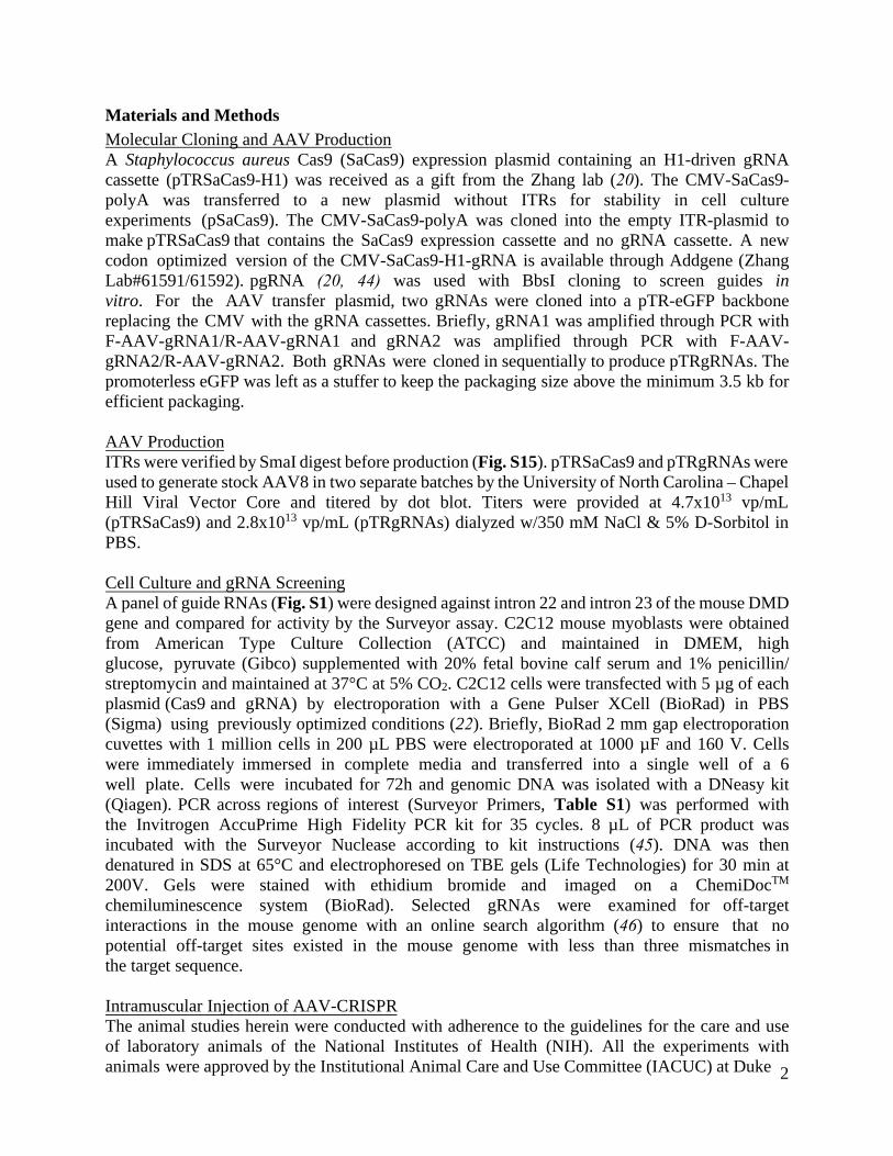
Materials and Methods Molecular Cloning and AAV Production A Staphylococcus aureus Cas9 (SaCas9) expression plasmid containing an H1-driven gRNA cassette (pTRSaCas9-H1) was received as a gift from the Zhang lab (20). The CMV-SaCas9-polyA was transferred to a new plasmid without ITRs for stability in cell culture experiments (pSaCas9). The CMV-SaCas9-polyA was cloned into the empty ITR-plasmid to make pTRSaCas9 that contains the SaCas9 expression cassette and no gRNA cassette. A new codon optimized version of the CMV-SaCas9-H1-gRNA is available through Addgene (Zhang Lab#61591/61592). pgRNA (20, 44) was used with BbsI cloning to screen guides in vitro. For the AAV transfer plasmid, two gRNAs were cloned into a pTR-eGFP backbone replacing the CMV with the gRNA cassettes. Briefly, gRNA1 was amplified through PCR with F-AAV-gRNA1/R-AAV-gRNA1 and gRNA2 was amplified through PCR with F-AAV-gRNA2/R-AAV-gRNA2. Both gRNAs were cloned in sequentially to produce pTRgRNAs. The promoterless eGFP was left as a stuffer to keep the packaging size above the minimum 3.5 kb for efficient packaging.
AAV Production ITRs were verified by SmaI digest before production (Fig. S15). pTRSaCas9 and pTRgRNAs were used to generate stock AAV8 in two separate batches by the University of North Carolina – Chapel Hill Viral Vector Core and titered by dot blot. Titers were provided at 4.7x10¹³ vp/mL (pTRSaCas9) and 2.8x10¹³ vp/mL (pTRgRNAs) dialyzed w/350 mM NaCl & 5% D-Sorbitol in PBS.
Cell Culture and gRNA ScreeningA panel of guide RNAs (Fig. S1) were designed against intron 22 and intron 23 of the mouse DMD gene and compared for activity by the Surveyor assay. C2C12 mouse myoblasts were obtained from American Type Culture Collection (ATCC) and maintained in DMEM, high glucose, pyruvate (Gibco) supplemented with 20% fetal bovine calf serum and 1% penicillin/streptomycin and maintained at 37°C at 5% CO2. C2C12 cells were transfected with 5 µg of each plasmid (Cas9 and gRNA) by electroporation with a Gene Pulser XCell (BioRad) in PBS (Sigma) using previously optimized conditions (22). Briefly, BioRad 2 mm gap electroporation cuvettes with 1 million cells in 200 µL PBS were electroporated at 1000 µF and 160 V. Cells were immediately immersed in complete media and transferred into a single well of a 6 well plate. Cells were incubated for 72h and genomic DNA was isolated with a DNeasy kit (Qiagen). PCR across regions of interest (Surveyor Primers, Table S1) was performed with the Invitrogen AccuPrime High Fidelity PCR kit for 35 cycles. 8 µL of PCR product was incubated with the Surveyor Nuclease according to kit instructions (45). DNA was then denatured in SDS at 65°C and electrophoresed on TBE gels (Life Technologies) for 30 min at 200V. Gels were stained with ethidium bromide and imaged on a ChemiDocTM chemiluminescence system (BioRad). Selected gRNAs were examined for off-target interactions in the mouse genome with an online search algorithm (46) to ensure that no potential off-target sites existed in the mouse genome with less than three mismatches in the target sequence.
Intramuscular Injection of AAV-CRISPRThe animal studies herein were conducted with adherence to the guidelines for the care and use of laboratory animals of the National Institutes of Health (NIH). All the experiments with animals were approved by the Institutional Animal Care and Use Committee (IACUC) at Duke
3
University or University of Missouri. 6-8 week old male mdx mice (Jackson Labs: 001801) were anesthetized and maintained at 37 °C. The tibialis anterior (TA) muscle was prepared and injected with 30-40 µL of AAV solution (5.6 x10¹¹ - 7.46 x10¹¹ vg/vector/mouse) or saline into the right and left TA, respectively. After 8 weeks or 6 months, mice were euthanized by CO2 inhalation and tissue was collected into RNALater® (Life Technologies) for DNA, RNA, or protein analysis.
Intraperitoneal Injection of AAV-CRISPR Two day-old male mdx neonates were injected into the intraperitoneal space with 15 µL of AAV8 solution (2.8 x10¹¹ vg/vector/mouse). After 7 weeks, mice were euthanized by CO2 inhalation and tissue was collected into RNALater® (Life Technologies) for DNA and protein analysis or 30% sucrose for frozen tissue sections.
Intravenous injection of AAV-CRISPR into adult mdx mice 6 week old male mdx mice were injected via the tail vein with 200 µL of AAV8 solution (2.7e12 vg/vector/mouse). After 8 weeks, mice were euthanized by CO2 inhalation and tissue was collected into RNALater® (Life Technologies) for DNA and protein analysis or 30% sucrose for frozen tissue sections.
Genomic DNA Analysis Mouse muscles were digested in ALT buffer and proteinase K for 4h at 56 °C with intermittent mixing. DNEasy kit (Qiagen) was used to collect genomic DNA. Endpoint PCR was performed with primers flanking CRISPR cut sites that amplify a 1638bp region (In22-gRNA1/6-F and In23-gRNA2-R). PCR products were electrophoresed in a 1% agarose gel and viewed on a BioRad GelDoc imager to observe the 1638 bp intact band and the expected 467 bp deletion product (full gel Fig. S16). Gel bands were extracted with a gel extraction kit (Qiagen) and further purified by PCR purification (Qiagen) for Sanger sequencing (Eton Bioscience). Quantitative droplet digital PCR (ddPCR) was performed on samples using a QX200™ Droplet Digital™ PCR System with QX200TM ddPCRTM EvaGreen Supermix (BioRad). Two primer sets were designed including primers specific for the intact dystrophin gene (dPCR-intact-F/dPCR-R) and primers specific the deletion product (dPCR-del-F/dPCR-R). The full data panel is available in Fig. S17.
Deep SequencingPCR of genomic DNA from 6 untreated control TA muscles and 6 treated TA muscles was completed using primers designed against the two target regions and 20 candidate off-target sites (Table S1). A second round of PCR was used to add Illumina flowcell binding sequences and experiment-specific barcodes on the 5’ end of the primer sequence (Table S1). The resulting PCR products were pooled and sequenced with 150 bp paired-end reads on an Illumina MiSeq instrument. Samples were demultiplexed according to assigned barcode sequences, and Illumina adapter and primer sequences were trimmed from reads. Indel analysis was performed as previously described (21, 24). Briefly, because expected PCR products were <250 bp in length, the 3’ ends of paired-end reads overlapped. This overlap was used to generate a consensus PCR amplicon for each paired-end read via single ungapped alignment (parameterized to score each match as 5 and each mismatch as -4). Trimmed fragments were aligned to the mm10 reference genome using BLAT. Each of the aligning fragments was then aligned to the reference genome sequence of the expected PCR product using a global affine alignment with the following parameterization: match=5, mismatch=−4, gap open=−5, gap extend=−2. Any fragment that did

4
not have greater than 60% identity with an expected PCR product or that had a top-scoring BLAT alignment that was different than the expected PCR product was discarded from downstream analysis. Alignments were then trimmed to a 20 bp window centered three base pairs 5’ of the PAM, the predicted site of SaCas9 nuclease activity. Indel (insertion and/or deletion) statistics were gathered from these windows separately for each treatment/control by counting gaps in the query and subject sequences of the resulting truncated alignments and tabulating numbers of fragments having any indels in these windows. RNA analysis RNA was extracted from tissues stabilized in RNALater® (Invitrogen) using a Tissue Lyser (Qiagen) and the RNeasy Plus Universal Kit (Qiagen). cDNA was synthesized using SuperScript® VILO cDNA Synthesis Kit and Master Mix (Life Technologies). Endpoint PCR was performed with Accuprime polymerase and electrophoresed in a 1% agarose gel (full gel Fig. S18). Gel bands were extracted with a gel extraction kit (Qiagen) and further purified by PCR purification kit (Qiagen) for Sanger sequencing. Quantitative digital drop PCR (ddPCR) was performed on samples using a QX200™ Droplet Digital™ PCR System with QX200TM ddPCRTM EvaGreen Supermix (BioRad). Two primer sets were designed including a primer set specific for the total dystrophin RNA (dPCR-ex22-F and dPCR-ex24/25-R) and the ∆23 transcript (dPCR-ex22/24-F and dPCR-ex24/25R). Full data panel is available in Fig. S19. Protein analysis and western blot Muscle biopsies were disrupted with a probe sonicator (Fisher Scientific FB50) in RIPA buffer (Sigma) with a proteinase inhibitor cocktail (Roche) and incubated for 30 min on ice with intermittent vortexing. Samples were centrifuged at 16000xg for 30 min at 4°C and the supernatant was isolated and quantified with a bicinchronic acid assay (Pierce). Protein isolate was mixed with NuPAGE loading buffer (Invitrogen) and 5% β-mercaptoethanol and boiled at 100°C for 10min. Samples were flash frozen in liquid nitrogen for future analysis. 25 µg total protein per lane were loaded into 4-12% NuPAGE Bis-Tris gels (Invitrogen) with MOPS buffer (Invitrogen) and electrophoresed for 30 min at 200 V. Protein was transferred to nitrocellulose membranes for 1 hour in 1X tris-glycine transfer buffer containing 10% methanol and 0.01% SDS at 4°C at 400 mA. The blot was blocked overnight at 4°C in 5% milk-TBST. Blots were probed with Dys-2 (1:100, clone Dy8/6C5, IgG1; Novocastra, Newcastle, UK), anti-HA (abcam), MANDYS8 (1:200, Sigma D8168), rabbit anti-GAPDH (1:5000, Cell Signaling 2118S), or anti-skeletal myosin heavy chain antibody (1:5000, Sigma MF20) for 1 hour in 5% milk-TBST at room temperature. Blots were then incubated with mouse or rabbit horseradish peroxidase-conjugated secondary antibodies (Santa Cruz) for 30 min in 5% milk-TBST. Blots were visualized using Western-C ECL substrate (Biorad) on a ChemiDoc chemiluminescent system (Biorad). The full blots are available in Fig. S20. Histological stains TA muscles were carefully dissected and embedded in OCT using liquid nitrogen-cooled isopentane. 10 µm sections were cut onto pre-treated histological slides. Hematoxylin and eosin was used to reveal general muscle histopathology. Macrophage and neutrophil infiltration were detected by immunohistochemical staining with a mouse macrophage-specific antibody (#RM2920, 1:200; clone CI:A3-1, IgG2b; Caltag Laboratories, Burlingame, CA) and a rat anti-mouse Ly6-G antibody (1:800; BD Pharmingen, San Diego, CA), respectively. Dystrophin was

5
detected with a rabbit polyclonal antibody (1:600) against the N-terminal domain of dystrophin. DGC components were evaluated with mouse monoclonal antibody against α-sarcoglycan (Ad1/20A6, 1:50, Vector Laboratories, Burlingame, CA), β-sarcoglycan (βSarc/5B1, 1:50; Novocastra, Buffalo Grove, IL), γ-sarcoglycan (35DAG/21B5, 1:50; Vector Laboratories, Burlingame, CA), β-dystroglycan (43DAG1/8D5, 1:50; Novocastra, Buffalo Grove, IL), syntrophin (ab11187, 1:200; Abcam, Cambridge, MA) and dystrobrevin (23/Dystrobrevin, 1:200; BD Biosciences, San Diego, CA). Laminin was detected with a rabbit polyclonal antibody (L9393, 1:200, Sigma, Saint Louis, MO). Utrophin was examined with a mouse monoclonal antibody against the N-terminus of utrophin (1:20, Vector Laboratories, Burlingame, CA). nNOS expression was revealed by nNOS activity staining as previously described (47) and by immunostaining with a rabbit polyclonal antibody against the C-terminal domain of nNOS (1:2000, Santa Cruz, Dallas, TX). Embryonic myofibers were detected with a monoclonal antibody against embryonic myosin heavy chain (eMHC) (1:250; Developmental Studies Hybridoma Bank, Iowa City, IA). The myosin heavy chain isoforms were identified using mouse monoclonal antibodies generated from hybridoma cells against Type I, IIa and IIb MyHC (Developmental Studies Hybridoma Bank, Iowa City, IA). The HA tag fused to C-terminus domain of the SaCas9 gene was detected with a rat monoclonal antibody (3F10, 1:50, Roche, Pleasanton, CA). Dystrophin-positive fibers were manually counted from the representative images in Fig. S21. Control injections of Cas9-AAV only or gRNA-AAV were stained for dystrophin and are reported in Fig. S22.
In situ evaluation of TA muscle contractile propertiesTA muscle contractile properties were measured as previously described (48) using the Aurora Scientific in situ muscle test system. Briefly, the mouse was anesthetized and the body core temperature was maintained at 37°C using a heated platform. The hind limb was shaved and the TA muscle was surgically exposed and the distal tendon was tied at the muscle tendon junction using a 4-0 suture line (SofSilk USSC Sutures, Norwalk, CT). Another suture line was attached to the patella ligament. The sciatic nerve was carefully dissected, tied to a suture, and cut at the proximal end. The mouse was placed prone on a custom made platform and the knee was secured to a fixed post using the patella suture line. The sciatic nerve was placed on custom made electrode and secured using the suture line. The distal end of the TA muscle was secured to the lever arm of the force transducer. The exposed hind limb muscles were covered with a warm Ringer’s buffer soaked Kimwipes. Throughout the experiment, the muscles were constantly superfused with warm oxygenated Ringer’s buffer. After 5 minutes equilibration, the muscle was warmed up and optimal length (Lo), current and stimulation time were determined. Twitch tension (Pt) and maximal isometric tetanic force (Po) were measured. The muscle was allowed to relax for 3 min before 10 cycles of eccentric contractions were applied with a 1 min rest between each cycle. Relative force loss was later determined for each cycle. Data were recorded and analyzed using the Lab-View based DMC and DMA software (Version 3.12, Aurora Scientific, Aurora, ON, Canada). At the end of each experiment, the distal TA tendon was removed and TA muscle weight was measured. The muscle cross sectional area was determined based on a muscle density of 1.06 g/cm3 and fiber length to Lo ratio of 0.6.
Statistical Analysis Sample sizes for animal studies were designed based on previous studies of dystrophin-mediated muscle function recovery (48). All intact samples were included in data analysis and no sample was excluded. Student t-tests were used to determine significance of single comparisons in Figures

6
1-3. Repeated measures ANOVA was used to validate overall treatment effect in the eccentriccontractility test (Figure 3d). All of the variation reported in Figures 1-3 show standard errorbetween biological replicates. Randomization and blinding were not used as each biologicalreplicate has a built-in negative treated contralateral control. Restoration of dystrophin protein hasbeen duplicated in multiple laboratory experiments. For muscle contractility experiments, micewere injected on different days and analyzed on different days (n=7).

7
Fig. S1. In vitro characterization of Staphylococcus aureus Cas9 (SaCas9) gRNAs. Plasmid electroporation of C2C12 mouse skeletal myoblasts was used to screen gRNAs. (a) 4 gRNAs were screened in intron 22 with gRNA1 displaying the highest activity at 12.1% indel formation. (b) For intron 23, 10 gRNAs were screened with gRNA2 displaying the highest activity at 12.2% indel formation. (c) gRNA sequences are shown in the table. (d) Combined activity of gRNA1 and gRNA2 creates genomic deletions. (e) Reverse transcription and PCR of the mRNA transcript also indicates the removal of exon 23 in C2C12 cells as evidenced by gel electrophoresis and Sanger sequencing of the extracted smaller band.

8
Fig. S2 Cas9-mediated deletion has a strong preference for perfect ligation of cut sites. a) The distribution of deletion products shows ~67% perfect deletions with ~30% deletions within the cut site and small fractions of insertions or substitutions. b) The perfect ligation product with cut site marked with arrows and the eight most prevalent resulting sequences.

9
Fig. S3 Distribution of indel sizes in selected target regions from mouse skeletal muscle by deep sequencing.

10
Fig. S4. Total dystrophin RNA levels relative to WT. ddPCR suggests that Cas9/gRNA treated mdx mice have higher overall levels of dystrophin RNA, indicating ∆23 transcripts avoid nonsense-mediated decay caused by the premature stop codon in exon 23.

11
Fig. S5. Detection of SaCas9 in treated muscle. (a) Two representative photomicrographs showing SaCas9 expression in the nucleus of AAV infected muscle. Laminin immunofluorescence staining marks the sarcolemma. Myonuclei are stained with DAPI. SaCas9 is revealed with immunofluorescence staining against the HA epitope tag on the C terminus of SaCas9. Scale bar, 50 µm. (b) Detection of the HA tag on SaCas9 in whole muscle lysate by western blot. 1- mdx mice treated with the AAV expressing HA-tagged Cas9, 2 – untreated mdx mice, 3 - untreated BL10 mice, 4 - mdx mice treated with an AAV vector encoding HA-tagged micro-dystrophin.

12
Fig. S6. Wild-type (WT) control for Figure 3a-c. Asterisk, same myofiber in the serial sections. Scale bar = 600 μm in full-view images, and 100 μm in high-power images.

13
Fig. S7. Representative photomicrographs of immunofluorescence staining for laminin, dystrophin, nNOS enzymatic activity, and nNOS - Serial muscle sections were stained for laminin (to mark muscle cell membrane), dystrophin, nNOS with the indicated rabbit polyclonal antibody. Asterisk, the same myofiber in serial sections. Scale bar: 100 µm.

14
Fig. S8. Representative photomicrographs of immunofluorescence staining for components of dystrophin-associated glycoprotein complex (α-sarcoglycan β-sarcoglycan γ-sarcoglycan, β-dystroglycan) using the indicated mouse monoclonal antibody. Arrow, Strong cytosolic staining reflecting accumulation of mouse immunoglobulin (IgG) in damaged myofibers. IgG reacts with the fluorescein-conjugated mouse anti-mouse antibody (secondary antibody). Asterisk, the same myofiber in serial sections. Scale bar, 100 µm.

15
Fig. S9. Representative photomicrographs of immunofluorescence staining for syntrophin, dystrobrevin and utrophin using the indicated mouse monoclonal antibody. The bottom row shows background staining with the secondary antibody alone (applicable to Supplemental Figure 6 and 7). Arrow, Strong cytosolic staining reflecting accumulation of mouse immunoglobulin (IgG) in damaged myofibers. IgG reacts with the fluorescein-conjugated mouse anti-mouse antibody (secondary antibody). Asterisk, the same myofiber in serial sections. Scale bar, 100 µm.

16
Cas9/gRNA-treated mice
Sham injected mice
Fig. S10. Representative photomicrographs of H&E stained muscle sections from CRISPR/gRNA treated muscles (top two rows) and sham injected muscles (bottom two rows).
17
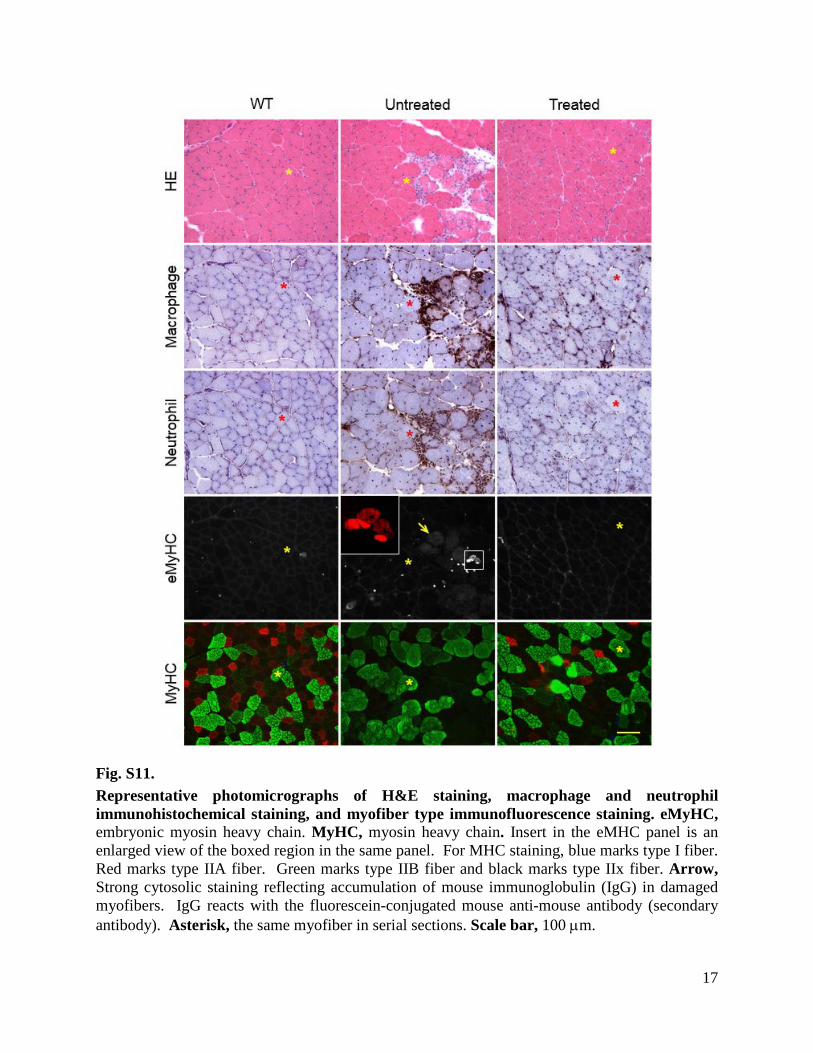
Fig. S11. Representative photomicrographs of H&E staining, macrophage and neutrophil immunohistochemical staining, and myofiber type immunofluorescence staining. eMyHC, embryonic myosin heavy chain. MyHC, myosin heavy chain. Insert in the eMHC panel is an enlarged view of the boxed region in the same panel. For MHC staining, blue marks type I fiber. Red marks type IIA fiber. Green marks type IIB fiber and black marks type IIx fiber. Arrow, Strong cytosolic staining reflecting accumulation of mouse immunoglobulin (IgG) in damaged myofibers. IgG reacts with the fluorescein-conjugated mouse anti-mouse antibody (secondary antibody). Asterisk, the same myofiber in serial sections. Scale bar, 100 µm.

18
Fig. S12. Systemic administration of CRISPR/Cas9 by intraperitoneal injection of AAV vectors recovers dystrophin in cardiac and skeletal muscles. (a) PCR across the genomic deletion shows removal of exon 23 from the genomic DNA in the liver, heart, and abdominal wall. (b) Removal of exon 23 from the mRNA transcript is shown strongly in the abdominal wall, heart, and diaphragm but not in distal muscles including the tibialis anterior. (c) Immunofluorescence staining of whole muscle sections shows abundant dystrophin expression in the heart, abdominal wall, and diaphragm. Distal muscles including the tibialis anterior (shown) were not significantly corrected. Scale bar = 200 µm.

19
Fig. S13. Full data panel for immunostaining for dystrophin from intraperitoneal administrated P2 neonates. Stained sections of heart, diaphragm, and abdominal wall muscles are shown from three independent mice. Left panels, full-view images (scale bar: 1 mm). Right panels, high power images (scale bar: 200 µm).

20
Fig. S14. Genome editing is preserved for 6 months after a single injection of AAV-CRISPR. (a) Genomic DNA maintains the 1171bp deletion containing exon 23 (b) RT-PCR shows preservation of the ∆23 transcript. (c) Immunoflourescence staining of dystrophin shows preservation of restored dystrophin protein. Scale bar: 100 µm.

21
Fig. S15. AAV transfer plasmid preparation. SmaI digestion indicates intact ITRs in preparation for AAV production. Surveyor and deletion PCR from mouse myoblasts using plasmids with ITRS (AAV plasmid) or without (conventional) shows slight reduction in Cas9 activity between plasmid architecture.

22
From Fig 1b
From Fig. 4a
Fig. S16. Full gel image from Fig. 1b and 4a. Genomic deletion represented by a 1638 bp parent band and a 467 bp deletion product.

23
Fig. S17. Complete ddPCR gDNA data panel from Fig. 1c. ddPCR for genomic DNA in 6 treated mice with primers specific for the intact dystrophin gene or the dystrophin gene missing the exon 23 region.

24
From Fig 1d
From Fig 4b
Fig. S18. Full gels From Fig. 1d. Exon 23 was removed from the transcript of mice treated with Cas9/gRNAs as indicated by the smaller band.

25
Fig. S19. Complete ddPCR panel for dystrophin cDNA from Fig. 1e. Complete ddPCR data showing the deletion product relative to total dystrophin RNA expression.

26
Fig. S20. (a) Representative dystrophin western blot using the Mandys-8 antibody. (b) High and lowexposure full blots are shown with arrows marking dystrophin and GAPDH expected protein sizes.

27
Cas9/gRNA-treated mice
Sham injected mice
Fig. S21. Representative photomicrographs of dystrophin stained muscle sections from CRISPR/gRNA treated muscles (top two rows) and sham injected muscles (bottom two rows).

28
Fig. S22. Injection of AAV.Cas9 or AAV.gRNA alone did not restore dystrophin expression. (a) Representative full-view dystrophin immunostaining of the tibialis anterior muscle that received either Cas9 or gRNA AAV vector. (b) Representative H&E and dystrophin immunostaining photomicrographs revealing no improvement in histology following single vector injection. Scale bar = 500 μm for panel a; scale bar = 100 μm for panel b.
29
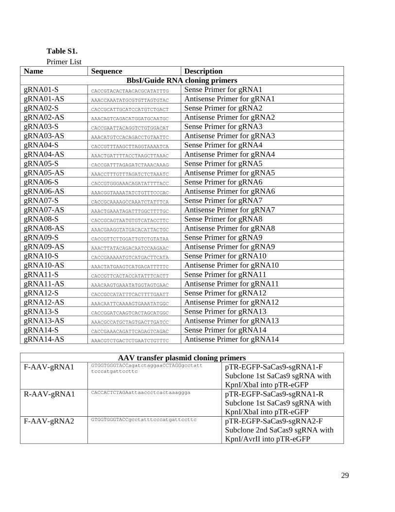
Table S1. Primer List
Name Sequence Description BbsI/Guide RNA cloning primers
gRNA01-S CACCGTACACTAACACGCATATTTG Sense Primer for gRNA1 gRNA01-AS AAACCAAATATGCGTGTTAGTGTAC Antisense Primer for gRNA1 gRNA02-S CACCGCATTGCATCCATGTCTGACT Sense Primer for gRNA2 gRNA02-AS AAACAGTCAGACATGGATGCAATGC Antisense Primer for gRNA2 gRNA03-S CACCGAATTACAGGTCTGTGGACAT Sense Primer for gRNA3 gRNA03-AS AAACATGTCCACAGACCTGTAATTC Antisense Primer for gRNA3 gRNA04-S CACCGTTTAAGCTTAGGTAAAATCA Sense Primer for gRNA4 gRNA04-AS AAACTGATTTTACCTAAGCTTAAAC Antisense Primer for gRNA4 gRNA05-S CACCGATTTAGAGATCTAAACAAAG Sense Primer for gRNA5 gRNA05-AS AAACCTTTGTTTAGATCTCTAAATC Antisense Primer for gRNA5 gRNA06-S CACCGTGGGAAACAGATATTTTACC Sense Primer for gRNA6 gRNA06-AS AAACGGTAAAATATCTGTTTCCCAC Antisense Primer for gRNA6 gRNA07-S CACCGCAAAAGCCAAATCTATTTCA Sense Primer for gRNA7 gRNA07-AS AAACTGAAATAGATTTGGCTTTTGC Antisense Primer for gRNA7 gRNA08-S CACCGCAGTAATGTGTCATACCTTC Sense Primer for gRNA8 gRNA08-AS AAACGAAGGTATGACACATTACTGC Antisense Primer for gRNA8 gRNA09-S CACCGTTCTTGGATTGTCTGTATAA Sense Primer for gRNA9 gRNA09-AS AAACTTATACAGACAATCCAAGAAC Antisense Primer for gRNA9 gRNA10-S CACCGAAAAATGTCATGACTTCATA Sense Primer for gRNA10 gRNA10-AS AAACTATGAAGTCATGACATTTTTC Antisense Primer for gRNA10 gRNA11-S CACCGTTCACTACCATATTTCACTT Sense Primer for gRNA11 gRNA11-AS AAACAAGTGAAATATGGTAGTGAAC Antisense Primer for gRNA11 gRNA12-S CACCGCCATATTTCACTTTTGAATT Sense Primer for gRNA12 gRNA12-AS AAACAATTCAAAAGTGAAATATGGC Antisense Primer for gRNA12 gRNA13-S CACCGGATCAAGTCACTAGCATGGC Sense Primer for gRNA13 gRNA13-AS AAACGCCATGCTAGTGACTTGATCC Antisense Primer for gRNA13 gRNA14-S CACCGAAACAGATTCAGAGTCAGAC Sense Primer for gRNA14 gRNA14-AS AAACGTCTGACTCTGAATCTGTTTC Antisense Primer for gRNA14
AAV transfer plasmid cloning primers F-AAV-gRNA1 GTGGTGGGTACCagatctaggaaCCTAGGgcctatt
tcccatgattccttc pTR-EGFP-SaCas9-sgRNA1-F Subclone 1st SaCas9 sgRNA with KpnI/XbaI into pTR-eGFP
R-AAV-gRNA1 CACCACTCTAGAattaaccctcactaaaggga pTR-EGFP-SaCas9-sgRNA1-R Subclone 1st SaCas9 sgRNA with KpnI/XbaI into pTR-eGFP
F-AAV-gRNA2 GTGGTGGGTACCgcctatttcccatgattccttc pTR-EGFP-SaCas9-sgRNA2-F Subclone 2nd SaCas9 sgRNA with KpnI/AvrII into pTR-eGFP

30
R-AAV-gRNA1 GTGGTGCCTAGGattaaccctcactaaaggga pTR-EGFP-SaCas9-sgRNA2-R Subclone 2nd SaCas9 sgRNA with KpnI/AvrII into pTR-eGFP
Surveyor/qPCR primers In22-gRNA1/6-F
tttgttgattctaaaaatcccatgt
Surveyor Primers for gRNA1, gRNA5
In22-gRNA1/6-R
atatttctgaaggtgctttcttgg
Rev-Survyor Primers for gRNA1 gRNA5
In23-gRNA2/13/14-F
gtgtttctcatagttggccatttg
FwdSurveyor Primers for gRNA2, gRNA13, gRNA14
In23-gRNA2/13/14-R Ggactgaagaacttggagaagga Rev- Surveyor Primers for gRNA2, gRNA13, gRNA14
In22-gRNA3-F TCCAGCAGTCAGAAAGCAAA
Surveyor Primers for gRNA3 In22-gRNA3-R
TGTTTTTGGTCAAATTGTTCTGTTSurveyor Primers for gRNA3
In22-gRNA4-F GAACAATTTGACCAAAAACATGA
Surveyor Primers for gRNA4 In22-gRNA4-R
TGTCCTCACATCACAGAAGTTTSurveyor Primers for gRNA4
dPCR-intact-F TCATAGTTGGCCATTTGTGAAA Fwd Primer gDNA dPCR intact dystrophin
dPCR-R GGTACAGTGTTAGGGAGCAGGA Rev Primer gDNA dPCR dPCR-del-F TTTCTGTCTAAATATAATATGCCCTGT Fwd Primer gDNA dPCR deleted
dystrophin dPCR-ex22-F ggatccagcagtcagaaagc Fwd Primer cDNA dPCR binds
exon 22 dPCR-ex22/24-F ctcgggaaattacagaatcaca Fwd Primer cDNA dPCR binds
exon22-24 junction dPCR-ex24/25-R tcaccaactaaaagtctgcattg Rev Primer cDNA dPCR binds
exon 24-25 junction
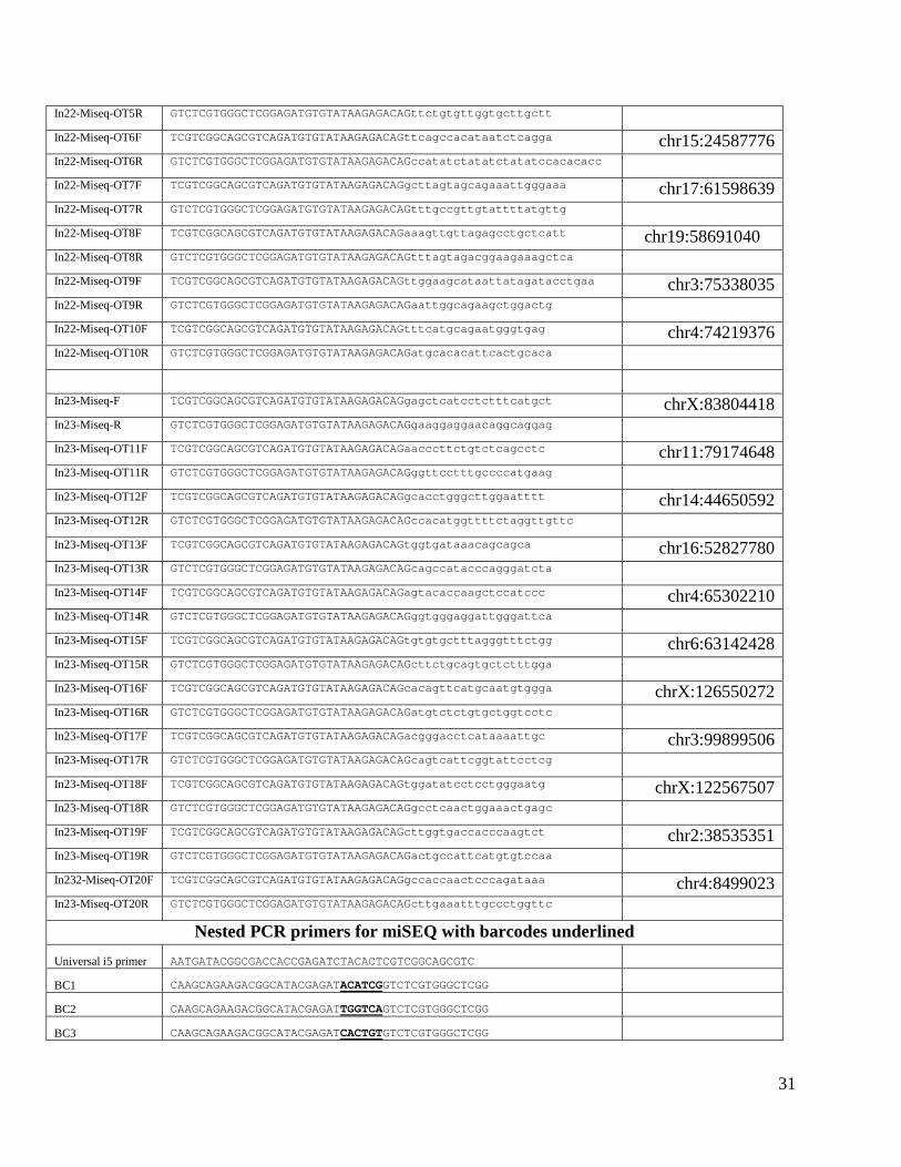
Miseq primers In22-Miseq-F TCGTCGGCAGCGTCAGATGTGTATAAGAGACAGtttctgtctaaatataatatgccctgt chrX:83803255 In22-Miseq-R GTCTCGTGGGCTCGGAGATGTGTATAAGAGACAGgcagagcctcaaaattaaatagaag
In22-Miseq-OT1F TCGTCGGCAGCGTCAGATGTGTATAAGAGACAGaatgttcttggagccaatttgt chr14:38227244 In22-Miseq-OT1R GTCTCGTGGGCTCGGAGATGTGTATAAGAGACAGagcttcaacattctccaacca
In22-Miseq-OT2F TCGTCGGCAGCGTCAGATGTGTATAAGAGACAGatcgacagacagatacacatcca chr6:37580259 In22-Miseq-OT2R GTCTCGTGGGCTCGGAGATGTGTATAAGAGACAGaaaatggttgaagccagctc
In22-Miseq-OT3F TCGTCGGCAGCGTCAGATGTGTATAAGAGACAGttgaaaggttttgctgggata chrX:93621693 In22-Miseq-OT3R GTCTCGTGGGCTCGGAGATGTGTATAAGAGACAGaggaacaatgctccaaccac
In22-Miseq-OT4F TCGTCGGCAGCGTCAGATGTGTATAAGAGACAGctgcttgtgcctgctgtaaa chr10:17880418 In22-Miseq-OT4R GTCTCGTGGGCTCGGAGATGTGTATAAGAGACAGccgtgttttggtgtctagga
In22-Miseq-OT5F TCGTCGGCAGCGTCAGATGTGTATAAGAGACAGtattcttgtcaggcccaacc chr13:68811666
31
In22-Miseq-OT5R GTCTCGTGGGCTCGGAGATGTGTATAAGAGACAGttctgtgttggtgcttgctt
In22-Miseq-OT6F TCGTCGGCAGCGTCAGATGTGTATAAGAGACAGttcagccacataatctcagga chr15:24587776 In22-Miseq-OT6R GTCTCGTGGGCTCGGAGATGTGTATAAGAGACAGccatatctatatctatatccacacacc
In22-Miseq-OT7F TCGTCGGCAGCGTCAGATGTGTATAAGAGACAGgcttagtagcagaaattgggaaa chr17:61598639 In22-Miseq-OT7R GTCTCGTGGGCTCGGAGATGTGTATAAGAGACAGtttgccgttgtattttatgttg
In22-Miseq-OT8F TCGTCGGCAGCGTCAGATGTGTATAAGAGACAGaaagttgttagagcctgctcatt chr19:58691040 In22-Miseq-OT8R GTCTCGTGGGCTCGGAGATGTGTATAAGAGACAGtttagtagacggaagaaagctca
In22-Miseq-OT9F TCGTCGGCAGCGTCAGATGTGTATAAGAGACAGttggaagcataattatagatacctgaa chr3:75338035 In22-Miseq-OT9R GTCTCGTGGGCTCGGAGATGTGTATAAGAGACAGaattggcagaagctggactg
In22-Miseq-OT10F TCGTCGGCAGCGTCAGATGTGTATAAGAGACAGtttcatgcagaatgggtgag chr4:74219376 In22-Miseq-OT10R GTCTCGTGGGCTCGGAGATGTGTATAAGAGACAGatgcacacattcactgcaca
In23-Miseq-F TCGTCGGCAGCGTCAGATGTGTATAAGAGACAGgagctcatcctctttcatgct chrX:83804418 In23-Miseq-R GTCTCGTGGGCTCGGAGATGTGTATAAGAGACAGgaaggaggaacaggcaggag
In23-Miseq-OT11F TCGTCGGCAGCGTCAGATGTGTATAAGAGACAGaacccttctgtctcagcctc chr11:79174648 In23-Miseq-OT11R GTCTCGTGGGCTCGGAGATGTGTATAAGAGACAGggttcctttgccccatgaag
In23-Miseq-OT12F TCGTCGGCAGCGTCAGATGTGTATAAGAGACAGgcacctgggcttggaatttt chr14:44650592 In23-Miseq-OT12R GTCTCGTGGGCTCGGAGATGTGTATAAGAGACAGccacatggttttctaggttgttc
In23-Miseq-OT13F TCGTCGGCAGCGTCAGATGTGTATAAGAGACAGtggtgataaacagcagca chr16:52827780 In23-Miseq-OT13R GTCTCGTGGGCTCGGAGATGTGTATAAGAGACAGcagccatacccagggatcta
In23-Miseq-OT14F TCGTCGGCAGCGTCAGATGTGTATAAGAGACAGagtacaccaagctccatccc chr4:65302210 In23-Miseq-OT14R GTCTCGTGGGCTCGGAGATGTGTATAAGAGACAGggtgggaggattgggattca
In23-Miseq-OT15F TCGTCGGCAGCGTCAGATGTGTATAAGAGACAGtgtgtgctttagggtttctgg chr6:63142428 In23-Miseq-OT15R GTCTCGTGGGCTCGGAGATGTGTATAAGAGACAGcttctgcagtgctctttgga
In23-Miseq-OT16F TCGTCGGCAGCGTCAGATGTGTATAAGAGACAGcacagttcatgcaatgtggga chrX:126550272 In23-Miseq-OT16R GTCTCGTGGGCTCGGAGATGTGTATAAGAGACAGatgtctctgtgctggtcctc
In23-Miseq-OT17F TCGTCGGCAGCGTCAGATGTGTATAAGAGACAGacgggacctcataaaattgc chr3:99899506 In23-Miseq-OT17R GTCTCGTGGGCTCGGAGATGTGTATAAGAGACAGcagtcattcggtattcctcg
In23-Miseq-OT18F TCGTCGGCAGCGTCAGATGTGTATAAGAGACAGtggatatcctcctgggaatg chrX:122567507 In23-Miseq-OT18R GTCTCGTGGGCTCGGAGATGTGTATAAGAGACAGgcctcaactggaaactgagc
In23-Miseq-OT19F TCGTCGGCAGCGTCAGATGTGTATAAGAGACAGcttggtgaccacccaagtct chr2:38535351 In23-Miseq-OT19R GTCTCGTGGGCTCGGAGATGTGTATAAGAGACAGactgccattcatgtgtccaa
In232-Miseq-OT20F TCGTCGGCAGCGTCAGATGTGTATAAGAGACAGgccaccaactcccagataaa chr4:8499023 In23-Miseq-OT20R GTCTCGTGGGCTCGGAGATGTGTATAAGAGACAGcttgaaatttgccctggttc
Nested PCR primers for miSEQ with barcodes underlined Universal i5 primer AATGATACGGCGACCACCGAGATCTACACTCGTCGGCAGCGTC
BC1 CAAGCAGAAGACGGCATACGAGATACATCGGTCTCGTGGGCTCGG
BC2 CAAGCAGAAGACGGCATACGAGATTGGTCAGTCTCGTGGGCTCGG
BC3 CAAGCAGAAGACGGCATACGAGATCACTGTGTCTCGTGGGCTCGG

32
BC4 CAAGCAGAAGACGGCATACGAGATATTGGCGTCTCGTGGGCTCGG
BC5 CAAGCAGAAGACGGCATACGAGATGATCTGGTCTCGTGGGCTCGG
BC6 CAAGCAGAAGACGGCATACGAGATTACAAGGTCTCGTGGGCTCGG
BC7 CAAGCAGAAGACGGCATACGAGATCGTGATGTCTCGTGGGCTCGG
BC8 CAAGCAGAAGACGGCATACGAGATGCCTAAGTCTCGTGGGCTCGG
BC9 CAAGCAGAAGACGGCATACGAGATTCAAGTGTCTCGTGGGCTCGG
BC10 CAAGCAGAAGACGGCATACGAGATAGCTAGGTCTCGTGGGCTCGG
BC11 CAAGCAGAAGACGGCATACGAGATGTCGTCGTCTCGTGGGCTCGG
BC12 CAAGCAGAAGACGGCATACGAGATCGATTAGTCTCGTGGGCTCGG
BC13 CAAGCAGAAGACGGCATACGAGATGAATGAGTCTCGTGGGCTCGG
BC14 CAAGCAGAAGACGGCATACGAGATCTTCGAGTCTCGTGGGCTCGG
BC15 CAAGCAGAAGACGGCATACGAGATCTCTACGTCTCGTGGGCTCGG
BC16 CAAGCAGAAGACGGCATACGAGATAGGAATGTCTCGTGGGCTCGG
BC17 CAAGCAGAAGACGGCATACGAGATGCTACCGTCTCGTGGGCTCGG
BC18 CAAGCAGAAGACGGCATACGAGATATCAGTGTCTCGTGGGCTCGG

33
Table S2. Summary of indel formation within the target cut site and the top 10 off target sites. Three target sites (gRNA1-OT10, gRNA-OT2, gRNA2-OT7) did not meet filtering criteria #
Sequence PAM Chr Gene #MM %Indels Treated/
Untreated
gRN
A1-In
tron
22
On TACACTAACACGCATATTTG ATGAGT chrX DMD-intron22 0 2.49% (± 0.96%) 12.84 OT1 TACACacACAtGCATATTTG GTGGAT chr14 None 3 0.63% (± 0.01%) 0.98
OT2 TACACatACACaCATATTTG GTGGAT chr6
5' of : Gm25062-201 ENSMUST00000104660 3 0.12% (± 0.00%) 1.02
OT3 TACACTtACACaCATAgTTG ATGGAT chrX
Intron 3 of MGI:99660, ENSMUSG000000006678 3 0.32% (± 0.02%) 1.27
OT4 TgCACTAtaAaGCATATTTG AAGGAT chr10 None 4 0.10% (± 0.01%) 1.05 OT5 cACACacACACaCATATTTG GTGGAT chr13 Intron 3 of Adcy2-201 4 1.23% (± 0.03%) 1.07 OT6 cACACccACACaCATATTTG GAGGGT chr15 None 4 1.28% (± 0.05%) 0.97 OT7 TgCttgAACACGCATATTTG AGGAAT chr17 None 4 0.15% (± 0.01%) 1.01 OT8 TACAtatACACaCATATTTG GTGGAT chr19 Intron 4 of Y1G0141A14 4 1.01% (± 0.33%) 6.66 OT9 TAaAgTAACcCaCATATTTG GTGGAT chr3 None 4 0.11% (± 0.01%) 0.97
OT10 cACACacACACaCATATTTG GTGGAT chr4 None 4 N/A N/A
gRN
A2-In
tron
23
On CATTGCATCCATGTCTGACT CTGAAT chrX DMD-intron23 0 3.12% (± 1.01%) 84.04 OT1 gAcTGCcTCCATGTCTGACT GAGGAT chr11 None 3 0.06% (± 0.00%) 0.91
OT2CATTGCAgCaATGTCTGAaT CTGAGT chr14
Intron 4 of NSMUST000000169583 3 N/A N/A
OT3 CATTGCATaCATGTCTtAgT CAGGGT chr16 Intron 2 of Mbi-105 3 0.12% (± 0.01%) 0.95 OT4 CATTtCATCCAgGTCTtACT TAGAAT chr4 Intron 13 of Pappa 3 0.15% (± 0.01%) 1.09 OT5 CATTtCATtCATGTCTGACa TGGAGT chr6 None 3 0.06% (± 0.01%) 1.12 OT6 CATTGCATCCAaGTgTGACc CTGAAT chrX None 3 0.11% (± 0.01%) 0.92 OT7 tATTaatTCCATGTCTGACT TAGGAT chr3 Intron 1 of Spag17 4 N/A N/A OT8 ggcTcCATCCATGTCTGACT TAGGGT chrX None 4 0.09% (± 0.01%) 0.99 OT9 CAgaGCAgCtATGTCTGACT ATGAAT chr2 Intron 1 Nek6 4 0.09% (± 0.00%) 1.22
OT10 gATgcCATCCATGaCTGACT CAGAAT chr4 None 4 0.14% (± 0.01%) 1.30

34
Table S3. Detailed summary of deep sequencing of target sequence and top 10 off-target sequences for each guide RNA.
Control Treated
gRNA1-Intron22 gRNA1-Intron22
%Indels Total Reads Deletion Insertions Mismatch %Indels
Total Reads Deletion Insertions Mismatch
ON 0.23% (± 0.19%)
21959 (± 1804)
0.17% (± 0.01%)
0.14% (± 0.01%)
2.83% (± 0.02%)
2.49% (± 0.96%)
20172 (± 22493)
2.14% (± 0.82%)
0.45% (± 0.14%)
2.73% (± 0.11%)
OT1 0.66% (± 0.64%)
22198 (± 2145)
0.62% (± 0.01%)
0.06% (± 0.01%)
2.62% (± 0.07%)
0.63% (± 0.01%)
22529 (± 22400)
0.62% (± 0.01%)
0.05% (± 0.01%)
2.66% (± 0.09%)
OT2 0.14% (± 0.12%)
25839 (± 3479)
0.10% (± 0.01%)
0.09% (± 0.01%)
6.94% (± 0.14%)
0.12% (± 0.00%)
29029 (± 30473)
0.11% (± 0.01%)
0.08% (± 0.01%)
7.04% (± 0.05%)
OT3 0.32% (± 0.25%)
20112 (± 1062)
0.23% (± 0.02%)
0.12% (± 0.01%)
3.38% (± 0.13%)
0.32% (± 0.02%)
13851 (± 18433)
0.30% (± 0.01%)
0.12% (± 0.01%)
3.38% (± 0.11%)
OT4 0.10% (± 0.10%)
26708 (± 2041)
0.09% (± 0.01%)
0.04% (± 0.00%)
2.05% (± 0.06%)
0.10% (± 0.01%)
17281 (± 20171)
0.09% (± 0.00%)
0.05% (± 0.01%)
2.13% (± 0.07%)
OT5 1.14% (± 1.14%)
27384 (± 1506)
1.03% (± 0.05%)
0.76% (± 0.05%)
7.66% (± 0.23%)
1.23% (± 0.03%)
16763 (± 19923)
1.08% (± 0.03%)
0.83% (± 0.04%)
7.73% (± 0.18%)
OT6 1.32% (± 1.32%)
23141 (± 1809)
1.29% (± 0.01%)
0.93% (± 0.03%)
35.50% (± 0.23%)
1.28% (± 0.05%)
20762 (± 21270)
1.25% (± 0.05%)
0.87% (± 0.03%)
35.37% (± 0.27%)
OT7 0.13% (± 0.15%)
40535 (± 5780)
0.13% (± 0.01%)
0.12% (± 0.01%)
2.67% (± 0.04%)
0.15% (± 0.01%)
25384 (± 27745)
0.13% (± 0.00%)
0.12% (± 0.01%)
2.69% (± 0.07%)
OT8 0.15% (± 0.15%)
25575 (± 2362)
0.14% (± 0.01%)
0.09% (± 0.01%)
2.30% (± 0.04%)
1.01% (± 0.33%)
12177 (± 19961)
1.00% (± 0.33%)
0.09% (± 0.01%)
1.91% (± 0.08%)
OT9 0.15% (± 0.11%)
20433 (± 4287)
1.13% (± 1.02%)
1.09% (± 1.03%)
4.66% (± 1.57%)
0.11% (± 0.01%)
9660 (± 21599)
0.10% (± 0.01%)
0.06% (± 0.01%)
2.99% (± 0.13%)
OT10 N/A N/A N/A N/A N/A N/A N/A N/A N/A N/A
Control Treated
gRNA2-Intron23 gRNA2-Intron23
%Indels Total Reads Deletion Insertions Mismatch %Indels
Total Reads Deletion Insertions Mismatch
ON 0.03% (± 0.04%)
24956 (± 1303)
0.03% (± 0.00%)
0.03% (± 0.00%)
2.30% (± 0.04%)
3.12% (± 1.01%)
23952 (± 2987)
2.77% (± 0.87%)
0.43% (± 0.17%)
2.33% (± 0.15%)
OT1 0.05% (± 0.07%)
31700 (± 2393)
0.06% (± 0.01%)
0.03% (± 0.00%)
7.60% (± 0.13%)
0.06% (± 0.00%)
27636 (± 1441)
0.06% (± 0.00%)
0.03% (± 0.00%)
7.77% (± 0.06%)
OT2 N/A N/A N/A N/A N/A N/A N/A N/A N/A N/A
OT3 0.14% (± 0.13%)
30882 (± 1343)
0.09% (± 0.00%)
0.09% (± 0.01%)
2.56% (± 0.08%)
0.12% (± 0.01%)
24231 (± 1068)
0.10% (± 0.01%)
0.08% (± 0.01%)
2.69% (± 0.07%)
OT4 0.12% (± 0.14%)
28762 (± 675)
0.12% (± 0.01%)
0.10% (± 0.01%)
4.07% (± 0.09%)
0.15% (± 0.01%)
24301 (± 1611)
0.14% (± 0.01%)
0.10% (± 0.01%)
4.21% (± 0.09%)
OT5 0.04% (± 0.06%)
29460 (± 1258)
0.04% (± 0.01%)
0.03% (± 0.00%)
2.64% (± 0.07%)
0.06% (± 0.01%)
26210 (± 1750)
0.05% (± 0.01%)
0.04% (± 0.01%)
2.75% (± 0.08%)
OT6 0.14% (± 0.12%)
30328 (± 3669)
0.11% (± 0.00%)
0.09% (± 0.01%)
2.52% (± 0.03%)
0.11% (± 0.01%)
30098 (± 1156)
0.10% (± 0.01%)
0.08% (± 0.01%)
2.41% (± 0.05%)
OT7 N/A N/A N/A N/A N/A N/A N/A N/A N/A N/A
OT8 0.06% (± 0.09%)
26860 (± 964)
0.07% (± 0.01%)
0.04% (± 0.01%)
3.09% (± 0.11%)
0.09% (± 0.01%)
20763 (± 2487)
0.07% (± 0.01%)
0.05% (± 0.01%)
3.04% (± 0.08%)
OT9 0.07% (± 0.07%)
29898 (± 1006)
0.06% (± 0.00%)
0.05% (± 0.01%)
2.88% (± 0.12%)
0.09% (± 0.00%)
19293 (± 2664)
0.07% (± 0.01%)
0.06% (± 0.00%)
3.03% (± 0.04%)
OT10 0.13% (± 0.11%)
26198 (± 4983)
0.09% (± 0.02%)
0.08% (± 0.02%)
3.46% (± 0.14%)
0.14% (± 0.01%)
23419 (± 2369)
0.12% (± 0.01%)
0.11% (± 0.01%)
3.34% (± 0.14%)

35
Table S4. Muscle weight, optimal length and cross-sectional area.
TA muscle weight (mg)
Lo (mm)
CSA (mm2)
WT 54.95 ± 1.51 14.37 ± 0.13 6.00 ± 0.14 Mdx (sham) 73.24 ± 2.09* 13.95 ± 0.08 8.28 ± 0.21* Mdx (treated) 54.42 ± 1.56 13.61 ± 0.07 6.32 ± 0.20
TA, tibialis anterior Lo, optimal muscle length CSA, cross-sectional area *significantly different from that of treated muscle

References and Notes 1. R. J. Fairclough, M. J. Wood, K. E. Davies, Therapy for Duchenne muscular dystrophy: Renewed optimism
from genetic approaches. Nat. Rev. Genet. 14, 373–378 (2013). Medline doi:10.1038/nrg3460
2. E. P. Hoffman, R. H. Brown Jr., L. M. Kunkel, Dystrophin: The protein product of the Duchenne musculardystrophy locus. Cell 51, 919–928 (1987). doi:10.1016/0092-8674(87)90579-4 Medline
3. S. B. England, L. V. Nicholson, M. A. Johnson, S. M. Forrest, D. R. Love, E. E. Zubrzycka-Gaarn, D. E.Bulman, J. B. Harris, K. E. Davies, Very mild muscular dystrophy associated with the deletion of 46% of dystrophin. Nature 343, 180–182 (1990). Medline doi:10.1038/343180a0
4. B. Wang, J. Li, X. Xiao, Adeno-associated virus vector carrying human minidystrophin genes effectivelyameliorates muscular dystrophy in mdx mouse model. Proc. Natl. Acad. Sci. U.S.A. 97, 13714–13719 (2000). Medline doi:10.1073/pnas.240335297
5. S. Q. Harper, M. A. Hauser, C. DelloRusso, D. Duan, R. W. Crawford, S. F. Phelps, H. A. Harper, A. S.Robinson, J. F. Engelhardt, S. V. Brooks, J. S. Chamberlain, Modular flexibility of dystrophin: Implications for gene therapy of Duchenne muscular dystrophy. Nat. Med. 8, 253–261 (2002). Medline doi:10.1038/nm0302-253
6. J. H. Shin, X. Pan, C. H. Hakim, H. T. Yang, Y. Yue, K. Zhang, R. L. Terjung, D. Duan, Microdystrophinameliorates muscular dystrophy in the canine model of duchenne muscular dystrophy. Mol. Ther. 21, 750–757 (2013). Medline doi:10.1038/mt.2012.283
7. J. R. Mendell, K. Campbell, L. Rodino-Klapac, Z. Sahenk, C. Shilling, S. Lewis, D. Bowles, S. Gray, C. Li,G. Galloway, V. Malik, B. Coley, K. R. Clark, J. Li, X. Xiao, J. Samulski, S. W. McPhee, R. J.Samulski, C. M. Walker, Dystrophin immunity in Duchenne’s muscular dystrophy. N. Engl. J. Med.363, 1429–1437 (2010). Medline doi:10.1056/NEJMoa1000228
8. S. Cirak, V. Arechavala-Gomeza, M. Guglieri, L. Feng, S. Torelli, K. Anthony, S. Abbs, M. E. Garralda, J.Bourke, D. J. Wells, G. Dickson, M. J. Wood, S. D. Wilton, V. Straub, R. Kole, S. B. Shrewsbury, C. Sewry, J. E. Morgan, K. Bushby, F. Muntoni, Exon skipping and dystrophin restoration in patients with Duchenne muscular dystrophy after systemic phosphorodiamidate morpholino oligomer treatment: An open-label, phase 2, dose-escalation study. Lancet 378, 595–605 (2011). Medline doi:10.1016/S0140-6736(11)60756-3
9. N. M. Goemans, M. Tulinius, J. T. van den Akker, B. E. Burm, P. F. Ekhart, N. Heuvelmans, T. Holling, A.A. Janson, G. J. Platenburg, J. A. Sipkens, J. M. Sitsen, A. Aartsma-Rus, G. J. van Ommen, G. Buyse,N. Darin, J. J. Verschuuren, G. V. Campion, S. J. de Kimpe, J. C. van Deutekom, Systemicadministration of PRO051 in Duchenne’s muscular dystrophy. N. Engl. J. Med. 364, 1513–1522 (2011).Medline doi:10.1056/NEJMoa1011367
10. A. Aartsma-Rus, I. Fokkema, J. Verschuuren, I. Ginjaar, J. van Deutekom, G. J. van Ommen, J. T. denDunnen, Theoretic applicability of antisense-mediated exon skipping for Duchenne muscular dystrophy mutations. Hum. Mutat. 30, 293–299 (2009). Medline doi:10.1002/humu.20918
11. D. B. Cox, R. J. Platt, F. Zhang, Therapeutic genome editing: Prospects and challenges. Nat. Med. 21, 121–131 (2015). Medline doi:10.1038/nm.3793
12. M. Jinek, K. Chylinski, I. Fonfara, M. Hauer, J. A. Doudna, E. Charpentier, A programmable dual-RNA-guided DNA endonuclease in adaptive bacterial immunity. Science 337, 816–821 (2012). Medline doi:10.1126/science.1225829
13. P. Mali, L. Yang, K. M. Esvelt, J. Aach, M. Guell, J. E. DiCarlo, J. E. Norville, G. M. Church, RNA-guidedhuman genome engineering via Cas9. Science 339, 823–826 (2013). Medline doi:10.1126/science.1232033
36

14. L. Cong, F. A. Ran, D. Cox, S. Lin, R. Barretto, N. Habib, P. D. Hsu, X. Wu, W. Jiang, L. A. Marraffini, F.Zhang, Multiplex genome engineering using CRISPR/Cas systems. Science 339, 819–823 (2013). Medline doi:10.1126/science.1231143
15. S. W. Cho, S. Kim, J. M. Kim, J. S. Kim, Targeted genome engineering in human cells with the Cas9 RNA-guided endonuclease. Nat. Biotechnol. 31, 230–232 (2013). Medline doi:10.1038/nbt.2507
16. M. Jinek, A. East, A. Cheng, S. Lin, E. Ma, J. Doudna, RNA-programmed genome editing in human cells.eLife 2, e00471 (2013). Medline doi:10.7554/eLife.00471
17. H. Yin, W. Xue, S. Chen, R. L. Bogorad, E. Benedetti, M. Grompe, V. Koteliansky, P. A. Sharp, T. Jacks,D. G. Anderson, Genome editing with Cas9 in adult mice corrects a disease mutation and phenotype.Nat. Biotechnol. 32, 551–553 (2014). Medline doi:10.1038/nbt.2884
18. L. Swiech, M. Heidenreich, A. Banerjee, N. Habib, Y. Li, J. Trombetta, M. Sur, F. Zhang, In vivointerrogation of gene function in the mammalian brain using CRISPR-Cas9. Nat. Biotechnol. 33, 102–106 (2015). Medline doi:10.1038/nbt.3055
19. R. J. Platt, S. Chen, Y. Zhou, M. J. Yim, L. Swiech, H. R. Kempton, J. E. Dahlman, O. Parnas, T. M.Eisenhaure, M. Jovanovic, D. B. Graham, S. Jhunjhunwala, M. Heidenreich, R. J. Xavier, R. Langer, D. G. Anderson, N. Hacohen, A. Regev, G. Feng, P. A. Sharp, F. Zhang, CRISPR-Cas9 knockin mice forgenome editing and cancer modeling. Cell 159, 440–455 (2014). Medline doi:10.1016/j.cell.2014.09.014
20. F. A. Ran, L. Cong, W. X. Yan, D. A. Scott, J. S. Gootenberg, A. J. Kriz, B. Zetsche, O. Shalem, X. Wu, K.S. Makarova, E. V. Koonin, P. A. Sharp, F. Zhang, In vivo genome editing using Staphylococcus aureusCas9. Nature 520, 186–191 (2015). Medline doi:10.1038/nature14299
21. D. G. Ousterout, A. M. Kabadi, P. I. Thakore, W. H. Majoros, T. E. Reddy, C. A. Gersbach, MultiplexCRISPR/Cas9-based genome editing for correction of dystrophin mutations that cause Duchenne muscular dystrophy. Nat. Commun. 6, 6244 (2015). Medline doi:10.1038/ncomms7244
22. D. G. Ousterout, P. Perez-Pinera, P. I. Thakore, A. M. Kabadi, M. T. Brown, X. Qin, O. Fedrigo, V. Mouly,J. P. Tremblay, C. A. Gersbach, Reading frame correction by targeted genome editing restores dystrophin expression in cells from Duchenne muscular dystrophy patients. Mol. Ther. 21, 1718–1726 (2013). Medline
23. L. Popplewell, T. Koo, X. Leclerc, A. Duclert, K. Mamchaoui, A. Gouble, V. Mouly, T. Voit, F. Pâques, F.Cédrone, O. Isman, R. J. Yáñez-Muñoz, G. Dickson, Gene correction of a duchenne muscular dystrophy mutation by meganuclease-enhanced exon knock-in. Hum. Gene Ther. 24, 692–701 (2013). Medline doi:10.1089/hum.2013.081
24. D. G. Ousterout, A. M. Kabadi, P. I. Thakore, P. Perez-Pinera, M. T. Brown, W. H. Majoros, T. E. Reddy,C. A. Gersbach, Correction of dystrophin expression in cells from Duchenne muscular dystrophypatients through genomic excision of exon 51 by zinc finger nucleases. Mol. Ther. 23, 523–532 (2015).Medline doi:10.1038/mt.2014.234
25. H. L. Li, N. Fujimoto, N. Sasakawa, S. Shirai, T. Ohkame, T. Sakuma, M. Tanaka, N. Amano, A.Watanabe, H. Sakurai, T. Yamamoto, S. Yamanaka, A. Hotta, Precise correction of the dystrophin gene in duchenne muscular dystrophy patient induced pluripotent stem cells by TALEN and CRISPR-Cas9. Stem Cell Rev. 4, 143–154 (2015).doi:10.1016/j.stemcr.2014.10.013 Medline
26. P. Chapdelaine, C. Pichavant, J. Rousseau, F. Pâques, J. P. Tremblay, Meganucleases can restore thereading frame of a mutated dystrophin. Gene Ther. 17, 846–858 (2010). Medline doi:10.1038/gt.2010.26
27. C. Long, J. R. McAnally, J. M. Shelton, A. A. Mireault, R. Bassel-Duby, E. N. Olson, Prevention ofmuscular dystrophy in mice by CRISPR/Cas9-mediated editing of germline DNA. Science 345, 1184–1188 (2014). Medline doi:10.1126/science.1254445
37
28. L. Xu et al., CRISPR-mediated genome editing restores dystrophin expression and function in mdx mice. Mol. Ther. 10.1038/mt.2015.192 (2015). doi:10.1038/mt.2015.192
29. P. Sicinski, Y. Geng, A. S. Ryder-Cook, E. A. Barnard, M. G. Darlison, P. J. Barnard, The molecular basis of muscular dystrophy in the mdx mouse: A point mutation. Science 244, 1578–1580 (1989). Medline doi:10.1126/science.2662404
30. C. J. Mann, K. Honeyman, A. J. Cheng, T. Ly, F. Lloyd, S. Fletcher, J. E. Morgan, T. A. Partridge, S. D. Wilton, Antisense-induced exon skipping and synthesis of dystrophin in the mdx mouse. Proc. Natl. Acad. Sci. U.S.A. 98, 42–47 (2001). Medline doi:10.1073/pnas.98.1.42
31. A. Goyenvalle, G. Griffith, A. Babbs, S. El Andaloussi, K. Ezzat, A. Avril, B. Dugovic, R. Chaussenot, A. Ferry, T. Voit, H. Amthor, C. Bühr, S. Schürch, M. J. Wood, K. E. Davies, C. Vaillend, C. Leumann, L. Garcia, Functional correction in mouse models of muscular dystrophy using exon-skipping tricyclo-DNA oligomers. Nat. Med. 21, 270–275 (2015). Medline
32. Z. Wang, T. Zhu, C. Qiao, L. Zhou, B. Wang, J. Zhang, C. Chen, J. Li, X. Xiao, Adeno-associated virus serotype 8 efficiently delivers genes to muscle and heart. Nat. Biotechnol. 23, 321–328 (2005). Medline doi:10.1038/nbt1073
33. D. Li, Y. Yue, D. Duan, Marginal level dystrophin expression improves clinical outcome in a strain of dystrophin/utrophin double knockout mice. PLOS ONE 5, e15286 (2010). Medline doi:10.1371/journal.pone.0015286
34. M. van Putten, M. Hulsker, C. Young, V. D. Nadarajah, H. Heemskerk, L. van der Weerd, P. A. ’t Hoen, G. J. van Ommen, A. M. Aartsma-Rus, Low dystrophin levels increase survival and improve muscle pathology and function in dystrophin/utrophin double-knockout mice. FASEB J. 27, 2484–2495 (2013). Medline doi:10.1096/fj.12-224170
35. M. Neri, S. Torelli, S. Brown, I. Ugo, P. Sabatelli, L. Merlini, P. Spitali, P. Rimessi, F. Gualandi, C. Sewry, A. Ferlini, F. Muntoni, Dystrophin levels as low as 30% are sufficient to avoid muscular dystrophy in the human. Neuromuscul. Disord. 17, 913–918 (2007). Medline doi:10.1016/j.nmd.2007.07.005
36. Y. M. Kobayashi, E. P. Rader, R. W. Crawford, N. K. Iyengar, D. R. Thedens, J. A. Faulkner, S. V. Parikh, R. M. Weiss, J. S. Chamberlain, S. A. Moore, K. P. Campbell, Sarcolemma-localized nNOS is required to maintain activity after mild exercise. Nature 456, 511–515 (2008). Medline
37. Y. Lai, G. D. Thomas, Y. Yue, H. T. Yang, D. Li, C. Long, L. Judge, B. Bostick, J. S. Chamberlain, R. L. Terjung, D. Duan, Dystrophins carrying spectrin-like repeats 16 and 17 anchor nNOS to the sarcolemma and enhance exercise performance in a mouse model of muscular dystrophy. J. Clin. Invest. 119, 624–635 (2009). Medline doi:10.1172/JCI36612
38. S. Al-Zaidy, L. Rodino-Klapac, J. R. Mendell, Gene therapy for muscular dystrophy: Moving the field forward. Pediatr. Neurol. 51, 607–618 (2014). Medline doi:10.1016/j.pediatrneurol.2014.08.002
39. D. Wang, H. Mou, S. Li, Y. Li, S. Hough, K. Tran, J. Li, H. Yin, D. G. Anderson, E. J. Sontheimer, Z. Weng, G. Gao, W. Xue, Adenovirus-Mediated Somatic Genome Editing of Pten by CRISPR/Cas9 in Mouse Liver in Spite of Cas9-Specific Immune Responses. Hum. Gene Ther. 26, 432–442 (2015). Medline doi:10.1089/hum.2015.087
40. F. Mingozzi, K. A. High, Immune responses to AAV vectors: Overcoming barriers to successful gene therapy. Blood 122, 23–36 (2013). Medline doi:10.1182/blood-2013-01-306647
41. M. Tabebordbar et al., In vivo gene editing in dystrophic mouse muscle and muscle stem cells. Science 10.1126/science.aad5177 (2015).
42. C. Long et al., Postnatal genome editing partially restores dystrophin expression in a mouse model of muscular dystrophy. Science 10.1126/science.aad5725 (2015).
38

43. Y. Lai, Y. Yue, M. Liu, A. Ghosh, J. F. Engelhardt, J. S. Chamberlain, D. Duan, Efficient in vivo gene expression by trans-splicing adeno-associated viral vectors. Nat. Biotechnol. 23, 1435–1439 (2005). Medline doi:10.1038/nbt1153
44. P. Mali, J. Aach, P. B. Stranges, K. M. Esvelt, M. Moosburner, S. Kosuri, L. Yang, G. M. Church, CAS9 transcriptional activators for target specificity screening and paired nickases for cooperative genome engineering. Nat. Biotechnol. 31, 833–838 (2013). Medline doi:10.1038/nbt.2675
45. D. Y. Guschin, A. J. Waite, G. E. Katibah, J. C. Miller, M. C. Holmes, E. J. Rebar, A rapid and general assay for monitoring endogenous gene modification. Methods Mol. Biol. 649, 247–256 (2010). Medline doi:10.1007/978-1-60761-753-2_15
46. S. Bae, J. Park, J. S. Kim, Cas-OFFinder: A fast and versatile algorithm that searches for potential off-target sites of Cas9 RNA-guided endonucleases. Bioinformatics 30, 1473–1475 (2014). Medline doi:10.1093/bioinformatics/btu048
47. D. Li, A. Bareja, L. Judge, Y. Yue, Y. Lai, R. Fairclough, K. E. Davies, J. S. Chamberlain, D. Duan, Sarcolemmal nNOS anchoring reveals a qualitative difference between dystrophin and utrophin. J. Cell Sci. 123, 2008–2013 (2010). Medline doi:10.1242/jcs.064808
48. C. H. Hakim, N. B. Wasala, D. Duan, Evaluation of muscle function of the extensor digitorum longus muscle ex vivo and tibialis anterior muscle in situ in mice. J. Vis. Exp. (72): (2013). Medline
39