supplementary information - nature · supplementary information ... nature chemical biology: ......
TRANSCRIPT

S1
Supplementary Information
Non-Immune Cells Equipped with T-Cell-Receptor-Like Signaling for Cancer Cell
Ablation
Ryosuke Kojima1, Leo Scheller1, Martin Fussenegger1,2
1ETH Zurich, Department of Biosystems Science and Engineering, Mattenstrasse 26, 4058
Basel, Switzerland. 2Faculty of Life Science, University of Basel, Mattenstrasse 26, 4058 Basel,
Switzerland.
Nature Chemical Biology: doi:10.1038/nchembio.2498

S2
Inventory
Supplementary Results
Supplementary Table 1. Table of plasmids used in this study ········································· 3
Supplementary Table 2. Table of oligonucleotides used in this study ································ 6
Supplementary Fig. 1. System performance with the hetero type receptor sets (combination of
ML39-IL4R & FRB-IL13R1, FKBP-IL4R & ML39-IL13R1) ··································· 7
Supplementary Fig. 2. Comparison of promoter strength (PhCMV vs PSV40) by FACS analysis ····· 7
Supplementary Fig. 3. Effect of sensor cell : target cell ratio on system performance ··············· 8
Supplementary Fig. 4. Generalizability of the cell contact sensing device ····························· 9
Supplementary Fig. 5. Evaluation of bystander effect of VP22-FCU1 ································ 10
Supporting references ························································································· 11
Nature Chemical Biology: doi:10.1038/nchembio.2498

S3
Supplementary Results
In Supplementary Table 1 and 2, the plasmids and oligonucleotides used in this study are shown. In
Supplementary Figure 1-5, additional data supporting that our system worked as designed and
showing detailed performance of the system are described. Please see online methods section for the
protocol of each experiment.
Plasmid Description and Cloning Strategy Reference/Source
pHCM-CD43ex
45int-mCherry
Constitutive expression vector of CD43ex-45int-mCherry (PhCMV-CD43ex-45int
-pA).
James et al1
pLeo26 An empty plasmid having SV40 promoter (having same structure as
pcDNA3.1(+)) (PSV40-MCS-pAbGH). SV40 promoter region was PCR amplified
from pSEAP2-Control Vector (Clontech) by oDA190 and oDA191. The
amplified DNA fragment was digested with MluI/NheI and inserted into
corresponding site of pcDNA3.1(+).
This work
pLeo49 Constitutive Ig leader sequence-FRB-MCS expression vector (PhCMV-FRB-
MCS-pAbGH). FRB (bearing Ig leader) was PCR amplified with oLeo97 and
oLeo98 from pMH2282. The amplified fragment was digested with NheI/EcoRI,
and was cloned into corresponding site of pcDNA3.1(+).
This work
pLeo50 Constitutive Ig leader sequence-FKBP-MCS expression vector (PhCMV-FKBP-
MCS-pAbGH). FKBP (bearing Igleader) was PCR amplified with oLeo99 and
oLeo100 from pMH2292. The amplified fragment was digested with NheI/EcoRI,
and was cloned into corresponding site of pcDNA3.1(+).
This work
pLeo52 Constitutive Ig leader sequence-FRB-hIL13R1 expression vector (PhCMV-FRB-
hIL13R1-pAbGH). hIL13R1 was PCR amplified from pCH023 (unpublished,
provided by C. Helene, ETH-Zurich) with oLeo133 and oLeo134. The amplified
fragment was digested with EcoRI/NotI, and was cloned into corresponding site
of pLeo49.
This work
pLeo53 Constitutive Ig leader sequence-FKBP-hIL4Rexpression vector (PhCMV-Ig-
FKBP-hIL4R-pAbGH). hIL4R wasPCR amplified from pCH024 (unpublished,
provided by C. Helene, ETH-Zurich) with oLeo135 and oLeo136. The amplified
fragment was digested with EcoRI/NotI, and was cloned into corresponding site
of pLeo50.
This work
pLeo56 Constitutive Ig leader sequence-FKBP-mIL10R expression vector (PhCMV-Ig -FKBP-mIL10R-pAbGH). mIL10R was cut out from pLS83 with EcoRI/NotI,
and was inserted into corresponding site of pLeo50.
This work
pLeo57 Constitutive Ig leader sequence-FRB-mIL10R expression vector (PhCMV-Ig -FRB-mIL10R-pAbGH). mIL10R was PCR amplified from pLS93 with oLeo103
and oLeo129. The amplified fragment was digested with EcoRV/NotI, and was
cloned into corresponding site of pLeo49.
This work
pLS12 PSTAT6 driven SEAP expression vector (PSTAT6-SEAP-pA). Schukur et al3
pLS13 PSTAT3-driven SEAP expression vector (PSTAT3-SEAP-pA). Schukur et al3
pLS15 Constitutive hSTAT3 expression vector (PhCMV-hSTAT3-pA). Schukur et al3
pLS16 Constitutive hSTAT6 expression vector (PhCMV-hSTAT6-pA). Schukur et al3
pRK14 Constitutive CD43ex-YFP expression vector (PhCMV-CD43ex-YFP-pASV40).
Extracellular and transmembrane domain of CD43 (CD43ex), whose NheI site is
silently mutated, was amplified from a DNASU plasmid (HsCD00446356) by 2 step
PCR. 1st PCR: By using a DNASU plasmid (HsCD00446356) as a template, One
fragment was amplified by oRK4 and oRK5, and another fragment was amplified by
oRK6 and oRK7. 2nd PCR: By using mixture of the 2 fragments obtained from 1st PCR
as templates, the CD43ex was amplified. The amplified fragment was digested with
NheI/AgeI, and was inserted into corresponding site of pEYFP-C1.
This work
pRK17 A plasmid having a multi-cloning site followed by linker-CFP (PhCMV-MCS
(multiple cloning site)-CFP-pAbGH). Linker-CFP was PCR amplified from
pECFP-C1 (Clontech) by oRK22 and oRK23. The amplified DNA fragment was
digested with EcoRI/NotI and inserted into corresponding site of pcDNA3.1(+).
This work
pRK21 Constitutive HER2-iRFP670 expression vector (PhCMV-HER2-iRFP-pAbGH).
HER2 was PCR amplified with oRK18 (having kozak) and oRK19 by using
addgene #16257 (a gift from Mien-Chie Hung4) as a template, and was digested
with HindIII/NotI. iRFP670 was PCR amplified with oRK20 (having linker
sequence) and oRK 21 by using pMM581 (unpublished) as a template, and was
This work
Nature Chemical Biology: doi:10.1038/nchembio.2498

S4
digested with NotI/XbaI. These 2 fragments were sequentially inserted in the
corresponding sites of pcDNA3.1(+).
pRK22 Constitutive iRFP670 expression vector (PhCMV-iRFP-pAbGH). iRFP670 was PCR
amplified with oRK30 (having kozak sequence) and oRK21 by using pMM581
(unpublished, a gift from Marius Muller, ETH Zurich) as a template, and was
digested with HindIII/XbaI. The fragment was inserted in the corresponding site
of pcDNA3.1(+).
This work
pRK96 Constitutive CD43ex-45int expression vector (PhCMV-CD43ex-45int-pAbGH).
CD43ex-45int was cut out from pHCM-CD43TMCD45Cyt (a gift from John
James and Ron Vale1) with BglII/NotI (additional treatment of the fragment with
HindIII/XbaI was necessary to remove another fragment from the vector), and
was inserted to the pcDNA 3.1 digested with BamHI/NotI.
This work
pRK114 Constitutive Ig leader signal-ML39-hIL13R expression vector (PhCMV-
ML39-hIL13R-pAbGH). DNA encoding ML39 (anti-HER2 scFv) (addgene
#10794, a gift from Judy Lieberman5) was PCR amplified with oRK41 (having
kozak and additional Ig leader) and oRK139 and was digested with NheI/EcoRI.
This fragment was inserted in the corresponding site of pLeo52.
This work
pRK115 Constitutive Ig leader signal-ML39-full hIL4R expression vector (PhCMV
-ML39-hIL4R-pAbGH). DNA encoding ML39 (anti HER2 scFv) (addgene
#10794) was PCR amplified with oRK41 (having kozak, additional Ig signal)
and oRK139, and was digested with NheI/EcoRI. Also, additional fragment cut
out from pLeo53 with EcoRI/XhoI (1216bp) was prepared. These 2 fragments
were inserted in pLeo52 digested with NheI/XhoI (3 piece ligation).
This work
pRK119 Constitutive Ig leader signal-ML39-CD28hinge-truncated hIL4R (trans-
membrane and cytosolic domain) expression vector (PhCMV-ML39-
CD28hin-hIL4Rex-pAbGH). DNA encoding transmembrane and cytosolic
domain of hIL4R was PCR amplified by oRK143 (having additional CD28
hinge domain) and oRK140 using pLeo53 as a template and was digested with
EcoRI/XbaI. This fragment was inserted in corresponding site of pRK115.
This work
pRK122 Plasmid vector expressing the same protein as pRK119 driven by SV40 promoter
(PSV40-ML39-hIL4Rex-pAbGH). Two fragments were prepared from pRK119
(1st fragment: 846bp digested with NheI/EcoRI, 2nd fragment: 3027 bp digested
with EcoRI/EagI) and were inserted into pLeo26 digested with NheI/EagI (3
piece ligation)
This work
pRK123 Plasmid vector expressing the same protein as pRK114 driven by SV40 promoter
(PSV40-ML39-hIL13R-pAbGH). Protein-coding region was cut out by NheI/ApaI
from pRK114. This fragment was inserted into corresponding site of pLeo26.
This work
pRK130 Constitutive FCU1 (Yeast cytosine deaminase (FCY1)- Yeast uracil
phosphoribosyltransferase (FUR1) conjugate) expression vector (PhCMV-FCU1
-pAbGH). FCY1 was PCR amplified by oRK148 (having EcoRI-KpnI-GGSGG
linker-kozak sequence for constructing pRK131 as well) and oRK149 (having
overlapping sequence with FUR1) from DNASU ScCD00009048. Also, FUR1
was PCR amplified with oRK150 (having overlapping sequence with FCY1) and
oRK151 from DNASU ScCD00009899. Then, these 2 fragments were connected
by PCR with oRK148/oRK151, yielding FCU1. This fragment was digested with
EcoRI/NotI and was inserted into corresponding site of pcDNA3.1(+).
This work
pRK131 Constitutive VP22-FCU1 expression vector (PhCMV-VP22-FCU1-pAbGH). The
DNA fragment encoding FCU1 produced while constructing pRK130 was
digested with KpnI/NotI, and was inserted into corresponding site of
pVP22/myc-His (Invitrogen).
This work
pRK132 Constitutive expression vector of secreted version of VP22 targeted to HER2 by
ML39. (PhCMV-Ig-ML39-FCU1-pAbGH) The DNA fragment encoding FCU1
produced when constructing pRK130 was digested with EcoRI/NotI, and was
inserted into pRK115 digested with EcoRI/NotI (replacement with hIL4R).
This work
pRK144 SV40-promoter-driven SP6 (irrelevant targeting moiety)-hIL13R expression
vector (PSV40-SP6-hIL13R-pAbGH). SP6 (including secretion signal) was PCR
amplified by oRK174 and oRK177 from pAT04 (unpublished, a gift from Aizhan
Tastan in ETH Zurich. This construct encodes SP6 provided by Steven A.
Rosenberg6). The amplified DNA fragment was digested with NheI/EcoRI and
was inserted into pRK 123 digested with NheI/EcoRI.
This work
pRK145 SV40-promoter-driven SP6 (irrelevant targeting moiety)-hIL4Rex expression
vector (PSV40-SP6-hIL4Rex-pAbGH). The DNA fragments encoding hIL4Rex
was cut out from pRK119 with EcoRI/XbaI. This fragment was inserted in
pRK144 digested with EcoRI/XbaI.
This work
pRK153 An empty plasmid having STAT6 promoter (having the same structure as This work
Nature Chemical Biology: doi:10.1038/nchembio.2498

S5
pcDNA3.1) (PSTAT6-MCS-pAbGH). PSTAT6 coding region was PCR amplified with
oRK182 and oRK183 using pLS12 as a template. The DNA fragment was
digested with NheI/MfeI and was inserted into corresponding site of
pcDNA3.1(+).
pRK163 Plasmid expressing the same construct as pRK122, whose NheI site in the
sequence of hIL4R is silently mutated. For the mutation, an one-step plasmid
mutagenesis protocol7 was used with oRK190 and oRK191 as primers, and
pRK122 as a template.
This work
pRK173 Plasmid vector expressing Ig leader sequence-9_26 (a DARPin against
HER28)-full hIL13R1 under SV40 promoter (PSV40-9_26-hIL13R-pAbGH). The
DARPin 9_26 was PCR amplified with oRK197 (having additional Ig leader
sequence) and oRK198 using 9_26_in_pQE30_2xstop_(corr31) (a gift from
Pluckthun lab, ETH Zurich) as a template. This fragment was digested with
NheI/EcoRI and was inserted into pRK123 digested with NheI/EcoRI.
This work
pRK174 Plasmid vector expressing Ig leader sequence-9_26- hIL4Rex under SV40
promoter (PSV40-9_26-hIL4Rex-pAbGH). The DNA fragment coding Ig-9_26
(the fragment produced while constructing pRK173) was digested with
NheI/EcoRI and was inserted into corresponding site of pRK163.
This work
pRK182 Plasmid vector expressing Ig leader sequence-Ec4 (a DARPin against
Epcam9)-hIL13R1 under SV40 promoter (PSV40-Ec4-hIL13R1-pAbGH). The
DARPin Ec4 was PCR amplified with oRK197 (having additional Ig leader
sequence) and oRK199 using pQE30ss_Ec4_corr31 (a gift from Pluckthun lab,
ETH Zurich) as a template. This fragment was digested with NheI/EcoRI and was
inserted into pRK123 digested with NheI/EcoRI.
This work
pRK183 Plasmid vector expressing Ig leader sequence-Ec4-hIL4Rex under SV40
promoter (PSV40-Ec4-hIL4Rex-pAbGH). The DNA fragment coding Ig-Ec4
(the fragment produced when constructing pRK182) was digested with
NheI/EcoRI and was inserted into pRK163 digested with NheI/EcoRI.
This work
pRK187 Lentivirus vector for constitutive expression of Epcam and ZsGreen
(PEF1-Epcam-IRES-ZsGreen). Epcam was PCR amplified with oRK200 (having
additional kozak sequence) and oRK201 by using addgene #32751 (a gift from
Alexander Sorkin10) as a template. This DNA fragment was digested with
NotI/XbaI, and was inserted into corresponding site of pHIV-Luc-ZsGreen
(addgene #39196, a gift from Bryan Welm) (replacement with Luc)
This work
pRK223 Plasmid vector expressing VP22-FCU1 under STAT6 promoter (PSTAT6
-VP22-FCU1-pAbGH). VP22-FCU1 was cut out from pRK131 with HindIII/XbaI
and was inserted into pRK153 digested with HindIII/XbaI.
This work
pRK290 Constitutive expression vector of CD43tm-45int. (PhCMV-CD43ex-45int-pAbGH)
The DNA fragment encoding CD43tm-45int was PCR amplified with oRK301
(bearing Ig leader sequence and Glycine) and oRK302 by using pRK96 as a
template. The DNA fragment was digested with NheI/ApaI, and was cloned into
corresponding site of pcDNA3.1(+).
This work
pRK291 Constitutive expression vector of Lyn-CD45int (PhCMV-Lyn-CD45int-pAbGH) The
DNA fragment encoding Lyn-CD45int was PCR amplified with oRK303 (bearing
Lyn) and oRK302 by using pRK96 as a template. The DNA fragment was
digested with NheI/ApaI, and was cloned into corresponding site of
pcDNA3.1(+).
This work
pRK292 Constitutive Ig leader signal-ML39-hIL4Rint-CFP expression vector
(PhCMV-ML39-hIL4Rint-CFP-pAbGH). DNA encoding ML39-hIL4Rint was
PCR amplified by oRK304 and oRK305 using pRK115 as a template. The DNA
fragment was digested with NheI/BstXI, and was inserted into corresponding site
of pRK17.
This work
pRK293 Constitutive Ig leader signal-ML39-hIL4Rtm-CFP expression vector
(PhCMV-ML39-hIL4Rtm-CFP-pAbGH) (tm: transmembrane domain). DNA
encoding ML39-hIL4Rtm was PCR amplified by oRK304 and oRK305 using
pRK122 as a template. The DNA fragment was digested with NheI/BstXI, and
was inserted into corresponding site of pRK17.
This work
pRK295 Expression vector having the same structure as pRK293 driven by PSV40 promoter
(PSV40-ML39-hIL4Rex&int-CFP-pAbGH). Protein coding region of pRK293
was digested out with NheI/ApaI, and was cloned into corresponding site of
pLeo26.
This work
Supplementary table 1: Table of plasmids used in this study.
Nature Chemical Biology: doi:10.1038/nchembio.2498
S6
Oligo number Sequence
oLeo97 AACCAAGCTAGCATGGAGACAGACACACTCCTGCTATGGGTACTGCTGCTCTGGGTTCCA
GGTTCCACTGGTGACGGAGATATACATATGGCCTCTCGC
oLeo98 TCTAGAGAATTCTTTGCTGATACGGCGGAACAC
oLeo99 AACCAAGCTAGCATGGAGACAGACACACTCCTGCTATGGGTACTGCTGCTCTGGGTTCCA
GGTTCCACTGGTGACATGGGCGTTCAGGTTGAAACC
oLeo100 TCTAGAGAATTCTTCCAGTTTCAGCAGTTCC
oLeo103 TTCTGCAGATATCCAATGATTCCACCCCCTGAGAAGG
oLeo129 TTCTGCAGATATCCAATGATTCCACCCCCTGAGAAGG
oLeo133 AACCAAGAATTCCCTACGGAAACTCAGCCACCT
oLeo134 AACCAAGCGGCCGCTCACTGAGAGGCTTTCTTCAGG
oLeo135 AACCAAGAATTCATGAAGGTCTTGCAGGAGCC
oLeo136 AACCAAGCGGCCGCCTAAGAGACCCTCATGTATGTGGG
oDA190 CCGATTACGCGT GATCTGCGATCTGCATCTCAATTAG
oDA191 GGCAGCTAGC GCGATTCGAAGCTTTTTGC
oRK4 ATCGgctagcGCCACCatggccacgcttctccttctccttg
oRK5 gtctccagagagctggctgccgtggt
oRK6 accacggcagccagctctctggagac
oRK7 ctgaACCGGTcctccagcgccaccagtccgccgcttctgccg
oRK18 actgAAGCTTgcCACCATGGAGCTGGCGGCCTTG
oRK19 ATTAgcggccgcCACTGGCACGTCCAGACCCAGG
oRK20 AAGTgcggccgcCGGCTCCGGAGGAATGACTAGTgcgcgtaaggtcgatctca
oRK21 ATGCtctagaCTAGAcACCGGTGGATCCGCTAGC
oRK22 atgaGAATTCggtggctccggaggaATGGTGAGCAAGGGCGAGGAGCTG
oRK23 ATTAgcggccgcCTTGTACAGCTCGTCCATGCCGAGAGT
oRK30 actgAAGCTTgcCACCATGACTAGTgcgcgtaaggtcgatctca
oRK41 taatGCTAGCgccaccATGGAGACAGACACACTCCTGCTATGGGTACTGCTGCTCTGGGTTCCA
GGTTCCACTGGTGACATGGCCCAGGTGCAGCTGGT
oRK139 ATTAgaattcACCTAGGACGGTCAGCTTGGTTCCTC
oRK140 ATTAtctagaCTAAGAGACCCTCATGTATGTGGGTCCCAC
oRK143 ATTTGAATTCGTGAAAGGGAAACACCTTTGTCCAAGTCCCCTATTTCCCGGACCTTCTAAG
CCCGGATCCCTCCTGCTGGGCGTCAGCGTTTC
oRK148 attaGAATTCGGTACCggtggctcaggtggcgccaccATGGTGACAGGGGGAATGGCAAGC
oRK149 GTTCTTAAATGGTTCCGAAGACTCACCAATATCTTCAAACCAATCCTGAGGTCTTTC
oRK150 GAAGATATTGGTGAGTCTTCGGAACCATTTAAGAACGTCTACTTGCTACC
oRK151 GATTgcggccgcTTAAACACAGTAGTATCTGTCACCAAAGTCACCCAAC
oRK174 TAATgctagcgccaccATGACCCAGTCTCCAAAATTCATGTCC
oRK177 AATAgaattcCGTAGTTCCTTGGCCCCAGTAAGCAAG
oRK182 ATTAcaattgccccgaaaagtgccacctgacgtC
oRK183 actgGCTAGCttaattaaCGCGGAGGCTGGATCGGTCCCGGTGTC
oRK190 TTGGGGCcAGCAGTGGGGAAGAGGGGTATAAGCCTTTCC
oRK191 CACTGCTgGCCCCAAACCCACATTTCTCTGGGGACACAGC
oRK197 attgGCTAGCTTAATTAAgccaccATGGAGACAGACACACTCCTGCTATGGGTACTGCTGCTCT
GGGTTCCAGGTTCCACTGGTGACAGAGGATCGCATCACCATCACCATCACG
oRK198 ATTAgaattcATTAAGCTTTTGCAGGATTTCAGCCAGGTCC
oRK199 ATTAgaattcATTAAGCTTCGCCGCTTTTTGCAGCAC
oRK200 ATTAgcggccgcGAATTCgccaccATGGCGCCCCCGCAGGTCCTC
oRK201 attaTCTAGActaTGCATTGAGTTCCCTATGCATCTCACCCATC
oRK301 ACTTgctagccaccATGGAGACAGACACACTCCTGCTATGGGTACTGCTGCTCTGGGTTCCAGG
TTCCACTGGTGACGGAggcatgctgccagtggctgtgcttg
oRK302 TAATgggccctcacgaaccttgatttaaagctggacttgcagg
oRK303 AATgctagccaccatgggatgtataaaatcaaaagggaaagacagcgcgggaggctcaggtggctcaggaggagatcctgatgaacagcagg
agcttgttg
oRK304 taatGCTAGCgccaccATGGAGACAGACACACTCCTGCTATGGG
oRK305 taatCCACCACACTGGcAATCTTGGTGATGCTGACATAGCACAACAGGC
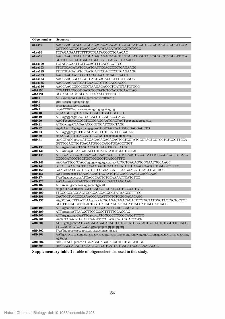
Supplementary table 2: Table of oligonucleotides used in this study.
Nature Chemical Biology: doi:10.1038/nchembio.2498

S7
Supplementary Fig. 1. Evaluation of hetero type receptor sets. The sensor HEK-293T cells (per well of
24-well plate) were transfected with 100 ng of pRK96 (PhCMV-CD43ex-45int-pA), 100 ng of pLS16
(PhCMV-STAT6-pA), and 100 ng of pLS12 (PSTAT6-SEAP-pA), as well as 100 ng each of following
interleukin receptors. Left: pRK115 (PhCMV-ML39-IL4R-pA) and pRK114 (PhCMV-ML39-IL13R1-pA),
middle: pRK115 and pLeo52 (PhCMV-FRB-IL13R1-pA), right: pLeo53 (PhCMV-FKBP-IL4R-pA) and
pRK114. After mixing the sensor cells with HEK-HER2 (target) or HEK-iRFP (non-target) cells, SEAP
secreted from the sensor cells was assayed (with the same method as for Fig. 2b). The data are the mean ±
SEM of three independent experiments measured in triplicate (n=3).
Supplementary Fig. 2. Comparison of promoter strength of hCMV promoter (PhCMV) and SV40 promoter
(PSV40). HEK-293T cells were transfected (per well of 24-well plate) with 250 ng of either pRK293
(PhCMV-ML39-IL4Rtm-CFP-pA) (tm: transmembrane domain) or pRK295 (PSV40-ML39-IL4Rtm-
CFP-pA) as well as 250 ng of pHCM-CD43ex-CD45int-mCherry (LTR-PhCMV-CD43ex-45int
-mCherry-LTR). Then, CFP fluorescence of mCherry positive cells was monitored by FACS BD
LSRFortessa (Negative control was normal HEK-293T cells).
Nature Chemical Biology: doi:10.1038/nchembio.2498

S8
Supplementary Fig. 3. Effect of sensor cell / target (non-target) cell ratio on the system performance.
The sensor HEK-293T cells were constructed by the same method as for Fig.3b (the optimized device
with truncated receptor). These sensor cells were mixed with HEK-HER2 cells or HEK-iRFP cells under
the following conditions in 1.5 mL tubes. Total number of sensor and opponent cells: 2.0x10⁵ cells. Ratio
of sensor cells to opponent cells: 90:1, 30:1, 10:1, 3:1, 1:1, or 1:2. Tubes were incubated at 37 °C for 30
min, then DMEM was added to make 500 µL, and the cell suspension was seeded on 24-well plates. 24
hours later, SEAP value in the supernatant was tested. (normalized to SEAP expression level when mixed
with HEK-iRFP cells in each condition. The data are the mean ± SEM of three independent experiments
measured in triplicate (n=3).
0
1
2
3
4
5
90:1 30:1 10:1 3:1 1:1 1:2
Norm
aliz
ed S
EA
P e
xpre
ssio
n
Sensor : opponent ratio
HEK-iRFP
HEK-HER2
Nature Chemical Biology: doi:10.1038/nchembio.2498

S9
Supplementary Fig. 4. Generalizability of the specific cell-contact-sensing system. (a) System
performance with an anti-HER2 DARPin, 9_268. The sensor cells were transfected (per well of 24-well
plate) with 200 ng of pRK96 (PhCMV-CD43ex-45int-pA), 50 ng of pRK174 (PSV40-9_26-hIL4Rex-pA),
50 ng of pRK173 (PSV40 -9_26-hIL13R1-pA), 100 ng of pLS16 (PhCMV-STAT6-pA), and 100 ng of
pLS12 (PSTAT6-SEAP-pA). Then, the sensor cells were mixed with HEK-iRFP or HEK-HER2 cells. SEAP
activity was measured at 24 hours after cell mixing (the same method as for Fig. 2b). (b) System
performance in hMSC-TERT cells (against HEK-293-HER2 (model) and SKBR3 (cancer cells)).
hMSC-TERT cells were transfected (per well of 24-well plate) with 200 ng of pRK96, 100 ng of pLS12,
50 ng of pRK122 (PSV40-ML39-hIL4Rex)-pA) and 50 ng of pRK123 (PSV40-ML39-hIL13R1)-pA).
Then, the transfected hMSC-TERT cells were mixed with HEK-iRFP, HEK-HER2, or SKBR3 cells.
SEAP activity was measured at 24 hours after cell mixing (the same method as for Fig. 2b). (c)
Confirmation of HER2 expression on HEK-HER2 cells (our model cell line) and SKBR3 cells. HEK-293,
HEK-HER2, SKBR3 cells were stained with a PE-labeled anti-HER2 antibody (Biolegend #324405) in
FACS buffer (DPBS containing 0.5 % BSA), and were analyzed by FACS BD LSRForsetta. HER2
expression level on SKBR3 cells is comparable to that on our model target cells, HEK-HER2. (d)
Performance of Epcam-sensing device. Sensor HEK-293T cells were transfected (per well of 24-well
plate) with Epcam-sensing components as follows. 50 ng of RK182 (PSV40-Ec4-hIL4Rex-pA)
(Ec4: anti-Epcam DARPin9), 50 ng of pRK183 (PSV40-Ec4-hIL13R1-pA), 200 ng of pRK96, 100 ng
of pLS16, and 100 ng of pLS12. Then, the sensor cells were mixed with HEK-293 or HEK-Epcam
cells (with the same cell number and dilution as for Fig. 2b). SEAP secreted from the sensor cells
was assayed.
Nature Chemical Biology: doi:10.1038/nchembio.2498

S10
Supplementary Fig. 5. Evaluation of bystander effect of VP22-FCU1 for cell-based enzyme-prodrug
cancer therapy in our setting. hMSC-TERT cells were transfected (per well of 24-well plate) with 50 ng of
pRK122 (PSV40-ML39-hIL4Rex-pA), 50 ng of pRK123 (PSV40-ML39-hIL13R1-pA), 200 ng of
pRK96 (PhCMV-CD43ex-45int-pA), 100 ng of pLS16 (PhCMV-STAT6-pA), together with 100 ng of one of
following plasmids: pcDNA3.1(+) (for non), pRK130 (PhCMV-FCU1-pA, for expressing cytosolic FCU1),
pRK 131 (PhCMV-VP22-FCU1-pA, for expressing cell penetrating FCU1), or pRK132 (PCMV-Ig secretion
signal (leader sequence from mouse immunoglobulin κ light chain)-ML39-FCU1-pA, for expressing
secreted and HER2-targeted FCU1). At 14 hours after transfection, the medium was changed to fresh
DMEM. At 24 hours after transfection, cells were detached in cell dissociation buffer, spun down, and
suspended in 40 µL of DMEM. Then, 10 µL of the cell suspension (containing 6.0x10⁴ cells) was mixed
with another cell suspension (40 µL) containing 2.4 x10⁵ cells of SKBR3 cells in 1.5 mL tubes. After
incubation for 30 min at 37 °C, fresh DMEM was added so that 2.5x10³ cells of the transfected
hMSC-TERT cells and 1.0x10⁴ cells of SKBR3 cells were suspended in 100 µL, and the cell suspension
was seeded on a flat bottom 96-well plate (Thermo Fischer Scientific) (100 µL/well). At 5 hrs after cell
seeding, various amounts of 5-FC were added. After 4 days, cell viability was determined with a CCK-8
assay (Dojindo) according to the manufacturer’s protocol. The data are the mean ± SEM of three
independent experiments measured in triplicate (n=3)
0
0.2
0.4
0.6
0.8
1
1.2
0 50 100 150 200 250
Rela
veabs.inCCK-8assay
5−FC(uM)
FCU1
VP22-FCU1
Igk-ML39-FCU1
non
Nature Chemical Biology: doi:10.1038/nchembio.2498
S11
Supporting references
1. James, J.R. & Vale, R.D. Biophysical mechanism of T-cell receptor triggering in a reconstituted system.
Nature 487, 64-69 (2012).
2. Horner, M. et al. A chemical switch for controlling viral infectivity. Chem. Commun. 50, 10319-10322
(2014).
3. Schukur, L., Geering, B., Charpin-El Hamri, G. & Fussenegger, M. Implantable synthetic cytokine
converter cells with AND-gate logic treat experimental psoriasis. Sci. Transl. Med. 7, 318ra201 (2015).
4. Li, Y.M. et al. Upregulation of CXCR4 is essential for HER2-mediated tumor metastasis. Cancer Cell 6,
459-469 (2004).
5. Song, E. et al. Antibody mediated in vivo delivery of small interfering RNAs via cell-surface receptors.
Nat. Biotechnol. 23, 709-717 (2005).
6. Chinnasamy, D. et al. Gene therapy using genetically modified lymphocytes targeting VEGFR-2
inhibits the growth of vascularized syngenic tumors in mice. J. Clin. Invest. 120, 3953-3968 (2010).
7. Liu, H. & Naismith, J.H. An efficient one-step site-directed deletion, insertion, single and multiple-site
plasmid mutagenesis protocol. BMC Biotechnol. 8, 91 (2008).
8. Steiner, D., Forrer, P. & Pluckthun, A. Efficient selection of DARPins with sub-nanomolar affinities
using SRP phage display. J. Mol. Biol. 382, 1211-1227 (2008).
9. Stefan, N. et al. DARPins recognizing the tumor-associated antigen EpCAM selected by phage and
ribosome display and engineered for multivalency. J. Mol. Biol. 413, 826-843 (2011).
10. Carter, R.E. & Sorkin, A. Endocytosis of functional epidermal growth factor receptor-green
fluorescent protein chimera. J. Biol. Chem. 273, 35000-35007 (1998).
Nature Chemical Biology: doi:10.1038/nchembio.2498