superior anti-tumor activity of the mdm2 antagonist ... · research open access superior anti-tumor...
TRANSCRIPT

RESEARCH Open Access
Superior anti-tumor activity of the MDM2antagonist idasanutlin and the Bcl-2inhibitor venetoclax in p53 wild-type acutemyeloid leukemia modelsChristian Lehmann1*, Thomas Friess1, Fabian Birzele2, Anna Kiialainen2 and Markus Dangl1,3
Abstract
Background: Venetoclax, a small molecule BH3 mimetic which inhibits the anti-apoptotic protein Bcl-2, andidasanutlin, a selective MDM2 antagonist, have both shown activity as single-agent treatments in pre-clinicaland clinical studies in acute myeloid leukemia (AML). In this study, we deliver the rationale and molecularbasis for the combination of idasanutlin and venetoclax for treatment of p53 wild-type AML.
Methods: The effect of idasanutlin and venetoclax combination on cell viability, apoptosis, and cell cycleprogression was investigated in vitro using established AML cell lines. In vivo efficacy was demonstrated insubcutaneous and orthotopic xenograft models generated in female nude or non-obese diabetic/severecombined immunodeficiency (NOD/SCID) mice. Mode-of-action analyses were performed by means of cellcycle kinetic studies, RNA sequencing as well as western blotting experiments.
Results: Combination treatment with venetoclax and idasanutlin results in synergistic anti-tumor activity comparedwith the respective single-agent treatments in vitro, in p53 wild-type AML cell lines, and leads to strongly superiorefficacy in vivo, in subcutaneous and orthotopic AML models. The inhibitory effects of idasanutlin were cell-cycledependent, with cells arresting in G1 in consecutive cycles and the induction of apoptosis only evident after cells hadgone through at least two cell cycles. Combination treatment with venetoclax removed this dependency, resulting inan acceleration of cell death kinetics. As expected, gene expression studies using RNA sequencing showed significantalterations to pathways associated with p53 signaling and cell cycle arrest (CCND1 pathway) in response to idasanutlintreatment. Only few gene expression changes were observed for venetoclax treatment and combination treatment,indicating that their effects are mediated mainly at the post-transcriptional level. Protein expression studiesdemonstrated that inhibition of the anti-apoptotic protein Mcl-1 contributed to the activity of venetoclax andidasanutlin, with earlier inhibition of Mcl-1 in response to combination treatment contributing to the superiorcombined activity. The role of Mcl-1 was confirmed by small hairpin RNA gene knockdown studies.
Conclusions: Our findings provide functional and molecular insight on the superior anti-tumor activity ofcombined idasanutlin and venetoclax treatment in AML and support its further exploration in clinical studies.
Keywords: BCL-2, MDM2, AML, Cell cycle kinetics, Apoptosis, Idasanutlin, Venetoclax, p53, MCL-1, Synergism
* Correspondence: [email protected] Pharma Research & Early Development, Roche Innovation CenterMunich, Roche Diagnostics GmbH, Nonnenwald 2, 82377 Penzberg, GermanyFull list of author information is available at the end of the article
© 2016 Lehmann et al. Open Access This article is distributed under the terms of the Creative Commons Attribution 4.0International License (http://creativecommons.org/licenses/by/4.0/), which permits unrestricted use, distribution, andreproduction in any medium, provided you give appropriate credit to the original author(s) and the source, provide a link tothe Creative Commons license, and indicate if changes were made. The Creative Commons Public Domain Dedication waiver(http://creativecommons.org/publicdomain/zero/1.0/) applies to the data made available in this article, unless otherwise stated.
Lehmann et al. Journal of Hematology & Oncology (2016) 9:50 DOI 10.1186/s13045-016-0280-3
BackgroundAcute myeloid leukemia (AML) is the most commonacute leukemia in adults and, although high remissionrates are achieved following induction chemotherapy,the majority of patients eventually relapses and becomesresistant to treatment [1]. There have been few changesin standard induction therapy and response rate over thepast three decades, particularly for older patients whoare ineligible for intensive chemotherapy or allogeneicstem cell transplantation [2–4]. Thus, the developmentof novel therapeutics based on an improved understand-ing of the molecular pathogenesis of AML has been amajor focus to address this unmet need.Idasanutlin (RG7388) and venetoclax (ABT-199/
GDC-0199) have each shown significant activity inAML models in pre-clinical studies [5, 6], and eachcompound is currently under evaluation in patientswith AML in phase I and II clinical trials as mono-therapy or in combination with chemotherapy (studyidentifiers: NCT01773408; NCT01994837; NCT02203773;NCT02287233). In addition, venetoclax has recently beengranted breakthrough designation by United States Foodand Drug Administration (FDA) for treatment of relapsedand refractory chronic lymphoid leukemia (CLL) with 17pdeletion [7].Idasanutlin and venetoclax have complementary mecha-
nisms of action. Idasanutlin is a second-generation,nutlin-class, orally bioavailable, selective mouse doubleminute 2 homolog (MDM2) antagonist [8]. MDM2 is animportant negative regulator of the p53 tumor suppressor[9] and is expressed at high levels in a large proportion ofAML [10, 11]. Wild-type p53 is expressed in over 80 % ofAML cases; thus, inhibition of the interaction betweenMDM2 and p53 can re-establish the p53 pathway in AMLcells resulting in cell cycle arrest and induction of apop-tosis [10, 12, 13]. Venetoclax (GDC-0199/ABT-199) is anorally bioavailable novel small-molecule BCL-2 homologdomain 3 (BH3) mimetic, designed to selectively inhibitthe anti-apoptotic protein Bcl-2. High Bcl-2 expression inAML is associated with resistance to chemotherapy andreduced likelihood of achieving complete remission[13–16]. Pre-clinical studies show that venetoclax hassignificant anti-tumor activity in AML models andthat venetoclax-dependent inhibition of Bcl-2 can primeAML cells for responsiveness to chemotherapy [17].The re-establishment of complementary key tumor
suppressor and apoptotic pathways through combinationtreatment with venetoclax and idasanutlin may increaseanti-tumor activity in AML cells. Direct activation ofmitochondrial apoptosis through Bcl-2 inhibition maybe enhanced by p53-dependent cell cycle arrest, p53-induced transcription of the pro-apoptotic Bcl-2 geneBax, and direct binding and inhibition of anti-apoptoticBcl-2 proteins.
However, combined MDM2 and Bcl-2 inhibition forAML treatment has not been clinically investigated yet.In this study, we sought to confirm the superior activityof this combination and to establish a pre-clinical ration-ale for its further exploration in clinical studies. Wetherefore assessed the anti-tumor effects of dualMDM2/Bcl-2 inhibition with the small-molecule inhibi-tors idasanutlin and venetoclax in p53 wild-type AMLmodels and explored the mechanism of action contribut-ing to their synergistic activity in vitro through cell-cycleanalysis, RNA sequencing (RNAseq), and time-courseprotein expression analysis. Moreover, we confirmed thesuperior activity of the combination treatment in sub-cutaneous and orthotopic AML xenograft models.
MethodsReagentsLyophilized idasanutlin and venetoclax were obtainedfrom Roche Innovation Center Basel and GenentechInc., South San Francisco, respectively. A stock solutionof each was prepared in dimethyl sulfoxide (DMSO),and serial dilutions were prepared in culture mediumprior to each experiment; the final DMSO concentrationwas less than 0.2 % (v/v) in all experiments.
Cell lines and culture conditionsThe MV4-11 and MOLM-13 p53 wild-type AML celllines and the p53 mutant HL-60 promyelocytic cellline were obtained from the Roche Pharma Research &Early Development (pRED) cell bank (Roche Diagnos-tics GmbH). The p53 wild-type/NPM mutant OCI-AML-3 cell line was obtained from the Roche pRED CELLO cellbank (Roche Diagnostics GmbH). MV4-11 and MOLM-13 cells were cultured in RPMI-1640 supplemented with20 % fetal calf serum (FCS; Gibco), HL-60 cells werecultured in RPMI-1640 supplemented with 10 % FCS, andOCI-AML-3 cells were cultured in alpha-MEM supple-mented with 20 % FCS. Cultures were maintained at37 °C, with 7 % CO2 in a humidified atmosphere. Cellline identity was confirmed by short tandem repeats-polymerase chain reaction (STR-PCR) genotyping usingthe QIAGEN Investigator IDplex Plus Kit (QIAGENcat. no. 381625).
Cell viability and apoptosis measurementCell cultures were treated with idasanutlin alone(0.6–2000 nM), venetoclax alone (0.6–2000 nM), orboth agents combined for 72 h. Viability was mea-sured using CellTiter-Glo® (Promega), as per manufac-turer’s instructions, with luminescence measuredusing the Infinite® F200 microplate reader (Tecan). Inshort hairpin RNA (shRNA) experiments, viability wasmeasured by trypan blue exclusion using the CedexHiRes automated analyzer (Roche Diagnostics).
Lehmann et al. Journal of Hematology & Oncology (2016) 9:50 Page 2 of 13
For measurement of apoptosis, cells were stainedwith Annexin-V-Fluos staining buffer (1 μL/sampleAnnexin-V-Fluos [Roche Diagnostics GmbH], 2 ng/μLHoechst 33258, and 400 nM CaCl2, prepared in culturemedium) and incubated on ice for 15 min in the dark.Fluorescence was analyzed at 4 °C on the LSRII flowcytometer (BD Biosciences).Data for viability and apoptosis measurements were
analyzed using XLfit® for Excel (Microsoft Corporation).Flow cytometric data were analyzed using FlowJo soft-ware versions 7.6.5 and 10.0.7 (Treestar).Relative and absolute half maximal inhibitory concentra-
tion (IC50) values were calculated for each treatment, withrelative IC50 corresponding to the mid-point between theupper and lower plateaus of the response curve and abso-lute IC50 representing 50 % of the control.To determine synergistic effects between idasanutlin and
venetoclax, combination indices were calculated as perLoewe and Muchnik [18] using the following equation:
Combination index ¼ CA
ICxAþ CB
ICxB
where ICxA and ICxB are the concentration of com-pounds A and B required individually for effect x (in thisstudy x = relative IC50) and CA and CB are the concen-tration of compounds A and B required in combinationto achieve the same effect x. Values less than 1 indicatesynergy.
AML xenograft modelsSubcutaneous and orthotopic xenograft models weregenerated in female nude or non-obese diabetic/severecombined immunodeficiency (NOD/SCID) mice. All ex-periments were conducted by Charles River DiscoveryResearch Services (CR-DRS) in accordance with the rec-ommendations of the Guide for Care and Use of Labora-tory Animals [19] with respect to restraint, husbandry,surgical procedures, feed and fluid regulation, and veter-inary care. The animal care and use program at CR-DRSis accredited by the Association for Assessment and Ac-creditation of Laboratory Animal Care International(AAALAC).For the subcutaneous model, MV4-11 cells (1 × 10⁷)
were injected subcutaneously with Matrigel™ BasementMembrane Matrix (BD Biosciences) into the right flanksof 7-week-old mice. On day 12 post-inoculation, mice(n = 10 per group) were stratified randomly based on pri-mary tumor size with a median tumor volume (prior totreatment) of approximately 100–150 mm3. Idasanutlin(30 mg/kg orally) and venetoclax (100 mg/kg orally)were administered daily for 21 days alone or in com-bination. Tumor volumes ([length × width]2/2) weremeasured by calipers twice weekly and tumor growth
inhibition relative to control animals was calculatedas follows:
Tumor growth inhibition ¼ 1− T−T0ð Þ= C−C0ð Þ½ �ð Þ � 100
where T is the tumor volume in the treated group atmeasurement, T0 is the tumor volume in the treatedgroup at baseline, C is the tumor volume in the controlgroup at measurement, and C0 is the tumor volume inthe control group at baseline.The MV4-11 and MOLM-13 orthotopic models were
established by intravenous inoculation of 1 × 10⁷ and5 × 106 cells, respectively. Ten-week-old mice were pre-treated with cyclophosphamide (150 mg/kg, intraperito-neally), cells were injected into the tail vein, and micewere distributed randomly into four groups (n = 10 pergroup). Treatment was initiated 22 days after inoculationof MV4-11 cells and 3 days after inoculation of MOLM-13 cells. In both tumor studies, idasanutlin (30 mg/kgorally) and venetoclax (100 mg/kg orally) were adminis-tered daily for 21 days alone or in combination. Animalswere monitored daily for clinical symptoms and adverseevents.A time-to-event (TTE) analysis was conducted until
day 78 post-inoculation in the MV4-11 model andday 48 post-inoculation in the MOLM-13 model. Theevent was death or moribundity due to disseminatedleukemia. Any animal that did not reach the endpointwas euthanized at the end of the study and assigneda TTE value of 78 days for the MV4-11 model or48 days for the MOLM-13 model. Any animal thatdied from non-tumor burden-related causes was ex-cluded from the analysis. A TTE value was recordedfor each assessable animal, and the median TTE wascalculated for each group.The percentage increase in lifespan (ILS) versus the
vehicle control was calculated as:
ILS ¼ T−Cð ÞC
� 100%
where T is the median survival of the treatment groupand C is the median survival of the control group.
Cell cycle analysisMV4-11 and MOLM-13 cells were treated with idasanu-tlin and venetoclax alone or in combination for 72 h(0.6–2000 nM). At the start of the final 24 h of incuba-tion, 5-bromo-2-deoxyuridine (BrdU; Sigma) was addedto cultures at a concentration of 80 μM. Culturemedium was also supplemented with 80 μM deoxycyti-dine (Sigma) at this point to minimize disturbance tothe nucleotide pathway. Prior to flow cytometric ana-lysis, cells were washed twice in ice-cold DNA-stainingbuffer (100 mM Tris pH 7.4, 154 mM NaCl, 1 mM
Lehmann et al. Journal of Hematology & Oncology (2016) 9:50 Page 3 of 13
CaCl2, 0.5 mM MgCl2, 0.1 % NP40, and 0.2 % bovineserum albumin) and incubated in DNA-staining buffercontaining 10 U/mL RNase (Roche Diagnostics GmbH)and 1.5 μg/mL Hoechst 33258 for 15 min at 37 °C. Pro-pidium iodide (PI) was added to a final concentration of1.5 μg/mL, and cells were incubated on ice for 15 min.Fluorescence was analyzed on the LSRII flow cytometer,and data were analyzed using FlowJo software versions7.6.5 and 10.0.7.
Gene expression analysisFor mRNA (poly-A) RNAseq studies, MOLM13 cellswere treated with idasanutlin (100 nM) and veneto-clax (100 nM) alone or in combination for 6 h. Highmolecular weight RNA (>200 base pairs) was ex-tracted from four biologic replicates using theRNeasy® Mini Kit (QIAGEN®) as per manufacturer’sinstructions. Residual genomic DNA was removedduring the extraction using the RNase-free DNase set(QIAGEN®). RNA quality was analyzed usingEukaryote Total RNA Nano chips (Agilent Technolo-gies), and all samples used for analysis had an RNAintegrity number >8. RNAseq libraries were generatedfrom 1 μg total RNA using the TruSeq® RNA SamplePreparation v2 kit (Illumina®) as per manufacturer’sinstructions. Sequencing libraries were quantifiedusing the Kapa Library Quantification kit (Kapa Bio-systems), and quality was assessed on the AgilentBioanalyzer using DNA 1000 chips (Agilent Technolo-gies). Libraries were sequenced on the HiSeq® 2500sequencer (Illumina) for 2 × 50 cycles using the Tru-Seq® PE Cluster Kit v3-cBot-HS and TruSeq® SBS Kitv3-HS sequencing reagents (Illumina®). Each lane wasspiked with the PhiX Control v3 library (Illumina®) ata final concentration of 1 % (v/v) as a sequencingcontrol.Sequencing reads were first aligned to the human
protein coding transcriptome (as defined by Ensemblv73 [http://www.ensembl.org]) using Bowtie 2 [20].In a second step, all reads not mapping to the hu-man transcriptome were aligned to the human gen-ome (hg19) using Bowtie 2. For both mapping steps,default sensitivity settings were used. Gene expres-sion was profiled using in-house tools, and reads perkilobase per million reads (RPKM) were computed aspreviously described using in-house software [21].Differential gene expression was computed usingDESeq [22]. Genes with an absolute log2 ratio >1(absolute fold change >2) versus the DMSO controland a false discovery rate corrected P value <0.01were considered differentially expressed. Functionalannotation and analysis of altered pathways andfunctions was performed using Ingenuity® PathwayAnalysis (QIAGEN®).
shRNA analysisMV4-11 cells (5 × 10⁵) were transduced with MIS-SION® non-specific or Mcl-1-targeting shRNA lenti-viral particles (Sigma) in the presence of polybrene(10 μg/mL); target sequences are listed in Additionalfile 1. Puromycin (2 μg/mL) was added after 48 h toselect for positive transductants, and cells were sam-pled for viability and protein expression analysis onthe following day.
Western blottingCells were lysed in RIPA buffer (Sigma), lysates wereloaded onto NuPAGE® 4–12 % Bis-Tris PrecastProtein Gels (Life Technologies), and proteins wereseparated and transferred to nitrocellulose mem-branes (Life Technologies). Membranes were blockedwith 5 % (w/v) milk powder in Tris-buffered salinewith Tween 20 and incubated with Mcl-1, cleavedcaspase-3 (Asp175), or ß-actin primary antibodies,followed by incubation with horseradish peroxidase(HRP)-conjugated secondary antibodies (Cell Signal-ing Technology). Bands were detected using Super-Signal® Chemiluminescent HRP Substrate (ThermoFisher Scientific).
Statistical analysesFor viability and apoptosis assays, at least three biologicreplicates were analyzed. The data presented are themean values of one representative experiment. Meanvalues and standard deviations were calculated usingExcel (Microsoft Corporation).For the MV4-11 subcutaneous model, primary tumor
growth data were baseline corrected against tumor vol-ume at the start of treatment, based on ratios. Adjusteddata were analyzed using non-parametric methods dueto an asymmetric distribution. Treatment-to-control ra-tios (TCRs) were calculated as follows:
TCR ¼�V treated
�V control
where �V is tumor volume. Two-sided non-parametricconfidence intervals (CIs; 1 − α) were also calculated [23].For the MV4-11 and MOLM-13 orthotopic models,
the date of the last measurement was used for analysisof median and overall survival and the generation ofKaplan-Meier plots. Treatment groups were comparedusing a pairwise log-rank test with a multiple test levelof 0.00833 considered statistically significant.Statistical analyses for all in vivo studies were per-
formed using SAS-JMP version 8.1 (SAS Inc., 2007).
Lehmann et al. Journal of Hematology & Oncology (2016) 9:50 Page 4 of 13

ResultsCombined treatment with idasanutlin and venetoclaxshows synergistic effects in AML cell lines in vitroThe effects of single-agent and combination therapywith idasanutlin and venetoclax were assessed in fourAML cell lines exposed to the inhibitors for 72 h. TheMV4-11 and MOLM-13 cell lines carry the wild-typeTP53 gene, and the OCI-AML-3 cell line carries wild-type TP53 and a nucleophosmin (NPM) mutation. NPMmutant AML accounts for ~35 % of AML in adults [24].The HL-60 AML cell line, which carries deletions inboth TP53 alleles, was used as a control to monitor non-specific effects of the MDM2 antagonist idasanutlin.Viability was assessed based on adenosine triphosphate(ATP) concentration, as a measure of metabolically activecells. In viability studies (Fig. 1), dose-dependent effects
were seen in the MV4-11 (Fig. 1a) and MOLM-13 (Fig. 1b)p53 wild-type cell lines for single-agent treatment withboth inhibitors. Relative and absolute IC50 values were cal-culated for each treatment. For MV4-11 cells, relative IC50
values for idasanutlin and venetoclax were 55 and 18 nM,respectively, and absolute IC50 values were 51 and 12 nM,respectively. For MOLM-13 cells, the respective relativeIC50 values were 35 and 20 nM, while absolute IC50 valueswere 31 and 16 nM. In both cell lines, combination treat-ment was synergistic, with a calculated combination indexof 0.72 for MV4-11 cells and 0.59 for MOLM-13 cells(values <1 indicate synergy), corresponding to relativeIC50 values of 8 and 7 nM, respectively.Venetoclax had little effect on the viability of the NPM
mutant OCI-AML-3 cell line (resistance to Bcl-2 inhibi-tors has been seen previously in this cell line; data not
Concentration (nM)
a
1 10 100 1000
Sig
nal r
educ
tion
rela
tive
to
cont
rol (
%)
Concentration (nM)
b
1 10 100 1000
Concentration (nM)
c
1 10 100 1000
Sig
nal r
educ
tion
rela
tive
to
cont
rol (
%)
1009080706050403020100
–10
100908070605040302010
0–10
Sig
nal r
educ
tion
rela
tive
to
cont
rol (
%)
Sig
nal r
educ
tion
rela
tive
to
cont
rol (
%)
Concentration (nM)
d
1 10 100 1000
Idasanutlin Venetoclax Idasanutlin + venetoclax
1009080706050403020100
–10
1009080706050403020100
–10
Fig. 1 Viability of a MV4-11, b MOLM-13, c OCI-AML-3, and d HL60 cells treated with idasanutlin and venetoclax alone and in combination for72 h. CellTiter-Glo® assay (Promega) was used to determine the cellular viability. Data are displayed as mean plus standard deviation calculatedfrom at least three biological replicates. In MV4-11 cells (a), combination treatment led to synergistic effects with a combination index (CI) of 0.72and a relative IC50 of 8 nM compared to 55 and 18 nM for idasanutlin and venetoclax single treatment, respectively. In MOLM-13 cells (b), thecalculated CI was 0.59 with an IC50 of 7 nM for the combination as opposed to single treatment with idasanutlin (IC50, 35 nM) and venetoclax (IC50,20 nM). OCI-AML-3 cells were resistant to venetoclax (c), while p53 mutated HL-60 cells did not react up to 2 μM idasanutlin as expected (d)
Lehmann et al. Journal of Hematology & Oncology (2016) 9:50 Page 5 of 13
shown); hence, there was no notable difference betweensingle-agent idasanutlin (relative/absolute IC50, 164/147nM) and the combination treatment for this cell line(relative/absolute IC50, 142/133 nM). Non-specific ef-fects were not observed for idasanutlin treatment in thep53 mutant HL-60 line, although venetoclax inducedsubstantial cell death (relative/absolute IC50, 4/4 nM).The results of cell viability studies were supported by
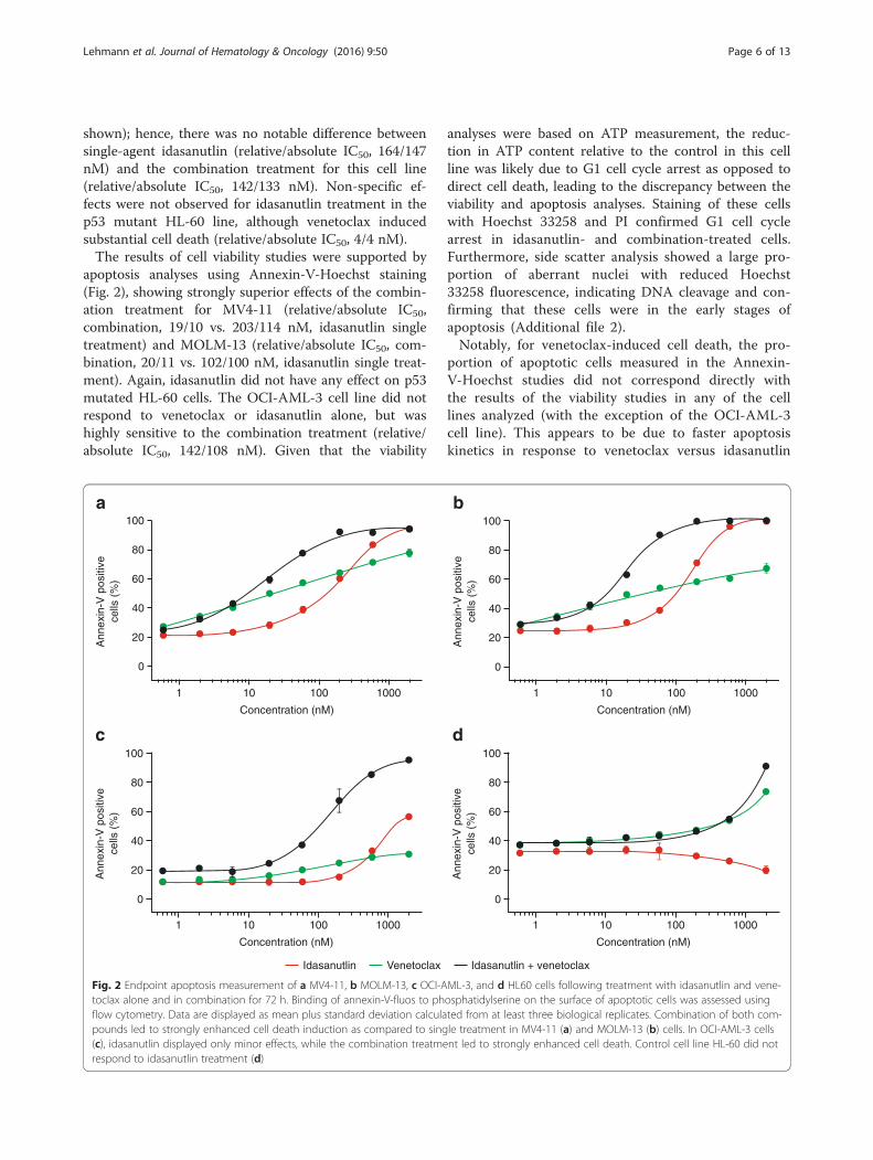
apoptosis analyses using Annexin-V-Hoechst staining(Fig. 2), showing strongly superior effects of the combin-ation treatment for MV4-11 (relative/absolute IC50,combination, 19/10 vs. 203/114 nM, idasanutlin singletreatment) and MOLM-13 (relative/absolute IC50, com-bination, 20/11 vs. 102/100 nM, idasanutlin single treat-ment). Again, idasanutlin did not have any effect on p53mutated HL-60 cells. The OCI-AML-3 cell line did notrespond to venetoclax or idasanutlin alone, but washighly sensitive to the combination treatment (relative/absolute IC50, 142/108 nM). Given that the viability
analyses were based on ATP measurement, the reduc-tion in ATP content relative to the control in this cellline was likely due to G1 cell cycle arrest as opposed todirect cell death, leading to the discrepancy between theviability and apoptosis analyses. Staining of these cellswith Hoechst 33258 and PI confirmed G1 cell cyclearrest in idasanutlin- and combination-treated cells.Furthermore, side scatter analysis showed a large pro-portion of aberrant nuclei with reduced Hoechst33258 fluorescence, indicating DNA cleavage and con-firming that these cells were in the early stages ofapoptosis (Additional file 2).Notably, for venetoclax-induced cell death, the pro-
portion of apoptotic cells measured in the Annexin-V-Hoechst studies did not correspond directly withthe results of the viability studies in any of the celllines analyzed (with the exception of the OCI-AML-3cell line). This appears to be due to faster apoptosiskinetics in response to venetoclax versus idasanutlin
Ann
exin
-V p
ositi
ve
cells
(%
)
Concentration (nM)
a
1 10 100 1000
Ann
exin
-V p
ositi
ve
cells
(%
)
Concentration (nM)
b
1 10 100 1000
Ann
exin
-V p
ositi
ve
cells
(%
)
Concentration (nM)
c
1 10 100 1000
100
80
60
40
20
0
100
80
60
40
20
0
100
80
60
40
20
0
100
80
60
40
20
0
Ann
exin
-V p
ositi
ve
cells
(%
)
Concentration (nM)
d
1 10 100 1000
Idasanutlin Venetoclax Idasanutlin + venetoclax
Fig. 2 Endpoint apoptosis measurement of a MV4-11, b MOLM-13, c OCI-AML-3, and d HL60 cells following treatment with idasanutlin and vene-toclax alone and in combination for 72 h. Binding of annexin-V-fluos to phosphatidylserine on the surface of apoptotic cells was assessed usingflow cytometry. Data are displayed as mean plus standard deviation calculated from at least three biological replicates. Combination of both com-pounds led to strongly enhanced cell death induction as compared to single treatment in MV4-11 (a) and MOLM-13 (b) cells. In OCI-AML-3 cells(c), idasanutlin displayed only minor effects, while the combination treatment led to strongly enhanced cell death. Control cell line HL-60 did notrespond to idasanutlin treatment (d)
Lehmann et al. Journal of Hematology & Oncology (2016) 9:50 Page 6 of 13

treatment. Given that apoptosis measurement is anendpoint determination, it is possible that Annexin-positive, and even Annexin-Hoechst-positive, cellswere missed in this analysis as they had already beendegraded.
Combined treatment with idasanutlin and venetoclaxshows superior anti-tumor effects in vivoThe activity of idasanutlin and venetoclax were furtherassessed in subcutaneous and orthotopic AML xenograftmodels. In the MV4-11 subcutaneous model, tumorgrowth inhibition relative to the vehicle control groupwas 0 % for venetoclax (no inhibition vs. control, TCR0.89, CI, 0.50–2.10) and 30 % for idasanutlin (TCR 0.5,CI, 0.26–0.98). Compared with the single-agent treat-ments, combination treatment resulted in superiortumor growth inhibition (>100 %) and notably partialtumor regression of 55 % (TCR 0.03, CI, 0.02–0.07) rela-tive to the control (Fig. 3a).In a TTE analysis of the MV4-11 orthotopic model,
the median survival of the mice post-inoculation was38.5 days for mice treated with the vehicle control(Fig. 3b). In comparison, the median survival was 38 dayswith venetoclax, 52 days with idasanutlin, and enhancedto 70 days with combination treatment. This corre-sponded to ILS values of −1 % for venetoclax, 35 % foridasanutlin, and 82 % for combination treatment. Themedian and overall survival was significantly better forcombination treatment than for each of the single-agenttreatments (P < 0.0001 for both).In the MOLM-13 orthotopic model, the median sur-
vival of the mice post-inoculation was 26 days withvenetoclax, 23 days with idasanutlin, and a superior45 days with combination treatment, compared with21 days for the vehicle control (Fig. 3c). The correspond-ing ILS values were 24 % for venetoclax, 10 % for idasa-nutlin, and 114 % for combination treatment. In termsof efficacy, median and overall survival was significantlybetter for combination treatment than single-agent treat-ment (P < 0.0001).For all in vivo studies, treatments were well tolerated
as indicated by no significant loss of body weight(>20 %) for the duration of the study.
Combination treatment leads to accelerated cell deathkinetics in vitroOnce the activity of idasanutlin and venetoclax had beenassessed in cell culture and human AML xenograftmodels, exploratory studies were conducted to deter-mine the cellular mechanisms contributing to their com-bined effects. BrdU analysis using Hoechst 33258 and PIwas conducted to analyze changes in cell cycle kineticsof MOLM-13 and MV4-11 cells in response to treat-ment. BrdU is incorporated into DNA during replication
and quenches fluorescence upon subsequent stainingwith Hoechst, giving a measure of cell division overtime. Simultaneous PI staining for DNA contentshows cell cycle progression. Figure 4 shows that cellstreated with the DMSO control (vehicle) progressedthrough up to three cell cycles over the 24 h studyperiod as measured by BrdU/Hoechst quenching.Treatment with idasanutlin (60 nM) induced G1 cellcycle arrest in the first and second cycles of
3000
300
30
1.0
0.8
0.6
0.4
0.2
0.0
Tum
or v
olum
e (m
edia
n, IQ
R [m
m3 ])
Time post inoculation (days)
a
11 13 15 17 19 21 23 25 27 29 31 33
Sur
viva
l (%
mic
e)
Time post-inoculation (days)
VehicleIdasanutlin (30 mg/kg)
b
20 30 40 50 60 70
Venetoclax (100 mg/kg)Idasanutlin (30 mg/kg)+ venetoclax (100 mg/kg)
1.0
0.8
0.6
0.4
0.2
0.0
Sur
viva
l (%
mic
e)
Time post-inoculation (days)
c
10 25 3015 20 35 40 45
Fig. 3 Tumor growth inhibition in a MV4-11 subcutaneous modeland TTE analysis of survival in b MV4-11 and c MOLM-13 orthotopicmodels (n = 10 mice per group). Combination treatment resulted insuperior anti-tumor activity and enhanced survival. See text fordetails. TTE time-to-event
Lehmann et al. Journal of Hematology & Oncology (2016) 9:50 Page 7 of 13

replication. Minor nuclear fragmentation indicatingapoptosis, evident from a sub-G1 population, wasonly observed in the G1 phase of the second cycleonwards (Fig. 4). However, treatment with 60 nMvenetoclax induced nuclear fragmentation in both thefirst and second cell cycles (Fig. 4). Combinationtreatment led to high levels of nuclear fragmentationfrom cells arrested in the G1 phase of the first cycleand, to a lesser extent, the second cycle (Fig. 4).Hence, viable but non-proliferating cells, arrested inG1 by idasanutlin, are more vulnerable to venetoclaxtreatment, resulting in enhanced and accelerated celldeath kinetics. The observed effects were similar be-tween MV4-11 and MOLM-13 cell lines (Fig. 4).
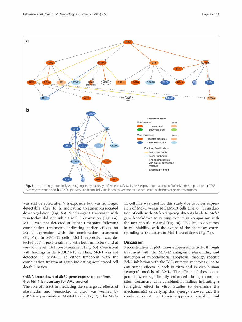
Gene expression analysis confirms TP53 pathwayactivation by idasantulin, while venetoclax treatmentdoes not result in transcriptional changesRNAseq analysis was performed to measure gene ex-pression changes in response to inhibitor treatment.MOLM-13 cells were exposed to idasanutlin (100 nM)and venetoclax (100 nM) alone or in combination for6 h, and differential gene expression was analyzed.Overall, more than 45 million reads were sequenced
for each sample. In comparison with the DMSO control,substantially more genes showed differential expressionfollowing idasanutlin and combination treatment thanvenetoclax treatment. Overall 46, 363, and 183 geneswere differentially expressed in response to venetoclax,
idasanutlin, and combination treatment, respectively.Upstream regulator analysis using Ingenuity pathwaysoftware predicted TP53 pathway activation as expectedand CCND1 pathway inhibition indicating G1 arrest asthe most significant changes following idasanutlin treat-ment and also following combination treatment (Fig. 5).No significantly altered pathways were identified in re-sponse to venetoclax treatment. There were no obviousdifferences in gene expression between the idasanutlin-treated cells and combination-treated cells that wouldexplain the synergistic effects of combination treatmentin vitro, confirming that venetoclax directly acts on Bcl-2 function rather than on gene expression.
Combination treatment leads to accelerated Mcl-1 down-regulation and caspase-3 activationAs expected, western blot analysis of MOLM-13 cellsshowed that p53 protein levels increased with the dur-ation of exposure to idasanutlin and combination treat-ment, but not with venetoclax treatment (Fig. 6a).Cleaved caspase-3, demonstrating execution of apop-tosis, was detected after 16 h of exposure to idasanutlinand after 7 h exposure to venetoclax, although expres-sion levels were lower following venetoclax treatmentcompared with idasanutlin-treated cells (16 h). Combin-ation treatment with idasanutlin and venetoclax resultedin increased detection of cleaved caspase-3 versussingle-agent therapy with either compound from 7 h ofexposure. In cells treated with idasanutlin alone, Mcl-1
Fig. 4 BrdU analysis of cell-cycle kinetics in MOLM-13 and MV4-11 cells treated with idasanutlin and venetoclax alone and in combination for72 h. BrdU was added for the last 24 h of the experiment as described in the “Methods” section. Populations residing in G1, S, and G2/M phasesare indicated as an example for MOLM-13 vehicle-treated cells. Sub G1 represents nuclear fragmentation as a consequence of cell death. Vehicle(DMSO)-treated cells displayed normal proliferation, with MOLM-13 cells reaching the third and MV4-11 cells reaching the second cycle. Treatmentwith idasanutlin induces cell cycle arrest and minor nuclear fragmentation from the G1 phase of the second cell cycle (sub G1, arrows). Treatmentwith venetoclax did not affect cell cycle progression, but induced nuclear fragmentation mainly from the G1 phase of the first cycle (arrows).Combination treatment results in remarkably enhanced nuclear fragmentation from the G1 phase of the first cell cycle onwards with few viablecells remaining
Lehmann et al. Journal of Hematology & Oncology (2016) 9:50 Page 8 of 13
was still detected after 7 h exposure but was no longerdetectable after 16 h, indicating treatment-associateddownregulation (Fig. 6a). Single-agent treatment withvenetoclax did not inhibit Mcl-1 expression (Fig. 6a).Mcl-1 was not detected at either timepoint followingcombination treatment, indicating earlier effects onMcl-1 expression with the combination treatment(Fig. 6a). In MV4-11 cells, Mcl-1 expression was de-tected at 7 h post-treatment with both inhibitors and atvery low levels 16 h post-treatment (Fig. 6b). Consistentwith findings in the MOLM-13 cell line, Mcl-1 was notdetected in MV4-11 at either timepoint with thecombination treatment again indicating accelerated celldeath kinetics.
shRNA knockdown of Mcl-1 gene expression confirmsthat Mcl-1 is necessary for AML survivalThe role of Mcl-1 in mediating the synergistic effects ofidasanutlin and venetoclax in vitro was verified byshRNA experiments in MV4-11 cells (Fig. 7). The MV4-
11 cell line was used for this study due to lower expres-sion of Mcl-1 versus MOLM-13 cells (Fig. 6). Transduc-tion of cells with Mcl-1-targeting shRNAs leads to Mcl-1gene knockdown to varying extents in comparison withthe non-specific control (Fig. 7a). This led to decreasesin cell viability, with the extent of the decreases corre-sponding to the extent of Mcl-1 knockdown (Fig. 7b).
DiscussionReconstitution of p53 tumor-suppressor activity, throughtreatment with the MDM2 antagonist idasanutlin, andinduction of mitochondrial apoptosis, through specificBcl-2 inhibition with the BH3 mimetic venetoclax, led toanti-tumor effects in both in vitro and in vivo humanxenograft models of AML. The effects of these com-pounds were significantly enhanced through combin-ation treatment, with combination indices indicating asynergistic effect in vitro. Studies to determine themechanism(s) underlying this synergy showed that thecombination of p53 tumor suppressor signaling and
a
b
BRCA1
TP53
SP1 CDKN1A
CDK2
CEBPB
Prediction Legend
Predicted Relationships
Leads to activation
Leeds to inhibition
Findings inconsistentwith state of downstreammolecule
Effect not predicted
Upregulated
Downregulated
More extreme Less
Predicted activation
Predicted inhibition
More confidence Less
CCNE1
TP63
TP73
TP53
RELA REL STAT3 MED1 ESR1 EGR1 CEBPB
MDM2
CCND1
MAXWT1MYC
E2F1
E2F1
E2F3
E2F4
E2F2 TFDP1
E2F4RBL1
RBL1 RB1
EP300
RB1
E2f
E2f
NFkB(complex)
Fig. 5 Upstream regulator analysis using Ingenuity pathway software in MOLM-13 cells exposed to idasanutlin (100 nM) for 6 h predicted a TP53pathway activation and b CCND1 pathway inhibition. Bcl-2 inhibition by venetoclax did not result in changes of gene transcription
Lehmann et al. Journal of Hematology & Oncology (2016) 9:50 Page 9 of 13

direct induction of mitochondrial apoptosis lead to im-mediate and potent cell death. Western blot analysisidentified that Mcl-1 inhibition is a key contributor tothese effects, and earlier Mcl-1 inhibition is seen in re-sponse to idasanutlin/venetoclax combination treatment,correlating with accelerated cell death kinetics.Both nutlins and BH3 mimetics have shown prom-
ise in pre-clinical and clinical studies [5, 6, 25–29].Idasanutlin and venetoclax represent clinically opti-mized molecules within their respective compoundclasses. Idasanutlin shows increased specificity and ac-tivity, with lower dosage requirements, compared withpreceding nutlin-class compounds [5, 8]. Venetoclaxhas enhanced specificity compared with earlier BH3mimetics (such as ABT-737 and the related clinical-grade compound navitoclax), which interact with theBH3 domains of Bcl-2, Bcl-xL, and Bcl-w. Venetoclaxspecifically inhibits Bcl-2 and therefore is not associ-ated with the dose-limiting thrombocytopenia result-ing from Bcl-xL inhibition [30].Our data show that exposure of human AML cell lines
in vitro to single-agent venetoclax or idasanutlin inducedcell death without off-target effects, with enhancedapoptotic activity seen when cells were treated with thetwo agents in combination. Discrepancies in the levels ofinhibition between the cell viability and apoptosis studiesare likely due to the nature of the assays used and
highlight the different modes of action of the two inhibi-tors. The viability studies (Fig. 1) were based on ATPmeasurement as an indicator of metabolic activity, whileapoptosis studies (Fig. 2) measured phosphatidyl serineexposure and membrane permeability. Thus, the viabilityassays will detect cell cycle arrest in addition to apop-tosis. For idasanutlin, cells became arrested in the G1phase of the cell cycle initially before induction of apop-tosis in subsequent cycles (Fig. 4). This is illustrated inthe OCI-AML-3 cell line where there was no differenceobserved between idasanutlin and combination treat-ment in viability studies based on ATP concentration.However, in apoptosis studies, there was little cell deathin response to idasanutlin, indicating that the effectsseen in the viability studies were due to cell cycle arrestas opposed to cell death (Figs. 1 and 2 and Additionalfile 2). In the MV4-11 and MOLM-13 cell lines, induc-tion of cell death was initiated immediately in responseto venetoclax treatment and this may actually have ledto some apoptotic cells being missed by the assay. Thisdifference in cell death kinetics between the two inhibi-tors was confirmed by Western blotting analysis ofcleaved caspase-3 expression, which shows acceleratedinduction of apoptosis in response to venetoclax versusidasanutlin (Fig. 6).
hours0 7 16 7 16 7 16
Cleaved caspase-3
p53
Mcl-1
Actin
Mcl-1
Actin
a
b
Idas
anut
lin
Venet
oclax
Idas
anut
lin
+ ve
neto
clax
hours0 7 16 7 16 7 16
Idas
anut
lin
Venet
oclax
Idas
anut
lin
+ ve
neto
clax
Fig. 6 Western blot analysis of a p53, cleaved caspase-3, and Mcl-1protein expression in MOLM-13 cells and b Mcl-1 protein expressionin MV4-11 cells exposed to venetoclax (100 nM) and idasanutlin(100 nM) alone or in combination for time periods indicated. Actinwas used as a loading control for both cell lines
Mcl-1
Actin
a
b
Null ve
ctor
shRNA 1
shRNA 2
shRNA 3
shRNA 4
shRNA 5
Null ve
ctor
shRNA 1
shRNA 2
shRNA 3
shRNA 4
shRNA 5
90
80
70
60
50
40
30
20
10
0V
iabi
lity
(%)
Fig. 7 shRNA inhibition of Mcl-1 gene expression in MV4-11 cells aWestern blot analysis of Mcl-1 protein expression and b viabilityof shRNA transduced cells measured by trypan blue exclusion,confirming that Mcl-1 contributes significantly to AML cell survival
Lehmann et al. Journal of Hematology & Oncology (2016) 9:50 Page 10 of 13

Notably, for the OCI-AML-3 cell line, little inhibitoryeffect was seen in response to venetoclax treatment andthis may be due to BH3 mimetic resistance previouslyobserved with this cell line (data not shown). However,combination treatment induced substantial cell death inthis cell line, overcoming resistance to venetoclax andremoving the cell cycle dependency of idasanutlin activ-ity (Figs. 1 and 2 and Additional file 2).In contrast to the in vitro studies, more modest
growth inhibition was seen when the single-agent inhibi-tors were administered to in vivo human AML xenograftmodels. Venetoclax had no effect in either the subcuta-neous or orthotopic MV4-11 models and neither inhibi-tor showed substantial improvements in survival versusthe vehicle control in the MOLM-13 model, althoughvenetoclax was marginally better than idasanutlin. Thislow activity for single-agent treatment may be due to theaggressive nature of these in vivo AML models, particu-larly the MOLM-13 orthotopic model, or due to poortumor penetration of the compounds. In spite of thisand in line with our results in vitro, combination treat-ment led to superior tumor growth inhibition and sub-stantial ILS compared with the vehicle control andsingle-agent therapies.Our data confirm and add on to previously pub-
lished findings. Saiki et al. [31] showed synergistic ef-fects when combining MDM2 antagonists and thedual Bcl-2/Bcl-xL inhibitors ABT-737 and ABT-263 insolid and hematologic tumor cell lines. Using the Bcl-2-selective inhibitor ABT-199 as combination partner,activity was mainly restricted to hematological celllines. The authors concluded that Bcl-xL inhibition isessential for synergy with MDM2 antagonists in solidtumor cell lines, while selective Bcl-2 inhibition inhematologic cell lines is sufficient [31]. Moreover, anearlier study of dual MDM2 and Bcl-2 inhibition withABT-737 and nutlin-3 (tool compounds used for invitro studies) showed the feasibility of combiningMDM2 and Bcl-2 inhibitors in AML [32]. In thatstudy, the individual effects of MDM2 and Bcl-2 in-hibition were shown to be cell cycle-specific withnutlin-3a inducing apoptosis predominantly in theG2/M phase of the cell cycle while ABT-737 inducedapoptosis predominantly while cells were in G1. Com-bination treatment removed the cell cycle-dependenteffects of treatment, with the authors proposing thatthis could account, in part, for the synergy observed.Similar cell cycle effects were seen in the currentstudy in response to single-agent exposure to theclinical-grade compounds idasanutlin and venetoclax.Treatment with idasanutlin alone led to G1 cell cyclearrest, with little evidence of apoptosis until subse-quent cycles of replication. With venetoclax treat-ment, cells continued to divide and apoptosis was
induced during each round of replication. Combin-ation treatment resulted in direct induction of celldeath without any further cell division. Thus, the cellcycle-independent activity of the combination treat-ment could contribute to the synergy, which mani-fests as an acceleration of cell death kinetics.Gene expression analysis in the MOLM-13 cell line
further highlighted the different mechanisms of action ofvenetoclax and idasanutlin. The expression of far fewergenes was altered in response to venetoclax treatmentcompared with idasanutlin or combination treatment,reflecting the fact that Bcl-2 inhibition and subsequentmitochondrial apoptosis is mediated mainly throughprotein-protein interactions while p53 signaling is medi-ated at the protein and transcriptional level. Upstreamregulator analysis of gene expression changes in re-sponse to idasanutlin and combination treatment identi-fied TP53 pathway activation and CCND1 pathwayinhibition as being significant (Fig. 5), which is as ex-pected based on the cell cycle inhibition observed in thecell cycle kinetic experiments (Fig. 4).The concept that Mcl-1 might be an important medi-
ator of AML cell survival has been described in previouswork [33]. By shRNA and Western blot studies, we con-firmed that Mcl-1 inhibition appears to play a significantrole in maintaining viability of AML cells (Figs. 6 and 7).Moreover, we demonstrated for the first time that thesynergistic effects seen in vitro in combination treatmentwith venetoclax and idasanutlin is due to acceleratedMcl-1 downregulation and subsequent induction ofapoptosis (Fig. 6). This is notable, as Mcl-1 expression isalso associated with resistance to BH3 mimetics [34].Suppression of Mcl-1 expression here in response to ida-sanutlin treatment may increase the sensitivity of AMLcells to venetoclax. Mcl-1 degradation through activatedcaspases at the timepoints investigated is rather unlikely,since Mcl-1 in MOLM-13 cells was still detectable aftercleaved-caspase-3 was already induced (Fig. 6a). Ourfindings are consistent with a previous study thatshowed decreased expression of Mcl-1 in response tocyclin-dependent kinase inhibition which led to in-creased sensitivity of human leukemia cells to the BH3mimetic ABT-737 [35].
ConclusionsIn this study, we provided evidence that in human AMLmodels, venetoclax and idasanutlin display a strongly su-perior effect when administered in combination to pro-vide substantial improvements in anti-tumor activityversus the respective single-agent treatments. Combin-ation treatment removed the cell cycle dependency ofidasanutlin response leading to an acceleration of celldeath kinetics versus single-agent treatment. It also ledto enhanced inhibition of the anti-apoptotic protein
Lehmann et al. Journal of Hematology & Oncology (2016) 9:50 Page 11 of 13

Mcl-1, a known resistance factor to BH3 mimetics in-cluding venetoclax. Given the clinical efficacy that hasalready been shown for idasanutlin and venetoclax assingle agents in AML [29, 30], these findings support thefurther testing of this novel combination in clinical stud-ies. In addition, pre-selecting patient populations on thebasis of p53 status and Bcl-2 expression might signifi-cantly accelerate clinical development of this combin-ation [36]. To this end, a clinical study with idasanutlinand venetoclax is being initiated currently.
Additional files
Additional file 1: Vector sequences for Mcl-1-targeting shRNA.(DOCX 13 kb)
Additional file 2: Flow cytometric analysis of OCI-AML-3 cells followingexposure to venetoclax and idasanutlin alone or in combination for 72 h.BrdU was added for the last 24 h as described in the “Methods” section.The top panel shows PI fluorescence versus SSC-A with gating for normalnuclei, abberant (SSC-A high) nuclei and subG1 events. The bottom panelshows cell cycle distribution for these subsets stained with Hoechst33258 and PI. As expected, idasanutlin induced cell cycle arrest in G1, firstcycle, while venetoclax had little or no effect on cell cycle progressionand viability. The combination of both compounds lead to an increase ofSSC-A high events. Events in subG1 were mainly derived from aberrantnuclei, while some SSC-A high nuclei are still intact. The decrease inHoechst 33258 fluorescence indicated that DNA degradation has alreadystarted, confirming that these cells were in the early stages of apoptosis.PI, propidium iodide; SSC-A, side scatter area. (PDF 73 kb)
AbbreviationsAML, acute lymphoblastic leukemiaBH3, BCL-2 homolog domain 3; BrdU, 5-bromo-2-deoxyuridine; CCND1, gene encoding cyclin-D1; CI, confidenceinterval; CLL, chronic lymphatic leukemia; DMSO, dimethyl sulfoxide; FCS,fetal calf serum; FDA, United States Food and Drug Administration; HRP,horseradish peroxidase; IC50, half maximal inhibitory concentration; ILS, in-crease in lifespan; MDM2, mouse double minute 2 homolog; NOD/SCID,non-obese diabetic/severe combined immunodeficiency; PCR, polymerasechain reaction; PI, propidium iodide; RNAseq, RNA sequencing; shRNA, shorthairpin RNA; STR, short tandem repeats; TCR, treatment-to-control-ratio; TTE,time-to-event
AcknowledgementsWe thank Katrin Schott and Nadine Kumpesa for the excellent technicalassistance and Sabine Bader for the support in the calculation of combinationindices. This study was sponsored by Roche Innovation Center Munich, RochePharma Research and Early Development, Roche Diagnostics GmbH, Germany.Writing support was provided by Susan Browne, PhD, at Gardiner-CaldwellCommunications (Macclesfield, UK).
FundingWriting support provided by Gardiner-Caldwell Communications (Macclesfield,UK) was funded by F. Hoffmann-La Roche Ltd.
Availability of data and materialsDue to our internal policy, raw data cannot be shared.
Authors’ contributionsCL designed the in vitro studies and drafted the manuscript. TF initiated andinstructed the in vivo experiments. FB analyzed the gene expression data. AKgenerated the gene expression data. MD initiated the study and helped toprepare the manuscript. All authors read and approved the final manuscript.
Competing interestsAll authors are current or former employees of F. Hoffmann-La Roche Ltd.,Switzerland (AK, FB), or Roche Diagnostics GmbH, Germany (CL, TF, MD). CL,TF, AK, and MD hold stock in F. Hoffmann-La Roche.
Ethics approval and consent to participateThe experimental protocol for animal studies was reviewed and approved bylocal government. All experiments were conducted by Charles River DiscoveryResearch Services (CR-DRS) in accordance with the recommendations of theGuide for Care and Use of Laboratory Animals [18] with respect to restraint,husbandry, surgical procedures, feed and fluid regulation, and veterinary care.The animal care and use program at CR-DRS is accredited by the Associationfor Assessment and Accreditation of Laboratory Animal Care International(AAALAC).
Author details1Roche Pharma Research & Early Development, Roche Innovation CenterMunich, Roche Diagnostics GmbH, Nonnenwald 2, 82377 Penzberg,Germany. 2Roche Pharma Research & Early Development, Roche InnovationCenter Basel, F-Hoffmann-La Roche Ltd, Basel, Switzerland. 3Present address:Medigene Immunotherapies GmbH, Planegg, Martinsried, Germany.
Received: 31 March 2016 Accepted: 16 June 2016
References1. Showel MM, Levis M. Advances in treating acute myeloid leukemia.
F1000Prime Rep 2014;6:96.2. Medeiros BC, Satram-Hoang S, Hurst D, Hoang KQ, Momin F, Reyes C. Big
data analysis of treatment patterns and outcomes among elderly acutemyeloid leukemia patients in the United States. Ann Hematol. 2015;94:1127–38.
3. National Cancer Institute. SEER stat fact sheets: acute myeloid leukemia.[Accessed July 2015]. Available from: http://seer.cancer.gov/statfacts/html/amyl.html.
4. Zeidner JF, Karp KE, Blackford A, Mejias J, Smith G, Ivy SP, et al. Acomprehensive assessment of phase 1 clinical trials in acute myeloidleukemia [abstract 2282]. 2014; 124.
5. Higgins B, Glenn K, Walz A, Tovar C, Filipovic Z, Hussain S, et al. Preclinicaloptimization of MDM2 antagonist scheduling for cancer treatment by usinga model-based approach. Clin Cancer Res. 2014;20:3742–52.
6. Pan R, Hogdal LJ, Benito JM, Bucci D, Han L, Borthakur G, et al. SelectiveBCL-2 inhibition by ABT-199 causes on-target cell death in acute myeloidleukemia. Cancer Discov. 2014;4:362–75.
7. Cang S, Iragavarapu C, Savooji J, Song Y, Liu D. ABT-199 (venetoclax) andBCL-2 inhibitors in clinical development. J Hemat Oncol. 2015;8:129.
8. Ding Q, Zhang Z, Liu JJ, Jiang N, Zhang J, Ross TM, Chu XJ. Discovery ofRG7388, a potent and selective p53-MDM2 inhibitor in clinical development.J Med Chem. 2013;56:5979–83.
9. Momand J, Zambetti GP, Olson DC, George D, Levine AJ. The mdm-2oncogene product forms a complex with the p53 protein and inhibits p53-mediated transactivation. Cell. 1992;69:1237–45.
10. Bueso-Ramos CE, Yang Y, de Leon E, McCown P, Stass SA, Albitar M. Thehuman MDM-2 oncogene is overexpressed in leukemias. Blood. 1993;82:2617–23.
11. Seliger B, Papadileris S, Vogel D, Hess G, Brendel C, Störkel S, et al. Analysisof the p53 and MDM-2 gene in acute myeloid leukemia. Eur J Haematol.1996;57:230–40.
12. Cancer Genome Atlas Research Network. Genomic and epigenomiclandscapes of adult de novo acute myeloid leukemia. N Engl J Med. 2013;368:2059–74.
13. Imamura J, Miyoshi I, Koeffler HP. p53 in hematologic malignancies. Blood.1994;84:2412–21.
14. Campos L, Rouault JP, Sabido O, Oriol P, Roubi N, Vasselon C, et al. Highexpression of bcl-2 protein in acute myeloid leukemia cells is associatedwith poor response to chemotherapy. Blood. 1993;81:3091–6.
15. Lauria F, Raspadori D, Rondelli D, Ventura MA, Fiacchini M, Visani G, et al.High bcl-2 expression in acute myeloid leukemia cells correlates with CD34positivity and complete remission rate. Leukemia. 1997;11:2075–8.
16. Wuchter C, Karawajew L, Ruppert V, Büchner T, Schoch C, Haferlach T, et al.Clinical significance of CD95, Bcl-2 and Bax expression and CD95 function in
Lehmann et al. Journal of Hematology & Oncology (2016) 9:50 Page 12 of 13
adult de novo acute myeloid leukemia in context of P-glycoproteinfunction, maturation stage, and cytogenetics. Leukemia. 1999;13:1943–53.
17. Niu X, Wang G, Wang Y, Caldwell JT, Edwards H, Xie C, et al. Acute myeloidleukemia cells harboring MLL fusion genes or with the acute promyelocyticleukemia phenotype are sensitive to the Bcl-2-selective inhibitor ABT-199.Leukemia. 2014;28:1557–60.
18. Loewe S, Muischnek H. Effect of combinations: mathematical basis ofproblem. Arch Exp Pathol Pharmacol. 1926;114:313–26.
19. Institute of Laboratory Animal Resources. Guide for the Care and Use ofLaboratory Animals. 7th ed. Commission on Life Sciences, National ResearchCouncil, Washington, 1996.
20. Langmead B, Salzberg SL. Fast gapped-read alignment with Bowtie 2. NatMethods. 2012;9:357–9.
21. Mortazavi A, Williams BA, McCue K, Schaeffer L, Wold B. Mapping andquantifying mammalian transcriptomes by RNA-Seq. Nat Methods. 2008;5:621–8.
22. Anders S, Huber W. Differential expression analysis for sequence count data.Genome Biol. 2010;11:R106.
23. Hothorn LA. Statistical analysis of in vivo anticancer experiments: tumorgrowth inhibition. Drug Inf J. 2006;40:229–38.
24. Quentmeier H, Martelli MP, Dirks WG, Bolli N, Liso A, Macleod RA, et al. Cellline OCI/AML3 bears exon-12 NPM gene mutation-A and cytoplasmicexpression of nucleophosmin. Leukemia. 2005;19:1760–7.
25. Ray-Coquard I, Blay JY, Italiano A, Le CA, Penel N, Zhi J, et al. Effect of theMDM2 antagonist RG7112 on the P53 pathway in patients with MDM2-amplified, well-differentiated or dedifferentiated liposarcoma: an exploratoryproof-of-mechanism study. Lancet Oncol. 2012;13:1133–40.
26. Roberts AW, Seymour JF, Brown JR, Wierda WG, Kipps TJ, Khaw SL, et al.Substantial susceptibility of chronic lymphocytic leukemia to BCL2inhibition: results of a phase I study of navitoclax in patients with relapsedor refractory disease. J Clin Oncol. 2012;30:488–96.
27. Tovar C, Graves B, Packman K, Filipovic Z, Higgins B, Xia M, et al. MDM2small-molecule antagonist RG7112 activates p53 signaling and regresseshuman tumors in preclinical cancer models. Cancer Res. 2013;73:2587–97.
28. Wilson WH, O’Connor OA, Czuczman MS, LaCasce AS, Gerecitano JF,Leonard JP, et al. Navitoclax, a targeted high-affinity inhibitor of BCL-2, inlymphoid malignancies: a phase 1 dose-escalation study of safety,pharmacokinetics, pharmacodynamics, and antitumour activity. LancetOncol. 2010;11:1149–59.
29. Yee K, Martinelli G, Vey N, Dickinson MJ, Seiter K, Assouline S, et al. Phase 1/1b study of RG7388, a potent MDM2 antagonist, inacute myelogenousleukemia (AML) patients (pts) [abstract 116]. Blood. 2014;124.
30. Souers AJ, Leverson JD, Boghaert ER, Ackler SL, Catron ND, Chen J, et al.ABT-199, a potent and selective BCL-2 inhibitor, achieves antitumor activitywhile sparing platelets. Nat Med. 2013;19:202–8.
31. Saiki AY, Caenepeel S, Yu D, Lofgren JA, Osgood T, Robertson R, et al.MDM2 antagonists synergize broadly and robustly with compoundstargeting fundamental oncogenic signaling pathways. Oncotarget. 2014;5(8):2030–43.
32. Kojima K, Konopleva M, Samudio IJ, Schober WD, Bornmann WG, AndreeffM. Concomitant inhibition of MDM2 and Bcl-2 protein functionsynergistically induce mitochondrial apoptosis in AML. Cell Cycle. 2006;5:2778–86.
33. Glaser SP, Lee EF, Trounson E, Bouillet P, Wei A, Fairlie WD, et al. Anti-apoptotic Mcl-1 is essential for the development and sustained growth ofacute myeloid leukemia. Genes Dev. 2012;26(2):120–5.
34. van Delft MF, Wei AH, Mason KD, Vandenberg CJ, Chen L, Czabotar PE, et al.The BH3 mimetic ABT-737 targets selective Bcl-2 proteins and efficientlyinduces apoptosis via Bak/Bax if Mcl-1 is neutralized. Cancer Cell. 2006;10:389–99.
35. Chen S, Dai Y, Harada H, Dent P, Grant S. Mcl-1 down-regulation potentiatesABT-737 lethality by cooperatively inducing Bak activation and Baxtranslocation. Cancer Res. 2007;67:782–91.
36. Smith AD, Roda D, Yap TA. Strategies for modern biomarker and drugdevelopment in oncology. J Hemat Oncol. 2014;7:70.
• We accept pre-submission inquiries
• Our selector tool helps you to find the most relevant journal
• We provide round the clock customer support
• Convenient online submission
• Thorough peer review
• Inclusion in PubMed and all major indexing services
• Maximum visibility for your research
Submit your manuscript atwww.biomedcentral.com/submit
Submit your next manuscript to BioMed Central and we will help you at every step:
Lehmann et al. Journal of Hematology & Oncology (2016) 9:50 Page 13 of 13